The Complex PrPc-Fyn Couples Human Oligomeric A with Pathological Tau Changes in Alzheimer's...
34
The complex PrP c -Fyn couples human oligomeric Aβ with pathological tau changes in Alzheimer’s disease Megan Larson 1,2,3 , Mathew A. Sherman 1,2,3 , Fatou Amar 1,2,3 , Mario Nuvolone 4,5 , Julie A. Schneider 6 , David A. Bennett 6 , Adriano Aguzzi 4 , and Sylvain E. Lesné 1,2,3,* 1 Department of Neuroscience, University of Minnesota, Minneapolis, MN 55414 2 N. Bud Grossman Center for Memory Research and Care, University of Minnesota, Minneapolis, MN 55414 3 Institute for Translational Neuroscience, University of Minnesota, Minneapolis, MN 55414 4 Institute of Neuropathology, University Hospital of Zurich, Switzerland 5 Amyloid Center, Fondazione IRCCS Policlinico San Matteo, Department of Molecular Medicine, University of Pavia and IUSS di Pavia, Pavia I-27100, Italy 6 Rush Alzheimer’s Disease Center, Rush University Medical Center, Chicago, IL 60612 Abstract Amidst controversy, the cellular form of the prion protein PrP c has been proposed to mediate oligomeric Aβ-induced deficits. In contrast, there is consistent evidence that the Src kinase Fyn is activated by Aβ oligomers and leads to synaptic and cognitive impairment in transgenic animals. However, the molecular mechanism by which soluble Aβ activates Fyn remains unknown. Combining the use of human and transgenic mouse brain tissue as well as primary cortical neurons, we demonstrate that soluble Aβ binds to PrP c at neuronal dendritic spines in vivo and in vitro where it forms a complex with Fyn, resulting in the activation of the kinase. Using the antibody 6D11 to prevent oligomeric Aβ from binding to PrP c , we abolished Fyn activation and Fyn-dependent tau hyperphosphorylation induced by endogenous oligomeric Aβ in vitro. Finally we showed that gene dosage of Prnp regulates Aβ-induced Fyn/tau alterations. Altogether, our findings identify a complete signaling cascade linking one specific endogenous Aβ oligomer, Fyn alteration and tau hyperphosphorylation in cellular and animal models modeling aspects of the molecular pathogenesis of Alzheimer’s disease. Keywords amyloid-beta; oligomer; prion protein; Fyn; Alzheimer’s disease; transgenic INTRODUCTION According to our current knowledge, Alzheimer’s disease (AD) may result from abnormal changes in soluble amyloid-β (Aβ) and tau proteins prior to the formation of amyloid plaques and neurofibrillary tangles in the cerebral parenchyma. With evidence indicating that soluble forms of Aβ, also called Aβ oligomers, may be the real culprits for AD (Walsh et al., 2002; Lesne et al., 2006; Shankar et al., 2008), there has been a paradigm shift in the approach to studying disease-associated proteins. Following these reports, synthetic * Corresponding author: Sylvain E. Lesné, Ph.D., Assistant Professor, Institute for Translational Neuroscience Scholar, University of Minnesota, Department of Neuroscience, Wallin Medical Biosciences Building, 5-180, 2101 Sixth Street SE, CDC 2641, Minneapolis, MN 55414, [email protected], Phone: 612-626-8341, Fax: 612-626-2639. The authors have no conflicts of interests in relation to this manuscript. NIH Public Access Author Manuscript J Neurosci. Author manuscript; available in PMC 2013 May 21. Published in final edited form as: J Neurosci. 2012 November 21; 32(47): 16857–1671a. doi:10.1523/JNEUROSCI.1858-12.2012. NIH-PA Author Manuscript NIH-PA Author Manuscript NIH-PA Author Manuscript
Transcript of The Complex PrPc-Fyn Couples Human Oligomeric A with Pathological Tau Changes in Alzheimer's...

The complex PrPc-Fyn couples human oligomeric Aβ withpathological tau changes in Alzheimer’s disease
Megan Larson1,2,3, Mathew A. Sherman1,2,3, Fatou Amar1,2,3, Mario Nuvolone4,5, Julie A.Schneider6, David A. Bennett6, Adriano Aguzzi4, and Sylvain E. Lesné1,2,3,*
1Department of Neuroscience, University of Minnesota, Minneapolis, MN 55414 2N. BudGrossman Center for Memory Research and Care, University of Minnesota, Minneapolis, MN55414 3Institute for Translational Neuroscience, University of Minnesota, Minneapolis, MN 554144Institute of Neuropathology, University Hospital of Zurich, Switzerland 5Amyloid Center,Fondazione IRCCS Policlinico San Matteo, Department of Molecular Medicine, University ofPavia and IUSS di Pavia, Pavia I-27100, Italy 6Rush Alzheimer’s Disease Center, Rush UniversityMedical Center, Chicago, IL 60612
AbstractAmidst controversy, the cellular form of the prion protein PrPc has been proposed to mediateoligomeric Aβ-induced deficits. In contrast, there is consistent evidence that the Src kinase Fyn isactivated by Aβ oligomers and leads to synaptic and cognitive impairment in transgenic animals.However, the molecular mechanism by which soluble Aβ activates Fyn remains unknown.Combining the use of human and transgenic mouse brain tissue as well as primary corticalneurons, we demonstrate that soluble Aβ binds to PrPc at neuronal dendritic spines in vivo and invitro where it forms a complex with Fyn, resulting in the activation of the kinase. Using theantibody 6D11 to prevent oligomeric Aβ from binding to PrPc, we abolished Fyn activation andFyn-dependent tau hyperphosphorylation induced by endogenous oligomeric Aβ in vitro. Finallywe showed that gene dosage of Prnp regulates Aβ-induced Fyn/tau alterations. Altogether, ourfindings identify a complete signaling cascade linking one specific endogenous Aβ oligomer, Fynalteration and tau hyperphosphorylation in cellular and animal models modeling aspects of themolecular pathogenesis of Alzheimer’s disease.
Keywordsamyloid-beta; oligomer; prion protein; Fyn; Alzheimer’s disease; transgenic
INTRODUCTIONAccording to our current knowledge, Alzheimer’s disease (AD) may result from abnormalchanges in soluble amyloid-β (Aβ) and tau proteins prior to the formation of amyloidplaques and neurofibrillary tangles in the cerebral parenchyma. With evidence indicatingthat soluble forms of Aβ, also called Aβ oligomers, may be the real culprits for AD (Walshet al., 2002; Lesne et al., 2006; Shankar et al., 2008), there has been a paradigm shift in theapproach to studying disease-associated proteins. Following these reports, synthetic
*Corresponding author: Sylvain E. Lesné, Ph.D., Assistant Professor, Institute for Translational Neuroscience Scholar, University ofMinnesota, Department of Neuroscience, Wallin Medical Biosciences Building, 5-180, 2101 Sixth Street SE, CDC 2641, Minneapolis,MN 55414, [email protected], Phone: 612-626-8341, Fax: 612-626-2639.
The authors have no conflicts of interests in relation to this manuscript.
NIH Public AccessAuthor ManuscriptJ Neurosci. Author manuscript; available in PMC 2013 May 21.
Published in final edited form as:J Neurosci. 2012 November 21; 32(47): 16857–1671a. doi:10.1523/JNEUROSCI.1858-12.2012.
NIH
-PA Author Manuscript
NIH
-PA Author Manuscript
NIH
-PA Author Manuscript

oligomeric forms of Aβ have been proposed to inhibit long-term potentiation (LTP) uponbinding to the cellular form of the prion protein, PrPc (Lauren et al., 2009). Several groups,however, failed to reach the conclusions that Aβ-induced LTP inhibition/cognitiveimpairment is dependent on PrPc (Balducci et al., 2010; Calella et al., 2010; Kessels et al.,2010) fueling an intense debate regarding the role of PrPc in AD (Benilova and De Strooper,2010). More recently, antibodies targeting the 94-104 domain of PrPc blocked the inhibitionof LTP triggered by soluble extracts of AD brain (Barry et al., 2011; Freir et al., 2011). Inthe latter study, both synthetic Aβ oligomers, or ADDLs, and soluble extracts of AD brainwere unsuccessful at blocking LTP in PrP-null hippocampal slices (Freir et al., 2011). Inaddition, insoluble monomeric Aβ was also proposed to bind to PrPc (Zou et al., 2011).Thus, PrPc appears to be required to mediate plasticity impairments triggered by certain Aβspecies, which remain to be identified.
In contrast, there is evidence documenting the implication of the Src tyrosine kinase Fyn inAβ-induced neuronal dysfunction: (i) genetic ablation of Fyn protects oligomeric Aβ-mediated toxicity (Lambert et al., 1998), (ii) alterations of Fyn expression and activationinduced by Aβ plays a role in synaptic and cognitive impairment in AD mouse models (Chinet al., 2004; Chin et al., 2005; Roberson et al., 2011), (iii) Fyn can mediate Aβ/tau-inducedtoxicity (Williamson et al., 2002; Williamson et al., 2008; Haass and Mandelkow, 2010;Ittner et al., 2010). Despite these studies linking Aβ and Fyn, the molecular mechanism bywhich endogenous soluble extracellular Aβ species activate the intracellular Fyn kinaseremains unknown.
A recent study demonstrated that synthetic Aβ oligomers bind to postsynaptic PrPc resultingin Fyn activation and neuronal demise (Um et al., 2012). However, while the authorsdescribed that their interaction relate to a “subset of Aβ peptide” and that different Aβoligomeric species might elicit different mechanisms, the type of endogenous Aβ moleculeinteracting with PrPc:Fyn is still an enigma. Resolving this mystery is particularly importantif one hopes to use this biological event as a possible diagnostic and therapeutic tool.
In this study, we report that affinity purified human Aβ oligomers form a complex with PrPc
and Fyn ex vivo, in situ and in vitro, leading to the abnormal phosphorylation of Fyn and tauin a PrPc-dependent manner. Finally, Prnp gene dosage differentially regulates Aβ-inducedFyn and tau phosphorylation in vivo.
MATERIALS AND METHODSHuman brain tissue
Brain tissue from the inferior temporal gyrus (Brodman Area 20) from 85 subjects enrolledin the Religious Order Study underwent biochemical analyses. One brain specimen showedsigns of protein degradation and was discarded from all analyses resulting in a total numberof 84 brain tissue samples used in this study. The demographic and clinical characteristics ofthe cases selected for these analyses were representative of all ROS cases available at thetime.
Each participant had undergone uniform structured baseline clinical evaluation and annualfollow-up evaluation until death (Bennett et al., 2012). Briefly, dementia and AD diagnosisrequired evidence of meaningful decline in cognitive function and impairment on at least 2cognitive domains (for AD, 1 domain had to have been episodic memory), based on theresults of 21 cognitive performance tests and their review by a clinical neuropsychologistand expert clinician. “MCI” refers to participants with cognitive impairment as assessed bythe neuropsychologist but without a diagnosis of dementia, as determined by the clinician.“NCI” refers to those individuals without dementia or MCI. At death, a neurologist, blinded
Larson et al. Page 2
J Neurosci. Author manuscript; available in PMC 2013 May 21.
NIH
-PA Author Manuscript
NIH
-PA Author Manuscript
NIH
-PA Author Manuscript

to all postmortem data, reviewed all available clinical data and rendered a summarydiagnostic opinion regarding the clinical diagnosis at the time of death (Bennett et al., 2002;Bennett et al., 2006).
Following death, each case was assigned a Braak score based on neuronal neurofibrillarytangle pathology (Braak and Braak, 1991), a neuritic plaque score based on the modifiedConsortium to Establish a Registry for Alzheimer Disease (CERAD) criteria (excluding ageand clinical diagnosis), and an AD pathologic diagnosis based on the National Institute onAging - Reagan criteria by examiners blinded to all clinical data (Bennett et al., 2005;Schneider et al., 2009). Neuritic plaques, diffuse plaques, and neurofibrillary tangles in theinferior temporal cortex were counted after Bielschowsky silver staining, as previouslydescribed (Bennett et al., 2003).
The characteristics of the three clinical diagnostic groups are summarized in Table 1.
Transgenic animalsMice from the APP line Tg2576, which express the human amyloid precursor protein withthe Swedish mutation (APPKM670/671NL), directed by the hamster PrP promoter (Hsiao etal., 1996), were purchased from Taconic Farms, Inc. and bred to obtain wild-type andhemizygous transgenic littermates.
APPPS1 mice (Radde et al., 2006) (kindly provided by Dr. Mathias Jucker), coexpressingthe same mutant APP and mutant presenilin-1 (PS1L166P) were crossed with mice lacking(Prnp-/-) or overexpressing PrPc under the mouse PrP promoter (tga20) as describedpreviously (Calella et al., 2010).
Both male and female mice were used in all experiments.
Primary cell culturesMouse cortical cultures of neurons were prepared from 14- to 15-d-old embryos as describedpreviously (Lesne et al., 2005) using 5×10⁵ cells/dish. After 3 d in vitro (DIV), neuronswere treated with 10 μM AraC to inhibit proliferation of non-neuronal cells. All experimentswere performed on near pure neuronal cultures (> 98% of microtubule associated protein-2immunoreactive cells) after 12-14 DIV. Three to six 35mm dishes per culture per conditionwere used across three independent experiments.
Following treatment(s), cells were harvested in an ice-cold lysis solution containing 50 mMTris-HCl, pH 7.4, 150 mM NaCl, 0.1% Triton X-100 (Sigma) with 1 mMphenylmethylsulfonyl fluoride (PMSF), 2 mM 1,10-phenanthroline monohydrate (1,10-PTH), 1% (v/v) mammalian protease inhibitor cocktail (Sigma), 0.1% (v/v) phosphataseinhibitor cocktails A (Santa Cruz Biotechnology, Inc.) and 2 (Sigma-Aldrich). Cell lysateswere centrifuged for 10 minutes at 13,000 × g, supernatants were isolated, andcorresponding pellets were resuspended with the protease/phosphatase inhibitor-containinglysis buffer to extract membrane-bound proteins. Plasma membranes were solubilized inRIPA lysis buffer (50 mM Tris-HCl, pH 7.4, 150 mM NaCl, 0.5% Triton X-100, 1 mMEGTA, 3% SDS, 1% deoxycholate, 1 mM PMSF, 2 mM 1,10-PTH, 1% (v/v) mammalianprotease inhibitor cocktail (Sigma), 0.1% (v/v) phosphatase inhibitor cocktails A (SantaCruz Biotechnology, Inc.) and 2 (Sigma-Aldrich). Membrane lysates were then subjected tocentrifugation for 10 minutes at 16,000 × g, and the soluble fraction was removed and storedfor analysis.
Larson et al. Page 3
J Neurosci. Author manuscript; available in PMC 2013 May 21.
NIH
-PA Author Manuscript
NIH
-PA Author Manuscript
NIH
-PA Author Manuscript

Protein extractionsFor analyzing Aβ species, we used two extractions protocols described elsewhere (Lesne etal., 2006; Shankar et al., 2008; Sherman and Lesne, 2011). In particular, membrane-enrichedprotein extracts (or MB extracts) refer to protein lysates obtained following the third step ofa serial extraction with a lysis RIPA buffer comprised of 50 mM Tris–HCl, pH 7.4, 150 mMNaCl, 0.5% Triton X-100, 1 mM EDTA, 3% SDS and 1% deoxycholate. As detailed inmethodology chapter recently published (Sherman & Lesne, 2011), samples are thencentrifugated at 16,100 × g for 90 minutes. Supernatants are collected and pellets furtherextracted with formic acid to analyze fibrillar/deposited proteins. It is possible that the use ofthe RIPA lysis buffer might strip loosely bound Aβ from plaques. Protein amounts weredetermined by the Bradford protein assay (BCA Protein Assay, Pierce). All supernatantswere ultra-centrifuged for 60 min at 100,000 × g. Finally, before analysis, fractions wereendogenous immunoglobulins were depleted by sequentially incubating extracts for onehour at room temperature with 50 μL of Protein A-Sepharose, Fast Flow® followed by 50μL of Protein G-Sepharose, Fast Flow® (GE Healthcare Life Sciences).
Affinity purification of human Aβ oligomersOne milligram of total proteins from AD brain TBS extracts (Shankar et al., 2008) wereincubated for 3 hours at 4°C with Protein G-coupled magnetic beads (MagG beads, GE LifeSciences) previously crosslinked with 200 μg of purified 6E10 antibody (Covance) or with200 μg of Mab13.1.1 and Mab2.1.3 (100 μg of each). Immunocaptured proteins were elutedfrom the immune complexes using 1% n-octyl beta-D-thioglucopyranoside (OTG; SigmaAldrich) in 100 mM Glycine, pH 2.8 for 1 minute (3 rounds).
Relative amounts of purified oligomeric Aβ were calculated based on synthetic Aβ1-42standards (0.001, 0.025, 0.05, 0.1, 0.25, 0.5, 1 and 2.5 ng) ran alongside the samples used forexperiments.
Size-exclusion chromatography (SEC)Immunoaffinity purified protein extracts were loaded on Tricorn Superdex® 75 columns(Amersham Life Sciences, Piscataway, NJ, USA) and run at a flow rate of ~0.3 mL/min.Fractions of 250 μL of eluate in 50 mM Tris-HCl, 150 mM NaCl, 0.01% Triton X-100, pH7.4, were collected using a BioLogic DuoFlow QuadTec 40 system (Bio-Rad) coupled to amicroplate-format fraction collector. A280 was determined live during the experiments andconfirmed following each run on a DTX800 Multimode microplate reader (BeckmanCoulter).
Western blotting and quantificationSDS-PAGE—Electrophoreses were done on pre-cast 10-20% SDS-polyacrylamide Tris-Tricine gels, 10.5-14% and 4-10.5% Tris-HCl gels (Bio-Rad). Protein levels werenormalized to 2-100 μg of protein per sample (depending on targeted protein) andresuspended with 4X Tricine loading buffer.
Transfer—Thereafter, proteins were transferred to 0.2 μm nitrocellulose membrane (Bio-Rad).
Blotting—Nitrocellulose membranes were boiled in 50 mL PBS by microwaving for 25and 15 sec with 3 min intervals. Membranes were blocked in TTBS (Tris-BufferedSaline-0.1%Tween®20) containing 5% bovine serum albumin (BSA) (Sigma) for 1-2 hoursat room temperature, and probed with appropriate antisera/antibodies diluted in 5%BSA-TTBS. Primary antibodies were probed either with anti-IgG immunoglobulins conjugated
Larson et al. Page 4
J Neurosci. Author manuscript; available in PMC 2013 May 21.
NIH
-PA Author Manuscript
NIH
-PA Author Manuscript
NIH
-PA Author Manuscript

with biotin or InfraRed dyes (Li-Cor Biosciences, USA). When biotin-conjugated secondaryantibodies were used, IR-conjugated Neutravidin® (Pierce) was added to amplify the signal.Blots were revealed on an Odyssey platform (Li-Cor Biosciences).
Stripping—When required, membranes were stripped using Restore™ Plus Strippingbuffer (Pierce) for 30-180 min at room temperature depending on antibody affinity.
Quantification—Densitometry analyses were performed using OptiQuant software(Packard Bioscience, Meriden, CT) or using the Odyssey software (Li-Cor). Each protein ofinterest was probed in three individual experiments under the same conditions, andquantified by software analysis, following determination of experimental conditionsascertaining linearity in the detection of the signal, and expressed as Density Light Units(DLU). The method used allows for a dynamic range of ~100-fold above background(0.01×10⁶ DLU). Respective averages were then determined across the triplicate westernblots. Normalization was performed against the neuron-specific nuclear protein NeuN, alsoperformed in triplicate. None of the protein brain levels measured correlated withpostmortem interval, arguing against potential protein degradation within human tissues(data not shown).
ImmunoprecipitationsAliquots (100 μg) of protein extracts were diluted to 1 mL with dilution buffer (50 mMTris–HCl, pH 7.4, 150 mM NaCl) and incubated with appropriate antibodies (10 μg ofeither C20 or 8B4; 5 μg of 6E10 or Mab2.1.3/13.1.1 antibodies) overnight at 4°C, and added50 μL of Protein G-Sepharose, Fast Flow® (GE Life Sciences) 1:1 (v:v) slurry solution withdilution buffer (50 mM Tris-HCl, pH 7.4, 150 mM NaCl, pH 7.4) for two hours. The beadswere washed twice in 1mL of dilution buffer and proteins were eluted in 25 μL of loadingSDS-PAGE buffer by boiling.
AntibodiesThe following primary antibodies were used in this study: 6E10 [1:2,500], 4G8 [1:2,500],biotinylated-6E10 [1:2,500], Tau5 [1:2,000], 6D11 [10 μg] (Covance, USA), DW6antiserum [1:1,000] (kind gift of Dr. Dominic Walsh), anti-α-tubulin [1:100,000] (Sigma,USA), anti-PrP(C-20) [1:200], anti-PrP(8B4) [1:1,000], anti-Fyn(Fyn3) [1:200], anti-PSD95[1:200] (Santa Cruz Biotechnology, USA), anti-Fyn [1:1,000] (BD Biosciences, USA), anti-pSrc (Y416) [1,1,000] (Cell Signaling, USA), anti-pY18 [1:2,500] (kind gift from GloriaLee), 40-/42-end specific Mab2.1.3 and Mab13.1.1 [1:1,000] (kind gift from Pritam Das),anti-NeuN [1:5,000] (Chemicon, USA), anti-MAP2 [1:500] (Novus Biologicals, USA).
Confocal imagingTriple or Quadruple-label immunofluorescence was performed as previously described(Lesne et al., 2005) using Alexa Fluor 488, 555, 635-conjugated secondary antibodies(Molecular Probes, Invitrogen). Human brain sections were treated for autofluorescencewith 1% Sudan Black solution (Schnell et al., 1999) and coverslipped with ProLong-DAPImounting medium (Molecular Probes). Digital images were obtained using an OlympusIX81 FluoView1000 microscope with laser intensities ranging from 7-11%. Raw image z-stacks (0.2 μm intervals) were analyzed using Bitplane’s Imaris7.x software suite. Framesize was maintained at 1024 × 800 and optical zoom of 1.00 was utilized to allow formaximum distribution of pixel size to tissue dimensions without over sampling. Six regionsof interest (ROIs) per brain section (6 sections/case) per case (3 cases per group) were usedfor voxel quantification.Z stacks were reconstructed using the Surpass module of the Imarissoftware package (version 7.x, Bitplane Inc., USA). Dendritic shafts and spines were
Larson et al. Page 5
J Neurosci. Author manuscript; available in PMC 2013 May 21.
NIH
-PA Author Manuscript
NIH
-PA Author Manuscript
NIH
-PA Author Manuscript

manually traced in the xy plane using the AutoDepth function of the Filament module. Aftertracing, accurate reconstruction of the diameter of the dendritic shaft and spines was madepossible using the diameter function with a constant contrast threshold.
Statistical AnalysesSince many variables were non-normally distributed, nonparametric statistics were used(Spearman rho correlation coefficients, Kruskal-Wallis nonparametric analysis of variancefollowed by Bonferroni-corrected two-group posthoc Mann-Whitney U tests). Analyseswere performed using StatView software, version 5.0.1 and JMP 8.0.1 (SAS Institute, USA).
RESULTSAbnormal expression of PrPc associated with Fyn phosphorylation in AD brains
Using an extremely well characterized cohort from the Religious Order Study (Table 1), wefirst sought to measure the protein expression of PrPc, Fyn and phosphorylated Fyn (pY416-Fyn) in human brain tissues by western blot/immunoprecipitation (Fig. 1A). To take intoconsideration neuronal cell loss across clinical groups, we also determined tissue levels ofthe neuron-specific nuclear protein NeuN (data not shown) and protein expressions of PrPc,Fyn and pFyn were normalized to NeuN values (Fig. 1B-D). Our results indicate anelevation of membrane-bound PrPc in AD brain tissue compared to MCI and NCI (Fig. 1B).Total Fyn levels (Fig. 1C) were unchanged across clinical groups, whereas pY416-Fyn brainlevels rose in AD compared to NCI (2.15-fold) and MCI (1.38-fold) (Fig. 1D). When testingwhether these modulations of protein expression were associated with each other, we foundthat PrPc and Fyn were highly correlated (Fig. 1E) in human brains regardless of clinicalstatus (data not shown). The observed relationship was not caused by the normalization toNeuN as regression analyses using absolute data still indicated a positive correlationbetween PrPc and Fyn levels (Spearman Rho = 0.383, P = 0.0004). Interestingly, pY416-Fynand PrPc protein expressions correlated positively in the AD group (Fig. 1F) but not in NCIor MCI groups, even though a weak trend for a possible positive correlation between PrPc
levels and pFyn levels in MCI subjects was noticed (Spearman Rho = 0.144, P = 0.0769)(data not shown). Of note, the accumulation of PrPc in AD was not linked to astroglialactivation as indicated by the absence of correlation between GFAP and PrPc levels (R =0.056, P not significant, data not shown).
Soluble Aβ binds to a PrPc/Fyn complex in vivoTo examine whether PrPc interacts with Fyn in AD brain tissue, we performed co-immunoprecipitations (co-IP) using membrane-enriched protein extracts (Sherman andLesne, 2011; Larson et al., 2012). Following pulldown of PrPc with either 8B4 (N-terminusPrP antibody) or C20 (C-terminus PrP antibody), we observed that using C20 yielded bettercapture efficiencies. Despite this difference, Fyn and caveolin-1 co-immunoprecipitated withPrPc indicating the presence of a PrPc/Fyn/Caveolin-1 complex in AD brain protein extracts(Fig. 2A). Our results suggest that the observation reported in cell lines by Mouillet-Richardand coworkers (2000) may also be true using human brain tissue. We then tested whetherPrPc could form a putative complex with Fyn and endogenous soluble Aβ species in brainsof subjects with AD or without cognitive impairment (Fig. 2B). Throughout this report, theterm “soluble Aβ” refers to Aβ molecules that remain soluble in aqueous buffers after tissuehomogenization and ultracentrifugation (see Methods). Following PrPc
immunoprecipitation, Fyn was detected readily in AD brain extracts but not in age-matchedcontrols (top insert, left panel). To identify potential Aβ species, nitrocellulose membraneswere stripped and probed with 6E10. Under these conditions, Aβ dimers were the onlyoligomeric Aβ assembly detected. Neither Aβ*56 nor large Aβ protofibrils (~150 kDa) werefound co-immunoprecipitating with PrPc using brain lysates from subjects with AD (data not
Larson et al. Page 6
J Neurosci. Author manuscript; available in PMC 2013 May 21.
NIH
-PA Author Manuscript
NIH
-PA Author Manuscript
NIH
-PA Author Manuscript

shown). These results were reproduced across a selection of 6 cases per group (5 additionalcases are shown) and importantly, without stripping the membranes (Fig. 2B, right panel).To confirm the specificity of our observations, we incubated human brain extractscontaining PrPc but with no detectable Aβ as assessed by western blot (Fig. 2C, lane 1) withaffinity purified Aβ oligomers (2.85 ng) from Tris-buffered saline (TBS) fractions of humanAD brains (Fig. 2C, lane 2 and Fig. 2D, lane 1 for left and right panels). PrPc was thenimmunoprecipitated and potential Aβ binding was determined with either 40-/42-endspecific Aβ antibodies or 6E10. In both cases, dimeric Aβ was the only assembly found tointeract with PrPc despite the clear presence of Aβ monomers, trimers and larger assemblies(including Aβ*56 and ~150kDa protofibrils) in the exogenous mixture (Fig. 2D). Our resultssuggested that endogenously produced Aβ dimers are specifically binding to PrPc/Fyn inlysed AD brain extracts. While forms composed of SDS-stable Aβ dimers appear to interactwith PrPc/Fyn, it is conceivable that higher molecular weight assemblies of Aβ existing inthe absence of denaturing agents may also interact with this pathway.
To test whether this potential association occurs in situ, we analyzed PrPc and Aβ cellulardistribution in AD brains by confocal immunofluorescence imaging (Fig. 3). While PrPc waslargely predominant in the ER in non-neuronal cells, PrPc was also present at apparentspecific synaptic sites and co-localized with Aβ (Fig. 3A,B), consistent with the notion thatAβ is a ligand for PrPc. At those areas (Fig. 3A, panels b-e and Fig. 3B), the respectivefluorescence for PrPc and for Aβ was overlapping possibly indicating that Aβ binding toPrPc at the neuronal surface occurs in vivo. In contrast, we did not observe any apparentdendritic puncta with PrPc and Aβ colocalizing in age-matched control brain sections (Fig.3A, panels g-h). We went on similarly to determine whether Fyn was also co-localized withPrPc and Aβ by performing a quadruple-channel image analysis (Fig. 3B). Fyn (in magenta)was preferentially observed at spines along the dendritic shaft (in blue), some of which werealso positive for PrPc and Aβ. Software-based voxel analyses revealed that while ~35% ofAβ colocalized with the postsynaptic protein Fyn at dendritic spines, less than 10% of Aβ(8.86 ± 1.27) colocalized with PrPc (Fig. 3C). These numbers are compatible with the datafrom Lauren and colleagues (2009) indicating that only a fraction of synthetic Aβ oligomersbinds to PrPc and with the knowledge that soluble Aβ molecules bind to synapses.Altogether, the colocalization of Aβ-PrPc-Fyn further supports our hypothesis that synapticPrPc might act as a potential receptor for endogenous soluble Aβ species in AD.
Soluble Aβ oligomers bind to a PrPc/Fyn complex in vitroTo confirm these results, we opted to test whether Aβ could activate Fyn in primary corticalneurons from the widely used AD mouse model Tg2576 (Hsiao et al., 1996). The solubleAβ species produced and secreted by Tg2576 neurons have been previously described(Lesne et al., 2006). To illustrate the oligomeric Aβ content in conditioned medium ofprimary cortical Tg2576 neurons, Aβ molecules were analyzed by western blot/immunoprecipitation (Fig.4A). Apparent SDS-resistant Aβ tetramers (18 kDa), trimers(13-14 kDa), dimers (8-9 kDa) and monomers (4 kDa) were readily detected, with trimericAβ seemingly the most abundant oligomeric Aβ species as detected with 6E10. Thisobserved profile is similar to the one previously reported (Lesne et al., 2006) at theexception of dimers, which were then not detected likely due to the increased sensitivity inour current system (lower detection limit of ~2 pg). We first confirmed the presence ofsoluble Aβ and PrPc at postsynaptic sites identified with the labeling of the postsynapticdensity protein 95, PSD95 (Fig. 5A). Quantification of dendritric sections (6 regions ofinterest, or R.O.I., per culture dish per experiment) indicated that 46.83% and 36.69% of Aβcolocalized with PSD95 and PrPc respectively in Tg2576 neurons (Fig.5B). If PrPc activatesFyn upon binding of soluble Aβ, we postulated that Aβ, PrPc and pFyn should be localizedat the same postsynaptic spines. Fig.5C shows that using antibodies specific to Aβx-42,
Larson et al. Page 7
J Neurosci. Author manuscript; available in PMC 2013 May 21.
NIH
-PA Author Manuscript
NIH
-PA Author Manuscript
NIH
-PA Author Manuscript

pY416-Src and PrPc, soluble Aβ42/pSrc/PrPc are actually sequestered at the samesubcellular space in 14-day-old cortical Tg2576 neurons. Interestingly, those sites alsoincluded enlarged varicosities (Fig.5C, arrow heads), seen when the tubulin/tau axis isabnormally disrupted by low molecular weight Aβ oligomers (Jin et al., 2011).
Since the oligomeric Aβ molecules secreted by Tg2576 neurons may differ from humanbrain-derived Aβ oligomers, we cultured primary cortical neurons from non-transgenicanimals under the same conditions used for Tg2576 neurons and exposed them briefly (1hour) to nanomolar concentrations (5-6 nM) of affinity-purified oligomeric Aβ species fromTBS AD brains extracts (Fig.4B). These purified assemblies were characterized usingmultiple Aβ antibodies (6E10, 4G8 and 40-/42-end specific Mab2.1.3 and Mab13.1.1, kindgifts from Dr. Pritam Das) as well as by size-exclusion chromatography (Fig.4C). Recoveredfractions were then reanalyzed by western blotting to ensure that the absorbance peaksobserved indeed contained the respective human Aβ oligomers (Fig. 4D). Both methodsconfirmed the presence of five predominant species including monomers, dimers, trimers,Aβ*56 and large (~150 kDa) soluble protofibrils (PF). Cells were then fixed and analyzedwith confocal microscopy and biochemistry techniques. Using a 42-end specific antibody(Mab2.1.3) to avoid cross-reactivity with APP, we found that soluble Aβ species colocalizedwith PrPc/Fyn at synaptic spines (Fig.5D), similarly to our previous observations in Tg2576neurons. To demonstrate that Fyn activation induced by affinity-purified human oligomericAβ molecules (oAβ) depends on PrPc/Aβ interaction, we used the differential ability of twoanti-PrPc antibodies to prevent Aβ binding to PrPc (Lauren et al., 2009). We therefore pre-incubated non-transgenic cortical neurons with either 6D11 (detecting the region 93-109 ofPrPc) or 8B4 (N-terminus PrPc antibody) for 2 hours at 37°C, then applied oAβ for 1 hour at37°C. Following this acute exposure with human brain-derived Aβ oligomers, Fyn wasimmunoprecipitated and pFyn levels were determined (Fig.5E). Upon quantification,pY416-Fyn was elevated in neurons treated with human oAβ (a ~300% increase comparedto controls). Pretreatment with 6D11, an antibody preventing Aβ from binding to the 95-105domain of PrPc, blocked Aβ-induced Fyn activation, whereas pretreatment with 8B4 had noeffect. Importantly either pretreatment did not modulate Fyn phosphorylation on its own(data not shown). Finally, we examined whether 6D11 could lower pY416-Fyn levels in amore chronic model of Aβ exposure using Tg2576 neurons (Fig.5F). Similarly to the acuteexposure paradigm, a 2-hour application of 6D11 but not 8B4 was sufficient to reduce Aβ-induced phosphorylation of Fyn to ~30% (28.58 ± 7.14) of control cells.
PrPc-dependent activation of Fyn leads to tau hyperphosphorylation in vitroSeveral reports suggested that Aβ, tau, and Fyn cooperate in AD-related pathogenesis(Lambert et al., 1998; Lee et al., 1998; Chin et al., 2004; Chin et al., 2005; Lee, 2005;Roberson et al., 2007). In follow-up studies, the genetic ablation of tau prevented cognitivedecline induced by synergistic effects of Aβ and Fyn, indicating that Aβ, Fyn and tau jointlyimpair synaptic function (Roberson et al., 2011). Since affinity-purified human oAβ speciescontaining Aβ dimers induced Fyn activation in a PrPc-dependent manner, we examinedpossible changes in tau phosphorylation at Y18, a Fyn-specific site (Lee, 2005), in primarycortical neurons (Fig. 6). Because the dendritic role of tau regulates the postsynaptictargeting of Fyn conferring Aβ toxicity (Ittner et al., 2010) and becausehyperphosphorylated tau abnormally accumulates in dendritic spines (Hoover et al., 2010),we also determined tau missorting to the postsynaptic density by determining itsbiochemical segregation in vitro. Two postsynaptic proteins, the glutamatergic N-Methyl D-Aspartate receptor subunit 2B, GluN2B, and the scaffolding protein PSD95 were used asinternal controls to ensure proper biochemical separation (Fig. 6A). In presence of oAβ (5nM for 1 hour), phosphorylation at Y18 sharply increased. In addition, taucompartmentalization was altered compared to controls (Fig. 6A,B). In addition to changes
Larson et al. Page 8
J Neurosci. Author manuscript; available in PMC 2013 May 21.
NIH
-PA Author Manuscript
NIH
-PA Author Manuscript
NIH
-PA Author Manuscript

its phosphorylation status, neuronal tau levels also showed a remarkable alteration of itscellular distribution (Fig.6C), consistent with earlier observations (Ittner et al., 2010; Zempelet al., 2010). Both changes were dependent on oAβ-PrPc interaction as 6D11 pre-treatmentpartially reduced pY18-tau and total tau levels in both cytosolic and PSD-containing proteinfractions. As shown for Fyn activation, pre-treatment with 8B4 did not reverse taualterations induced by oligomeric Aβ.
To determine which oligomeric Aβ species isolated from AD brain tissue was responsiblefor altering the Fyn/tau axis, isolated Aβ species (monomers, dimers, trimers, Aβ*56 andPFs; Fig. 4C,D) were then applied to primary neurons for 1 hour at equivalent concentration(5nM) calculated based on synthetic Aβ1-42 standards (not shown). While monomeric Aβ,Aβ*56 and PF preparations did not appear to induce Fyn activation under these conditions,phosphorylated Fyn levels were markedly increased in neurons treated with either dimers ortrimers (Fig.7A,B). When we examined the phosphorylation status of two downstreamtargets of Fyn, pY18-Tau and pY1472-GluN2B, we observed increases in tauphosphorylation in dimer- and trimer-treated neurons but no change in total GluN2B levelsin membrane extracts and in Y1472 status (Fig. 7B). Consistent with our co-immunoprecipitation results identifying Aβ dimers as an endogenous ligand to a PrPc:Fyncomplex, these findings indicate that purified Aβ oligomers from AD brain tissue (identifiedas Aβ dimers under denaturing conditions) alterthe Fyn/tau axis and suggest that thesignaling pathway induced by Aβ trimers converges onto Fyn likely through a PrPc-independent mechanism.
Prnp gene deletion uncouples oAβ and the Fyn/tau axis in vivoTo demonstrate that PrPc is mediating the deleterious effects induced by Aβ dimers, we useda genetic approach to examine the potential effects of Prnp gene dosage on Fyn/tauphosphorylation in vivo. Based on our previous findings, we hypothesized that Prnp geneablation would attenuate oAβ-induced Fyn activation and tau hyperphosphorylation. Incontrast we anticipated that Prnp overexpression would potentiate Fyn and tauphosphorylation in APPPS1+ mice.
We crossed APPPS1 transgenic mice (Radde et al., 2006) with PrPc deficient mice (Prnp-/-)or with PrPc overexpressing mice (tga20tg/-Prnp-/- thereafter called tga20+). All lines usedhere were previously described (Bueler et al., 1992; Fischer et al., 1996; Calella et al.,2010). However, the respective levels of low molecular weight Aβ oligomers in these lineswere not documented. Since these lines were previously used at 2-4 month of age (Calella etal., 2010), we first measured oligomeric Aβ levels in young and old mice (respectively 2 and14 months of age) when amyloid deposition is either quite modest or very abundant (Raddeet al., 2006). Fig.8A illustrates the respective levels of low-n Aβ oligomers across multipleanimals per genotype. Importantly, we found no overall differences in Aβ levels resultingfrom the reduction of the Prnp copy number at each age studied ((Calella et al., 2010) anddata not shown). Aβ monomers and trimers were faintly present in 2-month-oldAPPPS1+xPrnp+/+, APPPS1+xPrnp+/-, APPPS1+xPrnp-/- mice (Fig.8A, lanes 1-6). Incontrast, Aβ dimers were readily visible in aged APPPS1+xPrnp+/- or APPPS1+xPrnp-/- aswell as abundant monomers and trimers (Fig.8A, lanes 7-12). Following the determinationof the relative amounts of soluble Aβ oligomers in each animal, we examined the proteinexpression of Fyn and its activated phosphorylated form. Total Fyn levels were unchangedacross genotypes at 2 and 14 months of age. As previously shown for other APP transgenicmouse models, pFyn levels were markedly increased in old APPPS1+xPrnp+/- mice.Phosphorylated Fyn levels were ~10-fold lower in young APPPS1+ mice compared to agedAPPPS1+xPrnp+/- animals (Figure 7, lanes 1-6 vs. 7-9). When both copies of Prnp wereablated in 14 month-old APPPS1+xPrnp-/-, pY416-Fyn levels dropped to 19.25 ± 8.01 %compared to APPPS1 mice heterozygous for Prnp (Fig.8B). Since the inhibition of Fyn
Larson et al. Page 9
J Neurosci. Author manuscript; available in PMC 2013 May 21.
NIH
-PA Author Manuscript
NIH
-PA Author Manuscript
NIH
-PA Author Manuscript

phosphorylation was not restored to baseline levels (8-9% compared to 14-month-oldAPPPS1+xPrnp+/-), this result indicated that PrPc was required, in part, to mediate Fynactivation induced by the various oligomeric forms of Aβ present in aged APPPS1 mice.
To determine whether the reduction of Fyn activation observed in aged APPPS1+xPrnp-/-
mice had any effect on tau phosphorylation and tau missorting, we assessed total tau andpY18-Tau levels in intracellular- (IC) and membrane- (MB) enriched fractions of agedAPPPS1+xPrnp mice (Fig. 8C,D). Under physiological conditions (i.e. when the APPPS1transgene is not expressed), soluble tau is nearly exclusively found in the IC fraction and tauis not phosphorylated at Y18 (Fig. 8C, lanes 1-2 of right and left panels). In the presence ofsoluble Aβ/hAPP (Fig. 8A), hyperphosphorylated tau abnormally accumulated within MBextracts (Fig.8C, lanes 5-6) similarly to our in vitro findings (Fig. 7). When both copies ofPrnp were ablated (Fig. 8C, lanes 3-4), both hyperphosphorylation at Y18 and taumislocalization were reduced by 2.18- and 2.19-fold compared to APPPS1+xPrnp+/-
littermates (Fig.8C,D).
These data thus indicate that lowering Prnp gene expression reduces the activation of Fynand hyperphosphorylation of tau at Y18 induced by Aβ/APP molecules in aged APPPS1+
mice.
PrPc-overexpression potentiates oAβ-induced Fyn/tau toxicity in vivoTo provide further evidence that PrPc is involved in mediating the effects triggered bydimeric Aβ in vivo, we crossed PrPc overexpressing mice (thereafter called tga20+) withAPPPS1+ mice. Calella and coworkers (2010) previously described these mice. Analogouslyto the absence of alterations in Aβ levels across APPPS1+xPrnp genotypes, we did notobserve overt changes in low-molecular Aβ levels by quantitative western blotting inAPPPS1+xtga20+ mice compared to age-matched APPPS1+xtga20-animals (Fig.9A and datanot shown).
We then measured total Fyn, pY416-Fyn and actin brain levels by quantitative westernblotting (data not shown). Following image analyses, the pFyn/Fyn ratio was elevated in oldAPPPS1+xtga20+ mice compared to APPPS1-xtga20+ (Fig.9B). There was no statisticaldifference between APPPS1+xtga20- and APPPS1+xtga20+ animals at 14 months of age.Importantly, an abnormal elevation of the pFyn/Fyn ratio in aged APPPS1+xtga20+ micewas observed despite a ~33% reduction of total Fyn in these mice (Fig.9B).
Since exogenous application of AD brain-purified Aβ oligomers induced tau missorting andhyperphosphorylation at Y18 in primary cortical neurons, we determined whether similartau changes were found in vivo and whether PrPc overexpression led to a potentiation ofthese pathological alterationsal ready present in APP transgenic mice (Ittner et al., 2010). Asdescribed previously (Lesne et al., 2006; Larson et al., 2012) and earlier in this manuscript(Fig. 7), MB extracts selectively contain postsynaptic density proteins as illustrated in Fig.9C by the presence of PSD95 in MB but not in IC fractions. Comparing the biochemicaldistribution of pY18-tau and total tau (reflected by Tau5 immunoreactivity) in IC/MB brainlysates from APPPS1+xtga20-or APPPS1+xtga20+ mice (Fig. 9C), it was readily visible thatpY18-tau levels were elevated in MB extracts of APPPS1+xtga20+ mice while intracellularpY18 levels remained equivalent (Fig. 9D). Soluble total tau levels across IC and MBfractions also displayed a similar profile (Fig. 9C,D). Of interest, despite possible changes inthe respective amounts of full-length and cleaved tau products, the overall levels of total tauwere comparable between APPPS1 transgenic mice (Fig. 9D, right panel).
When focusing on tau molecules present in the PSD95 containing protein fraction, weconfirmed a potentiation of phosphorylation at Y18 for tau species of ~50-52 kDa and ~35
Larson et al. Page 10
J Neurosci. Author manuscript; available in PMC 2013 May 21.
NIH
-PA Author Manuscript
NIH
-PA Author Manuscript
NIH
-PA Author Manuscript

kDa in APPPS1+xtga20+ compared to APPPS1+xtga20- mice (Fig. 9E,F). In contrast, verylittle tau immunoreactivity was detected in APPPS1-xtga20+ littermates (Fig. 9E, lanes 4-5).Total actin levels remained unchanged across all genotypes tested (Fig.9E,F).
Intrigued by the apparent reduced expression of total Fyn in aged APPPS1+xtga20+ mice,we postulated that the reduction of this postsynaptic protein might simply reflect apotentiation of the synaptotoxic effects of Aβ dimers previously reported (Shankar et al.,2008; Jin et al., 2011). To test this hypothesis, we measured two additional well-known pre-and post-synaptic proteins, synaptophysin and PSD95 alongside Fyn in our 14-month-oldgroups (Fig.10A). Quantification of the data revealed a selective decrease in postsynapticproteins in APPPS1+xtga20+ mice compared to APPPS1+xtga20- andAPPPS1-xtga20+animals (Fig.10B), suggesting that PrPc overexpression in aged APPPS1mice may potentiate Aβ dimer-induced synaptotoxicity.
Altogether our genetic data provide further evidence for a cellular signaling pathway linkingsoluble Aβ, PrPc, Fyn and tau in AD pathogenesis.
DISCUSSIONThe Src kinase Fyn has been proposed to mediate oligomeric Aβ neurotoxicity since Kleinand coworkers first reported that synthetic globular Aβ species (or ADDLs) required theexpression of Fyn to be deleterious to cells (Lambert et al., 1998). Following this initialreport, several in vivo lines of evidence demonstrated that Aβ, Fyn and tau might constitutea deleterious triad in AD physiopathology (Chin et al., 2004; Chin et al., 2005; Ittner et al.,2010; Zempel et al., 2010; Roberson et al., 2011). With the recent discoveries that thecellular form of PrP can act as a surface binding partner for large synthetic Aβ oligomers(Lauren et al., 2009), that PrPc is required for memory impairment in APP transgenic mice(Gimbel et al., 2010) and that PrPc can form a complex with Fyn (Mouillet-Richard et al.,2000; Um et al., 2012), these accumulating observations point out Fyn as a crucial mediatorof synaptotoxicity triggered by soluble Aβ molecules.
Controversy between PrPc and Aβ oligomersThe controversy surrounding the potential interaction between PrPc and Aβ molecules(2011) may result from the source, state of aggregation and manipulation of Aβ oligomers.Dr. Strittmatter’s group previously established the potential functional relevance of PrPc inAlzheimer’s disease (Gimbel et al., 2010) using APPPS1xPrPnull mice (APPswe/PS1ΔE9line). Gimbel and coworkers showed that synaptotoxicity and behavior was rescued by Prnpdeletion, thereby demonstrating the functional relevance of PrPc in mediating Aβ/APP-induced deficits. This particular point was recently further validated by usingelectroencephalogram recordings (Um et al., 2012). The crucial questions that needed to beaddressed by the field were to determine: (i) what endogenous soluble form of Aβ is bindingto PrPc?, (ii) what signaling pathway is activated upon complex formation (if complex thereis)? (iii) is PrPc coupling oAβ and tau pathology? and (iv) why did some groups observed anmediation of oAβ toxicity by PrPc and why others did not?.
In this study, we deliberately chose to use Aβ oligomers endogenously produced or secretedin human brain tissue or in primary neurons. Since we do not possess oligomer-specific toolsto visualize each putative soluble Aβ form in situ at the present time, we define solubleendogenous Aβ oligomers as Aβ molecules that (i) remain soluble in aqueous buffersfollowing ultracentrifugation, (ii) are SDS-resistant following tissue lysis, (iii) are affinity-purified, (iv) are separated in liquid phase chromatography and (v) are immunoreactive to atleast 2 different Aβ antibodies. For these reasons, the terms “Aβ dimer” and “Aβ trimer” areused here to describe species that migrate as apparent dimer/trimer on SDS-PAGE even
Larson et al. Page 11
J Neurosci. Author manuscript; available in PMC 2013 May 21.
NIH
-PA Author Manuscript
NIH
-PA Author Manuscript
NIH
-PA Author Manuscript

though it is conceivable that these species may not necessarily exist or function as dimers/trimers in vivo (O’Nuallain et al., 2010). All preparations used in our experimentalparadigms are biochemically characterized and documented (Fig.4). Under these conditions,we report that Fyn expression and Fyn activation are correlated with PrPc levels in ADbrain. The elevation of PrPc observed in our AD cohort is further supported by recent studiesindicating that PrPc accumulates in dystrophic neurites in AD (Takahashi et al., 2011) andthat PrPc expression at the cell surface is increased by synthetic Aβ oligomers (Caetano etal., 2011). We also show that endogenous soluble Aβ species, specifically dimers, interactwith PrPc and Fyn in vivo and ex vivo combining biochemical approaches and confocalimaging. These findings were supported by colocalization studies performed in Tg2576primary cortical neurons used as a model of chronic exposure with endogenously producedAβ oligomers. In parallel, we applied affinity purified Aβ oligomers from Tris bufferedsaline (TBS) extracts of AD brain tissue to non-transgenic primary cortical neurons as amodel of acute exposure. Again, under these parameters, exogenous human Aβ oligomerscolocalized with PrPc and Fyn in certain dendritic spines. Moreover, we demonstrated thatpretreatment or treatment with 6D11 prevented oAβ-induced activation of Fyn. Finally, weshowed that Prnp gene dosage regulates Aβ-induced phosphorylation of Fyn and tau in vivoat ages when dimers are present. Altogether, these results support the earlier report that PrPc
acts as a receptor for oligomeric Aβ and links oAβ to disease-relevant tau changes. Incontrast to previous studies though (Lauren et al., 2009; Balducci et al., 2010; Barry et al.,2011; Freir et al., 2011; Um et al., 2012), we directly show that AD brain-purified Aβdimers are specifically binding to PrPc, activate Fyn which in turn triggers tau aberrantmissorting and hyperphosphorylation. Our results are in contrast with recent findingsindicating that insoluble monomeric Aβ is interacting with insoluble PrPc conformers, callediPrP, in the detergent-insoluble protein fraction of AD brain tissue (Zou et al., 2011). Usingfive antibodies raised against PrPc (8B4, C20, 6D11, M20 and 7D11) and four antibodiesagainst Aβ (6E10, 4G8 and 40-/42-end specific Mab2.1.3 and Mab13.1.1), we did not detectAβ monomers coimmunoprecipitating with PrPc (data not shown and Fig.2). Given theaggregation propensities of Aβ and PrP, it is conceivable that detergent-insoluble fibrillarforms of both proteins might interact with each other. This notion is supported by previousreports indicating that PrPc accumulates in amyloid deposits (Ferrer et al., 2001) and Aβ co-depositing with PrPSc aggregates in Creutzfeldt-Jakob disease (Debatin et al., 2008).
In human studies, apparent endogenous Aβ dimers were only found in brains of subjectswith AD (Shankar et al., 2008; Mc Donald et al., 2010) when comparing individuals withextensive amyloid and tau pathology and subjects with no detectable amyloid accumulation.In animal models of AD such as Tg2576 (Kawarabayashi et al., 2004) and J20 (Meilandt etal., 2009; Shankar et al., 2009), Aβ dimers can be detected at 10-12 and 10-14 months ofage, respectively. Interestingly, these ages correspond to amyloid plaques depositing incortical areas (Mucke et al., 2000; Kawarabayashi et al., 2001). Altogether, these findingssuggest that Aβ dimers are closely associated with fibrillar Aβ. Moreover, Aβ dimers can beextracted from insoluble Aβ deposits (Shankar et al., 2008; Mc Donald et al., 2010), furthersuggesting that amyloid burden and brain levels of Aβ dimers might be related. Our findingsand the point mentioned above could explain the lack of PrP-dependence in Aβ-inducedLTP impairment seen in young,2 to 4-month-old APPPS1 mice (Calella et al., 2010) and 4to 7-month-old J20 mice (Cisse et al., 2011a), which only display fair amyloid deposition atthe age tested (Mucke et al., 2000; Radde et al., 2006). Our own studies in 2-month-oldAPPPS1 mice seem to confirm this notion as we demonstrated that Aβ dimers were beyondthe sensitivity of our detection assay currently limited to less than 5 pg of human Aβ (Fig.8A), that Fyn was not activated at that age therefore suggesting that the PrPc:Fyn complex isnot engaged. Based onour findings, we also speculate that ≥14-month-old APPPS1+xPrnp-/-
mice would display a significant LTP rescue compared to age-matched APPPS1+ mice. Inagreement with our results, both synaptotoxicity and cognitive deficits were rescued in aged
Larson et al. Page 12
J Neurosci. Author manuscript; available in PMC 2013 May 21.
NIH
-PA Author Manuscript
NIH
-PA Author Manuscript
NIH
-PA Author Manuscript

12-month-old APP transgenic mice with abundant deposited amyloid (APPswe/PS1ΔE9mice) lacking PrPc compared to APPswe/PS1ΔE9 littermates (Gimbel et al., 2010). Wetherefore predict that the spatial reference memory deficit characterizing J20 mice wouldalso be rescued (in part) by the ablation of the Prnp gene in old J20 animals, when Aβdimers are readily detected (10-14 months of age). By extension, the mediation ofendogenous oligomeric Aβ-induced LTP/cognitive dysfunction by PrPc would greatlydepend on age and plaque-associated dimeric Aβ levels. Finally, while SDS-stable Aβdimers or forms composed of Aβ dimers appear to interact with PrPc/Fyn, it is also possiblethat higher molecular weight assemblies of Aβ existing in the absence of denaturing agentsmay also interact with this pathway.
PrPc:Fyn links Aβ oligomers and tauSeveral other receptors for Aβ oligomers have been proposed including mGluR5 (Renner etal., 2010), NMDA receptors (Decker et al., 2010) and EphB2 (Cisse et al., 2011b). However,none of these studies showed that specific, soluble, endogenously produced Aβ assembliesinteracted with these receptors by combining co-immunoprecipications and imagingtechniques using AD or APP transgenic brain tissue. Importantly, we used medicallyrelevant specimens to purify soluble Aβ oligomers in order to apply nanomolarconcentrations of these proteins for in vitro and ex vivo paradigms. Of notable interest,picomolar concentrations of the same Aβ assemblies did not trigger Fyn activation (data notshown).
In addition, the identification of PrPc as a molecular link between Aβ oligomers and Fynmay reveal new potential strategies to slow down the progression of AD. Contrary to Fyn,PrPc is a membrane-anchored protein, thus relatively accessible. Several PrP antibodiesbinding to the 95-105 domain of the protein have been shown to block Aβ binding (Laurenet al., 2009; Barry et al., 2011; Freir et al., 2011). Among them, the antibody 6D11 wasrecently used as a novel treatment for cognitive deficits in APP transgenic mice (Chung etal., 2010). We further extended these findings by reporting here that 6D11 also inhibits Fynactivation induced by acute and chronic exposure to human Aβ oligomers in primaryneurons. Even though the activation status of Fyn in APPPS1 mice used by Chung andcoworkers is unknown, we would expect pY416-Fyn to be downregulated in 6D11-treatedanimals based on our findings.
Lastly, we demonstrated that PrPc-dependent activation of Fyn induced by Aβ dimers leadsto tau missorting and tau hyperphosphorylation at tyrosine 18 both in vitro and in vivo.PY18-tau is found in neurofibrillary tangles in AD brain (Lee, 2005) and tangle-forming tautransgenic brains (Bhaskar et al., 2005; Bhaskar et al., 2010). Until now however, a directlink between this hyperphosphorylated tau species and oligomeric Aβ was only speculativeor fragmented. Despite the demonstration that tau and Fyn are mediating Aβ-inducedtoxicity in experimental mouse models (Roberson et al., 2007; Roberson et al., 2011), thestatus of Y18 phosphorylation had not been investigated in these studies. Our workcharacterized a signaling pathway coupling endogenous oAβ, Fyn and tau thereby providinga mechanistic link for the studies by Dr. Mucke’s group. In addition, we showed that taulocalization to dendritic spines is exacerbated in the presence of oAβ in a PrPc:Fyn-dependent fashion in vitro and in vivo, thereby further documenting the importance of thedendritic positioning of tau (Hoover et al., 2010; Ittner et al., 2010; Zempel et al., 2010).
To summarize, we believe that the findings presented here are the first to describe asignaling cascade induced by a specific, endogenous Aβ oligomer. Importantly, this study isdirectly relevant to the disease process as we used endogenous Aβ oligomers from ADbrain. Finally, we think this is the first description of a complete signaling pathway,including ligand/receptor/messenger/target, linking apparent Aβ dimers to tau
Larson et al. Page 13
J Neurosci. Author manuscript; available in PMC 2013 May 21.
NIH
-PA Author Manuscript
NIH
-PA Author Manuscript
NIH
-PA Author Manuscript

hyperphosphorylation. Overall, our data suggest that PrPc-mediated Fyn activation mightplay a determinant role in late stages of AD, when Aβ dimers levels are at their highest inthe brain.
AcknowledgmentsThis work was supported in part by NIH grants P30AG10161 and R01AG15819 to D.A.B. and startup funds fromthe University of Minnesota Medical Foundation to S.E.L. M.N. was partially supported by an investigatorfellowship from Collegio Ghislieri, Pavia, Italy. We thank Gloria Lee for the PY18 antibody, Michael K. Lee forcritical discussions, Harry Orr for comments on the manuscript and Kenji Kanamura, and Hoa Nguyen for technicalhelp. We are indebted to the participants in the Religious Orders Study.
ReferencesState of aggregation. Nat Neurosci. 2011; 14:399. [PubMed: 21445061]
Balducci C, Beeg M, Stravalaci M, Bastone A, Sclip A, Biasini E, Tapella L, Colombo L, Manzoni C,Borsello T, Chiesa R, Gobbi M, Salmona M, Forloni G. Synthetic amyloid-beta oligomers impairlong-term memory independently of cellular prion protein. Proc Natl Acad Sci U S A. 2010;107:2295–2300. [PubMed: 20133875]
Barry AE, Klyubin I, Mc Donald JM, Mably AJ, Farrell MA, Scott M, Walsh DM, Rowan MJ.Alzheimer’s Disease Brain-Derived Amyloid-{beta}-Mediated Inhibition of LTP In Vivo IsPrevented by Immunotargeting Cellular Prion Protein. J Neurosci. 2011; 31:7259–7263. [PubMed:21593310]
Benilova I, De Strooper B. Prion protein in Alzheimer’s pathogenesis: a hot and controversial issue.EMBO Mol Med. 2010; 2:289–290. [PubMed: 20698011]
Bennett DA, Schneider JA, Arvanitakis Z, Wilson RS. Overview and findings from the religiousorders study. Curr Alzheimer Res. 2012; 9:628–645. [PubMed: 22471860]
Bennett DA, Schneider JA, Bienias JL, Evans DA, Wilson RS. Mild cognitive impairment is related toAlzheimer disease pathology and cerebral infarctions. Neurology. 2005; 64:834–841. [PubMed:15753419]
Bennett DA, Wilson RS, Schneider JA, Evans DA, Mendes de Leon CF, Arnold SE, Barnes LL,Bienias JL. Education modifies the relation of AD pathology to level of cognitive function in olderpersons. Neurology. 2003; 60:1909–1915. [PubMed: 12821732]
Bennett DA, Wilson RS, Schneider JA, Evans DA, Beckett LA, Aggarwal NT, Barnes LL, Fox JH,Bach J. Natural history of mild cognitive impairment in older persons. Neurology. 2002; 59:198–205. [PubMed: 12136057]
Bennett DA, Schneider JA, Aggarwal NT, Arvanitakis Z, Shah RC, Kelly JF, Fox JH, Cochran EJ,Arends D, Treinkman AD, Wilson RS. Decision rules guiding the clinical diagnosis of Alzheimer’sdisease in two community-based cohort studies compared to standard practice in a clinic-basedcohort study. Neuroepidemiology. 2006; 27:169–176. [PubMed: 17035694]
Bhaskar K, Yen SH, Lee G. Disease-related modifications in tau affect the interaction between Fynand Tau. J Biol Chem. 2005; 280:35119–35125. [PubMed: 16115884]
Bhaskar K, Hobbs GA, Yen SH, Lee G. Tyrosine phosphorylation of tau accompanies diseaseprogression in transgenic mouse models of tauopathy. Neuropathol Appl Neurobiol. 2010;36:462–477. [PubMed: 20609109]
Braak H, Braak E. Neuropathological stageing of Alzheimer-related changes. Acta Neuropathol. 1991;82:239–259. [PubMed: 1759558]
Bueler H, Fischer M, Lang Y, Bluethmann H, Lipp HP, DeArmond SJ, Prusiner SB, Aguet M,Weissmann C. Normal development and behaviour of mice lacking the neuronal cell-surface PrPprotein. Nature. 1992; 356:577–582. [PubMed: 1373228]
Caetano FA, Beraldo FH, Hajj GN, Guimaraes AL, Jurgensen S, Wasilewska-Sampaio AP, Hirata PH,Souza I, Machado CF, Wong DY, De Felice FG, Ferreira ST, Prado VF, Rylett RJ, Martins VR,Prado MA. Amyloid-beta oligomers increase the localization of prion protein at the cell surface. JNeurochem. 2011; 117:538–553. [PubMed: 21352228]
Larson et al. Page 14
J Neurosci. Author manuscript; available in PMC 2013 May 21.
NIH
-PA Author Manuscript
NIH
-PA Author Manuscript
NIH
-PA Author Manuscript

Calella AM, Farinelli M, Nuvolone M, Mirante O, Moos R, Falsig J, Mansuy IM, Aguzzi A. Prionprotein and Abeta-related synaptic toxicity impairment. EMBO Mol Med. 2010; 2:306–314.[PubMed: 20665634]
Chin J, Palop JJ, Yu GQ, Kojima N, Masliah E, Mucke L. Fyn kinase modulates synaptotoxicity, butnot aberrant sprouting, in human amyloid precursor protein transgenic mice. J Neurosci. 2004;24:4692–4697. [PubMed: 15140940]
Chin J, Palop JJ, Puolivali J, Massaro C, Bien-Ly N, Gerstein H, Scearce-Levie K, Masliah E, MuckeL. Fyn kinase induces synaptic and cognitive impairments in a transgenic mouse model ofAlzheimer’s disease. J Neurosci. 2005; 25:9694–9703. [PubMed: 16237174]
Chung E, Ji Y, Sun Y, Kascsak RJ, Kascsak RB, Mehta PD, Strittmatter SM, Wisniewski T. Anti-PrPc
monoclonal antibody infusion as a novel treatment for cognitive deficits in an Alzheimer’s diseasemodel mouse. BMC Neurosci. 2010; 11:130. [PubMed: 20946660]
Cisse M, Sanchez PE, Kim DH, Ho K, Yu GQ, Mucke L. Ablation of cellular prion protein does notameliorate abnormal neural network activity or cognitive dysfunction in the J20 line of humanamyloid precursor protein transgenic mice. J Neurosci. 2011a; 31:10427–10431. [PubMed:21775587]
Cisse M, Halabisky B, Harris J, Devidze N, Dubal DB, Sun B, Orr A, Lotz G, Kim DH, Hamto P, HoK, Yu GQ, Mucke L. Reversing EphB2 depletion rescues cognitive functions in Alzheimer model.Nature. 2011b; 469:47–52. [PubMed: 21113149]
Debatin L, Streffer J, Geissen M, Matschke J, Aguzzi A, Glatzel M. Association between deposition ofbeta-amyloid and pathological prion protein in sporadic Creutzfeldt-Jakob disease. NeurodegenerDis. 2008; 5:347–354. [PubMed: 18349519]
Decker H, Lo KY, Unger SM, Ferreira ST, Silverman MA. Amyloid-beta peptide oligomers disruptaxonal transport through an NMDA receptor-dependent mechanism that is mediated by glycogensynthase kinase 3beta in primary cultured hippocampal neurons. J Neurosci. 2010; 30:9166–9171.[PubMed: 20610750]
Ferrer I, Blanco R, Carmona M, Puig B, Ribera R, Rey MJ, Ribalta T. Prion protein expression insenile plaques in Alzheimer’s disease. Acta Neuropathol. 2001; 101:49–56. [PubMed: 11194941]
Fischer M, Rulicke T, Raeber A, Sailer A, Moser M, Oesch B, Brandner S, Aguzzi A, Weissmann C.Prion protein (PrP) with amino-proximal deletions restoring susceptibility of PrP knockout mice toscrapie. Embo J. 1996; 15:1255–1264. [PubMed: 8635458]
Freir DB, Nicoll AJ, Klyubin I, Panico S, Mc Donald JM, Risse E, Asante EA, Farrow MA, SessionsRB, Saibil HR, Clarke AR, Rowan MJ, Walsh DM, Collinge J. Interaction between prion proteinand toxic amyloid beta assemblies can be therapeutically targeted at multiple sites. Nat Commun.2011; 2:336. [PubMed: 21654636]
Gimbel DA, Nygaard HB, Coffey EE, Gunther EC, Lauren J, Gimbel ZA, Strittmatter SM. Memoryimpairment in transgenic Alzheimer mice requires cellular prion protein. J Neurosci. 2010;30:6367–6374. [PubMed: 20445063]
Haass C, Mandelkow E. Fyn-tau-amyloid: a toxic triad. Cell. 2010; 142:356–358. [PubMed:20691893]
Hoover BR, Reed MN, Su J, Penrod RD, Kotilinek LA, Grant MK, Pitstick R, Carlson GA, LanierLM, Yuan LL, Ashe KH, Liao D. Tau mislocalization to dendritic spines mediates synapticdysfunction independently of neurodegeneration. Neuron. 2010; 68:1067–1081. [PubMed:21172610]
Hsiao K, Chapman P, Nilsen S, Eckman C, Harigaya Y, Younkin S, Yang F, Cole G. Correlativememory deficits, Abeta elevation, and amyloid plaques in transgenic mice. Science. 1996; 274:99–102. [PubMed: 8810256]
Ittner LM, Ke YD, Delerue F, Bi M, Gladbach A, van Eersel J, Wolfing H, Chieng BC, Christie MJ,Napier IA, Eckert A, Staufenbiel M, Hardeman E, Gotz J. Dendritic function of tau mediatesamyloid-beta toxicity in Alzheimer’s disease mouse models. Cell. 2010; 142:387–397. [PubMed:20655099]
Jin M, Shepardson N, Yang T, Chen G, Walsh D, Selkoe DJ. Soluble amyloid beta-protein dimersisolated from Alzheimer cortex directly induce Tau hyperphosphorylation and neuriticdegeneration. Proc Natl Acad Sci U S A. 2011; 108:5819–5824. [PubMed: 21421841]
Larson et al. Page 15
J Neurosci. Author manuscript; available in PMC 2013 May 21.
NIH
-PA Author Manuscript
NIH
-PA Author Manuscript
NIH
-PA Author Manuscript

Kawarabayashi T, Younkin LH, Saido TC, Shoji M, Ashe KH, Younkin SG. Age-dependent changesin brain, CSF, and plasma amyloid (beta) protein in the Tg2576 transgenic mouse model ofAlzheimer’s disease. J Neurosci. 2001; 21:372–381. [PubMed: 11160418]
Kawarabayashi T, Shoji M, Younkin LH, Wen-Lang L, Dickson DW, Murakami T, Matsubara E, AbeK, Ashe KH, Younkin SG. Dimeric amyloid beta protein rapidly accumulates in lipid raftsfollowed by apolipoprotein E and phosphorylated tau accumulation in the Tg2576 mouse model ofAlzheimer’s disease. J Neurosci. 2004; 24:3801–3809. [PubMed: 15084661]
Kessels HW, Nguyen LN, Nabavi S, Malinow R. The prion protein as a receptor for amyloid-beta.Nature. 2010; 466:E3–4. discussion E4-5. [PubMed: 20703260]
Lambert MP, Barlow AK, Chromy BA, Edwards C, Freed R, Liosatos M, Morgan TE, Rozovsky I,Trommer B, Viola KL, Wals P, Zhang C, Finch CE, Krafft GA, Klein WL. Diffusible, nonfibrillarligands derived from Abeta1-42 are potent central nervous system neurotoxins. Proc Natl Acad SciU S A. 1998; 95:6448–6453. [PubMed: 9600986]
Larson ME, Sherman MA, Greimel S, Kuskowski M, Schneider JA, Bennett DA, Lesne SE. Solublealpha-Synuclein Is a Novel Modulator of Alzheimer’s Disease Pathophysiology. J Neurosci. 2012;32:10253–10266. [PubMed: 22836259]
Lauren J, Gimbel DA, Nygaard HB, Gilbert JW, Strittmatter SM. Cellular prion protein mediatesimpairment of synaptic plasticity by amyloid-beta oligomers. Nature. 2009; 457:1128–1132.[PubMed: 19242475]
Lee G. Tau and src family tyrosine kinases. Biochim Biophys Acta. 2005; 1739:323–330. [PubMed:15615649]
Lee G, Newman ST, Gard DL, Band H, Panchamoorthy G. Tau interacts with src-family non-receptortyrosine kinases. J Cell Sci. 1998; 111(Pt 21):3167–3177. [PubMed: 9763511]
Lesne S, Koh MT, Kotilinek L, Kayed R, Glabe CG, Yang A, Gallagher M, Ashe KH. A specificamyloid-beta protein assembly in the brain impairs memory. Nature. 2006; 440:352–357.[PubMed: 16541076]
Lesne S, Ali C, Gabriel C, Croci N, MacKenzie ET, Glabe CG, Plotkine M, Marchand-Verrecchia C,Vivien D, Buisson A. NMDA receptor activation inhibits alpha-secretase and promotes neuronalamyloid-beta production. J Neurosci. 2005; 25:9367–9377. [PubMed: 16221845]
Mc Donald JM, Savva GM, Brayne C, Welzel AT, Forster G, Shankar GM, Selkoe DJ, Ince PG,Walsh DM. The presence of sodium dodecyl sulphate-stable Abeta dimers is strongly associatedwith Alzheimer-type dementia. Brain. 2010; 133:1328–1341. [PubMed: 20403962]
Meilandt WJ, Cisse M, Ho K, Wu T, Esposito LA, Scearce-Levie K, Cheng IH, Yu GQ, Mucke L.Neprilysin overexpression inhibits plaque formation but fails to reduce pathogenic Abetaoligomers and associated cognitive deficits in human amyloid precursor protein transgenic mice. JNeurosci. 2009; 29:1977–1986. [PubMed: 19228952]
Mouillet-Richard S, Ermonval M, Chebassier C, Laplanche JL, Lehmann S, Launay JM, KellermannO. Signal transduction through prion protein. Science. 2000; 289:1925–1928. [PubMed:10988071]
Mucke L, Masliah E, Yu GQ, Mallory M, Rockenstein EM, Tatsuno G, Hu K, Kholodenko D,Johnson-Wood K, McConlogue L. High-level neuronal expression of abeta 1-42 in wild-typehuman amyloid protein precursor transgenic mice: synaptotoxicity without plaque formation. JNeurosci. 2000; 20:4050–4058. [PubMed: 10818140]
O’Nuallain B, Freir DB, Nicoll AJ, Risse E, Ferguson N, Herron CE, Collinge J, Walsh DM. Amyloidbeta-protein dimers rapidly form stable synaptotoxic protofibrils. J Neurosci. 2010; 30:14411–14419. [PubMed: 20980598]
Radde R, Bolmont T, Kaeser SA, Coomaraswamy J, Lindau D, Stoltze L, Calhoun ME, Jaggi F,Wolburg H, Gengler S, Haass C, Ghetti B, Czech C, Holscher C, Mathews PM, Jucker M.Abeta42-driven cerebral amyloidosis in transgenic mice reveals early and robust pathology.EMBO Rep. 2006; 7:940–946. [PubMed: 16906128]
Renner M, Lacor PN, Velasco PT, Xu J, Contractor A, Klein WL, Triller A. Deleterious effects ofamyloid beta oligomers acting as an extracellular scaffold for mGluR5. Neuron. 2010; 66:739–754. [PubMed: 20547131]
Larson et al. Page 16
J Neurosci. Author manuscript; available in PMC 2013 May 21.
NIH
-PA Author Manuscript
NIH
-PA Author Manuscript
NIH
-PA Author Manuscript

Roberson ED, Scearce-Levie K, Palop JJ, Yan F, Cheng IH, Wu T, Gerstein H, Yu GQ, Mucke L.Reducing endogenous tau ameliorates amyloid beta-induced deficits in an Alzheimer’s diseasemouse model. Science. 2007; 316:750–754. [PubMed: 17478722]
Roberson ED, Halabisky B, Yoo JW, Yao J, Chin J, Yan F, Wu T, Hamto P, Devidze N, Yu GQ,Palop JJ, Noebels JL, Mucke L. Amyloid-beta/Fyn-induced synaptic, network, and cognitiveimpairments depend on tau levels in multiple mouse models of Alzheimer’s disease. J Neurosci.2011; 31:700–711. [PubMed: 21228179]
Schneider JA, Arvanitakis Z, Leurgans SE, Bennett DA. The neuropathology of probable Alzheimerdisease and mild cognitive impairment. Ann Neurol. 2009; 66:200–208. [PubMed: 19743450]
Schnell SA, Staines WA, Wessendorf MW. Reduction of lipofuscin-like autofluorescence influorescently labeled tissue. J Histochem Cytochem. 1999; 47:719–730. [PubMed: 10330448]
Shankar GM, Leissring MA, Adame A, Sun X, Spooner E, Masliah E, Selkoe DJ, Lemere CA, WalshDM. Biochemical and immunohistochemical analysis of an Alzheimer’s disease mouse modelreveals the presence of multiple cerebral Abeta assembly forms throughout life. Neurobiol Dis.2009; 36:293–302. [PubMed: 19660551]
Shankar GM, Li S, Mehta TH, Garcia-Munoz A, Shepardson NE, Smith I, Brett FM, Farrell MA,Rowan MJ, Lemere CA, Regan CM, Walsh DM, Sabatini BL, Selkoe DJ. Amyloid-beta proteindimers isolated directly from Alzheimer’s brains impair synaptic plasticity and memory. Nat Med.2008; 14:837–842. [PubMed: 18568035]
Sherman MA, Lesne SE. Detecting abeta*56 oligomers in brain tissues. Methods Mol Biol. 2011;670:45–56. [PubMed: 20967582]
Takahashi RH, Tobiume M, Sato Y, Sata T, Gouras GK, Takahashi H. Accumulation of cellular prionprotein within dystrophic neurites of amyloid plaques in the Alzheimer’s disease brain.Neuropathology. 2011; 31:208–214. [PubMed: 21062360]
Um JW, Nygaard HB, Heiss JK, Kostylev MA, Stagi M, Vortmeyer A, Wisniewski T, Gunther EC,Strittmatter SM. Alzheimer amyloid-beta oligomer bound to postsynaptic prion protein activatesFyn to impair neurons. Nat Neurosci. 2012
Walsh DM, Klyubin I, Fadeeva JV, Cullen WK, Anwyl R, Wolfe MS, Rowan MJ, Selkoe DJ.Naturally secreted oligomers of amyloid beta protein potently inhibit hippocampal long-termpotentiation in vivo. Nature. 2002; 416:535–539. [PubMed: 11932745]
Williamson R, Usardi A, Hanger DP, Anderton BH. Membrane-bound beta-amyloid oligomers arerecruited into lipid rafts by a fyn-dependent mechanism. Faseb J. 2008; 22:1552–1559. [PubMed:18096814]
Williamson R, Scales T, Clark BR, Gibb G, Reynolds CH, Kellie S, Bird IN, Varndell IM, SheppardPW, Everall I, Anderton BH. Rapid tyrosine phosphorylation of neuronal proteins including tauand focal adhesion kinase in response to amyloid-beta peptide exposure: involvement of Srcfamily protein kinases. J Neurosci. 2002; 22:10–20. [PubMed: 11756483]
Zempel H, Thies E, Mandelkow E, Mandelkow EM. Abeta oligomers cause localized Ca(2+)elevation, missorting of endogenous Tau into dendrites, Tau phosphorylation, and destruction ofmicrotubules and spines. J Neurosci. 2010; 30:11938–11950. [PubMed: 20826658]
Zou WQ, Xiao X, Yuan J, Puoti G, Fujioka H, Wang X, Richardson S, Zhou X, Zou R, Li S, Zhu X,McGeer PL, McGeehan J, Kneale G, Rincon-Limas DE, Fernandez-Funez P, Lee HG, Smith MA,Petersen RB, Guo JP. Amyloid-beta42 interacts mainly with insoluble prion protein in theAlzheimer brain. J Biol Chem. 2011; 286:15095–15105. [PubMed: 21393248]
Larson et al. Page 17
J Neurosci. Author manuscript; available in PMC 2013 May 21.
NIH
-PA Author Manuscript
NIH
-PA Author Manuscript
NIH
-PA Author Manuscript

Figure 1. Increased membrane-bound PrPc levels are associated with Fyn activation in AD(A) Representative western blot (WB) for PrPc, Fyn, pY416-Fyn, and NeuN in 10 out of 84specimens of NCI (N), MCI (M) and AD (A) composing our cohort. PrPc, Fyn, and α-tubulin were measured by direct WB, while pY416-Fyn levels were estimated followingtotal Fyn immunoprecipitation. (B-D) Box plots for PrPc (B), Fyn (C) and pY416-Fyn (D)protein levels in the MB fraction of NCI, MCI and AD groups. Total Fyn levels wereunchanged whereas the levels of its active phosphorylated form, pY416-Fyn, were increasedin the AD group compared to NCI and MCI (Kruskal-Wallis followed by Mann-Whitney’sU test and Bonferroni correction (P < 0.05)). (E,F) Linear regression analyses between PrPc/Fyn (E) and PrPc/pY416-Fyn (F) protein levels (Spearman rank correlation). PrPc and totalFyn showed a strong association regardless of clinical status (E). In contrast, a significantpositive correlation was only found in the AD group linking activated Fyn and PrPc levels.Italicized numbers between parentheses indicate group sizes. NCI is shown in green, MCI inyellow/orange and AD in red. In box plots, the bar inside the box indicates the median; theupper and lower limits of boxes represent the 75th and 25th percentiles, respectively, and
Larson et al. Page 18
J Neurosci. Author manuscript; available in PMC 2013 May 21.
NIH
-PA Author Manuscript
NIH
-PA Author Manuscript
NIH
-PA Author Manuscript

bars flanking the box represent 95th and 5th percentiles. Asterisks (*) indicate P< 0.05 while(**) indicates P< 0.01.Abbreviations: NCI: no cognitive impairment, MCI: mild cognitive impairment, AD:Alzheimer’s disease, EC: extracellular-enriched fraction, IC: intracellular-enriched fraction,MB: membrane-associated fraction, Tg: transgenic, AD, Alzheimer’s disease, DLU:Densitometry Light Units, A.U.: arbitrary units.
Larson et al. Page 19
J Neurosci. Author manuscript; available in PMC 2013 May 21.
NIH
-PA Author Manuscript
NIH
-PA Author Manuscript
NIH
-PA Author Manuscript

Figure 2. PrPc immunoprecipitates specifically with human soluble Aβ dimers(A) Potential interaction of PrPc with Fyn in AD brain tissues was assessed by SDS-PAGEanalysis of the PrPc/caveolin-1/Fyn complex following immunoprecipitation of PrPc (with8B4 or C20 antibodies) or Fyn. Grey arrowheads indicate exogenous antibodies used for IP.(B) PrPc forms a putative complex with Fyn and soluble Aβ dimers using AD brain tissue.Western blots for Aβ were performed with 6E10. Following membrane stripping, PrPc andFyn were revealed with PrP C20 and Fyn3 antibodies. Additional IPs with different casesare shown on the right panels to illustrate the reproducibility of the findings. (C) Westernblot analysis of TBS-soluble extracts from selected brains of subjects with AD or nocognitive impairment (N). Monomers, dimers and trimers are readily detected using 6E10.(D) Immunoprecipitation of PrPc with exogenous human Aβ dimers in a cell-free assay.Affinity-purified human soluble Aβx-40/42 species (lane 1, left and right panels, an estimatedtotal of 2.85 ng relative to Aβ1-42 standards) were added to brain protein extracts with nodetectable Aβ (N; same as panel A); PrPc was immunoprecipitated using the C20 antibodyand bound Aβ species were detected with 40/42-end specific antibodies (top left panel) or6E10 (lower left and right panels). Full western blots images indicated that isolated largesoluble Aβ assemblies (e.g. Aβ*56 and protofibrils) were not pulled down by C20 in ourassay (right panel).Abbreviations: IP: immunoprecipiration; No Ab: no antibody; N: non-impaired age-matchedcontrol brain; AD: Alzheimer’s disease brain; oAβ: endogenous oligomeric Aβ speciespurified from human AD brain tissue; sAβ1-42: 0.5 ng of human synthetic Aβ1-42 (Sigma).Italicized numbers between parentheses indicate group sizes. NCI is shown in green, MCI inyellow/orange and AD in red. In box plots, the bar inside the box indicates the median; theupper and lower limits of boxes represent the 75th and 25th percentiles, respectively, andbars flanking the box represent 95th and 5th percentiles. Asterisks (*) indicate P< 0.05 while(**) indicates P< 0.01.Abbreviations: NCI: no cognitive impairment, MCI: mild cognitive impairment, AD:Alzheimer’s disease, EC: extracellular-enriched fraction, IC: intracellular-enriched fraction,MB: membrane-associated fraction, Tg: transgenic, AD, Alzheimer’s disease, DLU:Densitometry Light Units, A.U.: arbitrary units.
Larson et al. Page 20
J Neurosci. Author manuscript; available in PMC 2013 May 21.
NIH
-PA Author Manuscript
NIH
-PA Author Manuscript
NIH
-PA Author Manuscript

Figure 3. PrPc and soluble Aβ colocalize at dendritic spines in human AD brain tissuesSurface rendering of triple-channel confocal immunofluorescence reveals that PrPc (in red,labeled using the C20 antibody) and Aβ (green, labeled using the DW6 antiserum)colocalize with the postsynaptic protein Fyn (magenta) along neuronal dendritic shafts (inblue, labeled using a MAP-2 antibody) in human brain tissues (AD: a-f; Age-matched NCI:g-h). Images were acquired using oil immersion 60x or 100x objectives and processed withImaris7.0 software. Transparency of the 405, 488, 536 nm channels (magenta, green, red)was increased to 40% in order to visualize the rendered volumes of all other fluorescentprobes conjointly. (A) Low- and high-power images of PrPc/Aβ puncta along dendriticshafts of inferior temporal gyrus neurons. Examples of colocalization between PrPc and Aβat higher magnification are shown in b-f. Please note the lack of colocalization of PrPc andAβ on dendrites of NCI brain sections (g,h). Scale bars = 10 (a, g), 3 μm (b,c,d, e), 4 μm (f)and 5 μm (h). (B) PrPc:Aβ complexes colocalize with the dendritic spine protein Fyn invivo. Scale bar = 1 μm. (C) Software-assisted quantification of colocalized voxels (N = 3subjects/ 6 sections/ 18 R.O.I.s). Bars represent the mean ± standard deviation.
Larson et al. Page 21
J Neurosci. Author manuscript; available in PMC 2013 May 21.
NIH
-PA Author Manuscript
NIH
-PA Author Manuscript
NIH
-PA Author Manuscript

Italicized numbers between parentheses indicate group sizes. NCI is shown in green, MCI inyellow/orange and AD in red. In box plots, the bar inside the box indicates the median; theupper and lower limits of boxes represent the 75th and 25th percentiles, respectively, andbars flanking the box represent 95th and 5th percentiles. Asterisks (*) indicate P< 0.05 while(**) indicates P< 0.01.Abbreviations: NCI: no cognitive impairment, MCI: mild cognitive impairment, AD:Alzheimer’s disease, EC: extracellular-enriched fraction, IC: intracellular-enriched fraction,MB: membrane-associated fraction, Tg: transgenic, AD, Alzheimer’s disease, DLU:Densitometry Light Units, A.U.: arbitrary units.
Larson et al. Page 22
J Neurosci. Author manuscript; available in PMC 2013 May 21.
NIH
-PA Author Manuscript
NIH
-PA Author Manuscript
NIH
-PA Author Manuscript
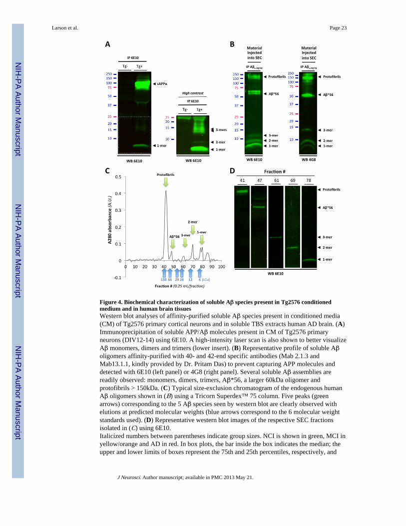
Figure 4. Biochemical characterization of soluble Aβ species present in Tg2576 conditionedmedium and in human brain tissuesWestern blot analyses of affinity-purified soluble Aβ species present in conditioned media(CM) of Tg2576 primary cortical neurons and in soluble TBS extracts human AD brain. (A)Immunoprecipitation of soluble APP/Aβ molecules present in CM of Tg2576 primaryneurons (DIV12-14) using 6E10. A high-intensity laser scan is also shown to better visualizeAβ monomers, dimers and trimers (lower insert). (B) Representative profile of soluble Aβoligomers affinity-purified with 40- and 42-end specific antibodies (Mab 2.1.3 andMab13.1.1, kindly provided by Dr. Pritam Das) to prevent capturing APP molecules anddetected with 6E10 (left panel) or 4G8 (right panel). Several soluble Aβ assemblies arereadily observed: monomers, dimers, trimers, Aβ*56, a larger 60kDa oligomer andprotofibrils > 150kDa. (C) Typical size-exclusion chromatogram of the endogenous humanAβ oligomers shown in (B) using a Tricorn Superdex™ 75 column. Five peaks (greenarrows) corresponding to the 5 Aβ species seen by western blot are clearly observed withelutions at predicted molecular weights (blue arrows correspond to the 6 molecular weightstandards used). (D) Representative western blot images of the respective SEC fractionsisolated in (C) using 6E10.Italicized numbers between parentheses indicate group sizes. NCI is shown in green, MCI inyellow/orange and AD in red. In box plots, the bar inside the box indicates the median; theupper and lower limits of boxes represent the 75th and 25th percentiles, respectively, and
Larson et al. Page 23
J Neurosci. Author manuscript; available in PMC 2013 May 21.
NIH
-PA Author Manuscript
NIH
-PA Author Manuscript
NIH
-PA Author Manuscript

bars flanking the box represent 95th and 5th percentiles. Asterisks (*) indicate P< 0.05 while(**) indicates P< 0.01.Abbreviations: NCI: no cognitive impairment, MCI: mild cognitive impairment, AD:Alzheimer’s disease, EC: extracellular-enriched fraction, IC: intracellular-enriched fraction,MB: membrane-associated fraction, Tg: transgenic, AD, Alzheimer’s disease, DLU:Densitometry Light Units, A.U.: arbitrary units.
Larson et al. Page 24
J Neurosci. Author manuscript; available in PMC 2013 May 21.
NIH
-PA Author Manuscript
NIH
-PA Author Manuscript
NIH
-PA Author Manuscript

Figure 5. Specific PrPc antibodies prevent soluble Aβ-induced Fyn activation in primaryneurons(A) Quadruple channel confocal immunofluorescence reveals that PrPc (labeled using theC20 antibody) and Aβ (labeled using the DW6 antiserum, a kind gift from Dr. Walsh)colocalize on neuronal dendritic spines (labeled using a PSD95 antibody) in 14 day-oldprimary cortical Tg2576 neurons. (B) Quantification of colocalized voxels for each channelpair of interest. Representative 3D-rendered images used for voxel counts are shown in theleft panel. Results for channel pairs are shown in the histogram. (C) Triple channel confocalimaging shows that Aβ42 species partly co-localize with pY416-Src and PrPc in Tg2576primary neurons. (D) Exogenous Aβ species purified from AD brain tissue colocalized withPrPc and Fyn at dendritic spines after application onto non-transgenic primary corticalneurons. (E-F) Fyn activation induced by human oligomeric Aβ is partially blocked by theantibody 6D11 targeting the 95-105 domain of PrPc. Preincubation with the N-terminus PrPc
antibody 8B4 failed to inhibit Fyn phosphorylation. 6D11 (10 μg) and 8B4 (10 μg) werepre-incubated for 2 hours onto cells prior to acute oAβ exposure (1 hour at 37°C). 6D11reduced Fyn activation in wild-type neurons acutely exposed to human-brain purified Aβoligomers (E) and in Tg2576 neurons chronically exposed to low-n Aβ oligomers (F).Images were acquired using an oil immersion 60x or 100x objective and processed withImaris7.1 software.Arrowheads indicate examples of colocalization between Aβ/PrPc while white asteriskscorrespond to colocalization between Aβ and PSD95 in absence of PrPc. Bars represent themean ± standard deviation (n > 3-6 dishes / experiment; independent experiments wereperformed in triplicate). *, P < 0.05, ANOVA followed by student t test.Italicized numbers between parentheses indicate group sizes. NCI is shown in green, MCI inyellow/orange and AD in red. In box plots, the bar inside the box indicates the median; theupper and lower limits of boxes represent the 75th and 25th percentiles, respectively, andbars flanking the box represent 95th and 5th percentiles. Asterisks (*) indicate P< 0.05 while(**) indicates P< 0.01.Abbreviations: NCI: no cognitive impairment, MCI: mild cognitive impairment, AD:Alzheimer’s disease, EC: extracellular-enriched fraction, IC: intracellular-enriched fraction,MB: membrane-associated fraction, Tg: transgenic, AD, Alzheimer’s disease, DLU:Densitometry Light Units, A.U.: arbitrary units.
Larson et al. Page 25
J Neurosci. Author manuscript; available in PMC 2013 May 21.
NIH
-PA Author Manuscript
NIH
-PA Author Manuscript
NIH
-PA Author Manuscript

Figure 6. PrPc-dependent phosphorylation of tau induced by endogenous Aβ oligomers inprimary neurons(A) Protein levels of hyperphosphorylated tau at Y18 (pY18-Tau) and total tau as assessedby western blot using PY18 and Tau5 antibodies in wild-type primary neurons exposed tohuman-brain purified oligomeric Aβ (5 nM for 1 hour). The glutamatergic NMDA receptorsubunit 2B (GluN2B) and the scaffolding protein PSD95 were used as internal control. (B,C) Quantitation of pY18-Tau (B) and total tau (C) across conditions and protein fractions(IC and MB) revealed an significant increase in tau hyperphosphorylation andmislocalization following oAβ exposure. This effect was partially inhibited by 6D11 but not8B4 (Bars represent the mean ± standard deviation; *, P < 0.05, ANOVA followed bystudent t test).Italicized numbers between parentheses indicate group sizes. NCI is shown in green, MCI inyellow/orange and AD in red. In box plots, the bar inside the box indicates the median; theupper and lower limits of boxes represent the 75th and 25th percentiles, respectively, andbars flanking the box represent 95th and 5th percentiles. Asterisks (*) indicate P< 0.05 while(**) indicates P< 0.01.Abbreviations: NCI: no cognitive impairment, MCI: mild cognitive impairment, AD:Alzheimer’s disease, EC: extracellular-enriched fraction, IC: intracellular-enriched fraction,
Larson et al. Page 26
J Neurosci. Author manuscript; available in PMC 2013 May 21.
NIH
-PA Author Manuscript
NIH
-PA Author Manuscript
NIH
-PA Author Manuscript

MB: membrane-associated fraction, Tg: transgenic, AD, Alzheimer’s disease, DLU:Densitometry Light Units, A.U.: arbitrary units.
Larson et al. Page 27
J Neurosci. Author manuscript; available in PMC 2013 May 21.
NIH
-PA Author Manuscript
NIH
-PA Author Manuscript
NIH
-PA Author Manuscript

Figure 7. Human AD brain-purified Aβ dimers and trimers activate Fyn in primary corticalneurons(A) Application of purified soluble Aβ dimers and trimers selectively phosphorylate Fynand tau as assessed by quantitative western blotting (duplicates are shown; n = 4 perexperiment). Sixty-minute long treatments were performed at the calculated concentration of5 nM for all species (estimated based on monomeric synthetic Aβ levels). Total proteinlevels of GluN2B and phosphorylation of GluN2B at Y1472 were also examined. Actinlevels are shown as control. (B) Quantification of the ratios for pY416-Src/Fyn (labeled aspFyn/Fyn), pY18-Tau/total tau (pTau/Tau) and pY1472-GluN2B/GluN2B (pGluN2B/GluN2B) after one hour-long exposure with the human oligomeric Aβ species listed. Bothbrain-purified Aβ dimers and trimers activate Fyn in vitro. Isolated monomeric Aβ, Aβ*56and soluble Aβ protofibrils (PF) did not induce Fyn phosphorylation under similarconditions. After 60 minutes, neither membrane-bound levels of GluN2B nor Y1472-
Larson et al. Page 28
J Neurosci. Author manuscript; available in PMC 2013 May 21.
NIH
-PA Author Manuscript
NIH
-PA Author Manuscript
NIH
-PA Author Manuscript

GluN2B phosphorylation were altered by AD brain-derived Aβ species. (Bars represent themean ± standard deviation; *, P < 0.05, ANOVA followed by student’s t test withBonferroni correction).Italicized numbers between parentheses indicate group sizes. NCI is shown in green, MCI inyellow/orange and AD in red. In box plots, the bar inside the box indicates the median; theupper and lower limits of boxes represent the 75th and 25th percentiles, respectively, andbars flanking the box represent 95th and 5th percentiles. Asterisks (*) indicate P< 0.05 while(**) indicates P< 0.01.Abbreviations: NCI: no cognitive impairment, MCI: mild cognitive impairment, AD:Alzheimer’s disease, EC: extracellular-enriched fraction, IC: intracellular-enriched fraction,MB: membrane-associated fraction, Tg: transgenic, AD, Alzheimer’s disease, DLU:Densitometry Light Units, A.U.: arbitrary units.
Larson et al. Page 29
J Neurosci. Author manuscript; available in PMC 2013 May 21.
NIH
-PA Author Manuscript
NIH
-PA Author Manuscript
NIH
-PA Author Manuscript

Figure 8. Prnp gene ablation attenuates Aβ-induced activation of Fyn and tauhyperphosphorylation in vivoGene ablation of Prnp modulates Fyn activation and tau phosphorylation at pY18 in brainsof aged transgenic APPPS1+xPrnp animals. (A) Protein levels of low-molecular weight Aβoligomers, total Fyn and pY416-Src were assessed by quantitative western blot (WB). 6E10was used for Aβ. A marked reduced of pY416 was revealed in APPPS1+xPrnp-/- comparedto APPPS1+xPrnp+/- mice. (B) Relative protein levels for total Fyn and pY416-Fyndetermined by densitometry analyses. (C, D) Prnp gene deletion limits tauhyperphorphorylation and missorting in vivo. PY18-tau and total tau levels were measuredby WB using either intracellular-enriched or membrane-enriched protein extracts (C) andquantified by software analysis (D). APPPS1+xPrnp-/- mice showed a >2-fold reduction intau phosphorylation at Y18 compared to APPPS1+xPrnp+/- littermates. (Bars represent themean ± standard deviation; *, P < 0.05, ANOVA followed by student’s t test, n ≥ 3 animals/genotype/age/experiment).Italicized numbers between parentheses indicate group sizes. NCI is shown in green, MCI inyellow/orange and AD in red. In box plots, the bar inside the box indicates the median; theupper and lower limits of boxes represent the 75th and 25th percentiles, respectively, andbars flanking the box represent 95th and 5th percentiles. Asterisks (*) indicate P< 0.05 while(**) indicates P< 0.01.Abbreviations: NCI: no cognitive impairment, MCI: mild cognitive impairment, AD:Alzheimer’s disease, EC: extracellular-enriched fraction, IC: intracellular-enriched fraction,MB: membrane-associated fraction, Tg: transgenic, AD, Alzheimer’s disease, DLU:Densitometry Light Units, A.U.: arbitrary units.
Larson et al. Page 30
J Neurosci. Author manuscript; available in PMC 2013 May 21.
NIH
-PA Author Manuscript
NIH
-PA Author Manuscript
NIH
-PA Author Manuscript

Figure 9. Aβ-induced activation of Fyn and tau hyperphosphorylation is enhanced in agedAPPS1 mice overexpressing PrP(A) Soluble brain levels of low-molecular weight Aβ oligomers in 14-month-oldAPPPS1xtga20 mice was assessed by quantitative western blot (WB) using 6E10. (B)Quantification of total Fyn, pY416-Src and actin protein levels in 14-month-old mice(APPPS1-xtga20-, APPPS1+xtga20-, APPPS1-xtga20+ and APPPS1+xtga20+) followingWB. A marked elevation of pY416-Fyn was observed in APPPS1+xtga20+ compared toAPPPS1-xtga20+ mice. (C) Tau missorting was illustrated by biochemical segregation ofpY18-Tau, total tau, PSD95 in IC vs. MB extracts using western blotting. (D) Quantificationof PY18-tau and total tau immunoreactivity across protein fractions and genotypes. AgedAPPPS1+xtga20+ mice showed a ~1.8-fold accumulation of tau and a ~1.6-fold increase inphosphorylation at Y18 compared to age-matched APPPS1+xtga20- mice. (E) Enhancedaccumulation of full-length tau and cleavage products phosphorylated at Y18 in PSD95-containing extracts of old APPPS1+xtga20+ mice. Actin was used as internal standard. (F)Quantification of tau species immunoreactive for Tau5 and PY18 in 14-month-oldAPPPS1-xtga20-, APPPS1+xtga20-, APPPS1-xtga20+ and APPPS1+xtga20+ mice. (Bars
Larson et al. Page 31
J Neurosci. Author manuscript; available in PMC 2013 May 21.
NIH
-PA Author Manuscript
NIH
-PA Author Manuscript
NIH
-PA Author Manuscript

represent the mean ± standard deviation; *, P < 0.05, ANOVA followed by student t test, n ≥3 animals/genotype/age/experiment).Italicized numbers between parentheses indicate group sizes. NCI is shown in green, MCI inyellow/orange and AD in red. In box plots, the bar inside the box indicates the median; theupper and lower limits of boxes represent the 75th and 25th percentiles, respectively, andbars flanking the box represent 95th and 5th percentiles. Asterisks (*) indicate P< 0.05 while(**) indicates P< 0.01.Abbreviations: NCI: no cognitive impairment, MCI: mild cognitive impairment, AD:Alzheimer’s disease, EC: extracellular-enriched fraction, IC: intracellular-enriched fraction,MB: membrane-associated fraction, Tg: transgenic, AD, Alzheimer’s disease, DLU:Densitometry Light Units, A.U.: arbitrary units.
Larson et al. Page 32
J Neurosci. Author manuscript; available in PMC 2013 May 21.
NIH
-PA Author Manuscript
NIH
-PA Author Manuscript
NIH
-PA Author Manuscript

Figure 10. PrPc overexpression in aged APPPS1 mice triggers a selective decrease inpostsynaptic proteins(A) Apparent selective decrease in postsynaptic proteins PSD95 and Fyn in 14-month-oldAPPPS1+xtga20+ mice. Representative WBs for the postsynaptic proteins PSD95 and Fynas well as for the presynaptic vesicle protein synaptophysin (SYP) are shown. Actin wasused as internal standard. (B) Quantification of PSD95, Fyn and Synaptophysin in 14-month-old APPPS1+xtga20-, APPPS1-xtga20+ and APPPS1+xtga20+ mice revealedsignificant reductions in postsynaptic proteins in APPPS1 mice overexpressing PrPc. (Barsrepresent the mean ± standard deviation; *, P < 0.05, ANOVA followed by student t test, n ≥3 animals/genotype/age/experiment). (C) Proposed model of tau regulation by the triadoAβ-PrPc-Fyn. In the presence of accumulating Aβ dimers, PrPc, Fyn and Cav-1 form acomplex at the plasma membrane (likely with receptors such GluN or integrins known tointeract with Cav-1 and Fyn). Upon phosphorylation of Fyn at Y416 (possibly by the kinasePyk2), this complex becomes biologically active. Two scenarios are possible depending onthe status of Fyn with respect to tau: (a) Fyn is not bound to tau and (b) Fyn is bound to tau.In (a), activated Fyn causes the hyperphosphorylation of tau at Y18 and its aberrantaccumulation at the postsynaptic density (PSD). It is possible that pFyn might be bound topTau at this stage and binds the SH3 domain of PSD95, where it is ideally located tomodulate GluN/NMDA receptor subunits (i.e. GluN2B at Y1472). In the model (b), Fyn isalready bound to tau in the dendrite, translocate to the PSD to interact with PSD95. There,Pyk2 could phosphorylate Fyn at Y416 resulting in Fyn activation and tau phosphorylationat Y18.Italicized numbers between parentheses indicate group sizes. NCI is shown in green, MCI inyellow/orange and AD in red. In box plots, the bar inside the box indicates the median; theupper and lower limits of boxes represent the 75th and 25th percentiles, respectively, andbars flanking the box represent 95th and 5th percentiles. Asterisks (*) indicate P< 0.05 while(**) indicates P< 0.01.Abbreviations: NCI: no cognitive impairment, MCI: mild cognitive impairment, AD:Alzheimer’s disease, EC: extracellular-enriched fraction, IC: intracellular-enriched fraction,MB: membrane-associated fraction, Tg: transgenic, AD, Alzheimer’s disease, DLU:Densitometry Light Units, A.U.: arbitrary units.
Larson et al. Page 33
J Neurosci. Author manuscript; available in PMC 2013 May 21.
NIH
-PA Author Manuscript
NIH
-PA Author Manuscript
NIH
-PA Author Manuscript

NIH
-PA Author Manuscript
NIH
-PA Author Manuscript
NIH
-PA Author Manuscript
Larson et al. Page 34
Table 1
Characteristics of the Religious Order Study Participants.
Group NCI (n = 26) MCI (n = 34) AD (n = 24) P values*
Age of death (years), Mean ± SD 82.97 ± 7.53 86.33 ± 5.69 90.27 ± 7.20 0.108
No. of M/F (%) 12/14 (46.1%) 14/20 (41.2%) 9/15 (37.5%) >0.99
Last MMSE Score, Mean ± SD 28.35 ± 1.38 26.41 ± 2.96 12.33 ± 8.79 <0.0001
PMI (hours), Mean ± SD (Range) 5.57 ± 2.25 (2-10) 4.90 ± 2.56 (1-9) 4.48 ± 1.66 (2-9) 0.191
Amyloid Burden (% of area), Mean ± SD 1.63 ± 0.40 1.57 ± 0.35 3.12 ± 0.42 0.0099
Tangle density (#/mm2), Mean ± SD 4.92 ± 3.93 5.81 ± 5.43 11.18 ± 10.08 0.002
Abbreviations: NCI, non cognitive impairment, MCI, mild-cognitive impairment, AD, Alzheimer’s disease, MMSE, mini-mental statusexamination, M/F, male/female ratio, PMI, post-mortem interval.
*Kruskal-Wallis test followed by Bonferroni-adjusted test for multiple comparisons.
J Neurosci. Author manuscript; available in PMC 2013 May 21.