Epileptogenesis in pediatric cortical dysplasia: The dysmature cerebral developmental hypothesis
Upload
independentCategory
view
4download
0
Am. J. Hum. Genet. 62:311–319, 1998
311
Diverse Mutations in the Gene for Cartilage Oligomeric Matrix Protein inthe Pseudoachondroplasia–Multiple Epiphyseal Dysplasia Disease SpectrumMichael D. Briggs,1,2,∗ Geert R. Mortier,4 William G. Cole,6 Lily M. King,1 Steven S. Golik,1Jacky Bonaventure,7 Lieve Nuytinck,4 Anne De Paepe,4 Jules G. Leroy,4,5 Leslie Biesecker,8Mark Lipson,9 William R. Wilcox,1,2 Ralph S. Lachman,3 David L. Rimoin,1,2
Robert G. Knowlton,10 and Daniel H. Cohn1,2
1Ahmanson Department of Pediatrics, Steven Spielberg Pediatric Research Center, Burns and Allen Cedars-Sinai Research Institute, andDepartments of 2Pediatrics and 3Radiology, UCLA School of Medicine, Los Angeles; Departments of 4Medical Genetics and 5Pediatrics,University of Gent Hospital, Gent, Belgium; 6Division of Orthopedics, Hospital for Sick Children, and University of Toronto, Toronto;7Hospital Necker, Paris; 8National Center for Human Genome Research, National Institutes of Health, Bethesda; 9Permanente Medical Group,Sacramento; and 10Department of Dermatology and Cutaneous Biology, Jefferson Medical College, Philadelphia
Summary
Pseudoachondroplasia (PSACH) and multiple epiphys-eal dysplasia (MED) are autosomal dominant osteo-chondrodysplasias that result in mild to severe short-limb dwarfism and early-onset osteoarthrosis. PSACHand some forms of MED result from mutations in thegene for cartilage oligomeric matrix protein (COMP;OMIM 600310 [http://www3.ncbi.nlm.nih.gov:80/htbin-post/Omim/dispmim?600310]). We report theidentification of COMP mutations in an additional 14families with PSACH or MED phenotypes. Mutationspredicted to result in single–amino acid deletions or sub-stitutions, all in the region of the COMP gene encodingthe calmodulin-like repeat elements, were identified inpatients with moderate to severe PSACH. We also iden-tified within this domain a missense mutation that pro-duced MED Fairbank. In two families, one with mildPSACH and the second with a form of MED, we iden-tified different substitutions for a residue in the carboxyl-terminal globular region of COMP. Both the clinicalpresentations of these two families and the identificationof COMP-gene mutations provide evidence of pheno-typic overlap between PSACH and MED. These dataalso reveal a role for the carboxyl-terminal domain inthe structure and/or function of COMP.
Received July 31, 1997; accepted for publication December 4, 1997;electronically published February 6, 1998.
Address for correspondence and reprints: Dr. Daniel H. Cohn, Med-ical Genetics, SSB-3, Cedars-Sinai Medical Center, 8700 Beverly Boul-evard, Los Angeles, CA 90048. E-mail: [email protected]
*Present affiliation: Department of Biochemistry, University of Man-chester, Manchester, England.
q 1998 by The American Society of Human Genetics. All rights reserved.0002-9297/98/6202-0015$02.00
Introduction
Pseudoachondroplasia (PSACH; OMIM 177170 [http://www3.ncbi.nlm.nih.gov:80/htbin-post/Omim/dispmim?177170]) and multiple epiphyseal dysplasia (MED;OMIM 132400 [http://www3.ncbi.nlm.nih.gov:80/htbin-post/Omim/dispmim?132400]) are autosomaldominant osteochondrodysplasias that result in mild tosevere short-limb dwarfism and early-onset osteoar-throsis (International Working Group on ConstitutionalDiseases of Bone 1992). PSACH is the clinically moresevere phenotype, characterized by marked short stature,short fingers, loose joints with ligamentous laxity, de-formity of the legs, and scoliosis. Radiographic featuresinclude small irregular epiphyses with delayed ossifica-tion, irregular metaphyses, and delayed ossification ofthe annular epiphyses of the vertebrae, resulting in acharacteristic anterior beaking of the vertebral bodiesthat is apparent on lateral projection. Milder cases ofPSACH exhibit similar radiographic features, but shortstature is less pronounced and there is less deformitythan in more severe cases (Maroteaux et al. 1980; Ri-moin et al. 1994). MED, which is divided into the moresevere Fairbank type (Fairbank 1947) and the milderRibbing type (Ribbing 1937), is less severe than PSACH.Patients with MED exhibit normal stature, or mild shortstature, and early-onset osteoarthrosis that becomes ap-parent in mid- to late childhood. Radiographically, theepiphyses are irregular and show poor ossification, butmetaphyseal abnormalities are mild or absent. The spineis typically radiographically normal. Morphologicalstudies of cartilage from PSACH and MED patientsshow accumulation of material in the rough endoplasmicreticulum (RER) of chondrocytes (Maynard et al. 1972;Stanescu et al. 1977, 1982, 1993; Rimoin et al. 1994).In most PSACH and in some MED cases, the accumu-lated material forms a unique lamellar structure (May-nard et al. 1972; Stanescu et al. 1977, 1982, 1993).

312 Am. J. Hum. Genet. 62:311–319, 1998
Initially, the mild and severe PSACH phenotypes werelinked to markers in the pericentromeric region of chro-mosome 19 (Briggs et al. 1993; Hecht et al. 1993), andthey were subsequently found to result from mutationsin the gene for cartilage oligomeric matrix protein(COMP) (Briggs et al. 1995a; Hecht et al. 1995). Someforms of MED were also linked to this region (Oehlmannet al. 1994), and COMP mutations were identified inpatients with the Fairbank (Briggs et al. 1995a) and Rib-bing (Ballo et al. 1997) types of MED. MED Fairbankcan also result from defects in the a2 chain of type IXcollagen (OMIM 600204 [http://www3.ncbi.nlm.nih.gov:80/htbin-post/Omim/dispmim?600204]) (Briggs etal. 1994; Muragaki et al. 1996) and from defects in atleast one additional gene (OMIM 600969 [http://www3.ncbi.nlm.nih.gov:80/htbin-post/Omim/dispmim?600969]) (Deere et al. 1995).
COMP is a 524-kD homopentameric glycoproteinthat is expressed prominently in the territorial matrixsurrounding chondrocytes (Hedbom et al. 1992). It is amember of the thrombospondin gene family and is alsoreferred to as “thrombospondin 5.” COMP is a modularprotein containing an amino-terminal domain, four ep-idermal growth factor–like repeats, eight calmodulin-like (CaM-like) repeats, and a large carboxyl-terminalglobular domain (Oldberg et al. 1992). To date, all ofthe reported COMP-gene mutations that result inPSACH or MED phenotypes have been in the region ofthe gene encoding the CaM-like domain (Briggs et al.1995a; Hecht et al. 1995; Ballo et al. 1997; Loughlin etal. [in press]). These have included point mutations thatresult in single–amino acid substitutions for conservedresidues and in-frame deletions that delete codon(s) forone or more residues.
We report the identification of mutations in an ad-ditional 14 families with phenotypes within the PSACH-MED disease spectrum. Twelve mutations were identi-fied in the CaM-like domain; this finding confirms theimportance of this region in the structure and/or functionof COMP. We have also identified two mutations in thecarboxyl-terminal domain of COMP, thereby extendingthe range of COMP-gene mutations that can producePSACH or MED phenotypes. These data provide evi-dence that the carboxyl terminal domain of COMP hasan important structural and/or functional role in thecartilage extracellular matrix.
Patients and Methods
Clinical Summary
The clinical and radiographic features of each patientor family were reviewed by at least two clinical genet-icists and/or radiologists. COMP mutations were iden-tified in eight affected individuals who were sporadic
products of nonconsanguinous unions (InternationalSkeletal Dysplasia Registry reference numbers R91-064,R92-207, R95-108, R95-109, and R95-110, and Uni-versity of Gent patients BW, CM, and WL). The probandin family R95-107 was the offspring of an affected fatherand an unaffected mother. All affected individuals inthese families exhibited the typical clinical features ofPSACH: short-limb dwarfism, small hands with shortfingers, loose joints, a waddling gait, and varum and/orvalgum deformity of the lower limbs. Childhood radi-ographs of all the patients were reviewed; they dem-onstrated small, irregular epiphyses in the hands andknees and irregular metaphyses. The hips exhibited mi-croepiphyses and acetabular irregularities. Anteriorbeaking of the vertebral bodies was apparent on lateralprojection. These features are diagnostic for typicalPSACH.
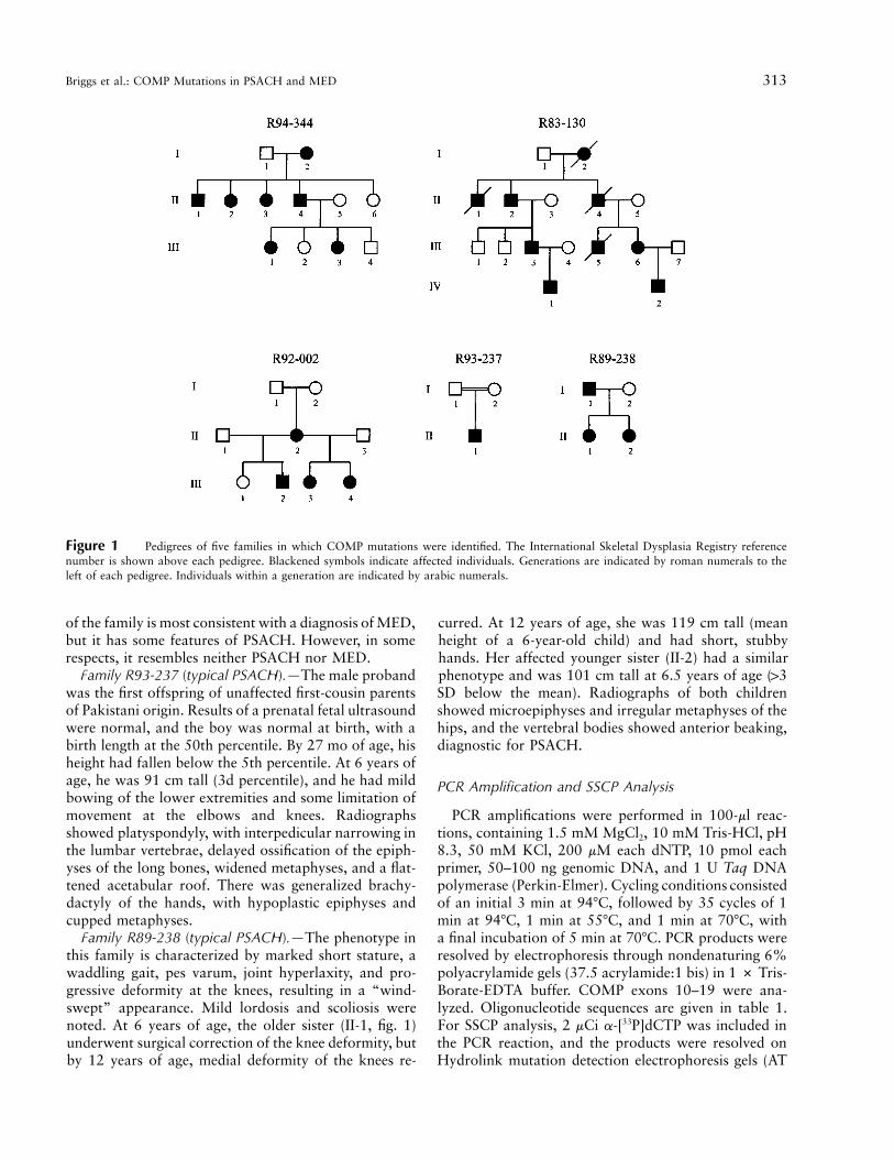
The pedigrees of the other five families in whichCOMP mutations were identified are shown in figure 1.The clinical features of these families are describedbelow.
Family R94-344 (MED Fairbank).—The proband, III-1 (fig. 1), was first seen at 15 years of age. She was shortin stature (height 144 cm), and she complained of painin the knees. Her hands were normal. Radiographsshowed normal hands and hips, but the tibial epiphyseswere irregular, with a squared aspect. Her affected sister,III-3, was also short in stature and had involvement ofthe knees and hips. The femoral head was small andirregular, but the severity of the deformities was some-what less striking than the deformities in other cases ofMED Fairbank. The affected father (II-4) of the probandhad knee involvement, but the hips were unaffected. Theaffected grandmother (I-2) had severe hip dysplasia thatrequired surgical replacement of the femoral head.
Family R83-130 (mild PSACH).—Affected individualswere identified initially on the basis of their waddlinggaits. In childhood, they developed chronic hip pain thatwas exacerbated by moderate exercise. Heights of theaffected individuals ranged from 145 cm (III-6, fig. 1)to 180 cm (IV-2). Radiographs showed significant epi-physeal and metaphyseal changes in the joints. The hipsshowed microepiphyses and irregular acetabulae. Thespines showed platyspondyly and anterior beaking ofthe vertebrae.
Family R92-002 (unclassified MED).—The earliestsymptoms included a waddling gait and lumbar lordosis.The height of the only affected adult in the family (II-2, fig. 1) was 147 cm. Radiographs showed small, ir-regular epiphyses, particularly at the proximal femur,and mild metaphyseal abnormalities. The hands wereatypical of PSACH or MED in that they were short, inall segments, with irregular metaphyses and large epiph-yses. In childhood, neither anterior beaking of the ver-tebrae nor platyspondyly was apparent. The phenotype
Briggs et al.: COMP Mutations in PSACH and MED 313
Figure 1 Pedigrees of five families in which COMP mutations were identified. The International Skeletal Dysplasia Registry referencenumber is shown above each pedigree. Blackened symbols indicate affected individuals. Generations are indicated by roman numerals to theleft of each pedigree. Individuals within a generation are indicated by arabic numerals.
of the family is most consistent with a diagnosis of MED,but it has some features of PSACH. However, in somerespects, it resembles neither PSACH nor MED.
Family R93-237 (typical PSACH).—The male probandwas the first offspring of unaffected first-cousin parentsof Pakistani origin. Results of a prenatal fetal ultrasoundwere normal, and the boy was normal at birth, with abirth length at the 50th percentile. By 27 mo of age, hisheight had fallen below the 5th percentile. At 6 years ofage, he was 91 cm tall (3d percentile), and he had mildbowing of the lower extremities and some limitation ofmovement at the elbows and knees. Radiographsshowed platyspondyly, with interpedicular narrowing inthe lumbar vertebrae, delayed ossification of the epiph-yses of the long bones, widened metaphyses, and a flat-tened acetabular roof. There was generalized brachy-dactyly of the hands, with hypoplastic epiphyses andcupped metaphyses.
Family R89-238 (typical PSACH).—The phenotype inthis family is characterized by marked short stature, awaddling gait, pes varum, joint hyperlaxity, and pro-gressive deformity at the knees, resulting in a “wind-swept” appearance. Mild lordosis and scoliosis werenoted. At 6 years of age, the older sister (II-1, fig. 1)underwent surgical correction of the knee deformity, butby 12 years of age, medial deformity of the knees re-
curred. At 12 years of age, she was 119 cm tall (meanheight of a 6-year-old child) and had short, stubbyhands. Her affected younger sister (II-2) had a similarphenotype and was 101 cm tall at 6.5 years of age (13SD below the mean). Radiographs of both childrenshowed microepiphyses and irregular metaphyses of thehips, and the vertebral bodies showed anterior beaking,diagnostic for PSACH.
PCR Amplification and SSCP Analysis
PCR amplifications were performed in 100-ml reac-tions, containing 1.5 mM MgCl2, 10 mM Tris-HCl, pH8.3, 50 mM KCl, 200 mM each dNTP, 10 pmol eachprimer, 50–100 ng genomic DNA, and 1 U Taq DNApolymerase (Perkin-Elmer). Cycling conditions consistedof an initial 3 min at 947C, followed by 35 cycles of 1min at 947C, 1 min at 557C, and 1 min at 707C, witha final incubation of 5 min at 707C. PCR products wereresolved by electrophoresis through nondenaturing 6%polyacrylamide gels (37.5 acrylamide:1 bis) in 1 # Tris-Borate-EDTA buffer. COMP exons 10–19 were ana-lyzed. Oligonucleotide sequences are given in table 1.For SSCP analysis, 2 mCi a-[33P]dCTP was included inthe PCR reaction, and the products were resolved onHydrolink mutation detection electrophoresis gels (AT

314 Am. J. Hum. Genet. 62:311–319, 1998
Table 1
Exons Amplified, Primer Names and Sequences, and Sizes of Amplified Products
Exon(s) Primer Names Primer SequencesProduct Size
(bp)
10 I9F1 5′-TGA GGA GTG TGA CCT TTG CC-3′ 279I10R1 5′-AGC CGA ATC CCG CCT TCG GTG-3′
11 I10F1 5′-CTT GGG CTC TGG TCC CGT GG-3′ 181I11R1 5′-GCT TAC CCA GCT GGA GTC TG-3′
12 I11F2 5′-ATT TCC TCT GTC TGA TTA TGG-3′ 168I12R1 5′-CCA GAG ACA ATG AGC TCT CCAG-3′
13 I12F2 5′-GGG TAG CCT TTG ACA AAA CG-3′ 2231492R 5′-GTT AGG CAC CAG GCG GCA G-3′
14 I13F1 5′-TGA CTT TAG CCC ACC GAG GG-3′ 281I14R1 5′-CTC AGC ATA GGC CTC ACT GTG-3′
15 I14F1 5′-CAC AGT GAG GCC TAT GCT GA-3′ 164I15R2 5′-GTG GCA GGA TAG CGC TGC TC-3′
16 I15F1 5′-GCG TTC GGA AAG GCC ACT GC-3′ 319I16R1 5′-CTA AGT GGC TGT AAA GGG TTT-3′
17 I16F1 5′-GCC CAC CGA GGT CTC TGA CC-3′ 275I17R1 5′-GGC ACT CCC ACC TGG GCC TG-3′
18/19 2116F 5′-GCG ATT CTA TGA GGG CCC TGA-3′ 3192342R 5′-GCG GTG AGG GTG GCT GTC AT-3′
Biochem), according to the manufacturer’s protocol, ex-cept that the gels were precooled and run for 2–3 h at47C, at 50 W.
DNA Sequence Analysis
PCR products were cleaned by use of the QIAquickkit (Qiagen) and were ligated into the pCRII TA cloningvector (Invitrogen). DNA was isolated from the clones,and sequences were determined by use of Sequenase andreagents from a kit (US Biochemical). Direct sequenceanalysis of PCR products was determined by means ofdye terminator chemistry followed by resolution on theABI PRISM 377 automated sequencer (Perkin-Elmer).
Results
The patient panel included 20 families with typicalPSACH, 2 families with mild PSACH, 6 families withMED Fairbank, 3 families with MED Ribbing, and 13families with unclassified forms of MED. By means ofheteroduplex, SSCP, and DNA sequence analysis, weidentified COMP-gene mutations in 14 individuals orfamilies who had phenotypes in the PSACH-MED clin-ical spectrum. The phenotypes are detailed in the clinicalsummary, the relevant pedigrees are shown in fig. 1, andthe mutations are summarized in table 2.
All individuals affected with typical PSACH, includingBW, WL, and members of families R89-238, R91-064,R92-207, R95-108, and R95-110, were heterozygousfor a 3-bp deletion identified previously in patients withthis phenotype (Hecht et al. 1995). The deletion removedone of five consecutive aspartic acid (GAC) codons cor-responding to residues 469–473 of the protein, within
the seventh CaM-like repeat. The repeated nature of thesequence (C GAC GAC GAC GAC GAC) does not allowprecise determination of the codon that is deleted inthese patients. We hypothesize that this repeat unit pre-disposes to mutations of this type, which may occur viaslipped base pairing during DNA replication (Kunkel1993; Ashley and Warren 1996). Ultrastructural studiesof cartilage from an individual in family R89-238showed the typical accumulation of material with a la-mellar appearance in the RER of chondrocytes that iscommon in cases of PSACH (Cohn et al. 1996). Al-though chondrocytes from R95-110 showed RER inclu-sions with a smooth to grainy (rather than lamellar)appearance (data not shown), cell preservation was poor,so few cells were examined.
A similar but shorter repeated sequence, C GAC GACGAC, is present in the portion of the COMP gene en-coding the fourth CaM-like repeat. Deletion of one ofthese aspartic acid codons, for residues 372–374 of theprotein, was reported previously in one patient with typ-ical PSACH (Briggs et al. 1995a). The same mutationwas identified in another patient, also with typicalPSACH, from family R95-109. Chondrocytes from thelatter patient showed the characteristic lamellar RERinclusions of PSACH (data not shown).
Patient CM and affected individuals in families R95-107 and R93-237, all with typical PSACH, were het-erozygous for point mutations predicted to result in asingle–amino acid substitution for glycine 440, withinthe sixth CaM-like repeat. In families R95-107 and R93-237, a GrA transition at the second position of thecodon predicted a G440E substitution (table 2). Themutation could be identified on the basis of a slowly

Briggs et al.: COMP Mutations in PSACH and MED 315
Table 2
Mutations and Polymorphisms Identified in the COMP Gene in Patients with PSACH or MED
Family or Patient Nucleotide Change Exon Residue Change Domain Phenotype
R95-109 DGAC 1139–1147 10 DD 372–374 CaM-like #2 PSACHR95-108a DGAC 1430–1444 13 DD 469–473 CaM-like #4 PSACHCM GrA at 1343 13 G440R CaM-like #6 PSACHR95-107b GrA at 1344 13 G440E CaM-like #6 PSACHR94-344 ArG at 1383 13 N453S CaM-like #7 MED FairbanksR83-130 CrT at 1779 16 T585M COOH Mild PSACHR92-002 CrG at 1779 16 T585R COOH MEDR83-130c GrA at 1780 16 Neutral polymorphism COOH . . .
a Also carried by families R89-238, R92-207, R95-110, and R91-64 and by patients BW and CM.b Also carried by family R93-237.c Also carried by family R92-002.
Figure 2 Sporadic COMP mutation (G440E) in family R93-237.A, Sequences of the normal and mutant alleles are summarized. B,PAGE of a PCR-amplified genomic DNA fragment containing the mu-tation in the proband and in the parents. Lane 1 (m) shows size mark-ers, with sizes (in bp) indicated on the left.
migrating heteroduplex in amplified genomic DNA (fig.2). Despite the fact that the proband in family R93-237was the offspring of a consanguinous union, the mu-tation was not detected in amplified DNA from the un-affected parents; this indicates that the PSACH pheno-type was the result of a new mutation. In the family ofCM, an individual patient, a GrA transition at the firstposition of the codon predicted a G440R substitution(data not shown). Sequence analysis of an amplified ge-nomic DNA fragment from both parents of CM showed
absence of the mutation; this demonstrates that thePSACH phenotype was produced by a new dominantmutation.
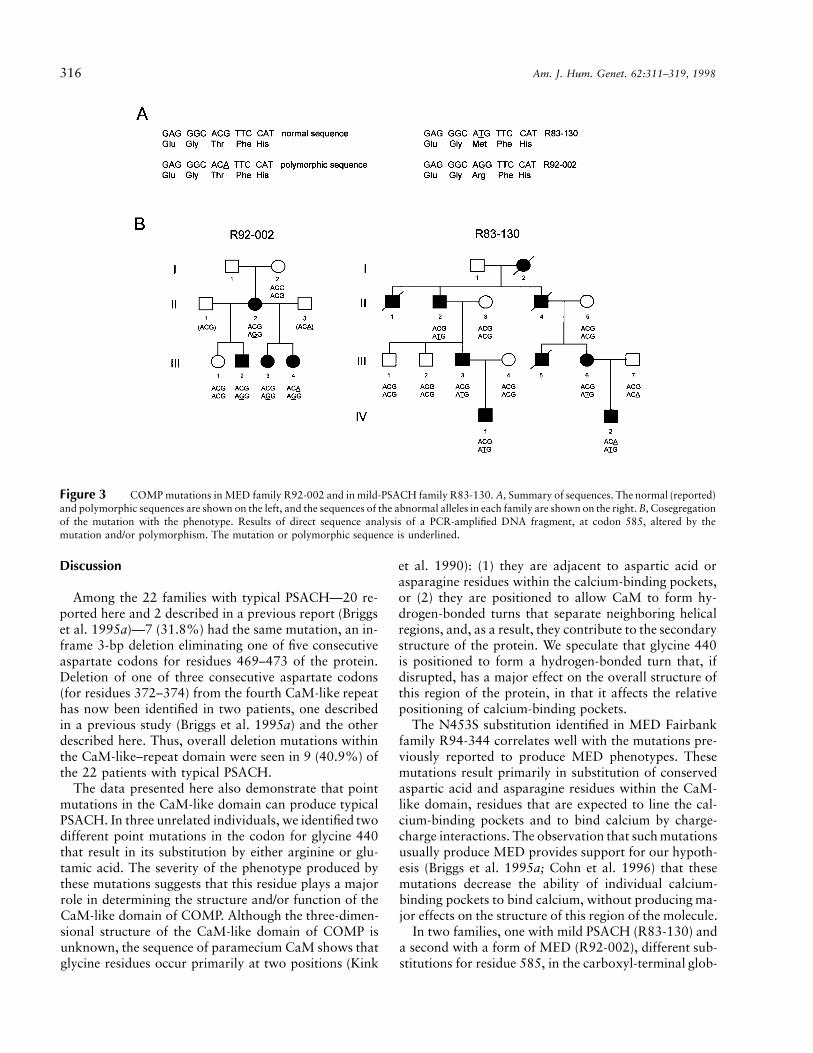
In families R83-130 and R92-002, who have domi-nantly inherited forms of mild PSACH and MED, re-spectively, we previously reported linkage of the diseasephenotypes to markers specific for the COMP locus onchromosome 19, and we excluded linkage to markersfor COL9A1 and COL9A2 (Briggs et al. 1995b). In bothfamilies, we identified point mutations that are predictedto result in the substitution of threonine 585 in the car-boxyl-terminal (COOH) domain of the protein. Ac-cording to the previously published sequence (Newtonet al. 1994) and our own data, the codon for threonine585 is ACG. In family R83-130 (individual IV-2), weidentified four clones with the sequence ATG and oneclone with the sequence ACA; the CrT transition pre-dicted a T585M substitution, whereas the GrA tran-sition was a silent polymorphism (fig. 3). In family R92-002 (individual II-2), we identified three clones with thesequence AGG and three clones with the sequence ACG;the CrG transversion predicted a T585R substitution(fig. 3). For all members of the two families, analysis ofPCR-amplified DNA from exon 16, by means of directsequence determination, confirmed the sequences of thepolymorphic alleles and demonstrated that the mutationin each family cosegregated with the phenotype, withoutexception (fig. 3).
In MED-Fairbank family R94-344, the disorder co-segregated with markers from the COMP region of chro-mosome 19, and linkage to COL9A1 and COL9A2 wasexcluded (Briggs et al. 1995b). We identified an ArGtransition within the COMP gene that implied the sub-stitution of a conserved asparagine residue by serine(N453S), in the seventh CaM-like repeat (fig. 4). Themutation created a BfaI restriction endonuclease cleav-age site, and analysis of amplified DNA from familymembers showed that the mutation cosegregated withthe phenotype (fig. 4).
316 Am. J. Hum. Genet. 62:311–319, 1998
Figure 3 COMP mutations in MED family R92-002 and in mild-PSACH family R83-130. A, Summary of sequences. The normal (reported)and polymorphic sequences are shown on the left, and the sequences of the abnormal alleles in each family are shown on the right. B, Cosegregationof the mutation with the phenotype. Results of direct sequence analysis of a PCR-amplified DNA fragment, at codon 585, altered by themutation and/or polymorphism. The mutation or polymorphic sequence is underlined.
Discussion
Among the 22 families with typical PSACH—20 re-ported here and 2 described in a previous report (Briggset al. 1995a)—7 (31.8%) had the same mutation, an in-frame 3-bp deletion eliminating one of five consecutiveaspartate codons for residues 469–473 of the protein.Deletion of one of three consecutive aspartate codons(for residues 372–374) from the fourth CaM-like repeathas now been identified in two patients, one describedin a previous study (Briggs et al. 1995a) and the otherdescribed here. Thus, overall deletion mutations withinthe CaM-like–repeat domain were seen in 9 (40.9%) ofthe 22 patients with typical PSACH.
The data presented here also demonstrate that pointmutations in the CaM-like domain can produce typicalPSACH. In three unrelated individuals, we identified twodifferent point mutations in the codon for glycine 440that result in its substitution by either arginine or glu-tamic acid. The severity of the phenotype produced bythese mutations suggests that this residue plays a majorrole in determining the structure and/or function of theCaM-like domain of COMP. Although the three-dimen-sional structure of the CaM-like domain of COMP isunknown, the sequence of paramecium CaM shows thatglycine residues occur primarily at two positions (Kink
et al. 1990): (1) they are adjacent to aspartic acid orasparagine residues within the calcium-binding pockets,or (2) they are positioned to allow CaM to form hy-drogen-bonded turns that separate neighboring helicalregions, and, as a result, they contribute to the secondarystructure of the protein. We speculate that glycine 440is positioned to form a hydrogen-bonded turn that, ifdisrupted, has a major effect on the overall structure ofthis region of the protein, in that it affects the relativepositioning of calcium-binding pockets.
The N453S substitution identified in MED Fairbankfamily R94-344 correlates well with the mutations pre-viously reported to produce MED phenotypes. Thesemutations result primarily in substitution of conservedaspartic acid and asparagine residues within the CaM-like domain, residues that are expected to line the cal-cium-binding pockets and to bind calcium by charge-charge interactions. The observation that such mutationsusually produce MED provides support for our hypoth-esis (Briggs et al. 1995a; Cohn et al. 1996) that thesemutations decrease the ability of individual calcium-binding pockets to bind calcium, without producing ma-jor effects on the structure of this region of the molecule.
In two families, one with mild PSACH (R83-130) anda second with a form of MED (R92-002), different sub-stitutions for residue 585, in the carboxyl-terminal glob-

Briggs et al.: COMP Mutations in PSACH and MED 317
Figure 4 COMP mutation in family R94-344. A, Summary ofsequence analysis predicting an N453S substitution. B, Cosegregationof the mutation with the phenotype. PAGE of a BfaI-cleaved ampli-fied–genomic DNA fragment containing the mutation. The pedigreeis shown above, with the numeric designations of generations andindividuals as described in the legend to figure 1. A line diagram ofthe amplified fragment, with the locations of cleavage sites for BfaIand the sizes of the resulting fragments, is shown below. The locationof the BfaI site created by the mutation is indicated by an asterisk (*).
ular domain of COMP, were identified. The mild-PSACH family has typical radiographic features of thespine, with anterior beaking of the vertebral bodies dur-ing childhood and both metaphyseal and epiphyseal ab-normalities. However, affected individuals in this familyexhibit either normal height or only mild short stature.In contrast, the phenotype in the family withMED—with primarily epiphyseal irregularities and anormal spine—does not exhibit the radiographic fea-tures of PSACH, but the only affected adult in the familyis of short stature. The clinical presentation of these fam-ilies and the identification of COMP-gene mutationsdemonstrate phenotypic overlap between MED and mildforms of PSACH.
The mutations in the carboxyl-terminal domain in-dicate that this region has an essential role in the struc-
ture and/or function of COMP. Although the precise roleis unknown, comparisons with the other members of thethrombospondin family of proteins provide some sug-gestions. Recent studies have shown that the carboxyl-terminal domain of thrombospondin 1 contains anon–RGD-dependent cell-binding domain based on thesequence RFYVVMWK (Gao and Frazier 1994; Tsaoand Mousa 1995). This interaction, between thrombo-spondin 1 and many cell types, appears to be mediatedby CD47 (Gao et al. 1996), an integrin-associated pro-tein (IAP) that can bind to integrin avb3. Tuckwell etal. (1994) determined the integrin profile of a humanchondrosarcoma cell line, HCS-2/8 (Takigawa et al.1989), identifying avb3 as one of the expressed integrins.The sequence SFYVVMWK is present in the carboxyl-terminal domain of COMP (residues 608–615), sug-gesting a similar role for this sequence in IAPavb3–mediated chondrocyte interactions. Interestingly,in vitro binding studies have shown that primary bovinechondrocytes can attach to purified bovine COMP(DiCesare et al. 1994), but the mechanism of this inter-action is unknown. Threonine 585 is 23 amino residuesterminal to this putative cell attachment site and maybe involved in determining the secondary structure ofthe domain. Future studies will examine the possibilitythat the threonine 585 mutations affect the ability ofCOMP to interact with chondrocytes.
Mutations were not identified in 8 PSACH patientsor in 20 MED patients. For PSACH, linkage studies haveshown absence of locus heterogeneity (Hecht et al.1993), so the mutations in the negative PSACH patientsare likely to result either from the known inefficienciesof mutation detection by SSCP or from the possibilitythat the mutations are in COMP exons not examinedin this study. For MED, the mutations may also be lo-cated in genes other than COMP, including the type IXcollagen genes (Briggs et al. 1994; Muragaki et al. 1996).
The data presented here show that a variety of COMPstructural mutations can produce a continuum of dis-ease, ranging from a typical PSACH to a mild MED-Ribbing phenotype. Mutations predicted to disrupt thesecondary structure of the CaM-like domain of COMPand to impede its secretion produce moderate to severeforms of PSACH. Mutations predicted to have mildereffects on the secondary structure of the protein producephenotypes at the MED end of the clinical spectrum.
Acknowledgments
We thank the patients and their families for their partici-pation in and their enthusiasm for these studies. We are mostgrateful to our clinical colleagues—A. Aylsworth, S. Braddock,B. Burton, E. Chen, D. Daentl, C. J. Epstein, F. Gilbert, J. C.Hoeffel, Y. E. Hsia, T. Jewett, R. Namba, W. Oppenheim, J.Rogers, J. Shively, M. Shohat, M. Stephan, D. Vine, G. Wilson,

318 Am. J. Hum. Genet. 62:311–319, 1998
and D. Yim—who provide clinical care to the families de-scribed here, for referring the families described in this study.Light and electron microscopy were expertly performed by J.Werkmeister and L. Nolasco. Excellent technical assistancewas provided by M. Van Thielen and J.-P. Renard. We ap-preciate the organizational assistance of M. Priore and S. Levinof the International Skeletal Dysplasia Registry; a critical read-ing of the manuscript, by E. Delot-Vilain; and a critical reviewof the figures, by Z. Cohn. This work was supported in partby NIH grants AR43139 (to D.H.C.) and HD22657 (toD.L.R.); by Arthritis and Rheumatism Council of Great Britainand the Commonwealth grant B0561 (to M.D.B.); by MedicalResearch Council of Canada, Canadian Arthritis Society, andSamuel Lunenfeld Foundation grants (to W.G.C.); and by aBelgian National Fund for Scientific Research grant (to G.R.M.and A.D.). Sequence reactions were run on the ABI PRISM377 DNA sequencer, by L. Hall (Biomolecules Laboratory,Wellcome Trust Centre for Cell Matrix Research, Universityof Manchester), and sequence analysis was supported by Well-come Trust grant 044327/Z/95/Z.
References
Ashley CT, Warren ST (1996) Trinucleotide repeat expansionsand human diseases. Annu Rev Genet 29:703–728
Ballo R, Briggs MD, Cohn DH, Knowlton RG, Beighton PH,Ramesar RS (1997) Multiple epiphyseal dysplasia, Ribbingtype: a novel point mutation in the COMP gene in a SouthAfrican family. Am J Med Genet 68:396–400
Briggs MD, Choi H, Warman ML, Loughlin JA, WordsworthP, Sykes BC, Irven CMM, et al (1994) Genetic mapping ofa locus for multiple epiphyseal dysplasia (EDM2) to a regionof chromosome 1 containing a type IX collagen gene. AmJ Hum Genet 55:678–684
Briggs MD, Hoffman SMG, King LM, Olsen AS, Mohren-weiser H, Leroy JG, Mortier GR, et al (1995a) Pseudo-achondroplasia and multiple epiphyseal dysplasia due tomutations in the cartilage oligomeric matrix protein gene.Nat Genet 10:330–336
Briggs MD, Rasmussen IM, Weber JL, Yuen J, Reinker K,Garber AP, Rimoin DL, et al (1993) Genetic linkage of pseu-doachondroplasia to markers in the pericentromeric regionof chromosome 19. Genomics 18:656–660
Briggs MD, Rimoin DL, Biesecker LG, Lipson MH, Bonaven-ture J, Lachman RS, Cohn DH (1995b) Genetic evidencefor a third multiple epiphyseal dysplasia locus. Second An-nual Meeting of the American College of Medical Genetics,Los Angeles, March 6–9, 1995 (abstract)
Cohn DH, Briggs MD, King LM, Rimoin DL, Wilcox WR,Lachman RS, Knowlton RG (1996) Mutations in the car-tilage oligomeric matrix protein (COMP) gene in pseu-doachondroplasia and multiple epiphyseal dysplasia. Ann NY Acad Sci 785:188–194
Deere M, Blanton SH, Scott CI, Langer LO, Pauli RM, HechtJT (1995) Genetic heterogeneity in multiple epiphyseal dys-plasia. Am J Hum Genet 56:698–704
DiCesare PE, Morgelin M, Mann K, Paulsson M (1994) Car-tilage oligomeric matrix protein and thrombospondin 1: pu-rification from articular cartilage, electron microscopic
structure and chondrocyte binding. Eur J Biochem 223:927–937
Fairbank T (1947) Dysplasia epiphysialis multiplex. Br J Surg34:225–232
Gao A, Frazier WA (1994) Identification of a receptor can-didate for the carboxyl-terminal cell binding domain ofthrombospondins. J Biol Chem 269:29650–29657
Gao A, Lindberg FP, Finn MB, Blystone SD, Brown EJ, FrazierWA (1996) Integrin-associated protein is a receptor for theC-terminal domain of thrombospondin. J Biol Chem 271:21–24
Hecht JT, Francomano CA, Briggs MD, Williams M, ConnerB, Horton WA, Warman ML, et al (1993) Linkage of typicalpseudoachondroplasia to chromosome 19. Genomics 18:661–666
Hecht JT, Nelson LD, Crowder E, Wang Y, Elder FFB, Har-rison WR, Francomano CA, et al (1995) Mutations in exon17B of cartilage oligomeric matrix protein (COMP) causepseudoachondroplasia. Nat Genet 10:325–329
Hedbom E, Antonsson P, Hjerpe A, Aeschlimann D, PaulssonM, Rosa-Pimental E, Sommarin Y, et al (1992) Cartilagematrix proteins: an acidic oligomeric protein (COMP) de-tected only in cartilage. J Biol Chem 267:6132–6136
International Working Group on Constitutional Diseases ofBone (1992) International classification of osteochondro-dysplasias. Am J Med Genet 44:223–229
Kink JA, Maley ME, Preston RR, Lig K, Wallen-Friedman MA,Saimi Y, Kung C (1990) Mutations in paramecium cal-modulin indicate functional differences between the C-ter-minal and N-terminal lobes in vitro. Cell 62:165–174
Kunkel TA (1993) Slippery DNA and diseases. Nature 365:207–208
Loughlin J, Irven C, Mustafa Z, Briggs MD, Carr A, LynchS, Knowlton RG, et al (1998) Identification of five novelmutations in the cartilage oligomeric matrix protein gene inpseudoachondroplasia and multiple epiphyseal dysplasia.Hum Mutat Suppl 11:S8–S9
Maroteaux P, Stanescu R, Stanescu V, Fontaine G (1980) Themild form of pseudoachondroplasia. Eur J Pediatr 133:227–231
Maynard JA, Cooper RR, Ponseti IV (1972) A unique roughsurfaced endoplasmic reticulum inclusion in pseudoachon-droplasia. Lab Invest 26:40–44
Muragaki Y, Mariman ECM, van Beersum SEC, Perala M, vanMourik JBA, Warman ML, Olsen BR, et al (1996) A mu-tation in the gene encoding the a2 chain of the fibril-asso-ciated collagen IX, COL9A2, causes multiple epiphyseal dys-plasia (EDM2). Nat Genet 12:103–105
Newton G, Weremowics S, Morton CC, Copeland NG, GilbertDJ, Jenkins NA, Lawler J (1994) Characterization of humanand mouse cartilage oligomeric matrix protein. Genomics24:435–439
Oehlmann R, Summerville GP, Yeh G, Weaver EJ, Jiminez SA,Knowlton RG (1994) Genetic linkage mapping of multipleepiphyseal dysplasia to the pericentromeric region of chro-mosome 19. Am J Hum Genet 54:3–10
Oldberg A, Antonsson P, Lindblom K, Heinegard D (1992)COMP (cartilage oligomeric matrix protein) is structurallyrelated to the thrombospondins. J Biol Chem 267:22346–22350
Briggs et al.: COMP Mutations in PSACH and MED 319
Ribbing S (1937) Studien uber hereditare multiple Epiphysen-storungen. Acta Radiol Suppl 34:1–107
Rimoin DL, Rasmussen IM, Briggs MD, Roughly PJ, GruberHE, Warman ML, Olsen BR, et al (1994) A large familywith features of pseudoachondroplasia and multiple epi-physeal dysplasia: exclusion of seven candidate loci that en-code proteins of the cartilage extracellular matrix. Hum Ge-net 93:236–242
Stanescu R, Stanescu V, Muriel M-P, Maroteaux P (1993) Mul-tiple epiphyseal dysplasia, Fairbank type: morphologic andbiochemical study of cartilage. Am J Med Genet 45:501–507
Stanescu V, Maroteaux P, Stanescu R (1982) The biochemicaldefect of pseudoachondroplasia. Eur J Pediatr 138:221–225
Stanescu V, Stanescu R, Maroteaux P (1977) Etude morphol-ogique et biochimique du cartilage de croissance dans lesosteochondrodysplasies. Arch Fr Pediatr Suppl 34:1–80
Takigawa M, Tajima K, Pan HO, Enomoto M, Kinoshita A,Suzuki F, Takano Y, et al (1989) Establishment of a clonalhuman chondrosarcoma cell line with cartilage phenotypes.Cancer Res 49:3996–4002
Tsao PW, Mousa SA (1995) Thrombospondin mediates cal-cium mobilization in fibroblasts via its Arg-Gly-Asp andcarboxyl-terminal domains. J Biol Chem 270:23747–23753
Tuckwell DS, Ayad S, Grant ME, Takigawa M, HumphriesMJ (1994) Conformation dependence of integrin–type II col-lagen binding. J Cell Sci 107:993–1005
Copyright © 2022 FDOKUMEN