Canine Elbow Dysplasia. Aetiopathogenesis, diagnosis and current treatment recommendations
19
8 Η δυσπλασία του αγκώνα στο σκύλο > Περίληψη Η δυσπλασία του αγκώνα είναι μία νόσος, η οποία προκαλεί πόνο και χωλότητα σε σκύλους μεγαλόσωμων και γιγαντόσωμων φυλών. Οφείλεται σε γενετικούς παράγοντες, οι οποίοι σε συνδυασμό με περιβαλλοντικούς παράγοντες, διαταράσσουν την ανάπτυξη της άρθρωσης του αγκώνα. Παλιότερα, ως κύρια αιτιολογία πρόκλησης της νόσου θεωρούνταν η οστεοχονδρίτιδα του αγκώνα. Από σύγχρονες όμως μελέτες υποστηρίζεται ότι η νόσος, οφείλεται, τις περισσότερες φορές, στις ποικίλες μορφές δυσαρμονίας μεταξύ των αρθρικών επιφανειών των τριών αρθρώσεων που σχηματίζουν την άρθρωση του αγκώνα. Η θεραπεία της είναι χειρουργική και πρέπει να εφαρμόζεται πριν από την εμφάνιση στην άρθρωση αλλοιώσεων οστεοαρθρίτιδας. Βιβλιογραφικά περιγράφονται διάφορες χειρουργικές τεχνικές αντιμετώπισης της νόσου. Σε περιπτώσεις που στην ακτινολογική εξέταση της άρθρωσης του αγκώνα απεικονίζονται βαριές αλλοιώσεις οστεοαρθρίτιδας, η επιλογή της χειρουργικής τεχνικής εξαρτάται από την κατανομή των αλλοιώσεων. > Εισαγωγή Η δυσπλασία του αγκώνα είναι κληρονομική νό- σος, η οποία χαρακτηρίζεται από τη μη φυσιολο- γική ανάπτυξη της άρθρωσης του αγκώνα. Από το 1993 η ∆ιεθνής Ομάδα Εργασίας για τον Αγκώνα (International Elbow Working Group, IEWG) όρι- σε, ότι οι κληρονομικές παθολογικές καταστάσεις που προσβάλλουν την άρθρωση του αγκώνα και αναφέρονται συνοπτικά με τον όρο «δυσπλασία του αγκώνα», είναι η αδυναμία συνοστέωσης του ράμφους του ωλεκράνου (ΑΣΡΩ), η διαχωριστι- κή οστεοχονδρίτιδα της τροχιλίας του βραχιόνι- ου οστού (∆Ο), η νόσος της έσω κορωνοειδούς απόφυσης της ωλένης (NEKA) και η δυσαρμονία μεταξύ των αρθρικών επιφανειών των οστών τα οποία απαρτίζουν τις διαρθρώσεις της άρθρωσης του αγκώνα (∆Α). 1 Η νόσος προσβάλλει κυρίως σκύλους μεγαλό- σωμων και γιγαντόσωμων φυλών. Συχνότερα προσβάλλονται οι φυλές Bernese Mountain dog, 2 Labrador retriever, 3 Golden retriever, Rottweiler και German shepherd dog, 4 ενώ οι ταχέως ανα- πτυσσόμενοι αρσενικοί σκύλοι προσβάλλονται σε διπλάσια συχνότητα σε σύγκριση με τους θη- λυκούς. 5,6 Με μικρότερη συχνότητα είναι δυνατό να προσβάλει και σκύλους μεσαίου μεγέθους, χονδροδυστροφικών φυλών (Dachshund, French bulldog). 4 Το κύριο σύμπτωμα του νοσήματος είναι η χωλό- τητα, η οποία μπορεί να εμφανιστεί από την ηλι- κία των 3-10 μηνών. Μερικοί σκύλοι εμφανίζουν χωλότητα μετά την ενηλικίωση (>6 ετών), λόγω ΝΕΚΑ, χωρίς να προϋπάρχει ιστορικό χωλότητας σε νεαρότερη ηλικία. 7 Σε ποσοστό 37-50% των περιστατικών προσβάλλονται και τα δύο πρόσθια άκρα .8 Επειδή η ΝΕΚΑ, η ∆Ο και η ∆Α προκαλούν αλλοι- ώσεις στα ανατομικά στοιχεία του εσωτερικού τμήματος της άρθρωσης του αγκώνα (έσω κορω- νοειδής απόφυση της ωλένης, τροχιλιακή εντομή της ωλένης και τροχιλία του βραχιόνιου), και επι- πλέον, επειδή οι τρεις αυτές παθολογικές κατα- στάσεις μπορεί να συνυπάρχουν στην άρθρωση, προτάθηκε να χρησιμοποιείται αντί των όρων αυ- τών, ο γενικότερος όρος «νόσος του εσωτερικού τμήματος της άρθρωσης του αγκώνα» (ΝΕΤΑ). 9 > Παθογένεια Η γενετική βάση της δυσπλασίας του αγκώνα έχει διερευνηθεί με αρκετές και μεγάλες επιδη- μιολογικές μελέτες. Βάση αυτών φαίνεται ότι η νόσος κληρονομείται με διαφορετικό τρόπο σε κάθε φυλή σκύλων. Επίσης, υπάρχει η σοβαρή υπόθεση, ότι κάθε μορφή δυσπλασίας του αγκώ- να κληρονομείται ανεξάρτητα από την άλλη. Το συμπέρασμα είναι, ότι η δυσπλασία του αγκώνα οφείλεται σε διαφορετικές γενετικές νόσους, οι οποίες διαταράσσουν την ανάπτυξη της άρθρω- σης του αγκώνα με διαφορετικούς μηχανισμούς. Λόγω της πολυπλοκότητας της κληρονομικής μεταβίβασης και της επίδρασης των περιβαλλο- Σκύλος ∆υσπλασία Αγκώνας Λέξεις- κλειδιά Υπεύθυνος αλληλογραφίας: Σ. Κλαδάκης, Στρατιωτικός Κτηνίατρος, Δαμασκηνού Γεωργάκου 3, 55132, Άγιος Ιωάννης, Καλαμαριά, Θεσσαλονίκη, Τηλ: 6944882220, Ε-mail: [email protected] Δανούρδης Α. Κτηνίατρος, Ελεύθερος Επαγγελματίας, Κτηνιατρική Κλινική, Παραδείσου 48, Χαλάνδρι, 15233, Αττική, Τηλ. 2106800758 Κλαδάκης Σ. Στρατιωτικός Κτηνίατρος, Γ΄ Κτηνιατρικό Νοσοκομείο, 570 01 Θέρμη, Θεσσαλονίκη, Τηλ. 2310462425 Λεβεντογιάννης Β. Κτηνίατρος, Ελεύθερος Επαγγελματίας, Κτηνιατρικό Απεικονιστικό Κέντρο, Σολωμού 7, Χαλάνδρι, Αττική, Τηλ. 2108015710 Λιονάκης Α. Κτηνίατρος, Ελεύθερος Επαγγελματίας, Ιατρείο Μικρών Ζώων, Πεντέλης 13, Μαρούσι, 151 26, Αττική, Τηλ. 2108060160
Transcript of Canine Elbow Dysplasia. Aetiopathogenesis, diagnosis and current treatment recommendations

8
Η δυσπλασία του αγκώνα στο σκύλο
> ΠερίληψηΗ δυσπλασία του αγκώνα είναι μία νόσος, η οποία προκαλεί πόνο και χωλότητα σε σκύλους μεγαλόσωμων και γιγαντόσωμων φυλών. Οφείλεται σε γενετικούς παράγοντες, οι οποίοι σε συνδυασμό με περιβαλλοντικούς παράγοντες, διαταράσσουν την ανάπτυξη της άρθρωσης του αγκώνα. Παλιότερα, ως κύρια αιτιολογία πρόκλησης της νόσου θεωρούνταν η οστεοχονδρίτιδα του αγκώνα. Από σύγχρονες όμως μελέτες υποστηρίζεται ότι η νόσος, οφείλεται, τις περισσότερες φορές, στις ποικίλες μορφές δυσαρμονίας μεταξύ των αρθρικών επιφανειών των τριών αρθρώσεων που σχηματίζουν την άρθρωση του αγκώνα. Η θεραπεία της είναι χειρουργική και πρέπει να εφαρμόζεται πριν από την εμφάνιση στην άρθρωση αλλοιώσεων οστεοαρθρίτιδας. Βιβλιογραφικά περιγράφονται διάφορες χειρουργικές τεχνικές αντιμετώπισης της νόσου. Σε περιπτώσεις που στην ακτινολογική εξέταση της άρθρωσης του αγκώνα απεικονίζονται βαριές αλλοιώσεις οστεοαρθρίτιδας, η επιλογή της χειρουργικής τεχνικής εξαρτάται από την κατανομή των αλλοιώσεων.
> ΕισαγωγήΗ δυσπλασία του αγκώνα είναι κληρονομική νό-σος, η οποία χαρακτηρίζεται από τη μη φυσιολο-γική ανάπτυξη της άρθρωσης του αγκώνα. Από το 1993 η ∆ιεθνής Ομάδα Εργασίας για τον Αγκώνα (International Elbow Working Group, IEWG) όρι-σε, ότι οι κληρονομικές παθολογικές καταστάσεις που προσβάλλουν την άρθρωση του αγκώνα και αναφέρονται συνοπτικά με τον όρο «δυσπλασία του αγκώνα», είναι η αδυναμία συνοστέωσης του ράμφους του ωλεκράνου (ΑΣΡΩ), η διαχωριστι-κή οστεοχονδρίτιδα της τροχιλίας του βραχιόνι-ου οστού (∆Ο), η νόσος της έσω κορωνοειδούς απόφυσης της ωλένης (NEKA) και η δυσαρμονία μεταξύ των αρθρικών επιφανειών των οστών τα οποία απαρτίζουν τις διαρθρώσεις της άρθρωσης του αγκώνα (∆Α).1
Η νόσος προσβάλλει κυρίως σκύλους μεγαλό-σωμων και γιγαντόσωμων φυλών. Συχνότερα προσβάλλονται οι φυλές Bernese Mountain dog,2
Labrador retriever,3 Golden retriever, Rottweiler και German shepherd dog,4 ενώ οι ταχέως ανα-πτυσσόμενοι αρσενικοί σκύλοι προσβάλλονται σε διπλάσια συχνότητα σε σύγκριση με τους θη-λυκούς.5,6 Με μικρότερη συχνότητα είναι δυνατό να προσβάλει και σκύλους μεσαίου μεγέθους, χονδροδυστροφικών φυλών (Dachshund, French bulldog).4
Το κύριο σύμπτωμα του νοσήματος είναι η χωλό-τητα, η οποία μπορεί να εμφανιστεί από την ηλι-κία των 3-10 μηνών. Μερικοί σκύλοι εμφανίζουν
χωλότητα μετά την ενηλικίωση (>6 ετών), λόγω ΝΕΚΑ, χωρίς να προϋπάρχει ιστορικό χωλότητας σε νεαρότερη ηλικία.7 Σε ποσοστό 37-50% των περιστατικών προσβάλλονται και τα δύο πρόσθια άκρα.8
Επειδή η ΝΕΚΑ, η ∆Ο και η ∆Α προκαλούν αλλοι-ώσεις στα ανατομικά στοιχεία του εσωτερικού τμήματος της άρθρωσης του αγκώνα (έσω κορω-νοειδής απόφυση της ωλένης, τροχιλιακή εντομή της ωλένης και τροχιλία του βραχιόνιου), και επι-πλέον, επειδή οι τρεις αυτές παθολογικές κατα-στάσεις μπορεί να συνυπάρχουν στην άρθρωση, προτάθηκε να χρησιμοποιείται αντί των όρων αυ-τών, ο γενικότερος όρος «νόσος του εσωτερικού τμήματος της άρθρωσης του αγκώνα» (ΝΕΤΑ).9
> ΠαθογένειαΗ γενετική βάση της δυσπλασίας του αγκώνα έχει διερευνηθεί με αρκετές και μεγάλες επιδη-μιολογικές μελέτες. Βάση αυτών φαίνεται ότι η νόσος κληρονομείται με διαφορετικό τρόπο σε κάθε φυλή σκύλων. Επίσης, υπάρχει η σοβαρή υπόθεση, ότι κάθε μορφή δυσπλασίας του αγκώ-να κληρονομείται ανεξάρτητα από την άλλη. Το συμπέρασμα είναι, ότι η δυσπλασία του αγκώνα οφείλεται σε διαφορετικές γενετικές νόσους, οι οποίες διαταράσσουν την ανάπτυξη της άρθρω-σης του αγκώνα με διαφορετικούς μηχανισμούς. Λόγω της πολυπλοκότητας της κληρονομικής μεταβίβασης και της επίδρασης των περιβαλλο-
Σκύλος∆υσπλασία Αγκώνας
Λέξεις- κλειδιά
Υπεύθυνος αλληλογραφίας: Σ. Κλαδάκης, Στρατιωτικός Κτηνίατρος, Δαμασκηνού Γεωργάκου 3, 55132, Άγιος Ιωάννης,Καλαμαριά, Θεσσαλονίκη, Τηλ: 6944882220, Ε-mail: [email protected]
Δανούρδης Α.Κτηνίατρος,
Ελεύθερος Επαγγελματίας,
Κτηνιατρική Κλινική, Παραδείσου 48, Χαλάνδρι,
15233, Αττική, Τηλ. 2106800758
Κλαδάκης Σ.Στρατιωτικός
Κτηνίατρος, Γ΄ Κτηνιατρικό
Νοσοκομείο, 570 01 Θέρμη, Θεσσαλονίκη,
Τηλ. 2310462425
Λεβεντογιάννης Β.Κτηνίατρος,
Ελεύθερος Επαγγελματίας,
Κτηνιατρικό Απεικονιστικό
Κέντρο, Σολωμού 7, Χαλάνδρι, Αττική, Τηλ. 2108015710
Λιονάκης Α.Κτηνίατρος,
Ελεύθερος Επαγγελματίας, Ιατρείο Μικρών
Ζώων, Πεντέλης 13, Μαρούσι,
151 26, Αττική, Τηλ. 2108060160

9
ντολογικών παραγόντων στην εκδήλωση της νό-σου, υποστηρίζεται ότι είναι αδύνατος ο γενετικός έλεγχος της δυσπλασίας του αγκώνα στο άμεσο μέλλον.10,11,12
Αναφέρονται τρεις αιτιολογικοί μηχανισμοί που εμπλέκονται στην πρόκληση της δυσπλασίας του αγκώνα. Η οστεοχόνδρωση,13,14 η δυσαρμο-νία μεταξύ των αρθρικών επιφανειών των οστών των επιμέρους διαρθρώσεων της άρθρωσης του αγκώνα15 και η στροφική αστάθεια της κερκιδω-λενικής διάρθρωσης.16,17
Οι αιτιολογικοί μηχανισμοί, οι οποίοι είναι απο-τέλεσμα γενετικής προδιάθεσης, σε συνδυασμό με δευτερογενείς προδιαθέτοντες παράγοντες, όπως είναι η υψηλοθερμιδική διατροφή και η υπερβολική άσκηση, αποτελούν τα κύρια αίτια κλινικής εκδήλωσης της νόσου.9
Σε σύγχρονες μελέτες υποστηρίζεται, ότι από τους τρεις αιτιολογικούς μηχανισμούς ο πιο πιθα-νός για την πρόκληση της νόσου είναι η δυσαρμο-νία μεταξύ των αρθρικών επιφανειών των οστών των επιμέρους διαρθρώσεων της άρθρωσης του αγκώνα. Σε μερικούς σκύλους η οστεοχόνδρωση παίζει σημαντικό αιτιολογικό ρόλο. Ο μηχανισμός της στροφικής αστάθειας της κερκιδωλενικής δι-άρθρωσης βρίσκεται υπό έρευνα.
Α. ΟστεοχόνδρωσηΣύμφωνα με την κλασική θεωρία, η οστεοχόν-δρωση είναι υπεύθυνη για τις αλλοιώσεις στην έσω κορωνοειδή απόφυση, στην τροχιλία του βραχιόνιου οστού και στο συζευκτικό χόνδρο του ράμφους του ωλεκράνου.13,18
Φυσιολογικά, η χόνδρινη έσω κορωνοειδής από-φυση οστεοποιείται από τη βάση προς τη κορυ-φή της. Έχει αποδειχθεί ότι στερείται ξεχωριστού πυρήνα οστέωσης, αλλά έχει τον ίδιο πυρήνα οστέωσης με το κεντρικό τμήμα της ωλένης. Η διαδικασία της οστεοποίησης ολοκληρώνεται στην ηλικία των 5-5½ μηνών. Εάν διαταραχθεί η ενδοχόνδρια οστέωση της χόνδρινης έσω κορω-νοειδούς απόφυσης προκαλείται θάνατος των βα-θύτερων κυττάρων του χόνδρου, με αποτέλεσμα τη χονδρομαλακία και τη δημιουργία ρωγμών. Τα τμήματα αυτά, της έσω κορωνοειδούς απόφυσης συνήθως ασβεστοποιούνται, επειδή διαταράσ-σεται η αιμάτωσή τους, η οποία γίνεται διαμέσου της ινώδους σύνδεσής τους με το δακτυλιοειδή σύνδεσμο.
Η θεωρία αυτή βρίσκεται σε αντίθεση με τα υπο-στηριζόμενα σε μία πιο σύγχρονη μελέτη, η οποία βασίστηκε σε ιστοπαθολογικές εξετάσεις της κορωνοειδούς απόφυσης σε κλινικά περιστατι-κά.19 Σύμφωνα με αυτή, δεν διαπιστώθηκαν αλ-λοιώσεις οστεοχόνδρωσης στα προσβεβλημένα τμήματα της έσω κορωνοειδούς απόφυσης, αλλά αλλοιώσεις όμοιες με αυτές που παρατηρούνται στα κατάγματα. Συμπεραίνεται, ότι οι αλλοιώσεις αυτές οφείλονται στη μεγάλη και συνεχή φόρτιση της χόνδρινης έσω κορωνοειδούς απόφυσης και τη δημιουργία καταγμάτων λόγω κόπωσης, με συ-
νέπεια την αδυναμία πώρωσης του ινώδη ιστού της.
Β. ∆υσαρμονία μεταξύ των αρθρικών επιφανειών των επιμέρους διαρθρώσεων της άρθρωσης του αγκώνα.Η άρθρωση του αγκώνα είναι μία σύνθετη διάρθρωση αποτελούμενη από τρεις επιμέρους διαρθρώσεις, οι οποίες είναι η βραχιονιοκερκιδική (οι αρθρικές επιφάνειες είναι ο κόνδυλος του βραχιόνιου και η κεφαλή της κερκίδας), η βραχιονιωλενική (οι αρθρικές επιφάνειες είναι η τροχιλία του βραχιόνιου, που συντάσσεται με τη τροχιλιακή ή μηνοειδή εντομή και την έσω κορωνεοειδή απόφυση της ωλένης) και η κερκιδωλενική διάρθρωση (οι αρθρικές επιφάνειες είναι η οπίσθια αρθρική επιφάνεια της κερκίδας και η κερκιδική εντομή της ωλένης).19
Έχουν διαπιστωθεί ως μορφές δυσαρμονίας η διαφορά μήκους μεταξύ κερκίδας και ωλένης, καθώς και η βραχιονιωλενική δυσαρμονία.
Β1. ∆ιαφορά μήκους μεταξύ κερκίδας και ωλένης
Οφείλεται σε ασύγχρονη κατά μήκος αύξηση των δύο οστών. Αναφέρονται δύο παραλλαγές. Στη πρώτη, η κερκίδα είναι βραχύτερη από την ωλένη ή το κεντρι-κό άκρο της βρίσκεται περιφερικά του επιπέδου της έσω κορωνοειδούς απόφυσης της ωλένης (σύνδρο-μο βραχείας κερκίδας).15,20 Στη δεύτερη παραλλαγή, η ωλένη είναι βραχύτερη της κερκίδας ή το κεντρικό άκρο της κερκίδας βρίσκεται κεντρικά του επιπέδου της έσω κορωνοειδούς απόφυσης της ωλένης (σύν-δρομο βραχείας ωλένης).21
Στο σύνδρομο της βραχείας κερκίδας, ολόκληρη η δύ-ναμη του βάρους του σώματος μεταφέρεται από την τροχιλία του βραχιόνιου στην έσω κορωνοειδή απόφυ-ση της ωλένης. Η έντονη μηχανική φόρτιση, προκαλεί βλάβη λόγω πίεσης στο υποχόνδριο οστό της έσω κο-ρωνοειδούς απόφυσης, με αποτέλεσμα τη δημιουργία σε αυτή ρωγμών ή κατάγματος (εικόνα 1).21
Κατά τη δεύτερη παραλλαγή, η κεφαλή της κερκίδας ασκεί ένα κεντρικά κατευθυνόμενο φορτίο στον κόν-δυλο του βραχιόνιου οστού, που στη συνέχεια μετα-φέρεται στο ράμφος του ωλεκράνου, εμποδίζοντας τη συνοστέωσή του με τη μετάφυση της ωλένης.22,23
Η δυσπλασία του αγκώνα στο σκύλο
Εικόνα 1. Σύνδρομο βραχείας κερκίδας απεικονίζεται η διαφορά ύψους των αρθρικών επιφανειών κερκίδας-ωλένης («σκαλοπάτι»). 1. Έσω κορωνοειδής απόφυση 2. Αρθρική επιφάνεια κερκίδας 3. Κερκιδική εντομή ωλένης 4. Ράμφος ωλεκράνου 5. Τροχιλιακή εντομή Τα κόκκινα βέλη απεικονίζουν τις μεγάλες φορτίσεις από το βάρος του σώματος που δέχεται η έσω κορωνοειδής από-φυση,ενώ τα μαύρα βέλη τις μικρές φορτίσεις που δέχεται η αρθρική επιφάνεια της κερκίδας.

10
Η δυσπλασία του αγκώνα στο σκύλο
Σύμφωνα με τα παραπάνω και με βάση τη δράση γραμμικών δυνάμεων στην άρθρωση, μπορεί να ερ-μηνευτούν η ΝΕΚΑ και η ΑΣΡΩ. Με τη θεωρία των γραμμικών δυνάμεων όμως, δεν μπορεί να ερμηνευ-θεί η συνύπαρξη ΝΕΚΑ και ΑΣΡΩ σε κλινικά περιστα-τικά.5
Για την ερμηνεία της συνύπαρξης αυτών των παθο-λογικών καταστάσεων προτάθηκε μια νέα θεωρία, η οποία βασίζεται στη δράση στροφικών δυνάμεων (θεωρία στροφικών δυνάμεων).24 Σύμφωνα με αυτή υφίστανται πάλι δύο παραλλαγές, σχετικά με τη δια-φορά μήκους μεταξύ κερκίδας και ωλένης. Στο σύν-δρομο της βραχείας κερκίδας, το οποίο παρατηρείται σπανιότερα, η θεωρία των γραμμικών δυνάμεων και η θεωρία των στροφικών δυνάμεων είναι σχετικά όμοιες. Αντίθετα, στο σύνδρομο της βραχείας ωλέ-νης, το οποίο παρατηρείται συχνότερα, η θεωρία των στροφικών διαφέρει από τη θεωρία των γραμμικών δυνάμεων.
Σύμφωνα με τη θεωρία των στροφικών δυνάμεων, στην περίπτωση που η κερκίδα αυξάνεται κατά μήκος με ρυθμό ταχύτερο από την ωλένη, η κεφαλή της κερ-κίδας ασκεί μία δύναμη με φορά κεντρική προς τον κόνδυλο του βραχιόνιου οστού. Αρχικά, η μετακίνηση του βραχιόνιου οστού κεντρικά, εμποδίζεται από το ράμφος του ωλεκράνου. Στη συνέχεια, καθώς η κερ-κίδα συνεχίζει να αυξάνει σε μήκος αυξάνει και η φόρ-τιση στον κόνδυλο του βραχιόνιου οστού, ο οποίος αρχίζει να στρέφεται προς τα έξω. Καθώς το βραχιό-νιο οστό στρέφεται προς τα έξω, η οπίσθια εσωτερική επιφάνεια του κονδύλου του βραχιόνιου οστού πιέζει την εξωτερική επιφάνεια της βάσης του ράμφους του ωλεκράνου. Εάν το γεγονός αυτό συμβεί πριν από την οστέωση του συζευκτικού χόνδρου του ράμφους του ωλεκράνου προκύπτει ΑΣΡΩ. Η συνεχιζόμενη προ-ώθηση του βραχιόνιου οστού κεντρικά, σε συνδυα-σμό με τη σύγχρονη στροφή του, προκαλεί έντονη φόρτιση και τριβή στο σημείο επαφής της οπίσθιας εσωτερικής επιφάνειας του κονδύλου του βραχιόνιου οστού με την εξωτερική επιφάνεια της κεντρικής μοί-ρας της τροχιλιακής εντομής της ωλένης. Στο σημείο αυτό δημιουργούνται αλλοιώσεις τριβής στο χόνδρο του κονδύλου του βραχιόνιου οστού, καθώς και στο χόνδρο της τροχιλιακής εντομής της ωλένης. Η συ-νεχιζόμενη φόρτιση της κεφαλής της κερκίδας στον κόνδυλο του βραχιόνιου οστού, σε συνδυασμό με τη δράση του ράμφους του ωλεκράνου ως υπομόχλιου, έχουν ως αποτέλεσμα τη μετακίνηση της τροχιλίας του βραχιόνιου οστού προς τα κάτω και τη συμπίεση της έσω κορωνοειδούς απόφυσης της ωλένης. Η συ-μπίεση και η τριβή, προκαλούν αλλοιώσεις στην έσω κορωνοειδή απόφυση, καθώς και στο κεντρικό τμήμα της τροχιλίας του βραχιόνιου οστού.
Τέλος, καθώς η στροφή του βραχιόνιου οστού γίνεται όλο και μεγαλύτερη, και ειδικότερα όταν δεν υφίσταται ΑΣΡΩ, προκαλείται ώθηση της τροχιλίας του βραχιόνιου οστού προς τα εμπρός, με συνέπεια να μετατοπίζεται έξω από την τροχιλιακή εντομή της ωλένης και να προκαλείται υπεξάρθρημα της άρθρωσης του αγκώνα.
Β2. Βραχιονιωλενική δυσαρμονία
Αυτή παρατηρείται όταν η ακτίνα καμπυλότητας της τροχιλιακής εντομής της ωλένης είναι μικρότερη της
ακτίνας καμπυλότητας της τροχιλίας του βραχιό-νιου οστού ή όταν η τροχιλιακή εντομή της ωλέ-νης έχει σχήμα ελλειπτικό. Η γεωμετρική αυτή δυ-σαρμονία έχει ως συνέπεια τη μη καλή προσαρ-μογή της τροχιλίας του βραχιόνιου οστού στην τροχιλιακή εντομή της ωλένης, με αποτέλεσμα την άσκηση μη φυσιολογικών φορτίσεων στην περιοχή της έσω κορωνοειδούς απόφυσης.25,26 Έχει διαπιστωθεί ότι στους σκύλους της φυλής Bernese mountain dog υπάρχει προδιάθεση σχηματισμού ελλειπτικού σχήματος τροχιλιακής εντομής της ωλένης.2
Γ. Στροφική αστάθεια της κερκιδωλενικής διάρθρωσηςΌταν υπάρχει δυσαρμονία μεταξύ της οπίσθιας αρθρικής επιφάνειας της κερκίδας με την κερκι-δική εντομής της ωλένης, η έκκεντρη έλξη του δικέφαλου βραχιόνιου και του πρόσθιου βραχιό-νιου μυ, προκαλεί κατά την κάμψη της άρθρωσης στροφική κίνηση της ωλένης προς τα έξω σε σχέ-ση με την κερκίδα. Η στροφική κίνηση προκαλεί συμπίεση των δύο αρθρικών επιφανειών και πρό-κληση κάκωσης στην έσω κορωνοειδή απόφυση και στην κερκιδική εντομή της ωλένης.27
> ∆ιάγνωσηΗ κάθε μορφή δυσπλασίας του αγκώνα μπορεί να εμφανίζεται μόνη της ή συνηθέστερα, σε συν-δυασμό και με κάποια άλλη. Ανεξάρτητα από τη μορφή της δυσπλασίας τα κλινικά συμπτώματα πάντοτε είναι όμοια, μη επιτρέποντας έτσι την αναγνώριση της μορφής της μόνο με κλινική εξέ-ταση. Τα συμπτώματα αρχίζουν συνήθως από την ηλικία των 3-10 μηνών.
Κατά την επισκόπηση των σκύλων με δυσπλασία του αγκώνα, διαπιστώνεται δύσκαμπτο βάδισμα μετά από ανάπαυση, ή και χωλότητα μετά από άσκηση. Όταν η νόσος προσβάλλει και τα δύο πρόσθια άκρα, η αναγνώριση της χωλότητας είναι δύσκολη. Στη θέση στάσης το πάσχον άκρο βρί-σκεται σε απαγωγή και το αντιβράχιο, μαζί με τον άκρο πόδα, σε στροφή προς τα έξω (υπτιασμός). Η θέση αυτή βοηθά στη μείωση των φορτίσεων που ασκούνται στο εσωτερικό τμήμα της άρθρω-σης του αγκώνα.28
Ένα από τα πλέον σταθερά ευρήματα της κλινικής εξέτασης, είναι ο πόνος που εκδηλώνεται κατά την ψηλάφηση, καθώς και κατά την εκτέλεση πα-θητικών κινήσεων στην άρθρωση του αγκώνα. Ο πόνος εκδηλώνεται στη βαθειά ψηλάφηση της κατάφυσης του δικεφάλου βραχιόνιου μυ στο ωλένιο όγκωμα, το οποίο εντοπίζεται στην εσω-τερική επιφάνεια της ωλένης, περιφερικά της έσω κορωνοειδούς απόφυσης.29 Επίσης, πόνος εκδη-λώνεται κατά την πλήρη κάμψη της άρθρωσης, σε συνδυασμό με στροφή προς τα έξω του αντι-βραχίου.
Η εκδήλωση πόνου κατά την κλινική εξέταση, σε συνδυασμό με απουσία κάποιας άλλης αναγνωρί-σιμης αιτίας χωλότητας ή πόνου, αποτελεί σημα-ντική ένδειξη δυσπλασίας αγκώνα. Σε περίπτωση

11
ΑΣΡΩ μπορεί να παρατηρηθεί ύδραθρος.30 Στην ΝΕΚΑ η παρουσία ύδραρθρου δεν είναι τυπική.
Σε σκύλους με χρόνια νόσο, η οποία συνοδεύεται από δευτερογενή οστεοαρθρίτιδα, διαπιστώνεται μείωση του εύρους των κινήσεων της άρθρωσης, κριγμός, μυϊκή ατροφία και διόγκωση της άρθρω-σης του αγκώνα.
Έχουν αναφερθεί περιστατικά ενήλικων σκύλων, οι οποίοι εμφανίζουν ετερόπλευρη ή αμφοτε-ρόπλευρη χωλότητα στα πρόσθια άκρα εξαιτίας ΝΕΚΑ, χωρίς ιστορικό χωλότητας σε νεαρή ηλι-κία.7
Η διαφορική διάγνωση πρέπει πάντα να περιλαμ-βάνει όλες τις πιθανές αιτίες πρόκλησης χωλό-τητας ενός πρόσθιου άκρου, όπως παθολογικές καταστάσεις της άρθρωσης του ώμου, κακώσεις τενόντων, χωλότητες νευρολογικής αιτιολογίας και νεοπλασίες.
Για την οριστική διάγνωση και την αναγνώριση της μορφής της δυσπλασίας είναι απαραίτητος ο απεικονιστικός έλεγχος της άρθρωσης του αγκώ-να. Η απεικόνιση μπορεί να γίνει με απλή ακτι-νογραφία και με αξονική τομογραφία (CT) ή μα-γνητική τομογραφία (MRI). Με την απλή ακτινο-γραφία μπορεί να μην απεικονιστούν πρωτογενή ευρήματα δυσπλασίας. Στις περιπτώσεις αυτές η διάγνωση βασίζεται στην απεικόνιση των δευτε-ρογενών ευρημάτων οστεοαρθρίτιδας, τα οποία που εμφανίζονται μετά την ηλικία των 7 μηνών.
Η απλή ακτινογράφηση δεν έχει καλή διαγνωστι-κή συνεισφορά στη διάγνωση της ΝΕΚΑ. Για πα-ράδειγμα, οι διαβρώσεις στον αρθρικό χόνδρο της τροχιλίας ή οι ρωγμές στο υποχόνδριο οστό της έσω κορωνοειδούς απόφυσης της ωλένης, δεν απεικονίζονται.31,32,33,34
Εάν σε ένα σκύλο με κλινικά συμπτώματα δυ-σπλασίας αγκώνα δεν απεικονιστούν αλλοιώσεις στην απλή ακτινογραφία ή αυτές είναι ασαφείς, προτείνεται περαιτέρω διερεύνηση με αξονική ή μαγνητική τομογραφία. Με την αξονική τομογρα-φία απεικονίζονται οι οστικές δομές της άρθρω-σης του αγκώνα χωρίς συμπροβολή των οστών.35 Ως εκ τούτου, έχει μεγαλύτερη ειδικότητα και ευ-αισθησία για τη διάγνωση της ΝΕΚΑ σε σύγκριση με τις απλές ακτινογραφίες (πίνακας 1).8,36
Παρόλα αυτά, είναι δυνατόν στην αξονική και στη μαγνητική τομογραφία να μην απεικονισθούν με σαφήνεια οι αλλοιώσεις στο υποχόνδριο οστό της έσω κορωνοειδούς απόφυσης. Στην περίπτωση που τα ευρήματα όλων των απεικονιστικών εξε-τάσεων είναι ασαφή συνιστάται η διενέργεια αρ-θροσκόπησης .
Σε ποσοστό 37-50% των περιστατικών η νόσος προσβάλλει και τα δύο πρόσθια άκρα, οπότε είναι απαραίτητη η απεικονιστική εξέταση και των δύο αρθρώσεων.8
Για την απλή ακτινολογική εξέταση απαιτούνται καλής ποιότητας ακτινογραφίες με το σκύλο το-ποθετημένο σε σωστή θέση. Είναι απαραίτητη η χορήγηση ηρέμησης ή γενικής αναισθησίας. Σε όλες τις θέσεις το άκρο τοποθετείται επάνω από την κασέτα χωρίς τη χρήση ακτινοδιαχυτικού
πλέγματος. Επίσης, πρέπει να γίνονται ακτινογραφίες στην άρθρωση του ώμου για πιθανή οστεοχονδρίτι-δα, που μπορεί να αποτελεί την αιτία της χωλότητας ή να συνυπάρχει με τη δυσπλασία του αγκώνα.
Για να απεικονιστούν όλα τα βασικά ανατομικά στοι-χεία της άρθρωσης και όλες οι πιθανές παθολογικές καταστάσεις, πρέπει να λαμβάνονται ακτινογραφίες με το άκρο σε 4 διαφορετικές θέσεις. Οι θέσεις αυτές είναι η πλαγιο-πλάγια με τον αγκώνα σε έκταση, η πλαγιο-πλάγια με τον αγκώνα σε κάμψη, η προσθιο-οπίσθια και η λοξή προσθιοεξωτερική ή οπισθιοεσω-τερική ακτινογραφία (150). Στις δύο πρώτες, ο σκύλος τοποθετείται σε πλάγια κατάκλιση και στις άλλες δύο σε στερνική.
Α. Πλαγιο-πλάγια ακτινογράφηση του αγκώνα με την άρθρωση σε έκταση.Στη θέση αυτή, η γωνία μεταξύ βραχιόνιου οστού και αντιβραχίου πρέπει να είναι περίπου 110ο. Το άκρο τοποθετείται σε τέτοια θέση ώστε να συμπροβάλλο-νται οι κόνδυλοι του βραχιόνιου οστού.
Η απεικόνιση της φυσιολογικής άρθρωσης του αγκώ-να, σε σκύλο ηλικίας μεγαλύτερης των 6 μηνών, χα-ρακτηρίζεται από αρμονία μεταξύ των αρθρικών επιφανειών και ομοιόμορφα λεπτού εύρους αρθρικά διαστήματα μεταξύ του βραχιόνιου οστού, της κερκί-δας και της ωλένης. Επίσης, η ακρολοφία της τροχιλι-ακής εντομής της ωλένης και η κεφαλή της κερκίδας πρέπει να σχηματίζουν τόξο με ομαλό περίγραμμα (εικόνα 2).
Πίνακας 1. Ευαισθησία, ειδικότητα και ακρίβεια της απλής ακτινογραφίας, της αξονικής το-μογραφίας (CT), της μαγνητικής τομογραφίας (MRI) και της αρθροσκόπησης για τη διάγνωση της ΝΕΚΑ 8,36
Ευαισθησία % Ειδικότητα % Ακρίβεια %
Απλή ακτινογραφία 23,5-28,5 100 56,7-77,2
Αξονική τομογραφία 71-88,2 84-84,6 86,7
Μαγνητική τομογραφία με ασβεστο-ποιημένη την κορωνοειδή απόφυση
100 93,3 95,5
Μαγνητική τομογραφία με χόνδρινη την κορωνοειδή απόφυση
83,3 93,7 91
Αρθροσκόπηση 82 100 _
Η δυσπλασία του αγκώνα στο σκύλο
Εικόνα 2. Φυσιολογική άρθρωση του αγκώνα˙ απεικονίζεται η αρ-μονία μεταξύ των αρθρικών επι-φανειών και τα ομοιόμορφα λε-πτού εύρους αρθρικά διαστήματα μεταξύ του βραχιόνιου οστού, της κερκίδας και της ωλένης.

12
Στη θέση αυτή, σε σκύλο με δυσπλασία του αγκώνα ηλικίας μικρότερης του ενός έτους, μπορεί να απει-κονιστούν ένα ή και περισσότερα από τα ακόλουθα παθολογικά ευρήματα:
1. ∆ιεύρυνση του αρθρικού διαστήματος της βραχιονιωλενικής διάρθρωσης στο κεντρικό τμήμα της τροχιλιακής εντομής.
2. ∆ιεύρυνση του αρθρικού διαστήματος της βραχιονιοκερκιδικής διάρθρωσης.
3. ∆ιακοπή του ομαλού περιγράμματος του τόξου που σχηματίζεται από την τροχιλιακή εντομή της ωλένης και την αρθρική επιφάνεια της κερκίδας. Το παθολογικό αυτό εύρημα ονομάζεται «σκαλοπάτι».34
4. Κάταγμα της έσω κορωνοειδούς απόφυσης, το οποίο μπορεί να είναι απλό ή με τη μορφή πολλαπλών παρασχίδων της έσω κορωνοειδούς απόφυσης. Η φυσιολογική έσω κορωνοειδής απόφυση απεικονίζεται ως μια τριγωνική περιοχή υποχόνδριου οστού, με οξύαιχμη περιφέρεια, που συμπροβάλλεται με την κεφαλή της κερκίδας. Το κάταγμα της έσω κορωνοειδούς απόφυσης απεικονίζεται σε ποσοστό μόλις 9,8% των περιστατικών.
5. Ήπια αύξηση της οστικής πυκνότητας (οστεοσκλήρυνση) και απώλεια των οστικών δοκίδων στο περιφερικό άκρο της τροχιλιακής εντομής. Το ακτινογραφικό αυτό εύρημα, αποτελεί πρώιμη ένδειξη άσκησης μη φυσιολογικών φορτίσεων στην άρθρωση του αγκώνα (εικόνα 3).
6. Παρουσία οστεοφύτων στη ραχιαία επιφάνεια του ράμφους του ωλεκράνου. Σε σκύλους ηλικίας μεγαλύτερης του ενός έτους, οι αλλοιώσεις της δευτερογενούς οστεοαρθρίτιδας συχνά επεκτείνονται και στο πρόσθιο αρθρικό χείλος της κεφαλής της κερκίδας.
Β. Πλαγιο-πλάγια ακτινογραφία με την άρθρωση του αγκώνα σε κάμψη.Στη θέση αυτή, η γωνία μεταξύ του βραχιόνιου οστού και του αντιβραχίου πρέπει να είναι περίπου 45ο. Το άκρο τοποθετείται έτσι, ώστε οι κόνδυλοι του βραχιό-νιου οστού να συμπροβάλλονται στο ακτινογραφικό φιλμ.
Η θέση αυτή είναι ειδική για τη διάγνωση της ΑΣΡΩ, επειδή έχει το πλεονέκτημα έναντι της πλαγιο-πλάγιας προβολής με την άρθρωση του αγκώνα σε έκταση, να αποφεύγεται η συμπροβο-λή του έσω επικόνδυλου του βραχιόνιου οστού με το ράμφος του ωλεκράνου. Έτσι, είναι ευκρινής η απεικόνιση του ράμφους, καθώς και των οστεφύ-των στη ραχιαία επιφάνεια του ωλεκράνου.
Στους σκύλους μεγαλόσωμων φυλών το ράμφος του ωλεκράνου έχει δικό του δευτερογενή πυρή-να οστέωσης. Ο συζευκτικός χόνδρος του πυρήνα αυτού οστέωσης του ράμφους του ωλεκράνου απεικονίζεται ακτινογραφικά μέχρι την ηλικία των 3½ - 5½ μηνών. Η ακτινογραφική απεικόνιση του συζευκτικού χόνδρου μετά την ηλικία αυτή θεω-ρείται παθογνωμονικό εύρημα ΑΣΡΩ. Ο παραμέ-νων συζευκτικός χόνδρος απεικονίζεται ως ένα ακτινοδιαυγές διάστημα με ασαφή όρια, μεταξύ του ράμφους του ωλεκράνου και του ωλεκράνου (εικόνα 4).
Γ. Προσθιο-οπίσθια ακτινογραφία της άρθρωσης του αγκώνα.Στη λήψη αυτή, ο σκύλος τοποθετείται σε στερ-νική κατάκλιση και το προς ακτινογράφηση άκρο συγκρατείται με, το βραχιόνιο οστό κάθετο προς στον άξονα της σπονδυλικής στήλης, η άρθρωση του αγκώνα σε επαφή με την στην ακτινογραφι-κή κασέτα και το αντιβράχιο σε έκταση προς τα εμπρός.
Στη θέση αυτή απεικονίζεται η χαρακτηριστική αλλοίωση της ∆Ο ήδη από την ηλικία των 5-6 μη-νών, ως μία ακτινοδιαφανής περιοχή ή ως επιπέ-δωση ή ακόμη και ως έλλειμμα στο υποχόνδριο οστό της αρθρικής επιφάνειας της τροχιλίας του βραχιόνιου οστού.33
∆. Λοξή προσθιοεξωτερική – οπισθιοεσωτερική ακτινογραφία (-150) του αγκώνα.
Σε αυτή τη λήψη ο σκύλος τοποθετείται σε στερ-νική κατάκλιση και το προς ακτινογράφηση άκρο
Εικόνα 3. Πλαγιο-πλάγια ακτι-νογραφία του αγκώνα σε κάμψη˙ απεικονίζεται η αύξηση της οστι-κής πυκνότητας στο περιφερικό άκρο της τροχιλιακής εντομής (βέλος).
Εικόνα 4. Πλαγιο-πλάγια ακτινογραφία της άρθρωσης του αγκώνα. Απεικονίζεται η ΑΣΡΩ (βέλος 1), η σκλήρυνση του υποχόνδριου οστού στο περιφερικό άκρο της τροχι-λιακής εντομής (βέλος 2) και η διεύρυνση του αρθρικού διαστήματος μεταξύ του βρα-χιόνιου οστού και της κερκίδας (βέλος 3).
Η δυσπλασία του αγκώνα στο σκύλο

13
του φέρεται σε θέση όπως στην προσθιο-οπί-σθια ακτινογράφηση με το αντιβράχιο όμως να στρέφεται προς τα έσω (πρηνισμός) κατά 150. Η ακτινογραφική δέσμη επικεντρώνεται στο κέντρο της άρθρωσης του αγκώνα. Στη θέση αυτή απει-κονίζεται ευκρινέστερα η αρθρική επιφάνεια της τροχιλίας του βραχιόνιου οστού και η έσω κορω-νοειδής απόφυση.
> Σύγχρονες θεραπευτικές επιλογές Η αντιμετώπιση της δυσπλασίας του αγκώνα εί-ναι χειρουργική και πρέπει να γίνεται πριν την εγκατάσταση αλλοιώσεων οστεοαρθρίτιδας στην άρθρωση. Εάν η χειρουργική αντιμετώπιση καθυστερήσει δεν μπορεί να ελεγχθεί συνήθως η εξέλιξη της οστεοαρθρίτιδας. ∆υστυχώς, η πο-λυπλοκότητα της παθογένειας, των κλινικών συ-μπτωμάτων και των απεικονιστικών ευρημάτων, καθιστά τη διάγνωση των πρώιμων σταδίων της νόσου δύσκολη.
Οι χειρουργικές τεχνικές αντιμετώπισης της δυ-σπλασίας του αγκώνα που περιγράφονται στην εργασία αυτή ταξινομούνται σε χειρουργικές τεχνικές για την αντιμετώπιση της ΝΕΤΑ σε σκύ-λους με ελαφριάς μορφής οστεοαρθρίτιδα, σε χειρουργικές τεχνικές για την αντιμετώπιση της ΝΕΤΑ σε σκύλους με μέτριας ή βαριάς μορφής οστεοαρθρίτιδα και σε χειρουργικές τεχνικές για την αντιμετώπιση της αδυναμίας συνοστέωσης του ράμφους του ωλεκράνου.44
1. Χειρουργικές τεχνικές αντιμετώπισης ΝΕΤΑ σε σκύλους με ελαφριά οστεοαρθρίτιδα.
α. ΝΕΚΑ.
Μολονότι για την επιλογή της κατάλληλης μεθό-δου αντιμετώπισης της ΝΕΚΑ έχουν γίνει αρκετές έρευνες, μέχρι σήμερα δεν υπάρχει ομόφωνη άποψη για τη μέθοδο εκλογής. Τα αποτελέσματα των ερευνών ποικίλουν, όμως στις περισσότερες από αυτές περιγράφεται φυσιολογική λειτουργία του άκρου για βραχύ χρονικό διάστημα μετά τη χειρουργική αντιμετώπιση και επιδείνωση της οστεοαρθρίτιδας σε μεσοχρόνια ή μακροχρόνια βάση.37,38 Ορισμένοι μάλιστα συγγραφείς αναφέ-ρουν, ότι η χειρουργική αντιμετώπιση δεν μετα-βάλει τη μακροχρόνια πρόγνωση και προτιμούν να αντιμετωπίζουν τη ΝΕΚΑ συντηρητικά.39
Οι χειρουργικές τεχνικές που αναφέρονται στην αντιμετώπιση της ΝΕΚΑ ταξινομούνται σε δύο κα-τηγορίες. Στην πρώτη κατηγορία κατατάσσονται οι εστιακές τεχνικές, στις οποίες η προσπέλαση μπορεί να γίνει με αρθροτομή ή με αρθροσκό-πιο, ενώ στη δεύτερη κατηγορία κατατάσσεται η οστεκτομή της ωλένης, που μπορεί να πραγμα-τοποιηθεί σε συνδυασμό με τις εστιακές τεχνικές.
Από τις εστιακές τεχνικές, έχουν μελετηθεί πε-ρισσότερο η απομάκρυνση του αποσπασμένου ή των αποσπασμένων τμημάτων της έσω κορω-
νοειδούς απόφυσης της ωλένης, καθώς και η μερική οστεκτομή της έσω κορωνοειδούς απόφυσης της ωλένης.
Η απομάκρυνση του αποσπασμένου ή των αποσπα-σμένων τμημάτων της έσω κορωνοειδούς απόφυσης της ωλένης οδηγεί πάντοτε σε οστεοαρθρίτιδα και δι-ατήρηση της χωλότητας, διότι παραμένουν οι εκφυλι-στικές αλλοιώσεις στο υποχόνδριο οστό. Για το λόγο αυτό προτάθηκε η ταυτόχρονη μερική οστεκτομή μεγάλου τμήματος της έσω κορωνοειδούς απόφυσης της ωλένης, στο οποίο περιλαμβάνεται το μεγαλύτε-ρο τμήμα του πάσχοντος υποχόνδριου οστού28,40,41 (εικόνα 5).
Η μερική οστεκτομή της έσω κορωνοειδούς απόφυ-σης της ωλένης, η οποία μπορεί να γίνει με οστεοτό-μο ή με αεροπρίονο, έχει φορά από το οπίσθιο-εσω-τερικό όριο της έσω κορωνοειδούς απόφυσης προς το πρόσθιο-εξωτερικό όριο της κερκιδικής εντομής της ωλένης. Από το οστεοτομημένο οστικό τεμάχιο απομακρύνονται οι προσφύσεις του δακτυλιοειδή συνδέσμου και αυτό απομακρύνεται.42,43 Ακολουθεί πλύση της άρθρωσης με φυσιολογικό ορό, έγχυση στην αρθρική κοιλότητα διαλύματος μπουπιβακαΐνης 0,5% σε δόση 1 mg/kg ΣΒ και συρραφή του εγχειρη-τικού τραύματος.
∆εν έχουν γίνει επαρκείς συγκριτικές μελέτες των αποτελέσματων της μερικής οστεκτομής της έσω κορωνοειδούς απόφυσης της ωλένης και της απλής απομάκρυνσης των αποσπασμένων τμημάτων της έσω κορωνοειδούς απόφυσης. Ως εκ τούτου, απαι-τείται μεγαλύτερη έρευνα για τον προσδιορισμό των ενδείξεων και της μετεγχειρητικής εξέλιξης από την εφαρμογή των παραπάνω τεχνικών στην καθημερινή πράξη.43
Στην περίπτωση που η ΝΕΚΑ συνυπάρχει με το σύν-δρομο της βραχείας κερκίδας, και προκειμένου να αντιμετωπιστούν οι μεγάλες φορτίσεις στο εσωτερικό τμήμα της άρθρωσης, η εστιακή αντιμετώπιση συν-δυάζεται με κεντρική οστεκτομή της ωλένης.44
Συχνά η ΝΕΚΑ συνοδεύεται με αλλοίωση του αρθρι-κού χόνδρου της τροχιλίας του βραχιόνιου οστού, η θεραπεία του οποίου συνίσταται στην απομάκρυνση του ελεύθερου τμήματός του και στην νεαροποίηση της περιοχής.
Εικόνα 5. Η μαύρη γραμμή απεικονίζει το σημείο από το οποίο απομακρύνεται το αποσπασμένο τμήμα της έσω κορωνοειδούς απόφυσης. Η κόκκινη γραμμή απεικονίζει τον προσανατολισμό της μερικής εκτομής της έσω κορωνοειδούς απόφυσης (φωτογρα-φικό αρχείο Ι. Πανόπουλου).
Η δυσπλασία του αγκώνα στο σκύλο

14
β. Σύνδρομο βραχείας κερκίδας
Η αντιμετώπιση του συνδρόμου της βραχείας κερκί-δας έχει σκοπό την αποκατάσταση της αρμονίας στις αρθρικές επιφάνειες της κερκιδωλενικής διάρθρω-σης, εξαλείφοντας την διαφορά ύψους μεταξύ των αρθρικών επιφανειών της κερκίδας και της ωλένης. Ο σκοπός αυτός επιτυγχάνεται χειρουργικά με κεντρική οστεκτομή της ωλένης.25,45
Μετά την οστεκτομή της ωλένης, από το βάρος του σώματος ωθείται η αρθρική επιφάνεια της ωλένης περιφερικά, με αποτέλεσμα η αρθρική επιφάνεια της κερκίδας και της ωλένης να ευθυγραμμίζονται στο ίδιο επίπεδο. Η τεχνική εφαρμόζεται όταν στην πλα-γιο-πλάγια ακτινογραφία της άρθρωσης του αγκώνα η διαφορά ύψους («σκαλοπάτι») μεταξύ των αρθρι-κών επιφανειών κερκίδας και ωλένης είναι μεγαλύτε-ρη από 2 mm.
Η οστεκτομή της ωλένης γίνεται με την εκτέλεση δύο παράλληλων οστεοτομών στην ωλένη, οι οποίες έχουν απόσταση η μία από την άλλη ίση με τη δια-φορά ύψους των αρθρικών επιφανειών κερκίδας – ωλένης όπως αυτή απεικονίζεται στην πλάγιο-πλάγια ακτινογραφία. Οι οστεοτομές έχουν λοξή φορά σε σχέση με τον επιμήκη άξονα της ωλένης και η κατεύ-θυνσή τους είναι από οπίσθιο-κεντρικά προς πρό-σθιο-περιφερικά με γωνία 400 σε σχέση με τον επιμή-κη άξονα και από κέντρο-εσωτερικά προς περιφέρι-κο-εξωτερικά με γωνία 500 σε σχέση με τον επιμήκη άξονα (εικόνα 6).20
Η ευθυγράμμιση της οστεοτομημένης ωλένης μπορεί να διασφαλιστεί με ενδομυελικό ήλο.46
γ. Στροφική αστάθεια της κερκιδωλενικής διάρθρωσης.
Για την εξουδετέρωση της διατμητικής δύναμης μετα-ξύ της κερκίδας και της ωλένης, η οποία δημιουργεί-ται κατά την κάμψη της άρθρωσης του αγκώνα λόγω της υφιστάμενης δυσαρμονίας μεταξύ της οπίσθιας αρθρικής επιφάνειας της κερκίδας με την κερκιδική εντομή της ωλένης, σε συνδυασμό με την έκκεντρη θέση των καταφύσεων των τενόντων του δικέφαλου βραχιόνιου και του πρόσθιου βραχιόνιου μυ σε σχέση με τον ανατομικό άξονα του αντιβραχίου, δημοσιεύ-τηκε πρόσφατα χειρουργική τεχνική απελευθέρωσης
των καταφυτικών τενόντων του δικέφαλου βρα-χιόνιου και του πρόσθιου βραχιόνιου μυ από την ωλένη (biceps-brachial ulnar release procedure, BURP).27
Ο καταφυτικός τένοντας του δικέφαλου βραχιό-νιου μυ είναι δισχιδής. Ο ένας κλάδος καταφύε-ται στο κερκιδικό όγκωμα (εσωτερικό τμήμα της πρόσθιας μοίρας του άνω άκρου της κερκίδας), ενώ ο άλλος κλάδος, μαζί με τον καταφυτικό τέ-νοντα του πρόσθιου βραχιόνιου μυ καταφύο-νται, υπό μορφή ευρείας «βεντάλιας», στο ωλένιο όγκωμα (ενδοαρθρική εσωτερική επιφάνεια της κορωνοειδούς απόφυσης). Κατά την τεχνική της απελευθέρωσης των καταφυτικών τενόντων του δικέφαλου βραχιόνιου και πρόσθιου βραχιόνιου μυ από την ωλένη, εκτελείται τενοντοτομή των καταφυτικών τενόντων στο σημείο της κατάφυ-σής τους στην εσωτερική επιφάνεια της έσω κο-ρωνοειδούς απόφυσης. Η προσπέλαση στο ση-μείο κατάφυσης των τενόντων γίνεται με ελάχιστα επεμβατική αρθροτομή ή με αρθροσκόπιο.
Η BURP ενδείκνυται σε σκύλους νεαρής ηλικίας, με χωλότητα και πόνο κατά την ψηλάφηση της άρθρωσης, στους οποίους τόσο απεικονιστικά ή και αρθροσκοπικά διαπιστώνεται ότι ο βαθ-μός της κάκωσης του αρθρικού χόνδρου και του υποχόνδριου οστού είναι μικρός (μικρός αριθμός ρωγμών και ελαφρού βαθμού σκλήρυνση στο υποχόνδριο οστό της περιοχής της κερκιδικής εντομής της ωλένης).28
Η κάμψη της άρθρωσης του αγκώνα δεν επηρε-άζεται μετά τη χειρουργική επέμβαση, επειδή η δεύτερη κατάφυση του τένοντα του δικέφαλου βραχιόνιου μυ στην κερκίδα παραμένει ανέπαφη.
δ. ∆ιαχωριστική οστεοχονδρίτιδα.
Η κλασσική χειρουργική αντιμετώπιση της ∆Ο της τροχιλίας του βραχιόνιου οστού συνίσταται στην απομάκρυνση τόσου του ελεύθερου τμήματος του αρθρικού χόνδρου, όσο και του τμήματος του αρθρικού χόνδρου από την περιφέρεια της αλ-λοίωσης, με σκοπό την απομάκρυνση της εστίας της φλεγμονής από την άρθρωση. Στη συνέχεια, τα χείλη της περιφέρειας της αλλοίωσης στρογγυ-λοποιούνται, για να μην αναχαιτιστεί η πλήρωση του ελλείμματος με ινοχόνδρινο ιστό.47 Πριν από τη συρραφή του αρθρικού θυλάκου, η άρθρωση εκπλύνεται με άφθονο φυσιολογικό ορό για την απομάκρυνση των ιζημάτων τριβής του ελεύθε-ρου τμήματος του αρθρικού χόνδρου.
Για την διέγερση και την επιτάχυνση της επούλω-σης του αρθρικού χόνδρου, μετά την αφαίρεση του ελεύθερου τμήματός του, έχουν περιγραφεί τεχνικές ανάπλασης και τεχνικές αποκατάστασης του χόνδρου.
Οι τεχνικές ανάπλασης του χόνδρου έχουν σκοπό τη δημιουργία καναλιών διείσδυσης του αίματος, από το υποκείμενο οστό στη θέση του ελλείμ-ματος. Η είσοδος αίματος στο έλλειμμα παρέχει αιμοποιητικά και μεσεγχυματικά προγονικά κύτ-ταρα, καθώς και παράγοντες ανάπτυξης, για την επιτάχυνση της επούλωσης και τη βελτίωση της ποιότητας του δημιουργούμενου ινοχόνδρινου
Εικόνα 6. Οι δύο παράλληλες κόκκινες γραμμές απεικονίζουν τον προσανατολισμό των δύο οστεοτομών της ωλένης (φωτογρα-φικό αρχείο Ι. Πανόπουλου).
Η δυσπλασία του αγκώνα στο σκύλο

15
ιστού, ώστε να προσομοιάζει περισσότερο με υα-λώδη χόνδρο. Στις τεχνικές ανάπλασης περιλαμ-βάνονται η απόξεση και η οστεοπαρακέντηση.
Απόξεση είναι η αφαίρεση με κοχλιάριο απόξε-σης του χαλαρού και νεκρωμένου χόνδρου, κα-θώς και του πάσχοντος υποχόνδριου οστού, μέχρι την αποκάλυψη υγιούς οστού που αιμορραγεί. Το κύριο μειονέκτημα της απόξεσης είναι η εκτετα-μένη κάκωση του υποχόνδριου οστού, καθώς και η δημιουργία αλλοίωσης μεγάλης έκτασης και βά-θους, με συνέπεια την καθυστέρηση της επούλω-σης και τη δημιουργία ινοχόνδρινου ιστού κακής ποιότητας.
Η οστεοπαρακέντηση συνίσταται στην δημιουρ-γία πολυάριθμων οπών στο υποχόνδριο οστό με λεπτό ήλο Kirschner, μέχρι την εμφάνιση αίματος. Σε σύγκριση με την τεχνική της απόξεσης η βλά-βη του υποχόνδριου οστού είναι μικρότερη.
Οι τεχνικές αποκατάστασης του αρθρικού χόν-δρου έχουν σκοπό την αποκατάσταση του ελλείμ-ματος με υαλώδη χόνδρο. Αυτό επιτυγχάνεται με την τοποθέτηση στο έλλειμμα οστεοχόνδρινου αυτομοσχεύματος. Ειδικότερα, μετά την απομά-κρυνση του οστεοχόνδρινου ελεύθερου τμήμα-τος, δημιουργούνται σημεία υποδοχής του οστεο-χόνδρινου μοσχεύματος, με το οποίο θα καλυφτεί η αλλοίωση (OATS, Arthex, Naples, FL). Το οστεο-χόνδρινο αυτομόσχευμα συλλέγεται με ειδική τε-χνική από την αρθρική επιφάνεια της άρθρωσης του γόνατος.1
2. Χειρουργικές τεχνικές αντιμετώπισης της ΝΕΤΑ, σε σκύλους με μέτρια ή βαριά οστεοαρθρίτιδα.Οι σύγχρονες τεχνικές, οι οποίες εφαρμόζονται σε περίπτωση αλλοιώσεων οστεοαρθρίτιδας μόνο στο εσωτερικό τμήμα της άρθρωσης είναι η συρταρωτή οστεοτομία του βραχιόνιου οστού (sliding humeral osteotomy, SHO) και η κεντρι-κή οστεοτομία της ωλένης και ακινητοποίηση στη συνέχεια των καταγματικών τμημάτων σε θέση πρόκλησης απαγωγής του άκρου (proximal abducting ulnar osteotomy, PAUL). Σκοπός των τεχνικών αυτών είναι η μείωση των ασκούμενων φορτίσεων στο πάσχον εσωτερικό τμήμα της άρ-
θρωσης και η μεταφορά των φορτίσεων στο υγιές εξωτερικό τμήμα.
Κατά την τεχνική SHO, εκτελείται εγκάρσια οστεοτο-μή στο μέσο της διάφυσης του βραχιόνιου οστού και τα δύο καταγματικά τμήματα ακινητοποιούνται σε νέα θέση με αυτασφαλιζόμενη μεταλλική πλάκα, στο μέσον της οποίας υπάρχει «σκαλοπάτι». Με τη βοή-θεια της παραπάνω ειδικής μεταλλικής πλάκας το πε-ριφερικό τμήμα της διάφυσης του βραχιόνιου οστού μετατοπίζεται προς τα έσω, σε σχέση με το μηχανικό άξονα βραχιόνιου οστού – αντιβραχίου. Η μετατόπι-ση του μηχανικού αυτού άξονα έχει ως αποτέλεσμα, την αποφόρτιση του πάσχοντος αρθρικού χόνδρου της βραχιονωλενικής διάρθρωσης και τη φόρτιση του φυσιολογικού αρθρικού χόνδρου της βραχιονιοκερ-κιδικής διάρθρωσης (εικόνες 7α & 7β).48,49
Σε μία μελέτη 59 περιστατικών δυσπλασίας αγκώνα, τα οποία χειρουργήθηκαν με την τεχνική της SHO διαπιστώθηκε καλή έως άριστη μεσοχρόνια μετεγ-χειρητική εξέλιξη.49 Αναλυτικότερα, 26 εβδομάδες μετά τη χειρουργική επέμβαση, σε ποσοστό 65,6% των περιπτώσεων δεν παρατηρήθηκε χωλότητα, ενώ σε ποσοστό 31,3% η χωλότητα ήταν ήπιου βαθμού. Ωστόσο, έχουν αναφερθεί βαριές επιπλοκές, με τη συγκράτηση της μεταλλικής πλάκας στο βραχιόνιο οστό και εκτιμάται ότι απαιτούνται και άλλες μελέτες με μεγαλύτερο αριθμό περιστατικών για τον έλεγχο της μετεγχειρητικής πορείας σε μακρύτερο διάστημα, πριν από την καθιέρωση της τεχνικής αυτής στην κα-θημερινή πράξη.
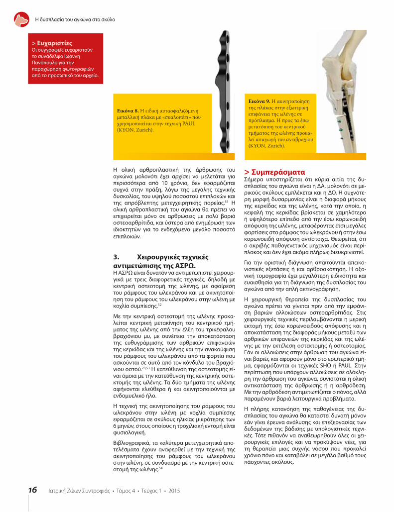
Κατά τη νεότερη τεχνική PAUL εκτελείται οστεκτομή στο κεντρικό τμήμα της ωλένης και τα δύο καταγμα-τικά τμήματα ακινητοποιούνται σε νέα θέση με ειδική αυτασφαλιζόμενη μεταλλική πλάκα, στο μέσον της οποίας υπάρχει «σκαλοπάτι» ύψους 2 ή 3 mm (KYON, Zurich, Switzerland) (εικόνα 8).50 Στη νέα αυτή θέση η ωλένη προκαλεί απαγωγή του αντιβραχίου, με συ-νέπεια τη μεταφορά των φορτίσεων του βάρους του σώματος στο εξωτερικό τμήμα της άρθρωσης του αγκώνα και την ανακούφιση του εσωτερικού τμήμα-τος (εικόνα 9). Μολονότι η τεχνική αυτή υπόσχεται καλά αποτελέσματα δεν έχει γίνει μέχρι σήμερα μεγά-λη αναλυτική μελέτη του ποσοστού της επιτυχίας και των επιπλοκών από την εφαρμογή της.
Όταν και στα δύο τμήματα της άρθρωσης του αγκώνα υπάρχουν βαριές αλλοιώσεις οστεοαρθρίτιδας, οι χει-ρουργικές θεραπευτικές επιλογές είναι η αρθρόδεση ή η ολική αντικατάσταση της άρθρωσης.
Με την αρθρόδεση της άρθρωσης του αγκώνα επι-τυγχάνεται ανακούφιση από τον πόνο της χρόνιας οστεοαρθρίτιδας, προκαλείται όμως σημαντική απώ-λεια της λειτουργικότητας του άκρου.49
Εικόνα 7(α). Με το βέλος απεικονίζεται ο μηχανικός άξονας βραχιόνιου οστού – αντιβραχίου. Τα μεγαλύτερα φορτία μεταφέρονται στο εσωτερικό τμήμα της άρθρωσης του αγκώνα.
Εικόνα 7(β). Μετά την οστεοτομή στη διάφυση του βραχιόνιου οστού και την ακινητοποίηση των δυο τμημάτων που προκύπτουν σε νέα θέση, μετατοπίζεται ο μηχανικός άξονας προς τα έξω (βέλος).
α β
Η δυσπλασία του αγκώνα στο σκύλο
16
Εικόνα 8. Η ειδική αυτασφαλιζόμενη μεταλλική πλάκα με «σκαλοπάτι» που χρησιμοποιείται στην τεχνική PAUL (KYON, Zurich).
Εικόνα 9. Η ακινητοποίηση της πλάκας στην εξωτερική επιφάνεια της ωλένης σε πρόπλασμα. Η προς τα έσω μετατόπιση του κεντρικού τμήματος της ωλένης προκα-λεί απαγωγή του αντιβραχίου (KYON, Zurich).
> ΕυχαριστίεςΟι συγγραφείς ευχαριστούν το συνάδελφο Ιωάννη Πανόπουλο για την παραχώρηση φωτογραφιών από το προσωπικό του αρχείο.
Η ολική αρθροπλαστική της άρθρωσης του αγκώνα μολονότι έχει αρχίσει να μελετάται για περισσότερα από 10 χρόνια, δεν εφαρμόζεται συχνά στην πράξη, λόγω της μεγάλης τεχνικής δυσκολίας, του υψηλού ποσοστού επιπλοκών και της απρόβλεπτης μετεγχειρητικής πορείας.51 Η ολική αρθροπλαστική του αγκώνα θα πρέπει να επιχειρείται μόνο σε αρθρώσεις με πολύ βαριά οστεοαρθρίτιδα, και ύστερα από ενημέρωση των ιδιοκτητών για το ενδεχόμενο μεγάλο ποσοστό επιπλοκών.
3. Χειρουργικές τεχνικές αντιμετώπισης της ΑΣΡΩ.Η ΑΣΡΩ είναι δυνατόν να αντιμετωπιστεί χειρουρ-γικά με τρεις διαφορετικές τεχνικές, δηλαδή με κεντρική οστεοτομή της ωλένης, με αφαίρεση του ράμφους του ωλεκράνου και με ακινητοποί-ηση του ράμφους του ωλεκράνου στην ωλένη με κοχλία συμπίεσης.52
Με την κεντρική οστεοτομή της ωλένης προκα-λείται κεντρική μετακίνηση του κεντρικού τμή-ματος της ωλένης από την έλξη του τρικέφαλου βραχιόνιου μυ, με συνέπεια την αποκατάσταση της ευθυγράμμισης των αρθρικών επιφανειών της κερκίδας και της ωλένης και την ανακούφιση του ράμφους του ωλεκράνου από τα φορτία που ασκούνται σε αυτό από τον κόνδυλο του βραχιό-νιου οστού.23,53 Η κατεύθυνση της οστεοτομής εί-ναι όμοια με την κατεύθυνση της κεντρικής οστε-κτομής της ωλένης. Τα δύο τμήματα της ωλένης αφήνονται ελεύθερα ή και ακινητοποιούνται με ενδομυελικό ήλο.
Η τεχνική της ακινητοποίησης του ράμφους του ωλεκράνου στην ωλένη με κοχλία συμπίεσης εφαρμόζεται σε σκύλους ηλικίας μικρότερης των 6 μηνών, στους οποίους η τροχιλιακή εντομή είναι φυσιολογική.
Βιβλιογραφικά, τα καλύτερα μετεγχειρητικά απο-τελέσματα έχουν αναφερθεί με την τεχνική της ακινητοποίησης του ράμφους του ωλεκράνου στην ωλένη, σε συνδυασμό με την κεντρική οστε-οτομή της ωλένης.54
> ΣυμπεράσματαΣήμερα υποστηρίζεται ότι κύρια αιτία της δυ-σπλασίας του αγκώνα είναι η ∆Α, μολονότι σε με-ρικούς σκύλους εμπλέκεται και η ∆Ο. Η συχνότε-ρη μορφή δυσαρμονίας είναι η διαφορά μήκους της κερκίδας και της ωλένης, κατά την οποία, η κεφαλή της κερκίδας βρίσκεται σε χαμηλότερο ή υψηλότερο επίπεδο από την έσω κορωνοειδή απόφυση της ωλένης, μεταφέροντας έτσι μεγάλες φορτίσεις στο ράμφος του ωλεκράνου ή στην έσω κορωνοειδή απόφυση αντίστοιχα. Θεωρείται, ότι ο ακριβής παθογενετικός μηχανισμός είναι περί-πλοκος και δεν έχει ακόμα πλήρως διευκρινιστεί.
Για την οριστική διάγνωση απαιτούνται απεικο-νιστικές εξετάσεις ή και αρθροσκόπηση. Η αξο-νική τομογραφία έχει μεγαλύτερη ειδικότητα και ευαισθησία για τη διάγνωση της δυσπλασίας του αγκώνα από την απλή ακτινογράφηση.
Η χειρουργική θεραπεία της δυσπλασίας του αγκώνα πρέπει να γίνεται πριν από την εμφάνι-ση βαριών αλλοιώσεων οστεοαρθρίτιδας. Στις χειρουργικές τεχνικές περιλαμβάνονται η μερική εκτομή της έσω κορωνοειδούς απόφυσης και η αποκατάσταση της διαφοράς μήκους μεταξύ των αρθρικών επιφανειών της κερκίδας και της ωλέ-νης με την εκτέλεση οστεκτομής ή οστεοτομίας. Εάν οι αλλοιώσεις στην άρθρωση του αγκώνα εί-ναι βαριές και αφορούν μόνο στο εσωτερικό τμή-μα, εφαρμόζονται οι τεχνικές SHO ή PAUL. Στην περίπτωση που υπάρχουν αλλοιώσεις σε ολόκλη-ρη την άρθρωση του αγκώνα, συνιστάται η ολική αντικατάσταση της άρθρωσης ή η αρθρόδεση. Με την αρθρόδεση αντιμετωπίζεται ο πόνος, αλλά παραμένουν βαριά λειτουργικά προβλήματα.
Η πλήρης κατανόηση της παθογένειας της δυ-σπλασίας του αγκώνα θα καταστεί δυνατή μόνον εάν γίνει έρευνα ανάλυσης και επεξεργασίας των δεδομένων της βάδισης με υπολογιστικές τεχνι-κές. Τότε πιθανόν να αναθεωρηθούν όλες οι χει-ρουργικές επιλογές και να προκύψουν νέες, για τη θεραπεία μιας συχνής νόσου που προκαλεί χρόνιο πόνο και καταβάλει σε μεγάλο βαθμό τους πάσχοντες σκύλους.
Η δυσπλασία του αγκώνα στο σκύλο

17
> Βιβλιογραφία1. Griffon DJ. Surgical diseases of the elbow. In: Small Animal Veterinary Surgery. Tobias KM, Johnson SA (eds). Elsevier Saunders, St Louis, MO, USA, 2012, pp.724-759.
2. Hazewinkel HAW, Meij BP, Nap RC, Ubbink GJ. Radiographic views for elbow dysplasia screening in Bernese Mountain dogs. Proceedings International Elbow Working Group 1995, 5: 32-37.
3. Morgan JP, Wind A, Davidson AP. Bone dysplasias in the labrador retriever: a radiographic study. J Am Anim Hosp Assoc 1999, 35: 332–340.
4. Morgan JP, Wind A, Davidson AP. Elbow Dysplasia. In: Hereditary Bone and Joint Diseases in the Dog. Morgan JP and Davidson AP, (eds). 1st ed. Hannover Schultersche: Germany, 2000, pp. 41-68.
5. Meyer-Lindberg A, Fehr M, Nolte I. Co-existance of UAP and FCP of the ulna in the dog. J Small Anim Pract 2006, 47: 61–65.
6. Kirberger RM, Fourie SL. Elbow dysplasia in the dog: pathophysiology, diagnosis and control. J S Afr Vet Assoc 1998, 69: 43–54.
7. Vermote KA, Bergenhuyzen AL, Gielen I, van Bree H, Duchateau L, Van Ryssen B. Elbow lameness in dogs of six years and older. arthroscopic and imaging findings of medial coronoid disease in 51 dogs. Vet Comp Orthop Traumatol 2010, 23: 43–50.
8. Snaps FR, Balligand MH, Saunders JH, Park RD, Dondelinger RF. Comparison of radiography, magnetic resonance imaging and surgical findings in dogs with elbow dysplasia. Am J Vet Res 1997, 58: 1367-1370.
9. Michelsen J. Canine Elbow dysplasia: aetiopathogenesis and current treatment recommendations. Vet J 2013, 196:12-19.
10. Clements DN. Gene expression in normal and diseased elbows. In: Proceedings of the Autumn Meeting of the British Veterinary Orthopaedic Association: Chester, UK, 2006, pp. 6–7.
11. Grandalen J, Lingaas F. Arthrosis in the elbow joint of young rapidly growing dogs: A genetic investigation. J Small Anim Pract 1991, 32: 460–464.
12. Lewis TW, Ilska JJ, Blott SC, Woolliams JA. Genetic evaluation of elbow scores and the relationship with hip scores in UK Labrador retrievers. Vet J 2011, 189: 227–233.
13. Nap RC. Pathophysiology and clinical aspects of canine elbow dysplasia. In: Proceedings of the 7th International Elbow Working Group Meeting, Constance, Germany, 1995, pp. 6–8.
14. Olsson SE. The early diagnosis of fragmented coronoid process and osteochondritis dissecans of the canine elbow joint. J Am Anim Hosp Assoc 1983, 19: 616–626.
15. Gemmill TJ, Mellor DJ, Clements DN, Clarke SP, Farrell M, Bennett D, Carmichael S. Evaluation of elbow incongruency using reconstructed CT in dogs suffering fragmented coronoid process. J Small Anim Pract 2005, 46: 327–333.
16. Kramer A, Holsworthy IG, Wisner ER, Kass PH, Schultz KS. Computed tomographic evaluation of canine radioulnar incongruence in vivo. Vet Surg 2006, 35: 24–29.
17. Hulse D. Co-contraction of the biceps/brachialis muscle complex produces a rotational moment which may induce fragmentation/microfracture of the medial coronoid. In: Congress proceedings of the American College of Veterinary Surgeons Symposium: San Diego, USA, 2008, pp. 466.
18. Bennett D, Duff SR, Kene RO, Lee R. Osteochondritis dissecans and fragmentation of the coronoid process in the elbow joint of the dog. Vet Rec 1981, 109: 329–336.
19. Guthrie S, Plummer JM, Vaughan LC. Post natal development of the canine elbow joint: a light and electron microscopic study. Res Vet Sci 1992, 52: 67-71.
20. Burton, NJ, Owen MR. Canine elbow dysplasia 2. Treatment and prognosis. In Practice 2008, 30: 552–557.
21. Bottcher P. Radio-ulnar incongruence in dogs with elbow dysplasia. In: Congress proceedings of the American College of Veterinary Surgeons Symposium, Chicago, USA, 2011b, pp. 110–112.
22. Preston, CA, Schulz KS, Kass PH. In vitro determination for contact areas in the normal elbow joint of dogs. Am J Vet Res 2000, 61: 1315–1321.
23. Sjstrom L, Kasstom H, Kllber M. Ununited anconeal process in the dog. Pathogenesis and treatment by osteotomy of the ulna. Vet Comp Orthop Traumatol 1995, 8: 170–176.
24. Lozier SM. How I treat elbows in the older canine patient and new prospective in elbow dysplasia. In: Congress proceedings of the 13th European Society of Veterinary Orthopaedics and Traumatology: Munich, Germany, 2006, pp. 93–96.
25. Wind AP, Packard ME. Elbow incongruity and developmental elbow diseases in the dog. Part II. J Am Anim Hosp Assoc 1986, 22: 725–730.
26. Proks P, Necas A, Stehlik L, Srnec R, Griffon DJ. Quantification of humeroulnar incongruity in Labrador retrievers with and without medial coronoid disease. Vet Surg 2011, 40: 981–986.
27. Fitzpatrick N. Biceps ulnar release procedure for treatment of medial coronoid disease in 49 elbows. In: Congress proceedings of the 36th Annual Conference, Veterinary Orthopaedic Society, Steamboat Springs: Colorado, USA, 2009, p. 44.
28. Fitzpatrick, N, Yeadon R. Working algorithm for treatment decision making for developmental disease of the medial compartment of the elbow in dogs. Vet Surg 2009, 38: 285–300.
29. Trostel C, McLaughlin R, Pool R. Canine lameness caused by developmental orthopedic diseases: FCP and UAP. Compend Cont Educ Pract 2003, 25: 112.
30. Demko J, McLaughlin R. Developmental orthopedic disease. Vet Clin North Am 2005, 35: 1111-1135.
31. Punke JP, Hulse DA, Kerwin SC, Peycke LE, Budsberg SC. Arthroscopic documentation of elbow cartilage pathology in dogs with clinical lameness without changes on standard radiographic projections. Vet Surg 2009, 38: 209
32. Boulay JP. Fragmented medial coronoid process of the ulna in the dog. Vet Clin North Am 1998, 28: 449-458.
33. Henry WB. Radiographic diagnosis and surgical management of fragmented medial coronoid process in dogs. J Am Vet Med Assoc 1984, 184: 799-805.
34. Wosar M, Lewis D, Neuwirth L, Parker RB, Spencer CP, Kubilis PS, Stubbs WP, Murphy ST, Shiroma JT, Stallings JT, Bertrand SG. Radiographic evaluation of elbow joints before and after surgery in dogs with possible fragmented medial coronoid process. J Am Vet Med Assoc 1999, 214: 52-58.
35. Rovesti G, Biasibetti M, Schumacher A, Fabiani M. The use of CT in the diagnostic protocol of the elbow in the dog: 24 joints. Vet Comp Orthop Traumatol 2002, 15: 35-39.
36. Carpenter LG, Schwarz PD, Lowry JE, Steyn PF. Comparison of radiologic imaging techniques for diagnosis of FCP of the cubital joint in dogs. J Am Vet Med Assoc 1993, 203: 78-83.
37. Ness M. Treatment of FCP in young dogs by proximal ulnar osteotomy. J Small Anim Pract 1998, 39:15-21.
38. Read RA, Armstrong SJ, O’Keef D, Eger CE. Fragmentation of the medial coronoid process of the ulna in dogs: a study of 109 cases. J Small Anim Pract 1990, 31: 330-334.
39. Book GR, Miller CW, Tavers CL. A comparison of surgical and medical treatment of fragmented coronoid process and osteochondritis dissecans of the canine elbow. Vet Comp Orthop Traumatol 1995, 8: 117-120.
40. Fitzpatrick N. Subtotal coronoid ostectomy (SCO) for the treatment of medial coronoid disease: A prospective study of 228 dogs (389 elbows) evaluating short and medium term outcome. In: Congress proceedings of the British Veterinary Orthopaedic Association, Autumn Scientific Meeting-Enigmas of the Canine Elbow, Chester, UK, 2006, pp. 22–29.
41. Danielson KC, Fitzpatrick N, Muir P, Manley PA. Histomorphometry of fragmented medial coronoid process in dogs: a comparison of affected and normal coronoid processes. Vet Surg 2006, 35: 501-512.
42. Tobias T, Miyabayashi T, Olmstead ML. Surgical removal of FCP in the dog: comparative effects of surgical approach and age at time of surgery. J Am Anim Hosp Assoc 1994, 30: 360-362.
43. Bοttcher, P. Accelerated cartilage loss following subtotal coronoid ostectomy. In: Congress proceedings of the American College of Veterinary Surgeons Sympo-sium: Chicago, USA, 2011, pp. 108–109.
44. Preston CA, Schulz KS, Taylor KT, Kass PH, Hagan CE, Stover SM. In vitro experimantal study of the effect of radial shortening and ulnar ostectomy on contact pat-terns in the elbow joint of dogs. Am J Vet Res 2001, 62: 1548–1556.
45. Holsworth IG. How I manage elbow incongruity. In: Congress proceedings of the 12th European Society of Veterinary Orthopaedics and Traumatology Congress: Munich, Germany, 2004, pp. 78-79.
46. Fox DJ. Radius and ulna. In: Small Animal Veterinary Surgery. Tobias KM, Johnson SA (eds). Elsevier Saunders: St. Louis, MO, USA, 2012, pp. 761–784.
47. Johnston SA. Osteochondritis dissecans of the humeral head. Vet Clin North Am Small Anim Pract 1998, 28: 33-40.
48. Schulz, KS, Fitzpatrick N, Young R. Theory and development of the sliding humeral osteotomy. In: Congress proceedings of the American College of Veterinary Surgeons Symposium, Chicago, USA, 2011, pp. 263–265.
49. Fitzpatrick N, Yeadon R, Smith TJ, Schulz K. Techniques of application and initial clinical experience with sliding humeral osteotomy for treatment of medial compartment disease of the canine elbow. Vet Surg 2009, 38: 261–278.
50. Pfeil I, Torrington A, Vezzoni A. In proceedings: Proximal abducting ulnar osteotomy. KYON Symposium, Zurich, 2014.
51. Acker R, Van Der Meulen GT. Tate elbow preliminary trials. In: Congress proceedings of the 35th Veterinary Orthopedic Society Annual Conference, MT, USA, March 2008 pp. 49-52.
52. Meyer-Lindenberg A, Fehr M, Nolte I. Short and longterm results after surgical treatment of an ununited anconeal process in the dog. Vet Comp Orthop Traumatol 2001, 14: 101–110.
53. Turner BM, Abercromby Rh, Innes J, McKee WM, Ness MG. Dynamic proximal ulnar osteotomy for the treatment of ununited anconeal process in 17 dogs. Vet Comp Orthop Traumatol 1998, 11: 76-80.
54. Krotscheck U, Hulse DA, Bahr A, Jerram RM. Ununited anconeal process: Lag-screw fixation with proximal ulnar osteotomy. Vet Comp Orthop Traumatol 2000, 13: 212-216.
Η δυσπλασία του αγκώνα στο σκύλο

18
Canine elbow dysplasia
current treatment recommenda-
> ABSTRACTElbow dysplasia is a condition that causes pain and lameness in large and giant breed dogs. Its origins are genetic and when combined with environmental factors, development of the elbow joint becomes abnormal. Originally, elbow osteochondrosis was considered to be the main cause of this condition. Modern studies claim that the condition, in most cases, is caused by various forms of incongruity between articular surfaces of the three joints forming the elbow. Treatment is surgical and should be performed before the development of osteoarthritic lesions in the joint. Multiple surgical techniques to correct this condition are described in the literature. In cases when radiological examination of the elbow joint reveals severe osteoarthritic lesions, the selection of surgical technique depends on lesion localization.
> IntroductionElbow dysplasia is a hereditary condition, char-acterized by the abnormal development of the elbow joint. Since 1993, the International Elbow Working Group (IEWG) has defined that heredi-tary pathological conditions affecting the elbow joint, reportedly referred to as «elbow dysplasia», include the ununited anconeal process (UAP), osteochondritis dissecans of the medial humeral condyle (OCD), medial coronoid disease (MCD) and incongruity between the articular surfaces of the bones forming the elbow joint (IC).1
The condition mainly affects large and giant breed dogs. Over-represented breeds include Bernese Mountain dogs,2 Labrador retrievers,3 Golden retrievers, Rottweilers and German shepherd dogs.4Rapidly-growing male dogs are affected at double the frequency of female dogs;5,6 medium-sized, chondrodystrophic dog breeds (Dachshund, French bulldog) are affected less commonly.4
The main clinical sign of this condition is lameness, which can develop between three and ten months of age. In some dogs, lameness manifests in adulthood (>6 years old), due to MCD, without any history of lameness at a younger age.7 Both forelimbs are affected in 37-50% of cases.8
Due to the fact that MCD, OCD and IC form lesions in the internal anatomical constituents of the elbow joint (medial coronoid process of the ulna, semilunar ulnar notch and medial part of the humeral condyle), and also because these three abnormal conditions may appear in combination, the more generic term «medial compartment disease» (MCoD) has been suggested as a replace-
ment for previous terms.9
> Aetiopathogenesis The genetic origins of elbow dysplasia have been investigated with several, large-scale epidemiologic papers. Based on the latter, the disease appears to be inherited differently in each dog breed. Moreover, it has been commonly hypothesized that each form of elbow dysplasia is inherited separately from the rest. The conclusion is that elbow dysplasia is caused by several genetic conditions, which disrupt the development of the elbow joint by several pathogenetic mechanisms. Due to the complexity of its hereditary transmission and the role of environmental factors in the development of the disease, it is maintained that the genetic investigation of elbow dysplasia is impossible in the immediate future.10,11,12
Three aetiopathogenic mechanisms have been reported for elbow dysplasia: osteochondritis dis-secans,13,14 elbow incongruity15 and rotatory insta-bility of the radioulnar joint.16,17
These mechanisms result from genetic predilection in combination with secondary predisposing factors, such as a diet of high caloric density and rigorous exercise, and constitute the main causes for the overt manifestation of the disease.9
In current studies, it is maintained that out of the three aetiopathogenic mechanisms, the most likely to cause overt disease is elbow incongruity. In some dogs, osteochondrosis is an important cause. The mechanism for distal radioulnar joint
DogDysplasia Elbow
Key words
Corresponding author: S.Kladakis, Army Veterinarian, 3 Damaskinou Georgakou Str., Postal code 55132, Agios Ioannis, Kalamaria, Thessaloniki, Tel:+30 6944882220, Ε-mail: [email protected]
Danourdis A.Veterinarian,
Private practitioner, Veterinary Clinic,
Paradeisou 48, Chalandri, 15233, Attiki,
Τel.+30 2106800758
Kladakis S.Army Veterinarian,
3rd Army Veterinary Hospital, Thermi,
57001, Thessaloniki, Tel.+30 2310462425
Leventogiannis V.Veterinarian,
Private practitioner, Veterinary Diagnos-
tic Imaging Center, Solomou 7,
Chalandri, Attiki, Tel.+30 2108015710
Lionakis A.Veterinarian,
Private practitioner, Companion
Animal Clinic, Pentelis 13,
Maroussi, 15126, Attiki, Tel.+30 2108060160

19
rotatory instability is currently investigated.
Α. Οsteochondrosis According to the standard theory, osteochondrosis is responsible for the formation of lesions in the medial coronoid process, the humeral condyle and the growth plate of the anconeal process.13,18
In normal animals, the cartilagenous medial coronoid process undergoes ossification from its base to its peak. It has been proven that although it lacks a separate ossification centre, it has the same ossification centre as the ulnar diaphysis. The ossification process is complete by the age of 5-5½ months. If the endochondral ossification of the cartilagenous medial coronoid process is disrupted, necrosis of the deeper cartilage cells is imminent, resulting in cartilage softening and the formation of erosions. These fragments of the medial coronoid process usually undergo calcification because their perfusion, which occurs through fibrous attachments to the annular ligament, is disrupted.
This theory contrasts with a more recent paper, which was based on the histopathologic exami-nation of the coronoid process in clinical cases.19 According to the latter, osteochondritis dissecans lesions were not discovered in the affected segments of the medial coronoid process; however, changes consistent with fractures were detected. It is concluded that such lesions are caused by powerful and constant forces on the cartilagenous medial coronoid process and the formation of stress fractures, which inhibit the ossification of its fibrous tissue.
Β. Elbow incongruity. The elbow joint is a composite joint, formed by three specific articulations, including the humeroradial joint (the capitulum of the humeral condyle articulates with the proximal surface of the head of the radius), the humeroulnar joint (the trochlea humeri articulates with the trochlear or semilunar ulnar notch and the medial coronoid process) and the radioulnar joint (the posterior articular surface of the head of the radius articulates with the radial notch of the ulna).19
A difference in length between the radius and the ulna and humeroulnar incongruity have been previously reported as forms of incongruity.
Β1. Difference in length between the radius and the ulna (radioulnar incongruity)
This difference is caused by asynchronous longitudinal growth of the two bones. Two forms have been reported: in the first form, the radius is shorter than the ulna or the radial head is located laterally to the medial coronoid process (short radius syndrome).15,20 In the second form, the ulna is shorter than the radius or the radial physis is located medially to the medial coronoid process (short ulna syndrome).21
In the shortened radius syndrome, the magnitude of weight-bearing forces are transported by the trochlea humeri to the medial coronoid process. The intense shearing forces lead to stress-induced damage in the subchondral bone of the medial coronoid process resulting in the formation of fissures or fragmentations (Figure 1).21
In the second form, the head of the radius exerts centrally-located pressure on the humeral condyle, which in turn is transmitted to the anconeal process, thus preventing its ossification and fusion with the ulnar physis.22,23
According to the above and based on linear forces being exerted on the joint, MCD and UAP can be explained. With the linear forces theory, however, the combination of MCD and UAP in clinical cases cannot be justified.5
In order to interpret the combination of these ab-normal conditions, a new theory has been sug-gested based on the action of rotational forces (rotational force theory).24 According to this theory, there are once more two variations related to the difference of length between the radius and the ulna. In the shortened radius syndrome, which is less frequently observed, the linear force theory and the rotational force theory are relatively similar. In contrast, in the shortened ulna syndrome, which is more commonly reported, the rotational theory differs from the linear force theory.
According to the rotational force theory, if the radius grows in length faster than the ulna, the radial head exerts forces with a central direction to the humeral condyle. Initially, the displacement of the humerus medially is prevented by the anconeal process. Later, as the radius continues to grow in length, so do the loads placed on the humeral condyle, which begins to externally rotate. As the humerus rotates externally, the caudal aspect of the humeral condyle impinges on the lateral surface of the base of the anconeal process. If this process occurs prior to the ossification of the growth plate of the anconeal process, UAP may develop. The continuous medial advancement of the humeral bone, combined with its simultaneous rotation, places intense loads and leads to abrasions upon the point of contact of the caudal medial surface of the humeral condyle with the lateral surface of the ulnar trochlear notch. At this point, lesions
Canine elbow dysplasia
Figure 1. Short radius syndrome. Presence of the articular surfaces of the radius and ulna on different levels («step») .1. Medial coronoid process2. Articular surface of the radius3. Radial notch of the ulna4. Anconeal process5. Trochlear notchΤhe red arrows represent severe weight-bearing loads that the medial coronoid process is subjected to, whereas the black arrows represent the lesser forces the articular surface of the radius is subjected to.

20
Canine elbow dysplasia
caused by friction are formed in the cartilage of the humeral condyle, as well as in the cartilage of the trochlear notch. The continuous loads placed by the radial head on the humeral condyle, combined with the effect of the anconeal process as a lever, lead to the downward displacement of the trochlea humeri and the compression of the medial coronoid process. The compression and friction result in the formation of lesions in the medial coronoid process, as well as the medial part of the humeral condyle.
Finally, as rotation of the humerus continues to increase, especially when there is no UAP, rostral displacement of the trochlea humeri follows, resulting in its displacement from the trochlear notch of the ulna, thus causing elbow subluxation.
Β2. Humeroulnar incongruity
This is noted when the radius of curvature of the trochlear notch of the ulna is smaller than the radius of curvature of the trochlea humeri or when the trochlear notch of the ulna has an elliptical shape. This geometrical abnormality subsequently results in malarticulation of the trochlear humeri with the trochlear notch, resulting in abnormal forces and loads on the medial coronoid process.25,26 It has been noted that in Bernese mountain dogs there is a predilection for the formation of an elliptical trochlear notch.2
C. Posterolateral rotatory instability of the proximal radioulnar joint When there is incongruity of the caudal articular surface of the radius with the radial notch of the ulna, the lateral pull of the biceps brachii and the brachialis muscle causes external rotation of the ulna (over the radius) during flexion of the joint. Rotation results in compression of the two articular surfaces and damage to the medial coronoid process and radial notch.27
Diagnosis Each form of elbow dysplasia may develop as a sin-gle condition or, more commonly, in combination. Unrelated to the form of dysplasia, clinical signs are always similar, thus inhibiting identification of the form by physical examination alone. Clinical signs usually begin from the age of 3-10 months.
During observation of dogs with elbow dysplasia, a stiff gait is noted following a period of rest, or lameness manifests after exercise. When the disorder affects both forelimbs, detection of lameness can be challenging. In a standing position, the affected limb is abducted and the antebrachium and toes are externally rotated (supination). This position helps to reduce the forces applied to the medial compartment of the elbow.28
One of the most reliable findings during physical examination is pain manifesting upon palpation as well as during passive range motions of the elbow joint. Pain manifests upon deep palpation of the insertion of the biceps brachii muscle into the ulnar tuberosity which is located on its internal surface, over the medial aspect of the coronoid process29. Moreover, pain manifests during extreme flexion of the joint in combination with external rotation of the antebrachium.
The manifestation of pain during physical exami-nation, combined with the absence of any other recognizable cause of lameness or pain, is an im-portant finding that supports elbow dysplasia. In the case of UAP, a synovial fluid effusion may be observed.30 In MCD, the presence of joint effusion is uncommon.
In dogs with the chronic form of the disorder and simultaneous presence of secondary osteoarthritis, a decrease in joint motion range is observed along with crepitation, muscular atrophy and swelling of the elbow joint.
Cases of adult dogs have been reported to show unilateral or bilateral lameness of the forelimbs due to MCD, with no history of lameness at a younger age.7
The differential diagnosis should always include ev-ery possible cause of frontal limb lameness such as disorders of the shoulder joint, tendon injury, neu-rological causes of lameness and neoplasia.
For a definitive diagnosis and identification of the form of elbow dysplasia, imaging investigation of the elbow joint is essential. Imaging can be per-formed with standard radiography, computed to-mography (CT) or magnetic resonance imaging (MRI). Primary findings of elbow dysplasia may be missed in radiographs. In such cases, diagnosis is based on detecting secondary findings of osteoar-thritis, which develop after the age of seven months.
Radiographs contribute poorly to the diagnosis of MCD. For example, erosions in the articular cartilage of the condyle or fissures in the subchondral bone of the medial coronoid process cannot be viewed with standard radiography.31,32,33,34
In a dog with clinical signs of elbow dysplasia, if no lesions are found on standard radiographs or if such lesions are unclear, further diagnostic investigation is warranted by CT or MRI. With CT, the bone structures of the elbow joint are viewed without superimposition.35 For this reason, its specificity and sensitivity are greater than those of radiography (Table 1).8,36
Table 1. Sensitivity, specificity and accuracy of plain radiography, computed tomography (CT), magnetic resonance imaging (MRI) and arthroscopy in the diagnosis of MCD8,36
Sensitivity % Specificity % Diagnostic Accuracy %
Radiography 23,5-28,5 100 56,7-77,2
Computed tomography 71-88,2 84-84,6 86,7
Magnetic resonance imaging with a calcified coronoid process
100 93,3 95,5
Magnetic resonance imaging with a cartilaginous medial coronoid process
83,3 93,7 91
Arthroscopy 82 100 _

21
Canine elbow dysplasia
Nevertheless, it is possible that CT and MRI may not clearly visualize subchondral bone lesions in the medial coronoid process. If the findings of all imaging modalities are unclear, arthroscopy is rec-ommended. In 37-50% of cases, the disorder affects both forelimbs, rendering imaging investigation of both joints necessary.8
For conventional radiographic evaluation, good quality radiographs are required with the dog cor-rectly placed. It is necessary to sedate or fully an-aesthetize the animals. In all positions, the limb is placed on top of the cassette without the use of a grid. Furthermore, radiography should include the shoulder joint for possible osteochondritis disse-cans which may be the cause of lameness or may coexist with elbow dysplasia.
In order to depict all of the basic anatomical segments of the joint and all the possible pathological conditions, radiographs should be obtained with the limb in four separate views. The latter include the mediolateral projection with the elbow extended, the maximally flexed mediolateral projection, the craniocaudal view and external oblique or craniolateral-caudomedial oblique projection (150). In the former two views, the dog is positioned in lateral recumbency and in the latter in sternal recumbency.
Α. Mediolateral projection with the elbow extended In this view, the angle between the humerus and the antebrachium should be about 110ο. Τhe limb is positioned in such a way that there is superimposi-tion of the humeral condyles.
Visualization of a normal elbow in a dog older than six months is characterized by articular surface congruity and uniform narrow joint spaces between the humerus, the radius and the ulna. Additionally, the edge of the trochlear notch of the ulna and the radial head should form an arch with a smooth outline (Figure 2).
With such positioning, in a dog less than one year of age with elbow dysplasia, one or more of the following abnormal findings may be noted:
1. Increased joint space in the humeroulnar artic-ulation along the internal surface of the troch-lear notch.
2. Increased joint space in the humeroradial ar-ticulation
3. Interruption of the smooth outline of the arch formed by the ulnar trochlear notch and the ar-ticular surface of the radius. This abnormal find-ing is called «step».34
4. Fragmented medial coronoid process, which can be simple or compound. The normal medial coronoid process is seen as a triangular area of subchondral bone with a sharp outline, super-imposing over the radial head. Fracture of the medial coronoid process is seen only in 9.8 % of cases.
5. Mild increase of bone radiopacity (osteosclero-sis) and loss of trabecular bone along the me-
dial surface of the semilunar notch of the ulna. This radiologic finding is an early indication of abnormal loads placed on the elbow joint (Fig-ure 3).
6. Presence of osteophytes along the dorsal as-pect of the anconeal process. In dogs older than one year of age, lesions of secondary osteoar-thritis usually extend to the proximal articular surface of the radial head.
Β. Maximally flexed mediolateral projection In this positioning, the angle between the humerus and the antebrachium should be more or less equal to 45ο. The limb is placed so that there is super-imposition of the humeral condyles on the radio-graphic film.
This positioning is ideal for the diagnosis of UAP, compared to the mediolateral view of the extended elbow, because it has the distinct advantage of being able to avoid superimposition of the medial epicondyle on the olecranon. Therefore, the anconeal process and any osteophytes along its dorsal aspect can be clearly visualized.
In large dog breeds, the anconeal process has its own separate secondary centre of ossification. The physis of this ossification centre of the anconeal process can be visualized in radiographs up until
Figure 2. Mediolateral view of a normal elbow. Articular surface congruity and uniform narrow width of the humeroradial joint and humeroulnar joint are noted.
Figure 3. Mediolateral flexed view of the elbow. Increased radiopacity is noted on the periphery of the trochlear notch (arrow).

22
the age of 3½ - 5½ months. If the physis remains visible in radiographs of older dogs, it is considered pathognomonic for UAP. The remaining physis is seen as a radiolucent space with unclear margins between the anconeal process and the olecranon (Figure 4).
C. Craniocaudal view of the elbow joint In this view, the dog is placed in sternal recumbency and the limb intended for radiographic evaluation is held with the humerus vertical to the axis of the spinal column, the elbow joint is held in contact with the cassette, and the antebrachium is rostrally extended.
In such a position, the characteristic OCD lesion can be visualized as early as 5-6 months of age as a ra-diolucent area or collapse of the medial compart-ment, or even as a defect in the subchondral bone of the articular surface of the humeral trochlea.33
D. Craniolateral-caudomedial oblique projection (-150) of the elbow. In this view, the dog is placed in sternal recumben-cy and the limb intended for radiographic evalua-tion is positioned as for the craniocaudal view ex-cept that the antebrachium is rotated externally by
150 (pronation). The radiographic beam is focused on the centre of the elbow joint. In this position, the articular surface of the trochlea humeri and the me-dial coronoid process are more clearly visualized.
Current treatment options Treatment for elbow dysplasia is surgical and should be performed before the development of osteoarthritic lesions in the joint. If surgical treatment is delayed, the development of osteoarthritis cannot usually be adequately controlled. Unfortunately, the complexity of aetiopathogenesis, physical examination and imaging findings render diagnosis in the early stages of the disease somewhat of a challenge.
Surgical techniques to correct elbow dysplasia described in this paper are classified as surgical procedures to correct MCoD in dogs with mild osteoarthritis or moderate or severe osteoarthritis, and to correct the UAP.44
1. Surgical procedures for MCoD in dogs with mild osteoarthritis.
a. MCD.
Even though several research studies have been undertaken to select the appropriate modality in order to correct MCD, controversy still surrounds the procedure of choice up to the present day. The results of research studies vary. However, normal function of the limb has been reported in most procedures for a short length of time after surgery, and exacerbation of osteoarthritis has been noted in the mid- to long-term outcome.37,38 In fact, some authors report that surgical treatment does not alter the long-term prognosis, and they prefer to manage MCD with conservative treatment.39
Surgical procedures that have been described in the treatment of MCD can be classified under two categories. Focal procedures via arthrotomy or arthroscopy can be placed under the first category, whereas the second category includes proximal ulnar osteotomy which can be performed in combination with focal procedures.
Among the focal techniques, those that have been researched the most are the removal of the osteochondral fragment or fragments of the medial coronoid process, as well as subtotal medial coronoid ostectomy.
Removal of the fragment or fragments of the medial coronoid process always leads to osteoarthritis and persistence of lameness because degenerative lesions remain in the subchondral bone. For that reason, simultaneous partial osteotomy of a large segment of the medial coronoid process was recommended, in which most of the diseased subchondral bone is included 28,40,41 (Figure 5).
Subtotal medial coronoid ostectomy, which can be performed with an osteotome or an air saw, has a caudal medial direction from the periphery of the medial coronoid process toward the external
Figure 4. Neutral mediolateral view of the elbow joint. UAP (arrow no 1), increased radiopacity of the subchondral bone on the rim of the trochlear notch (arrow no 2) and widening of the articular space between the humerus and the radial head (arrow no 3) can be observed.
Figure 5. The black line depicts the location from which the fragment of the medial coronoid process may detach.The red line represents the direction of subtotal medial coronoid ostectomy (photo courtesy of I.Panopoulos).
Canine elbow dysplasia

23
aspect of the radial notch of the ulna.
The attachments of the annular ligament are released from the osteotomized bone fragment and the fragment itself is removed.42,43 The joint is then irrigated with normal saline and subsequently injected with 1 mg/kg body weight of a 0.5% bupivacaine solution; the surgical site is sutured.
Insufficient comparative studies have been conducted concerning the results of partial ostectomy of the medial coronoid process and the simple removal of medial coronoid process fragments. As a consequence, more research is required in order to determine the indications and postoperative outcome of the aforementioned procedures in everyday clinical practice.43
If MCD coexists with short radius syndrome, in order to manage the severe load-bearing forces placed upon the medial compartment of the elbow joint, focal treatment is combined with proximal ulnar osteotomy.44
It is common for MCD to coexist with lesions on the articular cartilage of the humeral trochlea, treatment for which includes flap removal and debridement of the area.
b. Short radius syndrome
Treatment of the short radius syndrome aims to repair the congruity between the articular surfaces of the radioulnar articulation by bringing the articular surfaces of the radius and ulna on the same level. This objective is accomplished surgically by proximal ulnar osteotomy.25,45
After ulnar osteotomy, the articular surface of the ulna is transported laterally by weight-bearing forces, resulting in alignment of the articular surfaces of the radius and ulna on the same level. This procedure is selected when in the mediolateral radiograph of the elbow joint the difference of level (step) between the articular surfaces of the radius and ulna is wider than 2 mm.
Ulnar osteotomy is performed by two parallel oblique ulnar osteotomies with a distance between them that is equal to the difference in level between the ulnar-radial articular surfaces that is visualized on the mediolateral radiograph. The osteotomies are oblique to the longitudinal axis of the ulna and their direction is caudal medial to rostrolateral with a 40o angle to the longitudinal axis and mediolateral with a 500 angle to the longitudinal axis (Figure 6).20
Alignment of the osteotomized ulnar fragments may be secured with an intramedullary pin.46
c. Rotatory instability of the radioulnar joint.
In order to neutralize the shearing forces formed between the radius and the ulna during flexion of the elbow joint due to existing incongruity between the caudal articular surface of the radius and the radial notch of the ulna, combined with the lateral position of the insertions of the biceps brachii and brachialis muscles to the anatomical axis of the
antebrachium, a surgical technique was recently published that released the insertion tendons of the biceps brachii and brachialis muscles from the ulna (biceps-brachial ulnar release procedure, BURP).27
The insertion tendon of the biceps brachii muscle has two branches. The one branch is inserted into the radial tuberosity (medial part of the frontal as-pect of the radial head), whereas the other branch, along with the tendon of the brachialis muscle is inserted into the ulnar tuberosity in the form of a wide «fan» (intra-articular medial surface of the cor-onoid process). In the biceps-brachial ulnar release procedure, surgical release of the insertions is per-formed on the inner surface of the medial coronoid process. Topical surgical access can be provided via minimally invasive arthrotomy or arthroscopy.
The BURP technique is indicated in young dogs with lameness, manifestation of pain during joint palpation, in which either imaging or arthroscopy confirm that the degree of articular cartilage and subchondral bone damage is mild (few fissures present and mild subchondral bone sclerosis of the radial notch area) .28
Elbow joint flexion remains unaffected after the procedure because the remaining second insertion of the biceps brachii on the radius remains intact.
d. OCD.
The standard procedure for managing OCD of the humeral trochlea includes the removal of the detached cartilage as well as trimming part of the articular cartilage from the rim of the lesion, aiming at the removal of the inflammatory nidus from the joint. Subsequently, the rim of the defect site is debrided to create a vertical defect perimeter so that implanting fibrocartilage into the defect will not be impeded .47 Prior to suturing the articular capsule, the joint is irrigated thoroughly with normal saline to remove friction-made debris due to the cartilage flap.
In order to stimulate and promote the healing response of the articular cartilage after the removal of any detached cartilage segments, arthroplasty
Figure 6. The two parallel red lines represent the direction of double ulnar osteotomy (photo courtesy of Ι. Panopoulos).
Canine elbow dysplasia

24
and cartilage restoration procedures have been described.
The goal of arthroplasty procedures is to create vascular channels from the subchondral bone to the defect area. The introduction of blood in the defect provides haematopoietic and mesenchymal stem cells as well as developmental factors to promote the healing process and improve the quality of fibrocartilage tissue formation so as to resemble hyaline cartilage more closely.
The arthroplasty techniques include curettage and osteoparacentesis.
Curettage is the removal of detached or abnormal cartilage with a curette, as well as debridement of the affected subchondral bone to the level of the healthy bleeding bone. The main disadvantage of curettage is the extensive damage to the subchondral bone, as well as the formation of a wide and deep defect, resulting in delay of healing and the creation of poor quality fibrocartilage tissue.
Osteoparacentesis involves the formation of several canals in the subchondral bone with a narrow Kirschner pin, to the point of bleeding. Compared to the curettage technique, less subchondral bone damage is incurred.
The articular cartilage replacement procedures aim at repairing the defect with hyalinous cartilage. This is accomplished by placing an osteochondral autograft in the defect site. Specifically, after removal of the osteochondral flap, an appropriate recipient socket is formed in the chondral defect to accept the osteochondral graft, which will cover the eroded area (Osteochondral Autograft Transfer System, OATS, Arthex, Naples, FL). The osteochondral graft is collected by a special process from the articular surface of the knee joint.1
2. Surgical procedures
to manage MCoD in dogs with moderate or severe osteoarthritis. Modern procedures, applied in cases of os-teoarthritis lesions in the medial compart-ment only, include sliding humeral oste-otomy (SHO) and proximal ulnar osteotomy followed by fixation of bone fragments in a new position resulting in limb abduc-tion (proximal abducting ulnar osteotomy, PAUL). The aim of such procedures is to reduce load-bearing forces on the eroded medial compartment and transfer such forces to the healthy lateral compartment.
In the SHO procedure, a transverse osteot-omy is performed on the middle of the hu-meral diaphysis and the two resulting bone fragments are fixed in a new position with a sliding metal plate, which has a «step» at its middle. With the aid of the aforementioned special sliding metal plate, the peripheral section of the humerus is translocated me-dially to the mechanical axis of the humer-us-antebrachium. This lateral transposition of the mechanical axis results in shifting load-bearing forces away from the affected articular cartilage of the humeroulnar joint and towards the normal articular cartilage of the humeroradial joint (Figures 7a and 7b).48,49
In a study including 59 elbow dysplasia cases on which the SHO procedure was performed, a good to excellent mid-term outcome was noted.49 Specifically, no lame-ness was reported at 26 weeks post surgery in 65.6% of cases and in 31.3% of cases, the presence of lameness was mild. However, severe complications have been noted due to fixation of the metal plate on the hu-merus and it is estimated that more studies are required with a larger number of cases in order to evaluate the long-term postop-erative, before this procedure can be estab-lished in the clinical setting.
In the most recent PAUL procedure, a trans-verse proximal ulnar osteotomy is per-formed and the two ulnar fragments are fixed in a new position with a special sliding metal plate with a 2 to 3 mm «step» at its middle (KYON, Zurich, Switzerland) (Figure 8).50 In this new position, the ulna causes abduction of the antebrachium, enabling the unloading of the medial compartment and the transfer of load-bearing forces to
the lateral compartment of the elbow joint (Figure 9). Even though this is a promising procedure, there have been no extensive studies to analyze the success ratio and po-tential complications from its use.
In cases of severe osteoarthritis lesions in both compartments of the elbow joint, sur-gical options include arthrodesis or total elbow replacement.
Elbow arthrodesis offers relief from pain due to chronic osteoarthritis; however, there is severe loss of function in the limb.49
Even though total elbow arthroplasty has been studied for over 10 years, it is rarely used in a clinical setting due to several technical difficulties, a high complication rate and an unpredictable outcome.51 To-tal elbow replacement should only be at-tempted in elbows with severe osteoar-thritis, and only after the owner’s informed consent because there is a high risk of sev-eral complications.
3. Surgical procedures for UAPIt is possible to surgically correct UAP with three separate procedures, including proximal abduction ulnar osteotomy, removal of the anconeal process and lag screw fixation of the anconeal process on the ulna.52
Performing a proximal ulnar osteotomy results in relocating the proximal ulnar fragment medially due to the pull of the triceps brachii muscle, repairing radio-ulnar congruity as a consequence, and offering relief for the anconeal process from pressure exerted by the humeral condyle.23,53 The direction of ulnar osteotomy is similar to the direction of proximal ulnar ostectomy. The two ulnar fragments are then released or fixed with an intramedullary pin.
The procedure for lag screw fixation of the anconeal process is used on dogs younger than six months of age, with a normal trochlear notch.
The best postoperative results reported in the literature relate to screw fixation of the anconeal process combined with proximal ulnar osteotomy.54
Figure 7(a). The arrow represents the mechanical axis of the humerus-antebrachium. The heaviest loads are transported on the medial segment of the elbow joint.
Figure 7(b). Following osteotomy of the humeral diaphysis and fixation of bone segments at a new position, there is lateral transposition of the mechanical axis (arrow).
a b
Canine elbow dysplasia

25
Conclusions Currently, it is maintained that the main cause for elbow dysplasia is IC, even though in some dogs OCD is involved. The most common form of incongruity is the asynchronous growth of the radius and ulna, in which the radial head is at a lower or higher level than the medial coronoid process, thus re-distributing intense forces on the anconeal process or the medial coronoid process respectively. It is maintained that the exact aetiopathogenic mechanism is complex and has not yet been fully elucidated.
The definitive diagnosis requires imaging or even arthroscopy. Computed tomography has a higher specificity and sensitivity in the diagnosis of elbow dysplasia than standard radiography.
Surgical treatment for elbow dysplasia should be considered before the development of severe osteoarthritis lesions. Surgical procedures include
partial resection of the medial coronoid process and repair of length difference and congruity between the radial and ulnar articular surfaces by ostectomy or osteotomy. If joint lesions are severe and affect only the medial compartment, the SHO or PAUL techniques are used. If there are lesions across the entire elbow joint, total elbow replacement or arthrodesis is recommended. Arthrodesis is a solution for pain but severe functional issues remain.
A complete understanding of the pathogenesis of elbow dysplasia will become possible after research, analysis and processing of data collected from force plate analysis. Only then will it be possible for all surgical options to be reconsidered, possibly giving rise to new modalities for the treatment of this common disease that causes chronic pain and is severely debilitating for affected dogs.
> AcknowledgementsThe authors would like to thank their colleague Ioannis Panopoulos for offering pho-tos from his personal records.
> Βιβλιογραφία1. Griffon DJ. Surgical diseases of the elbow. In: Small Animal Veterinary Surgery. Tobias KM, Johnson SA (eds). Elsevier Saunders, St Louis, MO, USA, 2012, pp.724-759.
2. Hazewinkel HAW, Meij BP, Nap RC, Ubbink GJ. Radiographic views for elbow dysplasia screening in Bernese Mountain dogs. Proceedings International Elbow Working Group 1995, 5: 32-37.
3. Morgan JP, Wind A, Davidson AP. Bone dysplasias in the labrador retriever: a radiographic study. J Am Anim Hosp Assoc 1999, 35: 332–340.
4. Morgan JP, Wind A, Davidson AP. Elbow Dysplasia. In: Hereditary Bone and Joint Diseases in the Dog. Morgan JP and Davidson AP, (eds). 1st ed. Hannover Schultersche: Germany, 2000, pp. 41-68.
5. Meyer-Lindberg A, Fehr M, Nolte I. Co-existance of UAP and FCP of the ulna in the dog. J Small Anim Pract 2006, 47: 61–65.
6. Kirberger RM, Fourie SL. Elbow dysplasia in the dog: pathophysiology, diagnosis and control. J S Afr Vet Assoc 1998, 69: 43–54.
7. Vermote KA, Bergenhuyzen AL, Gielen I, van Bree H, Duchateau L, Van Ryssen B. Elbow lameness in dogs of six years and older. arthroscopic and imaging findings of medial coronoid disease in 51 dogs. Vet Comp Orthop Traumatol 2010, 23: 43–50.
8. Snaps FR, Balligand MH, Saunders JH, Park RD, Dondelinger RF. Comparison of radiography, magnetic resonance imaging and surgical findings in dogs with elbow dysplasia. Am J Vet Res 1997, 58: 1367-1370.
9. Michelsen J. Canine Elbow dysplasia: aetiopathogenesis and current treatment recommendations. Vet J 2013, 196:12-19.
10. Clements DN. Gene expression in normal and diseased elbows. In: Proceedings of the Autumn Meeting of the British Veterinary Orthopaedic Association: Chester, UK, 2006, pp. 6–7.
11. Grandalen J, Lingaas F. Arthrosis in the elbow joint of young rapidly growing dogs: A genetic investigation. J Small Anim Pract 1991, 32: 460–464.
12. Lewis TW, Ilska JJ, Blott SC, Woolliams JA. Genetic evaluation of elbow scores and the relationship with hip scores in UK Labrador retrievers. Vet J 2011, 189: 227–233.
13. Nap RC. Pathophysiology and clinical aspects of canine elbow dysplasia. In: Proceedings of the 7th International Elbow Working Group Meeting, Constance, Germany, 1995, pp. 6–8.
14. Olsson SE. The early diagnosis of fragmented coronoid process and osteochondritis dissecans of the canine elbow joint. J Am Anim Hosp Assoc 1983, 19: 616–626.
15. Gemmill TJ, Mellor DJ, Clements DN, Clarke SP, Farrell M, Bennett D, Carmichael S. Evaluation of elbow incongruency using reconstructed CT in dogs suffering fragmented coronoid process. J Small Anim Pract 2005, 46: 327–333.
16. Kramer A, Holsworthy IG, Wisner ER, Kass PH, Schultz KS. Computed tomographic evaluation of canine radioulnar incongruence in vivo. Vet Surg 2006, 35: 24–29.
17. Hulse D. Co-contraction of the biceps/brachialis muscle complex produces a rotational moment which may induce fragmentation/microfracture of the medial coronoid. In:
Congress proceedings of the American College of Veterinary Surgeons Symposium: San Diego, USA, 2008, pp. 466.
18. Bennett D, Duff SR, Kene RO, Lee R. Osteochondritis dissecans and fragmentation of the coronoid process in the elbow joint of the dog. Vet Rec 1981, 109: 329–336.
19. Guthrie S, Plummer JM, Vaughan LC. Post natal development of the canine elbow joint: a light and electron microscopic study. Res Vet Sci 1992, 52: 67-71.
20. Burton, NJ, Owen MR. Canine elbow dysplasia 2. Treatment and prognosis. In Practice 2008, 30: 552–557.
21. Bottcher P. Radio-ulnar incongruence in dogs with elbow dysplasia. In: Congress proceedings of the American College of Veterinary Surgeons Symposium, Chicago, USA, 2011b, pp. 110–112.
22. Preston, CA, Schulz KS, Kass PH. In vitro determination for contact areas in the normal elbow joint of dogs. Am J Vet Res 2000, 61: 1315–1321.
23. Sjstrom L, Kasstom H, Kllber M. Ununited anconeal process in the dog. Pathogenesis and treatment by osteotomy of the ulna. Vet Comp Orthop Traumatol 1995, 8: 170–176.
24. Lozier SM. How I treat elbows in the older canine patient and new prospective in elbow dysplasia. In: Congress proceedings of the 13th European Society of Veterinary Orthopaedics and Traumatology: Munich, Germany, 2006, pp. 93–96.
25. Wind AP, Packard ME. Elbow incongruity and developmental elbow diseases in the dog. Part II. J Am Anim Hosp Assoc 1986, 22: 725–730.
Figure 8. The specialized proximal abducting ulnar osteotomy metal plate with a «step» used in the PAUL technique (KYON, Zurich).
Figure 9. Fixing the plate on the internal ulnar surface on a cadaver. The central translocation of the proximal part of the ulna results in abduction of the antebrachium (KYON, Zurich).
Canine elbow dysplasia

26
26. Proks P, Necas A, Stehlik L, Srnec R, Griffon DJ. Quantification of humeroulnar incongruity in Labrador retrievers with and without medial coronoid disease. Vet Surg 2011, 40: 981–986.
27. Fitzpatrick N. Biceps ulnar release procedure for treatment of medial coronoid disease in 49 elbows. In: Congress proceedings of the 36th Annual Conference, Veterinary Orthopaedic Society, Steamboat Springs: Colorado, USA, 2009, p. 44.
28. Fitzpatrick, N, Yeadon R. Working algorithm for treatment decision making for developmental disease of the medial compartment of the elbow in dogs. Vet Surg 2009, 38: 285–300.
29. Trostel C, McLaughlin R, Pool R. Canine lameness caused by developmental orthopedic diseases: FCP and UAP. Compend Cont Educ Pract 2003, 25: 112.
30. Demko J, McLaughlin R. Developmental orthopedic disease. Vet Clin North Am 2005, 35: 1111-1135.
31. Punke JP, Hulse DA, Kerwin SC, Peycke LE, Budsberg SC. Arthroscopic documentation of elbow cartilage pathology in dogs with clinical lameness without changes on standard radiographic projections. Vet Surg 2009, 38: 209
32. Boulay JP. Fragmented medial coronoid process of the ulna in the dog. Vet Clin North Am 1998, 28: 449-458.
33. Henry WB. Radiographic diagnosis and surgical management of fragmented medial coronoid process in dogs. J Am Vet Med Assoc 1984, 184: 799-805.
34. Wosar M, Lewis D, Neuwirth L, Parker RB, Spencer CP, Kubilis PS, Stubbs WP, Murphy ST, Shiroma JT, Stallings JT, Bertrand SG. Radiographic evaluation of elbow joints before and after surgery in dogs with possible fragmented medial coronoid process. J Am Vet Med Assoc 1999, 214: 52-58.
35. Rovesti G, Biasibetti M, Schumacher A, Fabiani M. The use of CT in the diagnostic protocol of the elbow in the dog: 24 joints. Vet Comp Orthop Traumatol 2002, 15: 35-39.
36. Carpenter LG, Schwarz PD, Lowry JE, Steyn PF. Comparison of radiologic imaging techniques for diagnosis of FCP of the cubital joint in dogs. J Am Vet Med Assoc 1993, 203: 78-83.
37. Ness M. Treatment of FCP in young dogs by proximal ulnar osteotomy. J Small Anim Pract 1998, 39:15-21.
38. Read RA, Armstrong SJ, O’Keef D, Eger CE. Fragmentation of the medial coronoid process of the ulna in dogs: a study of 109 cases. J Small Anim Pract 1990, 31: 330-334.
39. Book GR, Miller CW, Tavers CL. A comparison of surgical and medical treatment of fragmented coronoid process and osteochondritis dissecans of the canine elbow. Vet Comp Orthop Traumatol 1995, 8: 117-120.
40. Fitzpatrick N. Subtotal coronoid ostectomy (SCO) for the treatment of medial coronoid disease: A prospective study of 228 dogs (389 elbows) evaluating short and medium term outcome. In: Congress proceedings of the British Veterinary Orthopaedic Association, Autumn Scientific Meeting-Enigmas of the Canine Elbow, Chester, UK, 2006, pp. 22–29.
41. Danielson KC, Fitzpatrick N, Muir P, Manley PA. Histomorphometry of fragmented medial coronoid process in dogs: a comparison of affected and normal coronoid processes. Vet Surg 2006, 35: 501-512.
42. Tobias T, Miyabayashi T, Olmstead ML. Surgical removal of FCP in the dog: comparative effects of surgical approach and age at time of surgery. J Am Anim Hosp Assoc 1994, 30: 360-362.
43. Bοttcher, P. Accelerated cartilage loss following subtotal coronoid ostectomy. In: Congress proceedings of the Ameri-can College of Veterinary Surgeons Symposium: Chicago, USA, 2011, pp. 108–109.
44. Preston CA, Schulz KS, Taylor KT, Kass PH, Hagan CE, Stover SM. In vitro experimantal study of the effect of radial shortening and ulnar ostectomy on contact patterns in the elbow joint of dogs. Am J Vet Res 2001, 62: 1548–1556.
45. Holsworth IG. How I manage elbow incongruity. In: Congress proceedings of the 12th European Society of Vet-erinary Orthopaedics and Traumatology Congress: Munich, Germany, 2004, pp. 78-79.
46. Fox DJ. Radius and ulna. In: Small Animal Veterinary Surgery. Tobias KM, Johnson SA (eds). Elsevier Saunders: St. Louis, MO, USA, 2012, pp. 761–784.
47. Johnston SA. Osteochondritis dissecans of the humeral head. Vet Clin North Am Small Anim Pract 1998, 28: 33-40.
48. Schulz, KS, Fitzpatrick N, Young R. Theory and development of the sliding humeral osteotomy. In: Congress proceedings of the American College of Veterinary Surgeons Symposium, Chicago, USA, 2011, pp. 263–265.
49. Fitzpatrick N, Yeadon R, Smith TJ, Schulz K. Techniques of application and initial clinical experience with sliding humeral osteotomy for treatment of medial compartment disease of the canine elbow. Vet Surg 2009, 38: 261–278.
50. Pfeil I, Torrington A, Vezzoni A. In proceedings: Proximal abducting ulnar osteotomy. KYON Symposium, Zurich, 2014.
51. Acker R, Van Der Meulen GT. Tate elbow preliminary trials. In: Congress proceedings of the 35th Veterinary Orthopedic Society Annual Conference, MT, USA, March 2008 pp. 49-52.
52. Meyer-Lindenberg A, Fehr M, Nolte I. Short and longterm results after surgical treatment of an ununited anconeal process in the dog. Vet Comp Orthop Traumatol 2001, 14: 101–110.
53. Turner BM, Abercromby Rh, Innes J, McKee WM, Ness MG. Dynamic proximal ulnar osteotomy for the treatment of ununited anconeal process in 17 dogs. Vet Comp Orthop Traumatol 1998, 11: 76-80.
54. Krotscheck U, Hulse DA, Bahr A, Jerram RM. Ununited anconeal process: Lag-screw fixation with proximal ulnar osteotomy. Vet Comp Orthop Traumatol 2000, 13: 212-216.
Εισαγωγήστην ΕστιασμένηΥπερηχογραφίαγια τον Ιατρό Μικρών Ζώων
Συγγραφέας: G. LISCIANDROΕπιμέλεια: Ι. ΠανόπουλοςΣελίδες: 350Διαστάσεις: 21x28
Kατάστηµα“Μαρία Γ. Παρισιάνου”Ναυαρίνου 20106 80 ΑθήναΤηλ.: 210 36 10 519 210 36 15 047Fax: 210 36 16 424
Υποκατάστηµα“Γρηγόριος Κ. Παρισιάνος”Μικράς Ασίας 76115 27 ΓουδήΤηλ./Fax: 210 74 75 275
ΥποκατάστηµαΘεσσαλονίκηςΠαναγιάς ∆έξιας 5546 35 ΘεσσαλονίκηΤηλ.: 2310 200 717Fax: 2310 200 767
Γραφικές ΤέχνεςΙωάννη Ράλλη 21144 52 ΜεταµόρφωσηΤηλ.: 210 28 15 902 210 28 55 183Fax: 210 28 17 264
ΠολυχώροςΠΑΡΙΣΙΑΝΟΥ Α.Ε.
Σύρου 2 & Τήνου144 52 ΜεταµόρφωσηΤηλ.: 210 28 47 711 6945 392 000Fax: 210 28 17 264
www.parisianou.gr [email protected]
Ακολουθήστε μαςΙατρική της Γάτας
Συγγραφείς: Α. HARVEY, S. TASKERΕπιμέλεια: Τ. Ράλλης
Σελίδες: 350Διαστάσεις: 21x29,7
ΕισαγωγήΕισαγωγήστην Εστιασμένηστην ΕστιασμένηΥπερηχογραφίαΥπερηχογραφίαγια τον Ιατρό Μικρών Ζώωνγια τον Ιατρό Μικρών Ζώων
Συγγραφέας: G. LISCIANDROΕπιμέλεια: Ι. ΠανόπουλοςΣελίδες: 350Διαστάσεις: 21x28
“Μαρία Γ. Παρισιάνου” “Γρηγόριος Κ. Παρισιάνος” ΘεσσαλονίκηςΓραφικές Τέχνες
144 52 Μεταµόρφωση
Ιατρική της ΓάταςΙατρική της ΓάταςΣυγγραφείς: Α. HARVEY, S. TASKER
Επιμέλεια: Τ. Ράλλης
Διαστάσεις: 21x29,7
ΕισαγωγήΕισαγωγήστην Εστιασμένηστην ΕστιασμένηΥπερηχογραφίαΥπερηχογραφίαγια τον Ιατρό Μικρών Ζώωνγια τον Ιατρό Μικρών Ζώων
Συγγραφέας: G. LISCIANDROΕπιμέλεια: Ι. ΠανόπουλοςΣελίδες: 350Διαστάσεις: 21x28
Kατάστηµα“Μαρία Γ. Παρισιάνου”
Υποκατάστηµα“Γρηγόριος Κ. Παρισιάνος”
ΥποκατάστηµαΘεσσαλονίκης
Γραφικές ΤέχνεςΙωάννη Ράλλη 21
Ιατρική της ΓάταςΙατρική της ΓάταςΣυγγραφείς: Α. HARVEY, S. TASKER
Επιμέλεια: Τ. Ράλλης
Διαστάσεις: 21x29,7
ΕισαγωγήΕισαγωγήστην Εστιασμένηστην ΕστιασμένηΥπερηχογραφίαΥπερηχογραφίαγια τον Ιατρό Μικρών Ζώωνγια τον Ιατρό Μικρών Ζώων
Συγγραφέας: G. LISCIANDROΕπιμέλεια: Ι. ΠανόπουλοςΣελίδες: 350Διαστάσεις: 21x28
Ιατρική της ΓάταςΙατρική της ΓάταςΣυγγραφείς: Α. HARVEY, S. TASKER
Επιμέλεια: Τ. Ράλλης
Διαστάσεις: 21x29,7
για τον Ιατρό Μικρών Ζώωνγια τον Ιατρό Μικρών Ζώωνγια τον Ιατρό Μικρών Ζώωνγια τον Ιατρό Μικρών Ζώωνγια τον Ιατρό Μικρών Ζώωνγια τον Ιατρό Μικρών Ζώων
www.parisianou.grwww.parisianou.grwww.parisianou.gr
Canine elbow dysplasia: