Epidemiology and control of canine leishmaniosis
250
Epidemiology and control of canine leishmaniosis: characterization of a previously undescribed endemic area in Catalonia and CaniLeish® vaccine field trial Maria Rita Perdigão Velez Aquesta tesi doctoral està subjecta a la llicència Reconeixement- NoComercial 4.0. Espanya de Creative Commons. Esta tesis doctoral está sujeta a la licencia Reconocimiento - NoComercial 4.0. España de Creative Commons. This doctoral thesis is licensed under the Creative Commons Attribution-NonCommercial 4.0. Spain License.
-
Upload
khangminh22 -
Category
Documents
-
view
2 -
download
0
Transcript of Epidemiology and control of canine leishmaniosis
Epidemiology and control of canine leishmaniosis: characterization of a previously undescribed endemic
area in Catalonia and CaniLeish® vaccine field trial
Maria Rita Perdigão Velez
Aquesta tesi doctoral està subjecta a la llicència Reconeixement- NoComercial 4.0. Espanya de Creative Commons. Esta tesis doctoral está sujeta a la licencia Reconocimiento - NoComercial 4.0. España de Creative Commons. This doctoral thesis is licensed under the Creative Commons Attribution-NonCommercial 4.0. Spain License.

UNIVERSITAT DE BARCELONA
FACULTAT DE FARMÀCIA I CIÈNCIES DE L’ALIMENTACIÓ
Programa de Doctorat en Recerca, Desenvolupament i Control de Medicaments
EPIDEMIOLOGY AND CONTROL OF CANINE LEISHMANIOSIS:
CHARACTERIZATION OF A PREVIOUSLY UNDESCRIBED
ENDEMIC AREA IN CATALONIA AND CANILEISH® VACCINE
FIELD TRIAL
Memòria presentada per Maria Rita Perdigão Velez per optar al títol de doctor per la
Universitat de Barcelona
Directora de tesi: Montserrat Gállego Culleré
Doctoranda: Maria Rita Perdigão Velez
Maria Rita Perdigão Velez
Barcelona, 2018


Montserrat Gállego Culleré, Doctora en Farmacia y Profesora Titular de la Sección de
Parasitología del Departamento de Biología, Sanidad y Medio Ambiente de la
Universitat de Barcelona y Associate Researcher en ISGlobal.
CERTIFICA:
Que el presente trabajo de investigación titulado: “Epidemiology and control of canine
leishmaniosis: characterization of a previously undescribed endemic area in Catalonia
and CaniLeish®
vaccine field trial”, presentado por la licenciada en Medicina
Veterinária Maria Rita Perdigão Velez, ha sido realizado en ISGlobal, en la Sección de
Parasitología del Departamento de Biología, Sanidad y Medio Ambiente de la
Universitat de Barcelona y en el Hospital Veterinari Canis bajo su dirección y cumple
las condiciones exigidas para ser presentado y defendido como Tesis Doctoral.
Barcelona, 12 de noviembre de 2018.
Directora de la Tesis Doctoral:
Dra. Monserrat Gállego Culleré
Doctoranda:
Maria Rita Perdigão Velez

FUNDING
This project was part of the EuroLeish.net network, a Marie Sklodowska-Curie Innovative
Training Network funded by the European Union’s Horizon 2020 research and innovation
programme under the grant agreement nº 642609. The canine leishmaniosis
seroprevalence study in Girona province was also partially supported by Ministerio de
Ciencia e Innovación, Spain (CG12010-22368-CO2-01). The research group receives
funding from Departament d'Universitats, Recerca i Societat de la Informació de la
Generalitat de Catalunya (grants 2014SGR026 and 2017SGR924), Plan Nacional de I+D+i
and Instituto de Salud Carlos III, Subdirección General de Redes y Centros de Investigación
Cooperativa, Ministerio de Economía y Competividad, Spanish Network for Research in
Infectious Diseases (grant REIPI RD12/0015), and European Development Regional Fund
(ERDF) “A way to achieve Europe”. Montserrat Gállego belongs to RICET, a Tropical
Disease Cooperative Research Network in Spain (grant RD12/0018/0010). ISGlobal is a
member of the CERCA Programme, Generalitat de Catalunya.


ACKNOWLEDGMENTS
The work presented in this thesis is the result of the contribution of several people,
without whom this research project would not have been possible. Therefore, I owe a
huge debt of gratitude to many people who helped me throughout these three years.
To my supervisor, Montserrat Gállego, and my co-supervisor, Jordi Cairó, for bringing me
into this project and for the constant support and guidance.
To Ester Domenech, my compass on the roads of Girona province, my field work partner
and the person responsible for a good laugh during and after working hours. Thank you
for opening your home to me and making me feel so welcome.
To Joan Picart, Pere Boada, Francesc Aulinas, Jordi Vallmajor, Marc Homs, Ramon
Brugulat, Ramon Dalemus, Jordi Noguera, Esteve, Albert Vinyals, Xavi Oliveras, Lluis
Noguer, Xavi Belloch, Joan and Susana Riera, Xevi Serra, Vicenç Planagumà, Jordi Barrot
and Joan Masó, for providing access to their dogs and for always being helpful and
patient.
To all the people working in Canis Hospital Veterinari, but especially to Elena Desoi and
Maria Garcia, who have worked directly on the project.
To Pilar Font, who welcomed me in her home and provided comfort and warm hospitality
after tiring days of fieldwork.
To all the people in Pharmacy School, UB, but especially to Silvia Tebar, who provided
essential help at the lab and without whom this thesis would not have been finished at
the expected time. Also to the other “pharmacy girls”, who were always of great scientific
and moral support: Anna Fernández, Cristina Ballart, Alba Abras, Magda Alcover, Carme
Guillén, Diana Berenguer and Roser Adalid.
To Carmen Muñoz and Montserrat Portús, for the helpful scientific discussions.
To all the people in CEK, who made me feel very welcome from the first day, but
especially to Carlota Dobaño, Ruth Aguilar and Laura Puyol. A special thank you to Diana
Barrios, who guided me during my stays in the lab and made sure everything was
prepared on time.

To all the people in ISGlobal, who received me first in Barcelona and were so welcoming.
Special thanks to Mària Sánchez, Rodney Ortiz, Pedro Corral and Adrián Somoza, who I
believe were the people who I “bothered” most frequently and were always available to
answer my queries.
To Sonia Ares and Céline Aerts, my project partners and good friends.
To Alhelí Rodríguez-Cortés and Jordi Alberola, for all the help provided and the shared
knowledge.
To the people working at Petr Volf’s lab in Charles University, especially to Petr Volf,
Tatiana Spitzová and Laura Willen, for the warm support during (and after) my stay in
Prague.
To all the people from Euroleish.net network, for the constructive discussions and useful
comments. Special thanks to Albert Picado and Orin Courtenay, for being always available
and providing their invaluable input whenever needed.
To Trelawny Bond-Taylor, the tireless English corrector of all the work I produced in the
last years, including this thesis. Thank you for being by my side, always.
Last, but certainly not least, I would like to thank my family and friends, both in Portugal
and in Barcelona. For the unconditional support and the endless source of wise words.
To all of you, my sincere gratitude.

i
TABLE OF CONTENTS
ABSTRACT…………………………………………………………………………………………………………………………………v
LIST OF ACRONYMS…………………………………………………………………………………………………………………vii
LIST OF FIGURES.………………………………………………………………………………………………………………………ix
LIST OF TABLES…………………………………………………………………………………………………………………………xi
1. INTRODUCTION ..................................................................................................................... 1
1.1. THE LEISHMANIASES ..................................................................................................... 3
1.2. THE PARASITE ................................................................................................................ 5
1.2.1. Transmission ............................................................................................................. 6
1.2.2. Epidemiological cycles .............................................................................................. 8
1.3. CANINE LEISHMANIOSIS .............................................................................................. 11
1.3.1. Clinical presentation ............................................................................................... 14
1.3.2. Diagnosis ................................................................................................................. 17
1.3.3. Control .................................................................................................................... 18
1.3.4. Epidemiological studies .......................................................................................... 21
1.3.5. Immune response to Leishmania infantum in dogs ............................................... 25
1.3.5.1. Host genetics ............................................................................................... 25
1.3.5.2. Innate immune response ............................................................................ 26
1.3.5.3. Acquired immune response ........................................................................ 27
a) Cellular immune response................................................................................... 27
b) Humoral immune response ................................................................................. 28
1.3.6. Commercially approved vaccines against canine leishmaniosis............................. 29
1.3.6.1. Leishmune® ................................................................................................. 30
1.3.6.2. Leish-Tec® .................................................................................................... 31
1.3.6.3. CaniLeish® .................................................................................................... 32
1.3.6.4. LetiFend® ..................................................................................................... 35
2. OBJECTIVES .......................................................................................................................... 37
3. MATERIALS AND METHODS ................................................................................................ 41
3.1. STUDY REGION ............................................................................................................ 43
3.2. STUDY LOCATIONS AND STUDY POPULATION ............................................................ 47
3.2.1. Preliminary assessment of Leishmania infantum infection in the study locations 47

ii
3.2.2. Location and individual selection for the study of humoral immune response to
vector saliva and CaniLeish® vaccine trial ........................................................................... 48
3.3. SEROPREVALENCE OF CANINE Leishmania infantum INFECTION IN GIRONA PROVINCE
AND IDENTIFICATION OF RISK FACTORS FOR THE INFECTION ................................................ 51
3.3.1. Study design ............................................................................................................ 51
3.3.2. Serological detection of Leishmania infantum infection ........................................ 52
3.3.3. Data collection ........................................................................................................ 53
3.3.4. Statistical analysis ................................................................................................... 53
3.4. EVALUATION OF DOG EXPOSURE TO Phlebotomus perniciosus THROUGH THE
DETECTION OF ANTI-SAND FLY SALIVA ANTIBODIES IN THE CANINE HOST ........................... 54
3.4.1. Study design ............................................................................................................ 54
3.4.2. Sand flies and salivary proteins .............................................................................. 55
3.4.3. Serological detection of dog exposure to sand flies ............................................... 55
3.4.4. Serological detection of Leishmania infantum infection ........................................ 56
3.4.5. Statistical analysis ................................................................................................... 56
3.5. CANILEISH® VACCINE FIELD TRIAL: IMPACT OF VACCINATION IN SEROPREVALENCE
STUDIES AND VACCINE EVALUATION IN NATIVE DOG POPULATIONS ................................... 57
3.5.1. Study design ............................................................................................................ 57
3.5.2. Vaccine safety assessment ..................................................................................... 60
3.5.3. Serological follow-up .............................................................................................. 60
3.5.4. Molecular assessment ............................................................................................ 60
3.5.5. Clinical follow-up .................................................................................................... 61
3.5.6. Evaluation of vaccine-induced cellular mediated immunity (CMI) ........................ 61
3.5.7. Definition of canine leishmaniosis case .................................................................. 62
3.5.8. Study endpoint ....................................................................................................... 63
3.5.9. Statistical analysis ................................................................................................... 63
4. RESULTS AND DISCUSSION .................................................................................................. 65
4.1. SEROPREVALENCE OF CANINE Leishmania infantum INFECTION IN GIRONA PROVINCE
AND IDENTIFICATION OF RISK FACTORS FOR THE INFECTION ................................................ 67
4.1.1. Resumen ................................................................................................................. 67
4.1.2. Background ............................................................................................................. 68
4.1.3. Results ..................................................................................................................... 69
4.1.4. Discussion ............................................................................................................... 75
4.2. EVALUATION OF DOG EXPOSURE TO Phlebotomus perniciosus THROUGH THE
DETECTION OF ANTI-SAND FLY SALIVA ANTIBODIES IN THE CANINE HOST ........................... 81
4.2.1. Resumen ................................................................................................................. 81

iii
4.2.2. Background ............................................................................................................. 82
4.2.3. Results ..................................................................................................................... 83
4.2.4. Discussion ............................................................................................................... 91
4.3. IMPACT OF CANILEISH® VACCINATION IN Leishmania infantum INFECTION
SEROPREVALENCE STUDIES ..................................................................................................... 94
4.3.1. Resumen ................................................................................................................. 94
4.3.2. Background ............................................................................................................. 95
4.3.3. Results ..................................................................................................................... 96
4.3.4. Discussion ............................................................................................................... 97
4.4. EVALUATION OF CANINE LEISHMANIOSIS VACCINE CANILEISH® UNDER FIELD
CONDITIONS IN NATIVE DOG POPULATIONS FROM AN ENDEMIC AREA OF SPAIN ............. 101
4.4.1. Resumen ............................................................................................................... 101
4.4.2. Background ........................................................................................................... 102
4.4.3. Results ................................................................................................................... 103
4.4.4. Discussion ............................................................................................................. 108
5. GENERAL DISCUSSION ....................................................................................................... 115
5.1. EPIDEMIOLOGICAL STUDIES OF CANINE Leishmania infantum INFECTION .............. 117
5.2. DOG VACCINATION AND THE CONTROL OF CANINE LEISHMANIOSIS ...................... 125
5.3. STUDY LIMITATIONS .................................................................................................. 131
5.4. FUTURE WORK .......................................................................................................... 132
6. CONCLUSIONS ................................................................................................................... 135
7. REFERENCES ...................................................................................................................... 141
8. ANNEXES ........................................................................................................................... 199
ANNEX 1. Results of serological Leishmania infantum infection surveys performed in the
study population for the vaccination studies in July 2015 and February 2016. Estimated point
seroprevalences and incidences obtained are reported. ..................................................... 201
ANNEX 2. Sand fly species identified at each dog kennel after one night capture with CDC
light traps. ............................................................................................................................. 203
ANNEX 4. Dataset used for the Leishmania infantum in-house ELISA sensitivity and specificity
analysis. ................................................................................................................................. 207
ANNEX 5. Leishmania infantum in-house ELISA sensitivity and specificity analysis (Stata
output) .................................................................................................................................. 209
ANNEX 6. Area under the ROC curve (95%CI) for the Leishmania infantum in-house ELISA
(Stata output) ........................................................................................................................ 210
ANNEX 7. Article accepted for publication in the journal Preventive Veterinary Medicine
(doi: 10.1016/j.prevetmed.2018.10.015) (uncorrected proof format)................................. 211

iv
ANNEX 8. Article published in the journal Parasites & Vectors (doi: 10.1186/s13071-018-
3123-y). ................................................................................................................................. 221

v
ABSTRACT
Leishmaniosis is an important vector-borne zoonosis caused by Leishmania infantum. The
disease is widespread across several continents and endemic in the Mediterranean region. The
domestic dog is the main vertebrate reservoir for the parasite and control of canine
leishmaniosis (CanL) is deemed to be essential for the control of human cases of the disease.
Due to the heterogeneous distribution of infection in endemic areas, epidemiological
surveillance should be carried out focally, including both screening of canine populations and
vector detection, the two determinant factors for parasite survival and expansion.
CanL control measures are usually directed at the canine reservoir through the detection and
treatment of infected individuals, as well as disease prevention through insecticide treatments
and/or canine immunoprophylaxis. Vaccination against CanL is relatively recent and evidence
of its impact in infection control at the community level is still insufficient. This is also the case
for CaniLeish® vaccine, the first CanL vaccine to be licensed in Europe, in 2011. Pre-licensing
studies were performed exclusively in homogeneous populations of beagle dogs,
experimentally infected or introduced in endemic areas, and very little is known regarding this
vaccine’s performance in native and heterogeneous dog populations from L. infantum endemic
areas.
The study presented in this thesis is divided into two parts. The first consists of a CanL
epidemiological study in Girona province, a previously uncharacterized region of north-eastern
Spain. The results obtained confirmed the endemicity of CanL in Girona province,
characterized by a high prevalence of L. infantum infection in dogs (19.5%), together with the
detection of a significant proportion of asymptomatic infected individuals (93.2%). The
increase of dogs’ age and lower altitude of the kennel location were identified as risk factors.
The two antigens tested to assess dog exposure to Phlebotomus perniciosus (SGH and rSP03B
salivary antigens) proved to be suitable, with specific antibodies showing a marked decrease
during the non-transmission season, which allowed detection of recent host exposure to
vectors. In addition, detected levels of antibodies against both SGH and rSP03B were
associated with seropositivity to L. infantum.
The second part of this thesis describes a one year field trial of CaniLeish® vaccine, performed
in a native heterogeneous canine population from Girona province. These dogs were kept in
their natural housing conditions throughout the study and were naturally exposed to an L.

vi
infantum transmission season. Results showed that CaniLeish® vaccine induces the production
of non-specific antibodies interfering with the serological diagnosis of L. infantum infection in
dogs and that this interference could have a greater impact between one and four months
post-vaccination. Vaccine trial results did not confirm CaniLeish® reported efficacy in
preventing active L. infantum infection or clinical disease in dogs during the first year post-
vaccination. These results were supported by an apparently short-lived vaccine-induced
cellular mediated immunity, assessed in this study through the quantification of gamma-
interferon (IFN-γ) produced by trial dogs at one and nine months post-vaccination.
The results presented in this thesis support the need for maintaining and extending
epidemiological surveillance in CanL endemic areas, in order to better characterize current
CanL distribution and to anticipate possible L. infantum expansion trends. Additionally, further
CaniLeish® evaluation studies are needed, together with active vaccine surveillance, to
definitely assess the utility of this vaccine in CanL control at the community level in L. infantum
endemic areas.

vii
LIST OF ACRONYMS
ALT Alanine aminotransferase
ALP Alkaline phosphatase
BCG Bacillus Calmette-Guérin
BUN Blood urea nitrogen
CanL CanL – Canine leishmaniosis
CBC Complete blood count
CLWG Canine Leishmaniasis Working Group
CMI Cellular mediated immunity
DNA Deoxyribonucleic acid
ELISA Enzyme linked immunosorbent assay
FML Fucose-mannose ligand
GGT Gamma-glutamyl transferase
HL Human leishmaniosis
ICT Immunochromatographic test
IFAT Immunofluorescence antibody test
Ig Immunoglobulin(s)
IFN-γ Gamma interferon
IL-2 Interleukin-2
iNOS Inducible nitric oxide synthase
kDNA Kinetoplast DNA
LiESP Purified excreted-secreted proteins of L. infantum
LST Leishmanin skin test
MDP Muramyl-dipeptide
MHC Major histocompatibility complex
MHC II Major histocompatibility complex class II
NO Nitric oxide
OD Optical density
PBMC Peripheral blood mononuclear cells
PCR PCR – Polymerase chain reaction
PSA Parasite surface antigen
qPCR Quantitative real time polymerase chain reaction
QuilA Saponin adjuvant isolated from Quillaja saponaria

viii
rSP03B 43-kDa yellow-related recombinant protein
SGH Salivary gland homogenate
SLA Soluble Leishmania antigens
SNOAPAD Standardized Nomenclature of Animal Parasitic Diseases
TLRs Toll-like receptors
TNF-α Tumour necrosis factor-α
UPC Urinary protein-to-creatinine (ratio)
VE Vaccine efficacy
ix
LIST OF FIGURES
Figure 1. Geographical distribution of the human leishmaniases……………………………………… 3
Figure 2. Distribution of HL in Spain…………………………………………………………………………………. 4
Figure 3. Taxonomic classification of the genus Leishmania……………………………………………… 5
Figure 4. Location of the province of Girona…………………………………………………………………….. 43
Figure 5. Flow chart of pre-vaccination screening and criteria followed for selection of individuals for the vaccine field trial……………………………………………………………….....
49
Figure 6. Map of Girona province. Field trial locations are marked in black circles; the number of study dogs per location (n) is presented………………………………………….
50
Figure 7. Map of altitudinal distribution in Girona province. Sampling locations are marked as black dots………………………………………………………………………………………….
52
Figure 8. Vaccine field trial chronogram…………………………………………………………………………… 59
Figure 9. Dynamics of anti-P. perniciosus salivary proteins IgG response in dogs from an endemic area during a sand fly activity season…………………………………………………..
84
Figure 10. Correlation between IgG recognizing SGH and rSP03B protein in dogs naturally exposed to P. perniciosus…………………………………………………………………………………..
85
Figure 11. Dynamics of dogs’ IgG recognizing SGH (a) and rSP03B protein (b) in the different sampling locations during a sand fly activity season……………………………
88
Figure 12. Median and interquartile range ELISA units observed in control and vaccine groups at each sampling point……………………………………………………………………………
97
Figure 13. Median and interquartile ranges of IFN-γ levels observed in the vaccine and control groups……………………………………………………………………………………………….…..
105

x

xi
LIST OF TABLES
Table 1: Alternative host species and possible reservoirs of L. infantum in Europe…… 9
Table 2. Leishmania species reported in the domestic dog (Canis familiaris)……………………. 12
Table 3. Clinical signs and lesions reported in CanL distributed by organic system……. 14
Table 4. Main laboratory findings associated with CanL…………………………………………….. 16
Table 5. Girona counties. Geographical position (2015) and demography (2017)……… 45
Table 6. Metereological data from Girona counties (2016)………………………………………… 46
Table 7. True seroprevalence for canine Leishmania infantum infection observed in each locality and overall true seroprelavence calculated per county and for Girona province….................................................................................................................
70
Table 8. Results of the questionnaire asked to dog owners regarding their knowledge of canine leishmaniosis and the methods used to prevent the infection (n=33)……….
71
Table 9. Number of dogs analysed and Leishmania infantum seropositivity observed for each category of the explanatory variables, followed by the results of the bivariate analysis expressed in odds ratios (OR)……………………………………………………
74
Table 10. Median values of normalized OD readings for SGH and rSP03B obtained per sampling month in all locations…………………………………………………………………………….
83
Table 11. Median values of normalized OD readings for SGH and rSP03B obtained per sampling location at all time points………………………………………………………………………
87
Table 12. Estimates of the multilevel linear regression model of the relationship between log transformed normalized SGH OD values and sampling time, location and dog seropositivity to L. infantum…………………………………………………………………………………
89
Table 13. Estimates of the multilevel linear regression model of the relationship between log transformed normalized rSP03B OD values and sampling time, location and dog seropositivity to L. infantum………………………………………………………………………….
90
Table 14. Profile of dogs diagnosed as confirmed cases of canine leishmaniosis………………. 107

xii

1. INTRODUCTION


Introduction
3
1. INTRODUCTION
1.1. THE LEISHMANIASES
The term “leishmaniases” is used to describe a wide spectrum of clinical presentations caused
by vector-transmitted protozoan parasites of the genus Leishmania Ross, 1903 (Kinetoplastida,
Trypanosomatidae) (Gradoni, 2018).
In humans, Leishmania infections have diverse clinical presentations: visceral leishmaniosis
(VL), post-kala-azar dermal leishmaniosis (PKDL), cutaneous leishmaniosis (CL), diffuse
cutaneous leishmaniosis (DCL), mucocutaneous leishmaniosis (MCL) and mucosal leishmaniosis
(ML), CL being the most common (Akhoundi et al., 2017).
Leishmania parasites have a worldwide distribution and leishmaniases are present on all
continents except Antarctica (Bañuls et al., 2007) (Figure 1). According to the WHO Global
Health Observatory, 94 countries and territories were considered to be endemic for
leishmaniases in 2016 (WHO, 2018a). An estimated 700.000 to 1 million new human cases and
20.000 to 30.000 deaths occur annually due to infections by Leishmania (WHO, 2018b).
Figure 1. Geographical distribution of the human leishmaniases (available at: http://apps.who.int/neglected_diseases/ntddata/leishmaniasis/leishmaniasis.html).

Introduction
4
In Spain, the notification of human leishmaniosis (HL) was mandatory at national level from
1992 to 1995. Following this, a new decentralized surveillance system based on the political
structure of the autonomous regions was implemented. HL was classified as a regional
endemic disease and no longer of mandatory notification in non-endemic Autonomous
Communities (Real Decreto 2210/1995). Since 2015, it is again of mandatory notification at
national level (Orden SSI/445/2015).
HL is hypoendemic in the country, with 0.41 cases registered per 100,000 inhabitants (Alvar et
al., 2012) (Figure 2). The disease is considered to be under-declared and the sub notification of
cases to the National Surveillance System is estimated to be approximately 50% (Suarez
Rodríguez et al., 2012). The Spanish Centralized Hospital Discharge Database recorded 3442
new cases of leishmaniosis amongst the 8010 HL hospitalization records between 1997 and
2011 (Herrador et al., 2015). In addition, there has been an epidemic outbreak in the
Community of Madrid that began in July 2009 (Boletín Epidemiológico Comunidad Madrid,
2011; CCAES, 2012; Molina et al., 2012b).
Figure 2. Distribution of HL in Spain (in Alvar et al., 2012)
Introduction
5
1.2. THE PARASITE
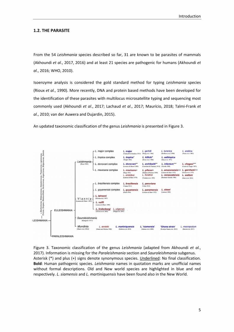
From the 54 Leishmania species described so far, 31 are known to be parasites of mammals
(Akhoundi et al., 2017, 2016) and at least 21 species are pathogenic for humans (Akhoundi et
al., 2016; WHO, 2010).
Isoenzyme analysis is considered the gold standard method for typing Leishmania species
(Rioux et al., 1990). More recently, DNA and protein based methods have been developed for
the identification of these parasites with multilocus microsatellite typing and sequencing most
commonly used (Akhoundi et al., 2017; Lachaud et al., 2017; Maurício, 2018; Talmi-Frank et
al., 2010; van der Auwera and Dujardin, 2015).
An updated taxonomic classification of the genus Leishmania is presented in Figure 3.
Figure 3. Taxonomic classification of the genus Leishmania (adapted from Akhoundi et al., 2017). Information is missing for the Paraleishmania section and Sauroleishmania subgenus. Asterisk (*) and plus (+) signs denote synonymous species. Underlined: No final classification. Bold: Human pathogenic species. Leishmania names in quotation marks are unofficial names without formal descriptions. Old and New world species are highlighted in blue and red respectively. L. siamensis and L. martiniquensis have been found also in the New World.

Introduction
6
The species included in the subgenus Leishmania multiply in the vectors’ midgut and foregut
and are present in Eurasia, Africa and the Americas whilst members of the Viannia subgenus
multiply in the vector’s hindgut and are restricted to the American continent (Lainson and
Shaw, 1987). The parasites most commonly responsible for VL belong to the Leishmania
subgenus, while both groups contain parasites causing CL. In recent years, new species have
been described in unexpected hosts and locations, including some being isolated from humans
(Cotton, 2017).
Leishmania infantum is the species with the widest geographical distribution. It is present in 47
countries throughout Europe, Africa, Asia and Central and South America (WHO, 2010), and is
the only autochthonous species in Spain (Alvar et al., 2012). Isoenzyme analysis shows that
there is a high diversity within L. infantum species, with 39 identified zymodemes (Pratlong et
al., 2013; Pratlong, pers.com.), although the most prevalent is MON-1 (Gallego et al., 2001). In
Spain, 32 zymodemes have been isolated from patients with leishmaniases, most of them HIV-
positive patients (Chicharro et al., 2003; Gallego et al., 2001; Martín-Sánchez et al., 2004;
Pratlong et al., 2013).
1.2.1. Transmission
Transmission of Leishmania parasites is mainly vectorial, through the bite of Phlebotomine
sand flies (Diptera, Psychodidae) (Gállego, 2004; Killick-Kendrick, 1999; Maroli et al., 2013).
Sand fly vectors in Eurasia and Africa belong mainly to the Phlebotomus genus, while
Lutzomyia spp. are responsible for transmission in the American continent (Akhoundi et al.,
2016; Maroli et al., 2013). In the New World, the vectorial role of Psychodopygus and
Nyssomyia genus is also considered (Maia and Depaquit, 2016). From the 800 known species,
98 are suspected or proven vectors of Leishmania parasites (Maroli et al., 2013).
Other phlebotominae (Sergentomyia genus) and arthropoda (ticks, biting midges and others)
have also been taken into account as possible vectors of Leishmania (Dantas-Torres, 2011;
Maia and Depaquit, 2016; Slama et al., 2014; Solano-Gallego et al., 2012). Nevertheless, due to
the sole molecular detection of the parasites in the majority of cases, the confirmation of
these species fulfilling all the criteria to be considered vectors of Leishmania parasites
pathogenic to mammals is yet to be proven (Maia and Depaquit, 2016).
Introduction
7
In the Mediterranean region, eight Phlebotomus species have been incriminated as vectors of
L. infantum according to conventional criteria (Killick-Kendrick, 1999; WHO, 2010): P. ariasi, P.
balcanicus, P. kandelakii, P. langeroni, P. neglectus, P. perfiliewi, P. perniciosus and P. tobbi
(reviewed in Alten et al., 2016). All belong to the subgenus Larroussius, except P. balcanicus,
which is a member of the Adlerius subgenus.
The vector species historically present in Spain are P. ariasi and P. perniciosus, from which the
parasite has been isolated in Andalusia, Aragon, Catalonia and Madrid regions (Alcover et al.,
2012; González et al., 2017; E. Guilvard et al., 1996; Lucientes-Curdi et al., 1988; Martín-
Sánchez et al., 2006, 1994; Morillas-Márquez et al., 1991; Rioux et al., 1986; Sanchez et al.,
1995). In addition, L. infantum DNA has been detected in P. perniciosus in Extremadura (Bravo-
Barriga et al., 2016). Furthermore, a stable population of P. langeroni infected with L. infantum
was recently detected by PCR in the south of the country (Sáez et al., 2018), showing that
additional overlooked vector species may exist.
Vectorial transmission is influenced by the presence and density of sand flies which, in turn, is
highly influenced by abiotic factors, showing a positive correlation with environmental
temperature and a negative association with relative humidity (Dantas-Torres et al., 2014;
Gálvez et al., 2010a; Tarallo et al., 2010). However, in a study carried out in Spain separately
analysing two vector species, P. ariasi and P. perniciosus, opposite correlations were observed
for each of the species (Ballart et al., 2014), highlighting the importance of performing
individual species analyses. P. ariasi favoured humid or sub-humid areas, whilst P. perniciosus
was more abundant in semi-arid zones (Aransay et al., 2004; Ballart et al., 2014; Gállego et al.,
1990).
The sand fly activity period in Spain is variable and can extend from the end of March to the
middle of December in the south of the country (Morillas-Márquez et al., 1983), although in
central and northern regions the sand fly activity season is considered to be from June to
October (González et al., 2017). This also corresponds to the period of higher potential risk for
L. infantum transmission in the Mediterranean region (Alten et al., 2016). P. perniciosus shows
a diphasic seasonal trend with two recognized abundance peaks in July and September, while
P. ariasi presents a monophasic abundance cycle, peaking in August (Alten et al., 2016; Gálvez
et al., 2010a).
Introduction
8
1.2.2. Epidemiological cycles
Depending on the species, Leishmania parasites can present an anthroponotic or a zoonotic
life cycle (Ashford, 2000; Bañuls et al., 2007; Gállego, 2004). Most Leishmania species known
to cause disease in humans are considered to present a zoonotic epidemiological cycle
(Gramiccia and Gradoni, 2005) or to have recent zoonotic origins (Ashford, 2000). In fact, the
possibility or demonstration of animal reservoirs for anthroponotic species has been reported
(Dereure et al., 1991; Kassahun et al., 2015; Singh et al., 2013).
A great number of animals have been found infected with Leishmania species and are
considered parasite hosts, but only a minority is suspected of having a possible role in parasite
maintenance and transmission in a particular scenario (i.e. parasite reservoirs). Leishmania
reservoirs show regional and temporal variation, and only a local study including ecological
and parasitological analysis could determine whether a species may serve as a reservoir in a
given environment (Roque and Jansen, 2014). In the Americas, Leishmania hosts belong to the
orders Didelphimorphia, Cingulata, Pilosa, Rodentia, Primata, Carnivora, and Chiroptera, whilst
in the Old World implicated animal orders are Carnivora, Hyracoidea, Rodentia and
Lagomorpha (Gramiccia and Gradoni, 2005; Roque and Jansen, 2014).
In the Mediterranean region, the cycle is zoonotic and domestic, with the dog acting as the
principal reservoir of L. infantum. Other animal species, proven or suspected reservoirs of L.
infantum in Europe have been revised by Millán et al. (2014) and Pennisi (2015) and are listed
on Table 1. For the large majority, there is no evidence that these can act as reservoirs (Portús
et al., 2002; Quinnell and Courtenay, 2009), but cats, black rats, foxes, hares and rabbits are
considered to be able to maintain a wild or domestic epidemiological cycle (Jiménez et al.,
2014; Maroli et al., 2007; Marín-Iniesta et al., 1982; Martín-Sánchez et al., 2007; Molina et al.,
2012b; Pozio et al., 1985; Zanet et al., 2014).

Introduction
9
Table 1. Alternative host species and possible reservoirs of L. infantum in Europe.
Species
Proven infectiousness to sand flies*/Isolation of parasites** References
Badger (Meles meles) del Río et al., 2014
Beech marten (Martes foina) Muñoz-Madrid et al., 2013
Black rat (Rattus rattus) */** Helhazar et al., 2013; Morillas-Márquez et al., 1985; Muñoz-Madrid et al., 2013; Navea-Pérez et al., 2015; Pozio et al., 1985; Zanet et al., 2014
Brown rat (Rattus norvegicus) Helhazar et al., 2013
Domestic cat (Felis catus domesticus)
*/** Ayllon et al., 2008; Hervás et al., 1999; Maia et al., 2010; Maroli et al., 2007; Martín-Sánchez et al., 2007; Millán et al., 2011; I. Moreno et al., 2014; Solano-Gallego et al., 2007
Domestic goat (Capra hircus) Fisa et al., 1999; Portús et al., 2002
Domestic ferret (Mustela putorius furo)
Brianti et al., 2005
Domestic horse (Equus caballus)
** Fernández-Bellon et al., 2006; Gama et al., 2014; Portús et al., 2002; Rolão et al., 2005; Solano-Gallego et al., 2003
Domestic sheep (Ovis aries) Fisa et al., 1999; Portús et al., 2002
Egyptian mongoose (Herpestes ichneumon)
Sobrino et al., 2008
European brown hare (Lepus europaeus)
Rocchigiani et al., 2018; Ruiz-Fons et al., 2013; Tsokana et al., 2016
European mink (Mustela lutreola)
del Río et al., 2014
European rabbits (Oryctolagus cuniculus)
*/** Díaz-Sáez et al., 2014; Jiménez et al., 2014; I. Moreno et al., 2014; Ortega et al., 2017; Risueño et al., 2018
Genet (Genetta genetta) del Río et al., 2014; Millán et al., 2011; Oleaga et al., 2018;
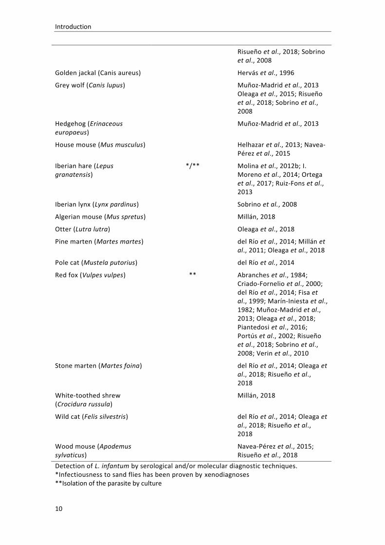
Introduction
10
Risueño et al., 2018; Sobrino et al., 2008
Golden jackal (Canis aureus) Hervás et al., 1996
Grey wolf (Canis lupus) Muñoz-Madrid et al., 2013 Oleaga et al., 2015; Risueño et al., 2018; Sobrino et al., 2008
Hedgehog (Erinaceous europaeus)
Muñoz-Madrid et al., 2013
House mouse (Mus musculus) Helhazar et al., 2013; Navea-Pérez et al., 2015
Iberian hare (Lepus granatensis)
*/** Molina et al., 2012b; I. Moreno et al., 2014; Ortega et al., 2017; Ruiz-Fons et al., 2013
Iberian lynx (Lynx pardinus) Sobrino et al., 2008
Algerian mouse (Mus spretus) Millán, 2018
Otter (Lutra lutra) Oleaga et al., 2018
Pine marten (Martes martes) del Río et al., 2014; Millán et al., 2011; Oleaga et al., 2018
Pole cat (Mustela putorius) del Río et al., 2014
Red fox (Vulpes vulpes) ** Abranches et al., 1984; Criado-Fornelio et al., 2000; del Río et al., 2014; Fisa et al., 1999; Marín-Iniesta et al., 1982; Muñoz-Madrid et al., 2013; Oleaga et al., 2018; Piantedosi et al., 2016; Portús et al., 2002; Risueño et al., 2018; Sobrino et al., 2008; Verin et al., 2010
Stone marten (Martes foina) del Río et al., 2014; Oleaga et al., 2018; Risueño et al., 2018
White-toothed shrew (Crocidura russula)
Millán, 2018
Wild cat (Felis silvestris) del Río et al., 2014; Oleaga et al., 2018; Risueño et al., 2018
Wood mouse (Apodemus sylvaticus)
Navea-Pérez et al., 2015; Risueño et al., 2018
Detection of L. infantum by serological and/or molecular diagnostic techniques. *Infectiousness to sand flies has been proven by xenodiagnoses **Isolation of the parasite by culture

Introduction
11
1.3. CANINE LEISHMANIOSIS
Canine leishmaniosis (CanL) is an important veterinary disease, present in at least 50 countries
(Alvar et al., 2004). Kaszak et al. (2015) have estimated that CanL affects more than 2.5 million
dogs in more than 70 countries, although other authors assert that these figures occur in the
Mediterranean basin alone (Moreno and Alvar, 2002). In this region, it has been estimated that
50 to 80% of the canine population is infected and that the prevalence of the disease ranges
between 2% and 5% (Saridomichelakis, 2009).
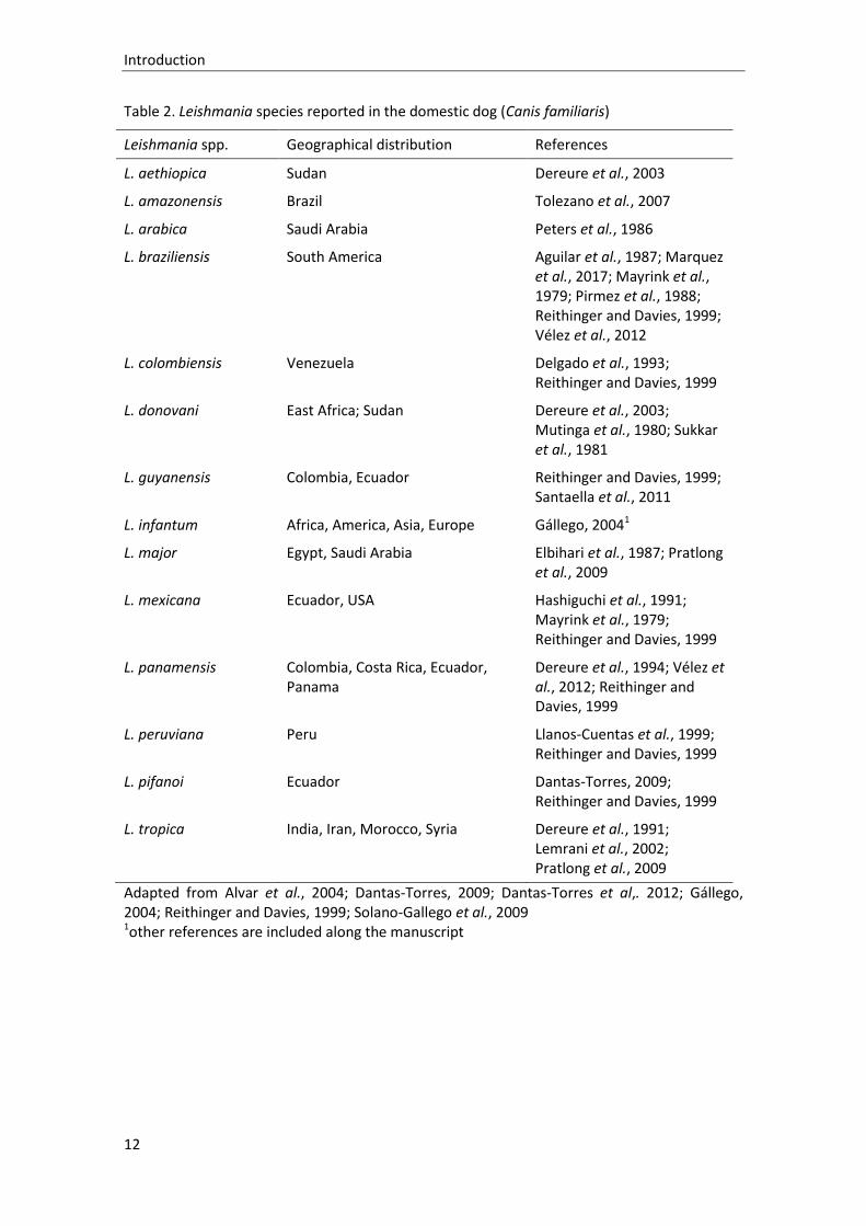
The term CanL is usually used to refer to the infection by L. infantum, although dogs can also
be infected by other Leishmania species, which are listed in Table 2. Being the main known
reservoir of L. infantum (Dantas-Torres, 2007; Gállego, 2004; Otranto et al., 2017; Ready,
2010), dogs assume a crucial role in the maintenance of the parasite’s life cycle. Likewise,
surveillance and control of infected canids in endemic regions is essential for the management
of CanL, as well as for the reduction of HL (Gavgani et al., 2002).
In dogs, the zymodeme MON-1 of L. infantum is the most prevalent, although 17 out of 39
zymodemes have been characterized in dogs’ strains (Aït-oudhia et al., 2011; Gallego et al.,
2001; Pratlong et al., 2013).
Introduction
12
Table 2. Leishmania species reported in the domestic dog (Canis familiaris)
Leishmania spp. Geographical distribution References
L. aethiopica Sudan Dereure et al., 2003
L. amazonensis Brazil Tolezano et al., 2007
L. arabica Saudi Arabia Peters et al., 1986
L. braziliensis South America Aguilar et al., 1987; Marquez et al., 2017; Mayrink et al., 1979; Pirmez et al., 1988; Reithinger and Davies, 1999; Vélez et al., 2012
L. colombiensis Venezuela Delgado et al., 1993; Reithinger and Davies, 1999
L. donovani East Africa; Sudan Dereure et al., 2003; Mutinga et al., 1980; Sukkar et al., 1981
L. guyanensis Colombia, Ecuador Reithinger and Davies, 1999; Santaella et al., 2011
L. infantum Africa, America, Asia, Europe Gállego, 20041
L. major Egypt, Saudi Arabia Elbihari et al., 1987; Pratlong et al., 2009
L. mexicana Ecuador, USA Hashiguchi et al., 1991; Mayrink et al., 1979; Reithinger and Davies, 1999
L. panamensis Colombia, Costa Rica, Ecuador, Panama
Dereure et al., 1994; Vélez et al., 2012; Reithinger and Davies, 1999
L. peruviana Peru Llanos-Cuentas et al., 1999; Reithinger and Davies, 1999
L. pifanoi Ecuador Dantas-Torres, 2009; Reithinger and Davies, 1999
L. tropica India, Iran, Morocco, Syria Dereure et al., 1991; Lemrani et al., 2002; Pratlong et al., 2009
Adapted from Alvar et al., 2004; Dantas-Torres, 2009; Dantas-Torres et al,. 2012; Gállego, 2004; Reithinger and Davies, 1999; Solano-Gallego et al., 2009 1other references are included along the manuscript

Introduction
13
The northward expansion of CanL in Europe, along with the possible spread of human cases, is
well documented (Ballart et al., 2013b; Gramiccia and Gradoni, 2005; Maroli et al., 2013;
Medlock et al., 2014). Surveys performed at border areas between CanL endemic and non-
endemic regions prove that the infection is present in locations where it had not been
documented before (Ballart et al., 2013a; Capelli et al., 2004; Cassini et al., 2013; Dumitrache
et al., 2016; Vaselek et al., 2017). Also, mathematical models and predictive risk maps forecast
an expansion of Leishmania vectors due to climate change and anthropogenic impact on the
landscape (Espejo et al., 2015; Fischer et al., 2010; Koch et al., 2017).
New cases of CanL have been registered in non-endemic areas where no vectorial transmission
exists, mainly as a result of dog movement to endemic countries (Dandrieux et al., 2018; Maia
and Cardoso, 2015). In some cases, however, autochthonous canine infection has been
proven, most certainly through direct contact (Boggiatto et al., 2011; Gibson-Corley et al.,
2008; Karkamo et al., 2014; Naucke and Lorentz, 2012; Svobodova et al., 2017; Tánczos et al.,
2012). Therefore, CanL in Europe is no longer only a problem of Mediterranean countries
(Dujardin et al., 2008; Pennisi, 2015; Ready, 2010). Although leishmaniosis is included in the
OIE list of notifiable diseases (OIE, 2018), this is not clearly reflected in the European or
country-level legislation (BOE, 2014; Official Journal of the European Union, 2012), meaning
that a common European strategy for leishmaniosis surveillance and control does not exist.
The work described in this thesis is set in a European Mediterranean country and refers
exclusively to canine infection by L. infantum; therefore, the term CanL will, for the reminder
of this dissertation, used to refer strictly to infection of dogs by this Leishmania species.
The terminology used to describe the disease caused by Leishmania parasites changes
according to authors’ personal preference. The term “leishmaniasis” is frequently used to refer
to the human disease, while “leishmaniosis” is more commonly applied to the veterinary
condition (Miró and López-Vélez, 2018). In this dissertation, the guidelines of the Standardized
Nomenclature of Animal Parasitic Diseases (SNOAPAD) will be followed, and the term
“leishmaniosis” will be applied both for the veterinary and the human diseases (Kassai, 2006;
Kassai et al., 1988). “Leishmaniosis” is also the term used by the World Organization for Animal
Health to refer to the canine infection by Leishmania species (OIE, 2014).

Introduction
14
1.3.1. Clinical presentation
The course of L. infantum infection in the canine host is highly influenced by the host’s
immune response (more details in section 1.3.5), which introduces a high individual variability
into the clinical outcome (Hosein et al., 2017). Likewise, factors such as parasitic burden,
virulence of Leishmania strain, previous infections or coinfections can also affect the polarity
of clinical manifestations (Saridomichelakis, 2009; Solano-Gallego et al., 2009). Consequently,
dogs with CanL can present a broad range of clinical signs and clinicopathological
abnormalities, which are usually nonspecific (Paltrinieri et al., 2016; Solano-Gallego et al.,
2011). The incubation period since infection and until the appearance of clinical disease in
dogs can last from three months to seven years (Alvar et al., 2004; Miró et al., 2008; Oliva et
al., 2006; Solano-Gallego et al., 2001a). CanL is a multisystemic disease that can potentially
affect any organ or tissue, as well as present a diffuse or localized progression. The most
common clinical signs observed in “classical” CanL are progressive weight loss, generalized
lymphadenomegaly, onycogryphosis and non-pruritic exfoliative dermatitis (Solano-Gallego et
al., 2011, 2009). Non regenerative anaemia, mild thrombocytopenia, leukogram changes and
indicators of renal dysfunction and/or inflammatory immune response are the most frequently
detected clinicopathological alterations (Foglia Manzillo et al., 2013; Paltrinieri et al., 2016).
Clinical signs and laboratory abnormalities observed in CanL are presented in Tables 3 and 4.
Table 3. Clinical signs and lesions reported in CanL distributed by organic system
Clinical signs and lesions References*
General
o Poor body condition o Exercise intolerance o Lethargy o Loss of appetite o Pale mucous membranes o Fever
Reticuloendothelial Santana et al., 2008
o Generalized lymphadenopathy o Splenomegaly
Cutaneous Ferrer et al., 1988; Lombardo et al., 2014
o Non-pruritic exfoliative dermatitis with or without alopecia o Onychogryphosis

Introduction
15
o Erosive-ulcerative dermatitis o Nodular dermatitis o Papular dermatitis o Pustular dermatitis o Mucosal ulceration o Nasal hyperkeratosis o Footpad hyperkeratosis
Ophthalmic Naranjo et al., 2005; Peña et al., 2008
o Blepharitis o Conjunctivitis o Keratoconjunctivitis o Anterior uveitis
Musculoskeletal
o Muscular atrophy o Atrophic masticatory myositis o Skeletal myositis
Renal
o Polyuria and polydipsia
Cardiac Martínez-Hernández et al., 2017
o Myocardial lesions
Coagulation/vascular
o Epistaxis
Articular Agut et al., 2003; Santos et al., 2006
o Arthritis o Lameness
Digestive Adamama-Moraitou et al., 2007; Pinto et al., 2011
o Vomiting o Diarrhoea
Neurological Giannuzzi et al., 2017; Márquez et al., 2013; Zobba et al., 2017
The most common clinical signs are marked in bold. *References presented in the table correspond to specific studies; general references for clinical signs and lesions were: Ciaramella et al., 1997; Foglia Manzillo et al., 2013; Koutinas and Koutinas, 2014; Noli and Saridomichelakis, 2014; Paltrinieri et al., 2016, 2010; Saridomichelakis, 2009; Solano-Gallego et al., 2011, 2009.

Introduction
16
Table 4. Main laboratory findings associated with CanL
Laboratory findings References
Hemogram Koutinas and Koutinas, 2014; Maia and Campino, 2018; Meléndez-Lazo et al., 2018; Noli and Saridomichelakis, 2014; Paltrinieri et al., 2016, 2010; Saridomichelakis, 2009; Solano-Gallego et al., 2011, 2009
o Mild to moderate non-regenerative anaemia o Thrombocytopenia o Leukocytosis or leukopenia
Biochemical parameters
o Renal azotaemia (↑BUN and creatinine) o Hepatic dysfunction (↑ALT, ALP and GGT) o Hyperglobulinemia o Hypoalbuminemia o Decreased albumin/globulin ratio
Serum protein electrophoresis
o Polyclonal gammopathy (less frequently, oligoclonal or monoclonal)
Immunological parameters
o Positive antinuclear antibody titres
Urinalysis
o Proteinuria o Increased UPC ratio o Decreased urine specific gravity
The most common laboratory findings are marked in bold.
Clinical staging systems for CanL have been proposed by two working groups, the Canine
Leishmaniasis Working Group (CLWG) and LeishVet (Roura et al., 2013; Solano-Gallego et al.,
2009). These have the purpose of providing clinically useful information for therapeutic
decisions and for prognostic purposes. However, independent peer validation of these
suggested systems is still lacking (Noli and Saridomichelakis, 2014).
An important feature of canine L. infantum infection is the high prevalence of asymptomatic
dogs, particularly in CanL endemic areas (Alvar et al., 2004; Ballart et al., 2013a; Baneth et al.,
2008; Fisa et al., 1999; Solano-Gallego et al., 2001a). Again, this is a result of the individual’s
immune response, which in some cases is capable of controlling the parasite (Solano-Gallego
et al., 2000). Asymptomatic infected dogs are characterized by parasite detection (either by

Introduction
17
direct or indirect techniques) in the absence of symptoms or clinicopathological abnormalities
(Molina et al., 1994; Paltrinieri et al., 2016). Apart from immunological resistance, the
asymptomatic state can also denote an early stage of infection, in which case a later
development of clinical signs is expected (Fisa et al., 2001; Miró et al., 2012). Although parasite
clearance has been mentioned in dogs (Fisa et al., 1999), there is no reliable evidence of it
(Cavaliero et al., 1999; Manna et al., 2008b; Solano-Gallego et al., 2016a), meaning that
asymptomatic infected individuals are at risk of developing clinical disease throughout their
lives (Alvar et al., 2004; Baneth et al., 2008). Furthermore, asymptomatic dogs assume a
particularly important role from an epidemiological perspective, as they represent overlooked
reservoirs of L. infantum in endemic areas. Previous studies have already demonstrated that
these dogs are infectious to sand flies (Borja et al., 2016; de Mendonça et al., 2017b; Laurenti
et al., 2013; Molina et al., 1994; Quinnell and Courtenay, 2009) and capable of maintaining the
infection in the canine population (eventually also being the source for human transmission).
1.3.2. Diagnosis
As mentioned, an important concept in L. infantum infection, especially in endemic areas, is
that parasite detection on the vertebrate host does not equate to active disease (Moreno and
Alvar, 2002). A vast number of dogs are capable of controlling the infection either in a latent
form or a transitory state, leading to eventual parasite clearance (Fisa et al., 1999; Miró et al.,
2012). This adds complexity to CanL diagnosis which, in some cases, will require the use of
multiple diagnostic tests (Morales-Yuste et al., 2012; Otranto et al., 2009).
Furthermore, the techniques used for CanL diagnosis must be adapted to each situation. Dogs
presented to veterinary practitioners are commonly symptomatic individuals, and the
challenge here may be to exclude differential diagnosis and to confirm that the clinical signs
observed are produced by L. infantum infection, regardless of parasite detection (Paltrinieri et
al., 2016; Solano-Gallego et al., 2011). Specific laboratory tests aimed at etiologic diagnosis
include direct methods (cytology, histopathology, parasite culture, molecular tests, and, less
frequently, xenodiagnosis) and indirect techniques (serology and cellular immune response
evaluation) (Gomes et al., 2008; Maia and Campino, 2008; Maroli et al., 2010; Noli and
Saridomichelakis, 2014; Rodríguez-Cortés et al., 2010; Solano-Gallego et al., 2009; Travi et al.,
2018). Description of these techniques, with a special emphasis on the ones used for

Introduction
18
prevalence surveys, will be addressed later, under the “Epidemiologic studies” chapter (section
1.3.4).
Due to the variety of possible outcomes after L. infantum infection (Noli and Saridomichelakis,
2014), the differentiation between exposure and disease through the available diagnostic
methods can be particularly difficult. In any case, clinical examination should be the first step
of CanL diagnosis (Solano-Gallego et al., 2009). In endemic areas, and due to the serious
consequences of a delayed diagnosis, it is recommended that the disease be investigated in
any dog presenting with even a single CanL-associated clinical sign (Noli and Saridomichelakis,
2014).
1.3.3. Control
Several methods have been proposed for CanL prevention and control, both at the individual
and at the population levels. These strategies are mainly focused at: 1) reducing the number of
infected and infectious animals by early detection and treatment of infected dogs, and 2)
avoiding new CanL infections by applying insecticides to both infectious and naïve dogs and/or
through immunomodulation and vaccination (Alvar et al., 2004; Maroli et al., 2010; Miró et al.,
2017b; Otranto and Dantas-Torres, 2013; Quinnell and Courtenay, 2009; Reguera et al., 2016;
Ribeiro et al., 2018; Travi et al., 2018).
First line treatment regimens are based on the association of a leishmanicidal drug
(pentavalent antimony meglumine antimoniate or miltefosine) and a leishmaniostatic
(allopurinol) (Solano-Gallego et al., 2009). More recently, immunomodulators aimed at
reducing parasite burden and boosting the host’s immune response [domperidone (Gómez-
Ochoa et al., 2009) or P-MAPA, a protein aggregate of magnesium-ammonium
phospholinoleate-palmitoleate anhydride (Melo et al., 2014; Santiago et al., 2013)] have also
been added to the classical therapeutic protocols.
Treatment of dogs with CanL has the aim of improving diseased dogs’ quality of life and
extending their life expectancy, while it also has an impact on the parasite load, thus reducing
dogs’ infectiousness to sand flies (Otranto and Dantas-Torres, 2013). Post-treatment decrease
in infectiousness has been shown by xenodiagnosis studies for a few chemotherapeutic
protocols (reviewed in Travi et al., 2018), proving that this is an effective method for reducing

Introduction
19
infection risk at the population level. However, no CanL treatment has proved to achieve
consistent parasite clearance and clinical improvement is only transitory (Manna et al., 2008b;
Torres et al., 2011), with affected dogs usually needing recurring treatment cycles (Solano-
Gallego et al., 2009). For this reason, and because the assessment of an individual’s
infectiousness for diagnostic purposes is not feasible, permanent use of topical insecticides is
recommended in animals known to be infected (Noli and Saridomichelakis, 2014).
Culling of infected dogs has been the approach used in some countries as a method for
leishmaniosis control. In Brazil, where cases of HL have been recently expanding to urban
areas, treatment of affected dogs is not allowed and CanL detection campaigns, followed by
culling of seropositive dogs, is the control method recommended by the Ministry of Health
(Ministério da Saúde Brasileiro, 2014). However, besides being an unethical procedure, there is
no real evidence supporting its efficacy in reducing the number of CanL or HL cases (Costa,
2011; Costa et al., 2013; Courtenay et al., 2002; Dye, 1996; Reithinger et al., 2004).
Furthermore, indiscriminate dog culling may have a detrimental effect if, by removing
seropositive resistant animals responsible for increasing herd immunity, it induces an increase
in disease transmission (Fox et al., 1971). It is currently known that only a small proportion of
dogs are responsible for most transmission (Courtenay et al., 2002) and that infectiousness
correlates with parasite load, especially on ear skin biopsies (Courtenay et al., 2014), while
serological tests seem to be unable to discriminate the most infectious dogs (de Mendonça et
al., 2017b). Therefore, the development of a diagnostic test able to differentiate between
infected (which may not be responsible for transmission) and infectious dogs is essential to
efficiently direct control efforts in areas of high transmission (Courtenay et al., 2014; de
Mendonça et al., 2017b; Duthie et al., 2018).
The application of topical insecticides with proven efficacy against sand flies is still considered
the most effective method for preventing L. infantum infection in dogs (Brianti et al., 2014;
Courtenay et al., 2009; Foglia Manzillo et al., 2006a; Goyena et al., 2016; Killick-Kendrick et al.,
1997; Maroli et al., 2001; Guadalupe Miró et al., 2007; Molina et al., 2012a; Otranto et al.,
2013). However, due to the need of repeated applications (spot-on preparations and sprays) or
product replacement (dog collars), owner compliance is critical to achieve a satisfactory
coverage and efficacy (Maia et al., 2018; Reithinger et al., 2004). The effect of systemic
insecticides administered to dogs in the reduction of CanL and HL is under research (Gomez et
al., 2018; Gomez and Picado, 2017) and it can prove to be an effective method to aid infection
control in endemic countries. Insecticides can also be used in the environment, through indoor
residual spraying (IRS) of houses and animal shelters (Alexander and Maroli, 2003; Maroli et

Introduction
20
al., 2010), and novel vector control methods, such as attractive toxic sugar baits, are under
development (Qualls et al., 2015).
Preventive treatments aimed at stimulating the dog’s immune system can be unspecific
(immunomodulators) or specific (vaccines). Domperidona, described before as a
complementary therapy for CanL, is also used as a preventive treatment in L. infantum
endemic regions (Sabaté et al., 2014). There are currently three licensed vaccines for CanL:
Leish-Tec® (Hertape) in Brazil and CaniLeish® (Virbac) and LetiFend® (LETI) in Europe (Reguera
et al., 2016). A fourth vaccine was commercialized in Brazil for 11 years, having been
withdrawn in 2014 by the Brazilian Ministry of Agriculture due to lack of evidence for its
effectiveness (MAPA, 2014). The vaccines available for CanL will be assessed later, under the
section “Commercially approved vaccines against CanL” (section 1.3.6).
Control of L. infantum infection directed at the domestic dog can prove insufficient in some
settings, where other animal species could be implicated in the maintenance and transmission
of zoonotic leishmaniosis (Antoniou et al., 2013; del Río et al., 2014; Millán et al., 2014; Navea-
Pérez et al., 2015; Zanet et al., 2014) (see Table 1). However, in the majority of species other
than dogs, a reservoir status for the parasite has not been proven (Courtenay et al., 2009;
Portús et al., 2002). Particularly noteworthy is the recent case of Fuenlabrada (Madrid region,
Spain), where Iberian hares (Lepus granatensis) and European rabbits (Oryctolagus cuniculus)
were identified as the animal reservoirs for L. infantum parasites (Jiménez et al., 2014; Molina
et al., 2012b) in the largest HL community outbreak in Europe (Arce et al., 2013).
Alternative routes have been confirmed for L. infantum transmission between dogs:
transplacental (Boggiatto et al., 2011; da Silva et al., 2009; Vida et al., 2016), venereal (Silva et
al., 2009) and blood transfusion (Owens et al., 2001). These routes assume importance mostly
in non-endemic countries (Karkamo et al., 2014; Naucke and Lorentz, 2012), since transmission
by sand fly bites in endemic areas is much more effective than any other route
(Saridomichelakis, 2009). Based on these findings, breeding dogs from endemic areas should
be tested and any infected animal should be excluded from reproduction. Likewise, any blood
donors should be regularly tested for L. infantum infection (Miró et al., 2017b).

Introduction
21
1.3.4. Epidemiological studies
According to the CLWG, an “exposed” dog is clinically healthy and presents low-titer anti-
Leishmania antibodies in the absence of parasite isolation (either by cytological, histological,
parasitological or molecular methods). The presence of specific anti-Leishmania antibody titers
together with parasite detection characterizes an “infected” dog (Paltrinieri et al., 2010).
However, in CanL endemic areas, molecular parasite detection in peripheral blood or skin
during the infection transmission season should be carefully interpreted and may not be
sufficient to classify a dog as infected (Solano-Gallego et al., 2011). Infected dogs can be
asymptomatic, which can be indicative of a “resistant” state or early infection, as mentioned
previously. In these cases, resistance can be more accurately ascertained through the
detection of an effective anti-Leishmania cellular-mediated immune response (Solano-Gallego
et al., 2000). Infected symptomatic dogs are considered “diseased”, which means they present
CanL (Paltrinieri et al., 2010). These definitions are essential to better understand the
information yielded by different epidemiological studies.
The distribution of L. infantum in canine populations from endemic regions is highly
heterogeneous (Maia et al., 2018; Pennisi, 2015). Reported infection prevalence can vary
substantially within a country, as described in a study which compiled published data from
France, Italy, Portugal and Spain (Franco et al., 2011). In this study, observed point CanL
prevalences within these endemic countries varied from 0 to more than 80%. In Spain,
reported canine L. infantum infection seroprevalence varies from 1.6% in the northwest (Miró
and Molina, 2006) to 34.6% in the south (Morillas-Márquez et al., 1996), with a range of
intermediate values reported across the territory (Acedo Sánchez et al., 1996; Alcover et al.,
2013; Amela et al., 1995; Amusategui et al., 2004; Arnedo Pena et al., 1994; Ballart et al.,
2013a; Botet and Portús, 1993; Cabezón et al., 2010; Couto et al., 2010; Encinas Grandes et al.,
1988; Fisa et al., 1999; Gálvez et al., 2010b; Goyena et al., 2016; Lepe et al., 2000; Martín-
Sánchez et al., 2009; G. Miró et al., 2007; Miró et al., 2017a, 2012; Morales-Yuste et al., 2011;
Morillas-Márquez et al., 1996; Nieto et al., 1992; Segovia and Martin-Luengo, 1985; Solano-
Gallego et al., 2001a).
Reasons for this heterogeneous distribution can be related to factors affecting parasite or
vector prevalence, as well as host-related factors. Additionally, study design and the diagnostic
techniques used can also influence the results of epidemiological surveys (Franco et al., 2011).

Introduction
22
Diagnostic methods most frequently used in epidemiological studies are serological and
molecular (Ballart et al., 2013a; Cabezón et al., 2010; Fisa et al., 1999; Maia et al., 2016; Miró
et al., 2012; Solano-Gallego et al., 2001a), but parasite detection by direct exam or culture, as
well as tests for specific cellular immunity assessment are also used (Fernández-Bellon et al.,
2008; Iniesta et al., 2002; Solano-Gallego et al., 2000). Epidemiological surveys usually include
large numbers of animals to be sampled under field conditions, whereby diagnostic tests used
for this purpose should be easy to perform and interpret, low-cost, and either applicable in
field settings or by making use of samples that can be easily transported.
Serological methods are one of the most commonly employed approaches for detection of L.
infantum infection (Solano-Gallego et al., 2014). Serology detects canine humoral response to
L. infantum, which can occur as early as one month after an infective phlebotomine bite
(Moreno and Alvar, 2002), although it has been reported to go up to 22 months in some cases
(Oliva et al., 2006). Therefore, serological tests are indicators of host-parasite contact, but not
of parasite presence (Solano-Gallego et al., 2009). These methods are considered to be good
predictors of the onset of clinical signs, and good indicators of active infection, as diseased
dogs tend to present significantly higher levels of anti-Leishmania antibodies (Oliva et al.,
2006). Enzyme-linked immunosorbent assay (ELISA) and indirect immunofluorescence (IFAT),
two of the most frequently used serological quantitative methods, are the screening tests
recommended by the World Organization for Animal Health for prevalence and surveillance
studies (OIE, 2014). Research on anti-L. infantum immunoglobulin (Ig) isotypes and IgG
subclasses has attempted to correlate serological profiles with protective cellular mediated
immunity (CMI) and infection outcome (reviewed in Maia and Campino, 2018). Results across
studies tend to provide evidence of an increase in all Leishmania-specific Ig subtypes in
symptomatic dogs, when compared to asymptomatic individuals (Iniesta et al., 2005; Reis et
al., 2006; Rodríguez-Cortés et al., 2007a). Similar trends are reported for IgG subclasses
(Iniesta et al., 2005; Laia Solano-Gallego et al., 2001b), although some authors have found a
clear correlation between IgG2 levels and disease progression (Cardoso et al., 2007; Iniesta et
al., 2007). Lack of consistency across IgG subclasses studies could be a result of the low
specificity of the polyclonal antisera commercially available and used in most laboratories
(Day, 2007). When monoclonal antibodies to IgG were used, no substantial difference between
subclasses was detected (Quinnell et al., 2003a; Strauss-Ayali et al., 2007). Serological
qualitative methods or rapid immunochromatographic tests (ICT), which are easy to perform
and provide immediate results, are also used in veterinary daily practice and in epidemiological
studies. However, positive qualitative results should be followed by a quantitative test to

Introduction
23
accurately assess the level of infection and for follow-up purposes (Noli and Saridomichelakis,
2014).
Molecular diagnostic techniques are used to detect parasite DNA in different organs or tissues.
Likewise, a positive result in a conventional polymerase chain reaction (PCR) test confirms the
presence of Leishmania, but has no predictive value on the infection outcome (Otranto et al.,
2009). Alternatively, quantitative PCR (qPCR) methods can be used to assess parasite loads in
different organs and to provide useful information for follow-up on positive animals (Francino
et al., 2006). Molecular techniques are highly specific, but their sensitivity depends on the
sample used (Solano-Gallego et al., 2016a) and on the target DNA sequence (Akhoundi et al.,
2017; Paltrinieri et al., 2016). Bone marrow, lymph node and skin usually harbour high parasite
loads and provide higher sensitivity for PCR techniques (Noli and Saridomichelakis, 2014;
Solano-Gallego et al., 2011). The use of non-invasive samples, such as conjunctival swabs (Di
Muccio et al., 2012; Strauss‐Ayali et al., 2004), cerumen (Belinchón-Lorenzo et al., 2016) and
hair (Belinchón-Lorenzo et al., 2013; Corpas-López et al., 2016), has proved to yield good
results and could be considered as a useful diagnostic alternative for large-scale field studies.
Peripheral blood is usually regarded as a less sensitive sample (Solano-Gallego et al., 2011), but
studies show that the detection of Leishmania DNA in blood is far more common than has
been previously recognized (Francino et al., 2006; Rodríguez-Cortés et al., 2007b). The
application of qPCR to blood specimens significantly increases the detection sensitivity
(Francino et al., 2006) and this method is considered adequate to complement serological
results in large-scale epidemiological field studies (Maia et al., 2009; Solano-Gallego et al.,
2016a).
Parasite detection by direct microscopic examination or parasite culture is highly specific, but
these techniques present low sensitivity. Although still frequently used in veterinary practice
(Bourdeau et al., 2014) and recommended by several authors as the first line diagnostic test
(Gharbi et al., 2015; Paltrinieri et al., 2010; Saridomichelakis, 2009), direct visualization of
amastigotes on lymph node smears or skin lesions imprints depends greatly on parasite load,
on the examiner’s experience and may be unfeasible in field settings. Parasite culture is
laborious, time-consuming and can take several weeks to provide definitive results (Maia and
Campino, 2008); for these reasons, it is generally used for research purposes only (Miró et al.,
2008).
The detection of Leishmania-specific CMI has also been used in epidemiological studies. In field
settings, it can be assessed by the leishmanin skin test (LST) (Cardoso et al., 1998). This

Introduction
24
technique identifies exposed and usually resistant animals (Solano-Gallego et al., 2000), as
dogs with active CanL (and the expected immunosuppression) frequently test negative to LST
(Solano-Gallego et al., 2005). Likewise, LST can be used to assess exposure and resistance to
the parasite at the community level, but cannot be used to identify infected or diseased
individuals (Iniesta et al., 2002). LST is easy to perform and inexpensive, which makes it
adequate for testing a large number of animals in field conditions. However, a follow-up is
needed after 72h and a possible iatrogenic induction of false positives can occur after repeated
inoculations (Fernández-Bellon et al., 2005). Additionally, leishmanin antigen for veterinary use
has not been internationally standardised and is not available commercially worldwide (OIE,
2014), which may hamper its application. Some of the most common laboratory-based CMI
techniques include the lymphoproliferation assay (LPA) and the quantification of specific
Leishmania-induced cytokines produced by canine lymphocytes. Although these techniques
also evaluate CMI, they seem to measure distinct parameters of the cellular-mediated
response, as their results only partially overlap with each other and when compared to LST
(Fernández-Bellon et al., 2005; Iniesta et al., 2002). Unlike LST, and being in vitro assays, there
is no risk of iatrogenic-induced false-positive results with repeated testing. However, these are
complex and time-consuming techniques that require access to laboratory facilities and several
days of sample processing. The recent development of easier-to-perform techniques that
make use of whole blood instead of isolated peripheral blood mononuclear cells (PBMC) may
allow its use in field settings (Zribi et al., 2017).
Detection of host-vector contact can provide useful information to complement studies of
vector population dynamics and host-vector interactions, to assess the risk of Leishmania
infection (Carvalho et al., 2015; Rohoušová et al., 2005; Vlkova et al., 2011) and to measure
the effectiveness of vector-control programmes (Clements et al., 2010; Gidwani et al., 2011).
Exposure of vertebrate hosts to sand flies can be assessed by the detection of antibodies
against sand fly saliva in the hosts’ blood, a method that has proven to be highly specific
(Vlkova et al., 2011) and was successfully used as a marker of exposure to L. infantum vectors
(Kostalova et al., 2015; Martín-Martín et al., 2014). The recent development of a rapid ICT
which detects host contact with P. perniciosus can provide a valuable tool for testing canine
populations in field settings (Willen et al., 2018).

Introduction
25
1.3.5. Immune response to Leishmania infantum in dogs
After the bite of an infected sand fly, L. infantum metacyclic promastigotes are inoculated
under the host’s skin. After first contact with the skin immune system, parasites are
phagocytized by macrophages, neutrophils and dendritic cells and transported to regional
lymph nodes (Hosein et al., 2017). From this point, the infection outcome will vary depending
on vector, parasite and host factors (Saridomichelakis, 2009).
Parasite virulence is determined by its species and strain, as well as the number of
promastigote forms inoculated (Saridomichelakis, 2009). Sand fly saliva injected during the
bite also stimulates the host’s immune system (Abdeladhim et al., 2014), although its role in
the establishment of infection or protection is still under debate (Collin et al., 2009; Rohoušová
et al., 2011). The host’s immune response is crucial to infection progression, although the
precise immune mechanisms responsible for resistance or susceptibility are still unknown
(Hosein et al., 2017). The wide spectrum of possible outcomes varies from an immediate local
elimination of the parasite by the host’s macrophages to a multisystemic overt disease
(Solano-Gallego et al., 2009).
1.3.5.1. Host genetics
It is currently accepted that a host’s genetics play an important role in susceptibility or
resistance to CanL (Baneth et al., 2008). Epidemiological studies in canine populations from
CanL endemic areas have shown that mongrels and autochthonous breeds tend to show a
higher resistance to infection than exotic breeds (Cortes et al., 2012; Fisa et al., 1999; Sanchez-
Robert et al., 2005; Solano-Gallego et al., 2000).
Genetic studies have identified genotypes that may be involved in susceptibility to L. infantum
infection, for example the DLA-DRB1*01502 allele of the major histocompatibility complex
class II (MHC II) (Quinnell et al., 2003b), the Slc11α1 gene (Altet et al., 2002), or the canine β-
defensin-1 (CBD1) gene (da Silva et al., 2017). TAG-8-141 haplotype of Slc11α1 gene was found
to be significantly associated with susceptibility to the disease in boxers (Sanchez-Robert et al.,
2005), as well as in other breeds (Sanchez-Robert et al., 2008), while other authors found no
difference in gene expression between phenotypically resistant and susceptible dogs (Bueno et

Introduction
26
al., 2009; Turchetti et al., 2015). Genome-wide studies have also found evidence of high
heritability and of genetic determinants for disease progression (Quilez et al., 2012;
Utsunomiya et al., 2015).
Other individual non-genetic factors, such as nutritional status, physiologic state (e.g.
pregnancy) and concomitant infections and parasitism, have a recognized role in the
susceptibility to CanL (Baneth et al., 2008). A recent study found a significant association
between low levels of serum vitamin D and symptomatic L. infantum infection in dogs,
although it has not yet been determined whether the avitaminosis is a cause or a consequence
of CanL (Rodríguez-Cortés et al., 2017).
1.3.5.2. Innate immune response
The immediate inflammatory reaction produced at the skin level after parasite inoculation by
sand flies is a non-specific immune response mediated by macrophages, dendritic cells and
neutrophils (Hosein et al., 2017). These cells are able to phagocytise and destroy Leishmania
parasites at the site of inoculation and, together with other molecular components of the
innate immune system, are capable of innitiating the development of a long lasting specific
adaptive immune response (Hosein et al., 2017; Kumar et al., 2009; Reis et al., 2010).
The role of neutrophils or polymorphonuclear cells (PMN) is still under debate; while it is
known that these cells are able to phagocytise and kill Leishmania promastigotes, the inability
of PMN to completely clear the parasite may favour the infection (Pereira et al., 2017).
Macrophages are the parasite’s main host cell and transporter, through which the parasite can
multiply and invade other organic systems. Simultaneously, macrophages are also the most
effective cells in parasite killing, whenever their effector mechanisms are activated
(Saridomichelakis, 2009).
Toll-like receptors (TLRs) are membrane receptors usually expressed on sentinel cells such as
macrophages and dendritic cells that recognize pathogen-associated molecular patterns
(PAMPs) from infectious agents, including parasites like Leishmania (Hosein et al., 2015). The
role of TLRs is not completely understood, but they seem to up-regulate pro-inflammatory
responses in infected macrophages resulting in parasite killing, while shaping adaptive
immunity (Kumar et al., 2009).

Introduction
27
1.3.5.3. Acquired immune response
a) Cellular immune response
L. infantum amastigotes are phagocytized and transported inside macrophages from the skin
inoculation point to regional lymph nodes. There, macrophages present parasite antigens to T
lymphocytes, via their MHC molecules (Saridomichelakis, 2009). An effective immune response
after L. infantum infection is assumed to be mediated by cytokines released by activated T
lymphocytes, namely gamma-interferon (IFN-γ), interleukin-2 (IL-2) and tumour necrosis
factor-α (TNF-α) (Carrillo and Moreno, 2009; Chamizo et al., 2005; Pinelli et al., 1995, 1994).
These cytokines will then upregulate macrophage ability to kill intracellular Leishmania
parasites through the activation of inducible nitric oxide synthase (iNOS) and consequent
production of nitric oxide (NO), responsible for amastigote apoptosis (Panaro et al., 2008). In
susceptible dogs, this mechanism is not elicited and parasite multiplication inside macrophages
goes unrestricted, resulting in the lysis of infected cells and the release of amastigotes, which
are then able to infect other macrophages (Saridomichelakis, 2009).
L. infantum-specific CMI is characterized by a mixed lymphocyte T helper type 1 (Th1) and type
2 (Th2) responses, in which a predominance of one type or the other will define host
susceptibility or resistance to infection (Hosein et al., 2017; Koutinas and Koutinas, 2014;
Panaro et al., 2009; Saridomichelakis, 2009). Resistant animals usually show a predominant
Th1 profile, with the corresponding production of protective cytokines (IFN-γ, TNF-α and IL-2),
while susceptible dogs seem to display a mixed Th1-Th2 response, together with a decrease of
CD4+ T-cells and reduced cytokine release (Saridomichelakis, 2009). CD8+ cytotoxic T-cells are
also considered important for host immunity, as they are able to lyse infected macrophages in
resistant dogs (Pinelli et al., 1995). However, higher levels of CD8+ T cells have also been found
in lymph nodes and peripheral blood of symptomatic dogs (Giunchetti et al., 2008; Moreno et
al., 1999). The CD4+/CD8+ ratio, which reflects the relative changes in both T-cell subset
population numbers, is usually diminished in diseased animals and is considered by some
authors as a good indicator of susceptibility (Papadogiannakis et al., 2010); nonetheless, other
studies have found no relationship between this ratio and disease progression (Miranda et al.,
2007; Rosypal et al., 2005).

Introduction
28
While the majority of studies tried to characterize Leishmania-specific CMI in peripheral blood,
some authors demonstrated that immune response to the parasite is organ-specific
(Alexandre-Pires et al., 2010; Barbosa et al., 2011; Foglia Manzillo et al., 2006b; Lage et al.,
2007; Quinnell et al., 2001; Reis et al., 2009; Rodríguez-Cortés et al., 2016; Sanchez et al.,
2004; Strauss-Ayali et al., 2007). Results revealed that different organs may simultaneously
present Th1, Th2 or mixed Th1-Th2 profiles, and that these may change throughout the course
of the infection (reviewed in Maia and Campino, 2012). These studies highlight the complexity
of the CMI developed by the canine host to L. infantum infection and provide a possible
explanation to the discrepancies observed across different studies which only focused on
circulating cytokines.
b) Humoral immune response
Production of specific antibodies against L. infantum is not protective and is frequently marked
in susceptible dogs, as opposed to resistant infected dogs, which usually present normal or
only a moderate increase in antibody levels (Paltrinieri et al., 2010). Amongst Ig isotypes, IgG
predominate, whereas IgM, IgE and IgA usually show lower concentrations (Saridomichelakis,
2009) or present contradictory results across studies (Reis et al., 2006; Rodríguez-Cortés et al.,
2007b). In addition to the direct correlation between humoral immune response and the host
clinical status, specific anti-Leishmania Ig levels are also directly related with tissue parasite
density (Reis et al., 2006). In addition to the absence of protective effect, a strong humoral
response is one of the main causes of CanL pathogenesis, due to type II (antibody-mediated)
and III (immune complex-mediated) hypersensitivity reactions (Brandonisio et al., 1990; Lopez
et al., 1996). Immune complex deposition frequently induces renal, muscular, articular, ocular,
dermal and vascular damage, while the production of autoantibodies can cause anaemia,
thrombocytopenia and myositis (Hosein et al., 2017; Saridomichelakis, 2009).

Introduction
29
1.3.6. Commercially approved vaccines against canine leishmaniosis
Given the limited efficacy of chemotherapy in CanL treatment, there is a pressing need for the
development of effective prophylactic measures. Mathematical models have identified canine
vaccination as the most effective method for reducing both CanL and HL in L. infantum
endemic areas (Dye, 1996).
The “ideal” CanL vaccine should induce a strong and long-lasting Th1-dominated immunity to
control infection progression, as well as a significant reduction in dogs’ infectiousness to sand
flies (Gradoni, 2015). Additionally, it should be able to protect equally against infection and
disease (Alvar et al., 2013). Although there are currently three licensed vaccines for CanL
(Leish-Tec® in Brazil and CaniLeish® and LetiFend® in Europe), these criteria are not fully met
or, in some cases, additional studies are needed to confirm them. Furthermore, the diversity of
study designs used to evaluate these vaccines, for which no standardization exists concerning
study population characteristics, intervention methods or techniques used to assess infection,
restrict any comparison of vaccine performance (Wylie et al., 2014). Assuming a high enough
efficacy of current vaccines, an important challenge to Leishmania control through vaccination
is uptake. To be most effective, vaccination coverage would need to be sufficient to induce
herd immunity, whereby a significant reduction in infection at the reservoir and vector levels
would induce protection of non-vaccinated individuals and humans (Fox et al., 1971). Finally,
CanL vaccination introduces a potential added challenge to the screening and diagnosis of a
complex disease (Solano-Gallego et al., 2017a). It is known that the strong seroconversion
induced by some CanL vaccines can be detected by current serological diagnostic tests
(Marcondes et al., 2013; Martin et al., 2014). The implications of this are the incorrect
classification of false-positive individuals (vaccinated but not infected) or, as no vaccine is
100% effective, the difficulty in correctly identifying vaccinated and infected dogs (Solano-
Gallego et al., 2017a). In this section, currently available information on each of the marketed
vaccines against CanL is outlined.

Introduction
30
1.3.6.1. Leishmune®
Leishmune® (Fort Dodge Wyeth, later Zoetis, Brazil) was the first licensed vaccine for CanL,
having been registered in Brazil in 2004. It is a second-generation vaccine, composed of the
GP36 glycoprotein fraction of L. donovani, which bears a fucose-mannose ligand (FML), and
the QuilA adjuvant (a plant-derived saponin isolated from the bark of Quillaja saponaria)
(Borja-Cabrera et al., 2002). Vaccination protocol consists of 3 vaccine doses administered
subcutaneously every 21 days to dogs 4 months old or older, followed by annual boosters
(Zoetis, n.d.).
Leishmune® vaccine proved to be well-tolerated in a large safety trial which included 600 dogs
and where no severe adverse reactions were observed; likewise, mild adverse reactions
registered after the first vaccine dose were transient and dissipated before the following
vaccine administration (Parra et al., 2007).
Several clinical trials have been conducted, showing vaccine-specific seroconversion and an
effective cellular response, either evaluated through LST (Borja-Cabrera et al., 2008, 2002; da
Silva et al., 2000) or by in vitro characterization of lymphocyte populations and cytokine
production in vaccinated dogs (Araújo et al., 2011; Costa-Pereira et al., 2015; de Lima et al.,
2010; Moreira et al., 2016). Phase III field studies were performed in CanL endemic areas and
included 117 (da Silva et al., 2000) and 85 dogs (Borja-Cabrera et al., 2002), although FML
antigen alone and not the complete Leishmune® formulation with the QuilA adjuvant was used
in the first study. Vaccine performance was evaluated through an FML-ELISA assay, LST and
CanL-induced morbidity and mortality rates. Vaccine efficacy (VE) and protection against
infection, severe disease and death due to CanL (Borja-Cabrera et al., 2008) were 76% and
92%, respectively, for the first study (da Silva et al., 2000) while 80% VE and 95% vaccine
protection were achieved in the second study (Borja-Cabrera et al., 2002).
When formulated with a higher adjuvant concentration, Leishmune® also proved to be
effective on the immunotherapy of experimental CanL (Borja-Cabrera et al., 2010, 2004;
Santos et al., 2007). Leishmune® was considered a transmission-blocking vaccine based on the
assumption that, as no CanL clinical signs or Leishmania DNA could be detected in vaccinated
dogs, these could not be infectious to sand flies (Nogueira et al., 2005). For the same purpose,
Saraiva et al. (2006) demonstrated that FML-induced antibodies present in dog sera were
capable of inhibiting L. donovani and L. chagasi procyclic promastigote-binding to dissected L.

Introduction
31
longipalpis midguts. In a comparative study of Leishmune® and Leish-Tec® vaccines, 5.1%
(2/39) of Leishmune®-vaccinated dogs were infectious to sand flies by xenodiagnosis, against a
36.6% (11/30) infectiousness in the control group (Fernandes et al., 2014).
Vaccination of dogs with Leishmune® was claimed to reduce CanL and HL incidence in Brazilian
endemic areas (Palatnik-de-Sousa et al., 2009), which in the case of an increased vaccine
coverage, could make it more effective in controlling Leishmania infection than dog culling, the
method currently adopted by the Brazilian Ministry of Health (Ministério da Saúde Brasileiro,
2014). Conversely, a possible cross-reaction of Leishmune®-vaccinated dogs with the official
Brazilian serological tests for CanL screening (Marcondes et al., 2013, 2011) would impede the
use of this vaccine in areas where seropositive dog culling is regularly performed; however, a
sero-cross-reactivity was not confirmed by all studies (Ribeiro et al., 2015).
In 2014, the Brazilian Ministry of Agriculture decided to withdraw Leishmune® vaccine’s
production and marketing license due to lack of effectiveness evidence in phase III trials
(MAPA, 2014).
1.3.6.2. Leish-Tec®
Leish-Tec® (Hertape Calier Saúde Animal, Brazil) is formulated with a recombinant protein A2
from L. donovani amastigotes and saponin as vaccine adjuvant. It was licensed in Brazil in 2007
and is currently the only authorized CanL vaccine in that country. It should be administered to
dogs of 4 months or older and the primary vaccination course consists of 3 doses,
administered subcutaneously at 21-day intervals, followed by annual boosters (Hertape, 2015).
Although Leish-Tec® was shown to induce protective immunity in beagle dogs against a high
dose intravenous infection with L. chagasi, the parasite was isolated in culture from bone
marrow samples of 4 out of 7 vaccinated dogs. The appearance of clinical signs was delayed in
the vaccine group (1 year post-infection) when compared with the control group (3-6 months)
(Fernandes et al., 2008). A comparative study between Leishmune® and Leish-Tec® found no
significant differences between vaccines in elicited humoral response or infection and
transmission rates; the only difference detected was a higher rate of adverse reactions in the
Leish-Tec® group (Fernandes et al., 2014).

Introduction
32
Side effects after Leish-Tec® administration were not found to be severe in a safety analysis,
which registered a 3.09% rate of mild, site-specific, adverse reactions in vaccinated dogs,
against a 0.68% rate in placebo animals (Toepp et al., 2017). Leish-Tec® did not induce
unspecific seroconversion in the large majority of vaccinated animals (69/70), showing no
cross-reactions with anti-Leishmania promastigote antigen (LPA) or the Dual Path Platform test
(DPP), based on the recombinant antigens K39 and K26 (de Souza Testasicca et al., 2014).
Vaccination also significantly reduced the infectiousness of dogs to sand flies, as demonstrated
by xenodiagnosis (Fernandes et al., 2014; Regina-Silva et al., 2016).
The first Leish-Tec® field trial included more than 500 dogs, equally allocated to vaccine and
control groups. Animals were followed up by clinical, serological and parasitological exams
over 18 months and a significant reduction in the number of CanL cases was observed in the
vaccine group. Different VE were estimated according to results in different diagnostic tests
and varied from 58.1% to 80.8% (Regina-Silva et al., 2016). A more recent efficacy trial, apart
from presenting several methodological inaccuracies, showed very few advantages in the use
of Leish-Tec® vaccine. Although reporting a significant difference in incidence of infection
between vaccine (27%; 40/151) and control (42%; 33/78) groups, described a two-fold higher
proportion of diseased dogs amongst the immunized animals (44%; 18/40) when compared to
the placebo group (21.2%; 7/33) (Grimaldi et al., 2017).
1.3.6.3. CaniLeish®
CaniLeish® vaccine (Virbac, France) was released in Europe in 2011 (EMA, 2011). It is a second
generation vaccine and is composed of purified excreted-secreted proteins of L. infantum
(LiESP) and a saponin adjuvant (QA-21) from a purified fraction of Quillaja saponaria (Moreno
et al., 2012). Vaccination protocol consists of one vaccine dose administered subcutaneously
to animals older than 6 months every 21 days for a total of 3 doses, followed by single dose
annual boosters (EMA, 2011). According to the pharmacovigilance data reported by Virbac in
October 2015, more than 1.8 million doses of CaniLeish® have been sold during the first 3.5
years of marketing in the European Economic Area, Switzerland and Tunisia (Breton et al.,
2015).
Several studies focusing on the LiESP antigen associated with another adjuvant (muramyl-
dipeptide, MDP) have been published prior to CaniLeish® release (Bourdoiseau et al., 2009;
Introduction
33
Holzmuller et al., 2005; Lemesre et al., 2007, 2005), demonstrating a good protective effect in
vaccinated dogs. The first study performed on CaniLeish® measured the impact of a primary
vaccination course in beagle dogs on selected humoral and cellular markers of immunity
(Moreno et al., 2012). Results showed that only vaccinated dogs produced antibodies to both
LiESP and parasite surface antigen (PSA), with a bias towards an IgG2 profile in the presence of
PSA. Vaccination also induced a proper CMI, with in vitro isolated PBMC from vaccinated
animals showing a specific T cell response and IFN-γ production when exposed to soluble
Leishmania antigens (SLA). Monocyte-derived macrophages from the vaccinated group
presented, when infected with L. infantum promastigotes and exposed to autologous
lymphocytes, an increased parasite killing capacity, inducible nitric oxide synthase (iNOS)
expression and NO2 production. The same immunity markers were evaluated at different time
points during the first year after vaccination (Moreno et al., 2014), showing that a similar
immune profile persisted during this period of time.
One year after completing the vaccine primary course and before the annual booster, an
infectious intravenous challenge was performed and animals were monitored for a year
(Martin et al., 2014). As in the previous studies, the same humoral and cellular patterns were
observed, showing that vaccinated dogs maintained a protective CMI throughout the study
and consistently presented seroconversion after exposure to total L. infantum antigens, while
in the control group this humoral recognition was only observed in infected animals.
The pre-licensing randomised efficacy field trial of CaniLeish® included 90 beagle dogs
introduced in two CanL endemic areas in Italy and Spain (Oliva et al., 2014). From these, 46
animals were randomly assigned to the vaccine group and 44 were kept as controls. The
vaccination phase was held in controlled conditions, during which vaccine safety was assessed
by regular clinical examinations and serological responses to vaccination were quantified.
Observed vaccine side effects were local oedema and crusting followed by local alopecia, all
resolving spontaneously. Animals were then transferred to the study sites and naturally
exposed to L. infantum vectors for 2 years. The results of this study demonstrated significant
differences between groups in the number of dogs showing active infection (33.3% in the
control group vs. 12.2% in the vaccine group) and the number of symptomatic cases (23.1% in
the control group vs. 7.3% in the vaccine group). However, no significant difference was
observed in the proportion of dogs presenting a positive PCR result on at least one occasion
throughout the trial, confirming that the vaccine does not prevent parasite invasion of “deep”
tissues (Martin et al., 2014). From the positive PCR dogs, some reverted to a Leishmania-free
status during the observation period, which was considered more frequent in the vaccine
Introduction
34
group than in placebo dogs. Based on these results, CaniLeish® VE in preventing clinical signs
was 68.4% and the vaccine protection level, defined as the percentage of non-symptomatic
vaccinated animals, was 92.7%. An odds ratio of 3.8 expressed the difference in the prevention
of clinical disease between the groups. An important additional conclusion of this study is that
IFAT alone cannot be used to test vaccinated dogs for Leishmania infection, as animals from
this group consistently presented positive titres due to vaccine-induced antibodies.
A more recent study, which evaluated the efficacy of two insecticide dog collars and
CaniLeish® vaccine in the prevention of CanL in highly endemic areas, found no statistically
significant differences in the frequency of active L. infantum infections between vaccinated
(n=54) and control (n=55) groups (Brianti et al., 2016). This trial enrolled mixed breed dogs that
were kept in 4 dog kennels in CanL endemic regions of Italy for one year. L. infantum infection
was assessed through PCR in bone marrow and skin samples. Positive detections were
followed by positive IFAT titres and asymptomatic cases were also registered. Similarly, no
differences were observed in the frequency of symptomatic infection between vaccine and
control individuals.
The infectiousness potential of infected CaniLeish®-vaccinated dogs was assessed through
xenodiagnosis (Bongiorno et al., 2013). Ten beagle dogs at different stages of L. infantum
infection were enrolled in the study (6 vaccinated animals and 4 controls). The results showed
no difference in the rate of sand fly infection in symptomatic dogs between groups, but
infectiousness levels were considered lower in the vaccinated cohort.
Starita et al. (2016) evaluated the impact of CaniLeish® vaccination in several haematological,
biochemical and serological parameters of healthy canine blood donors. Slight
hyperproteinaemia and a rise in some globulin fractions were the only observed changes
detected in vaccinated dogs. Serologic diagnosis of L. infantum infection with IFAT proved
unreliable, as it was not able to differentiate between vaccine and infection-induced
antibodies, confirming the results presented in previous studies (Martin et al., 2014; Oliva et
al., 2014).
Introduction
35
1.3.6.4. LetiFend®
LetiFend® (Laboratorios LETI, Spain) was licensed in Europe in February 2016 (EMA, 2016). It is
a recombinant vaccine containing a chimerical protein (protein Q) formed by five antigenic
fragments from four different L. infantum proteins (ribosomal proteins LiP2a, LiP2b and LiP0
and the histone H2A), to which no adjuvant has been added (Carcelén et al., 2009).
Vaccination protocol consists of one vaccine dose, followed by annual boosters, and should
only be used in dogs aged 6 months or older (EMA, 2016).
During phase II clinical trials, immunization results with protein Q were not consistent. Its use
with live bacillus Calmette-Guérin (BCG) adjuvant provided an estimated 90% protection
against infection and CanL-induced morbidity (Molano et al., 2003). However, BCG is not
considered a safe adjuvant in dogs, as it frequently induces local pain, skin irritation, abscesses,
ulcers and, occasionally, hypersensitivity reactions (Poot et al., 2009; Reguera et al., 2016).
Protein Q was then combined with 6 different adjuvants, and the results showed that
protection was not induced by any of the candidate vaccines, suggesting that live BCG could be
responsible for the protective effect against L. infantum infection in dogs (Poot et al., 2009).
Finally, the use of protein Q with no adjuvants (which corresponds to LetiFend® formulation)
was able to demonstrate a protective effect in vaccinated dogs in a third clinical trial (Carcelén
et al., 2009).
The LetiFend® phase III trial included 549 dogs (275 vaccinated and 274 controls) exposed to
natural infection in two CanL endemic areas in France and Spain during a two-year period.
Humoral response to protein Q antigen and SLA, prevalence of infection defined by presence
of the parasite in lymphoid organs and clinical assessment of all animals were performed at
pre-determined time-points. According to the results of this field study, LetiFend® showed a
72% VE in the prevention of CanL clinical signs and reduced the likelihood of confirmed CanL
cases or development of clinical signs in vaccinated dogs vs. placebo dogs in 5 and 9.8 times,
respectively (Fernández Cotrina et al., 2018). Additionally, no general or local adverse effects
were observed after LetiFend® administration during laboratory or field studies (Carcelén et
al., 2009; Fernández Cotrina et al., 2018) and vaccination does not seem to elicit false-positive
results in L. infantum serological diagnostic tests (Iniesta et al., 2016).


2. OBJECTIVES

Objectives
39
2. OBJECTIVES/OBJECTIVOS
The main objective of this thesis is to obtain independent results on the efficacy of canine
leishmaniosis vaccine CaniLeish® under field conditions in a population of native dogs from an
endemic region.
El objetivo general del presente trabajo es obtener datos independientes acerca de la eficacia de la
vacuna CaniLeish® frente a la leishmaniosis canina, dirigidos a supervisar el control de la
enfermedad, a través de un estudio independiente y en condiciones de campo en una población de
perros natural de zona endémica.
The specific objectives are:
Los objetivos específicos son:
1. To characterize the distribution of Leishmania infantum infection in the canine population of
Girona province (Catalonia, north-eastern Spain).
Estudiar la distribución de la leishmaniosis canina por Leishmania infantum en la provincia de
Girona (Cataluña, noreste de España).
2. To determine possible risk factors associated with canine leishmaniosis infection in dogs in
the study area
Determinar los posibles factores de riesgo asociados a la distribución de la leishmaniosis canina
en el área de estudio.
Objectives
40
3. To investigate the exposure of dogs to Leishmania infantum vectors through the detection
of anti-sand fly saliva antibodies in the canine host.
Analizar la exposición de los perros a los flebotomos vectores de Leishmania infantum a través
de la detección de anticuerpos frente a su saliva.
4. To assess the possible impact of vaccination with CaniLeish® in Leishmania infantum
serological surveillance of canine populations from endemic areas.
Determinar el posible impacto de la vacunación con CaniLeish® en estudios serológicos y en la
vigilancia de la leishmaniosis canina, causada por Leishmania infantum, en áreas endémicas.
5. To evaluate the performance of a vaccine against CanL (CaniLeish®) in preventing active
Leishmania infantum infection in dogs from endemic areas under field conditions.
Evaluar la eficacia de la vacuna CaniLeish® en la prevención de la leishmaniosis canina de una
población natural de perros de zona endémica.

3. MATERIALS AND METHODS


Materials and Methods
43
3. MATERIALS AND METHODS
3.1. STUDY REGION
The present study took place in Girona province (42°10′0″N, 2°40′0″E; area of 5,910 km2), the
most north-eastern province of Catalonia region (Spain). It is delimited by the Mediterranean
Sea (to the east), France (to the north), and by Barcelona and Lleida Catalan provinces (to the
south and west, respectively) (Figure 4). The province occupies a surface of 5,910 km² and it is
divided into nine counties with altitudes ranging from zero meters above sea level (m a.s.l.) to
2,910 m a.s.l. (Table 5).
Figure 4. Location of the province of Girona (available at: https://nn.wikipedia.org/wiki/Provinsen_Girona#/media/File:Location_Girona_province.png)

Materials and Methods
44
Habitats and climates vary from Mediterranean on the coast to Alpine in the Pyrenees. Annual
mean temperatures range from 16°C in the southern counties to 5°C in the north of the
province, though maximum and minimum temperatures can reach 39°C and -16°C,
respectively. Mean relative humidity varies from 61% to 81% and average annual rainfall
ranges from 550 mm to 1350 mm (Servei Meteorològic de Catalunya, 2016) (Table 6).

Materials and Methods
45
Table 5. Girona counties. Geographical position (2015) and demography (2017).
Counties Altitude Longitude (E) Latitude (N) Surface Population Population density
(inhab/km2) (m a.s.l.) Oriental
extrem Occidental
extrem Septentrional
extrem Meridional
extrem (km2) (inhabitants)
Girona counties 0-2,909 3°19'59,94" 1°57'27,27" 42°29'0,09" 41°39'4,42" 5,910.00 755,716 127.87
Alt Empordà 0-1,451 3°19'59,94" 2°32'37,13" 42°29'0,09" 42°5'57,10" 1,357.54 139,705 102.91
Baix Empordà 0-533 3°14'7,08" 2°53'13,71" 42°8'24,18" 41°45'14,81" 701.69 132,906 189.41
Cerdanya* 1,088-2,914 2°0'59,08" 1°34'57,72" 42°30'20,01" 42°16'51,03" 546.69 17,623 32.24
Garrotxa 146-1,557 2°46'53,44" 2°20'1,90" 42°20'7,35" 42°0'50,86" 734,62 56,184 76.48
Gironès 34-988 2°59'18,89" 2°37'55,45" 42°7'6,23" 41°45'32,00" 575.6 188,083 326.76
Pla de l'Estany 96-880 2°54'19,72" 2°38'17,22" 42°12'48,52" 42°1'59,10" 262.78 31,738 120.78
Ripollès 690-2,909 2°33'35,26" 1°57'27,27" 42°26'24,29" 42°6'30,27" 956.62 24,999 26.13
Selva 0-1,705 2°58'18,95" 2°24'28,49" 42°3'36,44" 41°39'4,42" 995.04 167,837 168.67
Osona** 400-1,697 2°30'29,01" 1°59'12,40” 42°10'38,55" 41°43'39,39” 1,245.20 156,572 125.7
Source: Institut d'Estadística de Catalunya (http://www.idescat.cat/pub/aec/200; https://www.idescat.cat/pub/?id=aec&n=208) and Diputació de Girona (https://www.enciclopedia.cat)
*Cerdanya county is part of Girona (eleven municipalities in the oriental region) and Lleida (six municipalities) provinces. **Osona county belongs principally to Barcelona province, but there are three municipalities in the oriental region that are within the boundary of Girona province.
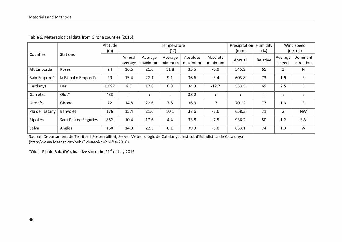
Materials and Methods
46
Table 6. Metereological data from Girona counties (2016).
Counties Stations
Altitude (m)
Temperature (°C)
Precipitation (mm)
Humidity (%)
Wind speed (m/seg)
Annual average
Average maximum
Average minimum
Absolute maximum
Absolute minimum
Annual Relative Average speed
Dominant direction
Alt Empordà Roses 24 16.6 21.6 11.8 35.5 -0.9 545.9 65 3 N
Baix Empordà la Bisbal d'Empordà 29 15.4 22.1 9.1 36.6 -3.4 603.8 73 1.9 S
Cerdanya Das 1.097 8.7 17.8 0.8 34.3 -12.7 553.5 69 2.5 E
Garrotxa Olot* 433 : : : 38.2 : : : : :
Gironès Girona 72 14.8 22.6 7.8 36.3 -7 701.2 77 1.3 S
Pla de l'Estany Banyoles 176 15.4 21.6 10.1 37.6 -2.6 658.3 71 2 NW
Ripollès Sant Pau de Segúries 852 10.4 17.6 4.4 33.8 -7.5 936.2 80 1.2 SW
Selva Anglès 150 14.8 22.3 8.1 39.3 -5.8 653.1 74 1.3 W
Source: Departament de Territori i Sostenibilitat, Servei Meteorològic de Catalunya, Institut d'Estadística de Catalunya (http://www.idescat.cat/pub/?id=aec&n=214&t=2016)
*Olot - Pla de Baix (DC), inactive since the 21st of July 2016

Materials and Methods
47
3.2. STUDY LOCATIONS AND STUDY POPULATION
For this work, a total of 593 dogs living in 36 kennels and 26 localities in the province of Girona
were sampled from 2012 to 2016. These dogs were all included in the L. infantum
seroprevalence study, described in section 3.3. The study of humoral immune response to
vector saliva and CaniLeish® vaccine trials only included animals sampled between 2015 and
2016, totalling 406 dogs distributed across 21 kennels and 16 localities. Sampling and
recruitment of dogs for these studies is described in sections 3.2.1 and 3.2.2. The work
reported in these sections does not include field work performed before 2015.
3.2.1. Preliminary assessment of Leishmania infantum infection in the study locations
Field work for the project started in July 2015 by a serological survey in 16 dog kennels
distributed over 11 localities.
A total of 300 dogs were sampled and tested for the detection of anti-L. infantum antibodies.
This cross-sectional study was intended to evaluate the level of infection at the different dog
kennels and to estimate point L. infantum infection prevalence (Annex 1).
During September 2015, sand fly surveys were performed in order to confirm the presence of
L. infantum vectors in the study locations. One to two CDC light traps were placed for one
night in each dog kennel. Collected sand flies were identified and the different phlebotomine
species abundance and frequency were registered. Sand fly species captured at each dog
kennel are described in Annex 2.
The following serological survey took place in February 2016 and included 293 dogs. From
these, 106 were dogs sampled for the first time, the majority of them belonging to five
additional dog kennels in four different localities. The remaining 187 corresponded to dogs
sampled for the second time. By comparison with the first survey, annual point L. infantum
infection incidences were estimated (Annex 1). Apart from the locations selection, the 2016
survey also allowed for a pre-selection of individuals to be enrolled in the vaccine field trial.

Materials and Methods
48
3.2.2. Location and individual selection for the study of humoral immune response to vector
saliva and CaniLeish® vaccine trial
In February 2016, dogs from 18 dog kennels and 12 localities were enrolled in the vaccine field
trial. The selection of dog kennels was based on the detection of canine L. infantum infection
cases and the presence of phlebotomine vectors. Thus, one dog kennel sampled in July 2015
(Annex 1; kennel 15) and one dog kennel sampled in February 2016 (kennel 21) were not
included in the study due to the absence of cases of L. infantum infection. A third kennel was
also excluded when the dog owner refused to participate in the vaccine trial (kennel 17).
Individuals were selected according to their serological result to L. infantum (only seronegative
dogs were enrolled in the vaccine study), followed by additional criteria, as described in Figure
5.

Materials and Methods
49
Figure 5. Flow chart of pre-vaccination screening and criteria followed for selection of individuals for the vaccine field trial.
Due to the reduced number of dogs in some dog kennels, their geographical proximity and the
similar infection prevalence and/or incidence estimated for kennels in the same localities, a
decision was made to group dogs by locality instead of dog kennels. This way, trial dogs were
divided by “locations” in the following studies, as shown in Figure 6. Dog density per study
location varied between 4 and 23.

Materials and Methods
50
Figure 6. Map of Girona province. Field trial locations are marked in black circles; the number of study dogs per location (n) is presented.
The vaccine study population was initially composed of 177 dogs (90 in the vaccine group and
87 in the control group), from which 168 (85 vaccine and 83 control) completed the first
vaccination phase and were included in the field trial. The majority of the dog population was
composed of hunting dogs (87.1% of the vaccine group and 83.9% of the control group), but
breeding (8.9% of the total dog population), racing (6.5%) or pet dogs (0.6%) were also
represented; no statistically significant differences in dog purpose between groups were
observed (χ2=3.66, p=0.3). Crossbred dogs represented 55.3% of the vaccine group and 45.8%
of the control group (χ2=1.52, p=0.218), while 55.3% and 65.1% of the vaccine and control
group, respectively, were males (χ2=1.67, p=0.196). In total, 60.1% of the dog population were
males and this high male/female ratio can be explained by the exclusion of gestating or
lactating females, either at the beginning of the study or expected to be so during the duration
of the trial. Mean dog age in the vaccine group was 3.3 years (SD=2.9) and in the control group
was 3.4 years (SD=3.0), ranging from 6 months to 11 years (χ2=6.58, p=0.832).
All animals were kept in open-air facilities, mostly in rural and periurban areas. Furthermore,
no specific anti-sand fly insecticide treatments were applied in any location, providing the ideal
conditions for dog exposure to L. infantum vectors.
Materials and Methods
51
3.3. SEROPREVALENCE OF CANINE Leishmania infantum INFECTION IN GIRONA
PROVINCE AND IDENTIFICATION OF RISK FACTORS FOR THE INFECTION
The L. infantum seroprevalence study performed in Girona province included data from canine
serological surveys performed throughout the province between 2012 and 2016. From these,
406 individuals were sampled in the context of this study, in July 2015 and February 2016, as
described in section 3.2.1.
3.3.1. Study design
Study individuals were recruited through local veterinarians registered in the regional
veterinary association (Col.legi Oficial de Veterinaris de Girona – COVGi). After an informative
talk about CanL, a number of professionals were willing to participate by being the link
between their clients and the project researchers. Dog owners consisted mostly of wild boar
hunters, who usually keep large packs of dogs, allowing the sampling of several animals in the
same location. Four cross-sectional surveys were conducted between April 2012 and March
2016 in different locations of north-eastern and Pyrenean areas of Spain, in Girona province,
including 36 sampling stations in 26 localities (Figure 7). Blood samples were collected from all
animals by cephalic or jugular venepuncture to 5 mL EDTA tubes and preserved at -40°C until
further processing.

Materials and Methods
52
Figure 7. Map of altitudinal distribution in Girona province. Sampling locations are marked as black dots.
3.3.2. Serological detection of Leishmania infantum infection
Plasma was obtained and preserved at -40°C. Samples were analysed by an in-house enzyme-
linked immunosorbent assay (ELISA) for the presence of anti-L. infantum antibodies, using a
technique previously described (Ballart et al., 2013a; Fisa et al., 2001; Iniesta et al., 2002; Riera
et al., 1999). Briefly, dog plasma samples diluted at 1:400 were incubated in titration plates
(Costar®) previously coated with sonicated whole promastigotes at a protein concentration of
20 µg/mL in 0.05 M carbonate buffer at pH 9.6. Protein A peroxidase ((1:30,000, Sigma®) was
used as conjugate and reactions were stopped with H2SO4 3M when a pre-determined positive
control serum reached an optical density of 450 read at 450 nm. Sample optical densities were
then read at 492 nm. All samples were run in duplicate and calibrator, positive and negative
serums were included in all plates. Results were expressed in standard units (U) compared to a
calibrator control sample set arbitrarily at 100U. The cut-off was established at 24U.

Materials and Methods
53
3.3.3. Data collection
In addition to sample collection, information was gathered from each sampling location
(geographical coordinates, altitude, county, nearest locality, type of habitat, and presence of
other domestic and farm animals) and each animal’s individual characteristics (sex, age, breed,
given use, type of night shelter, and presence of visible CanL clinical signs). Clinical exams were
performed by veterinarians and the criteria for classifying dogs as “symptomatic” were the
detection of the following clinical signs: weight loss, lymphadenomegaly, periocular or diffuse
alopecia, onychogryphosis, ocular lesions, and/or pale mucous membranes. Dog owners were
asked about their previous knowledge of CanL, as well as control measures regularly taken to
prevent the disease. This data was then used to determine possible risk factors associated with
CanL seroprevalence in the population studied. Data collected from each sampled dog and the
questionnaire completed by the dog owners is presented in Annex 3.
3.3.4. Statistical analysis
True seroprevalence was calculated following the method described in Cortes et al. (2012). The
formula used was: true prevalence (TP) = [apparent prevalence (AP) – 1 + test specificity (Sp)] /
[test sensitivity (Se) – 1 + Sp]. Confidence intervals for true prevalence were also calculated
with the following formula: TP 95%CI = 1.96 x √ [AP x (1-AP) / sample size (n) x (Se+Sp-1)]. The
L. infantum in-house ELISA has a specificity of 90% and a sensitivity of 85%, when the chosen
cut-off is used. These values were calculated based on a population of 77 dogs (Fisa et al.,
2001; Iniesta et al., 2002). Reference positivity status for L. infantum infection was determined
by parasite detection (culture and/or direct exam and/or PCR). ELISA sensitivity and specificity
analysis is provided in Annexes 4 to 6.
The relationship between CanL seropositivity and a series of individual and location variables
was assessed through a mixed logistic regression model. The choice of variables to analyse, as
well as the categories defined, were based on those used in previous publications (Ballart et
al., 2013a; Gálvez et al., 2010b; Martín-Sánchez et al., 2009) and adapted to the characteristics
of the present study. In summary, the covariates considered in the analysis were: altitude
(<800/>800 m.a.s.l.), type of habitat (rural or between villages/periurban or at the edge of
villages/urban or inside villages), presence of other animal species (yes/no), sex (male/female),

Materials and Methods
54
age (<1 to 13 years, introduced as a continuous variable), breed (purebred/crossbred), use
given (hunting/breeding/others, with “others” including racing and pet dogs), night shelter
(indoors/outdoors), dog owner knowledge of preventive methods against CanL (yes/no), dog
owner use of preventive methods against CanL (yes/no) and dog owner use of prophylactic
methods against other arthropods (yes/no).
A bivariate logistic regression analysis was performed, in which the relationship between the
outcome variable (“dog seropositivity”) and each explanatory variable listed above was
assessed individually. Statistical significance was set at p<0.05. This was followed by a
multivariable mixed logistic regression analysis, in which non-significant covariates (p>0.05)
were sequentially deleted through a backward stepwise selection method until a final model
was obtained. In this model, “Locality” was introduced as a random-effects variable to account
for geographic clustering of the data (Alonso et al., 2010; Ballart et al., 2013a) and the year of
the survey was included as a fixed-effects variable.
The association between CanL seroprevalence calculated per dog shelter and owner’s
perception of risk of infection (graded in percentage categories from 0 to 90-100%) was
assessed through a Spearman’s coefficient correlation.
All statistical analyses were performed using Stata 15 software (StataCorp LP, College Station,
TX, USA).
3.4. EVALUATION OF DOG EXPOSURE TO Phlebotomus perniciosus THROUGH THE
DETECTION OF ANTI-SAND FLY SALIVA ANTIBODIES IN THE CANINE HOST
3.4.1. Study design
The study included a population of 176 dogs distributed across 12 locations, as described in
section 3.2.2. Individuals were sampled before, during and after the L. infantum transmission
season, at 5 pre-determined time points: February, August and October 2016, and January and
April 2017.

Materials and Methods
55
3.4.2. Sand flies and salivary proteins
A colony of P. perniciosus was reared under standard conditions as described previously (Volf
and Volfova, 2011). Salivary glands were dissected from 4–6 day-old females, pooled at a
concentration of 1 salivary gland per 1 µl of 20 mM Tris buffer with 150 mM NaCl and stored at
-80 °C. The P. perniciosus 43 kDa yellow-related recombinant protein (rSP03B, Genbank accn.
DQ150622) was obtained from Apronex s.r.o. (Prague, Czech Republic) and quantified by the
Lowry method (Bio-Rad Laboratories, Inc.) following the manufacturer’s protocol.
3.4.3. Serological detection of dog exposure to sand flies
Anti-P. perniciosus IgG was measured by an in-house enzyme-linked immunosorbent assay
(ELISA) as described previously (Kostalova et al., 2015). All samples from a single dog were
processed in the same plate. Briefly, microtiter plates were coated either with salivary gland
homogenate (SGH) (40 ng per well, equivalent to 0.2 salivary gland) or with rSP03B (5 μg/ml) in
20 mM carbonate-bicarbonate buffer (pH 9.5) and incubated overnight at 4 °C. Plates were
then blocked with 6% (w/v) low fat dry milk in PBS with 0.05% Tween 20 (PBS-Tw). Canine
plasma were diluted 1:200 for SGH and 1:100 for rSP03B in 2% (w/v) low fat dry milk/PBS-Tw.
Secondary antibodies (anti-dog IgG, Bethyl laboratories) were diluted 1:9000 in PBS-Tw. The
reaction was stopped with 10% H2SO4 and absorbance was measured at 492 nm using a Tecan
Infinite M200 microplate reader (Tecan®). Each sample was tested in duplicate and positive
and negative controls were included in each plate. To account for the variability between
plates, sample OD readings were normalized by dividing them by the mean OD of positive
controls run in the same plate (Sanchez et al., 2002). The normalized OD values were
multiplied by 100. Positivity cut-offs were calculated as the mean plus 3 standard deviations
from 14 dog samples from a non-endemic area.

Materials and Methods
56
3.4.4. Serological detection of Leishmania infantum infection
The serological technique used to assess L. infantum infection was an in-house ELISA described
in section 3.3.2.
3.4.5. Statistical analysis
Statistical analyses were performed using R software (http://cran.r-project.org/) and Stata 15
software (StataCorp LP, College Station, TX, U.S.A.).
Correlations between IgG responses to P. perniciosus SGH and rSP03B and between each one
of the salivary antigens and anti-L. infantum IgG levels were tested by the Spearman rank
correlation test. Median OD values between time points were compared using the Wilcoxon
signed rank sum test.
The relationship between anti-SGH and anti-rSP03B antibodies and sampling month, L.
infantum infection status and location was tested by fitting multilevel linear regression models,
taking into account the correlation between repeated measures of the same dogs over time. In
the models, log-transformed anti-saliva or rSP03B normalized OD values were considered as
continuous dependent variables and sampling month, L. infantum infection and location as
categorical predictor variables. In order to assess variations in OD between the first sampling
month and those following, “February 2016” was set as reference level for this variable.
Likewise, the locality with the lowest median OD (“Aiguaviva”) was considered to be the
reference for the variable location. Finally, “seronegative” was set as the reference level for
the variable L. infantum infection. The random component included dog and time to allow for
variation at the intercept (between dogs) and the slope (over time). The inclusion of “dog” as a
random effects variable significantly improved both models, with a between dog variance of
48% for SGH and of 47% for the rSP03B model. A p<0.05 was considered to indicate statistical
significance.

Materials and Methods
57
3.5. CANILEISH® VACCINE FIELD TRIAL: IMPACT OF VACCINATION IN
SEROPREVALENCE STUDIES AND VACCINE EVALUATION IN NATIVE DOG
POPULATIONS
3.5.1. Study design
At the beginning of the trial, in March 2016, 168 dogs were selected from a population of 406
dogs, as described in section 3.2. Dog owners were previously informed of all details of the
study and signed an informed consent before the start of the trial.
Inclusion criteria for the vaccine study were those recommended by CaniLeish® manufacturer:
seronegativity to L. infantum, normal clinical exam, minimum age of 6 months, and non-
gestating or lactating females (either at the beginning of the study or expected to be so during
the trial). Furthermore, any dogs that had ever received a vaccine for CanL were also excluded
(Figure 5).
Sample size was calculated assuming a 1:1 ratio between the two experimental groups, an
expected 17.6% incidence of L. infantum infection in the control group, 3.6 times fewer cases
of active infection in the vaccine group, 10% estimated losses during 1 year trial, a power of
0.8 and a significance level of 0.05 in a two-sided test. Final sample size was constrained by the
number of individuals available and the limitations of the research team to follow a larger
group of dogs during the one year trial.
Selected animals were distributed over 12 locations, with the number of individuals per
location ranging from 4 to 23 (Figure 6). Dogs were randomly assigned to either vaccine (n=85)
or control (n=83) groups. As different locations had shown distinct infection levels, individual
allocation to study groups was first stratified per dog kennel and then randomized. This way,
an even proportion of dogs were assigned to each study group in each location, avoiding
possible result bias introduced by distinct infection pressures. After vaccination, dogs were
kept with their owners, under their usual housing and husbandry conditions, and naturally
exposed to an L. infantum transmission season.
As recommended by CaniLeish® vaccine manufacturer, all dogs from both groups were
dewormed with a mixture of febantel, pyrantel pamoate and praziquantel prior to vaccination.

Materials and Methods
58
Individuals from the vaccine group received a three dose vaccine course, 21 days apart. Dogs in
the control group did not receive any vaccine dose.
The vaccine field study took place between March 2016 and May 2017. During this period,
dogs were sampled at pre-determined time points, at which different samples were obtained.
Blood was collected from the cephalic or jugular veins to EDTA tubes (for serology and clinical
blood analysis) or heparin tubes (for peripheral blood mononuclear cells (PBMC) isolation and
cellular-mediated immunity tests). Lymph node samples were collected by fine needle
aspiration and placed in 100 µL of sterile 0.9% sodium chloride solution. Blood and lymph node
samples were frozen at -40°C and PBMC were preserved in liquid nitrogen until processing.
Follow-up samples from the same individual were analysed in parallel. The field study
chronogram, which describes sampling months, number of dogs sampled at each time point,
type of sample collected and laboratory tests performed, is presented in Figure 8

Materials and Methods
59
Figure 8. Vaccine field trial chronogram. Sampling months, number of dogs sampled, samples collected and tests performed at each time point are presented. Vac1: first vaccine dose (also T0); Vac2: second vaccine dose; Vac3: third vaccine dose; 1M: 1 month after the third vaccine dose; 4M, 6M, 9M and 12M: as in 1M; WB: whole blood; PBMC: peripheral blood mononuclear cells; CBC: complete blood count; BQ: biochemical plasma profile; PTG: protein electrophoretogram; LN aspirate: aspirate of popliteal lymph node; LN qPCR: real-time PCR on lymph node aspirate.

Materials and Methods
60
3.5.2. Vaccine safety assessment
After each vaccine dose, dog owners were asked to monitor their dogs and to report any
adverse clinical signs observed to the researchers. Periodical revisions by the veterinarians of
the team were also performed.
3.5.3. Serological follow-up
The serological technique used to assess L. infantum infection is described in section 3.3.2.
Serological assessments were performed at eight time points throughout the study (Figure 8).
An increase of 4-fold ELISA units when compared with the same individual’s basal values (ELISA
units measured at T0) was considered evidence of seroconversion to L. infantum (Paltrinieri et
al., 2016; Solano-Gallego et al., 2011, 2009).
3.5.4. Molecular assessment
L. infantum qPCR on lymph node samples was performed in suspected cases of CanL and in the
last sampling time point for seropositive dogs. DNA was extracted from lymph node aspirates
using the High Pure PCR Template Preparation Kit (Roche®), following the manufacturer’s
instructions. Quantitative PCR was performed in all samples as described elsewhere (Martín-
Ezquerra et al., 2009; Mary et al., 2004) with minor modifications. Briefly, PCR mix reaction
was prepared with 5 µL of DNA, 10 µL of master mix (FastStart Universal Probe Master (ROX),
Roche®), 10 µM of Leishmania primers (direct 5′-CTTTTCTGGTCCTCCGGGTAGG-3′ and reverse
5′-CCACCCGGCCCTATTTTACACCAA-3’, supplied by StabVida®) and 5 µM TaqMan® probe (FAM-
TTTTCGCAGAACGCCCCTACCCGC-TAMRA), designed to target a kinetoplast DNA (kDNA)
sequence, and 1 µL of H2O. Eukariotic 18S rRNA was used as endogenous control (VICTM/MGB
probe, primer limited, Thermo Fisher Scientific®). Amplifications and detection were
performed in an ABI7900 device (Applied Biosystems) and the thermal cycling profile was 50°C
for 2 min, 95°C for 10 min, 45 cycles at 95°C for 15 sec, and 60°C for 1 min. All samples were
analysed in triplicate and positive (DNA from L. infantum MHOM/FR/95/LEM3141 strain) and

Materials and Methods
61
negative controls were included in all qPCR reactions. Parasite quantification was performed
by comparison with a standard curve generated with L. infantum DNA extracted from 1 x 10⁶
parasites/mL by using serial dilutions from 105 to 1 parasites/mL.
3.5.5. Clinical follow-up
All dogs were clinically assessed before the beginning of the vaccine trial. This included a
clinical exam, complete blood count (CBC), renal and hepatic function assessment, and serum
protein electrophoretogram. In addition to allowing an assessment of each dog’s health status
at T0, these results were kept as a baseline to compare with subsequent exams throughout the
study.
The clinical exam included inspection of general body condition, hydration status, skin, hair
and nail condition, mucosae, external lymph nodes and ocular lesions. Owners were asked
about any recent disease, visible weight loss, anorexia, exercise intolerance,
polyuria/polydipsia, vomit or diarrhoea. Clinical assessments were repeated throughout the
field trial whenever there was a suspicion of CanL, either detected by the researchers during
follow-up visits or by the dog owners. At the end of the trial, a thorough clinical exam was
performed to all dogs. Likewise, blood analyses were repeated whenever needed to confirm a
CanL case and at the end of the study for all seropositive dogs.
Due to the nonspecific clinical presentation of CanL, dogs were considered symptomatic only if
two or more clinical signs compatible with the disease were observed (Fisa et al., 1999). The
same criterion was followed for any detected laboratory changes.
3.5.6. Evaluation of vaccine-induced cellular mediated immunity (CMI)
PBMC were obtained from each individual at three time points: before the first vaccine dose
(T0), 4 weeks after the 3rd vaccine dose (M1) and 9 months after vaccination completion (M9)
(Figure 8). Only dogs with samples for the 3 time points studied were included in the CMI
assessment, in a total of 152 individuals (75 in the vaccine group and 77 in the control group).

Materials and Methods
62
Samples of heparinized whole blood were processed within 4h of collection. PBMC were
isolated by centrifugation with a density gradient medium (LymphoprepTM; Stemcell
Technologies), frozen in foetal bovine serum (FBS) supplemented with 10% dimethyl sulfoxide
(DMSO), and stored in liquid nitrogen until processing (Requena et al., 2015).
For assessment of antigen-specific cytokine responses, samples from the same dog were
processed together. PBMC were slowly thawed, washed and left for an overnight rest at 37°C
in 5% CO2. On the following day, cells were counted on a TC20TM Automated Cell Counter (Bio-
Rad Laboratories, Inc.) and incubated in 96-well culture plates at a density of 106 cells/mL as
described elsewhere (Rodríguez-Cortés et al., 2017). Briefly, PBMC were incubated with 10
µg/mL soluble L. infantum antigen (LSA), or 2.5 µg/mL concanavalin A (ConA) (Sigma Aldrich®),
or culture media (unstimulated, negative control) for a period of 5 days at 37°C in 5% CO2. On
the fifth day, plates were centrifuged and supernatants were collected and stored at -40°C.
Interferon-γ (IFN-γ) concentration was determined on PBMC supernatants by a sandwich-
ELISA. The Canine IFN-γ DuoSet ELISA kit (R&D Systems) was used, following the
manufacturer’s instructions. All samples were processed in duplicate and a standard curve was
included in all plates, with a range of IFN-γ concentrations from 0 to 2000 pg/mL. Optical
densities were determined at 450 nm, with wavelength correction set to 570 nm. IFN-γ
concentrations were calculated using a four parameter logistic curve produced in GraphPad
Prism® version 5.3. To obtain the specific IFN-γ concentration for each sample, readings from
the unstimulated cell supernatant were subtracted from the LSA-stimulated cell supernatant.
All plates presented a coefficient of determination (R2) above 0.99.
3.5.7. Definition of canine leishmaniosis case
Classification of trial dogs’ infection status was determined based on the results of serological
tests, presence of clinical signs, and detection of CBC or plasma biochemical abnormalities
compatible with CanL. Any suspicion of L. infantum infection detected by the researchers
during sample collection or the dog owners throughout the trial period would be further
evaluated. Apart from these reported cases, and because all analyses were performed in
parallel at the end of the field study, identification of CanL cases was mainly performed in April
2017.
Materials and Methods
63
A confirmed case of leishmaniosis was defined as:
- Seroconversion to L. infantum, defined as a 4-fold increase in ELISA units when compared
with basal values (ELISA units measured at T0) for the same individual and,
- Detection of L. infantum DNA in lymph node samples and,
- Presence of clinical signs or laboratory abnormalities compatible with CanL.
In addition, animals without detectable clinical signs or laboratory findings, but showing
evidence of seroconversion and presence of Leishmania DNA in lymph nodes at the last time
point (12M) were also considered confirmed cases of active L. infantum infection.
3.5.8. Study endpoint
Cases of CanL confirmed during the field study were submitted to treatment and follow-up or
euthanasia, according to the dog owner’s decision.
3.5.9. Statistical analysis
All statistical analyses were performed using Stata 15 software (StataCorp LP, College Station,
TX, USA). Continuous variables included in this study did not present a normal distribution and
normality could not be achieved by data transformation. Therefore, non-parametric statistical
tests were used to compare between and within group results for continuous variables.
Comparisons between groups at each time point were tested by Mann-Whitney U test.
Longitudinal comparisons within groups were tested by Wilcoxon signed-rank test. Statistical
significance of difference in proportions between groups was tested by the Pearson Chi-square
test.


4. RESULTS AND DISCUSSION


Results and Discussion
67
4. RESULTS AND DISCUSSION
4.1. SEROPREVALENCE OF CANINE Leishmania infantum INFECTION IN GIRONA
PROVINCE AND IDENTIFICATION OF RISK FACTORS FOR THE INFECTION
This study has been accepted for publication in the journal Preventive Veterinary
Medicine and is available in article format in Annex 7.
4.1.1. Resumen
La Cuenca mediterránea es una región endémica de la leishmaniosis canina (LCan), donde
representa un importante problema veterinario y plantea problemas de salud humana.
Sin embargo, la distribución de la enfermedad es heterogénea y no todos los países y
lugares han sido estudiados y caracterizados por igual. Este trabajo describe la situación
de la LCan en la provincia de Girona (Cataluña, España), para la cual no existen datos
reportados y publicados. Entre 2012 y 2016, se realizaron cuatro encuestas transversales
de seroprevalencia en toda la provincia, incluyendo 36 estaciones de muestreo en 26
localidades y un total de 593 perros. Para cada animal también se recolectaron variables
individuales y de ubicación. Además, cada propietario respondió a un cuestionario sobre
sus conocimientos previos de la LCan y los métodos preventivos utilizados. Las muestras
de sangre se analizaron mediante un ELISA in house y se utilizó un modelo de regresión
logística mixta para evaluar la relación entre las variables predeterminadas y la
seropositividad del perro. Se utilizó la correlación de Spearman para evaluar la asociación
entre el riesgo de LCan percibido por los dueños y la seroprevalencia de L. infantum en un
lugar determinado. La seroprevalencia global estimada para la provincia de Girona fue del
32,8% (IC 95%: 28,2-37,5), de los cuales solo el 6,8% de los perros (10/146) se
consideraron sintomáticos. La edad del perro [OR = 1.21 (IC 95%: 1.11-1.31); p <0.001] y
la altitud [OR = 0.02 (IC 95%: 0.001-0.19); p = 0.001] fueron identificados como factores
de riesgo para la infección. Se espera que los resultados obtenidos en este estudio
ayuden a la implementación de programas de control dirigidos en áreas endémicas de
Results and Discussion
68
LCan en toda Europa, así como proporcionen datos adecuados para el diseño de mapas de
evaluación de riesgo de la enfermedad más idóneos.
4.1.2. Background
Spain is an endemic country for CanL and, as observed in other endemic areas, the
distribution of the infection is highly heterogeneous throughout the territory (Miró and
Molina, 2006). For this reason, Mediterranean endemic regions would benefit greatly
from CanL directed control efforts, targeted at areas with higher levels of infection. CanL
seroprevalence in owned dogs in Spain ranges from 1.6% in the northwest (Miró and
Molina, 2006) to 34.6% in the south (Morillas-Márquez et al., 1996), with a range of
intermediate values reported across the territory (Alcover et al., 2013; Ballart et al.,
2013a; Martín-Sánchez et al., 2009; Miró et al., 2017a; Solano-Gallego et al., 2001a).
Nevertheless, as in other Mediterranean countries, the map of CanL distribution in Spain
is far from complete, with many regions still lacking documented information.
Catalonia, in the north-east of Spain, is considered one such endemic area for CanL. Here,
like in other regions, identifying locations for the implementation of CanL directed control
programmes is constrained by the heterogeneous distribution of the infection and the
lack of published data on CanL prevalence. Historically, the south of Catalonia was known
for the presence of well-established and important foci of CanL (Fisa et al., 1999; Portús,
2007) but recent studies in northern areas such as the Lleida region (Ballart et al., 2013a)
showed that the infection is more widespread than previously thought. Furthermore,
results of a recently published questionnaire-based study suggest that Girona province, in
the north-east of Catalonia, may be an endemic area of CanL (Lladró et al., 2017). In a
survey of local veterinarians, the general opinion was that CanL is well established
throughout the province and the number of autochthonous cases has risen in the last
years. Additionally, Girona province shares a range of physical and climatic characteristics
with other Mediterranean locations and is therefore an appropriate example for CanL
epidemiological studies.
The objectives of this study were to provide the first data on CanL seroprevalence for
Girona province, which could also confirm the hypothesis of CanL endemicity in this
region suggested by Lladró et al. (2017), and to identify possible individual and location

Results and Discussion
69
risk factors associated with the infection in the study area which can be applied in the
control of CanL in other Mediterranean endemic regions.
4.1.3. Results
Descriptive analysis of the study population
A total of 593 blood samples were obtained from dogs distributed throughout the north-east
and Pyrenean areas of Spain, in Girona province, with sampling points ranging from 1 to 10 per
county (Table 7).
Sampling sites were mainly rural (corresponding to 50.1% of the dog sample) and periurban
(41.8% of sampled dogs), with dog density per site ranging from 3 to 34. Altitudes ranged from
50 to 1300 m a.s.l., with the majority of dogs living below 800 m a.s.l. (83%). Most dogs were
hunting animals (78.9%), but breeding (16%), shelter (2.5%), racing (2.2%) and pet dogs (0.3%)
were also represented. A large number of sampled dogs were born in Girona province (60.4%)
and were not reported to have left the region. All animals included in the study were kept with
other dogs in open kennels during the day time, and the majority were also kept outdoors at
night (87.9%). There were other animal species kept in close proximity to 49.6% of the
sampled dogs. These included cats, horses, cows, goats and pigs. Observed age average was
3.6 years (SD=2.9), 58.9% of the dogs were males and 55.4% were crossbred.

Results and Discussion
70
Table 7. True seroprevalence for canine Leishmania infantum infection observed in each locality and overall true seroprelavence calculated per county and for Girona province.
County Locality No. sampling points
No. sampled dogs (No. positive dogs)
No. seropositive dogs (%)
True seroprevalence % (95% CI)
Alt Empordà Cadaqués 2 29 (12) 41.4 41.8
Darnius 2 18 (6) 33.3 31.1
Ordis 1 31 (9) 29.0 25.4
Total 5 78 (27) 34.6 32.8 (20.6-45.0)
Baix Cerdanya Urtx 1 30 (1) 3.3 0
Baix Empordà Sant Feliu de Guíxols 1 19 (10) 52.6 56.8 (30.9-82.8)
Garrotxa Hostalnou de Bianya 2 21 (3) 14.3 5.7
Montagut 2 55 (16) 29.1 25.5
Olot 1 12 (5) 41.7 42.2
Sant Esteve de Llémena 1 21 (2) 9.5 0
Total 6 109 (26) 23.9 18.5 (9.2-27.7)
Gironès Aiguaviva 1 30 (4) 13.3 4.4
Canet d'Adri 3 44 (35) 79.6 92.7
Madremanya 1 20 (3) 15.0 6.7
Sant Gregori 1 24 (13) 54.2 58.9
Total 6 118 (55) 46.6 48.8 (38.4-59.2)
Plà de l’Estany Banyoles 1 15 (7) 46.7 48.9 (19.7 -78.0)
Ripollés Bruguera 1 9 (0) 0 0
Camprodon 1 8 (0) 0 0
Ogassa 1 32 (0) 0 0
Serrat 1 6 (0) 0 0
Setcases 1 3 (0) 0 0
Ventola 1 13 (0) 0 0
Total 6 71 (0) 0 0
Selva Brunyola 1 10 (0) 0 0
Maçanet de la Selva 1 17 (3) 17.7 10.2
Massanes 2 29 (3) 10.3 0.5
Riells i Viabrea 2 31 (7) 22.6 16.8
Sta Coloma de Farners 2 32 (0) 0 0
Vilobí d'Onyar 2 34 (7) 20.6 14.1
Total 10 153 (20) 13.1 4.1 (0-10.3)
Total for Girona province 36 593 (146) 24.6 19.5 (15.5-23.5)
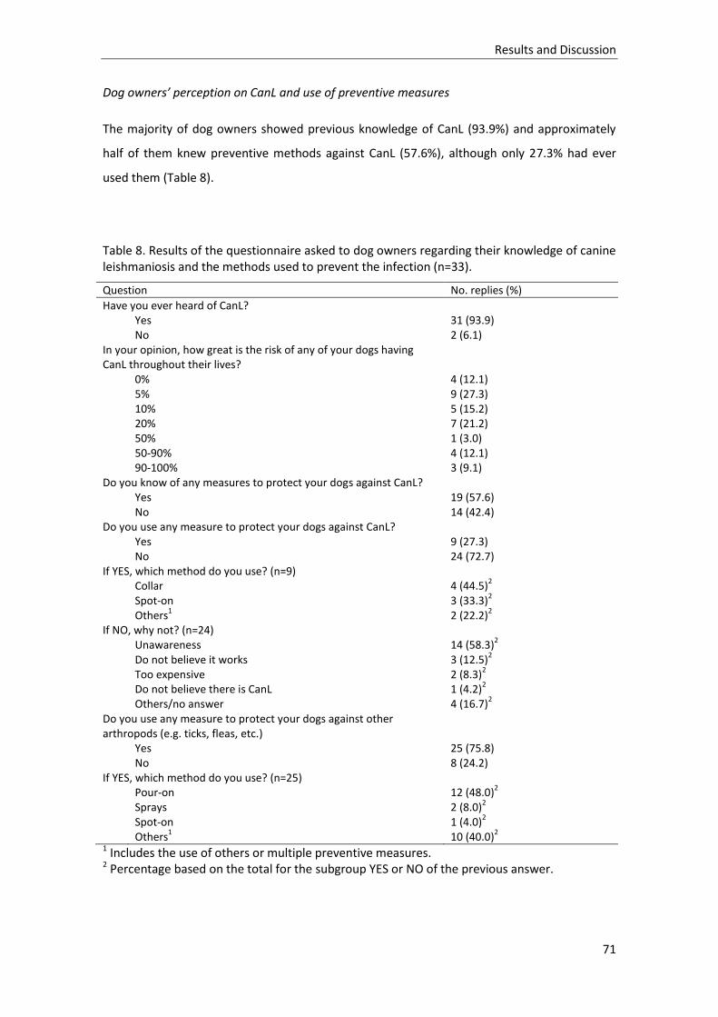
Results and Discussion
71
Dog owners’ perception on CanL and use of preventive measures
The majority of dog owners showed previous knowledge of CanL (93.9%) and approximately
half of them knew preventive methods against CanL (57.6%), although only 27.3% had ever
used them (Table 8).
Table 8. Results of the questionnaire asked to dog owners regarding their knowledge of canine leishmaniosis and the methods used to prevent the infection (n=33).
Question No. replies (%)
Have you ever heard of CanL? Yes 31 (93.9) No 2 (6.1) In your opinion, how great is the risk of any of your dogs having CanL throughout their lives?
0% 4 (12.1) 5% 9 (27.3) 10% 5 (15.2) 20% 7 (21.2) 50% 1 (3.0) 50-90% 4 (12.1) 90-100% 3 (9.1) Do you know of any measures to protect your dogs against CanL? Yes 19 (57.6) No 14 (42.4) Do you use any measure to protect your dogs against CanL? Yes 9 (27.3) No 24 (72.7) If YES, which method do you use? (n=9) Collar 4 (44.5)
2
Spot-on 3 (33.3)2
Others1 2 (22.2)
2
If NO, why not? (n=24) Unawareness 14 (58.3)
2
Do not believe it works 3 (12.5)2
Too expensive 2 (8.3)2
Do not believe there is CanL 1 (4.2)2
Others/no answer 4 (16.7)2
Do you use any measure to protect your dogs against other arthropods (e.g. ticks, fleas, etc.)
Yes 25 (75.8) No 8 (24.2) If YES, which method do you use? (n=25) Pour-on 12 (48.0)
2
Sprays 2 (8.0)2
Spot-on 1 (4.0)2
Others1 10 (40.0)
2
1 Includes the use of others or multiple preventive measures. 2 Percentage based on the total for the subgroup YES or NO of the previous answer.

Results and Discussion
72
Only a small number of dog owners believed that their dogs were not at risk of contracting
CanL during their lifetime (12.1 %), with the majority of them believing that the risk of CanL
ranged from 5 to 20% (63.7%). The result of the Spearman’s correlation showed a positive
association between dog owners’ perceived risk of CanL infection and CanL seroprevalence
(rs=0.5046; p=0.0027).
Prophylactic methods against CanL, when used, included dog collars (44.5%), spot-on (33.3%)
and combined insecticide treatments (22.2%). Vaccination against CanL or immunomodulatory
prophylactic treatments had not been used by any of the dog owners. The main reasons given
for not using any preventive method against CanL were unawareness (58.3%) and not believing
that prophylaxis worked (12.5%).
CanL study results
From the 593 dogs analysed, 146 were considered seropositive by ELISA. Apparent
seroprevalence at the sampling point level ranged from 0% to 79.6%, with a total apparent
seroprevalence calculated for Girona province of 24.6% (95% CI: 21.2-28.3). Considering these
values, the estimated true CanL seroprevalence for Girona province was 19.5% (95%CI: 15.5-
23.5). Estimated seroprevalence at the county level ranged from 0 to 56.8%. Results for all
localities and counties are summarized in Table 7.
Only 10 out of 146 seropositive dogs were considered symptomatic (6.8%). Observed clinical
signs included onychogryphosis (n=9), weight loss (3), skin wounds (3), diffuse alopecia (2),
popliteal lymphadenomegaly (2), periocular alopecia (2), and ocular lesions (2).
Bivariate statistical analysis
One of the dog kennels included in the seroprevalence study (Banyoles, Plà de l’Estany) was
excluded from the statistical analysis, following the criteria used in similar studies (Ballart et
al., 2013a). This is a shelter kennel that collects stray dogs, which means that some of the
individual data, as well as owners’ perception of CanL, could not be collected. Therefore, the
statistical analysis included 578 individuals and 25 localities. Results of the bivariate analysis
are summarized in Table 9. Dogs’ age and location altitude (p<0.001) showed a very strong
relationship with dog seropositivity. In our population, a bimodal CanL seroprevalence
distribution according to age was observed, with a first peak at 3-4 years and a second at 7-8
Results and Discussion
73
years old, with the risk of infection rising by each year of life [OR=1.18 (95%CI: 1.09-1.27)] and
decreasing at altitudes above 800 m a.s.l. [OR=0.012 (95%CI: 0.002-0.07)]. Also, according to
the results, being a crossbred dog raises the risk of infection [OR=2.19 (95%CI: 1.18-4.06);
p=0.013] and the use of unspecific insecticides against arthropods has a protective effect
[OR=2.94 (95%CI: 1.58-5.45); p=0.001]. All the other variables (sex of the dog, type of habitat,
dog purpose, type of nocturnal refuge, presence of other animal species, owner’s knowledge
of prophylactic measures against CanL and the regular application of these methods) showed
no statistically significant relationship with dog seropositivity.
Multivariable mixed model
The final multivariable mixed logistic regression model identified age of the dog and altitude of
the location as the explanatory variables that affect dog seropositivity. According to this
model, the odds of being infected rise in 1.21 per each year of life [(95%CI: 1.11-1.31);
p<0.001] and decrease at locations above 800 m a.s.l. [OR=0.02 (95%CI: 0.001-0.19); p=0.001].
The final model explains 53.7% of the total variance of the outcome variable, of which 42% is
explained by the fixed effects terms and 11.7% by the random effects variable.

Results and Discussion
74
Table 9. Number of dogs analysed and Leishmania infantum seropositivity observed for each category of the explanatory variables, followed by the results of the bivariate analysis expressed in odds ratios (OR). Statistically significant variables (p<0.05) are marked with (*).
Explanatory variables and categories
No. dogs analysed
No. seropositive dogs (% seropositive dogs)
Bivariate analysis
OR (95% CI) p-value
Altitude (m a.s.l.)
< 800 492 144 (29) Ref
>800 109 6 (6) 0.012 (0.002-0.07) <0.001*
Type of habitat
Rural 297 70 (24) Ref
Periurban 228 51 (22) 1.64 (0.93-2.88) 0.082
Urban 53 19 (36) 0.97 (0.42-2.22) 0.934
Presence of other animals (other than dogs)
Yes 302 73 (24) Ref
No 299 78 (26) 1.10 (0.58-2.08) 0.763
Sex
Male 338 87 (26) Ref
Female 240 53 (22) 0.88 (0.56-1.38) 0.581
Age (years) 1.18 (1.09-1.27) <0.001*
<1 53 6 (11)
1 83 12 (14)
2 91 13 (14)
3 79 20 (25)
4 51 16 (31)
5 42 10 (24)
6 42 12 (29)
7 33 15 (45)
8 33 14 (42)
9 15 3 (20)
10 18 5 (28)
11 10 4 (40)
12 3 0 (0)
13 1 1 (100)
Breed
Purebred 258 51 (20) Ref
Crossbred 320 89 (28) 2.19 (1.18-4.06) 0.013*
Use given
Hunting 468 118 (25) Ref
Breeding 95 21 (22) 2.28 (0.78-6.63) 0.130
Others1 15 1 (7) 0.20 (0.02-1.73) 0.145
Night shelter
Outdoors 506 126(25) Ref
Indoors 72 14 (19) 0.50 (0.20-1.23) 0.131
Owner knows preventive measures against CanL
Yes 362 103 (28) Ref

Results and Discussion
75
No 216 37 (17) 0.58 (0.29-1.19) 0.138
Owner has used preventive measures against CanL
Yes 140 46 (33) Ref
No 438 112 (26) 1.24 (0.63-2.43) 0.539
Owner has used prevention methods against other arthropods
Yes 472 112 (24) Ref
No 106 28 (26) 2.94 (1.58-5.45) 0.001* 1 Includes racing (n=13) and pet dogs (n=2).
4.1.4. Discussion
Until now, data on CanL in north-eastern and Pyrenean areas of Spain is scarce and
fragmented. The only published study regarding CanL in Girona province is a questionnaire-
based survey of veterinary practitioners working in the region (Lladró et al., 2017). This work
provided the first data from a previously recognized, but non-documented CanL endemic area
in north-east Spain and highlights gaps in the epidemiological picture in Mediterranean regions
considered to be endemic for CanL (Ready, 2017). The veterinary survey showed that new
cases of CanL in autochthonous animals were diagnosed annually, including some
asymptomatic cases detected by CanL pre-vaccination screening (Lladró et al., 2017). The
present study confirms the suspected endemicity of CanL in the region, providing results for
canine seroprevalence, as well as an overview of the infection distribution throughout Girona
province. Preliminary exploratory surveys showed the presence of phlebotomine vectors in the
surroundings of many of the sampling points (Annex 2), confirming that all conditions are
present for a complete L. infantum biologic cycle to be maintained in this region. In addition,
the characterisation of all individuals and locations included in the study allowed for the
identification of risk factors associated with CanL distribution.
As previously mentioned, there was an active search for individuals to be enrolled in the study,
assisted by local veterinarians. There was therefore a constraint in the distribution and type of
animals recruited, depending on the availability of veterinary practitioners’ clients willing to
participate. As a result, the dog population was mainly composed of hunting dogs. These
animals have inherent characteristics, such as the fact that they are usually kept with other
dogs in open kennels, in rural or periurban settings, and generally do not have the same type
of veterinarian monitoring as pet dogs. Therefore, this type of population is usually considered

Results and Discussion
76
a good sentinel for CanL (Ballart et al., 2013a; Cabezón et al., 2010). As similar hunting
activities take place throughout Mediterranean areas in Europe, it can be expected that
comparable dog populations are widespread. An overestimation of the overall infection
prevalence can however occur due to an expected lower incidence in urban centres, mostly
explained by a decreased probability of contact between dogs and sand fly vectors (Ballart et
al., 2013a). Additionally, there was an increased difficulty in recruiting dogs from higher
altitude regions, mainly because these areas are more inhospitable and less populated.
Consequently, dogs living at locations above 800 m a.s.l. are less represented.
Some degree of spatial clustering may have been introduced by sampling several dogs in the
same kennel or locality. This could also have had a clustering effect on the positive results, as
higher dog densities tend to favour the transmission of the parasite, especially if some of the
dogs are already infected (Alonso et al., 2010). Nevertheless, in the present study we have
used similar dog populations in the different sampling points, allowing comparison between
them. Additionally, this methodology has also been used in similar studies describing other
regions of Spain (Alcover et al., 2013; Ballart et al., 2013a). In the statistical analysis, the
potential clustering effect was dealt with by introducing “Locality” as a random-effects term in
the final multivariable mixed logistic regression model.
The serological technique used to measure antibody levels to L. infantum was an in-house
ELISA. ELISA is one of the methods recommended by the World Organization for Animal Health
for performing CanL surveillance studies and to determine prevalence of infection (OIE, 2014),
the other one being the indirect immunofluorescent antibody test (IFAT). Unlike IFAT, ELISA is
easy to perform and interpret, being particularly useful in field study settings, where a large
number of samples must be analysed (Maia and Campino, 2008). In addition, this ELISA has
been widely used for CanL diagnosis, as well as in other CanL epidemiological studies (Alcover
et al., 2013; Ballart et al., 2013a; Fernández-Bellon et al., 2008; Fisa et al., 2001; Iniesta et al.,
2002; Riera et al., 1999; Rodríguez-Cortés et al., 2010; Solano-Gallego et al., 2005).
The overall estimated seroprevalence for Girona province was 19.5%, ranging from 0 to 56.8%
across the different counties. These results are in accordance with previous reports for other
regions of Spain, as well as the Mediterranean basin (Ballart et al., 2013a; Cortes et al., 2012;
Maroli et al., 2008; Ntais et al., 2013). A series of CanL seroprevalence surveys undertaken in
France, Italy, Spain and Portugal between 1971 and 2006 showed an overall seroprevalence of
23.2%, with point prevalences of 0% and higher than 80% in some locations (Franco et al.,
2011). These values are comparable to the ones obtained in the present study and correspond

Results and Discussion
77
to the previous claims of the heterogeneous distribution of the disease. However, as pointed
out by Franco et al. (2011), caution must be taken when comparing studies with different
experimental designs and different criteria used in the selection of the target dog population,
as this can introduce significant variations in seroprevalence results. A common European
strategy for leishmaniosis surveillance and control would aid the implementation of
standardized methodology. However, although leishmaniosis is currently listed as a notifiable
disease by the World Organization for Animal Health (OIE, 2018), this is not clearly reflected in
the European or Spanish legislation (BOE, 2014; Official Journal of the European Union, 2012).
From the 146 seropositive dogs, only 10 (6.8%) showed clinical signs compatible with CanL and
more than 50% presented low standard ELISA units (inferior to 50U). This can be explained by
the cryptic nature of the infection and the wide clinical spectrum it can present, ranging from
asymptomatic or mild symptomatic cases to very severe clinical stages (Solano-Gallego et al.,
2009). There is also the possibility that some of the dogs are in an early stage of infection (Fisa
et al., 2001; Miró et al., 2012) or are immunologically resistant and only transiently
seropositive, eventually showing spontaneous clearance of the parasite (Fisa et al., 1999). In
such populations, serological techniques could have a lower sensitivity (Otranto et al., 2009). It
is also known that, in endemic areas, only a small proportion of dogs display symptoms of
CanL, while the majority of infected dogs do not show any clinical evidence of the disease
(Baneth et al., 2008). It is believed that the high prevalence of asymptomatic infected dogs,
comparable to that observed in Lleida province (other north-eastern and Pyrenean region
studied in Spain), is strong evidence for a well-established CanL focus in Girona province
(Ballart et al., 2013a). In the present study, clinical signs compatible with CanL were also
identified in 10 out of 447 seronegative dogs (2%), illustrating the lack of specificity of the
disease’s clinical presentation and the added difficulty in detecting affected dogs. As
mentioned before, the ability of serological tests to detect infected animals is limited,
especially in endemic settings, and a small number of seronegative asymptomatic infected
dogs should be expected, as previously reported in other studies (Iniesta et al., 2002; Otranto
et al., 2009; Solano-Gallego et al., 2011). These animals can harbour parasites in the skin,
detectable by PCR (Otranto et al., 2009), and could also be infectious to sand flies, as has been
demonstrated for asymptomatic seropositive dogs (Molina et al., 1994; Quinnell and
Courtenay, 2009). Considering this, any control programme for CanL should be based on
multiple diagnostic methods, as serology alone can prove to be insufficient in detecting all
infected and infectious dogs.

Results and Discussion
78
In the present study, the risk of infection increased with dogs’ age. This is an individual factor
commonly reported as being positively related with L. infantum infection (Alonso et al., 2010;
Ballart et al., 2013a; Cortes et al., 2012; Gálvez et al., 2010b; Maresca et al., 2009; Martín-
Sánchez et al., 2009; Miró et al., 2012), and which can be explained by an incremental risk of
exposure to infected sand flies. The bimodal CanL seroprevalence distribution observed has
been previously described by other authors (Gálvez et al., 2010b; Miró et al., 2012). This
pattern suggests that L. infantum may be able to infect immunologically vulnerable animals at
an earlier age, followed by a later infection of resistant animals either by cumulative exposure
or due to concomitant diseases that weaken the dogs’ immune system (Miranda et al., 2008).
According to the results, altitude shows a negative correlation with L. infantum infection. This
is mainly related to the bioclimatic needs of the phlebotomine vector species present in Spain,
Phlebotomus perniciosus and P. ariasi (Rioux et al., 1986). Altitude is known to be closely
linked to temperature, precipitation and land cover (Barón et al., 2011; Rivas-Martínez, 1983).
In temperate regions, as atmospheric temperature rises, a higher biting rate is expected
(Hartemink et al., 2011), therefore increasing the risk of sand fly bites to vertebrate hosts.
Simultaneously, a shorter extrinsic incubation period (Hartemink et al., 2011) and a more
effective development of the parasite inside the vector (Rioux et al., 1985) are observed,
raising the risk of L. infantum infection. Also, an increased altitude may provide a more hostile
environment for sand fly survival (Gálvez et al., 2010a), not only because of the more extreme
bioclimatic conditions, but also due to a possible scarcity of vertebrate hosts. However, a
relationship between altitude and risk of CanL infection was not observed in the neighbouring
province of Lleida (Ballart et al., 2014, 2013a) or in France (Chamaillé et al., 2010), where both
vector species are present and show different altitudinal preferences. In these areas, P.
perniciosus is known to occupy ecological niches commonly below 800 m a.s.l., while P. ariasi
shows a higher abundance above this altitude. Therefore, it would be of particular interest to
perform entomological studies and risk factor analysis associated with the vector populations
present in the study area. This could also help to improve the ability of the present model to
predict the outcome variable. One of the possible reasons for the moderate performance of
the final statistical model presented (which explains only 53.7% of variance of the outcome
variable) is the absence of data on the abiotic factors mentioned above, which are known to
have an important impact on sand fly populations, and indirectly on L. infantum infections
(Dantas-Torres et al., 2014; Gálvez et al., 2010a).
The present study failed to detect an effect of type of habitat (rural/urban) or access to night
shelter, which several other authors identified as significantly related to L. infantum infection
Results and Discussion
79
(Ballart et al., 2013a; Cortes et al., 2012; de Almeida et al., 2012; Gálvez et al., 2010b; Martín-
Sánchez et al., 2009; Oliveira et al., 2016). According to these studies, dogs that live in rural
habitats and are left outdoors at night show an increased risk of infection. In this study, the
high percentage of dogs living in rural/periurban areas and kept permanently outdoors may
not have allowed detection of such an effect. Also, periurban areas are increasingly described
as the most suitable ecosystems for sand flies, due to the ideal microclimate offered by house
gardens associated with the abundance of vertebrate hosts (Alvar et al., 2004; Ballart et al.,
2013a).
Results from the bivariate statistical analysis identified dog breed and the use of general
insecticide treatment against arthropods as variables associated with dog seropositivity. In the
first case, crossbred dogs would be at higher risk of infection [OR=2.19 (95%CI: 1.18-4.06);
p=0.013]. However, previous studies have shown that this should not be the case, as
crossbred, autochthonous dogs tend to be more resilient to L. infantum infection (Alvar et al.,
2004; Solano-Gallego et al., 2000). This is even more noticeable when the purebred dogs
belong to exotic breeds like boxers and beagles (both represented in this study), known for
their higher sensitivity to CanL (Solano-Gallego et al., 2009). The effect of dog breed was
absent in the mixed model, showing that the previous results were most probably induced by
confounding factors related to the kennel locations (e.g. altitude) or dog owners’ attitudes
(e.g. use of prophylactic measures against CanL). The non-use of generalist insecticide
preventive methods against arthropods was also identified as a risk factor for L. infantum
infection [OR=2.94 (95%CI: 1.58-5.45); p=0.001], while the use of specific prophylaxis against
CanL failed to show a protective effect (p=0.539). Again, this may be related to confounding
factors, such as a possible partial effect of some insecticides against phlebotomine vectors,
even though they may not be licensed for sand fly prevention. Additionally, the improper use
of specific sand fly prevention treatment, such as failure to apply it to all dogs or to maintain it
during the whole transmission season, may impair the protective effect of these products
(Courtenay et al., 2009). Once again, the effect of this variable lost significance in the
multivariable analysis and was not included in the final statistical model.
The majority of dog owners showed previous knowledge of CanL and to be aware of
preventive methods for the infection. Although a positive correlation was observed between
owners’ perceived risk of infection and CanL seropositivity at the dog kennel level, only 27.3%
of dog owners stated that they regularly used CanL prophylactic measures. This result is in
accordance with those reported by Lladró et al. (2017), in which all veterinary practitioners
working in Girona province recommended at least one preventive measure against CanL,

Results and Discussion
80
though the majority did not believe that dog owners protected their dogs properly. When
used, the most frequent prophylactic methods applied against CanL were dog collars and spot-
on insecticides, as recommended by veterinarians. However, most owners did not keep their
dogs indoors at night and did not report the use of vaccination against CanL or
immunomodulatory agents, as also suggested by veterinarians (Lladró et al., 2017). Our
studyshows that the implementation of prophylactic measures by dog owners should be
reinforced in order to reduce L. infantum transmission between dogs, as well as to reduce the
public health risk (Miró and López-Vélez, 2018).

Results and Discussion
81
4.2. EVALUATION OF DOG EXPOSURE TO Phlebotomus perniciosus THROUGH THE
DETECTION OF ANTI-SAND FLY SALIVA ANTIBODIES IN THE CANINE HOST
This study has been published in the journal Parasites & Vectors and is available in article
format in Annex 8.
4.2.1. Resumen
La transmisión de Leishmania infantum se hace a través de la picadura de vectores flebotomos.
Por lo tanto, el monitoreo del contacto entre el hospedador y el vector representa una
herramienta epidemiológica importante que podría usarse para evaluar la efectividad de los
programas de control de vectores en áreas endémicas. Estudios previos han demostrado que
los anticuerpos caninos contra la saliva de los flebotomos son marcadores específicos de la
exposición a los vectores de Leishmania. Sin embargo, la validación de este método debe
ampliarse a poblaciones heterogéneas de perros naturales de áreas endémicas de LCan. En
este estudio, se siguieron durante 14 meses 176 perros que viven en 12 lugares diferentes de
un área endémica para L. infantum en el noroeste de España (provincia de Girona). Las
muestras de sangre a utilizar para evaluar la respuesta inmune humoral canina al
homogeneizado de glándulas salivales (SGH) y a la proteína recombinante “yellow-related” de
43 kDa (rSP03B) de Phlebotomus perniciosus, uno de los vectores confirmados de L. infantum
presente de forma natural en esta región, fueron tomadas en 5 momentos de muestreo
predeterminados (Febrero, Agosto y Octubre de 2016, Enero y Abril de 2017).
Simultáneamente, se evaluó la infección por L. infantum en todos los perros por serología
(ELISA in house). La relación entre los anticuerpos anti-SGH y anti-rSP03B con el mes de
muestreo, la infección por L. infantum y la localidad se estudió mediante el ajuste de modelos
de regresión lineal multinivel. La dinámica de las IgG caninas anti-saliva de SGH y rSP03B siguió
las tendencias esperadas de actividad de P. perniciosus en la región. Se detectaron
asociaciones estadísticamente significativas para ambos antígenos salivales entre la exposición
al vector y el mes de muestreo o la seropositividad del perro a L. infantum. La correlación
entre anticuerpos caninos contra SGH y rSP03B fue moderada. Los resultados confirman la
presencia frecuente de vectores de LCan en el área de estudio así como la aplicabilidad de las

Results and Discussion
82
pruebas ELISA basadas en SGH y rSP03B para estudiar la exposición canina a P. perniciosus en
áreas endémicas de L. infantum.
4.2.2. Background
The detection of anti-sand fly salivary antibodies in the blood of vertebrate hosts has proven to
be highly specific (Rohoušová et al., 2005) and was successfully used as a marker of exposure
to L. infantum vectors (Martín-Martín et al., 2014; Vlkova et al., 2011). In CanL endemic areas,
monitoring the canine IgG response to sand fly saliva can be a useful epidemiological tool
(Kostalova et al., 2015; Vlkova et al., 2011), complementing studies of vector population
dynamics and host-vector interactions, as well as enabling the assessment of risk of
Leishmania infection (Carvalho et al., 2015; Marzouki et al., 2011; Rohoušová et al., 2005).
Furthermore, it can be used to measure the effectiveness of vector-control programmes and
to assist in the design of better control strategies for the disease (Clements et al., 2010;
Gidwani et al., 2011).
Originally, sand fly whole salivary gland homogenates (SGH) were used to investigate the
presence of anti-sand fly saliva antibodies in vertebrate hosts (Gidwani et al., 2011; Volf and
Rohoušova, 2001). However, its use in large-scale studies is impaired by technical limitations
(Lestinova et al., 2017). Additionally, the use of SGH in vector exposure tests may reduce the
specificity of detection due to a possible cross-reactivity with saliva of sympatric and closely
related sand fly species (Lestinova et al., 2015).
An alternative to the use of SGH is the identification of species-specific salivary proteins that
can be expressed in recombinant forms and produced in large quantities for use in large-scale
epidemiological studies (Drahota et al., 2014; Kostalova et al., 2017). Recent studies identified
P. perniciosus yellow-related protein rSP03B as the most promising candidate to replace SGH
in the detection of host markers of exposure to this vector species (Kostalova et al., 2017,
2015; Martín-Martín et al., 2014). This recombinant protein has been tested and validated in
dogs and other animals in cross-sectional studies (Kostalova et al., 2017; Martín-Martín et al.,
2014), as well as in a canine longitudinal study (Kostalova et al., 2015), but no information
exists on the seasonal dynamics of either SGH or rSP03B in natural heterogeneous dog
populations from endemic areas.

Results and Discussion
83
Therefore, the objectives of this study were (i) to investigate the dynamics of P. perniciosus
and their relative density in a previously uncharacterized CanL endemic area through the
detection of anti-saliva IgG in dogs; and (ii) to evaluate the performance of both SGH and
rSP03B antigens as markers of exposure to P. perniciosus in natural canine populations.
4.2.3. Results
Seasonal dynamics of IgG response against salivary proteins from Phlebotomus perniciosus
Median values of normalized ELISA OD values for SGH ranged from 9.04 (range: 3.94–66.23) in
January 2017 to 18.51 (7.93–100.58) in August 2016 (Table 10). For rSP03B, median OD values
varied between 12.21 (6.75–53.71) and 19.53 (10.64–124.01) in January 2017 and August
2016, respectively. With both antigens, median OD readings raised from basal values in
February 2016 (10.11 and 14.67 for SGH and rSP03B, respectively) to peak in August (18.51
and 19.53 for SGH and rSP03B, respectively), sustained higher readings in October (11.15 and
15.31 for SGH and rSP03B, respectively), and descended again to basal levels in January (9.04
and 12.21 for SGH and rSP03B, respectively) and April 2017 (9.54 and 13.44 for SGH and
rSP03B, respectively). Median normalized ELISA OD results obtained per month for both SGH
and rSP03B are described in Table 10 and plotted in Figure 9.
Table 10. Median values of normalized OD readings for SGH and rSP03B obtained per sampling month in all locations
Variable N SGH rSP03B
Median (Range) Median (Range)
February 2016 174 10.11 (5.49–49.62) 14.67 (7.36–41.24)
August 2016 33 18.51 (7.93–100.58) 19.53 (10.64–124.01)
October 2016 164 11.15 (5.56–86.44) 15.31 (6.15–112.54)
January 2017 154 9.04 (3.94–66.23) 12.21 (6.75–53.71)
April 2017 148 9.54 (5.25–62.59) 13.44 (6.27–36.22)
N: number of dogs sampled per sampling month
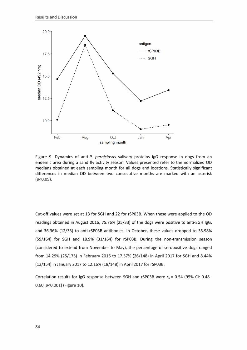
Results and Discussion
84
Figure 9. Dynamics of anti-P. perniciosus salivary proteins IgG response in dogs from an endemic area during a sand fly activity season. Values presented refer to the normalized OD medians obtained at each sampling month for all dogs and locations. Statistically significant differences in median OD between two consecutive months are marked with an asterisk (p<0.05).
Cut-off values were set at 13 for SGH and 22 for rSP03B. When these were applied to the OD
readings obtained in August 2016, 75.76% (25/33) of the dogs were positive to anti-SGH IgG,
and 36.36% (12/33) to anti-rSP03B antibodies. In October, these values dropped to 35.98%
(59/164) for SGH and 18.9% (31/164) for rSP03B. During the non-transmission season
(considered to extend from November to May), the percentage of seropositive dogs ranged
from 14.29% (25/175) in February 2016 to 17.57% (26/148) in April 2017 for SGH and 8.44%
(13/154) in January 2017 to 12.16% (18/148) in April 2017 for rSP03B.
Correlation results for IgG response between SGH and rSP03B were rS = 0.54 (95% CI: 0.48–
0.60, p<0.001) (Figure 10).

Results and Discussion
85
Figure 10. Correlation between IgG recognizing SGH and rSP03B protein in dogs naturally exposed to P. perniciosus. Results from both SGH and rSP03B are presented in normalized OD (rS = 0.54; 95% CI: 0.48–0.60, p<0.001)
Dogs’ exposure to Phlebotomus perniciosus in the study area
Exposure of dogs to phlebotomine vectors showed some variation according to the location.
Median OD readings varied from 9.11 (range: 5.25–20.57) to 14.14 (7.44–55.45) for SGH ELISA
and from 12.71 (7.53–64.44) to 17.87 (8.39–112.54) for rSP03B. Minimum median values of
response to both SGH and rSP03B corresponded to the same location (Aiguaviva), but
maximum median values were registered in different sites for each antigen (Sant Feliu de
Guíxols for SGH and Montagut for rSP03B) (Table 11). Figure 11 presents the dynamics of dogs’
IgG response to SGH (Figure 11a) and rSP03B (Figure 11b) in each locality.
The percentage of anti-sand fly saliva seropositive dogs per location, defined as the number of
dogs that showed a positive IgG titre at least once during the study period, ranged from

Results and Discussion
86
13.33% (1/8) in Ordis to 100% in Canet d’Adri (8/8) and Sant Feliu de Guíxols (4/4) for SGH, and
from 8.16% (1/12) in Hostalnou de Bianya to 100% (4/4) in Sant Feliu de Guíxols for rSP03B.
Total anti-sand fly saliva seropositivity calculated for the study area was 49.43% (87/176) for
anti-SGH IgG and 28.98% (51/176) for anti-rSP03B antibodies.
Dogs’ exposure to Phlebotomus perniciosus and Leishmania infantum infection
Correlation results between antibody response to P. perniciosus saliva and L. infantum were
low both for SGH (rS = 0.27, 95% CI: 0.19–0.35, p<0.001) and rSP03B protein (rS = 0.25, 95% CI:
0.18–0.32, p<0.001).

Results and Discussion
87
Table 11. Median values of normalized OD readings for SGH and rSP03B obtained per sampling location at all time points
Variable n (Range) Geographical
coordinates
SGH rSP03B
Median (Range) Median (Range)
Ordis 8 (7–9) 42°13'37.7''N,
2°54'24.1''E
9.14 (6.45–45.95) 15.16 (8.35–54.50)
Madremanya 14 (12–15) 41°58'47.0''N,
2°58'7.2''E
11.22 (6.79–49.84) 14.49 (8.95–43.99)
Vidreres 8 (7–9) 41°47'27.4''N,
2°45'0.4''E
10.59 (7.80–16.86) 13.46 (8.58–40.23)
Massanes 21 (20–23) 41°45'15.3''N,
2°38'44.0''E
9.31 (5.67–62.59) 16.35 (7.82–55.81)
Hostalnou de
Bianya
12 (11–14) 42°13'26.0''N,
2°26'9.7''E
8.75 (5.35–33.16) 13.19 (6.27–46.82)
Montagut 13 (7–15) 42°14'7.7''N,
2°35'57.6''E
12.01 (3.94–72.61) 17.87 (8.39–
112.54)
St. Esteve de
Llémena
9 (9–10) 42°3'35.1''N,
2°37'1.4''E
9.49 (6.23–22.40) 14.18 (9.12–22.46)
Canet d'Adri 8 (4–10) 42°1'53.7''N,
2°44'15.3''E
10.61 (6.52–
100.58)
14.03 (7.36–
124.01)
Aiguaviva 19 (16–22) 41°54'27.2''N,
2°46'19.0''E
9.11 (5.25–20.57) 12.71 (7.53–64.44)
St. Feliu de Guíxols 4 41°47'2.3''N,
2°59'58.7''E
14.14 (7.44–55.45) 16.73 (8.57–65.97)
Riells i Viabrea 20 (18–21) 41°43'59''N,
2°33'39.3''E
10.02 (6.07–66.23) 13.43 (8.59–35.31)
Vilobí d'Onyar 23 (22–23) 41°53'3.2''N,
2°43'38.6''E
9.13 (5.17–16.49) 13.05 (6.15–38.07)
n: mean number of dogs sampled in each location

Results and Discussion
88
Figure 11. Dynamics of dogs’ IgG recognizing SGH (a) and rSP03B protein (b) in the different sampling locations during a sand fly activity season. Values presented refer to the normalized OD medians obtained at each sampling month.
Multilevel analysis of the relationship between anti-Phlebotomus perniciosus salivary proteins,
month and location and Leishmania infantum seropositivity
The multilevel model results confirmed the annual dynamics of anti-salivary proteins IgG
responses. When compared to the first sampling month (February 2016), IgG responses to SGH
significantly rose in August (t = 8.55, df = 491, p<0.001) and October (t = 6.49, df = 491,
p<0.001) and dropped in January (t = -2.49, df = 491, p=0.013) and April 2017 (no significant
difference when compared to February 2016). As expected, the highest log OD estimate was
observed in August 2016 and the lowest in January 2017 (Table 12). The same trend was
observed in the model run for the rSP03B protein, with comparable levels of significance
(Table 13). There were no significant differences in IgG responses for both antigens between
each sampling location and the one set as reference, except for Montagut, where significantly
higher OD levels were observed for SGH (t = 2.28, df = 166, p=0.024) and rSP03B (t = 2.13, df =
164, p=0.035). According to the multilevel model, seropositivity to L. infantum proved to be
associated with a rise in anti-salivary proteins OD values for both SGH (t = 2.5, df = 491,
p=0.013) and rSP03B (t = 2.15, df = 493, p=0.032).

Results and Discussion
89
Table 12. Estimates of the multilevel linear regression model of the relationship between log transformed normalized SGH OD values and sampling time, location and dog seropositivity to L. infantum. “Dog” was included as a random effects variable
Variable Levels Estimate SE p-valuea
Intercept 2.40 0.06 < 0.001
Sampling month February 2016 Ref – –
August 2016 0.54 0.06 < 0.001
October 2016 0.20 0.03 < 0.001
January 2017 -0.06 0.03 0.013
April 2017 -0.01 0.03 0.666
Location Aiguaviva Ref – –
Ordis 0.07 0.11 0.562
Madremanya 0.08 0.10 0.427
Vidreres 0.10 0.11 0.393
Massanes 0.07 0.09 0.441
Hostalnou de Bianya -0.08 0.10 0.404
Montagut 0.22 0.10 0.024
St. Esteve de Llémena -0.03 0.11 0.786
Canet d'Adri -0.02 0.11 0.891
St. Feliu de Guíxols 0.16 0.15 0.308
Riells i Viabrea 0.03 0.09 0.703
Vilobí d'Onyar -0.02 0.09 0.791
L. infantum seropositivity Seronegative Ref – –
Seropositive 0.10 0.04 0.013
SE: standard error aLevel of significance of p<0.05 was used

Results and Discussion
90
Table 13. Estimates of the multilevel linear regression model of the relationship between log transformed normalized rSP03B OD values and sampling time, location and dog seropositivity to L. infantum. “Dog” was included as a random effects variable
Variable Levels Estimate SE p-valuea
Intercept 2.79 0.06 < 0.001
Sampling month February 2016 Ref – –
August 2016 0.39 0.06 < 0.001
October 2016 0.09 0.03 0.003
January 2017 -0.13 0.03 < 0.001
April 2017 -0.06 0.03 0.016
Location Aiguaviva Ref – –
Ordis 0.06 0.10 0.563
Madremanya -0.04 0.09 0.652
Vidreres -0.03 0.10 0.783
Massanes 0.05 0.08 0.533
Hostalnou de Bianya -0.16 0.09 0.074
Montagut 0.18 0.09 0.035
St. Esteve de Llémena -0.10 0.10 0.287
Canet d'Adri -0.05 0.10 0.641
St. Feliu de Guíxols -0.19 0.14 0.173
Riells i Viabrea -0.08 0.08 0.302
Vilobí d'Onyar -0.06 0.08 0.399
L. infantum seropositivity Seronegative Ref – –
Seropositive 0.07 0.03 0.032
SE: standard error aLevel of significance of p<0.05 was used
Results and Discussion
91
4.2.4. Discussion
The quantification of anti-sand fly saliva antibodies in vertebrate hosts of L. infantum has been
previously shown to be an effective way of measuring exposure to the parasite vectors
(Martín-Martín et al., 2014). In the case of dogs, the most frequent host and reservoir of L.
infantum, this has been proven for P. perniciosus (Kostalova et al., 2017; Vlkova et al., 2011),
as well as for other sand fly species (Hostomska et al., 2008; Rohoušová et al., 2015; Sima et
al., 2016). These markers of exposure can then be applied in host-vector epidemiological
studies, in L. infantum infection risk assessment, and to assist in the design of control
strategies for the disease. Therefore, it is important to validate these techniques in natural,
heterogeneous populations from endemic areas, in which a higher individual variability is
expected.
Phlebotomus perniciosus activity period in Spain shows two main peaks, the first in June-July
and the second in September-October. These peaks also correspond to the periods of highest
L. infantum transmission (Gálvez et al., 2010a; González et al., 2017; Morillas-Márquez et al.,
1983). This trend was identified in our study and corresponds to the rise in anti-saliva antibody
levels observed between August and October. Humoral immune response to P. perniciosus
saliva elicited in experimentally bitten dogs showed that antibody levels significantly rose after
2–4 weeks of continued exposure, peaking in week 5 (Vlkova et al., 2011). In our field study,
the highest IgG levels were in August, which clearly corresponded to the June-July P.
perniciosus expected activity peak. Similarly, the high IgG readings obtained in October are
likely to correspond to P. perniciosus second peak of activity. The lower rise in antibody levels
observed at this time point can be explained by an earlier sampling at the beginning of
October, which may have hindered the display of a complete seroconversion. The high overall
levels of seropositivity to anti-sand fly saliva antigens, especially for SGH (49.43%), strongly
support the CanL endemicity status for the region (Lladró et al., 2017). These results also
validate both SGH and rSP03B as suitable antigens to assess exposure to P. perniciosus in
natural canine populations from endemic areas.
An important remark when analysing the longitudinal dynamics of anti-sand fly saliva IgG in
the study dog population is that there was a clear basal antibody level before the transmission
season. After the expected rise in humoral response during summer months, IgG levels
returned again to basal levels. These results show that, though exposed to repetitive bites
during several months, dogs from endemic areas do not sustain high anti-saliva IgG levels
Results and Discussion
92
throughout the year, allowing the detection of recent exposure to sand flies in natural
populations. Similar results were recently reported in a longitudinal field study in Brazil, where
canine IgG against Lutzomyia longipalpis saliva were evaluated (Quinnell et al., 2018). Our
study identified the same trends for both SGH and rSP03B, which reinforces the suitability of
recombinant antigens in detecting recent exposure to phlebotomine vectors in endemic
settings, particularly when considering the use of these tests in large-scale studies for vector
control interventions (Marzouki et al., 2015; Souza et al., 2010).
Antibodies recognizing both SGH and rSP03B followed similar dynamics throughout the field
study. However, the correlation between the two antigens was only moderate (rS = 0.54; 95%
CI: 0.48–0.60, p<0.001). Even so, available studies show that rSP03B is the most promising
surrogate for SGH as a marker of exposure to P. perniciosus in the canine host. It has presented
high levels of correlation with SGH in both experimentally (Drahota et al., 2014) and naturally
bitten dogs (Kostalova et al., 2017, 2015; Martín-Martín et al., 2014). Two apyrase proteins
(rSP01B and rSP01) have also shown a good correlation with SGH (Drahota et al., 2014).
However, in a study where these three recombinant proteins presented similarly high
correlations with SGH, rSP03B presented the lowest data dispersion and was considered a
better option (Martín-Martín et al., 2014). These results were confirmed in a field trial, where
single rSP03B demonstrated a higher correlation coefficient with SGH than the combination of
rSP03B with rSP01 (Kostalova et al., 2015).
A similar correlation between SGH and rSP03B to the one obtained in the present study has
been observed before in Umbria region (central Italy) (rS=0.56; 95% CI: 0.38–0.71, p<0.001;
n=96), in a screening study of dog exposure to P. perniciosus across European CanL endemic
foci (Kostalova et al., 2017). A possible reason for these discordant results may be the
presence of other closely related phlebotomine species which could induce cross-reactivity
with the SGH (Volf and Rohoušova, 2001). In some parts of Catalonia, P. perniciosus is
sympatric with P. ariasi, also a proven vector of L. infantum (E Guilvard et al., 1996). Due to the
close relationship between P. perniciosus and P. ariasi, both belonging to the subgenus
Larroussius, it is expected that they share similar salivary antigens (Anderson et al., 2006).
When comparing the percentage of seropositive dogs detected by both methods during the
study, results for SGH are higher (49.43%) than for rSP03B (28.98%). Also, median results per
sampling location show differences between SGH and rSP03B: in some cases, the trend
between antigens is very similar (e.g. sera from Sant Feliu de Guíxols); in other cases, there is a
recognizable peak in anti-SGH IgG, while anti-rSP03B IgG shows no change (e.g. sera from
Madremanya). These differences can also be observed over time in the same location, with
Results and Discussion
93
humoral responses to SGH and rSP03B peaking in different months along the transmission
season (e.g. Canet d’Adri). We may hypothesize that SGH, because it contains more proteins
than the single-antigen rSP03B, will more likely cross-react with antibodies against P. ariasi,
inducing a stronger unspecific reaction to this vector species. It would also mean that the
prevalence of sand fly species responsible for L. infantum transmission in the province varies
according to the location, and possibly in the same location throughout the transmission
season, for which it would be interesting to perform further entomological studies in the
region.
Correlation indexes between levels of antibodies against both salivary antigens and L. infantum
infection were low [SGH: rS=0.27 (95% CI: 0.19–0.35, p<0.001); rSP03B: rS=0.25 (95% CI: 0.18–
0.32, p<0.001)]. Similar low correlations have been described before between sand fly bites
and human visceral leishmaniasis (VL), while stronger correlations are reported between
human cutaneous leishmaniasis (CL) and recent vector exposure (reviewed in Lestinova et al.,
2017). This can be explained by VL’s longer incubation period and/or the differences in host
immune responses to cutaneous and visceral infection (Kedzierski and Evans, 2014). Results
from some studies in human populations also suggest that the repeated contact with non-
infected sand flies could be correlated with markers of protection for VL (Andrade and
Teixeira, 2012). Partial protection against L. major, an agent of CL, has also been achieved in
immunized mice by the bites of uninfected sand flies (Kamhawi et al., 2000). However, another
study with BALB/c mice demonstrated that this type of immunity is limited to short-term
exposure and questioned the efficacy of sand fly saliva-induced protection against Leishmania
infection in CL endemic areas (Rohoušová et al., 2011). CanL follows a pattern which is more
similar to VL than to CL, therefore a low correlation between humoral responses to sand fly
saliva and Leishmania would be expected (Vlkova et al., 2011). However, results of the
multilevel linear regression model show a positive and statistically significant relationship
between P. perniciosus bites and a seropositive status for L. infantum, both for SGH and
rSP03B. Similar results have been described in other longitudinal field studies on both canine
anti-P. perniciosus and anti-L. longipalpis IgG dynamics (Kostalova et al., 2015; Quinnell et al.,
2018). Unlike cross-sectional surveys, longitudinal studies are able to detect the relationship
between a higher number of sand fly bites at a given time point and a subsequent L. infantum
infection. Therefore, this type of study is likely to better explain the relationship between
these two events, which can take place several months apart.

Results and Discussion
94
4.3. IMPACT OF CANILEISH® VACCINATION IN Leishmania infantum INFECTION
SEROPREVALENCE STUDIES
4.3.1. Resumen
El desarrollo de vacunas efectivas contra las leishmaniosis humana y canina es un objetivo para
la comunidad científica. Sin embargo, deben considerarse y evaluarse los posibles efectos
secundarios de la vacunación, preferiblemente antes de la autorización y comercialización de
la vacuna. Uno de estos posibles efectos es la reacción cruzada de los anticuerpos inducidos
por la vacuna con las pruebas serológicas comunes utilizadas para la detección de la infección
por Leishmania infantum. Se han realizado estudios longitudinales sobre el tipo de perfil
humoral inducido por las vacunas para la leishmaniosis canina comercializadas en Brasil, pero
poco se sabe sobre la situación en Europa. En este estudio, se realizó un seguimiento anual de
85 perros vacunados con CaniLeish® y 83 perros control no vacunados. Se tomaron muestras
de sangre de todos los animales en puntos de muestreo predeterminados: antes de iniciar la
vacunación, inmediatamente antes de cada una de las dos dosis de vacuna siguientes (a
intervalos de 21 días) y a los uno, cuatro, seis, nueve y 12 meses después de finalizar la
primovacunación. Todas las muestras se analizaron mediante un ELISA in house para
determinar la presencia de anticuerpos anti-L. infantum. La respuesta humoral detectable
mediante el método de diagnóstico serológico utilizado fue significativamente mayor en el
grupo de los perros vacunados en comparación con el grupo control (p <0,01) hasta un mes
después de la vacunación. Los resultados muestran que los anticuerpos inducidos por la
vacuna CaniLeish® reaccionan de forma cruzada con una prueba serológica de uso común para
el diagnóstico de la infección natural por L. infantum. Se discuten las implicaciones de esta
interferencia, con especial énfasis en un posible impacto negativo en los estudios de vigilancia
de la leishmaniosis canina.

Results and Discussion
95
4.3.2. Background
Vaccination is considered one of the most effective methods of controlling CanL and,
indirectly, human leishmaniosis (HL) (Palatnik-de-Sousa, 2012; Palatnik-de-Sousa et al., 2009).
Mathematical models have shown that this method is more effective than treatment or culling
of infected dogs (Dye, 1996). A vaccine for CanL should induce a strong, parasite-specific and
long-lasting cellular mediated immunity to control infection progression, as well as to block
Leishmania infantum transmission to sand fly vectors by significantly reducing parasite burden
at the vertebrate host level (Courtenay et al., 2014; Gradoni, 2015). A possible side effect of
most vaccines is the stimulation of humoral immunity and the consequent induction of
antibody production (Solano-Gallego et al., 2017a). These can be vaccine-specific, which would
not be detected by common serological tests for L. infantum infection diagnosis. However,
vaccines can also elicit the production of non-specific antibodies that cross-react with standard
diagnostic tests (Marcondes et al., 2013). In these cases, vaccinated individuals cannot be
differentiated from naturally infected ones (Marcondes et al., 2011).
CaniLeish® vaccine (Virbac) was licensed in Europe in 2011 (EMA, 2011). It is a second
generation vaccine, composed of purified excreted-secreted proteins of L. infantum (LiESP)
and a saponin adjuvant (QA-21) from a purified fraction of Quillaja saponaria (Moreno et al.,
2012). The pre-licensing CaniLeish® field trial (Oliva et al., 2014) showed that vaccine efficacy
in the prevention of CanL clinical signs is 68.4% and the risk of developing active disease is
reduced by 3.6 times in vaccinated dogs (EMA, 2011). This field trial also reported that vaccine-
induced antibodies were detected by a diagnostic IFAT, which was confirmed by two later
vaccine follow-up reports (Montoya et al., 2017; Oliva et al., 2014; Sagols et al., 2013).
Speed Leish KTM, a qualitative immunocromatographic test (ICT), is the pre-vaccination
screening method recommended by the CaniLeish® vaccine manufacturer (Virbac, n.d.).
Because in vaccine follow-up studies it showed no (Sagols et al., 2013) or low (Montoya et al.,
2017) cross-reactivity with vaccine-induced antibodies, its use as diagnostic tool able to
discriminate between vaccinated and infected individuals has been proposed. However,
reports of this ICT performance are not consistent (Ferroglio et al., 2013; Solano-Gallego et al.,
2014) and its sensitivity in L. infantum detection has been questioned (Solano-Gallego et al.,
2017a).

Results and Discussion
96
In the present study, a one year follow-up of CaniLeish® vaccinated dogs was performed and
an in-house ELISA test was used to measure anti-L. infantum antibodies at pre-determined
time points. The results reported are expected to provide information on the possible impact
of CaniLeish® vaccination on L. infantum seroprevalence studies and to motivate a reflection
on the need to rethink CanL research and control measures in endemic areas where
vaccination has been implemented.
4.3.3. Results
Humoral response to whole L. infantum antigen in the trial groups is presented in Figure 12.
During the immunization course, vaccinated dogs showed a progressive increase in anti-L.
infantum antibody levels, which peaked at 1 month post-vaccination. Differences between
groups at T2 [median (vaccine group) = 16.2; median (control group) = 14.0; z=-3.120;
p=0.002], T3 [median (vaccine) = 24.1; median (control) = 14.1; z=-7.149; p<0.001] and T4
[median (vaccine) = 32.3; median (control) = 16.6; z=-7.052; p<0.001] were considered
statistically significant. At T2, 27.1% (23/85) of vaccinated dogs were considered seropositive
[in comparison with 8.8% (7/80) in the control group], at T3 seropositivity was 50.6% (43/85) in
vaccinated dogs [against 10.8% (9/83) in controls], and at T4 the proportion of seropositive
vaccinated dogs was 74.1% (63/85) [20.7% (17/82) in controls]. Differences in the proportions
of seropositive individuals between vaccine and control groups at these sampling points (T2 to
T4) were considered statistically significant (p<0.01).
After this, antibody levels between groups followed a similar trend, with no statistically
significant differences detected between groups, except for T7 (in January 2017, 9 months
post-vaccination) [median (vaccine) = 16.7; median (control) = 14.2; z=-2.010; p=0.044].

Results and Discussion
97
Figure 12. Median and interquartile range ELISA units observed in control and vaccine groups at each sampling point. Statistically significant differences between groups assessed by a Mann-Whitney U test are marked with asterisks: *p <0.05; **p<0.01; ***p<0.001. The horizontal dashed line marks the ELISA cut-off, set at 24U. Outlier results are not represented in the figure.
4.3.4. Discussion
The development of effective vaccines for CanL and HL should be the ultimate goal for disease
control. (Dye, 1996; Palatnik-de-Sousa, 2012; Palatnik-de-Sousa et al., 2009). However, despite
the obvious advantages of vaccination, possible drawbacks of its implementation should also
be considered. Vaccines which are unable to block parasite transmission have the detrimental
effect of “masking” vaccinated asymptomatic carriers (Miró et al., 2017b). These animals,
although showing a lower susceptibility to developing active L. infantum infection or clinical
disease, can still harbour the parasite and play a potential role in maintaining its life cycle in
endemic areas (Solano-Gallego et al., 2001a). Furthermore, vaccines with low or only
moderate efficacy do not prevent disease in all vaccinated dogs, and these are a potential
diagnostic challenge (Solano-Gallego et al., 2017a).

Results and Discussion
98
“Differentiating between infected and vaccinated animals” (DIVA) is a well-known concept in
veterinary vaccinology (Liu et al., 2013; Solano-Gallego et al., 2017a). According to the DIVA
principle, veterinary vaccines should be produced in such a way that they allow serological
differentiation between vaccinated and infected animals. This differentiation can be achieved
by the non-interference with standard serological techniques or through the development of
specific diagnostic tests, which should present high specificity and sensitivity (Schmitt, 2005).
In any case, this aspect must be considered during the development of any new vaccine.
The impossibility of distinguishing between vaccinated and naturally infected dogs can
introduce considerable problems to disease or infection diagnosis and surveillance, especially
in endemic areas (Solano-Gallego et al., 2017a). CanL serves as a good example of this
situation. The diversity of possible infection outcomes and the high proportion of
asymptomatic infected animals (Baneth et al., 2008), make CanL a diagnostic challenge that
often requires the use of multiple diagnostic methods (Morales-Yuste et al., 2012; Otranto et
al., 2009). After the introduction of Leishmune® in Brazil, the first licensed CanL vaccine,
several studies pointed out the possibility of vaccine-induced antibodies cross-reacting with
CanL official diagnostic tests (Marcondes et al., 2013, 2011). In a country where detection and
culling of infected dogs is the control measure established by the Ministry of Health (Ministério
da Saúde Brasileiro, 2014), this would pose a risk to healthy vaccinated dogs, which could be
mistakenly identified as naturally infected individuals and removed (Marcondes et al., 2013). A
more recent study demonstrated that Leishmune® vaccinated dogs did not test positive with
the fast agglutination screening test (FAST) or the direct agglutination test (DAT) (Ribeiro et al.,
2015), which could then be used as confirmatory diagnostic methods for seropositive
vaccinated dogs. Meanwhile, Leishmune® vaccine was withdrawn from the market by the
Brazilian Ministry of Agriculture due to lack of effectiveness evidence in phase III trials (MAPA,
2014) and no other cross-reactivity studies were performed.
CaniLeish® vaccine was licensed in Europe in 2011. Results of preliminary studies on the
vaccine’s safety and efficacy showed that vaccine-induced antibodies could cross-react with
commonly used serological techniques like IFAT and ELISA (Martin et al., 2014; Oliva et al.,
2014). The use of IFAT to test for L. infantum infection in CaniLeish® vaccinated dogs was not
recommended, as these animals consistently presented positive titres due to vaccine-induced
antibodies (Oliva et al., 2014). This has also been confirmed by a long-term follow-up of owned
CaniLeish® vaccinated dogs, in which 31.9-40.3% and 3.2% of individuals tested positive on
IFAT one month and one year after vaccination, respectively (Montoya et al., 2017), while

Results and Discussion
99
another study reported 80% seropositivity with IFAT one month after the first annual vaccine
booster (Sagols et al., 2013).
Results from the longitudinal study presented here show that a similar situation occurs when
ELISA is used. In this study, vaccine-induced antibodies were detected by a commonly used L.
infantum diagnostic ELISA technique 3 weeks after the first vaccine dose and continued to rise
until they peaked one month after vaccination completion. At this time point, 74.1% of
vaccinated dogs would be classified as seropositive to L. infantum. Three months later (4
months after the third vaccine dose), antibody levels in the vaccine and control groups did not
show a statistically significant difference and the same non-significant results were observed at
6 and 12 months post-vaccination. These results show that CaniLeish® vaccinated dogs have a
high probability of testing positive by ELISA test until one month post-vaccination.
Unfortunately, the absence of data between this time point and the following one (4 months
post-vaccination), which showed a marked decrease in vaccine-induced humoral immunity (in
comparison with the control group), did not allow detection of an antibody inflexion point. It
should also be mentioned that the reduced number of animals tested 4 months after
vaccination (n=33) could be a possible reason for the failure in detecting a significant
difference between groups. Statistically significant differences in ELISA results between groups
were again detected 9 months after vaccination. However, because this assessment was
preceded by two sampling points where no significant differences between groups were
observed and since it corresponds to a post-transmission season sampling, the observed
difference cannot be clearly attributed to a vaccine effect.
As mentioned before, follow-up studies of CaniLeish® vaccinated dogs receiving their annual
vaccine boosters (up to the 4th annual booster in one of the studies) revealed that these
individuals also cross-reacted with IFAT testing (Montoya et al., 2017; Sagols et al., 2013).
Although it is expected that the same occurs with ELISA, it would be interesting to confirm it
and to evaluate the duration and magnitude of interference with this diagnostic test.
The impact of the interaction between vaccine and serological diagnostic tests at the individual
dog level is well documented. Several reports describing CanL cases in CaniLeish® vaccinated
dogs highlight the added complexity in the diagnosis of these animals (Ceccarelli et al., 2016;
Gavazza et al., 2016; Solano-Gallego et al., 2017b). Unfortunately, no information exists on the
impact of vaccination on L. infantum infection serological surveillance. Diagnostic techniques
for CanL large-scale surveys should be simple to perform and interpret, low-cost and highly
sensitive and specific. Due to the variable clinical presentation of L. infantum infection,

Results and Discussion
100
quantitative tests which can provide an assessment of infection stages across the community
should be favoured over qualitative ones. Finally, survey techniques must be applicable to the
whole studied population so that results can be compared and conclusions can be drawn.
Considering these points, quantitative serological methods remain the best tools for L.
infantum infection mass-screening surveys (OIE, 2014) and are commonly used in
epidemiological studies on CanL in Spain (Alcover et al., 2013; Amela et al., 1995; Ballart et al.,
2013a; Fisa et al., 1999; Gálvez et al., 2010b; Martín-Sánchez et al., 2009; Miró et al., 2012;
Morales-Yuste et al., 2011; Morillas-Márquez et al., 1996). Rapid qualitative serological
techniques, aside from only providing positive vs. negative results, can also show lower
sensitivity in infection detection (Maia and Campino, 2018). In the case of Speed Leish KTM, the
CaniLeish® recommended pre-vaccination screening and post-vaccination diagnostic test,
reported performance results are not consistent. Although a preliminary comparative study of
this ICT with IFAT and Western blot (WB) showed very high test sensitivity and specificity
(Ferroglio et al., 2013), a later study did not confirm these results, considering Speed Leish KTM
inferior to all the quantitative serological tests evaluated (Solano-Gallego et al., 2014). The use
of a less sensitive screening test in epidemiological studies, even when it holds the advantage
of not cross-reacting with vaccine-induced antibodies, compromises infection detection and
yields false lower prevalence and incidence rates. Likewise, the use of these tests in pre-
vaccination screening produces apparent vaccine failure due to vaccination of previously
infected dogs (Solano-Gallego et al., 2017a).
Taking into consideration the results presented here and others previously obtained, a critical
appraisal of the methods currently used for CanL epidemiological surveillance must be
performed. The need to either change or complement the currently used diagnostic
techniques and/or to develop new, more efficient, diagnostic methods capable of
differentiating between vaccinated and naturally infected individuals is urgently needed.
Results and Discussion
101
4.4. EVALUATION OF CANINE LEISHMANIOSIS VACCINE CANILEISH® UNDER FIELD
CONDITIONS IN NATIVE DOG POPULATIONS FROM AN ENDEMIC AREA OF SPAIN
4.4.1. Resumen
La vacunación es considerada como el mejor método para controlar la leishmaniosis, una
importante enfermedad parasitaria y zoonótica transmitida por vectores. Los perros
domésticos son el principal reservorio de Leishmania infantum, el agente etiológico de la
infección. Por lo tanto, la inmunización de perros contra el parásito podría ser una forma
efectiva de detener la transmisión del parásito y de reducir la incidencia de infección en la
población canina, así como la transmisión a humanos. Con este objetivo, en los últimos 14
años fueron autorizadas cuatro vacunas para la leishmaniosis canina, dos en Brasil y dos en
Europa. Sin embargo, el uso de vacunas solo parcialmente efectivas puede tener un efecto
perjudicial "enmascarando" a los animales portadores asintomáticos vacunados, que pueden
albergar al parásito y ser responsables de la transmisión de L. infantum a individuos
susceptibles. Después de siete años en el mercado europeo, se han publicado muy pocos
estudios sobre la seguridad y eficacia de la vacuna CaniLeish®. En el presente estudio, se
realizó un ensayo de campo aleatorio de dicha vacuna ® durante un año en un área endémica
de leishmaniosis canina. El estudio incluyó 168 individuos seleccionados de una población de
perros nativa y heterogénea. Los criterios de inclusión en el estudio fueron los recomendados
por el fabricante de la vacuna. El seguimiento serológico, molecular y clínico de los individuos
permitió la detección de casos de infección activa por L. infantum en ambos grupos.
Simultáneamente, se realizó la cuantificación de interferón-γ en tres puntos de muestreo para
evaluar la inmunidad celular inducida por la vacuna contra L. infantum. No se observaron
reacciones adversas graves en perros vacunados. Los resultados no mostraron diferencias en el
número o gravedad de los casos de infección activa por L. infantum entre el grupo de perros
vacunado y el grupo control. Un mes después de la vacunación, los niveles de interferón-γ
inducidos por el parásito presentaban valores significativamente más altos en el grupo
vacunado, en comparación con los niveles pre-vacunación (p <0,001) o con el grupo control en
el mismo punto de muestreo (p = 0,001). Sin embargo, 9 meses después de la finalización de la
vacunación, las diferencias en esta citoquina entre los dos grupos se consideraron
estadísticamente no significativas (p = 0,078). Los resultados obtenidos sugieren el fallo de la

Results and Discussion
102
vacuna CaniLeish® en la prevención de la infección activa en perros de áreas endémicas y
expuestos naturalmente al parásito. Esto podría explicarse por una reducción temprana en la
inmunidad celular inducida por la vacuna, que no protegería a los perros vacunados durante
toda la extensión de la presunta cobertura de la vacuna.
4.4.2. Background
Vaccination is seen as one of the optimal methods for controlling L. infantum infection (Dye,
1996; Palatnik-de-Sousa, 2012; Palatnik-de-Sousa et al., 2009) and the development of
effective vaccines against both CanL and human leishmaniosis (HL) has been a goal for the
scientific community. A vaccine for CanL should induce a strong and long-lasting Th1-
dominated cellular immunity to control infection progression, while simultaneously reducing
parasite burden in dogs in order to decrease their infectiousness to sand flies (Gradoni, 2015).
Furthermore, it should be equally effective in protecting against infection or disease (Alvar et
al., 2013).
The first vaccines for CanL were licensed in Brazil, where leishmaniosis has a significant impact
on human health. Leishmune® (Zoetis, Brazil) a second-generation vaccine composed of the
fucose-mannose ligand (FML) glycoprotein complex of L. donovani and a saponin adjuvant
(Borja-Cabrera et al., 2002) was licensed in 2004. Leish-Tec® (Hertape Calier, Brazil) is
formulated with a recombinant protein A2 from L. donovani amastigotes associated to saponin
and was released in 2007 (Fernandes et al., 2008). After 10 years of use, the Leishmune®
production and marketing licence was withdrawn by the Brazilian Ministry of Agriculture due
to lack of efficacy evidence in phase III trials (MAPA, 2014).
The European Medicines Agency (EMA) granted a marketing licence to the first CanL vaccine in
2011 (EMA, 2011). CaniLeish® (Virbac, France) is a second generation vaccine composed of
purified excreted-secreted proteins (LiESP) of L. infantum and a saponin adjuvant (Moreno et
al., 2012). In 2016, LetiFend® (Laboratorios LETI, Spain), a recombinant vaccine consisting of a
chimerical protein (protein Q) with no added adjuvants (Carcelén et al., 2009), also received
marketing permission for the European region (EMA, 2016). In both cases, licensing was
granted based on the safety and efficacy results of a single field study (Fernández Cotrina et
al., 2018; Oliva et al., 2014).

Results and Discussion
103
According to pharmacovigilance data reported by Virbac in October 2015, more than 1.8
million doses of CaniLeish® had been sold during the first 3.5 years of marketing in the
European Economic Area, Switzerland and Tunisia (Breton et al., 2015). However, few studies
have been published since the preliminary phase II research (Martin et al., 2014; Moreno et
al., 2014, 2012) and the only phase III trial performed before a licence was granted (Oliva et
al., 2014). Very little is known about the vaccine’s effect in heterogeneous dog populations
from endemic areas. In addition, after 7 years on the European market, CaniLeish® safety and
efficacy have been questioned by veterinarians and the general public. Cases of CanL in
vaccinated dogs have been reported (Ceccarelli et al., 2016; Gavazza et al., 2016), and the
performance of the recommended pre-vaccination screening method has presented
inconsistent results (Solano-Gallego et al., 2017b).
The present study consists of a one-year randomized CaniLeish® vaccine field trial performed
in a CanL endemic area with a heterogeneous and autochthonous canine population. Dogs of
both sexes, different ages and various breeds have been included. Inclusion criteria were the
same as recommended by the vaccine’s manufacturer for dog vaccination and were followed
for both experimental groups. The objective of this study was to evaluate CaniLeish® vaccine
under real field conditions in a representative population of dogs bred in a CanL endemic area.
4.4.3. Results
From the 177 dogs initially enrolled in the vaccine study, 168 completed the vaccination phase
(95%) [85 dogs in the vaccine group (94.4%) and 83 in the control group (95.4%)]. No
statistically significant differences in dogs’ characteristics were detected between groups, as
described in section 3.2.2 (pages 48-49).
Vaccine safety
No severe adverse reactions were observed in vaccinated dogs. One case of transient anorexia
and apathy following first vaccine dose administration was reported, which was not observed
again in the same animal following the second or third vaccination dose. No other adverse
reactions were reported.
Results and Discussion
104
Humoral and molecular detection of L. infantum
The dynamics in humoral response to whole L. infantum antigen during the one year vaccine
trial in both groups was previously described in section 4.3.3.
In April 2017 (one year post-vaccination), 35 individuals were seropositive for L. infantum and
were further tested by qPCR on lymph node samples (21 in the vaccine group and 14 in the
control group). From these, 19 (54.3%) presented a positive qPCR result [9 vaccine (42.9%) and
10 control (71.4%)], with parasite loads ranging from less than one parasite/mL to 1.24x10⁷
parasites/mL. No statistically significant differences were detected in lymph node parasite load
(z=1.31, p=0.1903) or in the incidence of positive results (χ2=2.76, p=0.096) between groups.
Vaccine-induced CMI
At the pre-vaccination sampling point, 28.1% of the trial dogs (43/153) presented L. infantum-
specific IFN-γ production (20 dogs in the vaccine group and 23 in the control group).
Measurable IFN-γ concentrations at this time point ranged from 2.50 to 7317.25 pg/mL.
Levels of IFN-γ in vaccine and control groups throughout the study are presented in Figure 13.
Median IFN-γ levels for the control group were equal to zero (range: 0 to 7317.25 pg/mL) in
the 3 sampling points tested and no differences were detected in this group between time
points (p>0.05). Dogs in the vaccine group showed a marked increase in IFN-γ levels one
month after vaccination completion (median=38.95 pg/mL; range: 0 to 5136.58 pg/mL),
considered to be significantly higher when compared to pre-vaccination results (z=-6.624,
p<0.001). At 9 months after vaccination, IFN-γ levels in the vaccine group had dropped
considerably (median=12.74 pg/mL; range: 0 to 6235.92 pg/mL), but maintained significantly
higher results than pre-vaccination levels (z=-2.931, p=0.003). Differences between vaccine
and control groups were only considered significant at the 1M time point (z=-3.297, p=0.001).
No statistically significant differences in IFN-γ levels were detected between groups at the pre-
vaccination (p=0.730) or 9M time points (p=0.078).

Results and Discussion
105
Figure 13. Median and interquartile ranges of IFN-γ levels observed in the vaccine and control groups at 3 time points: before vaccination (T0), one month after vaccination completion (1M) and 9 months after vaccination completion (9M). (a) indicates a statistically significant difference in the vaccine group between T0 and each time point; (b) indicates a statistically significant difference between trial groups at the same time point. ** p<0.01; *** p<0.001. Outside results (above the 3rd quartile) are not presented in the figure.
Clinical assessment of trial dogs
At the end of the vaccine trial, 87.6% of dogs (127/145) were considered asymptomatic for
CanL [62 dogs in the vaccine group (87.3%) and 65 in the control group (87.8%)]. The
remaining 18 individuals (12.4%) showed two or more clinical signs compatible with CanL [9 in
the vaccine group (12.7%) and 9 in the control group (12.2%)]. These were mainly
characterized by localized or multifocal lymphadenomegaly (detected in 100% of symptomatic
dogs) and pale mucous membranes (50% of symptomatic dogs). Other observed clinical signs
were dermatological lesions (38.9%), poor body condition (27.8%) and ocular alterations
(22.2%).
Laboratory exams after T0 were only performed in dogs suspected of CanL and in seropositive
dogs at the end of the trial. At 12M, 37.1% of the analysed dogs (13/35) were considered
healthy (zero or one laboratory changes compatible with CanL) (11 in the vaccine group and 2
in the control group), while 42.9% (7 dogs in the vaccine group and 8 dogs in the control

Results and Discussion
106
group) presented 2 or 3 analytical alterations, and 20.0% (3 vaccine and 4 control) showed 4 to
6 laboratory abnormalities.
Results of clinical exams and laboratory analysis were used as additional information for
defining L. infantum active infection in trial dogs. Table 14 describes clinical and laboratory
alterations found in confirmed cases of CanL.
Confirmed cases of CanL in the vaccine and control groups
During the vaccine field trial, two dogs were diagnosed with CanL (one in the vaccine group
and one in the control group). The remaining individuals were evaluated one year after
vaccination completion (April 2017) for seropositivity against L. infantum. From these, 33 dogs
showed positive anti-L. infantum antibody levels (20 in the vaccine (28.2%) and 13 in the
control (17.6%) groups) in one of the two post-transmission season serological assessments
(January and April 2017). These 35 dogs were further assessed by L. infantum DNA detection in
lymph node samples by qPCR and clinico-laboratory evaluation. Only dogs that met the
parameters previously defined for L. infantum active infection were considered to be
confirmed CanL cases. From these, 4 CanL cases were observed in vaccinated dogs (5.6%; 4/71)
and 4 in control individuals (5.4%; 4/74). The results showed no difference in the development
of active L. infantum infection between the two study groups (Table 14).

Results and Discussion
107
Table 14. Profile of dogs diagnosed as confirmed cases of canine leishmaniosis.
Treatment group Dog ID L. infantum ELISA units
a
Parasite detection in LNb
(parasites/mL) Clinical findings Laboratory findings
Control 5-009 109.81 Positive (3.05x10³) No clinical signs. - Anemia
- Hypoalbuminemia
8-004 227.61 Positive (1.24x10⁷) - Poor body condition
- Exfoliative dermatitis - Pale mucous
membranes - Lymphadenomegaly
- Anemia - Increased renal
parameters (BUN and CREA)
- Hypoalbuminemia - Decreased A/G ratio
12-003c 108.20 Positive (1.43x10
4) - Poor body condition
- Diffuse alopecia - Lymphadenomegaly
- Anemia - Leukocytosis - Hypoalbuminemia
23-022 151.13 Positive (2.11x10) - Ocular purulent discharge
- Lymphadenomegaly
- Hyperproteinemia - Hypoalbuminemia - Decreased A/G ratio
Vaccine 2-013 159.67 Positive (4.76x10²) - Poor body condition - Hypoalbuminemia
8-025 158.94 Positive (2.00x10⁴) - Lymphadenomegaly - Hypoalbuminemia
18-003d 173.62 Positive (1.35x10
4) - Lymphadenomegaly
- Skin lesions No laboratory findings.
22-003 204.06 Positive (1.39x10⁶) - Pale mucous
membranes - Anemia - Hyperproteinemia - Mild hypoalbuminemia
aELISA units measured at 12M, except when specified otherwise; the ELISA cut-off considered was 24U.
bqPCR performed in lymph node samples at 12M, except when specified otherwise.
cCanL diagnosis in May 2017
dCanL diagnosis in October 2016
BUN: blood urea nitrogen; CREA: creatinine; A/G ratio: albumin/globulin ratio

Results and Discussion
108
4.4.4. Discussion
The present study, a multi-site randomized vaccine trial, had the objective of evaluating
CaniLeish® vaccine in field conditions in a native heterogeneous population of dogs living in an L.
infantum endemic region.
Canine seropositivity to L. infantum at the end of the trial was detected in 75% (9/12) of the trial
locations, demonstrating the presence of infection in most dog kennels. The study of dog
exposure to sand fly saliva in the area showed a high incidence of reactivity to sand fly saliva
antigens and a homogeneous vector presence in the trial locations (Velez et al., 2018a).
Due to the duration of the present field trial, which included only one L. infantum transmission
season, detection of CanL clinical cases was not expected. The mean period between infection
and development of clinical disease was reported to be 7 months, ranging from 3 to 14 months
(Oliva et al., 2006), but it can extend to years in resistant individuals (Baneth et al., 2008). For
this reason, the studied outcome was active L. infantum infection and not clinical CanL.
Additionally, previous longitudinal studies of natural L. infantum infection have shown that once
an individual reaches a high specific antibody level, the evolution will be inevitably towards the
development of clinical disease (Oliva et al., 2014, 2006). The expected effect of vaccination on
the development of active infection is reported by the CaniLeish® vaccine manufacturer, which
states that the risk of developing active disease is reduced by 3.6 times in vaccinated dogs (EMA,
2011), and this was the parameter used to compare vaccine and control groups.
CaniLeish® vaccine proved to be safe in the dog population studied. Apart from one single case
of transient apathy and anorexia, no other adverse effects were reported by dog owners or
observed by the researchers. It should be noticed, however, that the study population was
mainly composed of robust crossbred or purebred hunting dogs weighing between 15 and 25 kg,
which may be less likely to show discomfort. In a questionnaire-based survey of veterinary
practitioners working in the Girona region, 82% of vaccine appliers reported adverse reactions,
ranging from the most commonly observed local swelling and pain, to cases of anaphylactic
shock. However, as also pointed out by the study’s authors, the attribution of these adverse
effects to vaccine administration was based on veterinarians’ criteria and confirmation of the
cause of clinical signs may not have been pursued on all occasions (Lladró et al., 2017). In the
present study, severe adverse effects were not observed, which is in accordance with previous
vaccine safety reports (Breton et al., 2015; Marino et al., 2017).

Results and Discussion
109
In the present study, a whole promastigote in-house ELISA was used as a diagnostic test for L.
infantum infection (Ballart et al., 2013a; Riera et al., 1999; Velez et al., 2018b). By measuring the
humoral immune response to L. infantum, quantitative serological tests are considered reliable
indicators of active infection and good predictors of the onset of clinical signs (Oliva et al., 2006).
In dogs, high levels of specific IgG antibodies against L. infantum have been related to the active
phase of the disease and to the onset of pathophysiological disorders (Oliva et al., 2006; Pinelli
et al., 1994). In a CanL longitudinal field study, it was observed that serology and parasite
culture were the best predictors of progression to active L. infantum infection, with highly
seropositive dogs showing no return to a parasite-free status (Oliva et al., 2006). Seroconversion
is defined as a 4-fold increase in sequential samples from the same dog (Paltrinieri et al., 2010)
or a 3-fold increase in the cut-off value of a well-standardized diagnostic test (Solano-Gallego et
al., 2011, 2009). In endemic areas, the median time between the establishment of progressive
infection and seroconversion was estimated to be 10.5 months (ranging from 4 to 22 months)
(Oliva et al., 2006).
Specific humoral response to vaccine antigens has not been evaluated in this trial for two
reasons. First, because it has been characterized in all previous CaniLeish® studies (Martin et al.,
2014; Moreno et al., 2014; Moreno et al., 2012; Oliva et al., 2014), showing consistent results.
Second, because no correlation was observed between CaniLeish®-induced IgG profile and
protection in vaccinated dogs (Oliva et al., 2014). Considering these points, and because the
main aim of this study was the characterization of L. infantum infection in the study population,
a complete description of the humoral immune profile induced by the vaccine was not
performed.
Molecular detection of the parasite was performed in lymph node samples to confirm the
diagnosis of active L. infantum infection in seropositive dogs at the end of the trial. Although the
levels of seropositivity considered for infection diagnosis in the study were very conservative
and clear indicators of progressive infection, the detection of the parasite in a target organ
validated the serological results. On the other hand, the detection of parasite DNA in lymph
nodes in the absence of seroconversion would not have been considered as a definitive
confirmation of infection. Reversion from a PCR positive result in “deep” tissues to a
Leishmania-free status has been documented before (Oliva et al., 2014, 2006; Paltrinieri et al.,
2010), showing that molecular detection alone, especially in the presence of low parasite loads,
cannot be used to perform a definitive CanL diagnosis.

Results and Discussion
110
IFN-γ is considered to be a high-quality biomarker of immunogenicity and protection against
Leishmania infection (Carrillo and Moreno, 2009; Reis et al., 2010). It is considered the key
cytokine involved in the activation of macrophages and the killing of intracellular L. infantum
amastigotes, in collaboration with other immune mechanisms (Carrillo and Moreno, 2009). High
levels of IFN-γ are associated with host resistance to L. infantum infection (Chamizo et al., 2005;
Reis et al., 2010; Solano-Gallego et al., 2016b) and it is used as a marker of response to CanL
therapy (Manna et al., 2008a; Martínez-Orellana et al., 2017). It has also been quantified as a
marker of protection in previous vaccine studies, both for CaniLeish® (Martin et al., 2014;
Moreno et al., 2014; Moreno et al., 2012), and for other vaccines (Costa-Pereira et al., 2015; de
Lima et al., 2010; Fernandes et al., 2008; Resende et al., 2013). Apart from providing an
indication of vaccine-induced CMI, the quantification of IFN-γ in this study also allowed
assessment of previous exposure to L. infantum in the trial population. According to the results
obtained in the pre-vaccination evaluation, almost 30% of dogs demonstrated a positive IFN-γ
response when exposed to SLA, which indicates recognition of L. infantum. It is also an indicator
of resistance in the trial dogs, showing that some of the individuals enrolled in the vaccine trial
were probably naturally immune to the parasite. This degree of resistance is expected in canine
populations from endemic areas (Baneth et al., 2008), although its effect may be difficult to
quantify and account for when setting a field trial.
The diversity of methods described for IFN-γ quantification may induce variability in reported
results (Cortese et al., 2013; Holzmuller et al., 2005; Moreno et al., 2012; Rodríguez-Cortés et
al., 2016, 2007a). Previous CaniLeish® studies have measured this cytokine production through
the ELISpot assay (R&D Systems), which quantifies the proportion of IFN-γ producing T cells after
stimulation with SLA. The commercial ELISA kit used in this study provides a direct quantification
of IFN-γ concentration in the supernatant of lymphocytes cultured with SLA and has been used
in previous CanL studies (Martínez-Orellana et al., 2017; Solano-Gallego et al., 2016b). Although
a direct comparison of results obtained with different methods is not possible, the detection of
significant changes at different time points or between trial groups can still be performed. Levels
of IFN-γ measured in the vaccine group one month after vaccination completion showed a
marked increase when compared to the pre-vaccination time point or to parallel results in the
control group. As mentioned before, this corresponds to the moment when vaccine-induced
immunity should be established (EMA, 2011), and illustrates the stimulation of cellular immune
response in vaccinated individuals. A similar significant difference was observed between
vaccine and control groups 3 weeks after vaccination completion in a previous CaniLeish® study
(Moreno et al., 2012). IFN-γ concentrations were measured again 9 months after vaccination,

Results and Discussion
111
showing that the cytokine levels in the vaccine group were still significantly higher than in the
pre-vaccination phase, but did not show a statistically significant difference when compared to
the control group at the same time point. Results from previous CaniLeish® studies have shown
a statistically significant difference in the proportion of IFN-γ producing cells between vaccine
and control dogs at 6 months post-vaccination (Moreno et al., 2014) and no detectable
difference one year post-vaccination (Martin et al., 2014; Moreno et al., 2014). In these studies,
the 9 month post-vaccination time point was not assessed. If the results of this and the two
CaniLeish® previous studies are gathered, it can be inferred that the levels of IFN-γ may drop to
non-significant levels between 6 and 9 months post-vaccination. However, unlike the two
studies mentioned, the present study was performed in field conditions and animals were
naturally exposed to one L. infantum transmission season, which may also have interfered in
IFN-γ levels. Nevertheless, 3 months after the end of the transmission season and of possible
interactions with L. infantum parasites, vaccinated dogs did not show evidence of significant
differences in IFN-γ production when compared to the control group. A short-lived vaccine-
induced CMI which fails to be protective during the whole period of expected vaccine coverage
could explain the lack of difference in detected CanL cases between vaccine and control groups
observed at the end of this study. However, care should be taken in the over-interpretation of a
single parameter as it is known that IFN-γ is only part of a complex network of regulatory and
counter-regulatory interactions involving multiple cells and cytokines (Hosein et al., 2017; Reis
et al., 2010). Further studies on the immune response developed by trial individuals would be
needed to fully characterize vaccine-induced CMI.
Cases of L. infantum active infection were defined by the assessment of multiple parameters.
Serological screening at the end of the vaccine trial allowed for the detection of potential cases.
These were further analysed by molecular detection of L. infantum in lymph nodes and by
characterization of general laboratory parameters (CBC, renal and hepatic biochemical profiles
and serum protein electrophoretogram). The combined information provided by all these assays
allowed for the identification of active L. infantum infection cases. Two individuals, one in each
trial group, were identified as diseased during the study. The remaining six (3 in each group)
were detected at the end of the trial. According to previous vaccine studies in natural
conditions, where a continued parasite challenge is present, it is unlikely that these individuals
reverted to a negative state (Oliva et al., 2014). The CaniLeish® vaccine manufacturer reports an
efficacy of 68.4% in the prevention of clinical signs of CanL and a protection level, defined as the
percentage of vaccinated animals which do not develop clinical signs, of 92.7%. These results
were obtained during the vaccine pre-licensing field study in a homogeneous population of
Results and Discussion
112
naïve beagle dogs, 5 to 7.5 months old (Oliva et al., 2014). In the Oliva et al. study, four cases of
active Leishmania infection were recorded at 12 months post-vaccination; one in the vaccine
group (2.4%) and three in the control group (7.7%). All these dogs progressed to symptomatic
active infection in the following months. In the present trial, no differences in the number or
severity of active infection cases were observed between vaccine and control groups one year
post-vaccination. Although the reduced number of CanL cases observed demands caution in the
interpretation of the results of this trial, they are supported by the results of a recent field study,
which compared the efficacy of the CaniLeish® vaccine and two insecticide dog collars in the
prevention of CanL. After one year of study, although different protection efficacies could be
determined for each insecticide collar, no difference was detected in the number of CanL cases
between CaniLeish® vaccinated dogs and the control group (Brianti et al., 2016). Again, the total
number of CanL cases detected in the trial presented by Brianti et al. was low, which may have
impaired the detection of a difference between groups. However, considering the results
obtained in both studies, if such a difference exists it is unlikely to be the one claimed by the
vaccine’s manufacturer.
The ultimate step to assess the efficacy of a vaccine against CanL is the phase III field trial, with
native canine populations from endemic areas, where vaccinated and control dogs are exposed
to natural infection by sand fly bites (Reis et al., 2010). However, in contrast to laboratory
experimental challenge, natural infection depends on many variable factors related to the
canine host, the vector and the parasite. According to Solano-Gallego et al. (2009), only an
estimated 1/3 of dogs living in CanL endemic areas will be susceptible to infection during the
course of their lives. This implies that, at the time of enrolment for a vaccine field trial, a high
proportion of individuals testing negative for L. infantum are already resistant to the parasite
and will be worthless in terms of vaccine effect assessment. Likewise, the proportion of
individuals lost during a field trial cannot be accurately ascertained beforehand. In the present
study, the expected loss to follow-up was 10%, based on preliminary assessments performed on
the same dog population. However, at the end of the study, 18% of the initial dog sample had
been lost. This was mainly due to deaths related to hunting activities and animal movement to
other dog kennels. Another important factor of variability in field trials is vector-related. Sand fly
populations are highly influenced by biotic and abiotic factors (Ballart et al., 2014; Gálvez et al.,
2010a; Hartemink et al., 2011), which change annually. Some of these factors, such as
temperature, are also known to influence L. infantum development inside the vector (Rioux et
al., 1985). Likewise, it is impossible to predict the success of natural parasite transmission in a
given area and year. For these reasons, field trials with privately owned dogs are challenging and

Results and Discussion
113
their success difficult to predict. Nevertheless, they represent the closest situation to a “real
life” scenario, allowing for a more realistic assessment of vaccine performance.


5. GENERAL DISCUSSION

General Discussion
117
5. GENERAL DISCUSSION
The work presented in this thesis aimed to extend the current knowledge of canine L. infantum
infection epidemiology and control. In a first phase, a survey of the dog population of Girona
province allowed the first description of L. infantum infection in a previously uncharacterized
region in the north-east of Spain, a CanL endemic country. An assessment of individual and
environmental risk factors associated with canine L. infantum infection in the studied
population provided additional information which can be applied to the design of directed
control measures. The indirect detection of host-vector contact through markers of exposure
to sand fly saliva completed the characterization of L. infantum infection in the province.
The epidemiological study was followed by a field trial of CaniLeish®, the first CanL vaccine
licensed in Europe in 2011. In addition to evaluating the possible role of CaniLeish® in CanL
control, this work also looked at the potential impact of vaccination on the
seroepidemiological vigilance of L. infantum infection.
5.1. EPIDEMIOLOGICAL STUDIES OF CANINE Leishmania infantum INFECTION
Epidemiological vigilance is essential for the study and monitoring of infectious diseases, as
well as for the planning and implementation of directed control measures (WHO, 2014). This is
certainly the case for L. infantum infection, the distribution of which in canine populations
from endemic regions is highly heterogeneous (Gradoni, 2018; Pennisi, 2015). The expected
existence of hyperendemic foci within an endemic region or country justifies the
implementation of epidemiological surveys at a finer scale, in order to identify these
“hotspots” of canine L. infantum infection (Ballart et al., 2013b; Capelli et al., 2004). This would
also be fundamental for controlling HL, as these CanL hyperendemic foci correspond to higher
reservoir concentration areas, thus increasing the risk of human infection (Sevá et al., 2016).
Surveys of L. infantum infection and vector presence are needed to predict possible parasite
geographic expansion (Ballart et al., 2013b; Maia and Cardoso, 2015; Maroli et al., 2008; Ntais
et al., 2013) and to anticipate and minimize the impact of disease outbreaks (Arce et al., 2013).
In addition to L. infantum infection incidence and prevalence, the characterization of
associated risk factors is essential for the design of accurate risk assessment maps for the

General Discussion
118
disease (Franco et al., 2011; Hartemink et al., 2011; Pigott et al., 2014). The information
gathered by these studies not only allows the selection of significant variables to introduce
into the maps, but also provides suitable data for them.
The study presented in section 4.1 of this thesis, an L. infantum infection prevalence study in
Girona province (Catalonia, north-eastern Spain), is an example of this type of epidemiological
survey. Although surrounded by proven CanL endemic areas (Ballart et al., 2013a; Pomares et
al., 2016; Portús et al., 2007), very little was known about the status of the disease in Girona
province. The only previously published study, by Lladró et al. (2017), presented results from a
questionnaire-based survey of veterinary practitioners working in the region and reported the
diagnosis of autochthonous cases of CanL in the province. Amongst the respondents, 75%
confirmed between one and 20 cases of CanL annually, most of which had not been diagnosed
before. Also, almost 50% of the veterinarians believed that the disease had been presenting a
rising trend in the previous 10 years. The study presented in this thesis confirmed the
endemicity of L. infantum infection in Girona and once again demonstrated the heterogeneity
in CanL distribution throughout an endemic region. Characteristics of a stable CanL focus were
identified, such as a significant infection prevalence detected in the canine population,
associated to a high number of asymptomatic individuals, which is indicative of the adaptation
of local dogs to the parasite (Baneth et al., 2008). Furthermore, contact between the canine
host with phlebotomine vectors was demonstrated (section 4.2), proving that all conditions for
the local maintenance of a complete L. infantum life cycle are gathered (Maroli et al., 2008).
Canine infection distribution in Girona province seems to be influenced by landscape and/or
climatic factors, as indicated by the close relationship found between altitude and infection.
Simultaneously, at the individual level, dog’s age showed a positive association with L.
infantum infection. The overall estimated seroprevalence for Girona province was 19.5%,
which is in accordance to others reported recently in Spain (Alcover et al., 2013; Ballart et al.,
2013a; Gálvez et al., 2010b; Morales-Yuste et al., 2011), as well as in other countries of the
Mediterranean basin (Cortes et al., 2012; Maroli et al., 2008; Ntais et al., 2013). Observed
point seroprevalences of 0 to 80% are comparable to those reported in a study which compiled
results from CanL seroprevalence surveys undertaken in France, Italy, Spain and Portugal
between 1971 and 2006 (Franco et al., 2011), confirming the previous claims of the
heterogeneous distribution of the disease.
Individual and location factors partially explain the differences in reported CanL prevalence in
endemic regions. Genetic factors can determine resistance or susceptibility to L. infantum
infection, and this can be correlated with specific breeds (Quilez et al., 2012; Sanchez-Robert

General Discussion
119
et al., 2008, 2005; Solano-Gallego et al., 2000). Age of the host can also be considered a risk
factor, as observed in Girona province and previously documented in several CanL studies
(Alonso et al., 2010; Cortes et al., 2012; Fisa et al., 1999; Gálvez et al., 2010b; Miranda et al.,
2008; Miró et al., 2012), as well as for HL, in which children under 5 years old are considered to
be at higher risk of L. infantum infection (Gradoni, 2018; WHO, 2010). Location risk factors are
usually determined by the edaphoclimatic conditions needed by sand flies (Barón et al., 2011;
Dantas-Torres et al., 2014; Gálvez et al., 2010a; Tarallo et al., 2010), as well as by vertebrate
host densities (Sharma and Singh, 2008). The interaction between these variables will
determine the presence and abundance of infected competent vectors, the main risk factor for
L. infantum transmission (Maroli et al., 2013; Suarez Rodríguez et al., 2012).
The design of epidemiological surveys can also introduce significant variations in L. infantum
infection seroprevalence results (Franco et al., 2011). Different criteria in the selection of the
target dog population or in its geographical distribution, as well as differences in the diagnostic
techniques used, usually allow little or no comparison between studies in different areas or in
the same region over a period of time (Morales-Yuste et al., 2012). Access to a representative
sample of a region’s canine population can prove to be difficult in countries where dog
registration is not mandatory and no population census exists. Alternative sources of study
individuals can be veterinary practices (Goyena et al., 2016; Maia et al., 2015; Morales-Yuste et
al., 2011), dog owners associations (hunters, breeders, etc.) or dog shelters (Cabezón et al.,
2010; Miró et al., 2012; Santi et al., 2014). Any of these options is not optimal and is expected
to introduce bias in CanL prevalence results (Botet and Portús, 1993; Franco et al., 2011). Since
the end of the compulsory anti-rabies vaccination in Catalonia (Escola d’Oficis Catalunya,
2017), for which campaigns were organized that gathered a significant proportion of the dog
population, it has become more difficult to perform large-scale surveys. For the work
presented in this thesis, and in order to obtain a representative sample of the province’s
canine population, dog recruitment was performed through local veterinarians. The target dog
population were not dogs attending the veterinary practice, but mostly composed of hunting
packs kept outdoors in rural or periurban settings, where CanL was expected to be present and
prevalent as shown in other studies (Alcover et al., 2013; Ballart et al., 2013). Their inherent
characteristics make them good sentinels for CanL (Ballart et al., 2013a; Cabezón et al., 2010)
and the consistent use of the same type of canine population throughout this study, as well as
in similar studies in Spain (Alcover et al., 2013; Ballart et al., 2013a), allowed for comparisons
to be made. Nevertheless, the bias introduced by dog selection and geographic constraints is

General Discussion
120
assumed in this work (discussed in detail in pages 75-76 of section 4.1) and should be
accounted for in future surveys.
As already mentioned, the choice of diagnostic methods also represents an important source
of variability, which should be considered when comparing results obtained in different studies
(Morales-Yuste et al., 2012). The most frequently used diagnostic methods in CanL
epidemiological surveys are serological and molecular. While the former identify antibodies
and are a reflection of the host’s humoral immune response to L. infantum, the latter detect
parasite DNA in a host’s tissues, regardless of the level of host-parasite interaction (Solano-
Gallego et al., 2009). Both present advantages and drawbacks but, most importantly, the two
methods provide different information that should be interpreted accordingly (Solano-Gallego
et al., 2011).
Serological methods are widely used and represent the most frequently reported screening
method in CanL epidemiological studies (Alcover et al., 2013; Ballart et al., 2013a; Cortes et al.,
2012; Gálvez et al., 2010b; Miró et al., 2017a). However, their performance is greatly
influenced by technical aspects and population-related parameters (de Mendonça et al.,
2017a; Dye et al., 1993; Greiner and Gardner, 2000; Morales-Yuste et al., 2012). By detecting
anti-L. infantum antibodies and not the parasite, serological tests are unable to differentiate
between infected, diseased, resistant and/or vaccinated individuals (Greiner and Gardner,
2000). Thus, in endemic areas, where 50% to 80% of the canine population is expected to be
infected or to have been exposed to the parasite (Noli and Saridomichelakis, 2014) and there is
a likely predominance of asymptomatic dogs (Baneth et al., 2008), a method capable of
discerning between the different levels of parasite exposure would certainly provide more
useful results. The higher prevalence of L. infantum infection observed in endemic areas,
together with the complexity of infection staging, can impact the sensitivity and specificity of
serological tests (de Mendonça et al., 2017a; Greiner and Gardner, 2000). A test’s specificity
will be reduced by the high number of false-positive results yielded by immune individuals (de
Mendonça et al., 2017a), either naturally exposed or vaccinated (Marcondes et al., 2011), and
possible cross-reactions with other common canine infectious agents, such as Ehrlichia canis,
Babesia canis or Hepatozoon canis (Morales-Yuste et al., 2012). Likewise, infected dogs with
prepatent infections, which have not yet seroconverted, will test negative (Dye et al., 1993;
Oliva et al., 2006; Otranto et al., 2009). The duration of this seronegative prepatent period
usually ranges between 1 and 22 months (Moreno and Alvar, 2002), but can be longer in dogs
showing a subpatent condition, characterized by an apparent “parasite silencing” (Oliva et al.,
2006). The impact of these factors in serological tests performance can vary across different
General Discussion
121
populations and subpopulations in an endemic area, as well as in the same population over
time, according to changes in infection dynamics (Greiner and Gardner, 2000). In addition to
these sources of variability, the accuracy of each serological method is distinct, and differences
in detection are observed when using whole Leishmania antigen or recombinant antigens,
either alone or combined (reviewed in Duthie et al., 2018). Finally, both quantitative and
qualitative serological methods are available, and the choice between these should be based
on the study purpose and resources. Quantitative methods provide a measurement of
antibody levels, which can be correlated with infection severity and used to identify diseased
individuals, as these tend to present significantly higher levels of anti-Leishmania antibodies
(Oliva et al., 2006). When only a dichotomous result is required, qualitative methods may
prove to be a better option, with the additional advantages of not requiring trained personnel
or laboratory facilities and providing almost immediate results (Ribeiro et al., 2018). Claims of
a possible lower diagnostic sensitivity provided by qualitative methods (Morales-Yuste et al.,
2012; Quinnell et al., 2013; Rodríguez-Cortés et al., 2013; Solano-Gallego et al., 2014) may not
apply to more recently developed techniques (Duthie et al., 2018; Travi et al., 2018).
Molecular techniques show high specificity which, unlike serological methods, is not affected
by the prevalence of infection in the study area (de Mendonça et al., 2017a). However, their
diagnostic sensitivity depends on the method and sample used (Solano-Gallego et al., 2016a)
and on the target DNA sequence (Lachaud et al., 2001, 2002a, 2002b; Paltrinieri et al., 2016).
Also, the use of PCR alone is not recommended for screening clinically healthy dogs in CanL
endemic areas, where it should be used and interpreted together with serological and clinical
findings for a correct diagnosis (Solano-Gallego et al., 2011). Kinetoplast DNA (kDNA) is the
most used target for detection of Leishmania because of its multicopy nature and high
sensitivity, but ribosomal DNA (rDNA) and protein-coding genes can also be used for diagnosis
(Akhoundi et al., 2017). The use of qPCR instead of conventional PCR methods allows the
reliable measurement of DNA products and provides higher detection sensitivity (Francino et
al., 2006). Finally, PCR performance is dependent on the sample used, as parasite burdens will
differ according to the organic tissue. Peripheral blood does not seem to provide enough
sensitivity to be used as a sole method (Maia et al., 2009; Solano-Gallego et al., 2011), while a
higher incidence of false-positives may be expected during the L. infantum transmission season
due to natural contamination or transient infection (Maia and Campino, 2008). Tissues
harbouring larger parasite burdens, for instance, bone marrow, lymph nodes or skin
(Courtenay et al., 2014; Noli and Saridomichelakis, 2014; Solano-Gallego et al., 2011), may be
more difficult to collect in field conditions and in a large number of animals. The validation of
General Discussion
122
non-invasive samples, such as conjunctival swabs (Di Muccio et al., 2012; Strauss‐Ayali et al.,
2004), would prove highly beneficial in large-scale epidemiological studies. Another significant
step towards the application of molecular techniques to field studies is the development of
methods capable of amplifying DNA at constant temperatures, avoiding the use of
sophisticated equipment or the need for specialized personnel (Travi et al., 2018). Techniques
such as loop-mediated isothermal amplification (LAMP) (Chaouch et al., 2013; Gao et al., 2015)
or recombinase polymerase amplification (RPA) (Castellanos-Gonzalez et al., 2015) may prove
to be effective ways of allowing access to molecular methods in the field and point-of-care.
The choice of diagnostic methods for CanL epidemiological studies must be based on the
proposed study objectives, cost, ease of sampling required and the characteristics of the target
canine population (Duthie et al., 2018; Gomes et al., 2008; Greiner and Gardner, 2000). In
general, the diagnostic tests needed for large-scale surveys of L. infantum infection in CanL
endemic areas should be easy to use, low-cost, highly sensitive (Duthie et al., 2018), and
showing high specificity in the differentiation between infected dogs (both symptomatic and
asymptomatic) and healthy immune individuals (either naturally exposed to the parasite or
vaccinated) (de Mendonça et al., 2017a). A “gold standard” diagnostic test for L. infantum
surveillance in natural populations from endemic areas does not exist and, depending on the
study goal, the possible use of more than one technique is advised (Maia and Campino, 2018,
2008; Morales-Yuste et al., 2012).
The L. infantum diagnostic method used in the prevalence study in Girona province (section
4.1) was a whole promastigote antigen in-house ELISA, a quantitative serological method.
There were several reasons for this choice. First, this method was developed by members of
this research group and was standardized and consistently used for CanL diagnosis and survey
of autochthonous canine populations (Alcover et al., 2013; Ballart et al., 2013a, 2012;
Fernández-Bellon et al., 2008; Fisa et al., 2001; Iniesta et al., 2002; Riera et al., 1999;
Rodríguez-Cortés et al., 2017, 2010, Solano-Gallego et al., 2016b, 2005, 2016a). Together with
IFAT, ELISA is one of the methods recommended by the World Organization for Animal Health
for performing CanL surveillance studies and to determine the prevalence of infection (OIE,
2014). However, unlike IFAT, the ELISA is easy to perform and provides objective results, which
are not conditioned by subjective operator interpretation (Solano-Gallego et al., 2014). Whole
parasite antigen ELISA is considered suitable for the serological diagnosis of CanL in both
symptomatic and asymptomatic dogs (Mettler et al., 2005); by contrast, IFAT shows very low
diagnostic sensitivity in asymptomatic individuals (Adel et al., 2016; Mettler et al., 2005;
Rodríguez-Cortés et al., 2013). These characteristics make ELISA particularly useful in endemic
General Discussion
123
field study settings and for processing large numbers of samples (Maia and Campino, 2008).
Additionally, techniques based on whole crude Leishmania antigen are usually associated with
a higher diagnostic sensitivity than monospecific tests, mainly due to a greater capacity in
detecting the heterogeneous range of individual immune responses expected to be present in
endemic settings (Morales-Yuste et al., 2012). However, this type of antigen is likely to exhibit
less specificity due to cross-reactivity with other canine infectious agents, mainly tick-
transmitted diseases (Solano-Gallego et al., 2014); nonetheless, the frequent cross-reactions
with other Leishmania or Trypanosomatidae species observed in other endemic regions
(Ferreira et al., 2007; Paz et al., 2018) are not as great a concern in Europe. Finally, an ELISA
was used to allow comparison with other CanL prevalence studies in the nearby regions of
Catalonia and Andorra and in the Balearic Islands (Alcover et al., 2013; Ballart et al., 2013a).
The use of molecular techniques is not yet sufficiently established in large-scale
epidemiological studies and few results have been published to allow extensive comparison.
Furthermore, as mentioned before, samples which provide higher sensitivity for PCR parasite
detection are either too invasive or complex to collect in field settings. Blood samples do not
provide enough diagnostic sensitivity for cross-sectional surveys (Maia et al., 2009; Solano-
Gallego et al., 2016a) and non-invasive samples, for example, hair or conjunctival swabs are
still under validation (Corpas-López et al., 2016; Di Muccio et al., 2012; Strauss‐Ayali et al.,
2004). Being an endemic region, the inherent characteristics of L. infantum infection in the
canine population of Girona province were anticipated to have an impact on ELISA
performance, as discussed above. This negative impact was expected to be minimized in an
assay standardized with very similar dog populations; nevertheless, as population
characteristics are not stable (Greiner and Gardner, 2000), there would always be potential
associated error. Likewise, assay-related characteristics would also influence the results. This
error source was accounted for by calculating and reporting true seroprevalence results, which
took into consideration the diagnostic test’s sensitivity and specificity (section 4.1).
The study presented in section 4.2 evaluated the contact of dog populations in Girona province
with P. perniciosus through the quantification of antibodies produced against sand fly saliva in
dogs. Besides confirming the presence of phlebotomine vectors in the surveyed locations, this
is the first longitudinal study performed with P. perniciosus saliva antigens in natural canine
populations from CanL endemic areas, with significant results contributing to the validation of
these techniques in field conditions. These methods are useful epidemiological tools and can
be applied in endemic areas to complement studies of vector population dynamics, as markers
of host exposure, to assess the risk of Leishmania transmission, or to measure the
General Discussion
124
effectiveness of vector-control programmes (reviewed in Lestinova et al., 2017). Two salivary
antigens were used in parallel: total salivary gland homogenate (SGH) and the recombinant
protein rSP03B, a yellow-related protein present in P. perniciosus saliva. Although SGH is
considered the “gold standard”, its use in large-scale epidemiological surveys is impaired by
technical limitations, such as the difficulty in maintaining a large enough sand fly colony and
the laborious work of dissecting large numbers of salivary glands (Lestinova et al., 2017).
Additionally, protein composition of SGH can vary depending on sand fly age and maintenance
temperature (Volf and Tesar, 2000), as well as individually and between colonies from
different geographic locations (Rohoušová et al., 2012). Finally, the use of SGH in diagnostic
tests may reduce the specificity of detection due to a higher probability of cross-reactivity with
saliva of sympatric sand fly species (Volf and Rohoušova, 2001). The recombinant rSP03B is
considered to be the most promising candidate to replace SGH as a marker of exposure to P.
perniciosus and studies have been conducted to validate this antigen in the field (Kostalova et
al., 2017, 2015; Martín-Martín et al., 2014). Results obtained in the longitudinal study
presented in this thesis (section 4.2) showed that canine humoral response (IgG) against SGH
and rSP03B followed the expected trends for P. perniciosus activity in the region, confirming
the applicability of both antigens as markers of exposure to L. infantum vectors in natural
populations from endemic areas. Notably, it was observed that, despite the repeated exposure
to sand flies during the vector activity season, dogs from endemic regions showed a marked
decrease in IgG levels during the non-transmission season. This finding demonstrates the
capability of salivary antigens in detecting recent host exposure, which can be particularly
relevant to studies assessing the effectiveness of vector-control programmes (Clements et al.,
2010).
The correlation observed between SGH and rSP03B was only moderate, which could be
explained by the co-occurrence of P. ariasi, the other L. infantum vector species present in
Spain. Due to its close relation to P. perniciosus (both belonging to the Larroussius subgenus),
IgG produced against the bite of P. ariasi could potentially be detected by P. perniciosus SGH
antigen. Unfortunately, due to the lack of colonies of sand fly species co-occurring with P.
perniciosus (Lawyer et al., 2017), this possible cross-reaction has not yet been tested.
Characterization of sand fly populations in the study area and comparison of SGH-rSP03B
correlations between locations of single P. perniciosus occurrence and co-occurrence of both
species could provide useful information to clarify this hypothesis. Nevertheless, because both
species are proven vectors of L. infantum, possible cross-reactions between P. perniciosus and
P. ariasi would not diminish the usefulness of both antigens in host-vector-parasite studies.

General Discussion
125
Finally, the results obtained in the study described in section 4.2 support the conclusions of
section 4.1 regarding the endemicity status of Girona province for CanL. The detection of L.
infantum vectors in an area where autochthonous CanL cases were confirmed and seropositive
asymptomatic individuals were also found, fulfil the criteria for an indisputable leishmaniosis
focus (Maroli et al., 2008). Despite some degree of variation observed across the different
locations, the exposure of dogs to phlebotomine vectors was detected throughout the study
area. Overall seropositivity to SGH and rSP03B during the trial was 49.4% and 29.0%,
respectively. Differences between the two antigens could be explained by the previously
suggested cross-reactivity with P. ariasi. Nevertheless, as also mentioned before, the observed
seropositivity would still indicate contact with one of the two L. infantum vector species
present in the region, which is equally significant for the proposed study objectives. The recent
development of a rapid ICT containing rSP03B antigen, which demonstrated a good correlation
with SGH ELISA, as well as a high diagnostic sensitivity and specificity in experimentally
exposed dogs (Willen et al., 2018), shows that this is a promising line of research in the field of
vector epidemiology.
5.2. DOG VACCINATION AND THE CONTROL OF CANINE LEISHMANIOSIS
Vaccination is considered to be one of the best methods for controlling leishmaniosis (Dye,
1996), either alone or in combination with insecticide treatments (Sevá et al., 2016). A vaccine
for CanL should be able to induce a strong and long-lasting Th1-dominated cellular immunity
to control infection progression, while simultaneously reducing parasite burden in dogs and
their infectiousness to sand flies (Courtenay et al., 2014; Gradoni, 2015). Additionally, it should
be equally effective in protecting against infection or disease (Alvar et al., 2013). The success
of vaccination at the population level is closely linked with vaccine efficacy and coverage,
which when high enough, would be able to induce herd immunity and indirect protection of
non-vaccinated individuals and humans (Fox et al., 1971). When effective, the advantages of
vaccination are well recognized; however, failure to provide proper protection against
infection can have the detrimental effect of “masking” vaccinated asymptomatic carriers,
which can harbour the parasite and represent a source for transmission to naïve individuals;
both other dogs and humans (Miró et al., 2017b). Another possible negative effect of
vaccination is the cross-reaction of vaccine-induced antibodies with serological techniques
commonly used to diagnose L. infantum infection (Marcondes et al., 2013). This interference
General Discussion
126
with standard tests hinders the diagnosis of vaccinated and infected individuals (which
assumes a greater relevance if vaccine efficacy is only moderate or low) and can have an
important impact on L. infantum seroprevalence surveys.
Unlike previous CaniLeish® vaccine studies, which were carried out under experimental
laboratory conditions or with beagle dogs introduced into endemic areas, the studies
described in sections 4.3 and 4.4 focused on an independent evaluation of CaniLeish® vaccine
in field conditions and with native dogs in an endemic region. This vaccine has been licensed in
Europe since 2011 (EMA, 2011) and very little has been published since the pre-licensing phase
II and III trials. The studies presented in this thesis were performed with native canine
populations from endemic areas, naturally exposed to phlebotomine vectors and to L.
infantum infection. The recommended procedures for pre-vaccination screening and
vaccination inclusion criteria provided by the vaccine’s manufacturer were followed. The
vaccine trial was designed to mimic the conditions of field vaccination as closely as possible.
The study of the possible impact of CaniLeish® vaccination in L. infantum infection
seroprevalence studies is presented in section 4.3. Humoral immune response to the parasite
was longitudinally assessed in the trial population over one year and antibody responses
obtained in vaccinated and non-vaccinated dogs were compared. Results demonstrated a clear
non-specific vaccine-induced seroconversion in the group treated with CaniLeish®, which
cross-reacted with a standard diagnostic in-house ELISA. Vaccine-induced humoral immune
response started increasing after the first vaccine dose, peaking one month after vaccination
completion; it is possible that the duration of this false-seropositive state lasted until 4 months
after vaccination. Similar results had been previously reported for IFAT and the use of this
method in the diagnosis of L. infantum in CaniLeish® vaccinated dogs is not recommended
(Martin et al., 2014; Montoya et al., 2017; Oliva et al., 2014 ; Sagols et al., 2013). A comparable
situation was identified after the introduction of Leishmune® in Brazil, the first licensed CanL
vaccine (Borja-Cabrera et al., 2002). Studies performed several years after its
commercialization showed evidence of the cross-reaction of vaccine-induced antibodies with
CanL official diagnostic tests (Marcondes et al., 2013, 2011). Detection and culling of infected
dogs is the control measure adopted by the Ministry of Health in Brazil (Ministério da Saúde
Brasileiro, 2014), meaning that, for a number of years, healthy Leishmune®-vaccinated dogs
could have been mistakenly identified as naturally infected individuals and removed
(Marcondes et al., 2013). This is not the case in Europe; however, there is now an added
complexity in CanL diagnosis introduced by vaccination, which is extensive to CanL surveillance
studies (Solano-Gallego et al., 2017a).

General Discussion
127
Vaccination with CaniLeish® is recommended for dogs living in CanL endemic areas. The fact
that vaccination could be extensively implemented in areas where the most common
diagnostic methods are unable to distinguish between vaccinated and infected individuals
raises a number of pertinent questions. The use of traditional quantitative serological methods
in epidemiological studies may need to be revised. Their use in association with other
diagnostic methods (e.g. molecular techniques) has been previously recommended (Maia and
Campino, 2018, 2008; Morales-Yuste et al., 2012), although this practice raises the cost and
duration of surveillance programmes. Bourdoiseau et al. (2009) suggested the monitoring of
LiESAP specific IgG2 to distinguish sera samples of LiESAP-MDP vaccinated dogs (a vaccine
formulation with the same antigen as CaniLeish®, but with a different adjuvant) from infected
individuals in large-scale studies in the field; however, the ability to diagnose vaccinated and
infected dogs would not be improved by this approach. The replacement of quantitative
serological methods by qualitative or molecular ones could also present some drawbacks,
already discussed in this chapter. In the specific case of Speed Leish KTM, the pre-vaccination
screening and post-vaccination diagnostic method recommended by CaniLeish®, reported
performance results are not consistent across studies (Ferroglio et al., 2013; Solano-Gallego et
al., 2014), raising doubts about its suitability for large-scale epidemiological surveys.
An additional aspect to consider when performing surveys in CanL endemic areas where
vaccination is implemented would be the need to collect extra information from vaccinated
individuals. For immunized dogs, it would be important to know which vaccine had been used,
if the initial vaccination protocol had been followed and when had the last vaccine booster
been administered. This information would then support the interpretation of diagnostic tests.
Finally, the development of new, highly sensitive serological diagnostic tests (or the
improvement of existing ones), capable of detecting infection but not vaccine-induced
humoral response, should be a priority. In such cases, the same detection method could be
applied to the whole canine population and used alone or in conjunction with other diagnostic
methods. However, the continuous development and possible licensing of new CanL vaccines
makes the development of a “universal” diagnostic test difficult to achieve. Since 2016,
another CanL vaccine, LetiFend® (Laboratorios LETI, Spain), was licensed in Europe (EMA,
2016). According to results available until now, vaccination with LetiFend® does not seem to
induce cross-reactions with the most common serological diagnostic tests (Iniesta et al., 2016).
The evaluation study of CaniLeish® vaccine in field conditions is provided in section 4.4. The
vaccine proved to be safe in the study population, in accordance with previous vaccine reports
(Breton et al, 2015; Marino et al., 2017). Nevertheless, it should be mentioned that the trial

General Discussion
128
canine population was mainly composed of 15 to 20 kg robust hunting dogs, which may not be
representative of the wide range of dog sizes and breeds presented for CanL vaccination in
veterinary practices. Study of vaccine-induced CMI was performed through the quantification
of L. infantum-specific IFN-γ production by canine PBMC. This cytokine is considered to be a
high-quality biomarker of a Th1-predominant cellular immune response, and an indicator of
immunogenicity and protection against Leishmania infection in dogs (Carrillo and Moreno,
2009; Reis et al., 2010). Vaccination with CaniLeish® induced a marked production of specific
IFN-γ, detectable one month after vaccination conclusion, with levels of this cytokine in the
vaccine group being significantly higher when compared to control dogs or with both groups at
T0. However, although higher levels of IFN-γ were still present in vaccinated dogs 9 months
after immunization, these were not considered statistically different from the ones observed in
the control group at the same time point. Similar results were reported in previous CaniLeish®
studies, where differences in the number of IFN-γ producing cells between vaccine and control
groups were detected 6 months post-vaccination, but not 12 months post-vaccination (Martin
et al., 2014; Moreno et al., 2014). This could mean that CaniLeish® vaccine may not able to
elicit a long enough protection against infection, but only a transiently effective CMI against L.
infantum. A similar situation is described by de Luca and Macedo (2016) regarding some HL
vaccine candidates, which although presenting satisfactory results in immunotherapeutic
interventions, mainly characterized by Th1-type effector cells, could not generate memory T
cells and an effective immunological memory. It could also be that IFN-γ quantification is
insufficient for characterizing the immune response to L. infantum, known to be modulated by
a complex network of regulatory and counter-regulatory interactions involving multiple cells
and cytokines (Hosein et al., 2017; Reis et al., 2010). Further studies would be needed to fully
assess the cellular immune response in vaccinated dogs and confirm its intensity and
persistence.
In the CaniLeish® vaccine study presented in section 4.4, no differences were found in number
or intensity of active L. infantum infections between vaccine and control groups one year post-
vaccination. In the context of this study, “active infection” was defined as an established and
progressive L. infantum infection, either symptomatic or asymptomatic, but that is expected to
inevitably lead to clinical disease. This state was characterized by the concomitant evidence of
specific-L. infantum seroconversion, defined as a 4-fold increase in ELISA units when compared
with the same individual’s basal values (Paltrinieri et al., 2010), detection of parasite’s DNA in
lymph node samples and, in the majority of cases, by the presence of clinical or laboratory
changes compatible with CanL. Using these criteria, 5.6% of vaccinated dogs (4/71) and 5.4%
General Discussion
129
of control dogs (4/74) were diagnosed with active L. infantum infection during one year follow-
up. These results confirm those presented in a previous field study, which compared the
efficacy of CaniLeish® and two insecticide dog collars in the prevention of CanL. After one year,
although different protection efficacies were determined for each insecticide collar, no
difference was detected in the number of CanL cases between CaniLeish® and control groups
(Brianti et al., 2016). As in the Brianti et al. (2016) study, the reduced sample size and trial
duration of the present study may have impaired the detection of a vaccine effect.
Another aspect deemed essential for an effective CanL vaccine is its capacity to reduce
infectiousness at the vertebrate host level, blocking L. infantum transmission to the sand fly
vector. The work presented in this thesis did not focus on this particular aspect of CaniLeish®
vaccination; however, results on the number and severity of active L. infantum infection cases
observed in the treated group strongly suggest that the vaccine does not stop or reduce
parasite transmission to the vector. Vaccinated dogs presented the same incidence of infection
as the control group, with similar levels of specific humoral response, parasite load in lymph
nodes and presence of clinical or laboratory abnormalities. Nevertheless, infectiousness was
not assessed in any group either by quantification of skin parasite loads (Courtenay et al.,
2014) or xenodiagnosis (Fernandes et al., 2014). The only previous xenodiagnosis study
performed in CaniLeish® vaccinated dogs did not find any difference in the proportion of
infectious vaccinated and non-vaccinated individuals progressing to active L. infantum
infection, although a reduction in the infectiousness burden was observed in the vaccine group
(Bongiorno et al., 2013). Nonetheless, the reduced number of dogs included in the mentioned
study (6 vaccinated and 4 controls) did not allow for any conclusions to be drawn regarding a
possible impact of vaccination on parasite transmission.
The results of CaniLeish® field study described in section 4.4 of this thesis do not corroborate a
significant role of this vaccine in the control of L. infantum infection in endemic areas.
According to these results, CaniLeish® vaccine does not seem to meet the criteria proposed for
an effective immunization against Leishmania parasites (Alvar et al., 2013; Gradoni, 2015). At
the individual level, there is little evidence that vaccination enhances a long enough Th1-
dominated cellular immune response capable of protecting against infection progression
during one year. This is suggested not only by the early decrease observed in IFN-γ levels in
vaccinated dogs, but also by a failure in detecting difference in frequency or severity of active
L. infantum infection cases between vaccinated and non-vaccinated animals. The apparent
vaccine inefficacy in protecting against infection or disease (previously reported by Brianti et
al., 2016), together with the proven infectiousness of infected vaccinated dogs to sand flies
General Discussion
130
(Bongiorno et al., 2013), is also suggestive of an apparently negligible impact of CaniLeish® in
blocking L. infantum transmission in canine populations from endemic areas, as well as to
humans. Further studies are needed to corroborate these findings and to unequivocally
determine whether recommendations for CaniLeish® vaccine use in canine L. infantum
infection prophylaxis in endemic areas should be maintained.
Field evidence of CanL vaccines effectiveness in reducing CanL and HL is essential to truly
assess the usefulness of such control measures (Weinberg and Szilagyi, 2010). Studies of
vaccine efficacy, designed to demonstrate the advantages of vaccination at the individual level,
do not provide clear information on the impact of such interventions in Leishmania infection
epidemiology. The only published example of population-level evaluation of vaccine impact
was performed for Leishmune® in Brazil, showing that in areas where vaccination had been
adopted, a decrease in the number of seropositive dogs and in the incidence of HL had been
observed (Palatnik-de-Sousa et al., 2009). This study reported results on CanL and HL cases
detected before and after vaccine introduction in regions subject to different vaccination
coverage rates. Official reports from the Ministry of Health’s Centre for Zoonosis Control and
data from the vaccine’s manufacturer and local veterinary practices were used. Surprisingly,
ten years later, the production and marketing license for Leishmune® vaccine would be
withdrawn by the Brazilian Ministry of Agriculture, under claims of lack of effectiveness
evidence in phase III trials (MAPA, 2014). Epidemiological surveillance should be considered an
essential procedure after any veterinary vaccine licensing to confirm safety and efficacy rates
reported in phase II and III trials, thus avoiding long-term commercialization of suboptimal or
ineffective medicines. Importantly, these results would also provide solid information to the
general public, who could then make informed decisions on whether to use these products. So
far, topical insecticides applied to the canine reservoir are the only prophylactic method
showing field evidence in the reduction of both CanL and HL (Gavgani et al., 2002).

General Discussion
131
5.3. STUDY LIMITATIONS
As already mentioned, the main limitation of the canine L. infantum serological survey
presented in section 4.1 was access to dogs. The selection of an unbiased and representative
sample of dogs from a whole province can prove to be problematic, given the cessation of anti-
rabies vaccination campaigns and the lack of systematic canine registration. As also discussed
before, alternative methods for recruitment of individuals will inevitably incur in some type of
selection bias. Furthermore, the analysis of individual and environmental risk factors
associated with the infection imposed the need for working with compliant dog owners. The
recruitment method chosen involved the local veterinarians, who were the link between the
research team and their clients. However, instead of sampling dogs attending the veterinary
practices, the study focused on dog populations living in rural and periurban areas, which were
considered to be a better sentinel for L. infantum infection. Sample selection and geographic
bias introduced by the study design were assumed by the research team and discussed in
section 4.1 (pages 75-76).
The follow-up study of vaccinated dogs presented in section 4.3 was performed with
individuals enrolled in the vaccine study presented in section 4.4. Likewise, the study was
designed as a case-control trial, where both groups were exposed to natural L. infantum
infection. Apart from a possible humoral response produced by vaccination, dogs could also
have developed a humoral response to infection, which is not possible to differentiate from
the vaccine-induced one. Interactions between vaccine-induced and parasite-induced humoral
responses are not studied and represent an added hindrance when analysing the data. The
study described in section 4.3 attempted to mimic possible cross-sectional seroprevalence
studies, this way providing an insight into the impact of the presence of vaccinated dogs in any
surveyed population. However, the only way of isolating a possible vaccine effect was by
comparing vaccine and control groups, and this method is not able to provide conclusive
results due to the confounding effect of natural infection. Furthermore, as observed in the
study results, even humoral responses from the control group failed to show stability during
the non-transmission season, when presence of the parasite is not expected, and may reflect
previous host-parasite contacts. Alternative study designs are described to evaluate antibody
levels in vaccinated dogs and their cross-reactivity with diagnostic methods (Marcondes et al.,
2013, 2011). In these studies, insecticide-impregnated collars were placed on trial dogs to
avoid infection during the follow-up period, this way isolating a possible effect of vaccination

General Discussion
132
on antibody titres at any given time point. Another possible way of performing such studies
would be by vaccinating dogs in non-endemic areas, this way reducing the chances of infection
and controlling for this confounding effect.
The vaccine field study presented in section 4.4 also endured some limitations. Sample size
was strongly conditioned by the number of available dogs, as well as by the research team’s
capacity to perform the follow-up on a large number of dogs dispersed over 12 different
localities in Girona province. Additionally, the one-year duration of the vaccine trial also
hampered the detection of CanL cases. The outcome of L. infantum infection in dogs is known
to be variable and to present long incubation periods, reported to range from 3 months to 7
years (Miró et al., 2008; Oliva et al., 2006; Solano-Gallego et al., 2001a). In canine populations
from endemic areas, which present some degree of immune adaptation to the parasite,
infection progression can be even less predictable. These factors placed marked constraints on
the study power to detect a possible vaccine effect.
Additional limitations were mainly related to those inherent in long-term field studies and with
the heterogeneity observed in autochthonous canine populations. Study samples selected
from natural communities are more representative of the general population and the results
obtained are more applicable to “real-life” conditions. However, they also represent an added
difficulty in controlling for external factors and confounders that may interfere in the study
results and are not directly related with the effect to be measured. Some of these limitations
were discussed in section 4.4 (pages 112-113). Solutions for overcoming the impact of these
interferences could be a considerable increase in sample size or by an exclusive recruitment of
young dogs, not exposed to previous L. infantum transmission seasons. However, while the
first option would make the study unfeasible, the second would diminish population
representativeness, because vaccination for CanL is recommended for dogs of all ages, and not
exclusively for 6-month-old puppies.
5.4. FUTURE WORK
Epidemiological characterization of canine L. infantum infection in Girona province should be
maintained and extended in the future. Sampling of urban dog populations and of dogs in
areas less represented in this survey could complement and improve the accuracy of reported
infection prevalence. Likewise, it would be important to maintain the surveillance in the
General Discussion
133
currently characterized locations, in order to identify future changes in vector or infection
distribution trends. Maintaining the same diagnostic method would be important to allow
comparisons to be made, even if additional tests were added to the surveys. Comparing
information from HL and CanL geographic incidence could be effectual in recognizing the
importance of implementing L. infantum control measures at the reservoir level.
The study of risk factors associated with L. infantum infection would also need further
investigation. Although an altitudinal trend was identified, observations in the field strongly
suggested that the infection is focally distributed, with the occurrence of clearly demarked
CanL “hotspots”. The high infection incidence observed in these locations, although certainly
influenced by altitudinal-related factors such as temperature, humidity and land cover, cannot
be explained solely by these conditions. Therefore, the inclusion of other environmental
variables, as well as information from entomological surveys and investigation of alternative
reservoir species should be added to the analysis of risk factors for L. infantum infection.
Extending the epidemiological surveillance to vector surveys would also assist in clarifying
some questions raised during the study of dog exposure to sand fly vectors. The hypothesis of
possible cross-reactions between P. perniciosus and P. ariasi in anti-vector saliva serological
studies should be clarified. Due to the difficulties in maintaining P. ariasi colonies, a possible
way of investigating this could be by the characterization of field sand fly populations in areas
where these diagnostic tests have been previously used. This way, comparisons in serological
results could be made between areas of single species occurrence and of co-occurrence of
both Phlebotomus species. The estimation of L. infantum burdens in sand fly vectors would
also provide meaningful information to assess the risk of dog and human infection and
compare it with individual sand fly exposure. Finally, entomological studies would also provide
data for building vector distribution and density maps in the region.
Concerning CaniLeish® vaccine studies, further research is needed to confirm the results
reported in this thesis. Amplifying the dog sample size and extending the duration of the
vaccine field trial would provide more robust conclusions. It would also be interesting to use
the PBMC samples collected during this field trial to better characterize a vaccine-induced
cellular immune response to L. infantum infection. Quantification of additional cytokines or
characterization of predominant lymphocyte populations at one and 9 months post-
vaccination would provide useful information and help to determine the duration of effective
vaccine immunity. The extension of the vaccine field trial would also allow for a follow-up of
non-specific vaccine-induced humoral immunity after annual vaccine boosters, and provide

General Discussion
134
information that is currently non-existent for ELISA and insufficient for IFAT. Nevertheless, as
mentioned before, possible interactions with natural exposure to L. infantum could impede
interpretation of results. An alternative study design could prove to be a better option for
assessing this parameter in field dog populations.

6. CONCLUSIONS


Conclusions
137
6. CONCLUSIONS/CONCLUSIONES
A. EPIDEMIOLOGICAL STUDIES OF CANINE Leishmania infantum INFECTION IN THE PROVINCE
OF GIRONA / ESTUDIOS EPIDEMIOLÓGICOS DE LA INFECCIÓN CANINA POR Leishmania
infantum EN LA PROVINCIA DE GERONA
1. Girona province shows evidence of a stable and endemic focus of CanL, characterized by a
high Leishmania infantum seroprevalence observed in dogs (19.5%), together with a large
number of asymptomatic cases (93.2%).
La provincia de Gerona muestra evidencias de ser un foco estable y endémico de
leishmaniosis canina, caracterizado por una elevada seroprevalencia de Leishmania
infantum observada en perros (19,5%) junto a una gran cantidad de casos asintomáticos
(93,2%).
2. The increase of dogs’ age and lower altitude of the kennel location were identified as risk
factors for Leishmania infantum infection in the population studied.
El incremento de la edad de los perros y la disminución de la altura en donde estaban
ubicadas las perreras se han identificado como factores de riesgo para la infección por
Leishmania infantum en la población estudiada.
3. The correlation found between SGH and rSP03B salivary antigens was moderate, but both
antigens proved to be suitable to assess exposure to Phlebotomus perniciosus in native and
heterogeneous canine populations from leishmaniosis endemic areas.
La correlación entre los antígenos salivales SGH y rSP03B ha sido moderada, pero ambos
antígenos han demostrado ser adecuados para evaluar la exposición a Phlebotomus
perniciosus en poblaciones caninas naturales y heterogéneas de un área endémicas de
leishmaniosis canina.
Conclusions
138
4. Despite exposure to repeated sand fly bites during their period of activity, anti-saliva
immunoglobulin G levels in the study dogs show a marked decrease during the non-
transmission season, which allows detection of recent host exposure to vectors and their
implementation in studies assessing the effectiveness of vector control programs.
A pesar de la exposición a repetidas picaduras de los flebotomos durante su período de
actividad, los niveles de inmunoglobulina G anti-saliva de los perros analizados en el área
endémica presentan una marcada disminución durante la temporada de no transmisión, lo
que permite la detección de la exposición reciente del hospedador a los vectores y su
implementación en estudios que evalúen la efectividad de los programas de control de
vectores.
5. Levels of antibodies against both SGH and rSP03B salivary antigens detected in the dog
population analyzed are associated with seropositivity to Leishmania infantum.
Los niveles de anticuerpos frente a los antígenos salivares SGH y y rSP03B detectados en
la población canina analizada están asociados a la seropositividad frente a Leishmania
infantum.
B. DOG VACCINATION AND THE CONTROL OF CANINE LEISHMANIOSIS / LA VACUNACIÓN DE
PERROS Y EL CONTROL DE LA LEISHMANIOSIS CANINA
1. Vaccination with CaniLeish® induces the production of non-specific antibodies after the first
vaccine dose administration, peaking one month after vaccination completion.
La vacunación con CaniLeish® induce la producción de anticuerpos inespecíficos después de la
primera dosis de administración de la vacuna y alcanza un máximo un mes después de finalizada
la vacunación.
2. The cross-reactivity shown with an in-house ELISA, a test commonly used for the detection
of Leishmania infantum infection, interferes with infection and disease diagnosis, as well as
with seroepidemiological surveys of canine leishmaniosis.

Conclusions
139
La reactividad cruzada mostrada al aplicar un ELISA convencional comúnmente utilizado para la
detección de la infección por Leishmania infantum interfiere en el diagnóstico de la infección y
enfermedad, así como en las encuestas seroepidemiológicas de la leishmaniosis canina.
3. The widespread use of canine leishmaniosis vaccines in endemic regions may impose the
need to change current surveillance methodologies.
El uso generalizado de las vacunas frente a la leishmaniosis canina en regiones endémicas
puede suponer la necesidad de cambiar las metodologías actuales de vigilancia.
4. CaniLeish® vaccine induces a marked and specific cellular mediated immunity in vaccinated
dogs, measured by the production of interferon gamma against soluble Leishmania
antigens, detectable one month after vaccination but not at nine month post-vaccination,
when no statistically significant differences between vaccinated and control groups were
observed; this suggests that the cellular immunity induced by the vaccine may not be
effective throughout the year of alleged vaccine coverage.
La vacuna CaniLeish® produce una marcada y específica inmunidad mediada por células en los
perros vacunados, medida por la producción de interferón gamma contra antígenos solubles de
Leishmania, un mes después de la vacunación y sin mostrar diferencias estadísticamente
significativas entre el grupo control y el vacunado a los nueves meses post-vacunación, lo que
sugiere que la inmunidad celular inducida por la vacuna no sería efectiva durante todo el año de
su supuesta cobertura.
5. Vaccination with CaniLeish® did not protect dogs against Leishmania infantum infection or
clinical disease during the first year post-vaccination and does not seem to meet the criteria
needed for an effective canine leishmaniosis vaccine.
La vacunación con CaniLeish® no protegió a los perros contra la infección por Leishmania
infantum o la enfermedad clínica durante el primer año post-vacunación y no cumpliría con los
criterios necesarios para ser considerada una vacuna eficaz contra la leishmaniosis canina.


7. REFERENCES


References
143
7. REFERENCES
Abdeladhim, M., Kamhawi, S., Valenzuela, J.G., 2014. What’s behind a sand fly bite? The
profound effect of sand fly saliva on host hemostasis, inflammation and immunity. Infect.
Genet. Evol. 28, 691–703. https://doi.org/10.1016/j.meegid.2014.07.028
Abranches, P., Conceição-Silva, F.M., Silva-Pereira, M.C., 1984. Kala-azar in Portugal. V. The
sylvatic cycle in the enzootic endemic focus of Arrabida. J. Trop. Med. Hyg. 87, 197–200.
Acedo Sánchez, C., Martín Sánchez, J., Vélez Bernal, I.D., Sanchís Marin, M.C., Louassini, M.,
Maldonado, J.A., Morillas-Márquez, F., 1996. Leishmaniasis eco-epidemiology in the
Alpujarra region (Granada province, southern Spain). Int. J. Parasitol. 26, 303–310.
https://doi.org/10.1016/0020-7519(95)00124-7
Adamama-Moraitou, K.K., Rallis, T.S., Koytinas, A.F., Tontis, D., Plevraki, K., Kritsepi, M., 2007.
Asymptomatic colitis in naturally infected dogs with Leishmania infantum: A prospective
study. Am. J. Trop. Med. Hyg. 76, 53–57. https://doi.org/10.4269/ajtmh.2007.76.53
Adel, A., Berkvens, D., Abatih, E., Soukehal, A., Bianchini, J., Saegerman, C., 2016. Evaluation of
immunofluorescence antibody test used for the diagnosis of canine leishmaniasis in the
mediterranean basin: A systematic review and meta-analysis. PLoS One 11, 1–16.
https://doi.org/10.1371/journal.pone.0161051
Aguilar, C.M., Rangel, E.F., Grimaldi, G., Momem, H., 1987. Human, canine and equine
leishmaniasis caused by Leishmania braziliensis braziliensis in an endemic area in the
state of Rio de Janeiro. Mem. Inst. Oswaldo Cruz 82(1):143.
Agut, A., Corzo, N., Murciano, J., Laredo, F.G., Soler, M., 2003. Clinical and radiographic study
of bone and joint lesions in 26 dogs with leishmaniasis. Vet. Rec. 153, 648–652.
http://doi.org/10.1136/vr.153.21.648
Aït-Oudhia, K., Harrat, Z., Benikhlef, R., Dedet, J.P., Pratlong, F., 2011. Canine Leishmania
infantum enzymatic polymorphism : A review including 1023 strains of the
Mediterranean area, with special reference to Algeria. Acta Trop. 118, 80–86.
https://doi.org/10.1016/j.actatropica.2011.03.001
Akhoundi, M., Downing, T., Votýpka, J., Kuhls, K., Lukeš, J., Cannet, A., Ravel, C., Marty, P.,

References
144
Delaunay, P., Kasbari, M., Granouillac, B., Gradoni, L., Sereno, D., 2017. Leishmania
infections: Molecular targets and diagnosis. Mol. Aspects Med. 57, 1–29.
https://doi.org/10.1016/j.mam.2016.11.012
Akhoundi, M., Kuhls, K., Cannet, A., Votýpka, J., Marty, P., Delaunay, P., Sereno, D., 2016. A
historical overview of the classification, evolution, and dispersion of Leishmania parasites
and sandflies. PLoS Negl. Trop. Dis. 10, e0004349.
https://doi.org/10.1371/journal.pntd.0004349
Alcover, M.M., Ballart, C., Serra, T., Castells, X., Scalone, A., Castillejo, S., Riera, C., Tebar, S.,
Gramiccia, M., Portús, M., Gállego, M., 2013. Temporal trends in canine leishmaniosis in
the Balearic Islands (Spain): A veterinary questionnaire. Prospective canine leishmaniosis
survey and entomological studies conducted on the Island of Minorca, 20 years after first
data were obtained. Acta Trop. 128, 642–651.
https://doi.org/10.1016/j.actatropica.2013.09.008
Alcover, M.M., Gramiccia, M., Di Muccio, T., Ballart, C., Castillejo, S., Picado, A., Portús, M.,
Gállego, M., 2012. Application of molecular techniques in the study of natural infection of
Leishmania infantum vectors and utility of sandfly blood meal digestion for
epidemiological surveys of leishmaniasis. Parasitol. Res. 111, 515–523.
https://doi.org/10.1007/s00436-012-2863-4
Alexander, B., Maroli, M., 2003. Control of phlebotomine sandflies. Med. Vet. Entomol. 17, 1–
18. https://doi.org/10.1046/j.1365-2915.2003.00420.x
Alexandre-Pires, G., de Brito, M.T.V., Algueró, C., Martins, C., Rodrigues, O.R., da Fonseca, I.P.,
Santos-Gomes, G., 2010. Canine leishmaniosis. Immunophenotypic profile of leukocytes
in different compartments of symptomatic, asymptomatic and treated dogs. Vet.
Immunol. Immunopathol. 137, 275–283. https://doi.org/10.1016/j.vetimm.2010.06.007
Alonso, F., Giménez Font, P., Manchón, M., Ruiz De Ybáñez, R., Segovia, M., Berriatua, E., 2010.
Geographical variation and factors associated to seroprevalence of canine leishmaniosis
in an endemic mediterranean area. Zoonoses Public Health 57, 318–328.
https://doi.org/10.1111/j.1863-2378.2008.01228.x
Alten, B., Maia, C., Afonso, M.O., Campino, L., Jiménez, M., González, E., Molina, R., Bañuls,
A.L., Prudhomme, J., Vergnes, B., Toty, C., Cassan, C., Rahola, N., Thierry, M., Sereno, D.,
Bongiorno, G., Bianchi, R., Khoury, C., Tsirigotakis, N., Dokianakis, E., Antoniou, M.,

References
145
Christodoulou, V., Mazeris, A., Karakus, M., Ozbel, Y., Arserim, S.K., Erisoz Kasap, O.,
Gunay, F., Oguz, G., Kaynas, S., Tsertsvadze, N., Tskhvaradze, L., Giorgobiani, E.,
Gramiccia, M., Volf, P., Gradoni, L., 2016. Seasonal dynamics of phlebotomine sand fly
species proven vectors of Mediterranean leishmaniasis caused by Leishmania infantum.
PLoS Negl. Trop. Dis. 10, 1–22. https://doi.org/10.1371/journal.pntd.0004458
Altet, L., Francino, O., Solano-Gallego, L., Renier, C., Sánchez, A., 2002. Mapping and
sequencing of the canine NRAMP1 gene and identification of mutations in leishmaniasis-
susceptible dogs. Infect. Immun. 70, 2763–2771. https://doi.org/10.1128/IAI.70.6.2763
Alvar, J., Cañavate, C., Molina, R., Moreno, J., Nieto, J., 2004. Canine leishmaniasis. Adv.
Parasitol. 57, 1–88. https://doi.org/10.1016/S0065-308X(04)57001-X
Alvar, J., Croft, S.L., Kaye, P., Khamesipour, A., Sundar, S., Reed, S.G., 2013. Case study for a
vaccine against leishmaniasis. Vaccine 31, 244–249.
https://doi.org/10.1016/j.vaccine.2012.11.080
Alvar, J., Vélez, I.D., Bern, C., Herrero, M., Desjeux, P., Cano, J., Jannin, J., de Boer, M., 2012.
Leishmaniasis worldwide and global estimates of its incidence. PLoS One 7.
https://doi.org/10.1371/journal.pone.0035671
Amela, C., Mendez, I., Torcal, J.M., Medina, G., Cafiavate, C., Alvar, J., 1995. Epidemiology of
canine leishmaniasis in the Madrid region, Spain. Eur. J. Epidemiol. 11, 157–161.
Amusategui, I., Sainz, A., Aguirre, E., Tesouro, M.A., 2004. Seroprevalence of Leishmania
infantum in northwestern Spain, an area traditionally considered free of leishmaniasis.
Ann. N. Y. Acad. Sci. 1026, 154–157. https://doi.org/10.1196/annals.1307.022
Anderson, J.M., Oliveira, F., Kamhawi, S., Mans, B.J., Reynoso, D., Seitz, A.E., Lawyer, P.,
Garfield, M., Pham, M., Valenzuela, J.G., 2006. Comparative salivary gland
transcriptomics of sandfly vectors of visceral leishmaniasis. BMC Genomics 7, 1–23.
https://doi.org/10.1186/1471-2164-7-52
Andrade, B.B., Teixeira, C.R., 2012. Biomarkers for exposure to sand flies bites as tools to aid
control of leishmaniasis. Front. Immunol. 3, 1–7.
https://doi.org/10.3389/fimmu.2012.00121
Antoniou, M., Gramiccia, M., Molina, R., Dvorak, V., Volf, P., 2013. The role of indigenous
phlebotomine sandflies and mammals in the spreading of leishmaniasis agents in the

References
146
Mediterranean region. Euro Surveill. 18, 1–9.
Aransay, A.M., Testa, J.M., Morillas-Marquez, F., Lucientes, J., Ready, P.D., 2004. Distribution
of sandfly species in relation to canine leishmaniasis from the Ebro Valley to Valencia,
northeastern Spain. Parasitol. Res. 94, 416–420. https://doi.org/10.1007/s00436-004-
1231-4
Araújo, M.S.S., de Andrade, R.A., Sathler-Avelar, R., Magalhães, C.P., Carvalho, A.T., Andrade,
M.C., Campolina, S.S., Mello, M.N., Vianna, L.R., Mayrink, W., Reis, A.B., Malaquias, L.C.C.,
Rocha, L.M., Martins-Filho, O.A., 2011. Immunological changes in canine peripheral blood
leukocytes triggered by immunization with first or second generation vaccines against
canine visceral leishmaniasis. Vet. Immunol. Immunopathol. 141, 64–75.
https://doi.org/10.1016/j.vetimm.2011.02.007
Arce, A., Estirado, A., Ordobas, M., Sevilla, S., García, N., Moratilla, L., de la Fuente, S.,
Martínez, A.M., Pérez, A.M., Aránguez, E., Iriso, A., Sevillano, O., Bernal, J., Vilas, F., 2013.
Re-emergence of leishmaniasis in Spain: community outbreak in Madrid, Spain, 2009 to
2012. Euro Surveill. 18, 20546. https://doi.org/10.2807/1560-7917.ES2013.18.30.20546
Arnedo Pena, A., Bellido Blasco, J.B., González Morán, F., Arias Sánchez, A., Calvo Más, C.,
Safont Adsuara, L., Fabra Peirat, E., Criado Juarez, J., Pons Roig, P., 1994. Leishmaniasis en
Castellón: estudio epidemiológico de los casos humanos, vector y reservorio canino. Rev.
Sanid. Hig. Pública 68, 481–491.
Ashford, R.W., 2000. The leishmaniases as emerging and reemerging zoonoses. Int. J. Parasitol.
30, 1269–1281.
Ayllon, T., Tesouro, M.A., Amusategui, I., Villaescusa, A., Rodriguez-Franco, F., 2008. Serologic
and molecular evaluation of Leishmania infantum in cats from central Spain. Anim.
Biodivers. Emerg. Dis. 1149, 361–364. https://doi.org/10.1196/annals.1428.019
Ballart, C., Alcover, M.M., Picado, A., Nieto, J., Castillejo, S., Portús, M., Gállego, M., 2013a.
First survey on canine leishmaniasis in a non classical area of the disease in Spain (Lleida,
Catalonia) based on a veterinary questionnaire and a cross-sectional study. Prev. Vet.
Med. 109, 116–127. https://doi.org/10.1016/j.prevetmed.2012.09.003
Ballart, C., Alcover, M.M., Portús, M., Gállego, M., 2012. Is leishmaniasis widespread in Spain?
First data on canine leishmaniasis in the province of Lleida, Catalonia, northeast Spain.
Trans. R. Soc. Trop. Med. Hyg. 106, 134–136.

References
147
https://doi.org/10.1016/j.trstmh.2011.11.001
Ballart, C., Guerrero, I., Castells, X., Barón, S., Castillejo, S., Alcover, M.M., Portús, M., Gállego,
M., 2014. Importance of individual analysis of environmental and climatic factors
affecting the density of Leishmania vectors living in the same geographical area: the
example of Phlebotomus ariasi and P. perniciosus in northeast Spain. Geospat. Health 8,
389–403. https://doi.org/10.4081/gh.2014.28
Ballart, C., Portús, M., Gállego, M., 2013b. Is leishmaniosis spreading to northern areas of the
Iberian Peninsula? The examples of Lleida (NE Spain) and Andorra, In: Recent Advances in
Pharmaceutical Sciences III. Transworld Research Network. Muñoz-Torrero, D., Cortés, A.,
Mariño, E.L. (Eds.), pp. 195–210.
Baneth, G., Koutinas, A.F., Solano-Gallego, L., Bourdeau, P., Ferrer, L., 2008. Canine
leishmaniosis – new concepts and insights on an expanding zoonosis: part one. Trends
Parasitol. 24, 325–330. https://doi.org/10.1016/j.pt.2008.04.001
Bañuls, A.L., Hide, M., Prugnolle, F., 2007. Leishmania and the leishmaniases: A parasite
genetic update and advances in taxonomy , epidemiology and pathogenicity in humans.
Adv. Parasitol. 64, 1–109. https://doi.org/10.1016/S0065-308X(06)64001-3
Barbosa, M.A.G., Alexandre-Pires, G., Soares-Clemente, M., Marques, C., Rodrigues, O.R., de
Brito, M.T.V., da Fonseca, I.P., Alves, L.C., Santos-Gomes, G.M., 2011. Cytokine gene
expression in the tissues of dogs infected by Leishmania infantum. J. Comp. Pathol. 145,
336–344. https://doi.org/10.1016/j.jcpa.2011.03.001
Barón, S.D., Morillas-Marquez, F., Morales-Yuste, M., Diaz-Saez, V., Irigaray, C., Martín-
Sánchez, J., 2011. Risk maps for the presence and absence of Phlebotomus perniciosus in
an endemic area of leishmaniasis in southern Spain: implications for the control of the
disease. Parasitology 138, 1234–1244. https://doi.org/10.1017/S0031182011000953
Belinchón-Lorenzo, S., Iniesta, V., Parejo, J.C., Fernández-Cotrina, J., Muñoz-Madrid, R., Soto,
M., Alonso, C., Gómez-Nieto, L.C., 2013. Detection of Leishmania infantum kinetoplast
minicircle DNA by Real Time PCR in hair of dogs with leishmaniosis. Vet. Parasitol. 192,
43–50. https://doi.org/10.1016/j.vetpar.2012.11.007
Belinchón-Lorenzo, S., Parejo, J.C., Iniesta, V., Fernández-Cotrina, J., Muñoz-Madrid, R.,
Monroy, I., Baz, V., Gómez-Luque, A., Serrano-Aguilera, F., Barneto, J.L., Gómez-Nieto,
L.C., 2016. First detection of Leishmania kDNA in canine cerumen samples by qPCR. Vet.
References
148
Parasitol. 228, 65–68. https://doi.org/10.1016/j.vetpar.2016.05.021
BOE, 2014. Real Decreto 526/2014. Boletín Oficial del Estado, N. 167, Sec. I. Pág. 54170.
Boggiatto, P.M., Gibson-Corley, K.N., Metz, K., Gallup, J.M., Hostetter, J.M., Mullin, K.,
Petersen, C.A., 2011. Transplacental transmission of Leishmania infantum as a means for
continued disease incidence in North America. PLoS Negl. Trop. Dis. 5.
https://doi.org/10.1371/journal.pntd.0001019
Boletín epidemiológico de la Comunidad de Madrid, Año 2011, Vol. 17, Número 12; ISSN-
e1135-3155, págs. 25-32
Bongiorno, G., Paparcone, R., Manzillo, V.F., Oliva, G., Cuisinier, A.M., Gradoni, L., 2013.
Vaccination with LiESP/QA-21 (CaniLeish®) reduces the intensity of infection in
Phlebotomus perniciosus fed on Leishmania infantum infected dogs. A preliminary
xenodiagnosis study. Vet. Parasitol. 197, 691–695.
https://doi.org/10.1016/j.vetpar.2013.05.008
Borja-Cabrera, G.P., Correia Pontes, N.N., Da Silva, V.O., Paraguai De Souza, E., Santos, W.R.,
Gomes, E.M., Luz, K.G., Palatnik, M., Palatnik De Sousa, C.B., 2002. Long lasting protection
against canine kala-azar using the FML-QuilA saponin vaccine in an endemic area of Brazil
(São Gonçalo do Amarante, RN). Vaccine 20, 3277–3284. https://doi.org/10.1016/S0264-
410X(02)00294-3
Borja-Cabrera, G.P., Mendes, A.C., Paraguai De Souza, E., Okada, L.Y.H., Trivellato, F.A.,
Kawasaki, J.K., Costa, A.C., Reis, A.B., Genaro, O., Batista, L.M., Palatnik, M., Palatnik-de-
Sousa, C.B., 2004. Effective immunotherapy against canine visceral leishmaniasis with the
FML-vaccine. Vaccine 22, 2234–2243. https://doi.org/10.1016/j.vaccine.2003.11.039
Borja-Cabrera, G.P., Santos, F.N., Bauer, F.S., Parra, L.E., Menz, I., Morgado, A. A., Soares, I.S.,
Batista, L.M., Palatnik-de-Sousa, C.B., 2008. Immunogenicity assay of the Leishmune®
vaccine against canine visceral leishmaniasis in Brazil. Vaccine 26, 4991–4997.
https://doi.org/10.1016/j.vaccine.2008.07.029
Borja-Cabrera, G.P., Santos, F.N., Santos, F.B., Trivellato, F.A., Kawasaki, J.K., Costa, A.C.,
Castro, T., Nogueira, F.S., Moreira, M.A., Luvizotto, M.C., Palatnik, M., Palatnik-de-Sousa,
C.B., 2010. Immunotherapy with the saponin enriched-Leishmune® vaccine versus
immunochemotherapy in dogs with natural canine visceral leishmaniasis. Vaccine 28,
597–603. https://doi.org/10.1016/j.vaccine.2009.09.071

References
149
Borja, L.S., Sousa, O.M.F. de, Solcà, M. da S., Bastos, L.A., Bordoni, M., Magalhães, J.T.,
Larangeira, D.F., Barrouin-Melo, S.M., Fraga, D.B.M., Veras, P.S.T., 2016. Parasite load in
the blood and skin of dogs naturally infected by Leishmania infantum is correlated with
their capacity to infect sand fly vectors. Vet. Parasitol. 229, 110–117.
https://doi.org/10.1016/j.vetpar.2016.10.004
Botet, J., Portús, M., 1993. La leishmaniosis en la España peninsular. Revisión histórico-
bibliográfica (1912-1985). Rev. San. Hig. Pública 67, 255–266.
Bourdeau, P., Saridomichelakis, M.N., Oliveira, A., Oliva, G., Kotnik, T., Gálvez, R., Foglia
Manzillo, V., Koutinas, A.F., Pereira da Fonseca, I., Miró, G., 2014. Management of canine
leishmaniosis in endemic SW European regions: A questionnaire-based multinational
survey. Parasit. Vectors 7, 1–14. https://doi.org/10.1186/1756-3305-7-110
Bourdoiseau, G., Hugnet, C., Gonçalves, R.B., Vézilier, F., Petit-Didier, E., Papierok, G., Lemesre,
J.L., 2009. Effective humoral and cellular immunoprotective responses in Li ESAp-MDP
vaccinated protected dogs. Vet. Immunol. Immunopathol. 128, 71–78.
https://doi.org/10.1016/j.vetimm.2008.10.309
Brandonisio, O., Carelli, G., Altamura, M., Varvara, B., Ceci, L., 1990. Circulating immune
complexes and autoantibodies in canine leishmaniasis. Parassitologia 32, 275–281.
Bravo-Barriga, D., Parreira, R., Maia, C., Blanco-Ciudad, J., Afonso, M.O., Frontera, E., Campino,
L., Pérez-Martín, J.E., Serrano Aguilera, F.J., Reina, D., 2016. First molecular detection of
Leishmania tarentolae-like DNA in Sergentomyia minuta in Spain. Parasitol. Res. 115,
1339–1344. https://doi.org/10.1007/s00436-015-4887-z
Breton, C., Frontczak, N., Gardey, L., 2015. Canileish® vaccine: a review of 3 and a half years of
pharmacovigilance data. In: Proceedings of the Southern European Veterinary
Conference (SEVC) 2015, Barcelona (Spain).
Brianti, E., Gaglio, G., Sorgi, C., Poglayen, G., Gramiccia, M., Giannetto, S., 2005. Report of a
natural case of leishmaniasis in a domestic ferret. In: Proceedings of Worldleish 3
International Congress, 10–15 April 2005, Palermo-Terrasini, Sicily (Italy), p. 119.
Brianti, E., Gaglio, G., Napoli, E., Falsone, L., Prudente, C., Basano, F.S., Latrofa, M.S., Tarallo,
V.D., Dantas-Torres, F., Capelli, G., Stanneck, D., Giannetto, S., Otranto, D., 2014. Efficacy
of a slow-release imidacloprid (10%)/flumethrin (4.5%) collar for the prevention of canine
leishmaniosis. Parasit. Vectors 7:327. doi: 10.1186/1756-3305-7-327.

References
150
Brianti, E., Napoli, E., Gaglio, G., Falsone, L., Gianetto, S., Basano, F.S., Nazzari, R., Latrofa, M.S.,
Annoscia, G., Tarallo, V.D., Stanneck, D., Dantas-Torres, F., Otranto, D., 2016. Field
evaluation of two different treatment approaches and their ability to control fleas and
prevent canine leishmaniosis in a highly endemic area. PLoS Negl. Trop. Dis. 10, 1–13.
https://doi.org/10.1371/journal.pntd.0004987
Bueno, R., Neta, A.V.C., Xavier, M.N., Oliveira, R.G., Diniz, S.A., Melo, M.N., Santos, R.L., 2009.
cDNA sequencing and expression of Nramp1 (Slc11a1) in dogs phenotypically resistant or
susceptible to visceral leishmaniasis. Vet. Immunol. Immunopathol. 127, 332–339.
https://doi.org/10.1016/j.vetimm.2008.10.335
Cabezón, O., Millán, J., Gomis, M., Dubey, J.P., Ferroglio, E., Almería, S., 2010. Kennel dogs as
sentinels of Leishmania infantum, Toxoplasma gondii, and Neospora caninum in Majorca
Island, Spain. Parasitol. Res. 107, 1505–1508. https://doi.org/10.1007/s00436-010-2015-
7
Capelli, G., Baldelli, R., Ferroglio, E., Genchi, C., Gradoni, L., Gramiccia, M., Maroli, M.,
Mortarino, M., Pietrobelli, M., Rossi, L., Ruggiero, M., 2004. Monitoring of canine
leishmaniasis in northern Italy: an update from a scientific network. Parassitologia 46,
193–197.
Carcelén, J., Iniesta, V., Fernández-Cotrina, J., Serrano, F., Parejo, J.C., Corraliza, I., Gallardo-
Soler, A., Marañón, F., Soto, M., Alonso, C., Gómez-Nieto, C., 2009. The chimerical multi-
component Q protein from Leishmania in the absence of adjuvant protects dogs against
an experimental Leishmania infantum infection. Vaccine 27, 5964–5973.
https://doi.org/10.1016/j.vaccine.2009.07.069
Cardoso, L., Neto, F., Sousa, J.C., Rodrigues, M., Cabral, M., 1998. Use of a leishmanin skin test
in the detection of canine Leishmania-specific cellular immunity. Vet. Parasitol. 79, 213–
220. https://doi.org/10.1016/S0304-4017(98)00169-1
Cardoso, L., Schallig, H.D.F.H., Cordeiro-da-Silva, A., Cabral, M., Alunda, J.M., Rodrigues, M.,
2007. Anti-Leishmania humoral and cellular immune responses in naturally infected
symptomatic and asymptomatic dogs. Vet. Immunol. Immunopathol. 117, 35–41.
https://doi.org/10.1016/j.vetimm.2007.01.014
Carrillo, E., Moreno, J., 2009. Cytokine profiles in canine visceral leishmaniasis. Vet. Immunol.
Immunopathol. 128, 67–70. https://doi.org/10.1016/j.vetimm.2008.10.310

References
151
Carvalho, A.M., Cristal, J.R., Muniz, A.C., Carvalho, L.P., Gomes, R., Miranda, J.C., Barral, A.,
Carvalho, E.M., De Oliveira, C.I., 2015. Interleukin 10-dominant immune response and
increased risk of cutaneous leishmaniasis after natural exposure to Lutzomyia intermedia
sand flies. J. Infect. Dis. 212, 157–165. https://doi.org/10.1093/infdis/jiv020
Cassini, R., Signorini, M., di Regalbono, A.F., Natale, A., Montarsi, F., Zanaica, M., Brichese, M.,
Simonato, G., Borgato, S., Babiker, A., Pietrobelli, M., 2013. Preliminary study of the
effects of preventive measures on the prevalence of canine leishmaniosis in a recently
established focus in northern Italy. Vet. Ital. 49, 157–161.
https://doi.org/10.12834/VetIt.2013.492.157.161
Castellanos-Gonzalez, A., Saldarriaga, O.A., Tartaglino, L., Gacek, R., Temple, E., Sparks, H.,
Melby, P.C., Travi, B.L., 2015. A novel molecular test to diagnose canine visceral
leishmaniasis at the point of care. Am. J. Trop. Med. Hyg. 93, 970–975.
https://doi.org/10.4269/ajtmh.15-0145
Cavaliero, T., Arnold, P., Mathis, A., Glaus, T., Hofmann-Lehmann, R., Deplazes, P., 1999.
Clinical, serologic, and parasitologic follow-up after long-term allopurinol therapy of dogs
naturally infected with Leishmania infantum. J. Vet. Intern. Med. 13, 330–334.
Ceccarelli, M., Galluzzi, L., Diotallevi, A., Gasparini, E., Migliazzo, A., Magnani, M., 2016. The
relevance of molecular diagnosis in a dog vaccinated against leishmaniasis. Vet. Med.
Anim. Sci. 4(4). https://doi.org/10.7243/2054-3425-4-4
Centro de Coordinación de Alertas y Emergencias sanitarias (CCAES). Evaluación del riesgo de
transmisión de Leishmania infantum en España. Ministerio de Sanidad, Servicios Sociales
e Igualdad. Octubre 2012.
Chamaillé, L., Tran, A., Meunier, A., Bourdoiseau, G., Ready, P., Dedet, J.P., 2010.
Environmental risk mapping of canine leishmaniasis in France. Parasit. Vectors 3, 31.
https://doi.org/10.1186/1756-3305-3-31
Chamizo, C., Moreno, J., Alvar, J., 2005. Semi-quantitative analysis of cytokine expression in
asymptomatic canine leishmaniasis. Vet. Immunol. Immunopathol. 103, 67–75.
https://doi.org/10.1016/j.vetimm.2004.08.010
Chaouch, M., Mhadhbi, M., Adams, E.R., Schoone, G.J., Limam, S., Gharbi, Z., Darghouth, M.A.,
Guizani, I., BenAbderrazak, S., 2013. Development and evaluation of a loop-mediated
isothermal amplification assay for rapid detection of Leishmania infantum in canine

References
152
leishmaniasis based on cysteine protease B genes. Vet. Parasitol. 198, 78–84.
https://doi.org/10.1016/j.vetpar.2013.07.038
Chicharro, C., Jiménez, M.I., Alvar, J., 2003. Iso-enzymatic variability of Leishmania infantum in
Spain. Ann. Trop. Med. Parasitol. 97, 57–64.
https://doi.org/10.1179/000349803225002534
Ciaramella, P., Oliva, G., de Luna, R., Gradoni, L., Ambrosio, R., Cortese, L., Scalone, A.,
Persechino, A., 1997. A retrospective clinical study of canine leishmaniasis in 150 dogs
naturally infected by Leishmania infantum. Vet. Rec. 141, 539–543.
https://doi.org/10.1136/vr.141.21.539
Clements, M.F., Gidwani, K., Kumar, R., Hostomska, J., Dinesh, D.S., Kumar, V., Das, P., Müller,
I., Hamilton, G., Volfova, V., Boelaert, M., Das, M., Rijal, S., Picado, A., Volf, P., Sundar, S.,
Davies, C.R., Rogers, M.E., 2010. Measurement of recent exposure to Phlebotomus
argentipes, the vector of Indian visceral leishmaniasis, by using human antibody
responses to sand fly saliva. Am. J. Trop. Med. Hyg. 82, 801–807.
https://doi.org/10.4269/ajtmh.2010.09-0336
Collin, N., Gomes, R., Teixeira, C., Cheng, L., Laughinghouse, A., Ward, J.M., Elnaiem, D.E.,
Fischer, L., Valenzuela, J.G., Kamhawi, S., 2009. Sand fly salivary proteins induce strong
cellular immunity in a natural reservoir of visceral leishmaniasis with adverse
consequences for Leishmania. PLoS Pathog. 5(5).
https://doi.org/10.1371/journal.ppat.1000441
Corpas-López, V., Merino-Espinosa, G., Acedo-Sánchez, C., Díaz-Sáez, V., Morillas-Márquez, F.,
Martín-Sánchez, J., 2016. Hair parasite load as a new biomarker for monitoring treatment
response in canine leishmaniasis. Vet. Parasitol. 223, 20–25.
https://doi.org/10.1016/j.vetpar.2016.04.001
Cortes, S., Vaz, Y., Neves, R., Maia, C., Cardoso, L., Campino, L., 2012. Risk factors for canine
leishmaniasis in an endemic Mediterranean region. Vet. Parasitol. 189, 189–196.
https://doi.org/10.1016/j.vetpar.2012.04.028
Cortese, L., Annunziatella, M., Palatucci, A.T., Rubino, V., Piantedosi, D., di Loria, A., Ruggiero,
G., Ciaramella, P., Terrazzano, G., 2013. Regulatory T cells, cytotoxic T lymphocytes and a
Th1 cytokine profile in dogs naturally infected by Leishmania infantum. Res. Vet. Sci. 95,
942–949. https://doi.org/10.1016/j.rvsc.2013.08.005

References
153
Costa-Pereira, C., Moreira, M.L., Soares, R.P., Marteleto, B.H., Ribeiro, V.M., França-Dias, M.H.,
Cardoso, L.M., Viana, K.F., Giunchetti, R.C., Martins-Filho, O.A., Araújo, M.S.S., 2015. One-
year timeline kinetics of cytokine-mediated cellular immunity in dogs vaccinated against
visceral leishmaniasis. BMC Vet. Res. 11, 1–10. https://doi.org/10.1186/s12917-015-
0397-6
Costa, C.H.N., 2011. How effective is dog culling in controlling zoonotic visceral leishmaniasis?
A critical evaluation of the science, politics and ethics behind this public health policy.
Rev. Soc. Bras. Med. Trop. 44, 232–242. https://doi.org/10.1590/S0037-
86822011005000014
Costa, D.N.C.C., Codeço, C.T., Silva, M.A., Werneck, G.L., 2013. Culling dogs in scenarios of
imperfect control: realistic impact on the prevalence of canine visceral leishmaniasis.
PLoS Negl. Trop. Dis. 7. https://doi.org/10.1371/journal.pntd.0002355
Cotton, J.A., 2017. The expanding world of human leishmaniasis. Trends Parasitol. 33, 341–
344. https://doi.org/10.1016/j.pt.2017.02.002
Courtenay, O., Carson, C., Calvo-Bado, L., Garcez, L.M., Quinnell, R.J., 2014. Heterogeneities in
Leishmania infantum infection: using skin parasite burdens to identify highly infectious
dogs. PLoS Negl. Trop. Dis. 8. https://doi.org/10.1371/journal.pntd.0002583
Courtenay, O., Kovacic, V., Gomes, P.A.F., Garcez, L.M., Quinnell, R.J., 2009. A long-lasting
topical deltamethrin treatment to protect dogs against visceral leishmaniasis. Med. Vet.
Entomol. 23, 245–256. https://doi.org/10.1111/j.1365-2915.2009.00815.x
Courtenay, O., Quinnell, R.J., Garcez, L.M., Shaw, J.J., Dye, C., 2002. Infectiousness in a cohort
of Brazilian dogs: why culling fails to control visceral leishmaniasis in areas of high
transmission. J. Infect. Dis. 186, 1314–1320. https://doi.org/10.1086/344312
Couto, C.G., Lorentzen, L., Beall, M.J., Shields, J., Bertolone, N., Couto, J.I., Couto, K.M., Nash,
S., Slack, J., Kvitko, H., Westendorf, N., Marin, L., Iazbik, M.C., Vicario, F.C., Sanz, P.,
Ruano, R., 2010. Serological study of selected vector-borne diseases in shelter dogs in
central Spain using point-of-care assays. Vector-Borne Zoonotic Dis. 10, 885–888.
https://doi.org/10.1089/vbz.2009.0063
Criado-Fornelio, A., Gutierrez-Garcia, L., Rodriguez-Caabeiro, F., Reus-Garcia, E., Roldan-
Soriano, M.A., Diaz-Sanchez, M.A., 2000. A parasitological survey of wild red foxes
(Vulpes vulpes) from the province of Guadalajara, Spain. Vet. Parasitol. 92, 245–251.

References
154
da Silva, L.G., Costa-Júnior, C., Figueiredo-Júnior, C., Leal-Balbino, T.C., Crovella, S., Otranto, D.,
Balbino, V.D.Q., Dantas-Torres, F., 2017. Canine β-defensin-1 (CBD1) gene as a possible
marker for Leishmania infantum infection in dogs. Parasit. Vectors 10, 1–7.
https://doi.org/10.1186/s13071-017-2130-8
da Silva, S.M., Ribeiro, V.M., Ribeiro, R.R., Tafuri, W.L., Melo, M.N., Michalick, M.S.M., 2009.
First report of vertical transmission of Leishmania (Leishmania) infantum in a naturally
infected bitch from Brazil. Vet. Parasitol. 166, 159–162.
https://doi.org/10.1016/j.vetpar.2009.08.011
da Silva, V.O., Borja-Cabrera, G.P., Correia Pontes, N.N., de Souza, E.P., Luz, K.G., Palatnik, M.,
Palatnik de Sousa, C.B., 2000. A phase III trial of efficacy of the FML-vaccine against
canine kala-azar in an endemic area of Brazil (São Gonçalo do Amaranto, RN). Vaccine 19,
1082–92.
Dandrieux, J.R.S., Sacchini, F., Harms, G., Globokar, M., Balzer, H.J., Pantchev, N., 2018. Canine
Leishmania infantum infection: an imported case in UK after staying in the Canary Islands.
Parasitol. Res. 117, 331–334. https://doi.org/10.1007/s00436-017-5681-x
Dantas-Torres, F., 2011. Ticks as vectors of Leishmania parasites. Trends Parasitol. 27, 155–
159. https://doi.org/10.1016/j.pt.2010.12.006
Dantas-Torres, F., 2009. Canine leishmaniosis in South America. Parasit. Vectors 2, 1–8.
https://doi.org/10.1186/1756-3305-2-S1-S1
Dantas-Torres, F., 2007. The role of dogs as reservoirs of Leishmania parasites, with emphasis
on Leishmania (Leishmania) infantum and Leishmania (Viannia) braziliensis. Vet.
Parasitol. 149, 139–146. https://doi.org/10.1016/j.vetpar.2007.07.007
Dantas-Torres, F., Solano-Gallego, L., Baneth, G., Ribeiro, V.M., de Paiva-Cavalcanti, M.,
Otranto, D., 2012. Canine leishmaniosis in the Old and New Worlds: Unveiled similarities
and differences. Trends Parasitol. 28, 531–538. https://doi.org/10.1016/j.pt.2012.08.007
Dantas-Torres, F., Tarallo, V.D., Latrofa, M.S., Falchi, A., Lia, R.P., Otranto, D., 2014. Ecology of
phlebotomine sand flies and Leishmania infantum infection in a rural area of southern
Italy. Acta Trop. 137, 67–73. https://doi.org/10.1016/j.actatropica.2014.04.034
Day, M.J., 2007. Immunoglobulin G subclass distribution in canine leishmaniosis: A review and
analysis of pitfalls in interpretation. Vet. Parasitol. 147, 2–8.
References
155
https://doi.org/10.1016/j.vetpar.2007.03.037
de Almeida, A.D.B.P.F., Sousa, V.R.F., da Cruz, F.A.C.S., Dahroug, M.A.A., Figueiredo, F.B.,
Madeira, M.D.F., 2012. Canine visceral leishmaniasis: seroprevalence and risk factors in
Cuiabá, Mato Grosso, Brazil. Rev. Bras. Parasitol. Vet. 21, 359–65.
de Lima, V.M.F., Ikeda, F.A., Rossi, C.N., Feitosa, M.M., de Oliveira Vasconcelos, R., Nunes,
C.M., Goto, H., 2010. Diminished CD4+/CD25+ T cell and increased IFN-γ levels occur in
dogs vaccinated with Leishmune® in an endemic area for visceral leishmaniasis. Vet.
Immunol. Immunopathol. 135, 296–302. https://doi.org/10.1016/j.vetimm.2009.12.008
de Luca, P.M., Macedo, A.B.B., 2016. Cutaneous leishmaniasis vaccination: a matter of quality.
Front. Immunol. 7, 151. https://doi.org/10.3389/fimmu.2016.00151
de Mendonça, I.L., Batista, J.F., Schallig, H., Cruz, M.D.S.P.E., Alonso, D.P., Ribolla, P.E.M.,
Costa, D.L., Costa, C.H.N., 2017a. The performance of serological tests for Leishmania
infantum infection screening in dogs depends on the prevalence of the disease. Rev. Inst.
Med. Trop. Sao Paulo 59. https://doi.org/10.1590/s1678-9946201759039
de Mendonça, I.L., Batista, J.F., Werneck, G.L., Soares, M.R.A., Costa, D.L., Costa, C.H.N., 2017b.
Serological tests fail to discriminate dogs with visceral leishmaniasis that transmit
Leishmania infantum to the vector Lutzomyia longipalpis. Rev. Soc. Bras. Med. Trop. 50,
483–488. https://doi.org/10.1590/0037-8682-0014-2017
de Souza Testasicca, M.C., dos Santos, M.S., Machado, L.M., Serufo, A.V., Doro, D., Avelar, D.,
Tibúrcio, A.M.L., de Freitas Abrantes, C., Machado-Coelho, G.L.L., Grimaldi, G., Gazzinelli,
R.T., Fernandes, A.P., 2014. Antibody responses induced by Leish-Tec®, an A2-based
vaccine for visceral leishmaniasis, in a heterogeneous canine population. Vet. Parasitol.
204, 169–176. https://doi.org/10.1016/j.vetpar.2014.04.025
del Río, L., Chitimia, L., Cubas, A., Victoriano, I., de la Rúa, P., Gerrikagoitia, X., Barral, M.,
Muñoz-García, C.I., Goyena, E., García-Martínez, D., Fisa, R., Riera, C., Murcia, L., Segovia,
M., Berriatua, E., 2014. Evidence for widespread Leishmania infantum infection among
wild carnivores in L. infantum periendemic northern Spain. Prev. Vet. Med. 113, 430–435.
https://doi.org/10.1016/j.prevetmed.2013.12.001
Delgado, O., Castes, M., White Jr., A.C., Kreutzer, R.D., 1993. Leishmania colombiensis in
Venezuela. Am. J. Trop. Med. Hyg. 48, 145–147.

References
156
Dereure, J., El-Safi, S.H., Bucheton, B., Boni, M., Kheir, M.M., Davoust, B., Pratlong, F., Feugier,
E., Lambert, M., Dessein, A., Dedet, J.P., 2003. Visceral leishmaniasis in eastern Sudan:
Parasite identification in humans and dogs; host-parasite relationships. Microbes Infect.
5, 1103–1108. https://doi.org/10.1016/j.micinf.2003.07.003
Dereure, J., Espinel, I., Barrera, C., Guerrini, F., Martini, A., Echeverria, R., Guderian, R.H., Le
Pont, F., 1994. Leishmaniasis in Ecuador. 4. Natural infestation of the dog by Leishmania
panamensis. Ann. Soc. Belg. Med. Trop. 74, 29–33.
Dereure, J., Rioux, J.A., Gallego, M., Perières, J., Pratlong, F., Mahjour, J., Saddiki, H., 1991.
Leishmania tropica in Morocco: infection in dogs. Trans. R. Soc. Trop. Med. Hyg. 85, 595.
Di Muccio, T., Veronesi, F., Antognoni, M.T., Onofri, A., Fioretti, D.P., Gramiccia, M., 2012.
Diagnostic value of conjunctival swab sampling associated with nested PCR for different
categories of dogs naturally exposed to Leishmania infantum infection. J. Clin. Microbiol.
50, 2651–2659. https://doi.org/10.1128/JCM.00558-12
Díaz-Sáez, V., Merino-Espinosa, G., Morales-Yuste, M., Corpas-López, V., Pratlong, F., Morillas-
Márquez, F., Martín-Sánchez, J., 2014. High rates of Leishmania infantum and
Trypanosoma nabiasi infection in wild rabbits (Oryctolagus cuniculus) in sympatric and
syntrophic conditions in an endemic canine leishmaniasis area: epidemiological
consequences. Vet. Parasitol. 202, 119–127.
https://doi.org/10.1016/j.vetpar.2014.03.029
Drahota, J., Martín-Martín, I., Sumova, P., Rohoušová, I., Jiménez, M., Molina, R., Volf, P., 2014.
Recombinant antigens from Phlebotomus perniciosus saliva as markers of canine
exposure to visceral leishmaniases vector. PLoS Negl. Trop. Dis. 8, 45.
https://doi.org/10.1371/journal.pntd.0002597
Dujardin, J.C., Campino, L., Cañavate, C., Dedet, J.P., Gradoni, L., Soteriadou, K., Mazeris, A.,
Ozbel, Y., Boelaert, M., 2008. Spread of vector-borne diseases and neglect of
leishmaniasis, Europe. Emerg. Infect. Dis. 14, 1013–1018.
https://doi.org/10.3201/eid1407.071589
Dumitrache, M.O., Nachum-Biala, Y., Gilad, M., Mircean, V., Cazan, C.D., Mihalca, A.D., Baneth,
G., 2016. The quest for canine leishmaniasis in Romania: the presence of an
autochthonous focus with subclinical infections in an area where disease occurred.
Parasit. Vectors 9, 1–7. https://doi.org/10.1186/s13071-016-1583-5
References
157
Duthie, M.S., Lison, A., Courtenay, O., 2018. Advances toward diagnostic tools for managing
zoonotic visceral leishmaniasis. Trends Parasitol. 34(10), 1–10.
https://doi.org/10.1016/j.pt.2018.07.012
Dye, C., 1996. The logic of visceral leishmaniasis control. Am. J. Trop. Med. Hyg. 55, 125–130.
Dye, C., Vidor, E., Dereure, J., 1993. Serological diagnosis of leishmaniasis: On detecting
infection as well as disease. Epidemiol. Infect. 110, 647–656.
https://doi.org/10.1017/S0950268800051074
Elbihari, S., Cheema, A.H., El-Hassan, A.M., 1987. Leishmania infecting man and wild animals in
Saudi Arabia. 4. Canine cutaneous leishmaniasis in the Eastern Province. Trans. R. Soc.
Trop. Med. Hyg. 81, 925–927.
Encinas Grandes, A., Gomez-Bautista, M., Novo, M.M., Martin, F.S., 1988. Leishmaniasis in the
province of Salamanca, Spain. Prevalence in dogs and seasonal dynamics of vectors. Ann.
Parasitol. Hum. Comp. 63, 387–397.
Escola d’Oficis Catalunya, 2017. Calendario de vacunación del perro [Online]. Available at:
http://escoladoficiscatalunya.com/calendario-de-vacunacion-del-perro/
[Accessed 16/09/2018]
Espejo, L.A., Costard, S., Zagmutt, F.J., 2015. Modelling canine leishmaniasis spread to non-
endemic areas of Europe. Epidemiol. Infect. 143, 1936–1949.
https://doi.org/10.1017/S0950268814002726
European Medicines Agency (EMA), 2011. CaniLeish®: Summary of Product Characteristics
[Online]. Available at:
http://www.ema.europa.eu/docs/en_GB/document_library/EPAR_-
_Summary_for_the_public/veterinary/002232/WC500104955.pdf [Accessed 29/07/2018]
European Medicines Agency (EMA), 2016. LetiFend®: Summary of Product Characteristics
[Online]. Available at:
http://www.ema.europa.eu/docs/en_GB/document_library/EPAR_-
_Summary_for_the_public/veterinary/003865/WC500207742.pdf [Accessed 29/07/2018]
Fernandes, A.P., Costa, M.M.S., Coelho, E.A.F., Michalick, M.S.M., de Freitas, E., Melo, M.N.,
Luiz Tafuri, W., Resende, D.D.M., Hermont, V., Abrantes, C.D.F., Gazzinelli, R.T., 2008.
Protective immunity against challenge with Leishmania (Leishmania) chagasi in beagle

References
158
dogs vaccinated with recombinant A2 protein. Vaccine 26, 5888–5895.
https://doi.org/10.1016/j.vaccine.2008.05.095
Fernandes, C.B., Junior, J.T.M., de Jesus, C., da Silva Souza, B.M.P., Larangeira, D.F., Fraga,
D.B.M., Tavares Veras, P.S., Barrouin-Melo, S.M., 2014. Comparison of two commercial
vaccines against visceral leishmaniasis in dogs from endemic areas: IgG, and subclasses,
parasitism, and parasite transmission by xenodiagnosis. Vaccine 32, 1287–1295.
https://doi.org/10.1016/j.vaccine.2013.12.046
Fernández-Bellon, H., Solano-Gallego, L., Bardagí, M., Alberola, J., Ramis, A., Ferrer, L., 2006.
Immune response to Leishmania infantum in healthy horses in Spain. Vet. Parasitol. 135,
181–185. https://doi.org/10.1016/j.vetpar.2005.09.007
Fernández-Bellon, H., Solano-Gallego, L., Rodríguez-Cortés, A., Ferrer, L., Gallego, M., Alberola,
J., Ramis, A., 2008. Little evidence of seasonal variation of natural infection by Leishmania
infantum in dogs in Spain. Vet. Parasitol. 155, 32–36.
https://doi.org/10.1016/j.vetpar.2008.04.009
Fernández-Bellon, H., Solano-Gallego, L., Rodríguez, A., Rutten, V.P.M.G., Hoek, A., Ramis, A.,
Alberola, J., Ferrer, L., 2005. Comparison of three assays for the evaluation of specific
cellular immunity to Leishmania infantum in dogs. Vet. Immunol. Immunopathol. 107,
163–169. https://doi.org/10.1016/j.vetimm.2005.04.002
Fernández Cotrina, J., Iniesta, V., Monroy, I., Baz, V., Hugnet, C., Marañon, F., Fabra, M.,
Gómez-Nieto, L.C., Alonso, C., 2018. A large-scale field randomized trial demonstrates
safety and efficacy of the vaccine LetiFend® against canine leishmaniosis. Vaccine 36,
1972–1982. https://doi.org/10.1016/j.vaccine.2018.02.111
Ferreira, E. de C., de Lana, M., Carneiro, M., Reis, A.B., Paes, D.V., Silva, E.S. da, Schallig, H.,
Gontijo, C.M.F., 2007. Comparison of serological assays for the diagnosis of canine
visceral leishmaniasis in animals presenting different clinical manifestations. Vet.
Parasitol. 146, 235–241. https://doi.org/10.1016/j.vetpar.2007.02.015
Ferrer, L., Rabanal, R., Fondevila, D., Ramos, J.A., Domingo, M., 1988. Skin lesions in canine
leishmaniasis. J. Small Anim. Pract. 381–388. https://doi.org/https://doi-
org.sire.ub.edu/10.1111/j.1748-5827.1988.tb02298.x
Ferroglio, E., Zanet, S., Mignone, W., Poggi, M., Trisciuoglio, A., Bianciardi, P., 2013. Evaluation
of a rapid device for serological diagnosis of Leishmania infantum infection in dogs as an

References
159
alternative to immunofluorescence assay and western blotting. Clin. Vaccine Immunol.
20, 657–659. https://doi.org/10.1128/CVI.00719-12
Fisa, R., Gállego, M., Castillejo, S., Aisa, M.J., Serra, T., Riera, C., Carrió, J., Gállego, J., Portús,
M., 1999. Epidemiology of canine leishmaniosis in Catalonia (Spain). The example of the
Priorat focus. Vet. Parasitol. 83, 87–97. https://doi.org/10.1016/S0304-4017(99)00074-6
Fisa, R., Riera, C., Gállego, M., Manubens, J., Portús, M., 2001. Nested PCR for diagnosis of
canine leishmaniosis in peripheral blood, lymph node and bone marrow aspirates. Vet
Parasitol 99, 105–111.
Fisa, R., Riera, C., Ribera, E., Gállego, M., Portús, M., 2002. A nested polymerase chain reaction
for diagnosis and follow-up of human visceral leishmaniasis patients using blood samples.
Trans. R. Soc. Trop. Med. Hyg. 96, S191–S194. https://doi.org/10.1016/S0035-
9203(02)90075-1
Fischer, D., Thomas, S.M., Beierkuhnlein, C., 2010. Temperature-derived potential for the
establishment of phlebotomine sandflies and visceral leishmaniasis in Germany. Geospat.
Health 5, 59–69. https://doi.org/10.4081/gh.2010.187
Foglia Manzillo, V., Di Muccio, T., Cappiello, S., Scalone, A., Paparcone, R., Fiorentino, E.,
Gizzarelli, M., Gramiccia, M., Gradoni, L., Oliva, G., 2013. Prospective study on the
incidence and progression of clinical signs in naïve dogs naturally infected by Leishmania
infantum. PLoS Negl. Trop. Dis. 7, 1–8. https://doi.org/10.1371/journal.pntd.0002225
Foglia Manzillo, V., Oliva, G., Pagano, A., Manna, L., Maroli, M., Gradoni, L., 2006a.
Deltamethrin-impregnated collars for the control of canine leishmaniasis: evaluation of
the protective effect and influence on the clinical outcome of Leishmania infection in
kennelled stray dogs. Vet. Parasitol. 142, 142–145.
https://doi.org/10.1016/j.vetpar.2006.06.029
Foglia Manzillo, V., Restucci, B., Pagano, A., Gradoni, L., Oliva, G., 2006b. Pathological changes
in the bone marrow of dogs with leishmaniosis. Vet. Rec. 158, 690–694.
Fox, J.P., Elveback, L., Scott, W., Gatewood, L., Ackerman, E., 1971. Herd immunity: basic
concept and relevance to public health immunization practices. Am. J. Epidemiol. 94,
179–189.
Francino, O., Altet, L., Sánchez-Robert, E., Rodríguez, A., Solano-Gallego, L., Alberola, J., Ferrer,
References
160
L., Sánchez, A., Roura, X., 2006. Advantages of real-time PCR assay for diagnosis and
monitoring of canine leishmaniosis. Vet. Parasitol. 137, 214–221.
https://doi.org/10.1016/j.vetpar.2006.01.011
Franco, A.O., Davies, C.R., Mylne, A., Dedet, J.P., Gállego, M., Ballart, C., Gramiccia, M.,
Gradoni, L., Molina, R., Gálvez, R., Morillas-Márquez, F., Barón-López, S., Pires, C.A.,
Afonso, M.O., Ready, P.D., Cox, J., 2011. Predicting the distribution of canine
leishmaniasis in western Europe based on environmental variables. Parasitology 138,
1878–1891. https://doi.org/10.1017/S003118201100148X
Gállego, M., 2004. Zoonosis emergentes por patógenos parásitos: las leishmaniosis. Rev. Sci.
Tech. Off. Int. Epiz 23, 661–676.
Gállego, M., Pratlong, F., Fisa, R., Riera, C., Rioux, J.A., Deder, J.P., Portús, M., 2001. The life-
cycle of Leishmania infantum MON-77 in the Priorat (Catalonia, Spain) involves human,
dogs and sandflies; also literature review of distribution and hosts of L. infantum
zymodemes in the Old World. Trans. R. Soc. Trop. Med. Hyg. 95, 269–271.
Gállego, M., Rioux, J.A., Rispail, P., Guilvard, E., Gállego, J., Portús, M., Delalbre, A., Bastien, P.,
Martínez-Ortega, E., Fisa, R., 1990. Primera denuncia de flebotomos (Diptera,
Psychodidae, Phlebotominae) en la provincia de Lérida (España, Cataluña). Rev. Ibér.
Parasitol. 50, 123–127.
Gálvez, R., Descalzo, M.A., Miró, G., Jiménez, M.I., Martín, O., dos Santos-Brandão, F.,
Guerrero, I., Cubero, E., Molina, R., 2010a. Seasonal trends and spatial relations between
environmental/meteorological factors and leishmaniosis sand fly vector abundances in
Central Spain. Acta Trop. 115, 95–102. https://doi.org/10.1016/j.actatropica.2010.02.009
Gálvez, R., Miró, G., Descalzo, M.A., Nieto, J., Dado, D., Martín, O., Cubero, E., Molina, R.,
2010b. Emerging trends in the seroprevalence of canine leishmaniosis in the Madrid
region (central Spain). Vet. Parasitol. 169, 327–334.
https://doi.org/10.1016/j.vetpar.2009.11.025
Gama, A., Elias, J., Ribeiro, A.J., Alegria, N., Schallig, H.D.F.H., Silva, F., Santarém, N., Cardoso,
L., Cotovio, M., 2014. Cutaneous leishmaniosis in a horse from northern Portugal. Vet.
Parasitol. 200, 189–192. https://doi.org/10.1016/j.vetpar.2013.12.005
Gao, C., Ding, D., Wang, J., Steverding, D., Wang, X., Yang, Y., Shi, F., 2015. Development of a
LAMP assay for detection of Leishmania infantum infection in dogs using conjunctival

References
161
swab samples. Parasit. Vectors 8, 1–8. https://doi.org/10.1186/s13071-015-0991-2
Gavazza, A., Valentin, A.A.M., Lubas, G., 2016. Typical leishmaniosis in a dog regularly
vaccinated with CaniLeish®. Veterinaria 65, 93–103.
Gavgani, A.S., Hodjati, M.H., Mohite, H., Davies, C.R., 2002. Effect of insecticide-impregnated
dog collars on incidence of zoonotic visceral leishmaniasis in Iranian children: a matched-
cluster randomised trial. Lancet 360, 374–379. https://doi.org/10.1016/S0140-
6736(02)09609-5
Gharbi, M., Mhadhbi, M., Rejeb, A., Jaouadi, K., Rouatbi, M., 2015. Leishmaniosis (Leishmania
infantum infection) in dogs. Rev. Sci. Tech. 34(2), 613–626.
Giannuzzi, A.P., Ricciardi, M., de Simone, A., Gernone, F., 2017. Neurological manifestations in
dogs naturally infected by Leishmania infantum: descriptions of 10 cases and a review of
the literature. J. Small Anim. Pract. 58, 125–138. https://doi.org/10.1111/jsap.12650
Gibson-Corley, K.N., Hostetter, J.M., Hostetter, S.J., Mullin, K., Ramer-Tait, A.E., Boggiatto,
P.M., Petersen, C.A., 2008. Disseminated Leishmania infantum infection in two sibling
foxhounds due to possible vertical transmission. Can. Vet. J. 49, 1005–1008.
Gidwani, K., Picado, A., Rijal, S., Singh, S.P., Roy, L., Volfova, V., Andersen, E.W., Uranw, S.,
Ostyn, B., Sudarshan, M., Chakravarty, J., Volf, P., Sundar, S., Boelaert, M., Rogers, M.E.,
2011. Serological markers of sand fly exposure to evaluate insecticidal nets against
visceral leishmaniasis in India and Nepal: a cluster-randomized trial. PLoS Negl. Trop. Dis.
5(9). https://doi.org/10.1371/journal.pntd.0001296
Giunchetti, R., Martins-Filho, O.A., Carneiro, C.M., Mayrink, W., Marques, M.J., Tafuri, W.L.,
Corrêa-Oliveira, R., Reis, A.B., 2008. Histopathology, parasite density and cell phenotypes
of the popliteal lymph node in canine visceral leishmaniasis. Vet. Immunol.
Immunopathol. 121, 23–33. https://doi.org/10.1016/j.vetimm.2007.07.009
Gomes, Y.M., Paiva Cavalcanti, M., Lira, R.A., Abath, F.G.C., Alves, L.C., 2008. Diagnosis of
canine visceral leishmaniasis: biotechnological advances. Vet. J. 175, 45–52.
https://doi.org/10.1016/j.tvjl.2006.10.019
Gómez-Ochoa, P., Castillo, J.A., Gascón, M., Zarate, J.J., Alvarez, F., Couto, C.G., 2009. Use of
domperidone in the treatment of canine visceral leishmaniasis: a clinical trial. Vet. J. 179,
259–263. https://doi.org/10.1016/j.tvjl.2007.09.014

References
162
Gomez, S.A., Curdi, J.L., Antonio, J., Hernandez, C., Peris, P.P., Gil, A.E., Vladimir, R., Velasquez,
O., Hernandez, P.O., Picado, A., 2018. Phlebotomine mortality effect of systemic
insecticides administered to dogs. Parasit. Vectors 11(1). doi: 10.1186/s13071-018-2820-x
Gomez, S.A., Picado, A., 2017. Systemic insecticides used in dogs: potential candidates for
phlebotomine vector control? Trop. Med. Int. Health 22(6), 755-764.
https://doi.org/10.1111/tmi.12870
González, E., Jiménez, M., Hernández, S., Martín-Martín, I., Molina, R., 2017. Phlebotomine
sand fly survey in the focus of leishmaniasis in Madrid, Spain (2012-2014): seasonal
dynamics, Leishmania infantum infection rates and blood meal preferences. Parasit.
Vectors 10, 1–14. https://doi.org/10.1186/s13071-017-2309-z
Goyena, E., Pérez-Cutillas, P., Chitimia, L., Risueño, J., García-Martínez, J.D., Bernal, L.J.,
Berriatua, E., 2016. A cross-sectional study of the impact of regular use of insecticides in
dogs on canine leishmaniosis seroprevalence in southeast Spain. Prev. Vet. Med. 124, 78–
84. https://doi.org/10.1016/j.prevetmed.2015.12.009
Gradoni, L., 2015. Canine Leishmania vaccines: Still a long way to go. Vet. Parasitol. 208, 94–
100. https://doi.org/10.1016/j.vetpar.2015.01.003
Gradoni L., 2018. A brief introduction to leishmaniasis epidemiology. In: The Leishmaniases:
Old Neglected Tropical Diseases. Bruschi, F. and Gradoni, L. (Eds.). Springer International
Publishing. doi:10.1007/978-3-319-72386-0: 1-14.
Gramiccia, M., Gradoni, L., 2005. The current status of zoonotic leishmaniases and approaches
to disease control. Int. J. Parasitol. 35, 1169–1180.
https://doi.org/10.1016/j.ijpara.2005.07.001
Greiner, M., Gardner, I.A., 2000. Epidemiologic issues in the validation of veterinary diagnostic
tests. Prev. Vet. Med. 45, 3–22. https://doi.org/10.1016/S0167-5877(00)00114-8
Grimaldi, G., Teva, A., Claudiney, B., Santos, N., Pinto, I., Fux, B., Leite, G.R., 2017. Field trial of
efficacy of the Leish-Tec® vaccine against canine leishmaniasis caused by Leishmania
infantum in an endemic area with high transmission rates. PLoS One 12, 1–18. doi:
10.1371/journal.pone.0185438
Guilvard, E., Gállego, M., Moreno, G., Fisa, R., Rispail, P., Pratlong, F., Martinez-Ortega, E.,
Gállego, J., Rioux, J.A., 1996. Infestation naturelle de Phlebotomus ariasi et Phlebotomus

References
163
perniciosus (Diptera-Psychodidae) par Leishmania infantum (Kinetoplastida-
Trypanosomatidae) en Catalogne (Espagne). Parasite 3, 191–192.
Hartemink, N., Vanwambeke, S.O., Heesterbeek, H., Rogers, D., Morley, D., Pesson, B., Davies,
C., Mahamdallie, S., Ready, P., 2011. Integrated mapping of establishment risk for
emerging vector-borne Infections: a case study of canine leishmaniasis in southwest
France. PLoS One 6(8). https://doi.org/10.1371/journal.pone.0020817
Hashiguchi, Y., Gomez, E.A., de Coronel, V.V., Mimori, T., Kawabata, M., Furuya, M., Nonaka,
S., Takaoka, H., Alexander, J.B., Quizhpe, A.M., Grimaldi, G., Kreutzer, R.D., Tesh, R.B.,
1991. Andean leishmaniasis in Ecuador caused by infection with Leishmania mexicana
and L. major-like parasites. Am. J. Trop. Med. Hyg. 44, 205–217.
Helhazar, M., Leitão, J., Duarte, A., Tavares, L., Pereira, I., 2013. Natural infection of
synanthropic rodent species Mus musculus and Rattus norvegicus by Leishmania
infantum in Sesimbra and Sintra – Portugal. Parasit. Vectors 6:88. doi: 10.1186/1756-
3305-6-88.
Herrador, Z., Gherasim, A., Jimenez, B.C., Granados, M., San Martín, J.V., Aparicio, P., 2015.
Epidemiological changes in leishmaniasis in Spain according to hospitalization-based
records, 1997–2011: raising awareness towards leishmaniasis in non-HIV patients. PLoS
Negl. Trop. Dis. 9, 1–14. https://doi.org/10.1371/journal.pntd.0003594
Hertape, 2015. Ficha Técnica de Produto Veterinário – Leish-Tec®. Hertape Saúde Animal S.A.
Hervás, J., Chacón-M. de Lara, F., Sánchez-lsarria, M.A., Pellicer, S., Carrasco, L., Castillo, J.A.,
Gómez-Villamandos, J.C., 1999. Two cases of feline visceral and cutaneous leishmaniosis
in Spain. J. Feline Med. Surg. 1, 101–105. https://doi.org/10.1016/S1098-612X(99)90066-
9
Hervás, J., Méndez, A., Carrasco, L., Gómez-Villamandos, J.C., 1996. Pathological study of
visceral leishmaniasis in a jackal (Canis aureus). Vet. Rec. 139, 293–295.
Holzmuller, P., Cavaleyra, M., Moreaux, J., Kovacic, R., Vincendeau, P., Papierok, G., Lemesre,
J.L., 2005. Lymphocytes of dogs immunised with purified excreted-secreted antigens of
Leishmania infantum co-incubated with Leishmania infected macrophages produce IFN
gamma resulting in nitric oxide-mediated amastigote apoptosis. Vet. Immunol.
Immunopathol. 106, 247–257. https://doi.org/10.1016/j.vetimm.2005.03.001

References
164
Hosein, S., Blake, D.P., Solano-Gallego, L., 2017. Insights on adaptive and innate immunity in
canine leishmaniosis. Parasitology 144, 95–115.
https://doi.org/10.1017/S003118201600055X
Hosein, S., Rodríguez-Cortés, A., Blake, D.P., Allenspach, K., Alberola, J., Solano-Gallego, L.,
2015. Transcription of toll-like receptors 2, 3, 4 and 9 , FoxP3 and Th17 cytokines in a
susceptible experimental model of canine Leishmania infantum infection. PLoS One 10,
1–19. https://doi.org/10.1371/journal.pone.0140325
Hostomska, J., Rohoušová, I., Volfova, V., Stanneck, D., Mencke, N., Volf, P., 2008. Kinetics of
canine antibody response to saliva of the sand fly Lutzomyia longipalpis. Vector Borne
Zoonotic Dis. 8, 443–450. https://doi.org/10.1089/vbz.2007.0214
Iniesta, L., Fernández-Barredo, S., Bulle, B., Gómez, M.T., Piarroux, R., Gállego, M., Alunda,
J.M., Portús, M., 2002. Diagnostic techniques to detect cryptic leishmaniasis in dogs. Clin.
Diagn. Lab. Immunol. 9, 1137–1141. https://doi.org/10.1128/CDLI.9.5.1137-1141.2002
Iniesta, L., Gállego, M., Portús, M., 2007. Idiotype expression of IgG1 and IgG2 in dogs naturally
infected with Leishmania infantum. Vet. Immunol. Immunopathol. 119, 189–197.
https://doi.org/10.1016/j.vetimm.2007.05.006
Iniesta, L., Gállego, M., Portús, M., 2005. Immunoglobulin G and E responses in various stages
of canine leishmaniosis. Vet. Immunol. Immunopathol. 103, 77–81.
https://doi.org/10.1016/j.vetimm.2004.08.011
Iniesta V., Fernández-Cotrina J., Solano-Gallego L., Monroy I., Gomez-Luque A., Muñoz-Madrid
R., 2016. Vaccination with LetiFend®, a novel canine leishmaniosis vaccine, does not
interfere with serological diagnostic tests. Proceedings of the X Southern European
Veterinary Conference/51º Congreso Nacional Avepa, 20-22 October 2016, Granada
(Spain) (Poster SEVC00678).
Jiménez, M., González, E., Martín-Martín, I., Hernández, S., Molina, R., 2014. Could wild rabbits
(Oryctolagus cuniculus) be reservoirs for Leishmania infantum in the focus of Madrid,
Spain? Vet. Parasitol. 202, 296–300. https://doi.org/10.1016/j.vetpar.2014.03.027
Kamhawi, S., Belkaid, Y., Modi, G., Rowton, E., Sacks, D., 2000. Protection against cutaneous
leishmaniasis resulting from bites of uninfected sand flies. Science 290(5495), 1351–
1354. https://doi.org/10.1126/science.290.5495.1351

References
165
Karkamo, V., Kaistinen, A., Näreaho, A., Dillard, K., Vainio-Siukola, K., Vidgrén, G., 2014. The
first report of autochthonous non-vector-borne transmission of canine leishmaniosis in
the Nordic countries. Acta Vet. Scand. 56–84. https://doi.org/10.1186/s13028-014-0084-
9
Kassahun, A., Sadlova, J., Dvorak, V., Kostalova, T., Rohoušová, I., Frynta, D., Aghova, T., Yasur-
landau, D., Lemma, W., Hailu, A., Baneth, G., Warburg, A., Volf, P., Votypka, J., 2015.
Detection of Leishmania donovani and L. tropica in Ethiopian wild rodents. Acta Trop.
145, 39–44. https://doi.org/10.1016/j.actatropica.2015.02.006
Kassai, T., 2006. Nomenclature for parasitic diseases: cohabitation with inconsistency for how
long and why? Vet. Parasitol. 138, 169–178.
https://doi.org/10.1016/j.vetpar.2006.02.019
Kassai, T., Campillo, M.C.D.E.L., Euzeby, J., Gaafar, S., Hiepe, T., Himonas, C.A., 1988.
Standardized nomenclature of animal parasitc diseases (SNOAPAD). Vet. Parasitol. 29,
299–326. https://doi.org/10.1016/0304-4017(88)90148-3
Kaszak, I., Planellas, M., Dworecka-Kaszak, B., 2015. Canine leishmaniosis - an emerging
disease. Ann. Parasitol. 61, 69–76.
Kedzierski, L., Evans, K.J., 2014. Immune responses during cutaneous and visceral
leishmaniasis. Parasitology 141, 1544–1562.
https://doi.org/10.1017/S003118201400095X
Killick-Kendrick, R., 1999. The biology and control of Phlebotomine sand flies. Clin. Dermatol.
17, 279–289. https://doi.org/10.1016/S0738-081X(99)00046-2
Killick-Kendrick, R., Killick-Kendrick, M., Focheux, C., Dereure, J., Puech, M.P., Cadiergues, M.C.,
1997. Protection of dogs from bites of phlebotomine sandflies by deltamethrin collars for
control of canine leishmaniasis. Med. Vet. Entomol. 11, 105–111.
https://doi.org/10.1111/j.1365-2915.1997.tb00298.x
Koch, L.K., Kochmann, J., Klimpel, S., Cunze, S., 2017. Modeling the climatic suitability of
leishmaniasis vector species in Europe. Sci. Rep. 7, 1–10. https://doi.org/10.1038/s41598-
017-13822-1
Kostalova, T., Lestinova, T., Maia, C., Sumova, P., Vlkova, M., Willen, L., Polanska, N.,
Fiorentino, E., Scalone, A., Oliva, G., Veronesi, F., Cristóvão, J.M., Courtenay, O., Campino,

References
166
L., Gradoni, L., Gramiccia, M., Volf, P., 2017. The recombinant protein rSP03B is a valid
antigen for screening dog exposure to Phlebotomus perniciosus across foci of canine
leishmaniasis. Med. Vet. Entomol. 31, 88–93. https://doi.org/10.1111/mve.12192
Kostalova, T., Lestinova, T., Sumova, P., Vlkova, M., Rohoušová, I., Berriatua, E., Oliva, G.,
Fiorentino, E., Scalone, A., Gramiccia, M., Gradoni, L., Volf, P., 2015. Canine antibodies
against salivary recombinant proteins of Phlebotomus perniciosus: a longitudinal study in
an endemic focus of canine leishmaniasis. PLoS Negl. Trop. Dis. 9, e0003855.
https://doi.org/10.1371/journal.pntd.0003855
Koutinas, A.F., Koutinas, C.K., 2014. Pathologic mechanisms underlying the clinical findings in
canine leishmaniosis due to Leishmania infantum/chagasi. Vet. Pathol. 51, 527–538.
https://doi.org/10.1177/0300985814521248
Kumar, H., Kawai, T., Akira, S., 2009. Toll-like receptors and innate immunity. Biochem.
Biophys. Res. Commun. 388, 621–625. https://doi.org/10.1016/j.bbrc.2009.08.062
Lachaud, L., Chabbert, E., Dubessay, P., Dereure, J., Lamothe, J., Dedet, J.P., Bastien, P., 2002a.
Value of two PCR methods for the diagnosis of canine visceral leishmaniasis and the
detection of asymptomatic carriers. Parasitology 125, 197–207.
Lachaud, L., Chabbert, E., Dubessay, P., Reynes, J., Lamothe, J., Bastien, P., 2001. Comparison
of various sample preparation methods for PCR diagnosis of visceral leishmaniasis using
peripheral blood. J. Clin. Microbiol. 39, 613–617. https://doi.org/10.1128/JCM.39.2.613
Lachaud, L., Fernández-Arévalo, A., Normand, A.C., Lami, P., Nabet, C., Donnadieu, J.L.,
Piarroux, M., Djenad, F., Cassagne, C., Ravel, C., Tebar, S., Llovet, T., Blanchet, D., Demar,
M., Harrat, Z., Aoun, K., Bastien, P., Muñoz, C., Gállego, M., Piarroux, R., 2017.
Identification of Leishmania by matrix-assisted laser desorption ionization-time of flight
(MALDI-TOF) mass spectometry using a free web-based application and a dedicated
mass-spectral library. J. Clin. Microbiol. 55, 2924–2933.
Lachaud, L., Marchergui-Hammami, S., Chabbert, E., Dereure, J., Dedet, J.P., Bastien, P., 2002b.
Comparison of six PCR methods using peripheral blood for detection of canine visceral
leishmaniasis. J. Clin. Microbiol. 40, 210–5. https://doi.org/10.1128/JCM.40.1.210
Lage, R.S., Oliveira, G.C., Busek, S.U., Guerra, L.L., Giunchetti, R.C., Corrêa-Oliveira, R., Reis, A.
B., 2007. Analysis of the cytokine profile in spleen cells from dogs naturally infected by
Leishmania chagasi. Vet. Immunol. Immunopathol. 115, 135–145.

References
167
https://doi.org/10.1016/j.vetimm.2006.10.001
Lainson, R. and Shaw, J.J. (1987). Evolution, classification and geographical distribution. In: The
Leishmaniasis in Biology and Medicine, vol. 1. Peters, W. and Killick-Kendrick, R. (Eds.), 1-
120.
Laurenti, M.D., Rossi, C.N., da Matta, V.L.R., Tomokane, T.Y., Corbett, C.E.P., Secundino, N.F.C.,
Pimenta, P.F.P., Marcondes, M., 2013. Asymptomatic dogs are highly competent to
transmit Leishmania (Leishmania) infantum chagasi to the natural vector. Vet. Parasitol.
196, 296–300. https://doi.org/10.1016/j.vetpar.2013.03.017
Lawyer, P., Killick-Kendrick, M., Rowland, T., Rowton, E., Volf, P., 2017. Laboratory colonization
and mass rearing of phlebotomine sand flies (Diptera, Psychodidae). Parasite 24, 42.
https://doi.org/10.1051/parasite/2017041
Lemesre, J.L., Holzmuller, P., Cavaleyra, M., Gonçalves, R.B., Hottin, G., Papierok, G., 2005.
Protection against experimental visceral leishmaniasis infection in dogs immunized with
purified excreted secreted antigens of Leishmania infantum promastigotes. Vaccine 23,
2825–2840. https://doi.org/10.1016/j.vaccine.2004.11.061
Lemesre, J.L., Holzmuller, P., Gonçalves, R.B., Bourdoiseau, G., Hugnet, C., Cavaleyra, M.,
Papierok, G., 2007. Long-lasting protection against canine visceral leishmaniasis using the
LiESAp-MDP vaccine in endemic areas of France: double-blind randomised efficacy field
trial. Vaccine 25, 4223–4234. https://doi.org/10.1016/j.vaccine.2007.02.083
Lemrani, M., Nejjar, R., Pratlong, F., 2002. A new Leishmania tropica zymodeme--causative
agent of canine visceral leishmaniasis in northern Morocco. Ann. Trop. Med. Parasitol. 96,
637–8. https://doi.org/10.1179/000349802125001645
Lepe, J.A., Ubeda, J.M., Morillas, F., Martín-Sánchez, J., Guerrero, F.J., Sanchis-Marín, M.C.,
Perea, R., 2000. Epidemiology of leishmaniosis in the nature reserve of Sierra de Aracena
and Picos de Aroche (Southwest Spain). Res. Rev. Parasitol. 60, 113–119.
Lestinova, T., Rohoušová, I., Sima, M., de Oliveira, C.I., Volf, P., 2017. Insights into the sand fly
saliva: blood-feeding and immune interactions between sand flies, hosts, and Leishmania.
PLoS Negl. Trop. Dis. 11, 1–26. https://doi.org/10.1371/journal.pntd.0005600
Lestinova, T., Vlkova, M., Votypka, J., Volf, P., Rohoušová, I., 2015. Phlebotomus papatasi
exposure cross-protects mice against Leishmania major co-inoculated with Phlebotomus

References
168
duboscqi salivary gland homogenate. Acta Trop. 144, 9–18.
https://doi.org/10.1016/j.actatropica.2015.01.005
Liu, F., Wu, X., Li, L., Ge, S., Liu, Z., Wang, Z., 2013. Virus-like particles: promising platforms with
characteristics of DIVA for veterinary vaccine design. Comp. Immunol. Microbiol. Infect.
Dis. 36, 343–352. https://doi.org/10.1016/j.cimid.2013.02.002
Lladró, S., Picado, A., Ballart, C., Portús, M., Gállego, M., 2017. Management, prevention and
treatment of canine leishmaniosis in north-eastern Spain: an online questionnaire-based
survey in the province of Girona with special emphasis on new preventive methods
(CaniLeish® vaccine and domperidone). Vet. Rec. 180, 47–47.
https://doi.org/10.1136/vr.103653
Llanos-Cuentas, E.A., Roncal, N., Villaseca, P., Paz, L., Ogusuku, E., Pérez, J.E., Cáceres, A.,
Davies, C.R., 1999. Natural infections of Leishmania peruviana in animals in the Peruvian
Andes. Trans. R. Soc. Trop. Med. Hyg. 93, 15–20.
Lombardo, G., Pennisi, M.G., Lupo, T., Chicharro, C., Solano-Gallego, L., 2014. Papular
dermatitis due to Leishmania infantum infection in seventeen dogs: diagnostic features,
extent of the infection and treatment outcome. Parasit. Vectors 7, 1–11.
https://doi.org/10.1186/1756-3305-7-120
Lopez, R., Lucena, R., Novales, M., Ginel, P.J., Martin, E., Molleda, J.M., 1996. Circulating
immune complexes and renal function in canine leishmaniasis. Zentralbl Veterinarmed B
43(8), 469–474.
Lucientes-Curdi, J., Acedo-Sánchez, C., Castillo, J.A., Estrada-Peña, A., 1988. Sobre la infección
natural por Leishmania en Phlebotomus perniciosus Newstead, 1991, y Phlebotomus
ariasi Tonnoir, 1921, en el foco de leishmaniosis de Zaragoza. Rev. Ibér. Parasitol. 48, 7–8.
Maia, C., Almeida, B., Coimbra, M., Fernandes, M.C., Cristóvão, J.M., Ramos, C., Martins, Â.,
Martinho, F., Silva, P., Neves, N., Nunes, M., Vieira, M.L., Cardoso, L., Campino, L., 2015.
Bacterial and protozoal agents of canine vector-borne diseases in the blood of domestic
and stray dogs from southern Portugal. Parasit. Vectors 8, 138.
https://doi.org/10.1186/s13071-015-0759-8
Maia, C., Altet, L., Serrano, L., Cristóvão, J.M., Tabar, M.D., Francino, O., Cardoso, L., Campino,
L., Roura, X., 2016. Molecular detection of Leishmania infantum, filariae and Wolbachia
spp. in dogs from southern Portugal. Parasit. Vectors 16–21.

References
169
https://doi.org/10.1186/s13071-016-1452-2
Maia, C., Campino, L., 2018. Biomarkers associated with Leishmania infantum exposure,
infection, and disease in dogs. Front. Cell. Infect. Microbiol. 8, 1–18.
https://doi.org/10.3389/fcimb.2018.00302
Maia, C., Campino, L., 2012. Cytokine and phenotypic cell profiles of Leishmania infantum
infection in the dog. J. Trop. Med. 2012. https://doi.org/10.1155/2012/541571
Maia, C., Campino, L., 2008. Methods for diagnosis of canine leishmaniasis and immune
response to infection. Vet. Parasitol. 158, 274–287.
https://doi.org/10.1016/j.vetpar.2008.07.028
Maia, C., Cardoso, L., 2015. Spread of Leishmania infantum in Europe with dog travelling. Vet.
Parasitol. 213, 2–11. https://doi.org/10.1016/j.vetpar.2015.05.003
Maia, C., Dantas-Torres, F., Campino, L., 2018. Parasite Biology: The Reservoir Hosts. In: The
Leishmaniases: Old Neglected Tropical Diseases. Bruschi, F. and Gradoni, L. (Eds.).
Springer International Publishing. doi:10.1007/978-3-319-72386-0:79-106.
Maia, C., Depaquit, J., 2016. Can Sergentomyia (Diptera, Psychodidae) play a role in the
transmission of mammal-infecting Leishmania? Parasite 23, 55.
https://doi.org/10.1051/parasite/2016062
Maia, C., Gomes, J., Cristóvão, J., Nunes, M., Martins, A., Rebêlo, E., Campino, L., 2010. Feline
Leishmania infection in a canine leishmaniasis endemic region, Portugal. Vet. Parasitol.
174, 336–340. https://doi.org/10.1016/j.vetpar.2010.08.030
Maia, C., Ramada, J., Cristóvão, J.M., Gonçalves, L., Campino, L., 2009. Diagnosis of canine
leishmaniasis: conventional and molecular techniques using different tissues. Vet. J. 179,
142–144. https://doi.org/10.1016/j.tvjl.2007.08.009
Manna, L., Reale, S., Picillo, E., Vitale, F., Gravino, A.E., 2008a. Interferon-gamma (INF-γ), IL4
expression levels and Leishmania DNA load as prognostic markers for monitoring
response to treatment of leishmaniotic dogs with miltefosine and allopurinol. Cytokine
44, 288–292. https://doi.org/10.1016/j.cyto.2008.08.017
Manna, L., Reale, S., Vitale, F., Picillo, E., Pavone, L.M., Gravino, A.E., 2008b. Real-time PCR
assay in Leishmania-infected dogs treated with meglumine antimoniate and allopurinol.
Vet. J. 177, 279–282. https://doi.org/10.1016/j.tvjl.2007.04.013
References
170
MAPA (2014). Suspensão da licença de fabricação e comercialização do produto Leishmune®.
Ministério da Agricultura, Pecuária e Abastecimento. Nota Técnica nº
038/2014/DFIP/SDA. Brasília.
Marcondes, M., de Lima, V.M.F., de Araújo, M.D.F.L., Hiramoto, R.M., Tolezano, J.E., Vieira,
R.F.C., Biondo, A.W., 2013. Longitudinal analysis of serological tests officially adopted by
the Brazilian Ministry of Health for the diagnosis of canine visceral leishmaniasis in dogs
vaccinated with Leishmune®. Vet. Parasitol. 197, 649–652.
https://doi.org/10.1016/j.vetpar.2013.07.013
Marcondes, M., Ikeda, F.A., Vieira, R.F.C., Day, M.J., Lima, V.M.F., Rossi, C.N., Perri, S.H.V.,
Biondo, A.W., 2011. Temporal IgG subclasses response in dogs following vaccination
against Leishmania with Leishmune®. Vet. Parasitol. 181, 153–159.
https://doi.org/10.1016/j.vetpar.2011.04.004
Maresca, C., Scoccia, E., Barizzone, F., Catalano, A., Mancini, S., Pagliacci, T., Porrini, M.,
Principato, M., Venditti, G., Grelloni, V., 2009. A survey on canine leishmaniasis and
phlebotomine sand flies in central Italy. Res. Vet. Sci. 87, 36–38.
https://doi.org/10.1016/j.rvsc.2008.11.009
Marín Iniesta, F., Marín Iniesta, E., Martín Luengo, F. (1982). Papel de perros y zorros como
reservorios de leishmaniosis en la región murciana. Resultados preliminares. Rev. Ibér.
Parasitol. 42: 307-313.
Marino V., Montoya A., Checa R., Galvez R., Fraile C., Marie K., Miro G., 2017. Tolerance of the
CaniLeish® vaccine in 314 healthy dogs in Spain: a clinical field study. Proceedings of
Worldleish 6 International Congress. Abstract C1462, p.1126.
Maroli, M., Feliciangeli, M.D., Bichaud, L., Charrel, R.N., Gradoni, L., 2013. Phlebotomine
sandflies and the spreading of leishmaniases and other diseases of public health concern.
Med. Vet. Entomol. 27, 123–147. https://doi.org/10.1111/j.1365-2915.2012.01034.x
Maroli, M., Gradoni, L., Oliva, G., Castagnaro, M., Crotti, A., Lubas, G., Paltrinieri, S., Roura, X.,
Zini, E., Zatelli, A., 2010. Guidelines for prevention of leishmaniasis in dogs. J. Am. Vet.
Med. Assoc. 236, 1200–1206. https://doi.org/10.2460/javma.236.11.1200
Maroli, M., Mizzoni, V., Siragusa, C., D’Orazi, A., Gradoni, L., 2001. Evidence for an impact on
the incidence of canine leishmaniasis by the mass use of deltamethrin-impregnated dog
collars in southern Italy. Med. Vet. Entomol. 15, 358–363.

References
171
https://doi.org/10.1046/j.0269-283X.2001.00321.x
Maroli, M., Pennisi, M.G., Di Muccio, T., Khoury, C., Gradoni, L., Gramiccia, M., 2007. Infection
of sandflies by a cat naturally infected with Leishmania infantum. Vet. Parasitol. 145,
357–360. https://doi.org/10.1016/j.vetpar.2006.11.009
Maroli, M., Rossi, L., Baldelli, R., Capelli, G., Ferroglio, E., Genchi, C., Gramiccia, M., Mortarino,
M., Pietrobelli, M., Gradoni, L., 2008. The northward spread of leishmaniasis in Italy:
evidence from retrospective and ongoing studies on the canine reservoir and
phlebotomine vectors. Trop. Med. Int. Health 13, 256–264.
https://doi.org/10.1111/j.1365-3156.2007.01998.x
Marquez, E.S., de Castro, E.A., Nabut, L.B., da Costa-Ribeiro, M.C.V., Araújo, L.D.C.T., Poubel,
S.B., Gonçalves, A.L., Cruz, M.F.R., Trad, A.P.M.E.S., Dias, R.A.F., Navarro, I.T., Thomaz-
Soccol, V., 2017. Cutaneous leishmaniosis in naturally infected dogs in Paraná, Brazil, and
the epidemiological implications of Leishmania (Viannia) braziliensis detection in internal
organs and intact skin. Vet. Parasitol. 243, 219–225.
https://doi.org/10.1016/j.vetpar.2017.07.003
Márquez, M., Pedregosa, J.R., López, J., Marco-Salazar, P., Fondevila, D., Pumarola, M., 2013.
Leishmania amastigotes in the central nervous system of a naturally infected dog. J. Vet.
Diagn. Investig. 25, 142–146. https://doi.org/10.1177/1040638712466728
Martín-Ezquerra, G., Fisa, R., Riera, C., Rocamora, V., Fernández-Casado, A., Barranco, C., Serra,
T., Baró, T., Pujol, R.M., 2009. Role of Leishmania spp. infestation in nondiagnostic
cutaneous granulomatous lesions: report of a series of patients from a western
Mediterranean area. Br. J. Dermatol. 161, 320–325. https://doi.org/10.1111/j.1365-
2133.2009.09282.x
Martín-Martín, I., Molina, R., Rohoušová, I., Drahota, J., Volf, P., Jiménez, M., 2014. High levels
of anti-Phlebotomus perniciosus saliva antibodies in different vertebrate hosts from the
re-emerging leishmaniosis focus in Madrid, Spain. Vet. Parasitol. 202, 207–216.
https://doi.org/10.1016/j.vetpar.2014.02.045
Martín-Sánchez, J., Acedo, C., Muñoz-Pérez, M., Pesson, B., Marchal, O., Morillas-Márquez, F.,
2007. Infection by Leishmania infantum in cats: epidemiological study in Spain. Vet.
Parasitol. 145, 267–273. https://doi.org/10.1016/j.vetpar.2006.11.005
Martín-Sánchez, J., Gállego, M., Barón, S., Castillejo, S., Morillas-Marquez, F., 2006. Pool screen

References
172
PCR for estimating the prevalence of Leishmania infantum infection in sandflies (Diptera:
Nematocera, Phlebotomidae). Trans. R. Soc. Trop. Med. Hyg. 100, 527–532.
https://doi.org/10.1016/j.trstmh.2005.08.005
Martín-Sánchez, J., Gramiccia, M., Di Muccio, T., Ludovisi, A., Morillas-Márquez, F., 2004.
Isoenzymatic polymorphism of Leishmania infantum in southern Spain. Trans. R. Soc.
Trop. Med. Hyg. 98, 228–232. https://doi.org/10.1016/S0035-9203(03)00060-9
Martín-Sánchez, J., Guilvard, E., Acedo-Sánchez, C., Wolf-Echeverri, M., Sanchiz-Marín, M.C.,
Morillas-Márquez, F., 1994. Phlebotomus perniciosus Newstead, 1911, infection by
various zymodemes of the Leishmania infantum complex in the Granada province
(Southern Spain). Int. J. Parasitol. 24, 405–408. https://doi.org/10.1016/0020-
7519(94)90089-2
Martín-Sánchez, J., Morales-Yuste, M., Acedo-Sánchez, C., Barón, S., Díaz, V., Morillas-
Márquez, F., 2009. Canine leishmaniasis in southeastern Spain. Emerg. Infect. Dis. 15,
795–798. https://doi.org/10.3201/eid1505.080969
Martin, V., Vouldoukis, I., Moreno, J., McGahie, D., Gueguen, S., Cuisinier, A.M., 2014. The
protective immune response produced in dogs after primary vaccination with the
LiESP/QA-21 vaccine (CaniLeish®) remains effective against an experimental challenge
one year later. Vet. Res. 45, 1–15. https://doi.org/10.1186/1297-9716-45-69
Martínez-Hernández, L., Casamian-Sorrosal, D., Barrera-Chacón, R., Cuesta-Gerveno, J.M.,
Belinchón-Lorenzo, S., Gómez Nieto, L.C., Duque-Carrasco, F.J., 2017. Comparison of
myocardial damage among dogs at different stages of clinical leishmaniasis and dogs with
idiopathic chronic kidney disease. Vet. J. 221, 1–5.
https://doi.org/10.1016/j.tvjl.2016.11.015
Martínez-Orellana, P., Marí-Martorell, D., Montserrat-Sangrà, S., Ordeix, L., 2017. Leishmania
infantum-specific IFN- γ production in stimulated blood from dogs with clinical
leishmaniosis at diagnosis and during treatment. Vet. Parasitol. 248, 39–47.
https://doi.org/10.1016/j.vetpar.2017.10.018
Mary, C., Faraut, F., Lascombe, L., Dumon, H., 2004. Quantification of Leishmania infantum
DNA by a real-time PCR assay with high sensitivity. J. Clin. Microbiol. 42, 5249–5255.
https://doi.org/10.1128/JCM.42.11.5249
Marzouki, S., Ben Ahmed, M., Boussoffara, T., Abdeladhim, M., Ben Aleya-Bouafif, N., Namane,

References
173
A., Hamida, N.B., Ben Salah, A., Louzir, H., 2011. Characterization of the antibody
response to the saliva of Phlebotomus papatasi in people living in endemic areas of
cutaneous leishmaniasis. Am. J. Trop. Med. Hyg. 84, 653–661.
https://doi.org/10.4269/ajtmh.2011.10-0598
Marzouki, S., Kammoun-Rebai, W., Bettaieb, J., Abdeladhim, M., 2015. Validation of
recombinant salivary protein PpSP32 as a suitable marker of human exposure to
Phlebotomus papatasi, the vector of Leishmania major in Tunisia. PLoS Negl. Trop. Dis.
9(9), 1–14. https://doi.org/10.1371/journal.pntd.0003991
Mauricio IL. Leishmania taxonomy. In: The Leishmaniases: Old Neglected Tropical Diseases.
Bruschi, F. and Gradoni, L. (Eds.). Springer International Publishing. doi:10.1007/978-3-
319-72386-0: 15-30.
Mayrink, W., Williams, P., Coelho, M.V., Dias, M., Martins, A.V., Magalhães, P.A., da Costa,
C.A., Falcão, A.R., Melo, M.N., Falcão, A.L., 1979. Epidemiology of dermal leishmaniasis in
the Rio Doce Valley, State of Minas Gerais, Brazil. Ann. Trop. Med. Parasitol. 73, 123–137.
Medlock, J.M., Hansford, K.M., Van Bortel, W., Zeller, H., Alten, B., 2014. A summary of the
evidence for the change in European distribution of phlebotomine sand flies (Diptera:
Psychodidae) of public health importance. J. Vector Ecol. 39, 72–7.
https://doi.org/10.1111/j.1948-7134.2014.12072.x
Meléndez-Lazo, A., Ordeix, L., Planellas, M., Pastor, J., Solano-Gallego, L., 2018.
Clinicopathological findings in sick dogs naturally infected with Leishmania infantum:
comparison of five different clinical classification systems. Res. Vet. Sci. 117, 18–27.
https://doi.org/10.1016/j.rvsc.2017.10.011
Melo, L.M., Perosso, J., Almeida, B.F.M., Silva, K.L.O., Somenzari, M.A., de Lima, V.M.F., 2014.
Effects of P-MAPA immunomodulator on Toll-like receptor 2, ROS, nitric oxide, MAPKp38
and IKK in PBMC and macrophages from dogs with visceral leishmaniasis. Int.
Immunopharmacol. 18, 373–378. https://doi.org/10.1016/j.intimp.2013.12.012
Mettler, M., Grimm, F., Capelli, G., Camp, H., 2005. Evaluation of enzyme-linked
immunosorbent assays, an immunofluorescent-antibody test, and two rapid tests
(immunochromatographic-dipstick and gel tests) for serological diagnosis of symptomatic
and asymptomatic Leishmania infections in dogs. J. Clin. Microbiol. 43, 5515–5519.
https://doi.org/10.1128/JCM.43.11.5515
References
174
Millán, J., 2018. Molecular investigation of vector-borne parasites in wild micromammals,
Barcelona (Spain). Parasitol Res. 117(9):3015-3018. doi: 10.1007/s00436-018-5971-y
Millán, J., Ferroglio, E., Solano-Gallego, L., 2014. Role of wildlife in the epidemiology of
Leishmania infantum infection in Europe. Parasitol. Res. 113, 2005–2014.
https://doi.org/10.1007/s00436-014-3929-2
Millán, J., Zanet, S., Gomis, M., Trisciuoglio, A., Negre, N., Ferroglio, E., 2011. An investigation
into alternative reservoirs of canine leishmaniasis on the endemic island of Mallorca
(Spain). Transbound. Emerg. Dis. 58, 352–357. https://doi.org/10.1111/j.1865-
1682.2011.01212.x
Ministério da Saúde Brasileiro (2014). Manual de Vigilância e Controle da Leishmaniose
Visceral. Ministério da Saúde; Secretaria de Vigilância em Saúde; Departamento de
Vigilância Epidemiológica. 1st ed. Brasília – DF.
Miranda, S., Martorell, S., Costa, M., Ferrer, L., Ramis, A., 2007. Characterization of circulating
lymphocyte subpopulations in canine leishmaniasis throughout treatment with
antimonials and allopurinol. Vet. Parasitol. 144, 251–260.
https://doi.org/10.1016/j.vetpar.2006.10.007
Miranda, S., Roura, X., Picado, A., Ferrer, L., Ramis, A., 2008. Characterization of sex, age, and
breed for a population of canine leishmaniosis diseased dogs. Res. Vet. Sci. 85, 35–38.
https://doi.org/10.1016/j.rvsc.2007.09.003
Miró, G., Cardoso, L., Pennisi, M.G., Oliva, G., Baneth, G., 2008. Canine leishmaniosis - new
concepts and insights on an expanding zoonosis: part two. Trends Parasitol. 24, 371–377.
https://doi.org/10.1016/j.pt.2008.05.003
Miró, G., Checa, R., Montoya, A., Hernández, L., Dado, D., Gálvez, R., 2012. Current situation of
Leishmania infantum infection in shelter dogs in northern Spain. Parasit. Vectors 5, 60.
https://doi.org/10.1186/1756-3305-5-60
Miró, G., Gálvez, R., Mateo, M., Montoya, A., Descalzo, M.A., Molina, R., 2007. Evaluation of
the efficacy of a topically administered combination of imidacloprid and permethrin
against Phlebotomus perniciosus in dog. Vet. Parasitol. 143, 375–379.
https://doi.org/10.1016/j.vetpar.2006.09.014
Miró, G., López-Vélez, R., 2018. Clinical management of canine leishmaniosis versus human

References
175
leishmaniasis due to Leishmania infantum : putting “One Health” principles into practice.
Vet. Parasitol. 254, 151–159. https://doi.org/10.1016/j.vetpar.2018.03.002
Miró, G., Molina, R., 2006. Leishmaniosis canina: manejo clínico y situación actual en España,
1a. ed. Química Farmacéutica Bayer, S.A.
Miró, G., Montoya, A., Mateo, M., Alonso, A., García, S., García, A., Caballero, M.J., Molina, R.,
2007. A leishmaniosis surveillance system among stray dogs in the region of Madrid: ten
years of serodiagnosis (1996-2006). Parasitol. Res. 101, 253–257.
https://doi.org/10.1007/s00436-007-0497-8
Miró, G., Müller, A., Montoya, A., Checa, R., Marino, V., Marino, E., Fuster, F., Escacena, C.,
Descalzo, M.A., Gálvez, R., 2017a. Epidemiological role of dogs since the human
leishmaniosis outbreak in Madrid. Parasit. Vectors 10, 209.
https://doi.org/10.1186/s13071-017-2147-z
Miró, G., Petersen, C., Cardoso, L., Bourdeau, P., Baneth, G., Solano-gallego, L., Pennisi, M.G.,
Ferrer, L., Oliva, G., 2017b. Novel areas for prevention and control of canine
leishmaniosis. Trends Parasitol. 34(5):445. https://doi.org/10.1016/j.pt.2017.05.005
Molano, I., García Alonso, M., Mirón, C., Redondo, E., Requena, J.M., Soto, M., Gómez Nieto,
C., Alonso, C., 2003. A Leishmania infantum multi-component antigenic protein mixed
with live BCG confers protection to dogs experimentally infected with L. infantum. Vet.
Immunol. Immunopathol. 92, 1–13. https://doi.org/10.1016/S0165-2427(02)00315-X
Molina, R., Amela, C., Nieto, J., San-Andrés, M., González, F., Castillo, J.A., Lucientes, J., Alvar,
J., 1994. Infectivity of dogs naturally infected with Leishmania infantum to colonized
Phlebotomus perniciosus. Trans. R. Soc. Trop. Med. Hyg. 88, 491–493.
https://doi.org/10.1016/0035-9203(94)90446-4
Molina, R., Espinosa-Góngora, C., Gálvez, R., Montoya, A., Descalzo, M.A., Jiménez, M.I., Dado,
D., Miró, G., 2012a. Efficacy of 65% permethrin applied to dogs as a spot-on against
Phlebotomus perniciosus. Vet. Parasitol. 187, 529–533.
https://doi.org/10.1016/j.vetpar.2012.01.024
Molina, R., Jiménez, M.I., Cruz, I., Iriso, A., Martín-Martín, I., Sevillano, O., Melero, S., Bernal, J.,
2012b. The hare (Lepus granatensis) as potential sylvatic reservoir of Leishmania
infantum in Spain. Vet. Parasitol. 190, 268–271.
https://doi.org/10.1016/j.vetpar.2012.05.006

References
176
Montoya, A., Checa, R., Marino, V., Galvez, R., Ruperez, C., Marie, K., Miró, G., 2017.
Antibodies elicited by primary vaccination and annual booster with CaniLeish® in dogs
from Spain: a clinical field study. Proceedings of Worldleish 6 International Congress.
Abstract C1460, p.1125.
Morales-Yuste, M., Acebedo-Sánchez, C., Barón, S., Morillas-Márquez, F., Díaz-Sáez, V., Corpas-
López, V., Martín-Sánchez, J., 2011. Leishmaniosis en la provincia de Cádiz (sur de
España): seroprevalencia y factores de riesgo de la leishmaniosis canina e incidencia en
humanos. Rev. Ibero-Latinoam. Parasitol 70, 138–144.
Morales-Yuste, M., Morillas-Márquez, F., Díaz-Sáez, V., Barón-López, S., Acedo-Sánchez, C.,
Martín-Sánchez, J., 2012. Epidemiological implications of the use of various methods for
the diagnosis of canine leishmaniasis in dogs with different characteristics and in differing
prevalence scenarios. Parasitol. Res. 111, 155–164. https://doi.org/10.1007/s00436-011-
2812-7
Moreira, M.L., Costa-Pereira, C., Alves, M.L.R., Marteleto, B.H., Ribeiro, V.M., Peruhype-
Magalhães, V., Giunchetti, R.C., Martins-Filho, O.A., Araújo, M.S.S., 2016. Vaccination
against canine leishmaniosis increases the phagocytic activity, nitric oxide production and
expression of cell activation/migration molecules in neutrophils and monocytes. Vet.
Parasitol. 220, 33–45. https://doi.org/10.1016/j.vetpar.2016.02.009
Moreno, I., Álvarez, J., García, N., de la Fuente, S., Martínez, I., Marino, E., Toraño, A., Goyache,
J., Vilas, F., Domínguez, L., Domínguez, M., 2014. Detection of anti-Leishmania infantum
antibodies in sylvatic lagomorphs from an epidemic area of Madrid using the indirect
immunofluorescence antibody test. Vet. Parasitol. 199, 264–267.
https://doi.org/10.1016/j.vetpar.2013.10.010
Moreno, J., Alvar, J., 2002. Canine leishmaniasis: epidemiological risk and the experimental
model. Trends Parasitol. 18, 399–405. https://doi.org/10.1016/S1471-4922(02)02347-4
Moreno, J., Nieto, J., Chamizo, C., Blanco, F., Barker, D.C., Alvar, J., 1999. The immune response
and PBMC subsets in canine visceral leishmaniasis before, and after, chemotherapy. Vet.
Immunol. Immunopathol. 71, 181–195.
Moreno, J., Vouldoukis, I., Martin, V., McGahie, D., Cuisinier, A.M., Gueguen, S., 2012. Use of a
LiESP/QA-21 vaccine (CaniLeish®) stimulates an appropriate Th1-dominated cell-
mediated immune response in dogs. PLoS Negl. Trop. Dis. 6.

References
177
https://doi.org/10.1371/journal.pntd.0001683
Moreno, J., Vouldoukis, I., Schreiber, P., Martin, V., Mcgahie, D., Gueguen, S., Cuisinier, A.M.,
2014. Primary vaccination with the LiESP/QA-21 vaccine (CaniLeish®) produces a cell-
mediated immune response which is still present 1 year later. Vet. Immunol.
Immunopathol. 158, 199–207. https://doi.org/10.1016/j.vetimm.2014.01.011
Morillas-Márquez, F., Benavides Delgado, I., González Castro, J., Reyes Magana, A., Valero
López, A., 1985. Découvert de Leishmania sp. dans des Rattus rattus de la province de
Grenade (Espagne). Ann. Parasitol. Hum. Comp. 60, 768–770.
https://doi.org/10.1051/parasite/1985606768
Morillas-Márquez, F., Guevara Benitez, D.C., Ubeda Ontiveros, J.M., Gonzalez Castro, J., 1983.
Fluctuations annuelles des populations de Phlébotomes (Diptera, Phlebotomidae) dans la
province de Grenade (Espagne). Ann. Parasitol. Hum. Comp. 58, 625–632.
https://doi.org/10.1051/parasite/1983586625
Morillas-Márquez, F., Sanchiz-Marín, M.C., Martín-Sánchez, J., Acedo Sanchez, C., 1991. On
Phlebotomus perniciosus Newstead, 1911 (Diptera, Phlebotomidae) in the province of
Almeria in southeastern Spain. Parassitologia 33 Suppl, 437–444.
Morillas-Márquez, F., Sanchez Rabasco, F., Ocaña, J., Martín-Sánchez, J., Ocaña-Wihelmi, J.,
Acedo, C., Sanchiz-Marín, M.C., 1996. Leishmaniosis in the focus of the Axarquía region,
Malaga province, southern Spain: a survey of the human, dog, and vector. Parasitol. Res.
82, 569–570.
Muñoz-Madrid, R., Belinchón-Lorenzo, S., Iniesta, V., Fernández-Cotrina, J., Parejo, J.C.,
Serrano, F.J., Monroy, I., Baz, V., Gómez-Luque, A., Gómez-Nieto, L.C., 2013. First
detection of Leishmania infantum kinetoplast DNA in hair of wild mammals: application
of qPCR method to determine potential parasite reservoirs. Acta Trop. 128(3):706-9. doi:
10.1016/j.actatropica.2013.08.009
Mutinga, M.J., Ngoka, J.M., Schnur, L.F., Chance, M.L., 1980. The isolation and identification of
leishmanial parasites from domestic dogs in the Machakos District of Kenya, and the
possible role of dogs as reservoirs of kala-azar in East Africa. Ann. Trop. Med. Parasitol.
74, 139–144.
Naranjo, C., Fondevila, D., Leiva, M., Roura, X., Peña, T., 2005. Characterization of lacrimal
gland lesions and possible pathogenic mechanisms of keratoconjunctivitis sicca in dogs

References
178
with leishmaniosis. Vet. Parasitol. 133, 37–47.
https://doi.org/10.1016/j.vetpar.2005.05.017
Naucke, T.J., Lorentz, S., 2012. First report of venereal and vertical transmission of canine
leishmaniosis from naturally infected dogs in Germany. Parasit. Vectors 5, 67.
https://doi.org/10.1186/1756-3305-5-67
Navea-Pérez, H.M., Díaz-Sáez, V., Corpas-López, V., Merino-Espinosa, G., Morillas-Márquez, F.,
Martín-Sánchez, J., 2015. Leishmania infantum in wild rodents: reservoirs or just
irrelevant incidental hosts? Parasitol. Res. 114, 2363–2370.
https://doi.org/10.1007/s00436-015-4434-y
Nieto, C.G., Navarrete, I., Habela, M., Hernández-Rodríguez, S., 1992. Seroprevalence of canine
leishmaniasis around Cáceres , Spain. Prev. Vet. Med. 13, 173–178.
Nogueira, F.S., Moreira, M.A.B., Borja-Cabrera, G.P., Santos, F.N., Menz, I., Parra, L.E., Xu, Z.,
Chu, H.J., Palatnik-de-Sousa, C.B., Luvizotto, M.C.R., 2005. Leishmune® vaccine blocks the
transmission of canine visceral leishmaniasis: absence of Leishmania parasites in blood,
skin and lymph nodes of vaccinated exposed dogs. Vaccine 23, 4805–4810.
https://doi.org/10.1016/j.vaccine.2005.05.011
Noli, C., Saridomichelakis, M.N., 2014. An update on the diagnosis and treatment of canine
leishmaniosis caused by Leishmania infantum (syn. L. chagasi). Vet. J. 202, 425–435.
https://doi.org/10.1016/j.tvjl.2014.09.002
Ntais, P., Sifaki-Pistola, D., Christodoulou, V., Messaritakis, I., Pratlong, F., Poupalos, G.,
Antoniou, M., 2013. Leishmaniases in Greece. Am. J. Trop. Med. Hyg. 89, 906–915.
https://doi.org/10.4269/ajtmh.13-0070
Official Journal of the European Union, 2012. Commission Implementing Decision of 27
November 2012 (2012/737/EU). L 329/19.
OIE, 2018. Listed diseases, infections and infestations in force in 2018 [Online]. Available at:
http://www.oie.int/en/animal-health-in-the-world/oie-listed-diseases-2018/ [Accessed
14/07/2018]
OIE, 2014. Chapter 2.1.11. Leishmaniosis. Manual of Diagnostic Tests and Vaccines for
Terrestrial Animals 2017. 1–12. [Online]. Available at:
http://www.oie.int/fileadmin/Home/eng/Health_standards/tahm/2.01.11_LEISHMANIOS

References
179
IS.pdf [Accessed 5/07/2018]
Oleaga, A., Vicente, J., Ferroglio, E., Pegoraro de Macedo, M.R., Casais, R., del Cerro, A., Espí,
A., García, E.J., Gortázar, C., 2015. Concomitance and interactions of pathogens in the
Iberian wolf (Canis lupus). Res. Vet. Sci. 101, 22–27.
https://doi.org/10.1016/j.rvsc.2015.05.010
Oleaga, A., Zanet, S., Espí, A., Pegoraro de Macedo, M.R., Gortázar, C., Ferroglio, E., 2018.
Leishmania in wolves in northern Spain: a spreading zoonosis evidenced by wildlife
sanitary surveillance. Vet Parasitol. 255:26-31. doi: 10.1016/j.vetpar.2018.03.015.
Oliva, G., Nieto, J., Foglia Manzillo, V., Cappiello, S., Fiorentino, E., Di Muccio, T., Scalone, A.,
Moreno, J., Chicharro, C., Carrillo, E., Butaud, T., Guegand, L., Martin, V., Cuisinier, A.-M.,
McGahie, D., Gueguen, S., Cañavate, C., Gradoni, L., 2014. A randomised, double-blind,
controlled efficacy trial of the LiESP/QA-21 vaccine in naïve dogs exposed to two
Leishmania infantum transmission seasons. PLoS Negl. Trop. Dis. 8, e3213.
https://doi.org/10.1371/journal.pntd.0003213
Oliva, G., Scalone, A., Manzillo, V.F., Gramiccia, M., Pagano, A., Di Muccio, T., Gradoni, L., 2006.
Incidence and time course of Leishmania infantum infections examined by
parasitological, serologic, and nested-PCR techniques in a cohort of naïve dogs exposed
to three consecutive transmission seasons. J. Clin. Microbiol. 44, 1318–1322.
https://doi.org/10.1128/JCM.44.4.1318-1322.2006
Oliveira, T.N.A., Guedes, P.E.B., Souza, G.B., Carvalho, F.S., Alberto Carlos, R.S., Albuquerque,
G.R., Munhoz, A.D., Silva, F.L., 2016. Diagnosis and epidemiology of canine leishmaniasis
in southeastern Bahia, Brazil. Genet. Mol. Res. 15(3).
https://doi.org/10.4238/gmr.15038684
Orden SSI/445/2015. Boletín Oficial del Estado. 2015, Nº 65: 24012-24015.
Ortega, M.V., Moreno, I., Domínguez, M., de la Cruz, M.L., Martín, A.B., Rodríguez-Bertos, A.,
López, R., Navarro, A., González, S., Mazariegos, M., Goyache, J., Domínguez, L., García,
N., 2017. Application of a specific quantitative real-time PCR (qPCR) to identify
Leishmania infantum DNA in spleen, skin and hair samples of wild Leporidae. Vet.
Parasitol. 243, 92–99. https://doi.org/10.1016/j.vetpar.2017.05.015
Otranto, D., Dantas-Torres, F., 2013. The prevention of canine leishmaniasis and its impact on
public health. Trends Parasitol. 29, 339–345. https://doi.org/10.1016/j.pt.2013.05.003

References
180
Otranto, D., Dantas-Torres, F., de Caprariis, D., Di Paola, G., Tarallo, V.D., Latrofa, M.S., Lia,
R.P., Annoscia, G., Breitshwerdt, E.B., Cantacessi, C., Capelli, G., Stanneck, D., 2013.
Prevention of canine leishmaniosis in a hyper-endemic area using a combination of 10%
imidacloprid/4.5% flumethrin. PLoS One 8.
https://doi.org/10.1371/journal.pone.0056374
Otranto, D., Dantas-Torres, F., Mihalca, A.D., Traub, R.J., Lappin, M., Baneth, G., 2017. Zoonotic
parasites of sheltered and stray dogs in the era of the global economic and political crisis.
Trends Parasitol. 33, 813–825. https://doi.org/10.1016/j.pt.2017.05.013
Otranto, D., Paradies, P., de Caprariis, D., Stanneck, D., Testini, G., Grimm, F., Deplazes, P.,
Capelli, G., 2009. Toward diagnosing Leishmania infantum infection in asymptomatic dogs
in an area where leishmaniasis is endemic. Clin. Vaccine Immunol. 16, 337–343.
https://doi.org/10.1128/CVI.00268-08
Owens, S., Oakley, D., Marryott, K., Hatchett, W., Walton, R., Nolan, T., Newton, A., Steurer, F.,
Schantz, P., Giger, U., 2001. Transmission of visceral leishmaniasis through blood
transfusions from infected English foxhounds to anemic dogs. J. Am. Vet. Med. Assoc.
219, 1076–1083.
Palatnik-de-Sousa, C.B., 2012. Vaccines for canine leishmaniasis. Front. Immunol. 3, 1–15.
https://doi.org/10.3389/fimmu.2012.00069
Palatnik-de-Sousa, C.B., Silva-Antunes, I., Morgado, A.D.A., Menz, I., Palatnik, M., Lavor, C.,
2009. Decrease of the incidence of human and canine visceral leishmaniasis after dog
vaccination with Leishmune® in Brazilian endemic areas. Vaccine 27, 3505–3512.
https://doi.org/10.1016/j.vaccine.2009.03.045
Paltrinieri, S., Gradoni, L., Roura, X., Zatelli, A., Zini, E., 2016. Laboratory tests for diagnosing
and monitoring canine leishmaniasis. Vet. Clin. Pathol. 45, 552–578.
https://doi.org/10.1111/vcp.12413
Paltrinieri, S., Solano-Gallego, L., Fondati, A., Lubas, G., Gradoni, L., Castagnaro, M., Crotti, A.,
Maroli, M., Oliva, G., Roura, X., Zatelli, A., Zini, E., 2010. Guidelines for diagnosis and
clinical classification of leishmaniasis in dogs. J. Am. Vet. Med. Assoc. 236, 1184–1191.
Panaro, M.A., Brandonisio, O., Cianciulli, A., Cavallo, P., Lacasella, V., Paradies, P., Testini, G.,
de Caprariis, D., Mitolo, V., Otranto, D., 2009. Cytokine expression in dogs with natural
Leishmania infantum infection. Parasitology 136, 823–831.

References
181
https://doi.org/10.1017/S0031182009006155
Panaro, M.A., Brandonisio, O., de Caprariis, D., Cavallo, P., Cianciulli, A., Mitolo, V., Otranto, D.,
2008. Canine leishmaniasis in southern Italy: a role for nitric oxide released from
activated macrophages in asymptomatic infection? Parasit. Vectors 1, 1–7.
https://doi.org/10.1186/1756-3305-1-10
Papadogiannakis, E., Andritsos, G., Kontos, V., Spanakos, G., Koutis, C., Velonakis, E., 2010.
Determination of CD4+ and CD8+ T cells in the peripheral blood of dogs with
leishmaniosis before and after prolonged allopurinol monotherapy. Vet. J. 186, 262–263.
https://doi.org/10.1016/j.tvjl.2009.08.001
Parra, L.E., Borja-Cabrera, G.P., Santos, F.N., Souza, L.O.P., Palatnik-de-Sousa, C.B., Menz, I.,
2007. Safety trial using the Leishmune® vaccine against canine visceral leishmaniasis in
Brazil. Vaccine 25, 2180–2186. https://doi.org/10.1016/j.vaccine.2006.11.057
Paz, G.F., Rugani, J.M.N., Marcelino, A.P., Gontijo, C.M.F., 2018. Implications of the use of
serological and molecular methods to detect infection by Leishmania spp. in urban pet
dogs. Acta Trop. 182, 198–201. https://doi.org/10.1016/j.actatropica.2018.03.018
Peña, M.T., Naranjo, C., Klauss, G., Fondevila, D., Leiva, M., Roura, X., Davidson, M.G.,
Dubielzig, R.R., 2008. Histopathological features of ocular leishmaniosis in the dog. J.
Comp. Pathol. 138, 32–39. https://doi.org/10.1016/j.jcpa.2007.09.004
Pennisi, M.G., 2015. Leishmaniosis of companion animals in Europe: an update. Vet. Parasitol.
208, 35–47. https://doi.org/10.1016/j.vetpar.2014.12.023
Pereira, M., Valério-Bolas, A., Santos-Mateus, D., Alexandre-Pires, G., Santos, M., Rodrigues,
A., Rocha, H., Santos, A., Martins, C., Tomas, A., Passero, F., Pereira, I., Santos-Gomes, G.,
2017. Canine neutrophils activate effector mechanisms in response to Leishmania
infantum. Vet. Parasitol. 248, 10–20. https://doi.org/10.1016/j.vetpar.2017.10.008
Peters, W., Elbihari, S., Evans, D.A., 1986. Leishmania infecting man and wild animals in Saudi
Arabia. 2. Leishmania arabica n. sp. Trans. R. Soc. Trop. Med. Hyg. 80, 497–502.
Piantedosi, D., Veneziano, V., Di Muccio, T., Manzillo, V.F., Fiorentino, E., Scalone, A., Neola, B.,
Di Prisco, F., D’Alessio, N., Gradoni, L., Oliva, G., Gramiccia, M., 2016. Epidemiological
survey on Leishmania infection in red foxes (Vulpes vulpes) and hunting dogs sharing the
same rural area in Southern Italy. Acta Parasitol. 61, 769–775.
References
182
Pigott, D.M., Bhatt, S., Golding, N., Duda, K.A., Battle, K.E., Brady, O.J., Messina, J.P., Balard, Y.,
Bastien, P., Pratlong, F., Brownstein, J.S., Freifeld, C.C., Mekaru, S.R., Gething, P.W.,
George, D.B., Myers, M.F., Reithinger, R., Hay, S.I., 2014. Global distribution maps of the
Leishmaniases. Elife 3, 1–21. https://doi.org/10.7554/eLife.02851.001
Pinelli, E., Gonzalo, R.M., Boog, C.J., Rutten, V.P., Gebhard, D., del Real, G., Ruitenberg, E.J.,
1995. Leishmania infantum-specific T cell lines derived from asymptomatic dogs that lyse
infected macrophages in a major histocompatibility complex-restricted manner. Eur. J.
Immunol. 25, 1594–1600. https://doi.org/10.1002/eji.1830250619
Pinelli, E., Killick-Kendrick, R., Wagenaar, J., Bernadina, W., Del Real, G., Ruitenberg, J., 1994.
Cellular and humoral immune responses in dogs experimentally and naturally infected
with Leishmania infantum. Infect. Immun. 62, 229–235.
Pinto, A.J.W., Figueiredo, M.M., Silva, F.L., Martins, T., Michalick, M.S.M., Tafuri, W.L., Tafuri,
W.L., 2011. Histopathological and parasitological study of the gastrointestinal tract of
dogs naturally infected with Leishmania infantum. Acta Vet. Scand. 53, 67.
https://doi.org/10.1186/1751-0147-53-67
Pirmez, C., Coutinho, S.G., Marzochi, M.C., Nunes, M.P., Grimaldi Jr., G., 1988. Canine
American cutaneous leishmaniasis: a clinical and immunological study in dogs naturally
infected with Leishmania braziliensis braziliensis in an endemic area of Rio de Janeiro,
Brazil. Am. J. Trop. Med. Hyg. 38, 52–58.
Pomares, C., Marty, P., Bañuls, A.L., Lemichez, E., Pratlong, F., Faucher, B., Jeddi, F., Moore, S.,
Michel, G., Aluru, S., Piarroux, R., Hide, M., 2016. Genetic diversity and population
structure of Leishmania infantum from southeastern France: evaluation using Multi-Locus
Microsatellite Typing. PLoS Negl. Trop. Dis. 10, 1–28.
https://doi.org/10.1371/journal.pntd.0004303
Poot, J., Janssen, L.H.M., van Kasteren-Westerneng, T.J., van der Heijden-Liefkens, K.H.A.,
Schijns, V.E.J.C., Heckeroth, A., 2009. Vaccination of dogs with six different candidate
leishmaniasis vaccines composed of a chimerical recombinant protein containing
ribosomal and histone protein epitopes in combination with different adjuvants. Vaccine
27, 4439–4446. https://doi.org/10.1016/j.vaccine.2009.05.043
Portús, M., Gállego, M., Riera, C., Aisa, M.J., Fisa, R., Castillejo, S., 2002. Wild and domestic
mammals in the life cycle of Leishmania infantum in southwest Europe. A literature

References
183
review and studies performed in Catalonia (Spain). Rev. Ibér. Parasitol. 62, 72–76.
Portús, M., Gállego, M., Riera, C., Fisa, R., Aisa, M.J., Botet, J., Carrió, J., Castillejo, S., Iniesta, L.,
López, P., Montoya, L., Muñoz, C., Serra, T., Gállego, J., 2007. A review of human and
canine leishmaniosis in Catalonia, and associated vector distribution. Rev. Ibér. Parasitol.
67, 59–67.
Pozio, E., Maroli, M., Gradoni, L., Gramiccia, M., 1985. Laboratory transmission of Leishmania
infantum to Rattus rattus by the bite of experimentally infected Phlebotomus perniciosus.
Trans. R. Soc. Trop. Med. Hyg. 79, 524–526.
Pratlong, F., Dereure, J., Ravel, C., Lami, P., Balard, Y., Serres, G., Lanotte, G., Rioux, J.A., Dedet,
J.P., 2009. Geographical distribution and epidemiological features of Old World
cutaneous leishmaniasis foci, based on the isoenzyme analysis of 1048 strains. Trop.
Med. Int. Health 14, 1071–1085. https://doi.org/10.1111/j.1365-3156.2009.02336.x
Pratlong, F., Lami, P., Ravel, C., Balard, Y., Dereure, J., Serres, G., Baidouri, F.E.L., Dedet, J.,
2013. Geographical distribution and epidemiological features of Old World Leishmania
infantum and Leishmania donovani foci, based on the isoenzyme analysis of 2277 strains.
Parasitology 140, 423–434. https://doi.org/10.1017/S0031182012001825
Qualls, W.A., Müller, G.C., Khallaayoune, K., Revay, E.E., Zhioua, E., Kravchenko, V.D., Arheart,
K.L., Xue, R., Schlein, Y., Hausmann, A., Kline, D.L., Beier, J.C., 2015. Control of sand flies
with attractive toxic sugar baits (ATSB) and potential impact on non-target organisms in
Morocco. Parasit. Vectors 8, 1–9. https://doi.org/10.1186/s13071-015-0671-2
Quilez, J., Martínez, V., Woolliams, J.A., Sanchez, A., Pong-Wong, R., Kennedy, L.J., Quinnell,
R.J., Ollier, W.E.R., Roura, X., Ferrer, L., Altet, L., Francino, O., 2012. Genetic control of
canine leishmaniasis: genome-wide association study and genomic selection analysis.
PLoS One 7, 1–10. https://doi.org/10.1371/journal.pone.0035349
Quinnell, R.J., Carson, C., Reithinger, R., Garcez, L.M., Courtenay, O., 2013. Evaluation of rK39
rapid diagnostic tests for canine visceral leishmaniasis: longitudinal study and meta-
analysis. PLoS Negl. Trop. Dis. 7. https://doi.org/10.1371/journal.pntd.0001992
Quinnell, R.J., Courtenay, O., 2009. Transmission, reservoir hosts and control of zoonotic
visceral leishmaniasis. Parasitology 136, 1915–1934.
https://doi.org/10.1017/S0031182009991156

References
184
Quinnell, R.J., Courtenay, O., Garcez, L.M., Kaye, P.M., Shaw, M.A., Dye, C., Day, M.J., 2003a.
IgG subclass responses in a longitudinal study of canine visceral leishmaniasis. Vet.
Immunol. Immunopathol. 91, 161–168. https://doi.org/10.1016/S0165-2427(02)00311-2
Quinnell, R.J., Courtenay, O., Shaw, M.A., Day, M.J., Garcez, L.M., Dye, C., Kaye, P.M., 2001.
Tissue cytokine responses in canine visceral leishmaniasis. J. Infect. Dis. 183(9):1421-
1424. doi: 10.1086/319869
Quinnell, R.J., Kennedy, L.J., Barnes, A., Courtenay, O., Dye, C., Garcez, L.M., Shaw, M.A.,
Carter, S.D., Thomson, W., Ollier, W.E.R., 2003b. Susceptibility to visceral leishmaniasis in
the domestic dog is associated with MHC class II polymorphism. Immunogenetics 55, 23–
28. https://doi.org/10.1007/s00251-003-0545-1
Quinnell, R.J., Soremekun, S., Bates, P.A., Rogers, M.E., Garcez, L.M., Courtenay, O., 2018.
Antibody response to sand fly saliva is a marker of transmission intensity but not disease
progression in dogs naturally infected with Leishmania infantum. Parasit. Vectors.
11(1):7. https://doi.org/10.1186/s13071-017-2587-5
Ready, P.D., 2017. Managing the spread of canine leishmaniosis in Europe. Vet. Rec. 44–46.
Ready, P.D., 2010. Leishmaniasis emergence in Europe. Euro Surveill. 15, 19505.
Real Decreto 2210/1995. Boletín Oficial del Estado. Nº 21/1502: 1996. 2153-2158.
Regina-Silva, S., Feres, A.M.L.T., França-Silva, J.C., Dias, E.S., Michalsky, É.M., de Andrade, H.M.,
Coelho, E.A.F., Ribeiro, G.M., Fernandes, A.P., Machado-Coelho, G.L.L., 2016. Field
randomized trial to evaluate the efficacy of the Leish-Tec® vaccine against canine visceral
leishmaniasis in an endemic area of Brazil. Vaccine 34, 2233–9.
https://doi.org/10.1016/j.vaccine.2016.03.019
Reguera, R.M., Morán, M., Pérez-Pertejo, Y., García-Estrada, C., Balaña-Fouce, R., 2016.
Current status on prevention and treatment of canine leishmaniasis. Vet. Parasitol. 227,
98–114. https://doi.org/10.1016/j.vetpar.2016.07.011
Reis, A.B., Giunchetti, R.C., Carrillo, E., Martins-Filho, O.A., Moreno, J., 2010. Immunity to
Leishmania and the rational search for vaccines against canine leishmaniasis. Trends
Parasitol. 26, 341–349. https://doi.org/10.1016/j.pt.2010.04.005
Reis, A.B., Martins-Filho, O.A., Teixeira-Carvalho, A., Giunchetti, R.C., Carneiro, C.M., Mayrink,
W., Tafuri, W.L., Corrêa-Oliveira, R., 2009. Systemic and compartmentalized immune

References
185
response in canine visceral leishmaniasis. Vet. Immunol. Immunopathol. 128, 87–95.
https://doi.org/10.1016/j.vetimm.2008.10.307
Reis, A.B., Teixeira-Carvalho, A., Vale, A.M., Marques, M.J., Giunchetti, R.C., Mayrink, W.,
Guerra, L.L., Andrade, R.A., Corrêa-Oliveira, R., Martins-Filho, O.A., 2006. Isotype patterns
of immunoglobulins: hallmarks for clinical status and tissue parasite density in Brazilian
dogs naturally infected by Leishmania (Leishmania) chagasi. Vet. Immunol.
Immunopathol. 112, 102–116. https://doi.org/10.1016/j.vetimm.2006.02.001
Reithinger, R., Coleman, P.G., Alexander, B., Vieira, E.P., Assis, G., Davies, C.R., 2004. Are
insecticide-impregnated dog collars a feasible alternative to dog culling as a strategy for
controlling canine visceral leishmaniasis in Brazil? Int. J. Parasitol. 34, 55–62.
https://doi.org/10.1016/j.ijpara.2003.09.006
Reithinger, R., Davies, C.R., 1999. Is the domestic dog (Canis familiaris) a reservoir host of
American cutaneous leishmaniasis? A critical review of the current evidence. Am. J. Trop.
Med. Hyg. 61, 530–541.
Requena, P., Barrios, D., Robinson, L.J., Samol, P., Umbers, A.J., Wangnapi, R., Ome-Kaius, M.,
Rosanas-Urgell, A., Mayor, A., López, M., Lazzari, E. De, Arévalo-Herrera, M., Fernández-
Becerra, C., Portillo, H., Chitnis, C.E., Siba, P.M., Rogerson, S., Mueller, I., Bardají, A.,
Menéndez, C., Dobaño, C., 2015. Proinflammatory responses and higher IL-10 production
by T cells correlate with protection against malaria during pregnancy and delivery
outcomes. J. Immunol. 194, 3275–3285.
https://doi.org/10.4049/jimmunol.1401038
Resende, L.A., Roatt, B.M., Aguiar-Soares, R.D.D.O., Viana, K.F., Mendonça, L.Z., Lanna, M.F.,
Silveira-Lemos, D., Corrêa-Oliveira, R., Martins-Filho, O.A., Fujiwara, R.T., Carneiro, C.M.,
Reis, A.B., Giunchetti, R.C., 2013. Cytokine and nitric oxide patterns in dogs immunized
with LBSap vaccine, before and after experimental challenge with Leishmania chagasi
plus saliva of Lutzomyia longipalpis. Vet. Parasitol. 198, 371–381.
https://doi.org/10.1016/j.vetpar.2013.09.011
Ribeiro, R.A.N., Teixeira-Neto, R.G., Belo, V.S., Ferreira, E.C., Schallig, H.D.F.H., Silva, E.S., 2015.
Ability of immunodiagnostic tests to differentiate between dogs naturally infected with
Leishmania infantum and Leishmune®-vaccinated dogs. Vet. Res. Commun. 87–95.
https://doi.org/10.1007/s11259-015-9625-6

References
186
Ribeiro, R.R., Michalick, M.S.M., da Silva, M.E., dos Santos, C.C.P., Frézard, F.J.G., da Silva, S.M.,
2018. Canine leishmaniasis: an overview of the current status and strategies for control.
Biomed Res. Int. 2018. https://doi.org/10.1155/2018/3296893
Riera, C., Valladares, J.E., Gállego, M., Aisa, M.J., Castillejo, S., Fisa, R., Ribas, N., Carrió, J.,
Alberola, J., Arboix, M., 1999. Serological and parasitological follow-up in dogs
experimentally infected with Leishmania infantum and treated with meglumine
antimoniate. Vet. Parasitol. 84, 33–47. https://doi.org/10.1016/S0304-4017(99)00084-9
Rioux, J.A., Aboulker, J.P., Lanotte, G., Killick-Kendrick, R., Martini-Dumas, A., 1985. Influence
de la température sur le développement de Leishmania infantum Nicolle, 1908 chez
Phlebotomus ariasi Tonnoir, 1921. Etude expérimentale. Ann. Parasitol. Hum. Comp. 60,
221–229.
Rioux, J.A., Guilvard, E., Gállego, J., Moreno, G., Pratlong, F., Portús, M., Rispail, P., Gállego, M.,
Bastien, P., 1986. Intervention simultanée de Phlebotomus ariasi Tonnoir, 1921 et P.
perniciosus Newstead 1911 dans un même foyer. Infestations par deux zymodemes
syntopiques. A propos d’une enquête en Catalogne (Espagne). In: Leishmania: Taxonomie
et Phylogenèse. Applications Éco-Épidémiologiques. IMEEE, Montpellier, pp. 439–444.
Rioux, J.A., Lanotte, G., Serres, E., Pratlong, F., Bastien, P., Perieres, J., 1990. Taxonomy of
Leishmania. Use of isoenzymes. Suggestions for a new classification. Ann. Parasitol. Hum.
Comp. 65, 111–125.
Risueño, J., Ortuño, M., Pérez-cutillas, P., Goyena, E., Maia, C., Cortes, S., Campino, L., Bernal,
L.J., Muñoz, C., Arcenillas, I., Martínez-Rondán, F.J., Gonzálvez, M., Collantes, F., Ortiz, J.,
Martínez-Carrasco, C., Berriatua, E., 2018. Epidemiological and genetic studies suggest a
common Leishmania infantum transmission cycle in wildlife, dogs and humans associated
to vector abundance in Southeast Spain. Vet. Parasitol. 259, 61–67.
https://doi.org/10.1016/j.vetpar.2018.05.012
Rivas-Martínez, S., 1983. Pisos bioclimáticos de España. Lazaroa 43, 33–43.
Rocchigiani, G., Ebani, V.V., Nardoni, S., Bertelloni, F., Bascherini, A., Leoni, A., Mancianti, F.,
Poli, A., 2018. Molecular survey on the occurrence of arthropod-borne pathogens in wild
brown hares (Lepus europaeus) from Central Italy. Infect Genet Evol. 59:142-147. doi:
10.1016/j.meegid.2018.02.005
Rodríguez-Cortés, A., Carrillo, E., Martorell, S., Todolí, F., Ojeda, A., Martínez-Flórez, A., Urniza,

References
187
A., Moreno, J., Alberola, J., 2016. Compartmentalized immune response in leishmaniasis:
changing patterns throughout the disease. PLoS One 11, 1–13.
https://doi.org/10.1371/journal.pone.0155224
Rodríguez-Cortés, A., Fernández-Bellón, H., Ramis, A., Ferrer, L., Alberola, J., Solano-Gallego, L.,
2007a. Leishmania-specific isotype levels and their relationship with specific cell-
mediated immunity parameters in canine leishmaniasis. Vet. Immunol. Immunopathol.
116, 190–198. https://doi.org/10.1016/j.vetimm.2007.01.015
Rodríguez-Cortés, A., Martori, C., Martinez-Florez, A., Clop, A., Amills, M., Kubejko, J., Llull, J.,
Nadal, J.M., Alberola, J., 2017. Canine leishmaniasis progression is associated with
Vitamin D deficiency. Sci. Rep. 7, 1–10. https://doi.org/10.1038/s41598-017-03662-4
Rodríguez-Cortés, A., Ojeda, A., Francino, O., López-Fuertes, L., Timón, M., Alberola, J., 2010.
Leishmania infection: laboratory diagnosing in the absence of a “gold standard”. Am. J.
Trop. Med. Hyg. 82, 251–256. https://doi.org/10.4269/ajtmh.2010.09-0366
Rodríguez-Cortés, A., Ojeda, A., López-Fuertes, L., Timón, M., Altet, L., Solano-Gallego, L.,
Sánchez-Robert, E., Francino, O., Alberola, J., 2007b. A long term experimental study of
canine visceral leishmaniasis. Int. J. Parasitol. 37, 683–693.
https://doi.org/10.1016/j.ijpara.2006.11.007
Rodríguez-Cortés, A., Ojeda, A., Todolí, F., Alberola, J., 2013. Performance of commercially
available serological diagnostic tests to detect Leishmania infantum infection on
experimentally infected dogs. Vet. Parasitol. 191, 363–366.
https://doi.org/10.1016/j.vetpar.2012.09.009
Rohoušová, I., Hostomská, J., Vlková, M., Kobets, T., Lipoldová, M., Volf, P., 2011. The
protective effect against Leishmania infection conferred by sand fly bites is limited to
short-term exposure. Int. J. Parasitol. 41, 481–485.
https://doi.org/10.1016/j.ijpara.2011.01.003
Rohoušová, I., Ozensoy, S., Ozbel, Y., Volf, P., 2005. Detection of species-specific antibody
response of humans and mice bitten by sand flies. Parasitology 130, 493–499.
https://doi.org/10.1017/S003118200400681X
Rohoušová, I., Talmi-Frank, D., Kostalova, T., Polanska, N., Lestinova, T., Kassahun, A., Yasur-
Landau, D., Maia, C., King, R., Votypka, J., Jaffe, C.L., Warburg, A., Hailu, A., Volf, P.,
Baneth, G., 2015. Exposure to Leishmania spp. and sand flies in domestic animals in

References
188
northwestern Ethiopia. Parasit. Vectors 8, 1–10. https://doi.org/10.1186/s13071-015-
0976-1
Rohoušová, I., Volfová, V., Nová, S., Volf, P., 2012. Individual variability of salivary gland
proteins in three Phlebotomus species. Acta Trop. 122, 80–86.
https://doi.org/10.1016/j.actatropica.2011.12.004
Rolão, N., Martins, M.J., João, A., Campino, L., 2005. Equine infection with Leishmania in
Portugal. Parasite 12, 183–186. doi: 10.1051/parasite/2005122183
Roque, A.L.R., Jansen, A.M., 2014. Wild and synanthropic reservoirs of Leishmania species in
the Americas. Int. J. Parasitol. Parasites Wildl. 3, 251–262.
https://doi.org/10.1016/j.ijppaw.2014.08.004
Rosypal, A.C., Gogal, R.M., Zajac, A.M., Troy, G.C., Lindsay, D.S., 2005. Flow cytometric analysis
of cellular immune responses in dogs experimentally infected with a North American
isolate of Leishmania infantum. Vet. Parasitol. 131, 45–51.
https://doi.org/10.1016/j.vetpar.2005.04.032
Roura, X., Fondati, A., Lubas, G., Gradoni, L., Maroli, M., Oliva, G., Paltrinieri, S., Zatelli, A., Zini,
E., 2013. Prognosis and monitoring of leishmaniasis in dogs: a working group report. Vet.
J. 198, 43–47. https://doi.org/10.1016/j.tvjl.2013.04.001
Ruiz-Fons, F., Ferroglio, E., Gortázar, C., 2013. Leishmania infantum in free-ranging hares,
Spain, 2004-2010. Euro Surveill. 18, 2004–2010. https://doi.org/10.2807/1560-
7917.ES2013.18.30.20541
Sabaté, D., Llinás, J., Homedes, J., Sust, M., Ferrer, L., 2014. A single-centre, open-label,
controlled, randomized clinical trial to assess the preventive efficacy of a domperidone-
based treatment programme against clinical canine leishmaniasis in a high prevalence
area. Prev. Vet. Med. 115, 56–63. https://doi.org/10.1016/j.prevetmed.2014.03.010
Sáez, V.D., Morillas-Márquez, F., Merino-Espinosa, G., Corpas-López, V., Morales-Yuste, M.,
Pesson, B., Barón-López, S., Lucientes-Curdi, J., Martín-Sánchez, J., 2018. Phlebotomus
langeroni Nitzulescu (Diptera, Psychodidae) a new vector for Leishmania infantum in
Europe. Parasitol. Res. 117, 1105–1113. https://doi.org/10.1007/s00436-018-5788-8
Sagols, E., Ferraz, F., Claret, E., McGahie, D., 2013. Evaluation of the humoral immune response
after the first annual Canileish® booster vaccination. Proceedings of the 79th SCIVAC

References
189
National Congress, p.155–156
Sanchez-Robert, E., Altet, L., Sanchez, A., Francino, O., 2005. Polymorphism of Slc11a1
(Nramp1) gene and canine leishmaniasis in a case-control study. J. Hered. 96, 755–758.
https://doi.org/10.1093/jhered/esi111
Sanchez-Robert, E., Altet, L., Utzet-Sadurni, M., Giger, U., Sanchez, A., Francino, O., 2008.
Slc11a1 (formerly Nramp1) and susceptibility to canine visceral leishmaniasis. Vet. Res.
39, 1–17. https://doi.org/10.1051/vetres
Sanchez, J., Dohoo, I.R., Markham, F., Leslie, K., Conboy, G., 2002. Evaluation of the
repeatability of a crude adult indirect Ostertagia ostertagi ELISA and methods of
expressing test results. Vet. Parasitol. 109, 75–90. https://doi.org/10.1016/S0304-
4017(02)00194-2
Sanchez, J.M., Marquez, F.M., Sanchez, C.A., Marin, M.C.S., 1995. The variability of the
etiological agent of leishmaniasis in the north-east of the Almería Region, south-east
Spain. Syst. Parasitol. 30, 233–238. https://doi.org/10.1007/BF00010474
Sanchez, M.A., Diaz, N.L., Zerpa, O., Negron, E., Convit, J., Tapia, F.J., 2004. Organ-specific
immunity in canine visceral leishmaniasis: analysis of symptomatic and asymptomatic
dogs naturally infected with Leishmania chagasi. Am. J. Trop. Med. Hyg. 70, 618–624.
https://doi.org/70/6/618 [pii]
Santaella, J., Ocampo, C.B., Saravia, N.G., Méndez, F., Góngora, R., Gomez, M.A.,
Munstermann, L.E., Quinnell, R.J., 2011. Leishmania (Viannia) infection in the domestic
dog in Chaparral, Colombia. Am. J. Trop. Med. Hyg. 84, 674–680.
https://doi.org/10.4269/ajtmh.2011.10-0159
Santana, C.C., Vassallo, J., de Freitas, L.A.R., Oliveira, G.G.S., Pontes-de-Carvalho, L.C., dos-
Santos, W.L.C., 2008. Inflammation and structural changes of splenic lymphoid tissue in
visceral leishmaniasis: a study on naturally infected dogs. Parasite Immunol. 30, 515–524.
https://doi.org/10.1111/j.1365-3024.2008.01051.x
Santi, A., Renzi, M., Baldelli, R., Calzolari, M., Caminiti, A., Dell’Anna, S., Galletti, G.,
Lombardini, A., Paternoster, G., Tamba, M., 2014. A surveillance program on canine
leishmaniasis in the public kennels of Emilia-Romagna region, northern Italy. Vector-
Borne Zoonotic Dis. 14, 206–211. https://doi.org/10.1089/vbz.2013.1362

References
190
Santiago, M.E., Neto, L.S., Alexandre, E.C., Munari, D.P., Andrade, M.M., Somenzari, M.A.,
Ciarlini, P.C., de Lima, V.M.F., 2013. Improvement in clinical signs and cellular immunity
of dogs with visceral leishmaniasis using the immunomodulator P-MAPA. Acta Trop. 127,
174–180. https://doi.org/10.1016/j.actatropica.2013.04.005
Santos, F.N., Borja-Cabrera, G.P., Miyashiro, L.M., Grechi, J., Reis, A.B., Moreira, M.A.B.,
Martins Filho, O.A., Luvizotto, M.C.R., Menz, I., Pessôa, L.M., Gonçalves, P.R., Palatnik, M.,
Palatnik-de-Sousa, C.B., 2007. Immunotherapy against experimental canine visceral
leishmaniasis with the saponin enriched-Leishmune® vaccine. Vaccine 25, 6176–6190.
https://doi.org/10.1016/j.vaccine.2007.06.005
Santos, M., Marcos, R., Assunção, M., Matos, A.J.F., 2006. Polyarthritis associated with visceral
leishmaniasis in a juvenile dog. Vet. Parasitol. 141, 340–344.
https://doi.org/10.1016/j.vetpar.2006.05.028
Saraiva, E.M., Barbosa, A.D.F., Santos, F.N., Borja-Cabrera, G.P., Nico, D., Souza, L.O.P.,
Mendes-Aguiar, C.D.O., de Souza, E.P., Fampa, P., Parra, L.E., Menz, I., Dias, J.G., de
Oliveira, S.M., Palatnik-De-Sousa, C.B., 2006. The FML-vaccine (Leishmune®) against
canine visceral leishmaniasis: a transmission blocking vaccine. Vaccine 24, 2423–2431.
https://doi.org/10.1016/j.vaccine.2005.11.061
Saridomichelakis, M.N., 2009. Advances in the pathogenesis of canine leishmaniosis:
epidemiologic and diagnostic implications. Vet Dermatol. 20(5-6), 471-89.
https://doi.org/10.1111/j.1365-3164.2009.00823.x
Schmitt, B., 2005. Differential diagnostic test technology: sensitivity and specificity, an OIE
validation perspective. Biologicals 33, 211–213.
https://doi.org/10.1016/j.biologicals.2005.08.007
Segovia, M., Martin-Luengo, F., 1985. Leishmaniasis in the south-east of Spain: preliminary
results of a serological and parasitological study in dogs. Ann. Trop. Med. Parasitol. 79,
337–338.
Servei Meteorològic de Catalunya. Anuari de dades meteorològiques 2016 [Online]. Available
at: http://static-m.meteo.cat/wordpressweb/wp-
content/uploads/2018/06/08090412/EMAresums_V22016.pdf [Accessed 28/09/2018]
Sevá, A.P., Ovallos, F.G., Amaku, M., Carrillo, E., Moreno, J., Galati, E.A.B., Lopes, E.G., Soares,
R.M., Ferreira, F., 2016. Canine-based strategies for prevention and control of visceral

References
191
leishmaniasis in Brazil. PLoS One 11, e0160058.
https://doi.org/10.1371/journal.pone.0160058
Sharma, U., Singh, S., 2008. Insect vectors of Leishmania: distribution , physiology and their
control. J. Vector Borne Dis. 45, 255–272.
Silva, F.L., Oliveira, R.G., Silva, T.M.A., Xavier, M.N., Nascimento, E.F., Santos, R.L., 2009.
Venereal transmission of canine visceral leishmaniasis. Vet. Parasitol. 160, 55–59.
https://doi.org/10.1016/j.vetpar.2008.10.079
Sima, M., Ferencova, B., Warburg, A., Rohoušová, I., Volf, P., 2016. Recombinant salivary
proteins of Phlebotomus orientalis are suitable antigens to measure exposure of
domestic animals to sand fly bites. PLoS Negl. Trop. Dis. 10, 1–14.
https://doi.org/10.1371/journal.pntd.0004553
Singh, N., Mishra, J., Singh, R., Singh, S., 2013. Animal reservoirs of visceral leishmaniasis in
India. J. Parasitol. 99, 64–67. https://doi.org/10.1645/GE-3085.1
Slama, D., Haouas, N., Remadi, L., Mezhoud, H., Babba, H., Chaker, E., 2014. First detection of
Leishmania infantum (Kinetoplastida: Trypanosomatidae) in Culicoides spp. (Diptera:
Ceratopogonidae). Parasit. Vectors 7, 51. https://doi.org/10.1186/1756-3305-7-51
Sobrino, R., Ferroglio, E., Oleaga, A., Romano, A., Millán, J., Revilla, M., Arnal, M.C.,
Trisciuoglio, A., Gortázar, C., 2008. Characterization of widespread canine leishmaniasis
among wild carnivores from Spain. Vet. Parasitol. 155, 198–203.
https://doi.org/10.1016/j.vetpar.2008.05.003
Solano-Gallego, L., Cardoso, L., Pennisi, M.G., Petersen, C., Bourdeau, P., Oliva, G., Miró, G.,
Ferrer, L., Baneth, G., 2017a. Diagnostic challenges in the era of canine Leishmania
infantum vaccines. Trends Parasitol. 33, 706–717.
https://doi.org/10.1016/j.pt.2017.06.004
Solano-Gallego, L., Di Filippo, L., Ordeix, L., Planellas, M., Roura, X., Altet, L., Martínez-Orellana,
P., Montserrat, S., 2016a. Early reduction of Leishmania infantum-specific antibodies and
blood parasitemia during treatment in dogs with moderate or severe disease. Parasit.
Vectors 9, 1–9. https://doi.org/10.1186/s13071-016-1519-0
Solano-Gallego, L., Fernández-Bellón, H., Serra, P., Gállego, M., Ramis, A., Fondevila, D., Ferrer,
L., 2003. Cutaneous leishmaniosis in three horses in Spain. Equine Vet. J. 35, 320–323.

References
192
Solano-Gallego, L., Koutinas, A., Miró, G., Cardoso, L., Pennisi, M.G., Ferrer, L., Bourdeau, P.,
Oliva, G., Baneth, G., 2009. Directions for the diagnosis, clinical staging, treatment and
prevention of canine leishmaniosis. Vet. Parasitol. 165, 1–18.
https://doi.org/10.1016/j.vetpar.2009.05.022
Solano-Gallego, L., Llull, J., Ramis, A., Fernández-Bellon, H., Rodríguez, A., Ferrer, L., Alberola,
J., 2005. Longitudinal study of dogs living in an area of Spain highly endemic for
leishmaniasis by serologic analysis and the leishmanin skin test. Am. J. Trop. Med. Hyg.
72, 815–818.
Solano-Gallego, L., Llull, J., Ramos, G., Riera, C., Arboix, M., Alberola, J., Ferrer, L., 2000. The
Ibizian hound presents a predominantly cellular immune response against natural
Leishmania infection. Vet. Parasitol. 90, 37–45. https://doi.org/10.1016/S0304-
4017(00)00223-5
Solano-Gallego, L., Miró, G., Koutinas, A., Cardoso, L., Pennisi, M.G., Ferrer, L., Bourdeau, P.,
Oliva, G., Baneth, G., 2011. LeishVet guidelines for the practical management of canine
leishmaniosis. Parasit. Vectors 4, 86. https://doi.org/10.1186/1756-3305-4-86
Solano-Gallego, L., Montserrrat-Sangra, S., Ordeix, L., Martínez-Orellana, P., 2016b. Leishmania
infantum-specific production of IFN-gamma and IL-10 in stimulated blood from dogs with
clinical leishmaniosis. Parasit. Vectors 9, 317. https://doi.org/10.1186/s13071-016-1598-y
Solano-Gallego, L., Morell, P., Arboix, M., Alberola, J., Ferrer, L., 2001a. Prevalence of
Leishmania infantum infection in dogs living in an area of canine leishmaniasis endemicity
using PCR on several tissues and serology. J. Clin. Microbiol. 39, 560–563.
https://doi.org/10.1128/JCM.39.2.560-563.2001
Solano-Gallego, L., Riera, C., Roura, X., Iniesta, L., Gallego, M., Valladares, J.E., Fisa, R.,
Castillejo, S., Alberola, J., Ferrer, L., Arboix, M., Portús, M., 2001b. Leishmania infantum-
specific IgG, IgG1 and IgG2 antibody responses in healthy and ill dogs from endemic
areas: evolution in the course of infection and after treatment. Vet. Parasitol. 96, 265–
276. https://doi.org/10.1016/S0304-4017(00)00446-5
Solano-Gallego, L., Rodríguez-Cortés, A., Iniesta, L., Quintana, J., Pastor, J., Espada, Y., Portús,
M., Alberola, J., 2007. Cross-sectional serosurvey of feline leishmaniasis in ecoregions
around the northwestern Mediterranean. Am. J. Trop. Med. Hyg. 76, 676–680.
Solano-Gallego, L., Rossi, L., Scroccaro, A.M., Montarsi, F., Caldin, M., Furlanello, T., Trotta, M.,
References
193
2012. Detection of Leishmania infantum DNA mainly in Rhipicephalus sanguineus male
ticks removed from dogs living in endemic areas of canine leishmaniosis. Parasit. Vectors
5, 1–6. https://doi.org/10.1186/1756-3305-5-98
Solano-Gallego, L., Tabar, M.D., Horno, F., Ordeix, L., 2017b. A descriptive study of clinical
canine leishmaniosis in dogs vaccinated with CaniLeish®. J. Vet. Intern. Med. 31, 238–239
Solano-Gallego, L., Villanueva-Saz, S., Carbonell, M., Trotta, M., Furlanello, T., Natale, A., 2014.
Serological diagnosis of canine leishmaniosis: comparison of three commercial ELISA tests
(Leiscan, ID Screen and Leishmania 96), a rapid test (Speed Leish K) and an in-house IFAT.
Parasit. Vectors 7, 111. https://doi.org/10.1186/1756-3305-7-111
Souza, A.P., Andrade, B.B., Aquino, D., Entringer, P., Miranda, J.C., Alcantara, R., Ruiz, D., Soto,
M., Teixeira, C.R., Valenzuela, J.G., Oliveira, C.I. De, Brodskin, C.I., Barral-Netto, M., Barral,
A., 2010. Using recombinant proteins from Lutzomyia longipalpis saliva to estimate
human vector exposure in visceral leishmaniasis endemic areas. PLoS Negl. Trop. Dis.
4(3), 1–8. https://doi.org/10.1371/journal.pntd.0000649
Starita, C., Gavazza, A., Lubas, G., 2016. Hematological, biochemical, and serological findings in
healthy canine blood donors after the administration of CaniLeish® vaccine. Vet. Med.
Int. 2016, 1–6. https://doi.org/10.1155/2016/4601893
Strauss-Ayali, D., Baneth, G., Jaffe, C.L., 2007. Splenic immune responses during canine visceral
leishmaniasis. Vet. Res. 38, 547–564.
Strauss‐Ayali, D., Jaffe, C.L., Burshtain, O., Gonen, L., Baneth, G., 2004. Polymerase chain
reaction using noninvasively obtained samples, for the detection of Leishmania infantum
DNA in dogs. J. Infect. Dis. 189, 1729–1733. https://doi.org/10.1086/383281
Suarez Rodríguez, B., Fernández, B.I., Sanz, S.S., Moros, M.J.S., Molina, R., Mochales, J.A.,
Heras, C.A., 2012. Situación epidemiológica y de los factores de riesgo de transmisión de
Leishmania infantum en España. Rev. Esp. Salud Pública 86, 555–564.
Sukkar, F., Al-Mahdawi, S.K., Al-Doori, N.A., Kadhum, J.A., 1981. Isolation of Leishmania from
the spleen of a dog in Iraq. Trans. R. Soc. Trop. Med. Hyg. 75, 859–860.
Svobodova, V., Svoboda, M., Friedlaenderova, L., Drahotsky, P., Bohacova, E., Baneth, G., 2017.
Canine leishmaniosis in three consecutive generations of dogs in Czech Republic. Vet.
Parasitol. 237, 122–124. https://doi.org/10.1016/j.vetpar.2017.02.025

References
194
Talmi-Frank, D., Nasereddin, A., Schnur, L.F., Schönian, G., Töz, S.Ö., Jaffe, C.L., Baneth, G.,
2010. Detection and identification of Old World Leishmania by high resolution melt
analysis. PLoS Negl. Trop. Dis. 4, 1–5. https://doi.org/10.1371/journal.pntd.0000581
Tánczos, B., Balogh, N., Király, L., Biksi, I., Szeredi, L., Gyurkovsky, M., Scalone, A., Fiorentino,
E., Gramiccia, M., Farkas, R., 2012. First record of autochthonous canine leishmaniasis in
Hungary. Vector-Borne Zoonotic Dis. 12, 588–594.
https://doi.org/10.1089/vbz.2011.0906
Tarallo, V.D., Dantas-Torres, F., Lia, R.P., Otranto, D., 2010. Phlebotomine sand fly population
dynamics in a leishmaniasis endemic peri-urban area in southern Italy. Acta Trop. 116,
227–234. https://doi.org/10.1016/j.actatropica.2010.08.013
Toepp, A., Larson, M., Grinnage-Pulley, T., Bennett, C., Parrish, M., Fowler, H., Wilson, G.,
Gharpure, R., Petersen, C., 2017. Safety analysis of Leishmania vaccine used in a
randomized canine vaccine/immunotherapy trial. Am. J. Trop. Med. Hyg. 98(5):1332-
1338. doi: 10.4269/ajtmh.17-0888.
Tolezano, J.E., Uliana, S.R.B., Taniguchi, H.H., Araújo, M.F.L., Barbosa, J.A.R., Barbosa, J.E.R.,
Floeter-Winter, L.M., Shaw, J.J., 2007. The first records of Leishmania (Leishmania)
amazonensis in dogs (Canis familiaris) diagnosed clinically as having canine visceral
leishmaniasis from Araçatuba County, São Paulo State, Brazil. Vet. Parasitol. 149, 280–
284. https://doi.org/10.1016/j.vetpar.2007.07.008
Torres, M., Bardagí, M., Roura, X., Zanna, G., Ravera, I., Ferrer, L., 2011. Long term follow-up of
dogs diagnosed with leishmaniosis (clinical stage II) and treated with meglumine
antimoniate and allopurinol. Vet. J. 188, 346–351.
https://doi.org/10.1016/j.tvjl.2010.05.025
Travi, B.L., Cordeiro-da-Silva, A., Dantas-Torres, F., Miró, G., 2018. Canine visceral
leishmaniasis: diagnosis and management of the reservoir living among us. PLoS Negl.
Trop. Dis. 12, 1–13. https://doi.org/10.1371/journal.pntd.0006082
Tsokana, C.N., Sokos. C., Giannakopoulos, A., Mamuris, Z., Birtsas, P., Papaspyropoulos, K.,
Valiakos, G., Spyrou, V., Lefkaditis, M., Chatzopoulos, D.C., Kantere, M., Manolakou, K.,
Touloudi, A., Burriel, A.R., Ferroglio, E., Hadjichristodoulou, C., Billinis, C., 2016. First
evidence of Leishmania infection in European brown hare (Lepus europaeus) in Greece:
GIS analysis and phylogenetic position within the Leishmania spp. Parasitol Res.
References
195
115(1):313-21. doi: 10.1007/s00436-015-4749-8
Turchetti, A.P., da Costa, L.F., Romão, E. de L., Fujiwara, R.T., da Paixão, T.A., Santos, R.L., 2015.
Transcription of innate immunity genes and cytokine secretion by canine macrophages
resistant or susceptible to intracellular survival of Leishmania infantum. Vet. Immunol.
Immunopathol. 163, 67–76. https://doi.org/10.1016/j.vetimm.2014.11.010
Utsunomiya, Y.T., Ribeiro, É.S., Quintal, A.P.N., Sangalli, J.R., 2015. Genome-wide scan for
visceral leishmaniasis in mixed-breed dogs identifies candidate genes involved in T helper
cells and macrophage signaling. PLoS One 10(9).
https://doi.org/10.1371/journal.pone.0136749
van der Auwera, G., Dujardin, J.C., 2015. Species typing in dermal leishmaniasis. Clin.
Microbiol. Rev. 28, 265–294. https://doi.org/10.1128/CMR.00104-14
Vaselek, S., Ayhan, N., Oguz, G., Erisoz Kasap, O., Savić, S., Di Muccio, T., Gradoni, L., Ozbel, Y.,
Alten, B., Petrić, D., 2017. Sand fly and Leishmania spp. survey in Vojvodina (Serbia): First
detection of Leishmania infantum DNA in sand flies and the first record of Phlebotomus
(Transphlebotomus) mascittii Grassi, 1908. Parasit. Vectors 10, 1–8.
https://doi.org/10.1186/s13071-017-2386-z
Vélez, I.D., Carrillo, L.M., López, L., Rodríguez, E., Robledo, S.M., 2012. An epidemic outbreak of
canine cutaneous leishmaniasis in Colombia caused by Leishmania braziliensis and
Leishmania panamensis. Am. J. Trop. Med. Hyg. 86, 807–811.
https://doi.org/10.4269/ajtmh.2012.11-0408
Velez, R., Ballart, C., Domenech, E., Abras, A., Fernández-Arévalo, A., Gómez, S.A., Tebar, S.,
Muñoz, C., Cairó, J., Gállego, M., 2018b. Seroprevalence of canine Leishmania infantum
infection in the Mediterranean region and identification of risk factors: the example of
North-Eastern and Pyrenean areas of Spain. Prev. Vet. Med.
https://doi.org/10.1016/j.prevetmed.2018.10.015
Velez, R., Spitzova, T., Domenech, E., Willen, L., Cairó, J., Volf, P., Gállego, M., 2018a. Seasonal
dynamics of canine antibody response to Phlebotomus perniciosus saliva in an endemic
area of Leishmania infantum. Parasit. Vectors 11(1):545. doi: 10.1186/s13071-018-3123-
y.
Verin, R., Poli, A., Ariti, G., Nardoni, S., Fanucchi, M.B., Mancianti, F., 2010. Detection of
Leishmania infantum DNA in tissues of free-ranging red foxes (Vulpes vulpes) in central
References
196
Italy. Eur. J. Wildl. Res. 56, 689–692. https://doi.org/10.1007/s10344-010-0395-8
Vida, B., Toepp, A., Schaut, R.G., Esch, K.J., Juelsgaard, R., Shimak, R.M., Petersen, C.A., 2016.
Immunologic progression of canine leishmaniosis following vertical transmission in
United States dogs. Vet. Immunol. Immunopathol. 169, 34–38.
https://doi.org/10.1016/j.vetimm.2015.11.008
Virbac, no date. CaniLeish®: Monografía de producto [Online]. Available at:
https://vet.es.virbac.com/files/live/sites/es-
vet/files/contributed/CAnileish/Canileish_prod_profile_sp.pdf [Accessed 05/08/2018]
Vlkova, M., Rohoušová, I., Drahota, J., Stanneck, D., Kruedewagen, E.M., Mencke, N., Otranto,
D., Volf, P., 2011. Canine antibody response to Phlebotomus perniciosus bites negatively
correlates with the risk of Leishmania infantum transmission. PLoS Negl. Trop. Dis. 5, 1–9.
https://doi.org/10.1371/journal.pntd.0001344
Volf, P., Rohoušova, I., 2001. Species-specific antigens in salivary glands of phlebotomine
sandflies. Parasitology 122, 37–41. https://doi.org/10.1017/S0031182000007046
Volf, P., Tesar, P., 2000. Salivary proteins and glycoproteins in phlebotomine sand flies of
various species , sex and age. Med. Vet. Entomol. 14(3):251-6.
https://doi.org/10.1046/j.1365-2915.2000.00240.x
Volf, P., Volfova, V., 2011. Establishment and maintenance of sand fly colonies. J. Vector Ecol.
36, S1–S9. https://doi.org/10.1111/j.1948-7134.2011.00106.x
Weinberg, G.A., Szilagyi, P.G., 2010. Vaccine epidemiology: efficacy, effectiveness, and the
translational research roadmap. J. Infect. Dis. 201, 1607–1610.
https://doi.org/10.1086/652404
Willen, L., Mertens, P., Volf, P., 2018. Evaluation of the rSP03B sero-strip, a newly proposed
rapid test for canine exposure to Phlebotomus perniciosus, vector of Leishmania
infantum. PLoS Negl. Trop. Dis. 12, e0006607.
https://doi.org/10.1371/journal.pntd.0006607
World Health Organization (WHO), 2018a. Global Health Observatory (GHO) data –
Leishmaniasis [Online]. Available at:
http://www.who.int/gho/neglected_diseases/leishmaniasis/en/ [Accessed 10/07/2018]

References
197
World Health Organization (WHO), 2018b. Leishmaniasis – Key facts [Online]. Available at:
http://www.who.int/news-room/fact-sheets/detail/leishmaniasis [Accessed 10/07/2018]
World Health Organization (WHO), 2014. Strategic framework for leishmaniasis control in the
WHO European Region 2014–2020 [Online]. Available at:
http://www.euro.who.int/__data/assets/pdf_file/0017/245330/Strategic-framework-for-
leishmaniasis-control-in-the-WHO-European-Region-20142020.pdf
[Accessed 11/09/2018]
World Health Organization (WHO), 2010. Control of the leishmaniases. World Health Organ.
Tech. Rep. Ser. 22–26.
Wylie, C.E., Carbonell-Antoñanzas, M., Aiassa, E., Dhollander, S., Zagmutt, F.J., Brodbelt, D.C.,
Solano-Gallego, L., 2014. A systematic review of the efficacy of prophylactic control
measures for naturally-occurring canine leishmaniosis, part I: Vaccinations. Prev. Vet.
Med. 117, 7–18. https://doi.org/10.1016/j.prevetmed.2014.06.015
Zanet, S., Sposimo, P., Trisciuoglio, A., Giannini, F., Strumia, F., Ferroglio, E., 2014.
Epidemiology of Leishmania infantum, Toxoplasma gondii, and Neospora caninum in
Rattus rattus in absence of domestic reservoir and definitive hosts. Vet. Parasitol. 199,
247–249. https://doi.org/10.1016/j.vetpar.2013.10.023
Zobba, R., Evangelisti, M.A., Manunta, M.L., Alberti, A., Zucca, D., Parpaglia, M.L.P., 2017. A
case of canine neurological leishmaniasis. Vet. Ital. 53, 321–326.
https://doi.org/10.12834/VetIt.307.1193.4
Zoetis (n.d.). Ficha Técnica Leishmune® [Online]. Available at:
https://www.zoetis.com.br/global-assets/private/leishmune_0.pdf
[Accessed 29/07/2018]
Zribi, L., El-Goulli, A.F., Ben-Abid, M., Gharbi, M., Ben-Sghaier, I., 2017. Use of an Interferon
Gamma Release Assay (IGRA) to test T-cell responsiveness to soluble Leishmania
infantum antigen in whole blood of dogs from endemic areas. Vet. Parasitol. 246, 88–92.
https://doi.org/10.1016/j.vetpar.2017.08.029


8. ANNEXES


Annexes
201
ANNEX 1. Results of serological Leishmania infantum infection surveys performed in the study population for the vaccination studies in July 2015 and February 2016. Estimated point seroprevalences and incidences obtained are reported.
July 2015 February 2016
Locality Dog kennel
Geographical coordinates
No. of dogs sampled
No. of seropositive
dogs
No. of seronegative
dogs
No. of doubtful results
1
Estimated seroprevalence
(%) (95%CI)
No. of dogs sampled
No. of seropositive
dogs
Estimated seroprevalence
(%) (95%CI)2
Estimated annual
incidence (%) (95%CI)
3
Ordis 1 N 42° 13' 37.7'' E 2° 54' 24.1''
30 5 21 4 16.7 (5.6-34.7)
24 5 20.0 (6.8-40.7)
Madremanya 2 N 41° 58' 47.0'' E 2° 58' 7.2''
18 0 14 4 0 19 2 11.1 (1.4-34.7)
Maçanet de la Selva
3 N 41° 47' 27.4'' E 2° 45' 0.4''
17 1 14 2 5.9 (0.1-28.7)
13 3 18.8 (4.0-45.6)
Massanes 4 N 41° 45' 15.3'' E 2° 38' 44.0''
11 1 10 0 9.1 (0.2-41.3)
12 0 0
5 N 41° 45' 53.8'' E 2° 39' 0.1''
14 1 12 1 7.1 (0.2-33.9)
13 1 7.7 (0.2-36.0)
Hostalnou de Bianya
6 N 42° 13' 26.0'' E 2° 26' 9.7''
5 0 4 1 0 7 2 40.0 (5.3-85.3)
7 N 42° 12' 39.2'' E 2° 28'35.0''
13 1 11 1 7.7 (0.2-36.0)
10 0 0
Montagut 8 N 42° 14' 7.7'' E 2° 35' 57.6''
30 7 20 3 23.3 (9.9-42.3)
21 3 13.0 (2.8-33.6)
9 N 42° 13' 41.8'' E 2° 35' 58.1''
21 4 16 1 19.0 (5.4-41.9)
15 6 35.3 (14.2-61.7)
Sant Esteve de Llémena
10 N 42° 3' 35.1'' E 2° 37' 1.4''
20 0 18 2 0 16 0 0
Canet d’Adri 11 N 42° 1' 53.7'' E 2° 44' 15.3''
16 8 4 4 50.0 (24.7-75.3)
8 4 50.0 (15.7-84.3)

Annexes
202
12 N 42° 2' 43.9'' E 2° 44' 59,2''
11 5 2 4 45.5 (16.7-76.6)
5 2 33.3 (4.3-77.7)
13 N 42° 1' 57.0'' E 2° 44' 8.6''
16 6 2 8 37.5 (15.2-64.6)
9 5 50.0 (18.7-81.3)
Aiguaviva 14 N 41° 54' 27.2'' E 2° 46' 19.0''
27 1 23 3 3.7 (0.1-19.0)
25 1 3.8 (0.1-19.6)
Sta. Coloma de Farners
15 N 41° 50' 39.4'' E 2° 40' 48.4''
32 0 32 0 0 Excluded from the vaccine study
St. Feliu de Guíxols
16 N 41° 47' 2.3'' E 2° 59' 58.7''
19 7 9 3 36.8 (16.3-61.6)
9 5 41.7 (15.2-72.3)
Olot 17 N 42° 10' 19'' E 2° 30' 29''
No information 12 5 41.7 (15.2-72.3)
Riells I Viabrea
18 N 41° 43' 59'' E 2° 33' 39.3''
No information 31 7 22.6 (9.6-41.1)
Vilobí d’Onyar
19 N 41° 53' 3.2'' E 2° 43' 38.6''
No information 23 5 21.7 (7.5-43.7)
20 N 41° 54' 15.4'' E 2° 42' 57.1''
No information 11 2 18.2 (2.3-51.8)
Brunyola 21 N 41° 54' 25.9'' E 2° 42' 9.9''
No information 10 0 0
TOTAL 21 300 47 212 41 15.7 (11.7-20.3)
293 58 21.8 (13.7-32.0)
17.6 (12.9-23.3)
1Doubtful results correspond to individuals who tested positive to an in-house ELISA and negative to a commercial ELISA assay (Leiscan®); these individuals were retested in February 2016. 2Point seroprevalences were not estimated for the second survey. 3Calculated as the no. of new cases (seropositive dogs in 2016) divided by the number of individuals at risk (seronegative and doubtful individuals in July 2015).
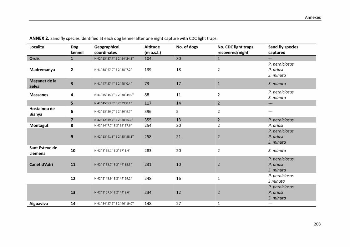
Annexes
203
ANNEX 2. Sand fly species identified at each dog kennel after one night capture with CDC light traps.
Locality Dog kennel
Geographical coordinates
Altitude (m a.s.l.)
No. of dogs No. CDC light traps recovered/night
Sand fly species captured
Ordis 1 N 42° 13' 37.7'' E 2° 54' 24.1'' 104 30 1 ---
Madremanya 2 N 41° 58' 47.0'' E 2° 58' 7.2'' 139 18 2 P. perniciosus P. ariasi S. minuta
Maçanet de la Selva
3 N 41° 47' 27.4'' E 2° 45' 0.4'' 73 17 1 S. minuta
Massanes 4 N 41° 45' 15.3'' E 2° 38' 44.0'' 88 11 2 P. perniciosus S. minuta
5 N 41° 45' 53.8'' E 2° 39' 0.1'' 117 14 2 --- Hostalnou de Bianya
6 N 42° 13' 26.0'' E 2° 26' 9.7'' 396 5 2 ---
7 N 42° 12' 39.2'' E 2° 28'35.0'' 355 13 2 P. perniciosus Montagut 8 N 42° 14' 7.7'' E 2° 35' 57.6'' 254 30 2 P. ariasi
9 N 42° 13' 41.8'' E 2° 35' 58.1'' 258 21 2 P. perniciosus P. ariasi S. minuta
Sant Esteve de Llémena
10 N 42° 3' 35.1'' E 2° 37' 1.4'' 283 20 2 S. minuta
Canet d'Adri 11 N 42° 1' 53.7'' E 2° 44' 15.3'' 231 10 2 P. perniciosus P. ariasi S. minuta
12 N 42° 2' 43.9'' E 2° 44' 59,2'' 248 16 1 P. perniciosus S minuta
13 N 42° 1' 57.0'' E 2° 44' 8.6'' 234 12 2 P. perniciosus P. ariasi S. minuta
Aiguaviva 14 N 41° 54' 27.2'' E 2° 46' 19.0'' 148 27 1 ---

Annexes
204
Sta. Coloma de Farners
15 N 41° 50' 39.4'' E 2° 40' 48.4'' 128 32 2 P. perniciosus
Sant Feliu de Guíxols
16 N 41° 47' 2.3'' E 2° 59' 58.7'' 84 19 2 P. perniciosus
P. perniciosus: Phlebotomus perniciosus; P. ariasi: Phlebotomus ariasi; S. minuta: Sergentomyia minuta
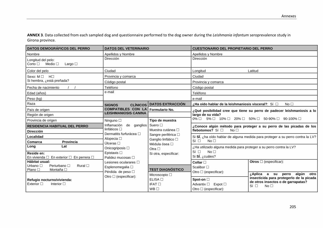
Annexes
205
ANNEX 3. Data collected from each sampled dog and questionnaire performed to the dog owner during the Leishmania infantum seroprevalence study in Girona province.
DATOS DEMOGRÁFICOS DEL PERRO DATOS DEL VETERINARIO CUESTIONARIO DEL PROPIETARIO DEL PERRO
Nombre Apellidos y Nombre Apellidos y Nombre
Longitud del pelo: Corto Medio Largo
Dirección Dirección
Color del pelo Ciudad Longitud Latitud
Sexo: M H Si hembra, ¿está preñada?
Provincia y comarca Ciudad
Código postal Provincia y comarca
Fecha de nacimiento / / Teléfono Código postal
Edad (años) e-mail Teléfono
Peso (kg) e-mail
Raza SIGNOS CLÍNICOS COMPATIBLES CON LA LEISHMANIOSIS CANINA
DATOS EXTRACCIÓN ¿Ha oído hablar de la leishmaniosis visceral?: Sí No
País de origen Formulario No. ¿Qué posibilidad cree que tiene su perro de padecer leishmaniosis a lo largo de su vida?
0% 5% 10% 20% 50% 50-90% 90-100% Región de origen
Provincia de origen Ninguno
Inflamación de ganglios linfáticos
Dermatitis furfurácea
Alopecia
Úlceras
Onicogriposis
Epistaxis
Palidez mucosas
Lesiones ocularares
Esplenomegalia
Pérdida de peso
Otro (especificar):
Tipo de muestra
Suero
Muestra cutánea
Sangre periférica
Ganglio linfático
Médula ósea
Otra
Si otra, especificar:
RESIDENCIA HABITUAL DEL PERRO ¿Conoce algún método para proteger a su perro de las picadas de los flebotomos? Sí No Dirección
Localidad Si SÍ, ¿ha oído hablar de alguna medida para proteger a su perro contra la LV?
Sí No Comarca Provincia
Long Lat ¿Ha utilizado alguna medida para proteger a su perro contra la LV?
Sí No
Si SÍ, ¿cuáles? Reside en:
En vivienda En exterior En perrera
Hábitat usual:
Urbano Periurbano Rural Plano Montaña
Collar
Scalibor
Otro (especificar):
Otros (especificar):
TEST DIAGNÓSTICO
Refugio nocturno/vivienda:
Exterior Interior
Microscopio
ELISA
IFAT
WB
¿Aplica a su perro algún otro insecticida para protegerlo de la picada de otros insectos o de garrapatas?
Sí No
Spot-on
Advantix Expot
Otro (especificar):

Annexes
206
Ocupación del perro:
Mascota Guardia Ovejero Cazador Perrera Vagabundo Otro (especificar):
Cultivo in vitro
PCR
Otro (especificar)
Spray
Duowin
Otro (especificar):
Si SÍ, proporcione detalles
Vive con otros perros: Sí No Si SÍ, ¿cuántos?
Resultado
Positivo Negativo
Título
Observaciones
Shampoo
(especificar):
Si NO aplica profilaxis, ¿por qué?
No lo puedo encontrar en la tienda
Demasiado caro
No creo que funcione
La LV no es importante
Otro (especificar)
Vive con otros animales: Sí No Si SÍ, ¿cuáles?
Jarabe LeisGuard®
Desplazamientos/viajes: Sí No Si SÍ, detalle de los lugares Si SÍ, periodo
Vacuna CaniLeish®

Annexes
207
ANNEX 4. Dataset used for the Leishmania infantum in-house ELISA sensitivity and specificity analysis.
Dog code Parasite detection result*
ELISA result (U) Reference
B1 Positive 337 Iniesta et al., 2002 B2 Positive 31 B3 Negative 40 B4 Positive 177 B5 Positive 58 B6 Positive 174 B7 Positive 247 B8 Positive 17 B9 Positive 13 B10 Positive 252 B11 Positive 209 B12 Negative 33 B13 Negative 8 B14 Negative 11 B15 Positive 30 B16 Negative 27 B17 Negative 2 B18 Negative 12 B19 Positive 118 B20 Positive 16 B21 Positive 24 B22 Positive 69 B23 Positive 42 B24 Positive 20 B25 Positive 40 B26 Positive 75 B27 Positive 29 B28 Positive 90 B29 Positive 24 B30 Positive 69 B31 Positive 12 B32 Negative 13 B33 Positive 258 B34 Positive 348 B35 Positive 11 B36 Positive 12 B37 Negative 18 B38 Positive 375
1 Positive 53 Fisa et al., 2001 2 Positive 100 3 Positive 38 4 Positive 100 5 Positive 107 6 Positive 84 7 Positive 147 8 Positive 27 9 Positive 86
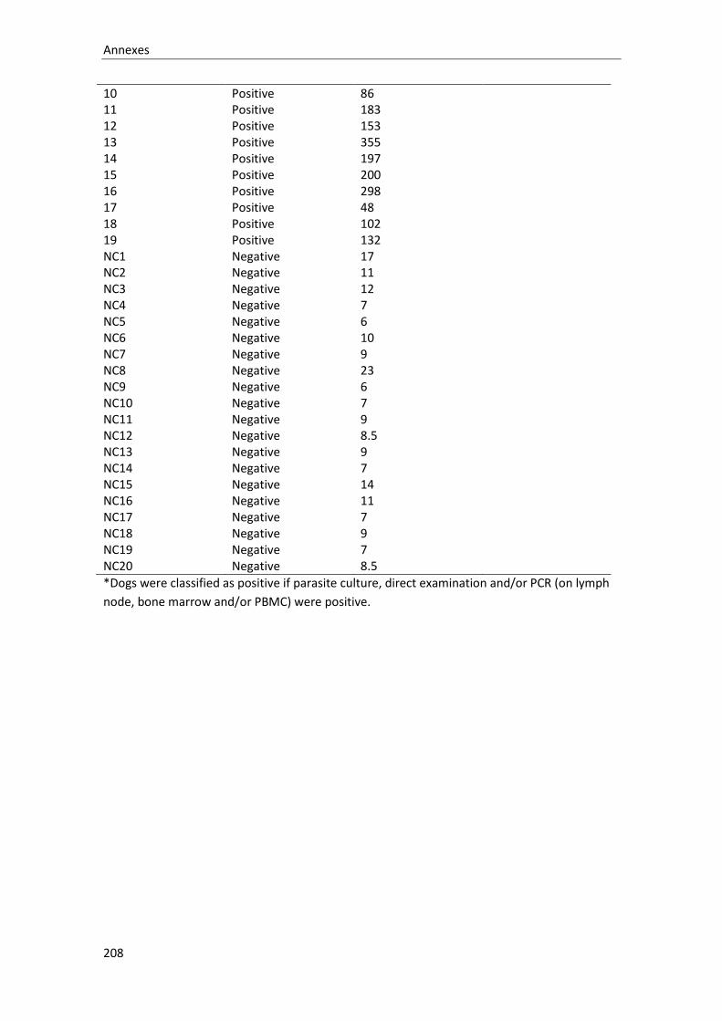
Annexes
208
10 Positive 86 11 Positive 183 12 Positive 153 13 Positive 355 14 Positive 197 15 Positive 200 16 Positive 298 17 Positive 48 18 Positive 102 19 Positive 132 NC1 Negative 17 NC2 Negative 11 NC3 Negative 12 NC4 Negative 7 NC5 Negative 6 NC6 Negative 10 NC7 Negative 9 NC8 Negative 23 NC9 Negative 6 NC10 Negative 7 NC11 Negative 9 NC12 Negative 8.5 NC13 Negative 9 NC14 Negative 7 NC15 Negative 14 NC16 Negative 11 NC17 Negative 7 NC18 Negative 9 NC19 Negative 7 NC20 Negative 8.5
*Dogs were classified as positive if parasite culture, direct examination and/or PCR (on lymph
node, bone marrow and/or PBMC) were positive.

Annexes
209
ANNEX 5. Leishmania infantum in-house ELISA sensitivity and specificity analysis (Stata output)
77 0.9508 0.0210 0.90958 0.99200
Obs Area Std. Err. [95% Conf. Interval]
ROC Asymptotic Normal
( > 375 ) 0.00% 100.00% 37.66% 1.0000
( >= 375 ) 2.08% 100.00% 38.96% 0.9792
( >= 355 ) 4.17% 100.00% 40.26% 0.9583
( >= 348 ) 6.25% 100.00% 41.56% 0.9375
( >= 337 ) 8.33% 100.00% 42.86% 0.9167
( >= 298 ) 10.42% 100.00% 44.16% 0.8958
( >= 258 ) 12.50% 100.00% 45.45% 0.8750
( >= 252 ) 14.58% 100.00% 46.75% 0.8542
( >= 247 ) 16.67% 100.00% 48.05% 0.8333
( >= 209 ) 18.75% 100.00% 49.35% 0.8125
( >= 200 ) 20.83% 100.00% 50.65% 0.7917
( >= 197 ) 22.92% 100.00% 51.95% 0.7708
( >= 183 ) 25.00% 100.00% 53.25% 0.7500
( >= 177 ) 27.08% 100.00% 54.55% 0.7292
( >= 174 ) 29.17% 100.00% 55.84% 0.7083
( >= 153 ) 31.25% 100.00% 57.14% 0.6875
( >= 147 ) 33.33% 100.00% 58.44% 0.6667
( >= 132 ) 35.42% 100.00% 59.74% 0.6458
( >= 118 ) 37.50% 100.00% 61.04% 0.6250
( >= 107 ) 39.58% 100.00% 62.34% 0.6042
( >= 102 ) 41.67% 100.00% 63.64% 0.5833
( >= 100 ) 45.83% 100.00% 66.23% 0.5417
( >= 90 ) 47.92% 100.00% 67.53% 0.5208
( >= 86 ) 52.08% 100.00% 70.13% 0.4792
( >= 84 ) 54.17% 100.00% 71.43% 0.4583
( >= 75 ) 56.25% 100.00% 72.73% 0.4375
( >= 69 ) 60.42% 100.00% 75.32% 0.3958
( >= 58 ) 62.50% 100.00% 76.62% 0.3750
( >= 53 ) 64.58% 100.00% 77.92% 0.3542
( >= 48 ) 66.67% 100.00% 79.22% 0.3333
( >= 42 ) 68.75% 100.00% 80.52% 0.3125
( >= 40 ) 70.83% 96.55% 80.52% 20.5416 0.3021
( >= 38 ) 72.92% 96.55% 81.82% 21.1458 0.2805
( >= 33 ) 72.92% 93.10% 80.52% 10.5729 0.2909
( >= 31 ) 75.00% 93.10% 81.82% 10.8750 0.2685
( >= 30 ) 77.08% 93.10% 83.12% 11.1771 0.2461
( >= 29 ) 79.17% 93.10% 84.42% 11.4792 0.2238
( >= 27 ) 81.25% 89.66% 84.42% 7.8542 0.2091
( >= 24 ) 85.42% 89.66% 87.01% 8.2569 0.1627
( >= 23 ) 85.42% 86.21% 85.71% 6.1927 0.1692
( >= 20 ) 87.50% 86.21% 87.01% 6.3437 0.1450
( >= 18 ) 87.50% 82.76% 85.71% 5.0750 0.1510
( >= 17 ) 89.58% 79.31% 85.71% 4.3299 0.1313
( >= 16 ) 91.67% 79.31% 87.01% 4.4306 0.1051
( >= 14 ) 91.67% 75.86% 85.71% 3.7976 0.1098
( >= 13 ) 93.75% 72.41% 85.71% 3.3984 0.0863
( >= 12 ) 97.92% 65.52% 85.71% 2.8396 0.0318
( >= 11 ) 100.00% 55.17% 83.12% 2.2308 0.0000
( >= 10 ) 100.00% 51.72% 81.82% 2.0714 0.0000
( >= 9 ) 100.00% 37.93% 76.62% 1.6111 0.0000
( >= 8.5 ) 100.00% 31.03% 74.03% 1.4500 0.0000
( >= 8 ) 100.00% 27.59% 72.73% 1.3810 0.0000
( >= 7 ) 100.00% 10.34% 66.23% 1.1154 0.0000
( >= 6 ) 100.00% 3.45% 63.64% 1.0357 0.0000
( >= 2 ) 100.00% 0.00% 62.34% 1.0000
Cutpoint Sensitivity Specificity Classified LR+ LR-
Correctly
Detailed report of sensitivity and specificity

Annexes
210
ANNEX 6. Area under the ROC curve (95%CI) for the Leishmania infantum in-house ELISA (Stata output)
77 0.9508 0.0210 0.90958 0.99200
Obs Area Std. Err. [95% Conf. Interval]
ROC Asymptotic Normal

Annexes
211
ANNEX 7. Article accepted for publication in the journal Preventive Veterinary Medicine (doi: 10.1016/j.prevetmed.2018.10.015) (uncorrected proof format)

Annexes
212
Annexes
213
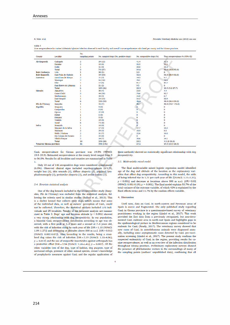
Annexes
214

Annexes
215

Annexes
216

Annexes
217

Annexes
218

Annexes
219


Annexes
221
ANNEX 8. Article published in the journal Parasites & Vectors (doi: 10.1186/s13071-018-3123-
y).

Annexes
222

Annexes
223

Annexes
224

Annexes
225

Annexes
226

Annexes
227

Annexes
228

Annexes
229