Regulation of Akt/PKB activity by P21-activated kinase in cardiomyocytes

Culture on Electrospun Polyurethane Scaffolds Decreases AtrialNatriuretic Peptide Expression by Cardiomyocytes In Vitro
Danielle N. Rockwood1, Robert E. Akins*,2, Ian Parrag3, Kimberly A. Woodhouse3, and JohnF. Rabolt11Department of Materials Science and Engineering, University of Delaware 201 Dupont Hall, Newark,Delaware 19711
2NemoursBiomedical Research, A.I. duPont Hospital for Children 1600 Rockland Road, Wilmington,Delaware 19803
3Department of Chemical Engineering and Applied Chemistry, University of Toronto 200 College Street,Toronto, Ontario M5S 3E5, Canada
AbstractThe function of the mammalian heart depends on the functional alignment of cardiomyocytes, andcontrolling cell alignment is an important consideration in biomaterial design for cardiac tissueengineering and research. The physical cues that guide functional cell alignment in vitro and theimpact of substrate-imposed alignment on cell phenotype, however, are only partially understood.In this report, primary cardiac ventricular cells were grown on electrospun, biodegradablepolyurethane (ES-PU) with either aligned or unaligned microfibers. ES-PU scaffolds supported high-density cultures, and cell subpopulations remained intact over two weeks in culture. ES-PU culturescontained electrically-coupled cardiomyocytes with connexin-43 localized to points of cell:cellcontact. Multi-cellular organization correlated with microfiber orientation, and aligned materialsyielded highly oriented cardiomyocyte groupings. Atrial natriuretic peptide, a molecular marker thathas decreasing expression during ventricular cell maturation, was significantly lower in culturesgrown on ES-PU scaffolds than in those grown on tissue culture polystyrene. Cells grown on alignedES-PU had significantly lower steady state levels of ANP and constitutively released less ANP overtime indicating that scaffold-imposed cell organization resulted in a shift in cell phenotype to a moremature state. We conclude that the physical organization of microfibers in ES-PU scaffolds impactsboth multi-cellular architecture and cardiac cell phenotype in vitro.
Keywordscardiac tissue engineering; electrospinning; biodegradable polyurethane; atrial natriuretic peptide(ANP); cardiomyocyte
*For correspondence: Robert E. Akins, Jr., Ph.D. Nemours Biomedical Research A. I. duPont Hospital for Children 1600 Rockland RoadWilmington, DE 19803 PH: (302) 651-6811 FAX: (302) 651-6897 Email: [email protected]'s Disclaimer: This is a PDF file of an unedited manuscript that has been accepted for publication. As a service to our customerswe are providing this early version of the manuscript. The manuscript will undergo copyediting, typesetting, and review of the resultingproof before it is published in its final citable form. Please note that during the production process errors may be discovered which couldaffect the content, and all legal disclaimers that apply to the journal pertain.
NIH Public AccessAuthor ManuscriptBiomaterials. Author manuscript; available in PMC 2009 December 1.
Published in final edited form as:Biomaterials. 2008 December ; 29(36): 4783–4791. doi:10.1016/j.biomaterials.2008.08.034.
NIH
-PA Author Manuscript
NIH
-PA Author Manuscript
NIH
-PA Author Manuscript
IntroductionCells in the mammalian heart communicate and contract in a highly directional manner, andcardiac ventricles contain ordered, multi-cellular structures comprising nearly parallelcardiomyocytes, which act in unison to provide efficient force production [1-3]. Controllingmulticellular alignment is, therefore, a central goal in the design of biomaterials for cardiactissue engineering, and understanding the effects of imposed alignment on cell phenotype is acritical area for research. In previous studies of tissue-level, multicellular organization invitro, cardiac cell alignment has been induced using oriented scaffolds [4], patterned substrates[5,6], or mechanical conditioning of established cultures [7-10]. In seminal work on patterning,Simpson et al. demonstrated that cultured cardiomyocytes grown on aligned gel matrices adopta morphologic phenotype typified by lateral cell alignment, elongated cell shape, and parallelmyofibrillar organization, which are hallmarks of tissue-level organization [11]. In more recentwork, McDevitt et al. demonstrated that cardiomyocytes could also be patterned using narrowstrips of laminin, which caused cells to align in series and form intercalated disk-like structureslocated at end-to-end contact points similar those seen in native tissue [5,6]. The transmissionof mechanical strain through the substratum to which cardiac cells are attached can also inducecellular alignment and subsequent morphologic adaptation consistent with tissue-levelorganization [7,9,10]. Significantly, although cells can be induced to align using matrixpatterning or mechanical strategies, the organization of multi-cellular cardiac tissue structuresseems to be principally driven by molecular responses to culture conditions [12]; unfortunately,the effects of induced cell alignment on cardiac cell molecular phenotype have been onlypartially described.
Atrial natriuretic peptide (ANP) is a critical marker for the molecular phenotype of cardiacmuscle cells. During structural development of the heart, maturational changes in cardiac geneexpression occur [13], including decreasing expression of ANP in the ventricle [14,15]. In themature heart, ANP expression is largely restricted to atrial cells, which release the peptide fromaccumulated stores through a regulated mechanisms triggered by increased intermittent stretchduring atrial filling. ANP is also produced in immature ventricular cells via a constitutivepathway, but production is down-regulated during normal maturation, and mature ventricularmycoytes only produce ANP under pathologic conditions associated with ventricularhypertrophy [14,15]. Since its production decreases with the structural maturation of thecardiac ventricle, ANP represents a useful marker for studying cardiac ventricular cellmolecular phenotype.
To investigate the relationship between structural organization and ANP expression in cardiacventricular cells in vitro, we prepared aligned and unaligned biodegradable polyurethane (PU)culture substrates by electrospinning. Electrospinning offers several advantages as a processingmethod for tissue engineered scaffolds including high surface area to volume ratios, formationof interconnected porous networks, and small diameter fibers that mimic the fibrousarchitecture of the extracellular matrix of soft tissue. In combination, these characteristicspromote cell adhesion and migration and enable the transport of nutrients and waste throughoutthe scaffold [16-18]. Additionally, the electrospinning process can be tailored to create highlyaligned scaffolds that can be used as templates for cell organization. In the present work,electrospun-PU (ES-PU) scaffolds were made from biodegradable, elastomeric material withtunable degradation properties [19] and used to culture primary neonatal rat heart cells. Cultureswere prepared using both aligned and unaligned scaffolds and assayed for cell survival,maintenance of cell populations, and differences in markers of molecular phenotype, includingANP.
Rockwood et al. Page 2
Biomaterials. Author manuscript; available in PMC 2009 December 1.
NIH
-PA Author Manuscript
NIH
-PA Author Manuscript
NIH
-PA Author Manuscript

MethodsPolyurethane Synthesis
PU was synthesized using polycaprolactone diol with a molecular weight of 1250 (Aldrich,Milwaukee, WI), 2,6-diisocyanate methylcaproate (LDI; Kyowa Hakko Kogyo, Tokyo, Japan)and a L-phenylalanine-based diester chain extender (Toronto Research Chemicals, Toronto,ON, Canada) following the methods of Skarja and Woodhouse [20].
Electrospinning and AlignmentThe PU was dissolved in dichloromethane (Sigma, St. Lo1uis, MO) at a concentration of 18%(w/v) as previously reported [19]. The electrospinning apparatus consisted of a variable speedsyringe pump (Orion Sage, Thermo Scientific; Waltham, MA) and a high voltage power supply(Glassman Series ED, High Bridge, NJ). A glass syringe (Popper & Sons, New Hyde Park,NY) was equipped with a blunt-tipped needle (23 gauge, Hamilton, Reno, NV) and filled withthe polymer solution. The syringe pump was set to a flow rate of 2.0 to 2.7 ml/hr to provide acontinuous flow of solution to the tip, and +10 kV of voltage was applied to the needle. Isotropicfibrous mats were collected using a stationary collector whereas a rotating mandrel assemblywas constructed to align the fibers for the anisotropic mats. A speed of 2000 RPM (10.6 m/sec) was used to align the fibers.
FE-SEMField emission-scanning electron microscope (FE-SEM) measurements were carried out on aJEOL JSM 7400F (Tokyo, Japan). Samples were coated with 10 Å of gold/palladium (Denton,Moorestown, NJ) prior to imaging. Images were obtained at a working distance of 8 millimetersat 1 kV. Fiber diameters for both aligned and isotropic mats were measured onboard JEOLsoftware. Fiber orientation was determined from digital images using Image Pro® Plussoftware (Media Cybernetics, Silver Spring, MD). Briefly, on each image, a reference line wasdrawn, and the acute angle formed between the reference and the principal axis of individualfibers was recorded.
Mechanical TestingUniaxial tensile testing was performed using an Instron 4301 (Instron, Norwood, MA) with acrosshead speed of 13 mm/min and a 10 N load cell running at 50% capacity. Samples of ES-PU for tensile testing were prepared at 60-70 μm in thickness and cut into strips 3 cm long and1 cm wide. Samples were stretched under ambient conditions until break in the preferreddirection of fiber orientation. Force-elongation data were used to generate stress-strain curves;stress and strain at break and initial moduli were determined from the curves.
Cell Isolation and CultureNeonatal rats (2-3 day old; Charles River Laboratories, Wilmington, MA) were anesthetizedand euthanized under an IACUC-approved protocol. The ventricular portion of the heart wasdissected, minced, and digested using a limited digestion with purified trypsin followed bycomplete digestion with collagenase using reagents from Worthington BiochemicalCorporation (Freehold, NJ) as previously described [21]. Cell harvests were collected bycentrifugation and filtered through 0.070 mm nylon mesh Nitex (Becton Dickinson, FranklinLakes, NJ), and statically inoculated at 2 × 10⁶ viable cells per 10 cm2 of surface area using6-well plates. Prior to inoculation, electrospun polyurethane (ES-PU) scaffolds and tissueculture polystyrene (TCPS) control wells were precoated with 100 μg/ml human fibronectin(FN, Becton Dickinson) for 18 hours followed by rinsing with Hank’s Balanced Salt Solution(HBSS, Invitrogen, Carlsbad, CA). Cells were allowed to adhere for 24 hours prior to the first
Rockwood et al. Page 3
Biomaterials. Author manuscript; available in PMC 2009 December 1.
NIH
-PA Author Manuscript
NIH
-PA Author Manuscript
NIH
-PA Author Manuscript

feeding. Cultures were maintained in a serum-free medium (SFM) as previously reported[21].
Staining and MicroscopyTo quantify the proportion of dead cells present in cultures, samples were rinsed with HBSSand stained for 20 minutes with a 1:500 dilution of ethidium homodimer-2 (1.29 mg/ml,EthH-2; Invitrogen) prior to fixation in 2% (w/v) paraformaldehyde (PFA, ElectronMicroscopy Sciences, Hatfield, PA) in Dulbecco's phosphate-buffered saline (PBS; pH 7.4;Mediatech, Inc., Manassas, VA) for 45 minutes. Cells were then permeabilized by the additionof Triton X-100 (Sigma) to 0.1% (v/v), rinsed in PBS, blocked with 3% (w/v) bovine serumalbumin (BSA, Sigma, St. Louis, MO). Subsequent evaluations were carried out to determinelive/dead cell ratio, the arrangement of sarcomeres, or the organization of gap junctions. Forlive/dead cell determination, cultures were counterstained with Hoechst 33258 (1 μg/ml,Sigma) and enumerated such that Eth-H2 positive nuclei indicated nuclei from dead cells outof total Hoechst 33258-positive nuclei. For sarcomeres, cells were stained with a primaryantibody that recognizes myosin heavy chain (MyHC; clone A4.1025, Developmental StudiesHybridoma Bank, Iowa City, IA) by incubating for 2 hours in hybridoma medium diluted 1:1with PBS followed by staining for 1 h with Texas Red conjugated anti-mouse IgG (JacksonImmunoResearch, Media, PA), Hoechst 33258 (1 μg/ml, Sigma) to stain nuclei and AlexaFluor 488-conjugated phalloidin (Invitrogen) to stain filamentous actin. For gap junctions,samples were incubated overnight in a 1:500 dilution of a monoclonal antibody againstconnexin43 (mouse anti-connexin-43 monoclonal, Chemicon, Temecula, CA) in PBS.Samples were then counter stained with Alexa Fluor 488-conjugated to anti-mouse IgG(Invitrogen), Hoechst 33258, and Alexa Fluor 546-conjugated phalloidin (Invitrogen). Imagesof fluorescently stained samples were acquired using an Evolution QEi, 12-bit, cooled CCDcamera (Media Cybernetics) mounted to an Olympus model BX-60 epi-fluorescencemicroscope and operated using Image Pro® Plus software (Media Cybernetics). Alignmentcalculations and live/dead counts were accomplished using Image Pro® Plus.
Flow CytometryCells for flow cytometry were collected via trypsinization (0.05% trypsin-EDTA, Invitrogen)of cultures. The collected cells were fixed in 2% PFA for 45 minutes, permeabilized with 0.1%(v/v) Triton X-100, and blocked with 3% BSA. Samples were stained with a primary antibodyrecognizing MyHC (A4.1025) and then labeled with a secondary antibody of Alexa Fluor 488-conjugated to anti-mouse IgG (Invitrogen). Samples were run through cell strainers (35μm,Becton Dickinson) and analyzed on a FACSCalibur flow cytometry instrument (BectonDickinson). MyHC-positive cells were considered myocytes.
Gene Expression AnalysisCells and tissue for real time, quantitative polymerase chain reaction (qPCR) were collectedvia trypsinization, rinsed with PBS, and stored in RNA-Later® at 4 °C for one week and then−20 °C, as per the manufacturer's recommendations (Ambion, Foster City, CA). RNA wasprepared using QIAshredder and RNeasy® kits (Qiagen, Valencia, CA) following the protocolsfrom the manufacturer. Potential DNA contamination of samples was eliminated by DNAsetreatment (Turbo DNAse™, Ambion). The RNA was reverse transcribed using a RT2 PCRArray First Strand Kit (SuperArray, Frederick, MD). QPCR was run on a MyiQ iCycler(BioRad, Hercules, CA) instrument using custom assays from SuperArray based on primerswith proprietary sequences to detect glyceraldehyde-3-phosphate dehydrogenase (Gapdh),which is expressed by all cells, cardiac troponin I (Tnni3), which is specific to cardiomyocytes,myosin light polypeptide 3 (Myl3), which is specific to ventricular cardiomyocytes, and
Rockwood et al. Page 4
Biomaterials. Author manuscript; available in PMC 2009 December 1.
NIH
-PA Author Manuscript
NIH
-PA Author Manuscript
NIH
-PA Author Manuscript

natriuretic peptide precursor type A (Nppa), which encodes the ANP precursor thataccumulates in atrial and immature ventricular cardiomyocytes.
Protein QuantificationTo determine atrial natriuretic peptide (ANP) levels, an enzyme immunoassay (EIA, ANF(1-28) rat, host rabbit, high sensitivity, Bachem, San Carlos, CA) were performed on cellularextracts following the manufacturer's recommendations. The results indicated the total levelof ANP, pro-ANP, and pre-pro-ANP in the sample. A bicinchoninic acid protein assay (PierceBiotechnology, Rockford, IL) was performed to determine overall protein concentration insamples. The level of ANP as a function of total protein was compared between samples.
Statistical AnalysisStatistical analysis was carried out with SPSS Graduate Pack 15 (Chicago, IL) and Primer ofBiostatistics (McGraw-Hill, Columbus, OH) software. Tests applied to individual datasets areindicated in the text.
ResultsCell Viability and Population Characteristics
To establish whether ES-PU scaffolds could support cardiac cell culture and to verify thestability of the primary cell populations over time, cell viability and flow cytometry assayswere carried out. Viability was assessed using a fluorescent live/dead staining protocol in whichthe number of live and dead cells in random microscope fields was tabulated manually. Theaverage percentage of live cells on isotropic ES-PU versus TCPS is shown over time in Figure1. Overall, the percentage of live cells was somewhat lower on the ES-PU mats than on TCPS,but the percentage of viable cells on the PU was relatively stable over the first week of culture(85.5% on day 1; 83.7% on day 2; and 85.2% on day 7, respectively) but increased somewhatby day 14 (92.6%). In contrast, the percentage of live cells on the TCPS started at 93.6% after24 hours, then decreased slightly to 89.5% and 88.3% on days 2 and 7, increasing to 95.3% atthe final time point. Despite these changes, overall the data indicate that ES-PU supportedviable cells to a level similar to that seen for TCPS without dramatic losses over the timeframeof the study.
Since primary cardiac cells comprise a mixed cell population predominated by non-proliferative cardiomyocytes and highly proliferative fibroblasts, we also determined whetherthe proportion of myocytes persisted over time in culture on ES-PU. Flow cytometry was usedto determine the ratio of myocytes to non-myocytes. This ratio was measured for the initialcell isolate and at intervals throughout the length of the experiment. Cells staining positive forMyHC, which is only present in the cardiomyocyte component of these cultures, werecategorized as myocytes while all remaining cells were considered non-myocytes. Figure 2shows the average proportion of myocytes (n = 3) for each material at four time points. Theinitial cell isolate used in this experiment contained 36.3% myocytes indicating a relativelyhigh proportion of potentially proliferative cells were present. The percentage of myocytesremained relatively unchanged after the cells had attached to the substrates such that by day14, the percentage of myocytes was 31.8%, 28.5%, and 38.4% on aligned ES-PU, TCPS, andisotropic ES-PU, respectively. These numbers were not significantly different between timepoints or across different materials by Two Way ANOVA (p>0.1), anddespite the relativelylow percentage of myocytes, all of these cultures beat throughout the duration of culture.
Rockwood et al. Page 5
Biomaterials. Author manuscript; available in PMC 2009 December 1.
NIH
-PA Author Manuscript
NIH
-PA Author Manuscript
NIH
-PA Author Manuscript
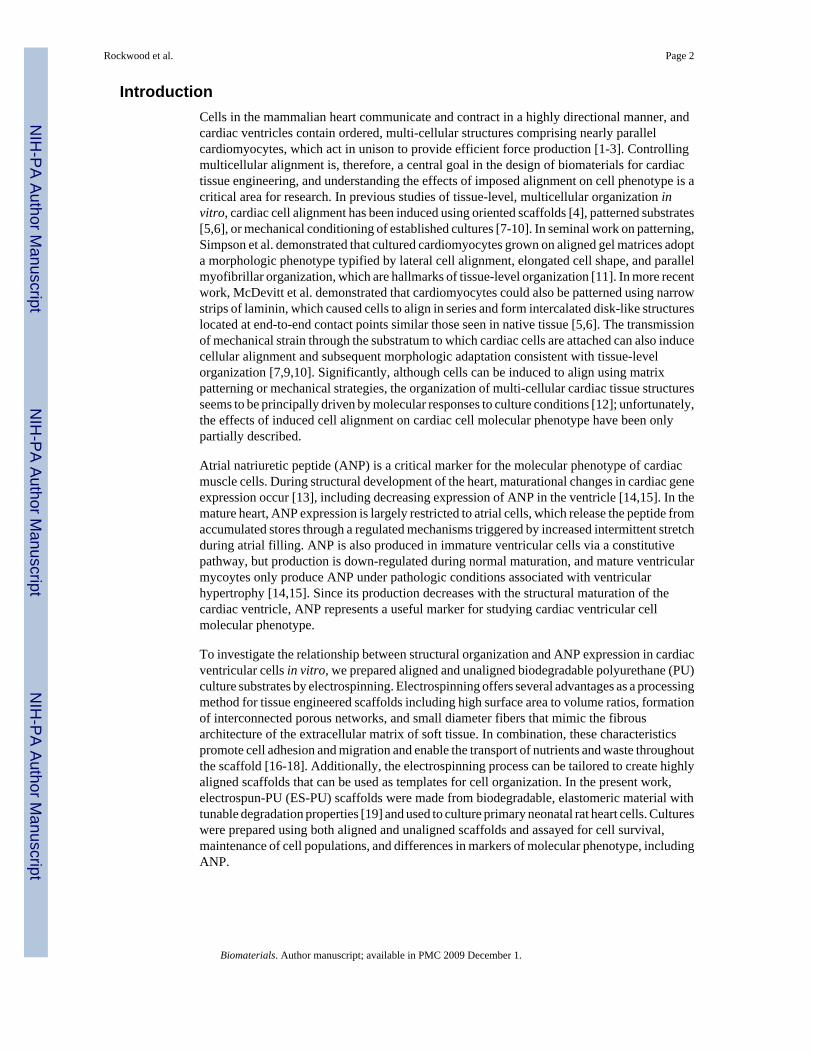
Alignment of polyurethane substrates and cultured cellsThe first step in evaluating cellular alignment in our study was to create a template to guidethe cells during seeding. This was done by fabricating isotropic (unaligned) and anisotropic(aligned) fibrous PU mats by electrospinning. Isotropic mats were prepared using a stationary,flat collector; whereas, aligned mats were collected on a rotating mandrel. The average fiberdiameter was 1.8 ± 1.26 μm and 2.0 ± 1.92 μm for aligned and isotropic mats, respectively. Asobserved previously, [19] there was evidence of polymer bead formation and a large range infiber diameters, from a few hundred nanometers to several microns that may have been causedby fiber splaying during the electrospinning process. Figure 3a-c shows FE-SEM images ofboth types of material and the relative fiber orientation in each. There was a clear differencein orientation of fibers within the isotropic and aligned mats that was quantified by assessingthe variance of the fiber angle distributions (Figure 3c) using Levene's test for Homogeneityof Variance (p ≤ 0.01).
Mechanical analysis of the different ES-PU mats indicated that the aligned scaffolds wereconsiderably stiffer than the isotropic mats. The isotropic fibers had an elastic modulus of 13.5MPa ± 1.2, ultimate tensile strain of 129% ± 26, and ultimate tensile stress of 1.53 MPa ± 0.06.In contrast, the aligned fibers showed an elastic modulus of 22.5 MPa ± 1.7, ultimate tensilestrain of 70% ± 5, and an ultimate tensile stress of 2.84 MPa ± 0.08. Therefore, as expected,the aligned materials were stiffer and less extensible than the unaligned substrates.
Cells seeded onto aligned and isotropic mats appeared to adopt the underlying orientation ofthe materials by orienting to the long-axes of the ES-PU microfibers. As a result, multicellulargroupings on the isotropic scaffolds appeared to be less organized than those on the alignedsubstrates, which appeared highly oriented relative to the material's axis. As expected (Figure3d), the degree of alignment was significantly higher for the cells cultured on the aligned ES-PU. Significant differences were not seen between the alignment scores of isotropic fibers andthat of cells grown on isotropic mats or between aligned fibers and cells grown on alignedmats.
Cell Morphology and OrganizationThe morphology and phenotype of the cultured cardiac cells were evaluated to determinewhether cells on aligned versus unaligned substrates were able to establish oriented electricalcommunication and whether substrate patterning affected cell maturation status evinced byANP production. In Figure 4a-c, cytoskeletal MyHC (red), which is specific to myocytes, andfilamentous actin (f-actin, green), which is present in all cell types but accumulate to aconsiderably higher level in cardiomyocytes due to the contractile apparatus [22], are shownin conjunction with the position of cell nuclei (blue). Cells on all of the substrates studiedshowed striated sarcomeres with organized f-actin and MyHC. In the TCPS and isotropic ES-PU cultures, the cells appeared relatively unaligned, and the contractile activity ofcardiomyocytes on these substrates was not directionally organized. In contrast, cells seededon aligned ES-PU microfibers were oriented and contractile in one principal direction with amore linear appearance, which is comparable to the organization of myocytes in ventriculartissue [22]. These observations were noted three days post-seeding and then throughout theduration of the experiments.
In vivo, the lateral alignment of cardiomyocyte contractile elements in ventricular tissue isaccompanied by the organization of gap-junctional components in the intercalated discs foundwhere myocytes join end-to-end [22]. Thus Cx43, which is the most prominent gap junctionprotein in ventricular tissue [23], demarks functional cell alignment; accordingly, weinvestigated the localization of Cx43 in our cultures. Figure 4d-f illustrates Cx43 distribution(red) relative to cell nuclei (blue), and f-actin (green). For all three substrata, Cx43 was found
Rockwood et al. Page 6
Biomaterials. Author manuscript; available in PMC 2009 December 1.
NIH
-PA Author Manuscript
NIH
-PA Author Manuscript
NIH
-PA Author Manuscript
around the cell periphery and did not specifically localize to end-to-end cardiomyocyte contactsalthough such structures were occasionally found (see black arrowhead in Figure 4f). Thesedata indicate that cells were electrically coupled in all culture types but that gap junctions werenot localized to intercalated disc-like structures at the ends of adjacent cells.
Molecular PhenotypeThe molecular phenotype of the cells in our cultures was investigated by measuring the relativelevels of mRNA for four genes via qPCR: Gapdh, Tnni3, Myl3, and Nppa. Gapdh levels, whichare commonly employed for normalizing gene expression data [24], were used as an indicatorof total cell transcription activity. Tnni3, which is the troponin I isoform expressed in atrialand ventricular cardiomyocytes, and Myl3, which is the ventricular myocyte specific isoformof essential myosin light chain [25,26], were used to indicate total cardiomyocyte andventricular cardiomyocyte subpopulations, respectively. Nppa, which is preferentiallyexpressed in atrial, immature ventricular, and hypertrophic ventricular tissue but not in mature,healthy ventricular tissue [27-29], was used as an indicator of cardiomyocyte phenotype.
As shown in Figure 5, mRNA levels for Gapdh, Tnni3, and Myl3 were not significantlydifferent across treatment groups indicating that the total cell transcriptional activity,cardiomyocyte-specific transcriptional activity, and ventricle-specific transcription weresimilar in TCPS and ES-PU cultures. Nppa, on the other hand, was relatively up-regulated incells cultured on TCPS such that steady state Nppa-mRNA levels were elevated from day oneonward. These data suggest that microfibrous ES-PU substrates supported cardiac cellpopulations that were similar to those grown on TCPS in terms of molecular phenotype butthat culture on ES-PU resulted in an alteration of cardiomyocyte maturational status.
To further investigate Nppa expression, levels of immunoreactive atrial natriuretic peptide(ANP), which is encoded by the Nppa gene and comprises pre-pro-ANP, pro-ANP and ANPitself, were determined in the cells and medium of 7 day old cultures by EIA. As shown inFigure 6, cells grown on TCPS contained the highest level of ANP (1.584 ± 0.116 ng per mgof extracted cellular protein). Cells on isotropic ES-PU had slightly lower but similar levels ofANP (1.481 ± 0.174 ng/mg); however, cells grown on aligned ES-PU had significantly lowerANP content (0.407 ± 0.114 ng/mg). For reference, the ANP content of adult (0.68 ng/mg) andneonatal (3.39 ng/mg) ventricular tissue were also determined. Levels of ANP released intothe medium during the 48 hours leading up to culture termination (i.e. between days 5 and 7)were also quantified. Cells grown on TCPS, isotropic ES-PU, and aligned ES-PU all releasedsignificantly different amounts of ANP (229.94 ± 16.80, 79.36 ± 28.87, and 22.35 ± 2.30 ngin 48 hours, respectively). These data suggest that compared to TCPS, culturing primary cardiaccells on aligned ES-PU induced cardiomyocytes to produce, store, and release less ANP, whichis indicative of a more mature phenotype, with cells cultured on isotropic ES-PU adopting anintermediate phenotype between TCPS and aligned ES-PU.
DiscussionWe have found that primary cardiomyocytes can be induced to align when cultured onmicrofibrous ES-PU substrates and that culture on ES-PU results in an alteration of cellphenotype such that aligned cultures produce less ANP than non-aligned cultures. Previousstudies have shown that cells can be aligned by patterning cues from the underlying substrate[4-6] or by imposed mechanical loads subsequent to attachment [7-10]; however, the effectsof alignment strategies on the molecular phenotype of component cells are not well understood.Our observations are consistent with a more mature phenotype being adopted by cells grownon aligned microfibrous ES-PU thus linking biomaterial patterning with cell phenotype.
Rockwood et al. Page 7
Biomaterials. Author manuscript; available in PMC 2009 December 1.
NIH
-PA Author Manuscript
NIH
-PA Author Manuscript
NIH
-PA Author Manuscript

A shift in the maturational phenotype of cultured cardiac cells is supported by assays of steadystate mRNA levels. Gapdh mRNA levels indicated that samples contained similar amounts ofoverall cellular material. The Tnni3 and Myl3 data indicated that contributions from ventricularcardiomyocytes were essentially the same across samples, and in combination with the Gapdhdata, that there were no significant differences in the hypertrophic status of cells cultured onthe different substrates. This latter interpretation is further supported by the flow cytometricdata, which indicated that the proportion of cardiomyocytes in the cell population of eachculture type was essentially the same. Thus, the decreased levels of Nppa seen in cultures grownon aligned ES-PU are not likely to be accounted for by broad differences in cellular content ortranscriptional activity or by more specific differences in cardiomyocyte hypertrophic status.
The isotropic and aligned ES-PU mats used in this study provided substrata that could betailored to provide two different cellular environments. The difference between these twosubstrates was quantified, as shown in Figure 3, and the aligned fibers were found to besignificantly more oriented than the isotropic mat. When cells were seeded on these materials,they aligned along the axes of the local fibers, and the organization of cells in culture mimickedthe underlying organization of the fibrous mats. This result suggests the ES-PU microfiberscan be used to pattern multi-cellular organization.
In addition, we observed through viability assays that ES-PU scaffolds supported primarycardiomyocyte cultures over time. In comparison to TCPS, there was a higher proportion ofdead to live cells on the ES-PU even at early time points, suggesting that the dead cells presentin the initial isolate may have been trapped in the fibrous ES-PU matrix during cell inoculation.Dead cells are typically washed away from culture dishes by medium exchanges, and it seemslikely that it would be easier to wash TCPS surfaces than ES-PU fibers, which would beexpected to entrap material seeded on it. As shown in Figure 1, there was a general trend ofthis type in our cultures. Critically, though, the number of dead cells did not increase over timein either TCPS or ES-PU cultures indicating that ES-PU supports cardiomyocyte cultures to adegree similar to that seen on TCPS. Additionally, the morphology of the cells, as shown byimmunofluorescence, demonstrates the survival of cardiac cells on ES-PU membranes.Myocytes with striated myosin heavy chain interlaced with filamentous actin were found onboth types of ES-PU mats and on TCPS (Figure 4). This staining pattern is indicative of healthy,contractile myocytes, and spontaneous contractility was evident on TCPS and ES-PU mats.
It is important to note that solid organs like the heart comprise of mixtures of cells and that thepresent experiments were carried out using a mixed cell population representative of what isfound in native heart tissue [30-32]. Although cardiomyocytes are generally nonproliferative[23], proliferation of other subpopulations of heart cells can result in dramatic changes inrelative cell contributions. Differential proliferation is, therefore, a critical concern in theinterpretation of primary culture experiments. Accordingly, we prepared our cultures to limitoverall proliferation: cells were inoculated at high density to elicit contact inhibition of celldivision and a serum-free medium was used to reduce serum-induced proliferation. To verifythat cell populations were uniform, we used flow cytometry to assess potential changes in thecardiomyocyte versus non-cardiomyocyte subpopulations through fourteen days in culture andfound that the overall ratio of myocytes to non-myocytes remained relatively unchanged overthe course of our experiments (see Figure 2). In combination with the live/dead assay resultsand immunostaining observations, these data indicate that cardiomyocytes survived two weeksin culture and that cell proliferation was severely restricted with no significant differencesfound between TCPS and ES-PU substrates. These data are further corroborated by themolecular analyses, especially Gapdh, Tnni3, which showed no differences across samples,and immunostaining observations.
Rockwood et al. Page 8
Biomaterials. Author manuscript; available in PMC 2009 December 1.
NIH
-PA Author Manuscript
NIH
-PA Author Manuscript
NIH
-PA Author Manuscript

Immunostaining results showed that multi-cellular organization differed among cultures ondifferent substrata. In particular, neighboring cells cultured on TCPS and isotropic ES-PUextended in different directions; whereas, cells grown on aligned ES-PU ran in parallelfollowing the template of the fibrous matrix, an organization that is similar to what is seen innative ventricular tissue [1]. To beat synchronously, cardiomyocytes must be electricallycoupled through gap junctions. As seen in Figure 4, the gap junctional protein Cx43 in ourcultures was distributed throughout the cells and not restricted to intercalated disks at end-to-end contacts. Interestingly, McDevitt et al. [5,6] found a similar Cx43 distribution when cellswere seeded on unpatterned substrates but found that Cx43 localized to intercalated disks incells restricted to 15-30 μm patterned lanes of laminin (spaced 20 μm apart), a geometry thatlimited side-to-side contact between cells and emphasized end-to-end contact of cells in series.In our experiments, cells were laterally aligned but formed side-to-side contacts, and Cx43 wasseen at contacts between cells organized in series and in parallel. Thus, it appears that thepresence of cell-to-cell contacts may influence the distribution of gap junctions on patternedmaterials, and to drive Cx43 organization to intercalated disks, it may be necessary toemphasize longitudinal contacts early in the culture but allow lateral contacts to form as cellsgrow later in culture. It should also be noted that mechanical load plays a key role in thedetermination of multi-cellular organization. In particular, mechanical load placed oncardiomyocytes in vitro results in morphological changes similar to those during heartdevelopment in vivo [7,9,33,34]. The application of controlled substrate patterning inconjunction with cyclic stretch may, therefore, allow engineers to drive cells to desirablearchitectures and cell phenotypes.
The molecular phenotype of cells was assessed by measuring the steady-state levels of mRNAexpressed for Gapdh, Tnni3, Myl3, and Nppa. Gapdh was used as a ubiquitous marker forexpression, Tnni3 as a marker for cardiac muscle cells generally, Myl3 as a marker forventricular cardiomyocytes specifically, and Nppa as a marker for ventricular cardiomyocytematuration. The amount of Gapdh, Tnni3, and Myl3 in the total mRNA isolates was notsignificantly different between samples, supporting the conclusion that the cell populations didnot differ across substrata. Interestingly, given the cell and molecular data we obtained,differences in Nppa expression could not easily be accounted for by differences in RNA yield/integrity (Gapdh), the proportion of cardiomyocytes present (Tnni3), or the phenotypicconversion of the neonatal ventricular myocytes to an atrial phenotype (Myl3). Thus, the shiftin ANP expression in cultures grown on ES-PU appears to be most likely accounted for by ashift in cardiomyocyte maturational status.
To further characterize the regulation of Nppa, ANP was measured at the protein level. Nppais the mRNA precursor to ANP, which is an endocrine factor that modulates blood volume[14,15]. In adults, ANP is primarily synthesized in the cardiac atria, which release ANP inresponse to increased filling volume [15,22,35]. ANP also appears to play a role in cardiacdevelopment [29], where it may act as an inhibitor of hypertrophic growth [36] or a modulatorof apoptosis [37]. Perhaps associated with this latter function, ventricular myocytes produceANP during development, but as ventricular tissue matures perinatally, ANP production fallsto levels <1% that seen in atria [15]. Elevated ANP production, therefore, is suggestive of eitheran atrial or immature ventricular cardiomyocyte phenotype. Since the cells used in our studywere of ventricular origin and uniformly expressed Myl3, ANP levels are best interpreted asindicating maturational status.
The processing of Nppa into ANP has been well studied [14,15], and although detailedmechanisms remain unelucidated, the general mechanisms by which ANP is released fromventricular and atrial tissues are known to differ. In both cell types, Nppa mRNA is initiallytranslated into a 152-amino acid pre-pro-ANP which is subsequently cleaved to form the 126-amino acid pro-ANP. Atrial myocytes subsequently release ANP through a regulated pathway
Rockwood et al. Page 9
Biomaterials. Author manuscript; available in PMC 2009 December 1.
NIH
-PA Author Manuscript
NIH
-PA Author Manuscript
NIH
-PA Author Manuscript

wherein the prohormone is stored in granules for rapid release after increased stretch. Incontrast, ventricular cardiomyocytes secrete ANP via a constitutive pathway in which ANP isreleased shortly after synthesis and proANP is generally not stored in granules [28,38].Importantly, immature cardiac ventricular tissue actively produces ANP and, therefore, hashigher steady state levels of Nppa mRNA and immunoreactive ANP and also constitutivelyreleases more ANP over time than mature ventricular tissue.
Detailed mechanisms to explain the observed shift in ANP metabolism are somewhat difficultto ascertain as details of ANP regulation remain unclear, especially how the expression of theNppa gene, which encodes the ANP precursor, is controlled and how intracellular ANP levelsare modulated [14,15]. Possible explanations for the observed effect include (i) biomechanicalinfluences and (ii) alteration of cell:cell interactions. Biomechanical influences play asignificant role in modulating ANP production in vivo. Specifically, ANP release is elevatedby increased atrial or ventricular stretch during diastole (i.e. pre-load) and under hypertrophicconditions [15,36], which are associated with increased resistance during systole (i.e.,afterload). In our experiments, however, there is no stretching/preloading of cells, and our dataindicate that there were no significant differences in the hypertrophic status of cells culturedon the different substrata. In addition, the aligned scaffolds were stiffer than isotropic scaffoldssuggesting that cardiomyocytes grown on aligned ES-PU should actually have higher ANPproduction than cells grown on the isotropic material if resistance to contraction were the majordeterminant of ANP production.Thus, although a role for biomechanical influences inregulating ANP cannot be ruled out, the biomechanical conditions associated with themodulation of ANP in vivo (stretch/preload or hypertrophy/afterload) are apparently notresponsible for the alteration of ANP levels we observe. Cellular interactions offer analternative explanation. Variations in substrate topography and texture are known to affectcell:cell interactions, and the Notch signaling pathway, which is triggered by particular cell:cellcontacts, is known to modulate ANP expression [39,40]. Thus, differences in the quantity andquality of cell:cell interactions on different substrates may account for differences in ANPproduction.
In our study, cultures grown on TCPS or on isotropic ES-PU had significantly higher steady-state ANP levels than those found in cells grown on aligned ES-PU (Figure 6). In addition,cells grown on the different substrata showed a decreasing progression of ANP release withTCPS cultures exhibiting the highest constitutive production of ANP, followed by isotropicES-PU, and finally aligned ES-PU. Viewed in conjunction with the mRNA data, these resultsindicate that culture on ES-PU influenced cells to adopt a more mature phenotype and that thealigned ES-PU provided the most mature cells. Interestingly, the isotropic ES-PU showedlower Nppa mRNA and reduced ANP release compared to the TCPS samples despite theobservation of similar steady-state peptide levels. Thus, cells on isotropic ES-PU adopted asomewhat intermediate phenotype where the rate of ANP expression and the rate of pro-ANPto ANP conversion by corin-like proteases may both be attenuated relative to cells on TCPSbut potentiated relative to cells on aligned ES-PU. Taken together, these results indicate thataligning cultured, primary, cardiac cells using physical cues from a biomaterial scaffold cannotonly induce organizational changes relative to unaligned materials, but can also inducephenotypic changes in the component myocytes.
ConclusionsWe demonstrate that biodegradable polyurethane can be electrospun into isotropic and alignedmicrofibrous templates capable of supporting primary cardiac cell culture in serum-freemedium. Since PU-based materials are non-toxic and erode slowly over time, aligned ES-PUmay provide useful scaffolds for engineering anisotropic cell sheets that are organized likenative cardiac tissue. We also found that primary cardiac cells attain aspects of a mature
Rockwood et al. Page 10
Biomaterials. Author manuscript; available in PMC 2009 December 1.
NIH
-PA Author Manuscript
NIH
-PA Author Manuscript
NIH
-PA Author Manuscript

phenotype when grown on aligned and isotropic ES-PU. Specifically, immunoreactive ANPlevels were significantly reduced in cultures grown on aligned ES-PU compared to otherwiseidentical cells grown on unaligned ES-PU or on TCPS controls. These results indicate that thephysical patterning of ES-PU scaffolds affected both cell organization and cell phenotype.
AcknowledgementsThe authors would like to thank Therese McLaughlin for help with cell culture, Frank Kriss and Dr. Chao Ni forassistance in electron microscopy, and Melissa Rhodes, Steve Beard, Alfred Lance and Dave Cowgill for help inmanufacturing the mandrel used for aligning the fibers during electrospinning. In addition, the authors greatlyappreciate the help of Dr. Chuanzhao Li and Robert Long with flow cytometry data collection. This work was fundedthrough NSF (NIRT and DMR - 0315461 to JFR), the National Center for Research Resources at NIH (RPG# 1P20-RR020173-01 to REA), the Nemours Foundation (REA), the Canadian Institutes for Health Research (KAW), andthe Natural Science and Engineering Research Council - Canada (KAW).
References1. Schulte RE, Sachse FB, Werner CD, Dossel O. Rule Based Assignment of Myocardial Sheet
Orientation. Biomed Tech (Berl) 2000;45(2):97–102.2. LeGrice IJ, Smaill BH, Chai LZ, Edgar SG, Gavin JB, Hunter PJ. Laminar structure of the heart:
Ventricular myocyte arrangement of connective tissue architecture in the dog. Am J Physiol1995;269:H571. [PubMed: 7653621]
3. Hooks DA, Trew ML, Caldwell BJ, Sands GB, LeGrice IJ, Smaill BH. Laminar Arrangement ofVentricular Myocytes Influences Electrical Behavior of the Heart. Circ Res 2007;101:e103–e12.[PubMed: 17947797]
4. Riboldi SA, Sadr N, Pigini L, Neuenschwander P, Siomonet M, Mognol P, et al. Skeletal myogenesison highly orientated microfibrous polyesterurethane scaffolds. J Biomed Mater Res Part A 2008;84A(4):1094–101.
5. McDevitt TC, Angello JC, Whitney ML, Reinecke H, Hauschka SD, Murry CE, et al. In vitro generationof differentiated cardiac myofibers on micropatterned laminin surfaces. J Biomed Mater Res 2002;60(3):472–9. [PubMed: 11920672]
6. McDevitt TC, Woodhouse KA, Hauschka SD, Murry CE, Stayton PS. Spatially organized layers ofcardiomyocytes on biodegradable polyurethane films for myocardial repair. J Biomed Mater Res2003;66A(3):586–95.
7. Vandenburgh HH, Solerssi R, Shansky J, Adams JW, Henderson SA. Mechanical stimulation oforganogenic cardiomyocyte growth in vitro. American Journal of Physiology-Cell Physiology 1996;39(5):C1284–C92.
8. Carrier RL, Rupnick M, Langer R, Schoen FJ, Freed LE, Vunjak-Novakovic G. Perfusion improvestissue architecture of engineered cardiac muscle. Tissue Eng 2002;8(2):175–88. [PubMed: 12031108]
9. Zimmermann W-H, Schneiderbanger K, Schubert P, Didie M, Munzel F, Heubach JF, et al. TissueEngineering of a Differentiated Cardiac Muscle Construct. Circ Res 2002;90(2):223–30. [PubMed:11834716]
10. Fink C, Ergun S, Kralisch D, Remmers UTE, Weil J, Eschenhagen T. Chronic stretch of engineeredheart tissue induces hypertrophy and functional improvement. FASEB J 2000;14(5):669–79.[PubMed: 10744624]
11. Simpson DG, Terracio L, Terracio M, Price RL, Turner DC, Borg TK. Modulation of cardiac myocytephenotype in vitro by the composition and orientation of the extracellular matrix. J Cell Physiol1994;161(1):89–105. [PubMed: 7929612]
12. Baudino TA, McFadden A, Fix C, Hastings J, Price R, Borg TK. Cell patterning: interaction of cardiacmyocytes and fibroblasts in three-dimensional culture. Microscopy and microanalysis 2008;14(2):117–25. [PubMed: 18312716]
13. Nemer M. Genetic insights into normal and abnormal heart development. Cardiovascular Pathology2008;17(1):48–54. [PubMed: 18160060]
14. McGrath MF, de Bold AJ. Determinants of natriuretic peptide gene expression. Peptides 2005;26(6):933–43. [PubMed: 15911063]
Rockwood et al. Page 11
Biomaterials. Author manuscript; available in PMC 2009 December 1.
NIH
-PA Author Manuscript
NIH
-PA Author Manuscript
NIH
-PA Author Manuscript
15. Pandey KN. Biology of natriuretic peptides and their receptors. Peptides 2005;26(6):901–32.[PubMed: 15911062]
16. Lu L, Mikos AG. The Importance of New Processing in Tissue Engineering. MRS Bulletin 1996;31(11):28–32. [PubMed: 11541498]
17. Zong X, Bien H, Chung C-Y, Yin L, Fang D, Hsiao BS, et al. Electrospun fine-textured scaffolds forheart tissue constructs. Biomaterials 2005;26(26):5330–8. [PubMed: 15814131]
18. Riboldi SA, Sampaolesi M, Neuenschwander P, Cossu G, Mantero S. Electrospun degradablepolyesterurethane membranes: potential scaffolds for skeletal muscle tissue engineering.Biomaterials 2005;26(22):4606–15. [PubMed: 15722130]
19. Rockwood DN, Woodhouse KA, Fromstein JD, Chase DB, Rabolt JF. Characterization ofbiodegradable polyurethane microfibers for tissue engineering. Journal of Biomaterials Science-Polymer Edition 2007;18(6):743–58. [PubMed: 17623555]
20. Skarja GA, Woodhouse KA. Synthesis and Characterization of degradable polyurethane elastomerscontaining an amino acid-based chain extender. J Biomater Sci Polymer Edn 1998;9(3):271–95.
21. Akins RE, Gratton K, Quezada E, Rutter H, Tsuda T, Soteropoulos P. Gene expression profile ofbioreactor-cultured cardiac cells: Activation of morphogenetic pathways for tissue engineering. DNACell Biol 2007;26(6):425–34. [PubMed: 17570766]
22. Opie, LH. Heart Physiology: From Cell to Circulation. Vol. 4th Edition ed.. Lippincott Williams &Wilkins; Philadelphia: 2004.
23. Reinecke H, MacDonald GH, Hauschka SD, Murry C. Electromechanical Coupling between Skeletaland Cardiac Muscle: Implications for Infarct Repair. J Cell Biol 2000;149(3):731–40. [PubMed:10791985]
24. Barber RD, Harmer DW, Coleman RA, Clark BJ. GAPDH as a housekeeping gene: analysis ofGAPDH mRNA expression in a panel of 72 human tissues. Physiol Genomics 2005;21(3):389–95.[PubMed: 15769908]
25. Guenet JL, SimonChazottes D, Gravel M, Hastings KEM, Schiaffino S. Cardiac and skeletal muscletroponin I isoforms are encoded by a dispersed gene family on mouse Chromosomes 1 and 7. MammGenome 1996;7(1):13–5. [PubMed: 8903721]
26. Barth AS, Merk S, Arnoldi E, Zwermann L, Kloos P, Gebauer M, et al. Functional profiling of humanatrial and ventricular gene expression. Pflugers Archiv-European Journal of Physiology 2005;450(4):201–8. [PubMed: 15877233]
27. Claycomb WC. Atrial-Natriuretic-Factor Messenger-Rna Is Developmentally Regulated in HeartVentricles and Actively Expressed in Cultured Ventricular Cardiac-Muscle Cells of Rat and Human.Biochem J 1988;255(2):617–20. [PubMed: 2974280]
28. Bloch KD, Seidman JG, Naftilan JD, Fallon JT, Seidman CE. Neonatal Atria and Ventricles SecreteAtrial Natriuretic Factor via Tissue-Specific Secretory Pathways. Cell 1986;47:695–702. [PubMed:2946416]
29. Cameron VA, Ellmers LJ. Minireview: Natriuretic Peptides during Development of the Fetal Heartand Circulation. Endocrinology 2003;144(6):2191–4. [PubMed: 12746273]
30. Harding SE, Davies CH, Wynne DG, Poolewilson PA. Contractile Function and Response to Agonistsin Myocytes from Failing Human Heart. Eur Heart J 1994;15:35–6. [PubMed: 7713111]
31. Sperelakis N. Cultured Heart Cell Reaggregate Model for Studying Cardiac Toxicology. EnvironHealth Perspect 1978;26:243–67. [PubMed: 214299]
32. Wollenberger A. Isolated Heart-Cells as a Model of the Myocardium. Basic Res Cardiol 1985;80:9–13. [PubMed: 3904721]
33. Yamane M, Matsuda T, Ito T, Fujio Y, Takahashi K, Azuma J. Rac1 activity is required for cardiacmyocyte alignment in response to mechanical stress. Biochem Biophys Res Commun2007;353:1023–7. [PubMed: 17207463]
34. Matsuda T, Takahashi K, Nariai T, Ito T, Takatani T, Fujio Y, et al. N-cadherin-mediated cell adhesiondetermines the plasticity for cell alignment in response to mechanical stretch in culturedcardiomyocytes. Biochem Biophys Res Commun 2005;326(1):228–32. [PubMed: 15567175]
35. de Bold AJ, Borenstein HB, Veress AT, Sonnenberg H. A rapid and potent natriuretic response tointravenous injection of atrial myocardial extract in rats. Life Sci 1981;28(1):89–94. [PubMed:7219045]
Rockwood et al. Page 12
Biomaterials. Author manuscript; available in PMC 2009 December 1.
NIH
-PA Author Manuscript
NIH
-PA Author Manuscript
NIH
-PA Author Manuscript

36. Horio T, Nishikimi T, Yoshihara F, Matsuo H, Takishita S, Kangawa K. Inhibitory Regulation ofHypertrophy by Endogenous Atrial Natriuretic Peptide in Cultured Cardiac Myocytes. Hypertension2000;35(1):19–24. [PubMed: 10642269]
37. Wu C-F, Bishopric NH, Pratt RE. Atrial Natriuretic Peptide Induces Apoptosis in Neonatal RatCardiac Myocytes. J Biol Chem 1997;272(23):14860–6. [PubMed: 9169455]
38. Irons CE, Sei CA, Glembotski CC. Regulated secretion of atrial natriuretic factor from culturedventricular myocytes. Am J Physiol Heart Circ Physiol 1993;264(1):H282–5.
39. Xin M, Small EM, van Rooij E, Qi X, Richardson JA, Srivastava D, et al. Essential roles of the bHLHtranscription factor Hrt2 in repression of atrial gene expression and maintenance of postnatal cardiacfunction. Proceedings of the National Academy of Sciences 2007;104(19):7975–80.
40. Koibuchi N, Chin MT. CHF1/Hey2 plays a pivotal role in left ventricular maturation throughsuppression of ectopic atrial gene expression. Circ Res 2007;100(6):850–5. [PubMed: 17332425]
Rockwood et al. Page 13
Biomaterials. Author manuscript; available in PMC 2009 December 1.
NIH
-PA Author Manuscript
NIH
-PA Author Manuscript
NIH
-PA Author Manuscript

Figure 1. Cell survival during cultureLive and dead cells were enumerated in cultures grown on ES-PU fibers and on TCPS.Although the proportion of live cells was similar in all samples, statistically significantdifferences were found between ES-PU and TCPS samples taken on days 1, 2, and 14 by Chisquare test (p≤0.05). Data shown are mean ± S.D., n = 3.
Rockwood et al. Page 14
Biomaterials. Author manuscript; available in PMC 2009 December 1.
NIH
-PA Author Manuscript
NIH
-PA Author Manuscript
NIH
-PA Author Manuscript
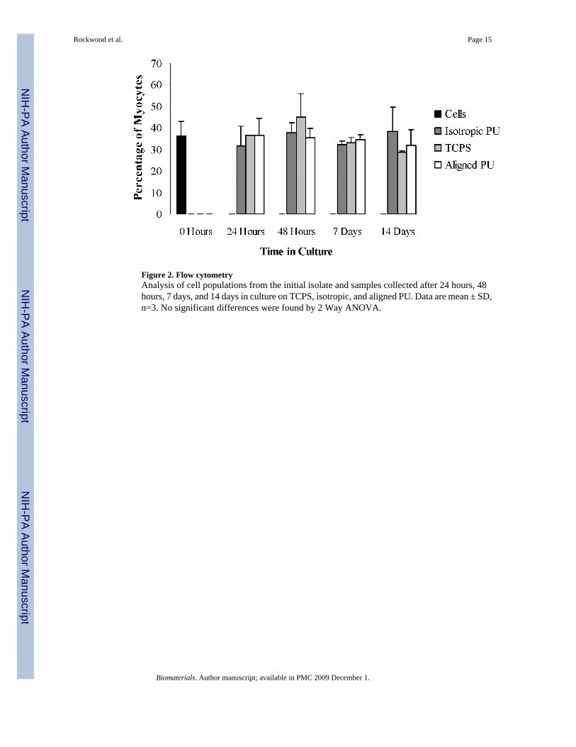
Figure 2. Flow cytometryAnalysis of cell populations from the initial isolate and samples collected after 24 hours, 48hours, 7 days, and 14 days in culture on TCPS, isotropic, and aligned PU. Data are mean ± SD,n=3. No significant differences were found by 2 Way ANOVA.
Rockwood et al. Page 15
Biomaterials. Author manuscript; available in PMC 2009 December 1.
NIH
-PA Author Manuscript
NIH
-PA Author Manuscript
NIH
-PA Author Manuscript
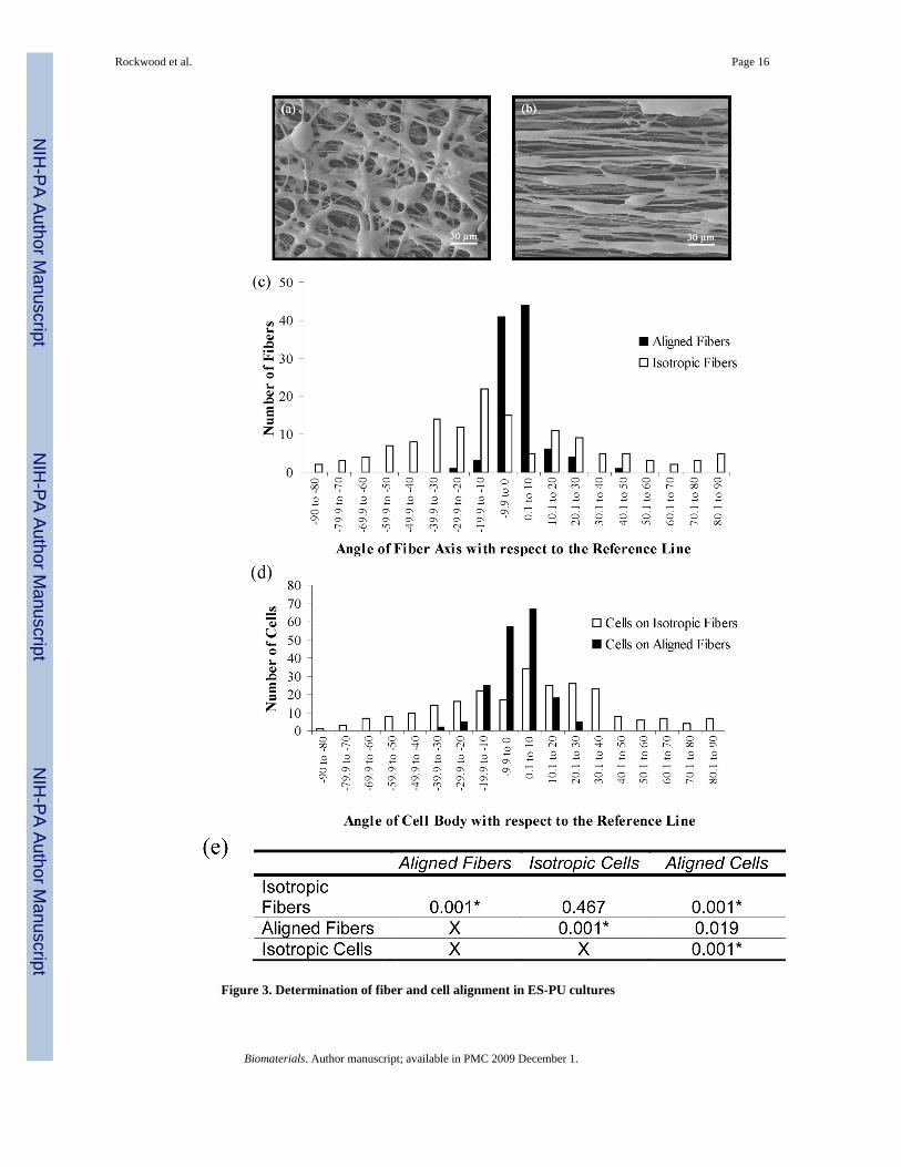
Figure 3. Determination of fiber and cell alignment in ES-PU cultures
Rockwood et al. Page 16
Biomaterials. Author manuscript; available in PMC 2009 December 1.
NIH
-PA Author Manuscript
NIH
-PA Author Manuscript
NIH
-PA Author Manuscript
Isotropic (a) and aligned fibrous mats (b) were used as templates for cellular organization. Thealignment of the (c) fibers and (d) cells was quantified with respect to a reference line and isrepresented as the angle from the reference line ± 90°. (e) Statistical analysis of fiber and cellalignment was done via a Levene's tests for homogeneity of variance and subsequent tests.Here the p-values are shown and significant differences (p≤0.01) are indicated by an asterisk.
Rockwood et al. Page 17
Biomaterials. Author manuscript; available in PMC 2009 December 1.
NIH
-PA Author Manuscript
NIH
-PA Author Manuscript
NIH
-PA Author Manuscript

Figure 4. Cellular organizationSamples were stained for f-actin (green), nuclei (blue), and either muscle-specific myosinheavy chain (red in panels a, b, and c) or gap-junction-specific connexin-43 (red in d, e, andf) to visualize cellular structures. The top image in each pair shows a merged, three-colorfluorescence micrograph obtained at low magnification to show typical staining patterns ofcells cultured on TCPS (a, d), isotropic ES-PU microfibers, and aligned ES-PU microfibers.The bottom image in each pair shows a 4x zoomed view of the region indicated by the rectanglein each upper image to allow visualization of sub-cellular structures. Cells seeded on TCPS (a,d) and isotropic ES-PU fibers (b, e) were substantially less oriented than those grown on alignedES-PU microfibers (c, f). In all cases, muscle cells (black asterisks) and non-muscle cells (whiteasterisks) were present. Striations from organized sarcomeric proteins in cardiomyocytes(white arrow heads) were prevalent in all samples; however, these were generally more difficultto resolve in ES-PU samples due to apparent lensing effects from the underlying biomaterial.
Rockwood et al. Page 18
Biomaterials. Author manuscript; available in PMC 2009 December 1.
NIH
-PA Author Manuscript
NIH
-PA Author Manuscript
NIH
-PA Author Manuscript

Cx-43 staining of gap junctions was also prevalent (black arrowheads). Scale bars represent20 μm.
Rockwood et al. Page 19
Biomaterials. Author manuscript; available in PMC 2009 December 1.
NIH
-PA Author Manuscript
NIH
-PA Author Manuscript
NIH
-PA Author Manuscript
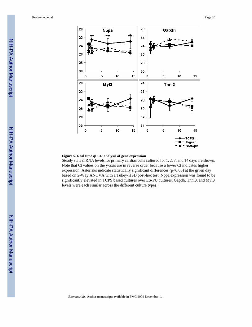
Figure 5. Real time qPCR analysis of gene expressionSteady state mRNA levels for primary cardiac cells cultured for 1, 2, 7, and 14 days are shown.Note that Ct values on the y-axis are in reverse order because a lower Ct indicates higherexpression. Asterisks indicate statistically significant differences (p<0.05) at the given daybased on 2-Way ANOVA with a Tukey-HSD post-hoc test. Nppa expression was found to besignificantly elevated in TCPS based cultures over ES-PU cultures. Gapdh, Tnni3, and Myl3levels were each similar across the different culture types.
Rockwood et al. Page 20
Biomaterials. Author manuscript; available in PMC 2009 December 1.
NIH
-PA Author Manuscript
NIH
-PA Author Manuscript
NIH
-PA Author Manuscript
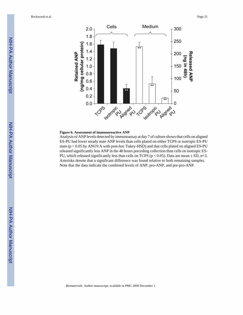
Figure 6. Assessment of immunoreactive ANPAnalysis of ANP levels detected by immunoassay at day 7 of culture shows that cells on alignedES-PU had lower steady state ANP levels than cells plated on either TCPS or isotropic ES-PUmats (p < 0.05 by ANOVA with post-hoc Tukey-HSD) and that cells plated on aligned ES-PUreleased significantly less ANP in the 48 hours preceding collection than cells on isotropic ES-PU, which released significantly less than cells on TCPS (p < 0.05). Data are mean ± SD, n=3.Asterisks denote that a significant difference was found relative to both remaining samples.Note that the data indicate the combined levels of ANP, pro-ANP, and pre-pro-ANP.
Rockwood et al. Page 21
Biomaterials. Author manuscript; available in PMC 2009 December 1.
NIH
-PA Author Manuscript
NIH
-PA Author Manuscript
NIH
-PA Author Manuscript
Copyright © 2022 FDOKUMEN