
Nanoimmunoassay onto a screen printed electrode for HER2 breast cancer biomarker determination
7
Nanoimmunoassay onto a screen printed electrode for HER2 breast cancer biomarker determination Stéphanie Patris a , Pieter De Pauw b,e , Marie Vandeput a , Joëlle Huet c , Pierre Van Antwerpen d , Serge Muyldermans b,e , Jean-Michel Kauffmann a,n a Laboratory of Instrumental Analysis and Bioelectrochemistry, Faculty of Pharmacy, Université Libre de Bruxelles, Boulevard du Triomphe, Campus Plaine, CP 205/06, 1050 Brussels, Belgium b Laboratory of Cellular and Molecular Immunology, Vrije Universiteit Brussel, Pleinlaan 2, 1050 Brussels, Belgium c Laboratory of Biopolymers and Supramolecular Nanomaterials, Faculty of Pharmacy, Université Libre de Bruxelles, Boulevard du Triomphe, Campus Plaine, CP 206/04, 1050 Brussels, Belgium d Analytical Platform, Faculty of Pharmacy, Université Libre de Bruxelles, Boulevard du Triomphe, Campus Plaine, CP 205/05, 1050 Brussels, Belgium e Structural Biology Research Center, VIB, Vrije Universiteit Brussel, 1050 Brussels, Belgium article info Article history: Received 26 May 2014 Received in revised form 27 June 2014 Accepted 28 June 2014 Available online 7 July 2014 Keywords: Screen printed electrode Nanobody HER2 Immunoassay Amperometric immunosensor abstract A chip format sandwich-type immunoassay based on Nanobodies s (Nbs) with the Human Epidermal Growth Factor Receptor (HER2) extracellular domain as antigen model has been developed. The HER2 is considered as an important biomarker because its overexpression causes an aggressive type of breast cancer. Nbs are single domain antigen-binding fragments derived from camelid heavy-chain antibodies. The strategy of the presently developed sandwich immunoassay takes advantage of the small size of Nbs for the detection of the electroactive redox tracer onto the screen printed electrode (SPE). A capture anti HER2 Nb was covalently immobilized onto the SPE, and the detection Nb, raised against another epitope of HER2, was labeled with horseradish peroxidase (HRP). The biosensor signal corresponded to the electroreduction of para-quinone generated at the SPE by the HRP in the presence of hydroquinone and hydrogen peroxide. The best performing and optimized immunoassay conditions consisted of 2 and 20 min for the first and the second incubation times, respectively. The amperometric signal obtained was proportional to the logarithm of HER2 concentration between 1 and 200 mg/mL and the modified SPE storage stability lasted for at least three weeks. Determination of HER2 in human cells has been realized. & 2014 Elsevier B.V. All rights reserved. 1. Introduction Breast cancer is the most common cancer in women worldwide. The disease can be subdivided in four main classes, i.e. (i) hormone (estrogen or progesterone) receptor positive, (ii) HER2 positive, (iii) hormone receptor and HER2 positive and (iv) hormone receptor and HER2 negative breast cancers. The hormone receptor positive cancer responds to endocrine therapy and the prognosis is generally better than the other type of breast cancers [1]. Human epidermal growth factor receptors (HER/erbB) are involved in the normal growth and differentiation of cells. HER2 is the only receptor of this family which has no known ligand. The association of a specific ligand to HER1, HER3 or HER4 provokes a receptor dimerization, preferentially with HER2 as partner and induces cell signaling. A malignant growth occurs when HER2 is overexpressed giving rise to multiple HER2 heterodimers and to a stronger cell signaling, resulting in enhanced responsiveness to growth factors [2]. The HER2 overexpression is present in some cases of breast, ovarian, gastric, prostate and other cancers [3]. HER2 is a trans-membrane glycoprotein of 185 kDa with tyrosine kinase activity, encoded by a gene located on the long arm of chromosome 17 [4]. The amplifica- tion of this chromosome region leads to over-expression of the HER2 [5]. The HER2 is over-expressed in around 20–25% of invasive breast cancers and is associated with poor-prognosis as well as reduced survival [4]. The HER2 status of a breast cancer should be evaluated because therapeutic compounds that specifically target HER2 are available [5]. Trastuzumab (Herceptin s , Roche), a huma- nized monoclonal antibody against HER2 extracellular domain, is currently used but a significant proportion of patients with HER2 positive breast cancer display either primary or secondary resis- tance [6]. Trastuzumab combined with paclitaxel after a treatment with doxorubicin and cyclophosphamide reduce the rate of recur- rence by half among women with operable HER2 positive breast cancer and the mortality was reduced by one third. The therapeutic results were similar for women with hormone-receptor-negative Contents lists available at ScienceDirect journal homepage: www.elsevier.com/locate/talanta Talanta http://dx.doi.org/10.1016/j.talanta.2014.06.069 0039-9140/& 2014 Elsevier B.V. All rights reserved. n Corresponding author. E-mail addresses: [email protected] (S. Patris), [email protected] (J.-M. Kauffmann). Talanta 130 (2014) 164–170
Transcript of Nanoimmunoassay onto a screen printed electrode for HER2 breast cancer biomarker determination

Nanoimmunoassay onto a screen printed electrode for HER2 breastcancer biomarker determination
Stéphanie Patris a, Pieter De Pauwb,e, Marie Vandeput a, Joëlle Huet c, Pierre VanAntwerpen d, Serge Muyldermans b,e, Jean-Michel Kauffmann a,n
a Laboratory of Instrumental Analysis and Bioelectrochemistry, Faculty of Pharmacy, Université Libre de Bruxelles, Boulevard du Triomphe, Campus Plaine, CP205/06, 1050 Brussels, Belgiumb Laboratory of Cellular and Molecular Immunology, Vrije Universiteit Brussel, Pleinlaan 2, 1050 Brussels, Belgiumc Laboratory of Biopolymers and Supramolecular Nanomaterials, Faculty of Pharmacy, Université Libre de Bruxelles, Boulevard du Triomphe, Campus Plaine,CP 206/04, 1050 Brussels, Belgiumd Analytical Platform, Faculty of Pharmacy, Université Libre de Bruxelles, Boulevard du Triomphe, Campus Plaine, CP 205/05, 1050 Brussels, Belgiume Structural Biology Research Center, VIB, Vrije Universiteit Brussel, 1050 Brussels, Belgium
a r t i c l e i n f o
Article history:Received 26 May 2014Received in revised form27 June 2014Accepted 28 June 2014Available online 7 July 2014
Keywords:Screen printed electrodeNanobodyHER2ImmunoassayAmperometric immunosensor
a b s t r a c t
A chip format sandwich-type immunoassay based on Nanobodiess (Nbs) with the Human EpidermalGrowth Factor Receptor (HER2) extracellular domain as antigen model has been developed. The HER2 isconsidered as an important biomarker because its overexpression causes an aggressive type of breastcancer. Nbs are single domain antigen-binding fragments derived from camelid heavy-chain antibodies.The strategy of the presently developed sandwich immunoassay takes advantage of the small size of Nbsfor the detection of the electroactive redox tracer onto the screen printed electrode (SPE). A capture antiHER2 Nb was covalently immobilized onto the SPE, and the detection Nb, raised against another epitopeof HER2, was labeled with horseradish peroxidase (HRP). The biosensor signal corresponded to theelectroreduction of para-quinone generated at the SPE by the HRP in the presence of hydroquinone andhydrogen peroxide. The best performing and optimized immunoassay conditions consisted of 2 and20 min for the first and the second incubation times, respectively. The amperometric signal obtained wasproportional to the logarithm of HER2 concentration between 1 and 200 mg/mL and the modified SPEstorage stability lasted for at least three weeks. Determination of HER2 in human cells has been realized.
& 2014 Elsevier B.V. All rights reserved.
1. Introduction
Breast cancer is the most common cancer in women worldwide.The disease can be subdivided in four main classes, i.e. (i) hormone(estrogen or progesterone) receptor positive, (ii) HER2 positive, (iii)hormone receptor and HER2 positive and (iv) hormone receptorand HER2 negative breast cancers. The hormone receptor positivecancer responds to endocrine therapy and the prognosis is generallybetter than the other type of breast cancers [1]. Human epidermalgrowth factor receptors (HER/erbB) are involved in the normalgrowth and differentiation of cells. HER2 is the only receptor of thisfamily which has no known ligand. The association of a specificligand to HER1, HER3 or HER4 provokes a receptor dimerization,preferentially with HER2 as partner and induces cell signaling. Amalignant growth occurs when HER2 is overexpressed giving rise to
multiple HER2 heterodimers and to a stronger cell signaling,resulting in enhanced responsiveness to growth factors [2]. TheHER2 overexpression is present in some cases of breast, ovarian,gastric, prostate and other cancers [3]. HER2 is a trans-membraneglycoprotein of 185 kDa with tyrosine kinase activity, encoded by agene located on the long arm of chromosome 17 [4]. The amplifica-tion of this chromosome region leads to over-expression of theHER2 [5]. The HER2 is over-expressed in around 20–25% of invasivebreast cancers and is associated with poor-prognosis as well asreduced survival [4]. The HER2 status of a breast cancer should beevaluated because therapeutic compounds that specifically targetHER2 are available [5]. Trastuzumab (Herceptins, Roche), a huma-nized monoclonal antibody against HER2 extracellular domain, iscurrently used but a significant proportion of patients with HER2positive breast cancer display either primary or secondary resis-tance [6]. Trastuzumab combined with paclitaxel after a treatmentwith doxorubicin and cyclophosphamide reduce the rate of recur-rence by half among women with operable HER2 positive breastcancer and the mortality was reduced by one third. The therapeuticresults were similar for women with hormone-receptor-negative
Contents lists available at ScienceDirect
journal homepage: www.elsevier.com/locate/talanta
Talanta
http://dx.doi.org/10.1016/j.talanta.2014.06.0690039-9140/& 2014 Elsevier B.V. All rights reserved.
n Corresponding author.E-mail addresses: [email protected] (S. Patris),
[email protected] (J.-M. Kauffmann).
Talanta 130 (2014) 164–170

tumors and women with hormone-receptor-positive tumors [7].Only patients with HER2 overexpression receive the anti-HER2treatment because of the therapeutic cost and the side-effects. Itis important to avoid false negatives so that only HER2 positivepatients receive a treatment [8]. Immunohistochemistry is theprimary technique applied to determine the HER2-status. Theextracellular domain of HER2 (110 kDa) is released into the systemiccirculation after HER2 cleavage by matrix metalloproteinase. Sub-sequently it can be determined in serum of patients. An enzymelinked immuno sorbent assay (ELISA) has been commercialized bySiemens, which has been approved by the FDA in 2000 [8]. Theserum extracellular HER2 normal level limit is 15 ng/mL and it is agood indicator of the antitumor treatment efficiency. The extra-cellular domain level of HER2, however, is not always in correlationwith the tumor response [9]. A high serum level of extracellulardomain of HER2 could indicate resistance against trastuzumab [10].
Another HER2 immunoassay has been developed using twomonoclonal antibodies directed against independent but adjacenttarget epitopes on the HER2 receptor [11]. One antibody is labeledwith a fluorescent VeraTagTM and the other with a photosensitivemolecule that releases free oxygen radicals upon photoactivation.The free oxygen radicals cleave the nearby VeraTagTM reporterwhich is subsequently collected and quantified using capillaryelectrophoresis [11]. Techniques such as real time PCR and micro-array based sensors have been described to quantify the RNA levelcoding for HER2. Two techniques to measure DNA are approved bythe FDA: a fluorescence in situ hybridization test and a chromo-genic hybridization test [12].
An electrochemical immunosensor has been developed byimmobilizing trastuzumab on gold nanodisk electrodes. The inter-action between HER2 and antibody is detected by suitable sec-ondary antibodies labeled with HRP and methylene blue as theredox mediator. This immunosensor is used to determine HER2 incell lysates and tumor lysates [13]. Another electrochemicalimmunoassay for HER2 detection is based on a sandwich formatin which a primary monoclonal anti-HER2 antibody is coupled toprotein A modified magnetic beads and the secondary antibody islabeled with biotin. After the two immunoreactions, alkalinephosphatase conjugated with streptavidin and 1-naphtyl-phos-phate are used for the differential pulse voltammetry detection atscreen printed electrodes [14]. An opto-fluidic ring resonatorbiosensor has also been developed to determine the HER2 extra-cellular domain in spiked serum [15]. A label free electrochemicalimmunosensor for HER2 based on antiHER2-iron oxide nanopar-ticle bioconjugates has recently been published. After the immu-noreaction the nanoparticles are laid over the gold electrodesurface for differential pulse voltammetry detection [16].
With respect to the development of a rapid immunoassay forHER 2 determination, it was of interest to exploit the advantagesprovided by Nanobodiess (Nbs). Also referred to as “VHH” becausethey consist of the variable part of heavy chain-only antibodies,they are the smallest known single entity with full antigen bindingproperties that are derived from functional antibodies [17]. Heavychain-only antibodies were first discovered in serum of camel, butall species of the Camelidae contain these unique IgGs' in theirserum[18]. Nbs have similar antigen binding properties comparedto whole antibodies, but they offer multiple advantages over otherantibody fragments e.g. they are readily produced by E. coli and areresistant to heat and reducing agents and are highly water soluble[17]. Their stability, size (ca 15 kDa) and antigen affinity andspecificity make them highly attractive for developing novelanalytical tools. In the biosensor field, the first reported applica-tion is a Nb-based chip for prostate-specific antigen immunoassayusing a commercially available surface plasmon resonance (SPR)device [19]. An electrochemiluminescent immunosensor with Nbsimmobilized onto a glassy carbon electrode was described for the
highly sensitive assay of human procalcitonin in serum samples[20]. Subsequently we reported an amperometric immunoassaywith nanobodies immobilized onto SPE for HER2 detection [21].Two amperometric magnetoimmunosensors using nanobodies forfibrinogen detection in plasma [22] have also been described. Thesmall size of the Nbs was instrumental for easy access to theirtarget and for signal detection in close proximity of the transducer.
Here, we report for the first time the development of a sandwichtype immunoassay (nanoimmunoassay) using Nbs linked to a carbonbased screen printed electrode (SPE) for the HER2 extracellulardomain determination. HER2 antigen was selected as a biomarkertarget model because of its availability together with a series of non-competitive anti HER2 Nbs [23]. The HER2 level was determined inhuman cells spiked with HER2 to mimic a cancerous lysate.
2. Material and methods
2.1. Reagents
N-(3-Dimethylaminopropyl)-N0-ethylcarbodiimide hydrochlor-ide (EDC), N-Hydroxysuccinimide (NHS), polysorbate 20 (tween20), hydroquinone, Tris–Cl (Trizmas base) and Tergitol-type nonylphenoxypolyethoxylethanol 40 (Nonidet P-40, NP40) were pur-chased from Sigma (Bornem, Belgium). EZ-link Plus ActivatedPeroxidase kit and BCA protein assay kit were from PierceBiotechnology /Thermo scientific (Rockford, USA). Albumin frombovine serum (fraction V) (BSA) was from Fluka (Neu-Ulm,Germany). Recombinant Human ErbB2/Fc Chimera (HER2) wasobtained from ImmunoSource (Schilde, Belgium). Hydrogen per-oxide (30%) was from VWR International Inc (Leuven, Belgium).Citric acid monohydrate and sodium carbonate anhydrous werefrom Janssen Chimica (Beerse, Belgium). Sodium hydrogen carbo-nate was from Fisher Chemicals (Loughborough, United Kingdom).Potassium chloride and di-sodium hydrogen phosphate dihydratewere from Merck (Darmstadt, Germany). Potassium dihydrogenphosphate dihydrate was from Vel (Leuven, Belgium). Sodiumchloride was from Carlo Erba (Rodano, Italy). The protease inhi-bitor cocktail tablets were from Roche (Mannheim).
Following buffers and solutions were prepared:
– Phosphate buffered saline pH 7.4 (PBS) containing 8 g/L ofsodium chloride, 0.2 g/L of potassium chloride, 8 mM di-sodium hydrogen phosphate and 1.5 mM potassium dihydro-gen phosphate for immunochemical reactions.
– MES buffer containing 10.6 g/L 2-(4-morpholino) ethanesul-phonic acid adjusted to pH 5.5 for the formation of the peptidebond with EDC and NHS.
– PBS containing 0.5 g/L of polysorbate 20 as washing buffer.– A 0.05 M citric acid solutionwas adjusted to pH 5.5 with a solution
of sodium hydroxide for the electrochemical mea-surements.
– Lysis buffer containing 50 mM Tris–HCl pH 8.0, 0.4 M sodiumchloride, 1% (v/v) NP40 and protease inhibitor cocktail (1 tablet for10 mL) was used to lyse sample cells [13].
2.2. Instrumentation
The electrochemical measurements were performed using an LCEpsilon computer-controlled potentiostat with Epsilon EC software.The screen printed electrodes (SPE) and the adapted connector werepurchased from DropSens (Oviedo, Spain). SPE had the followingdimensions: 3.4�1.0�0.05 cm3 (length�width�height). Working(4 mm diameter) and counter electrodes were made of carbon,pseudoreference electrode and electric contacts were made of silver.
S. Patris et al. / Talanta 130 (2014) 164–170 165

The LC-QTOF analyzes were performed with a QTOF 6520 (Agilenttechnologies, Palo Alto, CA, USA).
2.3. Anti-HER2 Nb production
The anti-HER2 Nbs 2Rs15d, 1R143c and 1R59b were producedand purified as described previously [23]. In short, their DNAfragments were re-cloned in vector pHEN6 and expressed in E. coliWK6 after isopropyl-β-D-thiogalactoside (IPTG) induction toobtain His6-tagged Nbs in the periplasm. The Nbs were extractedfrom the periplasm by osmotic shock and purified by immobilizedmetal affinity chromatography (IMAC) on Ni-NTA resin (Sigma-Aldrich, St Louis, MO, USA), eluting with 0.5 M imidazole in PBS pH7.5 and containing 80 g/L NaCl, and further purified by molecularsieving on Superdex 75 h 16/60 (Pharmacia, Gaithersburg, USA) inPBS. Molecular weights, the number of lysines and the extinctioncoefficients at 280 nm were calculated from their amino acidsequence analysis, and binding properties were determined bySPR (Table 1).
2.4. Surface plasmon resonance (SPR)
To confirm the mutual non-competitiveness of the utilized anti-HER2 Nbs, single solutions and paired mixtures were subjected to SPRperformed on a Biacore T100 instrument (GE Healthcare, LittleChalfont, UK), with the recombinant human ErbB2/Fc fusion proteinimmobilized on a CM-5 gold layer chip. In practice, the first 600 sbinding phase occurred with a single Nb, and was followed by asecond 600 s binding phase with a mixture of the Nb and one otherHER2 Nb. Control curves were measured with the single Nb in bothphases and analyzed using Biacore's evaluation software (not shown).
2.5. Peroxidase labeled anti-HER2 Nbs
The procedure recommended by the kit supplier was followedexcept that glycine was used instead of ethanolamine (see reasonbelow) to neutralize the excess of EZ-LinkTM Plus activated peroxidase,and was optimized in terms of pH and incubation time. The sugars ofnative HRP were oxidized by periodate to generate aldehyde groups,and the oxidized HRP was then crosslinked to the anti-HER2 Nbs byforming Shiff-bases between the aldehydes on HRP and the primaryamines present on the Nb. Sodium cyanoborohydride was added toreduce the link and form a stable HRP-Nb conjugate. (i) A volume of100 ml of oxidized HRP (1 mg/100 mL) and 10 mL of sodium cyanobor-ohydride (5.0 M) were added to 500 mL PBS containing 0.3 mg of Nb;(ii) this solution was incubated overnight under stirring at roomtemperature; (iii) a 20 mL volume of glycine 3 M was added andreacted during 15 min at room temperature. At the end, the HRPlabeled Nb (Nb-HRP) was purified as described above for free Nbs byIMAC (to remove free HRP) and size exclusion chromatography (toremove free Nb). The efficiency of the labeling was tested by SDS-PAGE. The SDS-PAGE experiments were carried out on precast gels(NuPAGE Novex 4–12% Bis–tris with MES SDS running buffer) usingthe X-cell SureLock Mini-cell from Invitrogen. The running conditions
were 200 V, 50 mA at constant temperature 25 1C during 45 min. TheNovex Sharp Protein standard from Invitrogen consisted of a mixtureof 12 pre-stained standards (3.5, 10, 15, 20, 30, 40, 50, 60, 80, 110, 160,260 kDa) for protein molecular weight comparison. Protein detectionwas performed using Coomassie Blue staining (ethanol 40% (v/v),acetic acid 10% (v/v) and Coomassie Brillant R250 0.2% (w/v)).
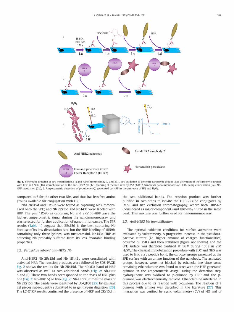
2.6. Immobilizing capture anti-HER2 on the SPE (Fig. 1.1)
The SPE surface was modified according to the followingprocedure: (i) the SPE was activated in the presence of 50 mL of2 M H2SO4 dropped onto the SPE and by applying a voltage ofþ1.60 V for 150 s, then the SPE was washed with distillated water,(ii) 10 mL of 40 mM EDC and 40 mM NHS solution in MES bufferwas spiked onto the pretreated SPE surface during 1 h, (iii) the SPEwas again thoroughly washed with distilled water and dried toremove excess reagent and 10 mL of 10 mg/mL (6.6�10�7 M) anti-HER2 Nb were spiked onto the electrode surface, (iv) after 1 h thenon-linked Nbs were eliminated by gently washing the SPE withdistillated water. Any remaining surface free sites were blocked by10 mL of BSA solution in PBS (1% m/v) for 30 min. Finally the SPEwas washed with water. All the incubations and reactions occurredat room temperature and in a humid atmosphere.
2.7. Nanoimmunoassay (Fig. 1.2 and 1.3)
The SPE surface modified with anti-HER2 Nb was spiked with10 mL of HER2 solution in PBS for 2 min at room temperature(Fig. 1.2a). After this incubation period, the SPE was rinsed withwater and 10 mL Nb-HRP solution was added and left reacting for20 min (Fig. 1.2b). The SPE was washed twice with 1 mL ofwashing buffer then, 40 mL of citrate buffer containing 2.5 mMH2O2 was added onto the SPE for the amperometric detection.A potential of �280 mV was applied and 10 mL HQ (0.1 M) wasspiked onto the electrode and the reduction current was mon-itored (Fig. 1.3). All the electrochemical parameters were opti-mized in a previous study [24].
2.8. Sample assays
HER2 negative (MCF7) cells were cultured in a T80 flask in15 mL of the following medium: RPMi, 10% FBS heat inactivated,2% Pen-strep (10,000 Um/L), 2% L-glutamine (200 mM) and 0.2%Gentamicine (50 mg/mL). The cells were collected when theconfluence reached 85–90%. The cell lysates were prepared using500 mL of lysis buffer. After an incubation time of 30 min at 4 1C,the lysates were collected, sonicated, and centrifuged. A volume of70 mL of lysate was spiked with 30 mL HER2 and 10 mL of thissample served for the HER2 nanoimmunoassay (n¼3). The totalprotein concentration was estimated by a Bradford assay beforeadding HER2. The HER2 concentration in the samples was deter-mined by referring to a calibration curve realized daily in PBS.
3. Results and discussion
3.1. Anti-HER2 Nb selection
Three different Her2-specific Nbs were selected based on theirknown binding rate constants and mutual non-competitiveness(Table 1). For stability reasons, the Nb 2Rs15d having the lowest kd,would be the first choice as capture Nb (immobilized onto theSPE), to be combined with Nb 1R59b having the second best kdand KD as detecting Nb (HRP conjugated) in a in HER2 nanoim-munoassay. The 1R59b Nb however contains only three lysines
Table 1Properties of selected Nbs.
Nanobody SPR MW Lys # ε (M�1cm�1)
Ka (M�1s�1) kd (s�1) KD (nM)
2Rs15da 2.14�105 5.71�10�4 2.70 13453 6 256901R59ba 5.01�105 2.43�10�3 4.90 13361 3 117101R143ca 6.27�105 4.23�10�3 6.80 14540 6 20650
a Non-competitiveness between these Nbs was demonstrated with SPR.
S. Patris et al. / Talanta 130 (2014) 164–170166
compared to 6 for the other two Nbs, and thus has less free aminegroups available for conjugation with HRP.
Nbs 2Rs15d and 1R59b were tested as capturing Nb (immobi-lized onto the SPE) and Nb 2Rs15d and Nb143c were labeled withHRP. The pair 1R59b as capturing Nb and 2Rs15d-HRP gave thehighest amperometric signal during the nanoimmunoassay, andwas selected for further application of nanoimmunoassay. The SPRresults (Table 1) suggest that 2Rs15d is the best capturing Nbbecause of its low dissociation rate, but the HRP labeling of 1R59b,containing only three lysines, was unsuccessful. Nb143c-HRP asdetecting Nb probably suffered from its less favorable bindingproperties.
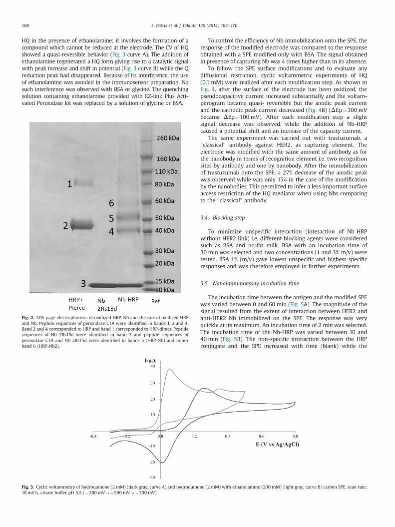
3.2. Peroxidase labeled anti-HER2 Nb
Anti-HER2 Nb 2Rs15d and Nb 1R143c were crosslinked withactivated HRP. The reaction products were followed by SDS-PAGE.Fig. 2 shows the results for Nb 2Rs15d. The 40 kDa band of HRPwas observed as well as two additional bands (Fig. 2: Nb-HRP5 and 6). These two bands corresponded to the mass of HRP plusone (Fig. 2: Nb-HRP 5) or two (Fig. 2: Nb-HRP 6) times the mass ofNb 2Rs15d. The bands were identified by LC-QTOF [25] by excisinggel pieces subsequently submitted to in gel trypsin digestion [26].The LC-QTOF results confirmed the presence of HRP and 2Rs15d in
the two additional bands. The reaction product was furtherpurified in two steps to isolate the HRP-2Rs15d conjugates byIMAC and size exclusion chromatography, where both HRP-Nb(considered as major component) and HRP-Nb2 eluted in the samepeak. This mixture was further used for nanoimmunoassay.
3.3. Anti-HER2 Nb immobilization
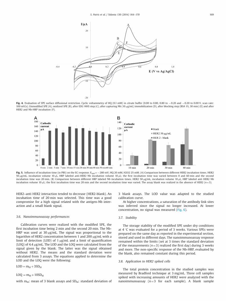
The optimal oxidation conditions for surface activation wereevaluated by voltammetry. A progressive increase in the pseudoca-pacitive current (i.e. higher amount of charged functionalities)occurred till 150 s and then stabilized (figure not shown), and theSPE surface was therefore oxidized at 1.6 V during 150 s in 2 MH2SO4.The classical immobilization procedure with EDC and NHS wasused to link, via a peptide bond, the carboxyl groups generated at theSPE surface with an amine function of the nanobody. The activatedgroups, however, were not blocked by ethanolamine since someremaining ethanolamine was found to react with the HRP generatedquinone in the amperometric assay. During the detection step,hydroquinone was oxidized to p-quinone by HRP and the p-quinone was electrochemically reduced. Ethanolamine interfered inthis process due to its reaction with p-quinone. The reaction of aquinone with amines was described in the literature [27]. Thisinteraction was verified by cyclic voltammetry (CV) of HQ and of
Fig. 1. Schematic drawing of SPE modification. (1) and nanoimmunoassay (2 and 3). 1. SPE oxidation to generate carboxylic groups (1a), activation of the carboxylic groupswith EDC and NHS (1b), immobilization of the anti-HER2 Nb (1c), blocking of the free sites by BSA (1d); 2. Sandwich nanoimmunoassay: HER2 sample incubation (2a), Nb-HRP incubation (2b); 3. Amperometric detection of p-quinone (Q) generated by HRP in the presence of HQ and H2O2.
S. Patris et al. / Talanta 130 (2014) 164–170 167
HQ in the presence of ethanolamine; it involves the formation of acompound which cannot be reduced at the electrode. The CV of HQshowed a quasi-reversible behavior (Fig. 3 curve A). The addition ofethanolamine regenerated a HQ form giving rise to a catalytic signalwith peak increase and shift in potential (Fig. 3 curve B) while the Qreduction peak had disappeared. Because of its interference, the useof ethanolamine was avoided in the immunosensor preparation. Nosuch interference was observed with BSA or glycine. The quenchingsolution containing ethanolamine provided with EZ-link Plus Acti-vated Peroxidase kit was replaced by a solution of glycine or BSA.
To control the efficiency of Nb immobilization onto the SPE, theresponse of the modified electrode was compared to the responseobtained with a SPE modified only with BSA. The signal obtainedin presence of capturing Nb was 4 times higher than in its absence.
To follow the SPE surface modifications and to evaluate anydiffusional restriction, cyclic voltammetric experiments of HQ(0.1 mM) were realized after each modification step. As shown inFig. 4, after the surface of the electrode has been oxidized, thepseudocapacitive current increased substantially and the voltam-perogram became quasi- reversible but the anodic peak currentand the cathodic peak current decreased (Fig. 4B) (ΔEp¼300 mVbecame ΔEp¼100 mV). After each modification step a slightsignal decrease was observed, while the addition of Nb-HRPcaused a potential shift and an increase of the capacity current.
The same experiment was carried out with trastuzumab, a“classical” antibody against HER2, as capturing element. Theelectrode was modified with the same amount of antibody as forthe nanobody in terms of recognition element i.e. two recognitionsites by antibody and one by nanobody. After the immobilizationof trastuzumab onto the SPE, a 27% decrease of the anodic peakwas observed while was only 15% in the case of the modificationby the nanobodies. This permitted to infer a less important surfaceaccess restriction of the HQ mediator when using Nbs comparingto the “classical” antibody.
3.4. Blocking step
To minimize unspecific interaction (interaction of Nb-HRPwithout HER2 link) i.e. different blocking agents were consideredsuch as BSA and no-fat milk. BSA with an incubation time of30 min was selected and two concentrations (1 and 3% m/v) weretested. BSA 1% (m/v) gave lowest unspecific and highest specificresponses and was therefore employed in further experiments.
3.5. Nanoimmunoassay incubation time
The incubation time between the antigen and the modified SPEwas varied between 0 and 60 min (Fig. 5A). The magnitude of thesignal resulted from the extent of interaction between HER2 andanti-HER2 Nb immobilized on the SPE. The response was veryquickly at its maximum. An incubation time of 2 min was selected.The incubation time of the Nb-HRP was varied between 10 and40 min (Fig. 5B). The non-specific interaction between the HRPconjugate and the SPE increased with time (blank) while the
Fig. 2. SDS-page electrophoresis of oxidized HRP, Nb and the mix of oxidized HRPand Nb. Peptide sequences of peroxidase C1A were identified in bands 1, 2 and 4.Band 2 and 4 corresponded to HRP and band 1 corresponded to HRP-dimer. Peptidesequences of Nb 2Rs15d were identified in band 3 and peptide sequences ofperoxidase C1A and Nb 2Rs15d were identified in bands 5 (HRP-Nb) and minorband 6 (HRP-Nb2).
I/µA
20
10
-20
-10 E (V vs Ag/AgCl)
30
40
-30
Fig. 3. Cyclic voltammetry of hydroquinone (2 mM) (dark gray, curve A) and hydroquinone (2 mM) with ethanolamine (200 mM) (light gray, curve B) carbon SPE, scan rate:10 mV/s, citrate buffer pH 5.5 (�300 mV-þ500 mV-�300 mV).
S. Patris et al. / Talanta 130 (2014) 164–170168
HER2–anti HER2 interaction tended to decrease (HER2-blank). Anincubation time of 20 min was selected. This time was a goodcompromise for a high signal related with the antigen-Nb inter-action and a small blank signal.
3.6. Nanoimmunoassay performances
Calibration curves were realized with the modified SPE, thefirst incubation time being 2 min and the second 20 min. The Nb-HRP was used at 30 mg/mL. The signal was proportional to thelogarithm of HER2 concentration between 1 and 200 mg/mL with alimit of detection (LOD) of 1 mg/mL and a limit of quantification(LOQ) of 4.4 mg/mL. The LOD and the LOQ were calculated from thesignal given by the blank. The latter was the signal obtainedwithout HER2. The means and the standard deviation werecalculated from 3 assays. The equations applied to determine theLOD and the LOQ were the following:
LOD¼mblþ3SDbl
LOQ ¼mblþ10SDbl
with mbl: mean of 3 blank assays and SDbl: standard deviation of
3 blank assays. The LOD value was adapted to the studiedcalibration curve.
At higher concentrations, a saturation of the antibody link siteswas inferred since the signal no longer increased. At lowerconcentration, no signal was measured (Fig. 6).
3.7. Stability
The storage stability of the modified SPE under dry conditionsat 4 1C was evaluated for a period of 3 weeks. Various SPEs wereprepared on the same day as reported in the experimental section,stored and used in different days. The nanoimmunoassay responseremained within the limits (set at 3 times the standard deviationof the measurements (n¼3) realized the first day) during 3 weeksand more. The non-specific response of the Nb-HRP, evaluated bythe blank, also remained constant during this period.
3.8. Application to HER2 spiked cells
The total protein concentration in the studied samples wasmeasured by Bradford technique at 3 mg/mL. Three cell samplesspiked with increasing amounts of HER2 were analyzed with thenanoimmunoassay (n¼3 for each sample). A blank sample
I/µA
E (V vs Ag/AgCl)
20
10
-20
-10
A
BC FDE
Fig. 4. Evaluation of SPE surface diffusional restriction. Cyclic voltammetry of HQ (0.1 mM) in citrate buffer (0.00 to 0.80, 0.80 to �0.20 and �0.20 to 0.00 V, scan rate:100 mV/s). Unmodified SPE (A), oxidized SPE (B), after EDC-NHS step (C), after capturing Nb (10 mg/mL) immobilization (D), after blocking step (BSA 1%, 30 min) (E) and afterHER2 and Nb-HRP incubation (F).
Fig. 5. Influence of incubation time (in PBS) on the EC response. Eapp¼�280 mV, HQ 20 mM, H2O2 25 mM. (A) Comparison between different HER2 incubation times. HER250 mg/mL, incubation volume 10 mL, HRP labeled anti-HER2 Nb incubation volume 10 mL; the first incubation time was varied between 0 and 60 min and the secondincubation time was 20 min. (B) Comparison between different HRP labeled Nb incubation times. HER2 50 mg/mL, incubation volume 10 mL, HRP labeled anti HER2 Nbincubation volume 10 mL; the first incubation time was 20 min and the second incubation time was varied. The assay blank was realized in the absence of HER2 (n¼3).
S. Patris et al. / Talanta 130 (2014) 164–170 169

containing no HER2 was also tested. The signal obtained with thissample was similar to the signal obtained with the PBS buffer. Thisindicated an absence of interference in our nanoimmunoassay andreflects the specificity of the test. The results obtained with thespiked samples are shown in Table 2. An acceptable negative biaswas found for all samples, possibly due to the high proteinconcentration in tested cell lysates.
4. Conclusion
This work reported the successful SPE modification by Nb. Itpermitted a new type of immunoassay called nanoimmunoassayonto the SPE. A high stability of Nb was inferred since the storagestability of the SPE was higher than 3 weeks. Very short incubationtimes were sufficient to obtain a satisfactory response. Thenanoimmunoassay allowed determining HER2 spiked in celllysates. Future work, however, is needed in order to increase thesensitivity of the nanoimmunoassay for serum sample applicationsat physiological relevant values and for cell lysates assays aftereventual dilution.
Acknowledgments
Thanks are expressed to Institut Jules Bordet, Université Librede Bruxelles (ULB) and especially to Ms. Zéna Wimana fortrastuzumab supply, Ms. Aude Ingels from the Cancerology andExperimental Toxicology Laboratory, Faculty of Pharmacy of ULBfor cell samples handling and Mr. Damien Dufour from AnalyticalPlatform, Faculty of Pharmacy of ULB for the LC-QTOF analysis.
References
[1] M.-J. Kim, J.Y. Ro, S.-H. Ahn, H.H. Kim, S.-B. Kim, G. Gong, Hum. Pathol. 37(2006) 1217–1226.
[2] R.A. George, J. Heringa, Protein Eng. Des. Sel. 15 (2002) 871–879.[3] W. Tai, R. Mahato, K. Cheng, J. Control. Release 146 (2010) 264–275.[4] F. Révillion, J. Bonneterre, J. Peyrat, Eur. J. Cancer 34 (1998) 791–808.[5] N. Pathmanathan, P.J. Provan, H. Mahajan, G. Hall, K. Byth, A.M. Bilous, et al.,
Breast 21 (2012) 724–729.[6] K.S. Saini, H.A. Azim, O. Metzger-Filho, S. Loi, C. Sotiriou, E. de Azambuja,
Breast 20 (3) (2011) S20–S27.[7] E.H. Romond, E.A. Perez, J. Bryant, V.J. Suman, C.E. Geyer, N.E. Davidson, et al.,
N. Engl. J. Med. 353 (2005) 1673–1684.[8] C.B. Moelans, R.A. de Weger, E. Van der Wall, P.J. van Diest, Crit. Rev. Oncol.
Hematol. 80 (2011) 380–392.[9] S. Lennon, C. Barton, L. Banken, L. Gianni, M. Marty, J. Baselga, et al., J. Clin.
Oncol. 27 (2009) 1685–1693.[10] M. Scaltriti, F. Rojo, A. Ocaña, J. Anido, M. Guzman, J. Cortes, et al., J. Natl.
Cancer Inst. 99 (2007) 628–638.[11] Y. Shi, W. Huang, Y. Tan, X. Jin, R. Dua, E. Penuel, et al., Diagn. Mol. Pathol. 18
(2009) 11–21.[12] S. Di Palma, N. Collins, C. Faulkes, B. Ping, G. Ferns, B. Haagsma, et al., J. Clin.
Pathol. 60 (2007) 1067–1068.[13] S.P. Mucelli, M. Zamuner, M. Tormen, G. Stanta, P. Ugo, Biosens. Bioelectron. 23
(2008) 1900–1903.[14] Q.A.M. Al-Khafaji, M. Harris, S. Tombelli, S. Laschi, A.P.F. Turner, M. Mascini,
et al., Electroanalysis 24 (2012) 735–742.[15] J.T. Gohring, P.S. Dale, X. Fan, Sens. Actuators B Chem. 146 (2010) 226–230.[16] M. Emami, M. Shamsipur, R. Saber, R. Irajirad, Analyst 139 (2014) 2858–2866.[17] S. Muyldermans, Annu. Rev. Biochem. 82 (2013) 775–797.[18] C. Hamers-Casterman, T. Atarhouch, S. Muyldermans, G. Robinson, C. Hamers,
E.B. Songa, et al., Nature 363 (1993) 446–448.[19] L. Huang, G. Reekmans, D. Saerens, J.-M. Friedt, F. Frederix, L. Francis, et al.,
Biosens. Bioelectron. 21 (2005) 483–490.[20] H. Li, Y. Sun, J. Elseviers, S. Muyldermans, S. Liu, Y. Wan, Analyst (2014).[21] S. Patris, P. De Pauw, S. Muyldermans, J.-M. Kauffmann, Nano-immunoassays
for the determination of HER2, a breast cancer market, using a screen printedelectrode. 2012. Abstracts book ⟨https://www.ulb.ac.be/facs/pharma/docs/abstract.pdf⟩.
[22] S. Campuzano, V. Salema, M. Moreno-Guzmán, M. Gamella, P. Yáñez-Sedeño,L.A. Fernández, et al., Biosens. Bioelectron. 52 (2014) 255–260.
[23] I. Vaneycken, N. Devoogdt, N. Van Gassen, C. Vincke, C. Xavier, U. Wernery,et al., Fad. Am. Soc. Exp. Biol. J. 25 (2011) 2433–2446.
[24] S. Patris, C. De Vriese, F. Prohoroff, E.B. Calvo, J.A. Martínez, J.-M. Kauffmann,Electroanalysis 22 (2010) 41–48.
[25] C. Delporte, P. Van Antwerpen, K. Zouaoui Boudjeltia, C. Noyon, F. Abts,F. Métral, et al., Anal. Biochem. 411 (2011) 129–138.
[26] M.-C. Slomianny, A. Dupont, F. Bouanou, O. Beseme, A.-L. Guihot, P. Amouyel,et al., Proteomics 6 (2006) 2365–2375.
[27] M. Morrison, W. Steele, D.J. Danner, Arch. Biochem. Biophys. 134 (1969)515–523.
Fig. 6. Calibration curve. Representative results obtained with SPE modified byEDC/NHS, anti-HER2 (1R59b) Nb and BSA 1%, incubation HER2 2 min; Nb-HRP(2Rs15d) incubation 20 min. Blank signal was subtracted.
Table 2Recoveries, biases, RSDs for HER2 determination in cell lysates using the developedHER2 nanoimmunosensor.
HER2 spiked (mg/mL) HER2 found (mg/mL) Recovery (%) Bias (%) RSD (%)
25 22 88 �12 850 41 82 �18 675 66 88 �12 14
S. Patris et al. / Talanta 130 (2014) 164–170170