Basophils Produce IL4 and Accumulate in Tissues after Infection with a Th2-inducing Parasite
11
The Journal of Experimental Medicine The Journal of Experimental Medicine • Volume 200, Number 4, August 16, 2004 507–517 http://www.jem.org/cgi/doi/10.1084/jem.20040590 507 Basophils Produce IL-4 and Accumulate in Tissues after Infection with a Th2-inducing Parasite Booki Min, 1 Melanie Prout, 3 Jane Hu-Li, 1 Jinfang Zhu, 1 Dragana Jankovic, 2 Ellen S. Morgan, 4 Joseph F. Urban, Jr., 5 Ann M. Dvorak, 4 Fred D. Finkelman, 6 Graham LeGros, 3 and William E. Paul 1 1 Laboratory of Immunology and 2 Laboratory of Parasitic Diseases, National Institute of Allergy and Infectious Diseases, National Institutes of Health, Bethesda, MD 20892 3 Malaghan Institute of Medical Research,Wellington, New Zealand 4 Department of Pathology, Beth Israel Deaconess Medical Center and Harvard Medical School, Boston, MA 02215 5 Nutrient Requirements and Functions Laboratory, Beltsville Human Nutrition Research Center, U.S. Department of Agriculture, Beltsville, MD 20705 6 Department of Medicine, University of Cincinnati College of Medicine, Cincinnati, OH 45267 Abstract Using mice in which the eGfp gene replaced the first exon of the Il4 gene (G4 mice), we examined production of interleukin (IL)-4 during infection by the intestinal nematode Nippostrongylus brasiliensis (Nb). Nb infection induced green fluorescent protein (GFP) pos cells that were FcRI pos , CD49b bright , c-kit neg , and Gr1 neg . These cells had lobulated nuclei and granules char- acteristic of basophils. They were found mainly in the liver and lung, to a lesser degree in the spleen, but not in the lymph nodes. Although some liver basophils from naive mice express GFP, Nb infection enhanced GFP expression and increased the number of tissue basophils. Similar basophil GFP expression was found in infected Stat6 mice. Basophils did not increase in number in infected Rag2 mice; Rag2 mice reconstituted with CD4 T cells allowed significant basophil accumulation, indicating that CD4 T cells can direct both tissue migration of basophils and enhanced IL-4 production. IL-4 production was immunoglobulin independent and only partially dependent on IL-3. Thus, infection with a parasite that induces a “Th2-type response” resulted in accumulation of tissue basophils, and these cells, stimulated by a non-FcR cross-linking mechanism, are a principal source of in vivo IL-4 production. Key words: CD4 T cells • cytokine • green fluorescent protein • Nippostrongylus brasiliensis • liver Introduction IL-4 plays a crucial role in the development of Th2-type immune responses and the regulation of immunoglobulin isotype switching to IgE (1, 2). An analysis of the in vivo role of IL-4 and its congener IL-13 has been particularly important in understanding the pathogenesis of allergic dis- eases and of parasitic infections (1–3). Nonetheless, such experiments have been hampered by the difficulty of mea- suring cytokine production in situ. Current techniques that use in vitro stimulation (TCR dependent or independent) to detect cytokine production measure cytokine producing potential rather than actual in situ cytokine production. In an effort to overcome such limitations, mice that express surrogates for cytokines have been generated. Among the type I cytokines and the IFNs, mice that express GFP in lieu of IL-2 and IL-4 have been described previously (4,5), as have mice that make GFP in addition to IL-4 or yellow fluorescent protein in addition to IFN (6, 7). We have generated a mouse (the G4 mouse) in which the first exon and a portion of the first intron of the Il4 gene have been replaced by the gene encoding enhanced GFP. Cells from G4 mice possessing a Gfp allele of Il4 express GFP instead of IL-4 from that allele (5). Because GFP is not secreted, it is detectable by flow cytometry without need for restimula- The online version of this article contains supplemental material. Address correspondence to William E. Paul, Laboratory of Immunology, National Institute of Allergy and Infectious Diseases, National Institutes of Health, 10 Center Dr., Bldg. 10, Rm. 11N314, Bethesda, MD 20892. Phone: (301) 496-5046; Fax: (301) 496-0222; email: [email protected] Abbreviation used in this paper: Nb, Nippostrongylus brasiliensis. on March 24, 2014 jem.rupress.org Downloaded from Published August 16, 2004 http://jem.rupress.org/content/suppl/2004/08/16/jem.20040590.DC1.html Supplemental Material can be found at:
Transcript of Basophils Produce IL4 and Accumulate in Tissues after Infection with a Th2-inducing Parasite

The
Journ
al o
f Exp
erim
enta
l M
edic
ine
The Journal of Experimental Medicine • Volume 200, Number 4, August 16, 2004 507–517http://www.jem.org/cgi/doi/10.1084/jem.20040590
507
Basophils Produce IL-4 and Accumulate in Tissues after Infection with a Th2-inducing Parasite
Booki Min,
1
Melanie Prout,
3
Jane Hu-Li,
1
Jinfang Zhu,
1
Dragana Jankovic,
2
Ellen S. Morgan,
4
Joseph F. Urban, Jr.,
5
Ann M. Dvorak,
4
Fred D. Finkelman,
6
Graham LeGros,
3
and William E. Paul
1
1
Laboratory of Immunology and
2
Laboratory of Parasitic Diseases, National Institute of Allergy and Infectious Diseases, National Institutes of Health, Bethesda, MD 20892
3
Malaghan Institute of Medical Research, Wellington, New Zealand
4
Department of Pathology, Beth Israel Deaconess Medical Center and Harvard Medical School, Boston, MA 02215
5
Nutrient Requirements and Functions Laboratory, Beltsville Human Nutrition Research Center, U.S. Department of Agriculture, Beltsville, MD 20705
6
Department of Medicine, University of Cincinnati College of Medicine, Cincinnati, OH 45267
Abstract
Using mice in which the
eGfp
gene replaced the first exon of the
Il4
gene (G4 mice), we examinedproduction of interleukin (IL)-4 during infection by the intestinal nematode
Nippostrongylusbrasiliensis
(Nb). Nb infection induced green fluorescent protein (GFP)
pos
cells that wereFc
�
RI
pos
, CD49b
bright
, c-kit
neg
, and Gr1
neg
. These cells had lobulated nuclei and granules char-acteristic of basophils. They were found mainly in the liver and lung, to a lesser degree in thespleen, but not in the lymph nodes. Although some liver basophils from naive mice expressGFP, Nb infection enhanced GFP expression and increased the number of tissue basophils.Similar basophil GFP expression was found in infected Stat6
���
mice. Basophils did not increasein number in infected Rag2
���
mice; Rag2
���
mice reconstituted with CD4 T cells allowedsignificant basophil accumulation, indicating that CD4 T cells can direct both tissue migrationof basophils and enhanced IL-4 production. IL-4 production was immunoglobulin independentand only partially dependent on IL-3. Thus, infection with a parasite that induces a “Th2-typeresponse” resulted in accumulation of tissue basophils, and these cells, stimulated by a non-FcRcross-linking mechanism, are a principal source of in vivo IL-4 production.
Key words: CD4 T cells • cytokine • green fluorescent protein •
Nippostrongylus brasiliensis
• liver
Introduction
IL-4 plays a crucial role in the development of Th2-typeimmune responses and the regulation of immunoglobulinisotype switching to IgE (1, 2). An analysis of the in vivorole of IL-4 and its congener IL-13 has been particularlyimportant in understanding the pathogenesis of allergic dis-eases and of parasitic infections (1–3). Nonetheless, suchexperiments have been hampered by the difficulty of mea-suring cytokine production in situ. Current techniques thatuse in vitro stimulation (TCR dependent or independent)to detect cytokine production measure cytokine producing
potential rather than actual in situ cytokine production. Inan effort to overcome such limitations, mice that expresssurrogates for cytokines have been generated. Among thetype I cytokines and the IFNs, mice that express GFP inlieu of IL-2 and IL-4 have been described previously (4,5),as have mice that make GFP in addition to IL-4 or yellowfluorescent protein in addition to IFN
�
(6, 7). We havegenerated a mouse (the G4 mouse) in which the first exonand a portion of the first intron of the
Il4
gene have beenreplaced by the gene encoding enhanced GFP. Cells fromG4 mice possessing a
Gfp
allele of
Il4
express GFP insteadof IL-4 from that allele (5). Because GFP is not secreted, itis detectable by flow cytometry without need for restimula-
The online version of this article contains supplemental material.Address correspondence to William E. Paul, Laboratory of Immunology,
National Institute of Allergy and Infectious Diseases, National Institutes ofHealth, 10 Center Dr., Bldg. 10, Rm. 11N314, Bethesda, MD 20892.Phone: (301) 496-5046; Fax: (301) 496-0222; email: [email protected]
Abbreviation used in this paper:
Nb,
Nippostrongylus brasiliensis
.
on March 24, 2014
jem.rupress.org
Dow
nloaded from
Published August 16, 2004
http://jem.rupress.org/content/suppl/2004/08/16/jem.20040590.DC1.html Supplemental Material can be found at:

Basophil IL-4 Production in Parasite Infection
508
tion; furthermore, GFP has a relatively long half-life and
Gfp
mRNA is more stable than
Il4
mRNA. These charac-teristics make GFP a sensitive reporter of IL-4 productionand, thus, its expression should be representative of in situIL-4 production.
Infection of mice with the nematode
Nippostrongylus bra-siliensis
(Nb) is a widely used experimental system to studyin vivo Th2 immune responses. Nb infection is associatedwith a highly polarized Th2-type immune responses char-acterized by high levels of IgE, IL-4, and IL-13 production(3, 8–10). Differentiated Th2 cells producing those cyto-kines play an important role in the development of hostprotective immune responses (9, 10). Earlier studies havealso demonstrated that IL-4 could be produced fromsplenic non–B, non–T cells and that such production wasincreased as a result of Nb infection as well as in the anti-IgD injection model (11). It was shown that Fc
�
RI
�
cellsstimulated by FcR cross-linkage or by treatment with ion-omycin produced IL-4 and that this IL-4 production wasenhanced by IL-3 (11–14). Phenotypic and electron micro-scopic examination indicated that the IL-4–producing cellpopulation was enriched in basophils and purified Fc
�
RI
�
,c-kit
�
cells were shown to be excellent IL-4 producers(15–17). Human basophils have also been demonstrated tobe IL-4 producers (18–22). Recent studies using
4get
re-porter mice were interpreted to indicate that eosinophilswere the main IL-4 producers in the lungs of Nb-infectedmice (23).
4get
mice, unlike G4 mice, were generated byinserting the
Gfp
gene 3
�
of the
Il4
gene behind a highlyefficient internal ribosomal entry sequence; therefore, theresulting cells produce both IL-4 and GFP (6). AlthoughGFP expression in cells from these mice appears to Th2specific, the frequency of GFP
�
T cells is substantiallygreater than that of IL-4 positive cells. In addition, NKTcells express GFP constitutively, whereas in conventionalmice NKT cells only express substantial amounts of IL-4mRNA and protein after stimulation with anti-CD3 or
�
GalCer (24, 25). Thus, it is possible that GFP expressionin
4get
mice may reflect basal transcription of the IL-4 locusand may report the capacity of a cell to make IL-4 ratherthan actual IL-4 production.
To clarify the identification of IL-4–producing nonlym-phocytes in response to parasite infection, we investigatedIL-4 production using Nb-infected G4 mice. We observedthat the principal non–T cells that produce IL-4 wereFc
�
RI
�
, CD49b
�
, c-kit
�
, and Gr1
�
. Electron microscopicanalysis of these cells identified them as basophils, consis-tent with the aforementioned set of markers. Basophilswere mainly found in tissues such as liver and lung, as wellas in spleen and blood, but not in the lymph nodes. Baso-phil GFP expression was observed even in the liver in naivemice, indicating constitutive IL-4 expression by basophils.The extent of IL-4 production and, more importantly,their recruitment to the tissue was strikingly correlatedwith Nb infection. Furthermore, basophil responses wereindependent of IL-4, Stat6, and immunoglobulin, partiallydependent on IL-3, but highly dependent on the presence
of antigen-activated CD4 T cells. Our results demonstratethat basophils are the primary IL-4–producing non–T cellsthat accumulate in tissues such as liver and lung during par-asitic infection, suggesting an immunoregulatory or effectorfunction in Th2-related immune responses.
Materials and Methods
Mice.
The generation of GFP/IL-4 knockin (G4) mice hasbeen reported previously (5). G4 mice were backcrossed toBALB/c mice and Rag2
���
5C.C7 TCR transgenic mice for
�
10 generations; they were obtained from the National Instituteof Allergy and Infectious Diseases contract facility at TaconicFarms. BALB/c G4 Stat6
���
mice were obtained by crossing G4mice with BALB/c Stat6
���
mice. BALB/c mice were purchasedfrom Division of Cancer Treatment, National Cancer Institute.BALB/c Rag2
���
mice were purchased from Taconic Farms. Allmice were maintained under pathogen-free conditions.
Nb Infection.
Third stage infectious larvae (L3) of Nb wereinoculated by subcutaneous injection. Lymph nodes (draining ornondraining), spleen, Peyer’s patches, lung, and liver cells wereharvested 10 d after infection, and GFP expression was measuredby flow cytometry. In some experiments, infected mice weretreated with 2 mg anti–IL-3 (8F8; reference 26) or control anti-body (GL113) every 3 d after infection.
Liver and Lung Cell Preparation.
Isolation of cells from liverwas performed as reported previously (27). Perfused lung wasminced and incubated for 1 h in culture media containing 2.4mg/ml collagenase type I (Invitrogen) and 0.1% DNase I (Roche).A single cell suspension was layered over a 30/70% Percoll gradi-ent. Cells were spun at 2,000 revolutions/min for 20 min atroom temperature. Cells at the interface were recovered andwashed before counting. Processed cells were stained for surfacemarkers and analyzed by FACS
®
analysis, or stimulated with 10ng/ml PMA plus 1
M ionomycin for 4 h. 2
M monensin wasadded in the culture during the last 2 h of culture.
Flow Cytometry.
FITC or PE-labeled anti-Fc
�
RI (MAR-1)antibody was purchased from eBioscience. All the other antibod-ies used in this work were purchased from BD Biosciences. Datawere acquired using a FACSCalibur™ (Becton Dickinson), andanalyzed with Flowjo Software (Treestar, Inc.). In the cell sortingexperiment, cells were sorted using a FACSVantage SE™ (Bec-ton Dickinson).
Quantitative RT-PCR.
Total RNA was isolated using TRI-zol (Invitrogen). First strand cDNAs were made using the Su-perScript
TM
Preamplification System (Invitrogen). Quantitativereal-time PCR was performed on a 7900HT sequence detection sys-tem (Applied Biosystems). The sequences of the primers andMGB probe for GATA-3 are 5
�
-CAGAACCGGCCCCT-TATCA-3
�
, 5
�
-CATTAGCGTTCCTCCTCCAGA-3
�
, and 5
�
-FAM-CGAAGGCTGTCGGCA-3
�
, respectively. GFP was de-tected by adding SYBR green to the PCR reaction. The primersfor GFP are 5
�
-TGAACCGCATCGAGCTGAAG-3
�
and 5
�
-GATGTTGTGGCGGATCTTGAAG-3
�
(28). The primer andprobe set for detecting murine IL-4 (FAM MGB Probe) andTaqMan
®
Ribosomal RNA Control Reagents for detecting the18S ribosomal RNA (VIC MGB Probe) were purchased fromApplied Biosystems.
Serum IL-4 Measurement.
In vivo IL-4 production was mea-sured by the in vivo cytokine capture assay as described previ-ously (29). In brief, mice were injected i.v. with 10
g of biotin-BVD4-1D11 antibody 10 d after infection. Mice were bled the
on March 24, 2014
jem.rupress.org
Dow
nloaded from
Published August 16, 2004

Min et al.
509
next day, and serum levels of IL-4–biotin-anti–IL-4 complexeswere determined by ELISA using microtiter plates coated withBVD6-24G2.3.
Electron Microscopy.
Cells in suspension were fixed for 2 h in amixture of aldehydes containing 2.0% paraformaldehyde, 2.5%gluteraldehyde, and 0.025% CaCl
2
in 0.1 M sodium cacodylatebuffer, pH 7.4. Cells were washed in the same buffer, embeddedin 2% agar, postfixed in 2% collidine-buffered osmium tetroxidefor 2 h at room temperature, and stained en bloc with uranyl ac-etate for 2 h at room temperature. A graded series of alcohols wasused for dehydration before infiltration and embedding inEponate. Plastic 1-
m sections were stained with alkaline-Giemsaand studied by light microscopy. Thin sections were stained withlead citrate and examined by electron microscopy.
Online Supplemental Material.
The frequency of GFP express-ing CD4 T cells and non-CD4 T cells in each lymphoid or non-lymphoid organ from Nb-infected G4 homozygous, heterozy-gous, and wild-type mice is illustrated in Fig. S1. In Fig. S2,kinetics of GFP expression from lung non-CD4 T cells after Nbinfection is compared between G4 homozygous and heterozy-gous mice. Lung cells from Nb-infected G4 homozygous andheterozygous mice were in vitro stimulated. GFP and IL-4 ex-pression of granulated (SSC
high
) cells from homozygous, het-erozygous, and wild-type mice were compared (Fig. S3). Adop-tive transfer of wild-type CD4 T cells into G4 5C.C7 TCRtransgenic mice at the time of Nb infection induces basophil ac-cumulation and GFP expression (Fig. S4). Online supplementalmaterial is available at http://www.jem.org/cgi/content/full/jem.20040590/DC1.
Results
GFP Expression by Cells from G4 Mice Faithfully RepresentsIn Vitro and In Vivo IL-4 Production.
To test if GFP ex-pression by cells of G4 mice is a surrogate for IL-4 produc-tion, CD4 T cells from G4 homozygous (
G4/G4
) and het-erozygous (
G4/Il4
) mice were stimulated with plate-bound
anti-CD3 plus anti-CD28 antibodies. Appropriate antibod-ies and cytokines were added in the culture to promote ei-ther Th1 or Th2 differentiation. When restimulated withimmobilized anti-CD3/anti-CD28, both homozygous andheterozygous cells that had been cultured under Th2 con-ditions expressed GFP (5). In heterozygous cells, both IL-4and GFP were produced as has been described previously(5); cells produced both IL-4 and GFP (7%), IL-4 only(14%), GFP only (11%), or neither (Fig. 1 A). Cells cul-tured under Th1 conditions expressed IFN
�
, but not GFPor IL-4. Homozygous cells cultured under Th2 conditionsproduced GFP only (
�
19%), consistent with these animalsbeing knockouts for
Il4
, whereas those cultured under Th1conditions produced only IFN
�
(Fig. 1 A). Unlike
4get
mice, developed by others (6), GFP expression was not de-tected until 48 h of Th2 priming of naive G4 CD4 T cells,and GFP was essentially undetectable by
�
36 h after re-moval from anti-CD3/anti-CD28 (unpublished data).Upon restimulation, these G4 Th2 cells reexpressed GFP(Fig. 1 B). It is important to note that the kinetics of GFPexpression after restimulation is different from that of IL-4.This can be accounted for by the lack of the mRNA insta-bility element in the 3
�
untranslated region of the GFPgene and the stability of the GFP protein, which contrastswith the instability of IL-4 mRNA and with the rapid se-cretion of the IL-4 protein (30).
Very few (
�
5%) NKT (CD4
�
, NK1.1
�
) cells isolatedfrom untreated G4 mice were GFP
�
, but injection of 2
ganti-CD3 caused
�
19% of these cells to be GFP positive90 min later, without in vitro stimulation (Fig. 1 C). Theseresults differ from those reported with
4get
mice in whichNKT cells were GFP
�
before stimulation, but accord withthe finding that NKT cells from untreated wild-type micedo not express IL-4 mRNA, yet IL-4 mRNA peaks at 90min after injection of anti-CD3 (24). Therefore, our results
Figure 1. GFP faithfully represents IL-4expression in G4 knockin mice. (A) Lymphnode CD4 T cells were purified from eitherG4 heterozygous (G4/Il4) or homozygous(G4/G4) mice and stimulated with plate-bound anti-CD3 plus anti-CD28 antibodiesfor 72 h. IL-12 and anti–IL-4, or IL-4, anti–IL-12, and anti-IFN� were added in theculture to promote Th1 or Th2 differentia-tion, respectively. At the end of culture,cells were stimulated with plate-bound anti-CD3 plus anti-CD28 for 6 h. The represen-tative result of intracellular IL-4 and IFN�staining after stimulation is shown. (B) RestingTh2 CD4 T cells (from G4 homozygousmice) were restimulated with plate-boundantibodies and harvested at indicated times;GFP expresion was measured by flow cy-tometry. (C) G4 homozygous mice wereinjected intravenously with either PBS or 2g anti-CD3 antibody, and killed 90 minlater. GFP expression by splenic NKT cells(NK1.1pos CD4pos) was measured by flowcytometry (bold line). GFP expression byCD4 T cells (NK1.1neg) was used to deter-mine background levels of GFP (shaded area).
on March 24, 2014
jem.rupress.org
Dow
nloaded from
Published August 16, 2004

Basophil IL-4 Production in Parasite Infection510
suggest that GFP expression in G4 homozygous and het-erozygous mice is a faithful surrogate for both in vitro andin vivo IL-4 production in contrast to 4get mice, in whichGFP expression appears to correlate not with the actualproduction of IL-4 but with the capacity to make IL-4upon subsequent stimulation.
Infection with Nb Induces GFP-expressing, non-CD4 T Cellsin Liver and Lung. To study in vivo Th2-type immuneresponses using G4 mice, 600 Nb L3 were subcutaneouslyinjected into heterozygous or homozygous G4 mice, andGFP expression was measured ex vivo without restimula-tion. The frequency of GFP-expressing cells in draininglymph nodes and in the liver (Fig. 2 A) and lung (not de-picted) peaked at �day 10 after infection. The majority ofGFP-expressing CD4 T cells were CD44high (Fig. 2 A). Al-though the proportion of GFP� T cells in different tissuesvaried, we did not find any differences between homozy-gous and heterozygous mice, indicating that the inductionof the IL-4 surrogate (and thus presumably of IL-4) wasIL-4 independent as has been reported previously (refer-ence 31 and Fig S1, available at http://www.jem.org/cgi/content/full/jem.20040590/DC1).
We noted that a substantial population of the non-CD4T cells (�15%) expressed GFP, particularly in the liver(Fig. 2 A) and the lung (Fig. S1). Such cells were also foundin the spleen (3–5%), but not in the lymph nodes (bothdraining and nondraining) or Peyer’s patches (Fig. 2 A andFig. S1). GFP expression in liver non–T cells peaked 9 dafter infection, similar to that of CD4 T cells (Fig. 2 B).Approximately 104 cells (�1% of total non-CD4 T cells)expressed low levels of GFP in the liver from naive G4 ho-mozygous and heterozygous mice. The number of GFP�
liver non-CD4 cells increased �50-fold at the peak of theresponse. The infection also enhanced the intensity ofGFP, indicating increased IL-4 production on a per cell ba-
sis. A similar response (both in terms of kinetics and degreeof increase) was found in the lung after infection (Figs. S1and S2, available at http://www.jem.org/cgi/content/full/jem.20040590/DC1). GFP� non-CD4 T cells were alsoobserved in the blood (Fig. 2 C). Similar to CD4 T cell re-sponses, the level of GFP expression of non-CD4 T cellswas similar in heterozygous and homozygous mice, indi-cating that the induction of GFP (IL-4) in these cells is alsoindependent of IL-4 (Fig. 2 and Figs. S1 and S2).
The GFP� Non-CD4 T Cells in Nb-infected Mice Are Ba-sophils. Non–B, non–T cells capable of producing IL-4upon ex vivo stimulation have been reported previously(11–15, 32, 33). However, identification of such cells wasdifficult due to the lack of defined surface markers. Using4get mice, Shinkai et al. have shown recently that Nb in-fection is associated with the appearance of GFP� non–B,non–T cells (23). Their GFP� population included cellswith the morphology of eosinophils as well as cells withlow side scatter, interpreted to be degranulated eosinophils.To identify the GFP-expressing non-CD4 T cells fromNb-infected G4 mice, we studied the cells by flow cytom-etry using a panel of antibodies. As shown in Fig. 3 A,GFP� non-CD4 T cells were negative for CD3, �/, andV�8.1, implying that they were not conventional, �/, orNKT cells. In addition, they were negative for B220,CD19, CD11c, CD80, CD14, Gr-1, CD23, CD122,Ly49C, and c-kit. This indicates that they were not B cells,dendritic cells, macrophages, neutrophils, eosinophils, orNK cells. Virtually all the GFP� cells show a low side scat-ter pattern and expressed high levels of CD49b (alsoknown as DX5; reference 34) and of the high affinity IgEreceptor, Fc�RI. Their expression of Fc�RI but not c-kitstrongly suggests they are basophils, not mast cells. Whencells were costained for CD49b and Fc�RI, most of doublepositive cells were GFP positive (unpublished data). More
Figure 2. Induction of GFP� non-CD4 T cells inresponse to Nb infection. G4/Il4 and G4/G4 mice weresubcutaneously inoculated with 600 Nb L3. (A) Nb-infected mice were killed 10 d later, and GFP expressionfrom both CD4 and non-CD4 T cells was measured.Representative results of GFP expression from thedraining lymph nodes and liver are shown. Indicatedpercentages represent the proportion of GFP� cellsamong CD44bright CD4 T cells or among total non-CD4 T cells. Data are representative of more thanthree independent experiments. (B) Total numbers ofnon-CD4 GFP� cells from liver cells were counted.The data were collected from three to four mice foreach time point. (C) The percentage of blood GFP�
non-CD4 T cells from individual mice on the indi-cated date after infection is shown. The dotted linerepresents blood GFP� non-CD4 T cells from unin-fected mice.
on March 24, 2014
jem.rupress.org
Dow
nloaded from
Published August 16, 2004

Min et al.511
recently, the GFP� degranulated non–T cells in Nb-infected4get mice have been also identified as basophils (35).
Cells expressing both GFP and CD49b were isolated bycell sorting (Fig. 3 D), and subsequently analyzed by mi-croscopy. As shown in Fig. 3 B, their nuclei were lobulatedand they had a limited amount cytoplasm and few granules(36). Electron microscopic examination further confirmedtheir identification as basophils based on their lobulated nu-clei and characteristic granules (Fig. 3 C). These cells, aswell as purified GFP�CD4 T cells, contained mRNA forboth IL-4 and GFP measured by real-time PCR (Fig. 3 E).However, unlike CD4 T cells, basophils were negative forGata3 (Fig. 3 E) and c-maf mRNA (not depicted), imply-ing that different mechanisms regulate IL-4 production inbasophils than in T cells. GFP�CD49b� cells, which arepresumably NK cells, were positive for Gata3 mRNA,consistent with a previous paper (37). Electron microscopicexamination of those populations confirmed an NK cell–like morphology (unpublished data).
The non-CD4 GFP� cells found in the blood of Nb-infected mice exhibited similar characteristics (CD49b� andFc�RI�; unpublished data). A particularly interesting pointwas the presence of GFP/CD49b double positive cells inthe bone marrow. These cells were �0.6% of the nucleatedbone marrow cells. In Nb-infected mice, their frequencyrose to �1.5% without any discernable change in GFPmean fluorescence intensity (Fig. 3 F). This implies thatnormal basophils are already producing IL-4 and that the
major affect of Nb infection is to increase their numbersand their accumulation in the tissues.
Together, our results indicate that Nb infection inducesaccumulation of IL-4–producing basophils in tissues (liverand lung). Because most of GFP� cells are CD49b� andFc�RI�, but c-kit�, it appears that basophils are the mainpopulation of nonlymphocytes in G4 mice that expressGFP and that produce IL-4 in response to Nb infection.
Basophils Are Main IL-4 Producers after Restimulation. Totest if GFP expression by basophils from Nb-infected miceand their possession of IL-4 mRNA indicate that theyhave the capacity to produce IL-4 in vitro, liver cells fromNb-infected G4 homozygous or heterozygous mice werestimulated in vitro with PMA plus ionomycin, and testedfor IL-4 production by intracellular staining. As shown inFig. 4 A, essentially all GFP-expressing cells from het-erozygous mice also were positive for intracellular IL-4,and there were a small portion of IL-4–producing non-GFP cells. Most Fc�RI� cells produced IL-4 upon stimu-lation (unpublished data). We did not detect cells contain-ing cytosolic IL-5, IL-13, or IFN� in these populations(unpublished data). The level of Fc�RI expressed on baso-phils was slightly higher in GFP homozygous mice than inheterozygous mice. Although it has been shown that IgEregulates Fc�RI expression on murine basophils (38), thedifference in levels of detected Fc�RI most likely repre-sents competition by IgE bound to Fc�RI in heterozygousG4 mice, which does not occur in homozygous G4 mice;
Figure 3. GFPpos non-CD4 Tcells are basophils. (A) Non-CD4T cells from liver cells from Nb-infected mice were stained for var-ious surface markers (y axis). (B)GFP�CD49b� non-CD4 T cellsfrom Nb-infected liver were iso-lated by cell sorting. A cytospinpreparation of sorted cells wasstained with Wright-Giemsa(100�). (C) An electron micro-graph of a GPF� basophil revealsa multilobed nucleus (N) andcharacteristic granules (arrows;25,000�). (D) Cell sorting pro-files for GFP�CD49b�CD4� andGFP�CD49b�CD4� cells. (E)mRNA levels for IL-4, GFP, andGata3 were measured by real-timePCR from sorted populations ofbasophils (GFP�CD49b�CD4�),of GFP�CD49b�CD4� cells, andof GFP� CD4 T cells from Nb-infected mice. (F) The frequencyof GFP�CD49b� cells from bonemarrow cells obtained from Nb-infected and noninfected G4 ho-mozygous mice was compared.
on March 24, 2014
jem.rupress.org
Dow
nloaded from
Published August 16, 2004

Basophil IL-4 Production in Parasite Infection512
the latter, being IL-4 knockout, would not be expected toproduce IgE.
Because the majority of basophils from G4 mice expressGFP ex vivo and can produce IL-4 after stimulation, wetested normal BALB/c mice to see if a comparable situationheld for basophils from non-GFP mice. Liver cells fromBALB/c mice infected with Nb were stimulated withPMA plus ionomycin and stained for CD49b, Fc�RI, andIL-4. As shown in Fig. 4 B, 87% of basophils (cells express-ing both CD49b and Fc�RI) are positive for IL-4. AmongCD49b�Fc�RI�, mainly NK-type cells, few (�2%) pro-duce IL-4. Only 5% of Fc�RI� CD49b��� (presumably in-cluding eosinophils) produced IL-4 upon stimulation withPMA and ionomycin. We also tested granulated cells(SSChigh) from lungs of Nb-infected G4 homozygous andheterozygous mice and from wild-type mice to see if theyexpress GFP ex vivo and are capable of making IL-4 uponstimulation. We did not detect any GFP expression ex vivo(Fig. S3, available at http://www.jem.org/cgi/content/full/jem.20040590/DC1) or IL-4 after stimulation withPMA and ionomycin (unpublished data).
Basophil IL-4 (GFP) Expression Is Stat6 Independent. Toinvestigate the mechanisms involved in IL-4 production,we tested whether basophil GFP expression is dependenton Stat6. Stat6��� G4 homozygous mice (which are thusboth IL-4 and Stat6 deficient) were infected with Nb. Lit-termate mice (Stat6���, G4 homozygous) were used ascontrols. At the peak of the response, GFP expression bynon-CD4 CD44bright liver or lung cells from Stat6��� andStat6��� mice was comparable (Fig. 5). These results indi-
cate that in vivo IL-4 expression by basophils is indepen-dent of both Stat6 and IL-4. It has been reported previouslythat serum levels of IL-4 production were similar in wild-type and Stat6��� Nb-infected mice (39).
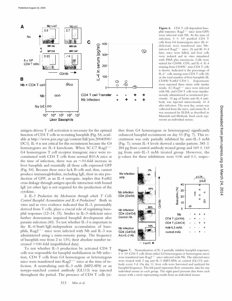
CD4 T Cells Are Required for the Basophil Recruitment tothe Liver, but Basophil GFP Expression Is B Cell Independent.To test if the presence of CD4 T cells affects IL-4 produc-tion and/or recruitment of basophils, Rag2��� mice wereinfected with Nb; on the day of infection, some animals re-ceived 5 � 106 purified CD4 T cells. At day 10, liver cellswere harvested and stimulated with PMA and ionomycin.As shown in Fig. 6 A, 1% of non-CD4 CD49b� cellsfrom noninfected Rag2��� mice produced IL-4 upon stim-ulation. Nb infection increased the frequency of IL-4–pro-ducing cells to 2.7% and there was an �10-fold increase inthe absolute number of liver basophils, measured by ex-pression of both CD49b and Fc�RI among non-CD4 Tcells (Fig. 6 B). Transfer of purified CD4 T cells intoRag2��� mice infected with Nb increased the proportionof IL-4–producing non-CD4 CD49b� cells to 7–8%, andthe total number of basophils increased �10-fold comparedwith infected nonreconstituted mice (Fig. 6 B). Further-more, there was an increase in the mean fluorescenceintensity of IL-4 staining, after stimulation, in the T cell–reconstituted infected group compared with the nonrecon-stituted infected group, indicating that CD4 T cells bothrecruit basophils to the tissues and increase IL-4–producingcapacity on a per cell level in infected mice. To testwhether T cell reconstitution also increased in situ IL-4production, Rag2��� mice were reconstituted with CD4 Tcells from G4 homozygous donors; some were infectedwith Nb. On day 11, serum IL-4 levels were measured withthe in vivo cytokine capture assay. Reconstitution withT cells increased IL-4 production in Nb-infected animals�10-fold (Fig. 6 C). Because the only source of IL-4 inthese mice is non–T cells (the transferred T cells are IL-4deficient) and basophils are the dominant non–T cells pro-ducing GFP in G4 mice without restimulation, this resultstrongly argues that these cells are also producing IL-4 insitu in response to Nb infection.
Basophil accumulation in the tissues was not observedafter Nb infection of 5C.C7 Rag2��� G4 homozygous Tcell receptor transgenic mice, which express a TCR spe-cific for cytochrome c peptide presented on I-Ek class IImolecule. Their failure to support the mobilization of ba-sophils in response to Nb infection suggests it is not simplyT cell number that is important, but the capacity of those Tcells to recognize Nb-associated antigens, indicating that
Figure 4. Basophils are major IL-4 producers. (A) Liver cells from Nb-infected G4 heterozygous and homozygous mice were stimulated in vitrowith PMA and ionomycin for 4 h. Cells were stained for intracellular IL-4.The FACS® profile for GFP and IL-4 expression from non-CD4 T cells isshown. (B) Liver cells from Nb-infected BALB/c (non-GFP) mice werestimulated in vitro as mentioned previously. Cells were stained for Fc�RI,CD49b, IL-4, and CD4. The IL-4 staining from each gated population ofnon-CD4 T cells is shown. Percentages of IL-4 positive cells are indi-cated. Experiments were repeated three times with similar results.
Figure 5. Basophil GFP induc-tion is independent of IL-4 andStat6. Liver cells from Nb-infectedG4 homozygous (IL-4 deficient)Stat6��� or littermates Stat6���
mice were stained for CD44 andCD4. The GFP profile fromnon-CD4 T cells is shown. Ex-periments were repeated threetimes with similar results.
on March 24, 2014
jem.rupress.org
Dow
nloaded from
Published August 16, 2004
Min et al.513
antigen-driven T cell activation is necessary for the optimalfunction of CD4 T cells in recruiting basophils (Fig. S4, avail-able at http://www.jem.org/cgi/content/full/jem.20040590/DC1). IL-4 is not critical for this recruitment because the G4homozygotes are IL-4 knockouts. When 5C.C7 Rag2���
G4 homozygous T cell receptor transgenic mice were re-constituted with CD4 T cells from normal B10.A mice atthe time of infection, there was an �10-fold increase inliver basophils and essentially all those cells expressed GFP(Fig. S4). Because these mice lack B cells and, thus, cannotproduce immunoglobulins, including IgE, their in situ pro-duction of GFP, as an IL-4 surrogate, implies that Fc�RIcross-linkage through antigen-specific interaction with boundIgE (or other Igs) is not required for the production of thecytokine.
Is IL-3 Production the Mechanism through which T CellsControl Basophil Accumulation and IL-4 Production? Both invitro and in vivo evidence indicated that IL-3, presumablyderived from T cells, plays a crucial role of regulating baso-phil responses (12–14, 33). Studies in IL-3–deficient micefurther demonstrate impaired basophil development afterparasite infection (40). To test whether IL-3 is important inthe IL-4/Stat6/IgE-independent accumulation of baso-phils, Rag2��� mice were infected with Nb and IL-3 wasadministered using a mini-osmotic pump. The frequencyof basophils rose from 2 to 13%; their absolute number in-creased �100-fold (unpublished data).
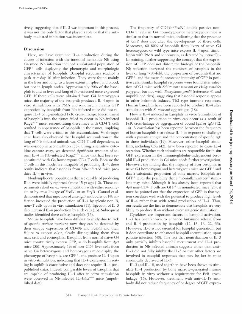
To test whether IL-3 production by activated CD4 Tcells was responsible for basophil mobilization in Nb infec-tion, CD4 T cells from G4 homozygous or heterozygousmice were transferred into Rag2��� mice at the time of in-fection. A neutralizing anti–IL-3 mAb (MP2-8F8) or anisotype-matched control antibody (GL113) was injectedthroughout the period. The presence of CD4 T cells (ei-
ther from G4 homozygous or heterozygous) significantlyenhanced basophil recruitment on day 10 (Fig. 7). This re-cruitment was only partially inhibited by anti–IL-3 mAb(Fig. 7); serum IL-4 levels showed a similar pattern: 385 �284 pg from control antibody treated group and 169 � 143pg from anti–IL-3 mAb treated group. However, thep-values for these inhibitions were 0.06 and 0.1, respec-
Figure 6. CD4 T cell–dependent baso-phil responses. Rag2��� mice (non-GFP)were infected with Nb. At the time ofinfection, 5 � 106 purified CD4 Tcells from G4 homozygous mice (IL-4–deficient) were transferred into Nb-infected Rag2��� mice. (A and B) 10 dlater, mice were killed, and liver cellswere isolated and in vitro stimulatedwith PMA plus ionomycin. Cells werestained for CD49b, CD4, and IL-4. IL-4staining from CD49b� non-CD4 T cellsis shown. Indicated is the percentage ofIL-4� cells among non-CD4 T cells (A)or the total number of liver basophils (B,CD49b�Fc�RI�CD4�). Experimentswere repeated three times with similarresults. (C) Rag2��� mice were infectedwith Nb, and CD4 T cells were simulta-neously administered as mentioned pre-viously. 10 g of biotin anti–IL-4 anti-body was injected intravenously 10 dafter infection. The next day, serum wascollected from the mice, and serum IL-4was measured by ELISA as described inMaterials and Methods. Each circle rep-resents an individual mouse.
Figure 7. Neutralization of IL-3 partially inhibits basophil responses.5 � 106 CD4 T cells (from either G4 heterozygous or homozygous mice)were transferred into Rag2��� mice infected with Nb. The infected micewere treated with 2 mg anti–IL-3 (MP2-8F8) or control (GL113) anti-body every 3 d. On day 11, liver cells were harvested and analyzed forbasophil frequency. The left panel represents flow cytometric data for oneindividual mouse in each group. The right panel presents data from eachmouse with a circle representing results from an individual mouse.
on March 24, 2014
jem.rupress.org
Dow
nloaded from
Published August 16, 2004
Basophil IL-4 Production in Parasite Infection514
tively, suggesting that if IL-3 was important in this process,it was not the only factor that played a role or that the anti-body-mediated inhibition was incomplete.
DiscussionHere, we have examined IL-4 production during the
course of infection with the intestinal nematode Nb usingG4 mice. Nb infection induced a substantial population ofGFP� cells displaying the phenotypic and morphologiccharacteristics of basophils. Basophil responses reached apeak at �day 10 after infection. They were found mainlyin the liver and lung, to a lesser extent in spleen and blood,but not in lymph nodes. Approximately 90% of the baso-phils found in liver and lung of Nb-infected mice expressedGFP. If these cells were obtained from G4 heterozygousmice, the majority of the basophils produced IL-4 upon invitro stimulation with PMA and ionomycin. In situ GFPexpression by basophils from Nb-infected mice did not re-quire IL-4 or Ig-mediated FcR cross-linkage. Recruitmentof basophils into the tissues failed to occur in Nb-infectedRag2��� mice; reconstituting these mice with CD4 T cellsresulted in appearance of basophils in the tissues, implyingthat T cells were critical to this accumulation. Voehringeret al. have also observed that basophil accumulation in thelung of Nb-infected animals was CD4 T cell dependent, aswas eosinophil accumulation (35). Using a sensitive cyto-kine capture assay, we detected significant amounts of se-rum IL-4 in Nb-infected Rag2��� mice that had been re-constituted with G4 homozygous CD4 T cells. Because theT cells in this model are incapable of producing IL-4, theseresults indicate that basophils from Nb-infected mice pro-duce IL-4 in vivo.
Nonlymphocyte populations that are capable of producingIL-4 were initially reported almost 15 yr ago (12). These ex-periments relied on ex vivo stimulation with either ionomy-cin or by cross-linkage of Fc�RI or an Fc�R. Conrad et al.demonstrated that injection of anti-IgD antibodies or Nb in-fection increased the production of IL-4 by splenic non–B,non–T cells upon in vitro stimulation (11). Injection of IL-3also increased IL-4 production by such cells (13). Subsequentstudies identified these cells as basophils (15).
Mouse basophils have been difficult to study due to lackof specific surface markers; now they can be detected bytheir unique expression of CD49b and Fc�RI and theirfailure to express c-kit, clearly distinguishing them frommast cells and eosinophils. Basophils from normal naive G4mice constitutively express GFP, as do basophils from 4getmice (35). Approximately 1% of non-CD4 liver cells fromnaive G4 heterozygous and homozygous mice display thephenotype of basophils, are GFP�, and produce IL-4 uponin vitro stimulation, indicating that IL-4 expression in rest-ing basophils is constitutive and does not require IL-4 (un-published data). Indeed, comparable levels of basophils thatare capable of producing IL-4 after in vitro stimulationwere observed in Nb-infected IL-4R���� mice (unpub-lished data).
The frequency of CD49b/Fc�RI double positive non-CD4 T cells in G4 homozygous or heterozygous mice issimilar to that in normal mice, indicating that the presenceof GFP does not alter the development of these cells.Moreover, 60–80% of basophils from livers of naive G4heterozygotes or wild-type mice express IL-4 upon stimu-lation with PMA and ionomycin, as detected by intracellu-lar staining, further supporting the concept that the expres-sion of GFP does not distort the biology of the basophils.Nb infection increased the numbers of basophils in theliver or lung �50-fold, the proportion of basophils that areGFP�, and the mean fluorescence intensity of GFP in posi-tive cells. Similar basophil responses were found after infec-tion of G4 mice with Schistosoma mansoni or Heligosomoidespolygyrus, but not with Toxoplasma gondii (reference 41 andunpublished data), suggesting that basophil response appearin other helminth induced Th2 type immune responses.Human basophils have been reported to produce IL-4 afterstimulation with S. mansoni egg antigen (18).
How is IL-4 induced in basophils in vivo? Stimulation ofbasophil IL-4 production in vitro can occur as a result ofFcR cross-linkage by aggregation of bound IgE or IgG (12,14). A correlation has been reported between the frequencyof human basophils that release IL-4 in response to challengewith a parasite antigen and the concentration of specific IgEin those individuals (19). However, other basophil stimu-lants, including C5a (42), have been reported to cause IL-4secretion. Whether such stimulants are responsible for in situGFP expression in the immunoglobulin-independent baso-phil IL-4 production in G4 mice needs further investigation.However, the finding that the majority of liver basophils innaive G4 homozygous and heterozygous mice are GFP� andthat a substantial proportion of bone marrow basophils areGFP� raises the possibility that a “noninflammatory” stimu-lant may exist. Although it has already been reported that4get non-CD4 T cells are GFP� in noninfected mice (23), itmust be pointed out that the expression of GFP in that sys-tem correlates well with the potential to produce high levelsof IL-4 rather than with actual production of IL-4. Thus,our results are the first to demonstrate that basophils are verylikely to produce IL-4 without overt antigenic stimulation.
Cytokines are important factors in basophil activation.IL-3 has been shown to enhance histamine release fromand IL-4 production by basophils (12, 13, 16, 22, 33).However, IL-3 is not essential for basophil generation, butit does contribute to enhanced basophil accumulation uponparasite infection (40). The fact that neutralization of IL-3only partially inhibits basophil recruitment and IL-4 pro-duction in Nb-infected animals suggests either than anti–IL-3 did not fully inhibit the IL-3 or that other factors areinvolved in basophil responses that may be lost in micechronically deprived of IL-3.
IL-3 and IL-18, used together, have been shown to stim-ulate IL-4 production by bone marrow–generated murinebasophils in vitro without a requirement for FcR cross-linkage (16). However, treatment with anti–IL-18 anti-body did not reduce frequency of or degree of GFP expres-
on March 24, 2014
jem.rupress.org
Dow
nloaded from
Published August 16, 2004

Min et al.515
sion in basophils from Nb-infected mice (unpublisheddata). Similarly, it has been observed that caspase-1 knock-out mice, which are severely deficient in IL-18, normallyrecruit basophils when Nb infected (unpublished data). Itmay very well be that cytokines other than IL-18 play arole in basophil IL-4 responses in response to Nb infection.Alternatively, those Nb-associated molecules that act asTh2-driving adjuvants may directly activate and trigger ba-sophil responses (43, 44). We are currently investigatingwhether such mechanisms could induce basophil responses.
What is particularly striking is that CD4 T cells are nec-essary for optimal migration of, and IL-4 production by,basophils. The fact that Nb-infected 5C.C7 TCR trans-genic CD4 T cells failed to induce basophil responsesstrongly suggests that T cell activation by Nb antigen is re-quired. How the help from CD4 T cells is linked to baso-phil recruitment is not yet clear at this moment. IL-3 pro-duced by T cells may enhance basophil production/responses (33), but it is not absolutely required for IL-4production (unpublished data). How, then, could T cellsinduce basophil responses? CD4 T cells responsive to para-site antigens could produce other cytokines or chemokinesthat recruit and/or activate basophils (43, 45).
What is the role of IL-4 made by basophils? BasophilIL-4, or alternatively IL-13, could have important effectorfunctions. Particularly in the lung where Th2 responses areassociated with airway hyperresponsiveness, endothelial meta-plasia, and mucus production, which are mediated largelyby IL-13, the rich source of this cytokine provided bybasophils could be of particular pathogenic significance.
Because tissue basophils in Nb-infected mice are GFP�
without overt stimulation and these cells contain IL-4mRNA, they appear to provide a source of IL-4 evenwithout antigen-driven stimulation. However, upon stim-ulation, they very likely increase their production. Indeed,basophils may produce more total IL-4 in the tissues in re-sponse to antigenic challenge than any other cell type, in-cluding CD4 T cells (19, 32, 46).
Although in Nb infection, the mobilization of IL-4–pro-ducing basophils may well be too slow for them to act suffi-ciently early in the response to bias the first cohort of re-sponding CD4 T cells in the Th2 direction, they may havea very important role in propagating Th2 responses by pro-viding a source of IL-4 to prime newly emerging or newlyrecruited naive CD4 T cells to become IL-4 producers (46).
Earlier studies have shown basophil and eosinophil mi-gration to sites of inflammation in tick infection and thatdepleting basophils diminished eosinophil recruitment, sug-gesting that secretion of IL-4 or other type 2 cytokines atthese sites may be important as immune modulators (47).Other examples of basophil responses include increasedblood basophil levels in Nb-infected rat (48), basophils inskin reactions after infestation with Dermacentor varibilis(49), and, more recently, basophil IL-4 production in amouse model of allergic pulmonary inflammation (50).
In vitro–cultured basophils have been reported to induceIgE synthesis by B cells (51, 52). Thus, basophils might
promote and/or amplify antibody responses associated withparasite infection, but the degree to which basophils can actas class-specific helper cells for IgE production in vivo hasstill to be determined.
In summary, our results using G4 mice reveal evidencethat murine basophils are the primary, if not sole, IL-4–producing nonlymphocytes during parasitic infection. Con-stitutive production of IL-4 by these cells and their tissuelocalization patterns implies that they play important rolesin the development of adaptive immune responses and/orin host effector function.
The authors thank S. Tanksley, T. Moyer, and C. Henry for theirexcellent cell sorting; R. Monahan-Earley for helping to prepareEM samples; Dr. T. Yoshimoto for helpful discussions, particularlyabout the possible role of IL-18; Dr. A. Sher for critical review ofthe manuscript, Dr. S. Galli for sharing unpublished data; andmembers of the Paul laboratory for helpful discussion.
M. Prout and G. LeGros are supported by the Health ResearchCouncil of New Zealand. The ultrastructural studies were supportedby National Institutes of Health grant AI33372 (to A.M. Dvorak).
The authors have no conflicting financial interests.
Submitted: 26 March 2004Accepted: 8 July 2004
References1. Seder, R.A., and W.E. Paul. 1994. Acquisition of lympho-
kine-producing phenotype by CD4� T cells. Annu. Rev. Im-munol. 12:635–673.
2. Finkelman, F.D., J. Holmes, I.M. Katona, J.F. Urban Jr.,M.P. Beckmann, L.S. Park, K.A. Schooley, R.L. Coffman,T.R. Mosmann, and W.E. Paul. 1990. Lymphokine controlof in vivo immunoglobulin isotype selection. Annu. Rev. Im-munol. 8:303–333.
3. Finkelman, F.D., T. Shea-Donohue, J. Goldhill, C.A. Sulli-van, S.C. Morris, K.B. Madden, W.C. Gause, and J.F. UrbanJr. 1997. Cytokine regulation of host defense against parasiticgastrointestinal nematodes: lessens from studies with rodentmodels. Annu. Rev. Immunol. 15:505–533.
4. Naramura, M., R.J. Hu, and H. Gu. 1998. Mice with a fluo-rescent marker for interleukin 2 gene activation. Immunity. 9:209–216.
5. Hu-li, J., C. Pannetier, L. Guo, M. Lohning, H. Gu, C.Watson, M. Assenmacher, A. Radbruch, and W.E. Paul.2001. Regulation of expression of IL-4 alleles: analysis usinga chimeric GFP/IL-4 gene. Immunity. 14:1–11.
6. Mohrs, M., K. Shinkai, K. Mohrs, and R.M. Locksley. 2001.Analysis of type 2 immunity in vivo with a bicistronic IL-4reporter. Immunity. 15:303–311.
7. Stetson, D.B., M. Mohrs, R. Lee Reinhardt, J.L. Baron, Z.Wang, L. Gapin, M. Kronenberg, and R.M. Locksley. 2003.Constitutive cytokine mRNAs mark natural killer (NK) andNK T cells poised for rapid effector function. J. Exp. Med.198:1069–1076.
8. Gause, W.C., J.F. Urban Jr., and M.J. Stadecker. 2003. Theimmune response to parasitic helminths: insights from mu-rine models. Trends Immunol. 24:269–277.
9. McKenzie, G.J., C.L. Emson, S.E. Bell, S. Anderson, P. Fal-lon, G. Zurawski, R. Murrays, R. Grencis, and A.N.J. Mc-Kenzie. 1998. Impaired development of Th2 cells in IL-13-
on March 24, 2014
jem.rupress.org
Dow
nloaded from
Published August 16, 2004

Basophil IL-4 Production in Parasite Infection516
deficient mice. Immunity. 9:423–432.10. Urban, J.F., Jr., N. Noben-Trauth, D.D. Donaldson, K.B.
Madden, S.C. Morris, M. Collins, and F.D. Finkelman.1998. IL-13, IL-4R�, and Stat6 are required for the expul-sion of the gastrointestinal nematode parasite Nippostrongylusbrasiliensis. Immunity. 8:255–264.
11. Conrad, D.H., S.Z. Ben-Sasson, G. LeGros, F.D. Finkelman,and W.E. Paul. 1990. Infection with Nippostrongylus brasilien-sis or injection of anti-IgD antibodies markedly enhances Fc-receptor–mediated interleukin 4 production by non–B,non–T cells. J. Exp. Med. 171:1497–1508.
12. Ben-Sasson, S.Z., G. LeGros, D.H. Conrad, F.D. Finkelman,and W.E. Paul. 1990. Cross-linking Fc receptors stimulatesplenic non-B, non-T cells to secrete interleukin 4 and otherlymphokines. Proc. Natl. Acad. Sci. USA. 87:1421–1425.
13. LeGros, G., S.Z. Ben-Sasson, D.H. Conrad, I. Clark-Lewis,F.D. Finkelman, M. Plaut, and W.E. Paul. 1990. IL-3 promotesproduction of IL-4 by splenic non-B, non-T cells in response toFc receptor cross-linkage. J. Immunol. 145:2500–2506.
14. Seder, R.A., M. Plaut, S. Barbieri, J. Urban Jr., F.D. Finkel-man, and W.E. Paul. 1991. Purified Fc�R� bone marrowand splenic non-B, non-T cells are highly enriched in the ca-pacity to produce IL-4 in response to immobilized IgE,IgG2a, or ionomycin. J. Immunol. 147:903–909.
15. Seder, R.A., W.E. Paul, A.M. Dvorak, S.J. Sharkis, A.Kagey-Sobotka, Y. Niv, F.D. Finkelman, S.A. Barbieri, S.J.Galli, and M. Plaut. 1991. Mouse splenic and bone marrowcell populations that express high-affinity Fc� receptors andproduce interleukin 4 are highly enriched in basophils. Proc.Natl. Acad. Sci. USA. 88:2835–2839.
16. Yoshimoto, T., H. Tsutsui, K. Tominaga, K. Hoshino, H.Okamura, S. Akira, W.E. Paul, and K. Nakanishi. 1999. IL-18, although antiallergic when administered with IL-12,stimulates IL-4 and histamine release by basophils. Proc. Natl.Acad. Sci. USA. 96:13962–13966.
17. Paul, W.E. 1991. Interleukin-4 production by Fc epsilonR� cells. Skin Pharmacol. 4:8–14.
18. Falcone, F.H., C.A. Dahinden, B.F. Gibbs, T. Noll, U.Amon, H. Hebestreit, O. Abrahamsen, J. Klaucke, M.Schlaak, and H. Haas. 1996. Human basophils release inter-leukin-4 after stimulation with Schistosoma mansoni egg anti-gen. Eur. J. Immunol. 26:1147–1155.
19. Mitre, E., R.T. Taylor, J. Kunofcik, and T.B. Nutman.2004. Parasite antigen-driven basophils are a major source ofIL-4 in human filarial infections. J. Immunol. 172:2439–2445.
20. Philips, Clair, W.R. Coward, D.I. Pritchard, and C.R.A.Hewitt. 2003. Basophils express a type 2 cytokine profile onexposure to protease from helminthes and house dust mites.J. Leukoc. Biol. 73:165-171.
21. Dahinden, C.A., S. Rihs, and B. Ochsensberger. 1997. Reg-ulation of cytokine expression by human blood basophils. Int.Arch. Allergy Immunol. 113:134–137.
22. MacGlashan, D., Jr., J.M. White, S.-K. Huang, S.J. Ono,J.T. Schroeder, and L.M. Lichtenstein. 1994. Secretion ofIL-4 from human basophils. The relationship between IL-4mRNA and protein in resting and stimulated basophils. J. Im-munol. 152:3006–3016.
23. Shinkai, K., M. Mohrs, and R.M. Locksley. 2002. Helper Tcells regulate type-2 innate immunity in vivo. Nature. 420:825–829.
24. Yoshimoto, T., and W.E. Paul. 1994. CD4pos, NK1.1pos Tcells promptly produce interleukin 4 in response to in vivochallenge with anti-CD3. J. Exp. Med. 179:1285–1295.
25. Matsuda, J.L., L. Gapin, J.L. Baron, S. Sidobre, D.B. Stetson,M. Mohrs, R.M. Locksley, and M. Kronenberg. 2003. MouseV�14i natural killer T cells are resistant to cytokine polariza-tion in vivo. Proc. Natl. Acad. Sci. USA. 100:8395–8400.
26. Abrams, J.S., and M.K. Pearce. 1988. Development of ratanti-mouse interleukin 3 monoclonal antibodies which neu-tralize bioactivity in vitro. J. Immunol. 140:131–137.
27. Matsui, K., T. Yoshimoto, H. Tsutsui, Y. Hyodo, N. Ha-yashi, K. Hiroishi, N. Kawada, H. Okamura, K. Nakanishi,and K. Higashino. 1997. Propionibacterium acnes treatmentdiminishes CD4�NK1.1� T cells but induces type I T cellsin the liver by induction of IL-12 and IL-18 production fromKupffer cells. J. Immunol. 159:97–106.
28. Guo, L., J. Hu-Li, J. Zhu, C.J. Watson, M.J. Difilippantonio,C. Pannetier, and W.E. Paul. 2002. In Th2 cells the Il4 genehas a series of accessibility states associated with distinctiveprobabilities of IL-4 production. Proc. Natl. Acad. Sci. USA.99:10623–10628.
29. Finkelman, F.D., and S.C. Morris. 1999. Development of anassay to measure in vivo cytokine production in the mouse.Int. Immunol. 11:1811–1818.
30. Guhaniyogi, J., and G. Brewer. 2001. Regulation of mRNAstability in mammalian cells. Gene. 265:11–23.
31. Noben-Trauth, N., L.D. Shultz, F. Brombacher, J.F. UrbanJr., H. Gu, and W.E. Paul. 1997. An interleukin 4 (IL-4)-independent pathway for CD4� T cell IL-4 production is re-vealed in IL-4 receptor-deficient mice. Proc. Natl. Acad. Sci.USA. 94:10838–10843.
32. Aoki, I., C. Kinzer, A. Shirai, W.E. Paul, and D.M. Klin-man. 1995. IgE receptor-positive non-B/non-T cells domi-nate the production of interleukin 4 and interleukin 6 in im-munized mice. Proc. Natl. Acad. Sci. USA. 92:2534–2538.
33. Kullberg, M.C., J.A. Berzofsky, D. Jankovic, S. Barbieri,M.E. Williams, P. Perlmann, A. Sher, and M. Troye-Blomberg. 1996. T cell-derived IL-3 induces the productionof IL-4 by non-B, non-T cells to amplify the Th2-cytokineresponse to a non-parasite antigen in Schistosoma mansoni-infected mice. J. Immunol. 156:1482–1489.
34. Arase, H., T. Saito, J.H. Phillips, and L.L. Lanier. 2001. Cut-ting edge: the mouse NK cell-associated antigen recognizedby DX5 monoclonal antibody is CD49b (alpha 2 integrin,very late antigen-s). J. Immunol. 167:1141–1144.
35. Voehringer, D., K. Shinkai, and R. Locksley. 2004. Type 2immunity reflects orchestrated recruitment of cells commit-ted to IL-4 production. Immunity. 20:267–277.
36. Dvorak, A.M., G. Nabel, K. Pyne, H. Cantor, H.F. Dvorak,and S.J. Galli. 1982. Ultrastructural identification of themouse basophil. Blood. 59:1279–1285.
37. Samson, S.I., O. Richard, M. Tavian, T. Ranson, C.A.J.Vosshenrich, F. Colucci, J. Buer, F. Grosveld, I. Godin, andJ.P. Di Santo. 2003. Gata-3 promotes maturation, IFN-�production, and liver-specific homing of NK cells. Immunity.19:701–711.
38. Lantz, C.S., M. Yamaguchi, H.C. Oettgen, I.M. Katona, I.Miyajima, J.-P. Kinet, and S.J. Galli. 1997. IgE regulatesmouse basophil Fc�RI expression in vivo. J. Immunol. 158:2517–2521.
39. Finkelman, F.D., S.C. Morris, T. Orekhova, M. Mori, D.Donaldson, S.L. Reiner, N.L. Reilly, L. Schopf, and J.F. Ur-ban Jr. 2000. Stat6 regulation of in vivo IL-4 responses. J. Im-munol. 164:2303–2310.
40. Lantz, C.S., J. Boesiger, C.H. Song, N. Mach, T. Kobayashi,R.C. Mulligans, Y. Nawa, G. Dranoff, and S.J. Galli. 1998.
on March 24, 2014
jem.rupress.org
Dow
nloaded from
Published August 16, 2004

Min et al.517
Role for interleukin-3 in mast-cell and basophil developmentand in immunity to parasite. Nature. 392:90–93.
41. Williams, M.E., M.C. Kullberg, S. Barbieri, P. Caspar, J.A.Berzofsky, R.A. Seder, and A. Sher. 1993. Fc epsilon recep-tor-positive cells are a major source of antigen-induced inter-leukin-4 in spleens of mice infected with Schistosoma mansoni.Eur. J. Immunol. 23:1910-1916.
42. Ochensberger, B., S. Rihs, T. Brunner, and C.A. Dahinden.1995. IgE-independent interleukin-4 expression and induc-tion of a late phase of leukotriene C4 formation in humanblood basophils. Blood. 86:4039–4049.
43. Maizels, R.M., and M. Yazdanbakhsh. 2003. Immune regu-lation by helminth parasites: cellular and molecular mecha-nisms. Nat. Rev. Immunol. 3:733–744.
44. Holland, M.J., Y.M. Harcus, P.L. Riches, and R.M. Maizels.2000. Proteins secreted by the parasitic nematode Nippo-strongylus brasiliensis act as adjuvant for Th2 responses. Eur. J.Immunol. 30:1977–1987.
45. Devouassoux, G., D.D. Metcalfe, and C. Prussin. 1999. Eo-taxin potentiates antigen-dependent basophil IL-4 produc-tion. J. Immunol. 163:2877–2882.
46. Poorafshar, M., H. Helmby, M. Troye-Blomberg, and L.Hellman. 2000. MMCP-8, the first lineage-specific differen-tiation marker for mouse basophils. Elevated numbers of po-tent IL-4-producing and MMCP-8-positive cells in spleensof malaria-infected mice. Eur. J. Immunol. 30:2660–2668.
47. Brown, S.J., S.J. Galli, G.J. Gleich, and P.W. Askenase. 1982.Ablation of immunity to Amblyomma americanum by anti-basophil serum: cooperation between basophils and eosino-phils in expression of immunity to ectoparasites (ticks) in guineapigs. J. Immunol. 129:790–796.
48. Ogilvie, B.M., P.W. Askenase, and M. Elaine Rose. 1979. Ba-sophils and eosinophils in three strains of rats and in athymic(nude) rats following infection with the nematodes Nippostrongy-lus brasiliensis or Trichinella spiralis. Immunology. 39:385–389.
49. Steeves, E.B.T., and J.R. Allen. 1990. Basophils in skin reac-tions of mast cell-deficient mice infested with Dermacentorvariabilis. Int. J. Parasitol. 20:655–667.
50. Luccioli, S., D.T. Brody, S. Hasan, A. Keane-Myers, C.Prussin, and D.D. Metcalfe. 2002. IgE�, Kit-, I-A/I-E- my-eloid cells are the initial source of IL-4 after antigen challengein a mouse model of allergic pulmonary inflammation. J. Al-lergy Clin. Immunol. 110:117–124.
51. Gauchat, J.-F., S. Henchoz, G. Mazzei, J.-P. Aubry, T.Brunner, H. Blasey, P. Life, D. Talabot, L. Flores-Romo, J.Thompson, et al. 1993. Induction of human IgE synthesis inB cells by mast cells and basophils. Nature. 365:340–343.
52. Yanagihara, Y., K. Kajiwara, Y. Basaki, K. Ikizawa, M. Ebi-sawa, C. Ra, H. Tachimoto, and H. Saito. 1998. Culturedbasophils but not cultured mast cells induce human IgE syn-thesis in B cells after immunologic stimulation. Clin. Exp. Im-munol. 111:136–143.
on March 24, 2014
jem.rupress.org
Dow
nloaded from
Published August 16, 2004