IL4 Utilizes an Alternative Receptor to Drive Apoptosis of Th1 Cells and Skews Neonatal Immunity...
12
Immunity, Vol. 20, 429–440, April, 2004, Copyright 2004 by Cell Press IL-4 Utilizes an Alternative Receptor to Drive Apoptosis of Th1 Cells and Skews Neonatal Immunity toward Th2 munity (Siegrist, 2001). Thus, understanding how neona- tal exposure to antigen skews Th1 and favors Th2 cells remains the key feature for progress toward the develop- ment of effective pediatric vaccines. The lack of an adaptable model for investigation of primary neonatal Lequn Li, 1,4,5 Hyun-Hee Lee, 1,4 J. Jeremiah Bell, 1 Randal K. Gregg, 1 Jason S. Ellis, 1 Andre Gessner, 2 and Habib Zaghouani 1,3 1 Department of Molecular Microbiology and Immunology University of Missouri School of Medicine responses has been a major limitation in neonatal immu- nity. Transfer of TCR transgenic (Tg) T cells into animals M616 Medical Sciences Building Columbia, Missouri 65212 allowed for proper homing to lymphoid organs and facili- tated analysis of T cell responses in vivo (Kearney et 2 Institut fu ¨ r Klinische Mikrobiologie Immunologie und Hygiene al., 1994). Recently, we have used the ovalbumin (OVA) 323-339-specific TCR transgenic DO11.10 T cells and Wasserturmstrasse 3 91054 Erlangen developed a T cell transfer system suitable for tracking neonatal T cells and analyzing their responses upon Germany exposure to antigen (Li et al., 2001). Accordingly, trans- fer of neonatal (1 day old) DO11.10 T cells into 1 day old Balb/c mice sustains homing of the DO11.10 T cells Summary to the hosts’ lymphoid organs. Moreover, priming of the Balb/c hosts with Ig-OVA, an Ig chimera genetically Primary neonatal Th1 cells develop alongside of Th2 engineered to express the OVA peptide, circumvents upon priming of the newborn but undergo apoptosis the use of adjuvant and promotes a secondary response upon recall with antigen. These Th1 cells were iso- skewed toward Th2 upon challenge with OVA peptide lated, and their death was correlated with elevated IL- at an adult age (Li et al., 2001). Herein, the neonatal 13R1 chain expression. Strikingly, neutralization of DO11.10 TCR Tg T cell transfer system was used to Th2s IL-4 reduced apoptosis, sustained recall re- investigate the primary neonatal response to determine sponses, and the live Th1 cells displayed a decrease whether the Th2 bias was due to a lack of development in IL-13R1 expression. Blockade of IL-13R1 or IL- of primary Th1 cells or to a defective recall of neonatally 4R also restores recall and secondary Th1 re- primed Th1 cells. The results indicate that primary Th1 sponses. Adult T cells primed within the neonatal envi- cells do develop alongside of Th2 counterparts upon ronment did not upregulate IL-13R1 chain or undergo neonatal priming. However, recall with antigen drives the apoptosis and developed recall Th1 responses. These Th2 cells to respond but the Th1 lymphocytes undergo observations indicate that developmental expression apoptosis. Moreover, when the Th2 cytokines are neu- of IL-13R1 along with IL-4R provides a receptor tralized either during neonatal priming or recall with anti- through which IL-4 induces death of Th1 cells and gen, Th1 responses develop alongside of Th2. Interest- skews neonatal immunity toward Th2. ingly, adult T cells primed within the neonatal environment give rise to primary Th1 and Th2 that are Introduction both able to respond to a recall with antigen. Finally, gene expression analysis indicated a developmental Neonatal exposure to antigen usually leads to tolerance upregulation of IL-13R1 chain in neonatal Th1 that cor- (Billingham et al., 1956; Gammon et al., 1986; Garza related with an inability of the cells to balance pro and et al., 1997). Initially, T cell deletion/inactivation was antiapoptotic factors leading to death of the Th1 subset. considered the leading mechanism for such tolerance. However, blockade of IL-13R1 or IL-4R during either Lately, however, it has been shown that immunity devel- priming or rechallenge with antigen prevents apoptosis ops in the newborn (Ridge et al., 1996; Sarzotti et al., and restores Th1 responses. 1996), but in most cases the responses manifest an indubitable bias toward Th2 (Forsthuber et al., 1996; Results Chen and Field, 1995; Singh et al., 1996). Furthermore, when immunoglobulins (Igs) were used for peptide deliv- Exposure of Neonatal T Cells to Antigen Induces ery to enhance presentation and provide adjuvanticity, Cell Activation and Division biased Th2 immunity remained dominant (Min et al., Figure 1 shows that neonatal DO11.10 T cells transferred 1998; Pack et al., 2001). Although this outcome could into newborn Balb/c mice are activated and undergo be beneficial for transplantation and suppression of Th1- cell division upon exposure to antigen. Indeed, ex vivo mediated autoimmunity, it may place the neonate at risk cell surface staining analysis indicated that only 9% of for microbial infections, which require Th1 cells. In fact, DO11.10 T cells stained positive for CD25 expression a great deal of interest has been devoted lately toward in animals that were not given the antigen, as opposed defining regimens that could promote neonatal Th1 im- to those injected with OVA peptide or Ig-OVA where upregulation of CD25 was seen in 35% and 28% of 3 Correspondence: [email protected] their splenic DO11.10 T cells, respectively (Figure 1A). 4 These authors contributed equally to this work. Moreover, both CD62L and CD45RB (Figures 1B and 5 Present address: Dana Farber Cancer Institute, 44 Binney Street, Mayer Building 547, Boston, Massachusetts 02115. 1C) were downregulated, indicating that contact with
-
Upload
independent -
Category
Documents
-
view
3 -
download
0
Transcript of IL4 Utilizes an Alternative Receptor to Drive Apoptosis of Th1 Cells and Skews Neonatal Immunity...
Immunity, Vol. 20, 429–440, April, 2004, Copyright 2004 by Cell Press
IL-4 Utilizes an Alternative Receptorto Drive Apoptosis of Th1 Cellsand Skews Neonatal Immunity toward Th2
munity (Siegrist, 2001). Thus, understanding how neona-tal exposure to antigen skews Th1 and favors Th2 cellsremains the key feature for progress toward the develop-ment of effective pediatric vaccines. The lack of anadaptable model for investigation of primary neonatal
Lequn Li,1,4,5 Hyun-Hee Lee,1,4 J. Jeremiah Bell,1
Randal K. Gregg,1 Jason S. Ellis,1 Andre Gessner,2
and Habib Zaghouani1,3
1Department of Molecular Microbiologyand Immunology
University of Missouri School of Medicine responses has been a major limitation in neonatal immu-nity. Transfer of TCR transgenic (Tg) T cells into animalsM616 Medical Sciences Building
Columbia, Missouri 65212 allowed for proper homing to lymphoid organs and facili-tated analysis of T cell responses in vivo (Kearney et2 Institut fur Klinische Mikrobiologie
Immunologie und Hygiene al., 1994). Recently, we have used the ovalbumin (OVA)323-339-specific TCR transgenic DO11.10 T cells andWasserturmstrasse 3
91054 Erlangen developed a T cell transfer system suitable for trackingneonatal T cells and analyzing their responses uponGermanyexposure to antigen (Li et al., 2001). Accordingly, trans-fer of neonatal (1 day old) DO11.10 T cells into 1 dayold Balb/c mice sustains homing of the DO11.10 T cellsSummaryto the hosts’ lymphoid organs. Moreover, priming ofthe Balb/c hosts with Ig-OVA, an Ig chimera geneticallyPrimary neonatal Th1 cells develop alongside of Th2engineered to express the OVA peptide, circumventsupon priming of the newborn but undergo apoptosisthe use of adjuvant and promotes a secondary responseupon recall with antigen. These Th1 cells were iso-skewed toward Th2 upon challenge with OVA peptidelated, and their death was correlated with elevated IL-at an adult age (Li et al., 2001). Herein, the neonatal13R�1 chain expression. Strikingly, neutralization ofDO11.10 TCR Tg T cell transfer system was used toTh2s� IL-4 reduced apoptosis, sustained recall re-investigate the primary neonatal response to determinesponses, and the live Th1 cells displayed a decreasewhether the Th2 bias was due to a lack of developmentin IL-13R�1 expression. Blockade of IL-13R�1 or IL-of primary Th1 cells or to a defective recall of neonatally4R� also restores recall and secondary Th1 re-primed Th1 cells. The results indicate that primary Th1sponses. Adult T cells primed within the neonatal envi-cells do develop alongside of Th2 counterparts uponronment did not upregulate IL-13R�1 chain or undergoneonatal priming. However, recall with antigen drives theapoptosis and developed recall Th1 responses. TheseTh2 cells to respond but the Th1 lymphocytes undergoobservations indicate that developmental expressionapoptosis. Moreover, when the Th2 cytokines are neu-of IL-13R�1 along with IL-4R� provides a receptortralized either during neonatal priming or recall with anti-through which IL-4 induces death of Th1 cells andgen, Th1 responses develop alongside of Th2. Interest-skews neonatal immunity toward Th2.ingly, adult T cells primed within the neonatalenvironment give rise to primary Th1 and Th2 that areIntroductionboth able to respond to a recall with antigen. Finally,gene expression analysis indicated a developmentalNeonatal exposure to antigen usually leads to toleranceupregulation of IL-13R�1 chain in neonatal Th1 that cor-(Billingham et al., 1956; Gammon et al., 1986; Garzarelated with an inability of the cells to balance pro andet al., 1997). Initially, T cell deletion/inactivation wasantiapoptotic factors leading to death of the Th1 subset.considered the leading mechanism for such tolerance.However, blockade of IL-13R�1 or IL-4R� during eitherLately, however, it has been shown that immunity devel-priming or rechallenge with antigen prevents apoptosisops in the newborn (Ridge et al., 1996; Sarzotti et al.,and restores Th1 responses.1996), but in most cases the responses manifest an
indubitable bias toward Th2 (Forsthuber et al., 1996;ResultsChen and Field, 1995; Singh et al., 1996). Furthermore,
when immunoglobulins (Igs) were used for peptide deliv-Exposure of Neonatal T Cells to Antigen Inducesery to enhance presentation and provide adjuvanticity,Cell Activation and Divisionbiased Th2 immunity remained dominant (Min et al.,Figure 1 shows that neonatal DO11.10 T cells transferred1998; Pack et al., 2001). Although this outcome couldinto newborn Balb/c mice are activated and undergobe beneficial for transplantation and suppression of Th1-cell division upon exposure to antigen. Indeed, ex vivomediated autoimmunity, it may place the neonate at riskcell surface staining analysis indicated that only 9% offor microbial infections, which require Th1 cells. In fact,DO11.10 T cells stained positive for CD25 expressiona great deal of interest has been devoted lately towardin animals that were not given the antigen, as opposeddefining regimens that could promote neonatal Th1 im-to those injected with OVA peptide or Ig-OVA whereupregulation of CD25 was seen in 35% and 28% of3Correspondence: [email protected] splenic DO11.10 T cells, respectively (Figure 1A).4These authors contributed equally to this work.Moreover, both CD62L and CD45RB (Figures 1B and5Present address: Dana Farber Cancer Institute, 44 Binney Street,
Mayer Building 547, Boston, Massachusetts 02115. 1C) were downregulated, indicating that contact with
Immunity430
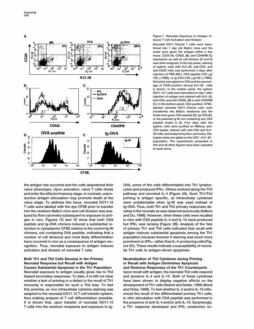
Figure 1. Neonatal Exposure to Antigen In-duces T Cell Activation and Division
Neonatal DO11.10/scid T cells were trans-ferred into 1 day old Balb/c mice and thehosts were given the antigen within a fewhours. CD25 (A), CD62L (B), and CD45RB (C)expression as well as cell division (D and E)were then analyzed. In the top panel, stainingof splenic cells with KJ1-26, anti-CD4, andanti-CD25 mAb was performed 2 days afterinjection of PBS (NIL), OVA peptide (100 �g/100 �l PBS), or Ig-OVA (100 �g/100 �l PBS).Dot plots were gated on CD4 and the percent-age of CD25-positive among KJ1-26� cellsis shown. In the median panel, the splenicDO11.10 T cells were harvested on day 7 afterinjection of antigen and stained with KJ1-26,anti-CD4, and anti-CD62L (B) or anti-CD45RB(C). In the bottom panel, CD4-purified, CFSE-labeled neonatal DO11.10/scid cells weretransferred into Balb/c newborns and thehosts were given OVA peptide (D), Ig-OVA (E),or the parental Ig-W not containing any OVApeptide (insert in E). Four days later thesplenic cells were purified on Miltenyi anti-CD4 beads, stained with anti-CD4 and KJ1-26 mAb, and analyzed by flow cytometry. His-togram plots are gated on the CD4�-KJ1-26�
population. The experiments presented inthis and all other figures have been repeatedat least twice.
the antigen has occurred and the cells abandoned their OVA, some of the cells differentiated into Th1 lympho-cytes and produced IFN�. Others evolved along the Th2naive phenotype. Upon activation, naive T cells divide
and enter the effector/memory stage. In contrast, unpro- pathway and secreted IL-4 (Figure 2A). Such Th1/Th2priming is antigen specific, as intracellular cytokinesductive antigen stimulation may promote death at the
naive stage. To address this issue, neonatal DO11.10 were undetectable when Ig-W was used instead ofIg-OVA. Thus, both Th1 and Th2 primary responses de-T cells were labeled with the dye CFSE prior to transfer
into the newborn Balb/c mice and cell division was ana- velop in the neonate as was observed previously (Adkinsand Du, 1998). However, when these cells were recalledlyzed by flow cytometry subsequent to exposure to anti-
gen in vivo. Figures 1D and 1E show that both OVA in vitro with OVA peptide IL-4 and IL-10 were producedbut IFN� was lacking (Figure 2B). Analysis of the fatepeptide and Ig-OVA chimera induced a substantial re-
duction in cytoplasmic CFSE relative to the control Ig-W of primary Th1 and Th2 cells indicated that recall withantigen induces substantial apoptosis among the Th1chimera, not containing OVA peptide, indicating that a
number of cell divisions and most likely differentiation population because Annexin V staining was much moreprominent on IFN�- rather than IL-4-producing cells (Fig-have occurred in vivo as a consequence of antigen rec-
ognition. Thus, neonatal exposure to antigen induces ure 2C). These results indicate a susceptibility of neona-tal Th1 cells to antigen-driven apoptosis.activation and division of neonatal T cells.
Both Th1 and Th2 Cells Develop in the Primary Neutralization of Th2 Cytokines during Primingor Recall with Antigen Diminishes ApoptosisNeonatal Response but Recall with Antigen
Causes Substantial Apoptosis in the Th1 Population and Restores Responses of the Th1 CounterpartsUpon recall with antigen, the neonatal Th2 cells respondNeonatal exposure to antigen usually gives rise to Th2
biased secondary responses. To date, it is still not clear and produce IL-4 and IL-10. Both of these cytokineshave been shown to display negative effects on thewhether a lack of priming or a defect in the recall of Th1
immunity is responsible for such a Th2 bias. To test development of Th1 cells (Reiner and Seder, 1999; Moreland Oriss, 1998). To test whether IL-4 and/or IL-10 influ-this premise, ex vivo intracellular cytokine staining was
adapted to the neonatal DO11.10 T cell transfer system, enced the recall of the differentiated primary Th1 cells,in vitro stimulation with OVA peptide was performed inthus making analysis of T cell differentiation possible.
It is shown that, upon transfer of neonatal DO11.10 the presence of anti-IL-4 and/or anti-IL-10. Surprisingly,a Th1 response developed and IFN� production oc-T cells into the newborn recipients and exposure to Ig-
The Role of IL-13R�1 in Neonatal Immunity431
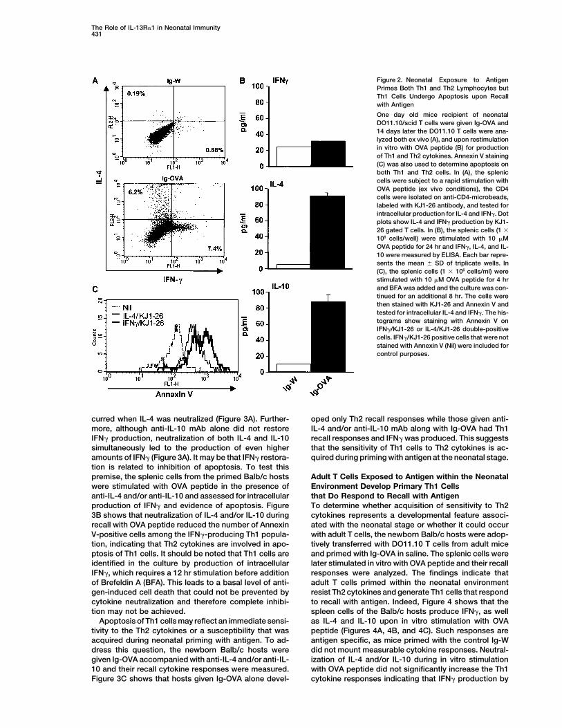
Figure 2. Neonatal Exposure to AntigenPrimes Both Th1 and Th2 Lymphocytes butTh1 Cells Undergo Apoptosis upon Recallwith Antigen
One day old mice recipient of neonatalDO11.10/scid T cells were given Ig-OVA and14 days later the DO11.10 T cells were ana-lyzed both ex vivo (A), and upon restimulationin vitro with OVA peptide (B) for productionof Th1 and Th2 cytokines. Annexin V staining(C) was also used to determine apoptosis onboth Th1 and Th2 cells. In (A), the spleniccells were subject to a rapid stimulation withOVA peptide (ex vivo conditions), the CD4cells were isolated on anti-CD4-microbeads,labeled with KJ1-26 antibody, and tested forintracellular production for IL-4 and IFN�. Dotplots show IL-4 and IFN� production by KJ1-26 gated T cells. In (B), the splenic cells (1 �
106 cells/well) were stimulated with 10 �MOVA peptide for 24 hr and IFN�, IL-4, and IL-10 were measured by ELISA. Each bar repre-sents the mean � SD of triplicate wells. In(C), the splenic cells (1 � 106 cells/ml) werestimulated with 10 �M OVA peptide for 4 hrand BFA was added and the culture was con-tinued for an additional 8 hr. The cells werethen stained with KJ1-26 and Annexin V andtested for intracellular IL-4 and IFN�. The his-tograms show staining with Annexin V onIFN�/KJ1-26 or IL-4/KJ1-26 double-positivecells. IFN�/KJ1-26 positive cells that were notstained with Annexin V (Nil) were included forcontrol purposes.
curred when IL-4 was neutralized (Figure 3A). Further- oped only Th2 recall responses while those given anti-IL-4 and/or anti-IL-10 mAb along with Ig-OVA had Th1more, although anti-IL-10 mAb alone did not restore
IFN� production, neutralization of both IL-4 and IL-10 recall responses and IFN� was produced. This suggeststhat the sensitivity of Th1 cells to Th2 cytokines is ac-simultaneously led to the production of even higher
amounts of IFN� (Figure 3A). It may be that IFN� restora- quired during priming with antigen at the neonatal stage.tion is related to inhibition of apoptosis. To test thispremise, the splenic cells from the primed Balb/c hosts Adult T Cells Exposed to Antigen within the Neonatal
Environment Develop Primary Th1 Cellswere stimulated with OVA peptide in the presence ofanti-IL-4 and/or anti-IL-10 and assessed for intracellular that Do Respond to Recall with Antigen
To determine whether acquisition of sensitivity to Th2production of IFN� and evidence of apoptosis. Figure3B shows that neutralization of IL-4 and/or IL-10 during cytokines represents a developmental feature associ-
ated with the neonatal stage or whether it could occurrecall with OVA peptide reduced the number of AnnexinV-positive cells among the IFN�-producing Th1 popula- with adult T cells, the newborn Balb/c hosts were adop-
tively transferred with DO11.10 T cells from adult micetion, indicating that Th2 cytokines are involved in apo-ptosis of Th1 cells. It should be noted that Th1 cells are and primed with Ig-OVA in saline. The splenic cells were
later stimulated in vitro with OVA peptide and their recallidentified in the culture by production of intracellularIFN�, which requires a 12 hr stimulation before addition responses were analyzed. The findings indicate that
adult T cells primed within the neonatal environmentof Brefeldin A (BFA). This leads to a basal level of anti-gen-induced cell death that could not be prevented by resist Th2 cytokines and generate Th1 cells that respond
to recall with antigen. Indeed, Figure 4 shows that thecytokine neutralization and therefore complete inhibi-tion may not be achieved. spleen cells of the Balb/c hosts produce IFN�, as well
as IL-4 and IL-10 upon in vitro stimulation with OVAApoptosis of Th1 cells may reflect an immediate sensi-tivity to the Th2 cytokines or a susceptibility that was peptide (Figures 4A, 4B, and 4C). Such responses are
antigen specific, as mice primed with the control Ig-Wacquired during neonatal priming with antigen. To ad-dress this question, the newborn Balb/c hosts were did not mount measurable cytokine responses. Neutral-
ization of IL-4 and/or IL-10 during in vitro stimulationgiven Ig-OVA accompanied with anti-IL-4 and/or anti-IL-10 and their recall cytokine responses were measured. with OVA peptide did not significantly increase the Th1
cytokine responses indicating that IFN� production byFigure 3C shows that hosts given Ig-OVA alone devel-

Immunity432
Figure 3. Neutralization of IL-4 and IL-10 during Neonatal Priming or Recall with Antigen Decreases Apoptosis and Restores Response inNeonatal Th1 Cells
One day old Balb/c mice recipient of neonatal DO11.10 T cells were given 100 �g Ig-OVA alone (A and B) or in combination with 100 �g anti-IL-4 and/or 100 �g anti-IL-10 (C). Fourteen days later, the splenic cells were stimulated in vitro with 10 �M OVA peptide (C) alone or (A andB) in the presence of 20 �g anti-IL-4 and/or 20 �g anti-IL-10 antibody. Cytokine production was measured by ELISA 24 hr later. Each barrepresents the mean � SD of triplicate wells. For cell surface staining with Annexin V (B), the splenic cells (1 � 106 cells/ml) were stimulatedfor 24 hr with 10 �M OVA peptide in the presence of 20 �g/ml of anti-IL-4 and/or 20 �g anti-IL-10 mAb with addition of BFA during the last8 hr of incubation. The cells were then labeled with KJ1-26 and Annexin V and stained for production of intracellular IFN�. Dot plots weregated on IFN�-producing cells.
the Th1 cells was optimal and not subject to regulation sponding adult splenic cells indicated that IFN�-produc-ing Th1 cells stained with Annexin V to the same extentby the cytokines of the Th2 counterparts (Figures 4D,
4E, and 4F). Analysis of early apoptosis of the re- as their IL-4-producing Th2 counterparts (Figure 4G).
The Role of IL-13R�1 in Neonatal Immunity433
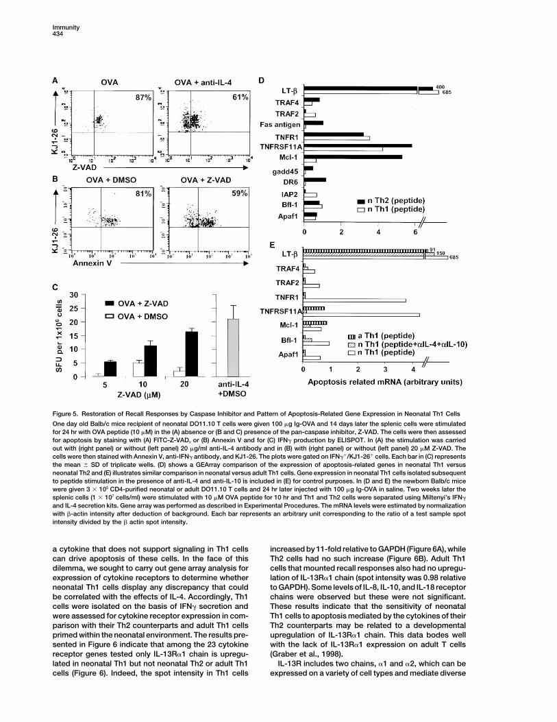
in the Th1 cells and 87% of the detectable lymphocytesbound Z-VAD, a synthetic substrate that binds to acti-vated caspases. However, when IL-4 of the Th2 cellswas neutralized with anti-IL-4 antibody, many more cellsremained alive and a significant number (39%) of themshowed reduced Z-VAD binding activity. Thus, caspasesare activated during OVA stimulation and such activationis inhibitable by anti-IL-4. Subsequently, unlabeledZ-VAD was used to block caspase activity and the cellswere tested for IFN� production and Annexin V binding.Figure 5B shows that addition of Z-VAD during stimula-tion with OVA peptide rescues a significant number ofcells from apoptosis. Indeed while 81% of IFN�-produc-ing cells bound Annexin V during OVA-stimulation, only59% displayed Annexin V binding activity when Z-VADwas added to the primary cell culture. Moreover, therescued Th1 cells secreted IFN� and such cytokine pro-duction was proportional to the amount of Z-VAD added(Figure 5C). These results provide direct evidence thatapoptosis is responsible for the lack of IFN� productionand the bias of neonatal recall responses toward Th2.
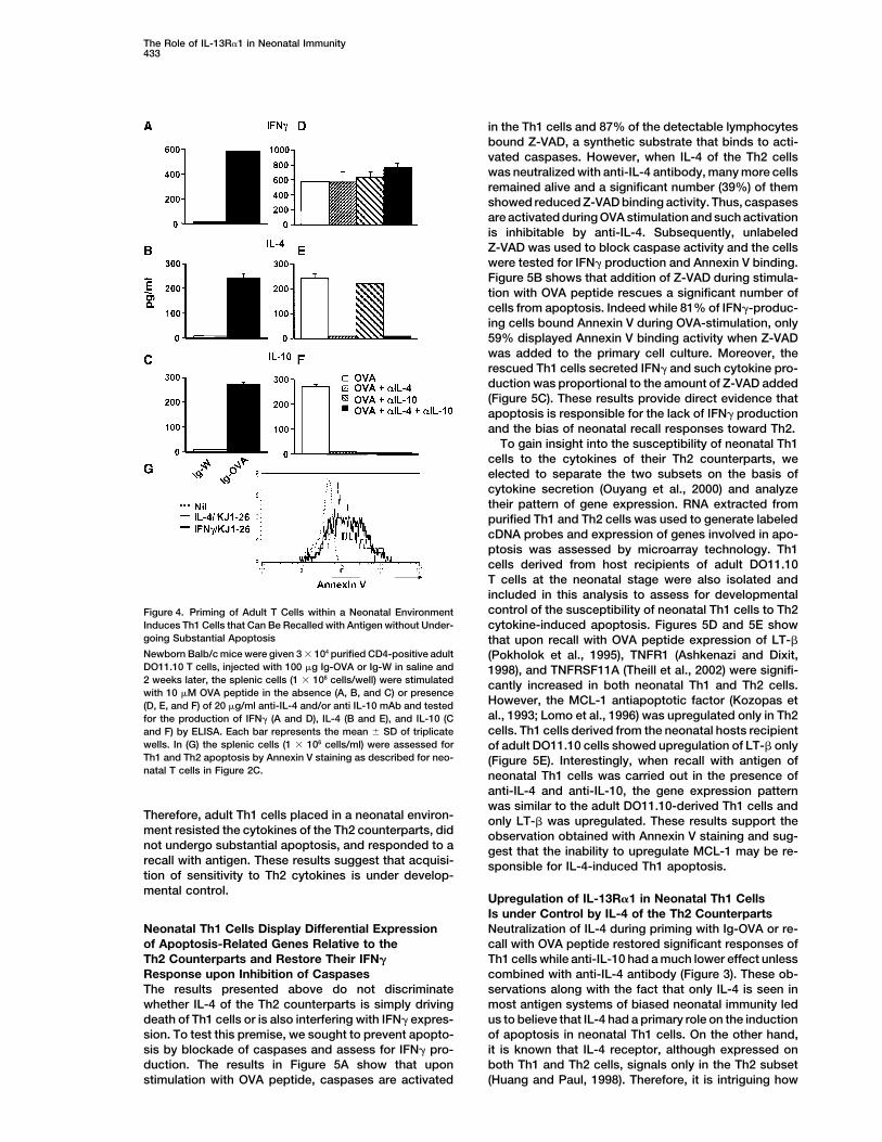
To gain insight into the susceptibility of neonatal Th1cells to the cytokines of their Th2 counterparts, weelected to separate the two subsets on the basis ofcytokine secretion (Ouyang et al., 2000) and analyzetheir pattern of gene expression. RNA extracted frompurified Th1 and Th2 cells was used to generate labeledcDNA probes and expression of genes involved in apo-ptosis was assessed by microarray technology. Th1cells derived from host recipients of adult DO11.10T cells at the neonatal stage were also isolated andincluded in this analysis to assess for developmentalcontrol of the susceptibility of neonatal Th1 cells to Th2Figure 4. Priming of Adult T Cells within a Neonatal Environment
Induces Th1 Cells that Can Be Recalled with Antigen without Under- cytokine-induced apoptosis. Figures 5D and 5E showgoing Substantial Apoptosis that upon recall with OVA peptide expression of LT-�Newborn Balb/c mice were given 3 � 104 purified CD4-positive adult (Pokholok et al., 1995), TNFR1 (Ashkenazi and Dixit,DO11.10 T cells, injected with 100 �g Ig-OVA or Ig-W in saline and 1998), and TNFRSF11A (Theill et al., 2002) were signifi-2 weeks later, the splenic cells (1 � 106 cells/well) were stimulated cantly increased in both neonatal Th1 and Th2 cells.with 10 �M OVA peptide in the absence (A, B, and C) or presence
However, the MCL-1 antiapoptotic factor (Kozopas et(D, E, and F) of 20 �g/ml anti-IL-4 and/or anti IL-10 mAb and testedal., 1993; Lomo et al., 1996) was upregulated only in Th2for the production of IFN� (A and D), IL-4 (B and E), and IL-10 (Ccells. Th1 cells derived from the neonatal hosts recipientand F) by ELISA. Each bar represents the mean � SD of triplicate
wells. In (G) the splenic cells (1 � 106 cells/ml) were assessed for of adult DO11.10 cells showed upregulation of LT-� onlyTh1 and Th2 apoptosis by Annexin V staining as described for neo- (Figure 5E). Interestingly, when recall with antigen ofnatal T cells in Figure 2C. neonatal Th1 cells was carried out in the presence of
anti-IL-4 and anti-IL-10, the gene expression patternwas similar to the adult DO11.10-derived Th1 cells and
Therefore, adult Th1 cells placed in a neonatal environ- only LT-� was upregulated. These results support thement resisted the cytokines of the Th2 counterparts, did observation obtained with Annexin V staining and sug-not undergo substantial apoptosis, and responded to a gest that the inability to upregulate MCL-1 may be re-recall with antigen. These results suggest that acquisi- sponsible for IL-4-induced Th1 apoptosis.tion of sensitivity to Th2 cytokines is under develop-mental control.
Upregulation of IL-13R�1 in Neonatal Th1 CellsIs under Control by IL-4 of the Th2 CounterpartsNeutralization of IL-4 during priming with Ig-OVA or re-Neonatal Th1 Cells Display Differential Expression
of Apoptosis-Related Genes Relative to the call with OVA peptide restored significant responses ofTh1 cells while anti-IL-10 had a much lower effect unlessTh2 Counterparts and Restore Their IFN�
Response upon Inhibition of Caspases combined with anti-IL-4 antibody (Figure 3). These ob-servations along with the fact that only IL-4 is seen inThe results presented above do not discriminate
whether IL-4 of the Th2 counterparts is simply driving most antigen systems of biased neonatal immunity ledus to believe that IL-4 had a primary role on the inductiondeath of Th1 cells or is also interfering with IFN� expres-
sion. To test this premise, we sought to prevent apopto- of apoptosis in neonatal Th1 cells. On the other hand,it is known that IL-4 receptor, although expressed onsis by blockade of caspases and assess for IFN� pro-
duction. The results in Figure 5A show that upon both Th1 and Th2 cells, signals only in the Th2 subset(Huang and Paul, 1998). Therefore, it is intriguing howstimulation with OVA peptide, caspases are activated
Immunity434
Figure 5. Restoration of Recall Responses by Caspase Inhibitor and Pattern of Apoptosis-Related Gene Expression in Neonatal Th1 Cells
One day old Balb/c mice recipient of neonatal DO11.10 T cells were given 100 �g Ig-OVA and 14 days later the splenic cells were stimulatedfor 24 hr with OVA peptide (10 �M) in the (A) absence or (B and C) presence of the pan-caspase inhibitor, Z-VAD. The cells were then assessedfor apoptosis by staining with (A) FITC-Z-VAD, or (B) Annexin V and for (C) IFN� production by ELISPOT. In (A) the stimulation was carriedout with (right panel) or without (left panel) 20 �g/ml anti-IL-4 antibody and in (B) with (right panel) or without (left panel) 20 �M Z-VAD. Thecells were then stained with Annexin V, anti-IFN� antibody, and KJ1-26. The plots were gated on IFN��/KJ1-26� cells. Each bar in (C) representsthe mean � SD of triplicate wells. (D) shows a GEArray comparison of the expression of apoptosis-related genes in neonatal Th1 versusneonatal Th2 and (E) illustrates similar comparison in neonatal versus adult Th1 cells. Gene expression in neonatal Th1 cells isolated subsequentto peptide stimulation in the presence of anti-IL-4 and anti-IL-10 is included in (E) for control purposes. In (D and E) the newborn Balb/c micewere given 3 � 105 CD4-purified neonatal or adult DO11.10 T cells and 24 hr later injected with 100 �g Ig-OVA in saline. Two weeks later thesplenic cells (1 � 107 cells/ml) were stimulated with 10 �M OVA peptide for 10 hr and Th1 and Th2 cells were separated using Miltenyi’s IFN�
and IL-4 secretion kits. Gene array was performed as described in Experimental Procedures. The mRNA levels were estimated by normalizationwith �-actin intensity after deduction of background. Each bar represents an arbitrary unit corresponding to the ratio of a test sample spotintensity divided by the � actin spot intensity.
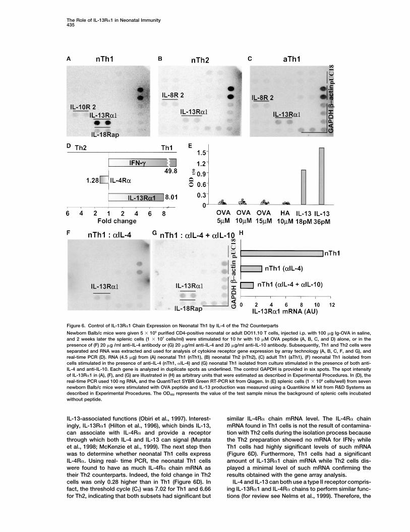
a cytokine that does not support signaling in Th1 cells increased by 11-fold relative to GAPDH (Figure 6A), whileTh2 cells had no such increase (Figure 6B). Adult Th1can drive apoptosis of these cells. In the face of this
dilemma, we sought to carry out gene array analysis for cells that mounted recall responses also had no upregu-lation of IL-13R�1 chain (spot intensity was 0.98 relativeexpression of cytokine receptors to determine whether
neonatal Th1 cells display any discrepancy that could to GAPDH). Some levels of IL-8, IL-10, and IL-18 receptorchains were observed but these were not significant.be correlated with the effects of IL-4. Accordingly, Th1
cells were isolated on the basis of IFN� secretion and These results indicate that the sensitivity of neonatalTh1 cells to apoptosis mediated by the cytokines of theirwere assessed for cytokine receptor expression in com-
parison with their Th2 counterparts and adult Th1 cells Th2 counterparts may be related to a developmentalupregulation of IL-13R�1 chain. This data bodes wellprimed within the neonatal environment. The results pre-
sented in Figure 6 indicate that among the 23 cytokine with the lack of IL-13R�1 expression on adult T cells(Graber et al., 1998).receptor genes tested only IL-13R�1 chain is upregu-
lated in neonatal Th1 but not neonatal Th2 or adult Th1 IL-13R includes two chains, �1 and �2, which can beexpressed on a variety of cell types and mediate diversecells (Figure 6). Indeed, the spot intensity in Th1 cells
The Role of IL-13R�1 in Neonatal Immunity435
Figure 6. Control of IL-13R�1 Chain Expression on Neonatal Th1 by IL-4 of the Th2 Counterparts
Newborn Balb/c mice were given 5 � 105 purified CD4-positive neonatal or adult DO11.10 T cells, injected i.p. with 100 �g Ig-OVA in saline,and 2 weeks later the splenic cells (1 � 107 cells/ml) were stimulated for 10 hr with 10 �M OVA peptide (A, B, C, and D) alone, or in thepresence of (F) 20 �g /ml anti-IL-4 antibody or (G) 20 �g/ml anti-IL-4 and 20 �g/ml anti-IL-10 antibody. Subsequently, Th1 and Th2 cells wereseparated and RNA was extracted and used for analysis of cytokine receptor gene expression by array technology (A, B, C, F, and G), andreal-time PCR (D). RNA (4.5 �g) from (A) neonatal Th1 (nTh1), (B) neonatal Th2 (nTh2), (C) adult Th1 (aTh1), (F) neonatal Th1 isolated fromcells stimulated in the presence of anti-IL-4 (nTh1, �IL-4) and (G) neonatal Th1 isolated from culture stimulated in the presence of both anti-IL-4 and anti-IL-10. Each gene is analyzed in duplicate spots as underlined. The control GAPDH is provided in six spots. The spot intensityof IL-13R�1 in (A), (F), and (G) are illustrated in (H) as arbitrary units that were estimated as described in Experimental Procedures. In (D), thereal-time PCR used 100 ng RNA, and the QuantiTect SYBR Green RT-PCR kit from Qiagen. In (E) splenic cells (1 � 106 cells/well) from sevennewborn Balb/c mice were stimulated with OVA peptide and IL-13 production was measured using a Quantikine M kit from R&D Systems asdescribed in Experimental Procedures. The OD450 represents the value of the test sample minus the background of splenic cells incubatedwithout peptide.
IL-13-associated functions (Obiri et al., 1997). Interest- similar IL-4R� chain mRNA level. The IL-4R� chainmRNA found in Th1 cells is not the result of contamina-ingly, IL-13R�1 (Hilton et al., 1996), which binds IL-13,
can associate with IL-4R� and provide a receptor tion with Th2 cells during the isolation process becausethe Th2 preparation showed no mRNA for IFN� whilethrough which both IL-4 and IL-13 can signal (Murata
et al., 1998; McKenzie et al., 1999). The next step then Th1 cells had highly significant levels of such mRNA(Figure 6D). Furthermore, Th1 cells had a significantwas to determine whether neonatal Th1 cells express
IL-4R�. Using real- time PCR, the neonatal Th1 cells amount of IL-13R�1 chain mRNA while Th2 cells dis-played a minimal level of such mRNA confirming thewere found to have as much IL-4R� chain mRNA as
their Th2 counterparts. Indeed, the fold change in Th2 results obtained with the gene array analysis.IL-4 and IL-13 can both use a type II receptor compris-cells was only 0.28 higher than in Th1 (Figure 6D). In
fact, the threshold cycle (CT) was 7.02 for Th1 and 6.66 ing IL-13R�1 and IL-4R� chains to perform similar func-tions (for review see Nelms et al., 1999). Therefore, thefor Th2, indicating that both subsets had significant but
Immunity436
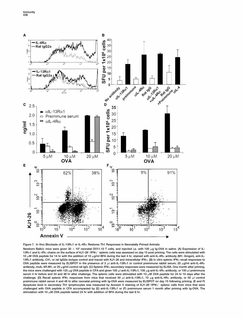
Figure 7. In Vivo Blockade of IL-13R�1 or IL-4R� Restores Th1 Responses in Neonatally Primed Animals
Newborn Balb/c mice were given 30 � 103 neonatal DO11.10 T cells, and injected i.p. with 100 �g Ig-OVA in saline. (A) Expression of IL-13R�1 and IL-4R� chains on the surface of KJ1-26�/IFN�� splenic cells was assessed on day 15 post priming. The cells were stimulated with10 �M OVA peptide for 14 hr with the addition of 10 �g/ml BFA during the last 4 hr, stained with anti-IL-4R� antibody (M1: Amgen), anti-IL-13R�1 antibody, C41, or rat IgG2a isotype control and traced with KJ1-26 and intracellular IFN�. (B) In vitro splenic IFN� recall responses toOVA peptide were measured by ELISPOT in the presence of 2 �l anti-IL-13R�1 or control preimmune rabbit serum; 20 �g/ml anti-IL-4R�
antibody, muIL-4R-M1, or 20 �g/ml control rat IgG. (C) Splenic IFN� secondary responses were measured by ELISA. One month after priming,the mice were challenged with 125 �g OVA peptide in CFA and given 100 �l anti-IL-13R�1, 100 �g anti-IL-4R� antibody, or 100 �l preimmuneserum 4 hr before and 24 and 48 hr after challenge. The splenic cells were stimulated with 10 �M OVA peptide for 24 hr 10 days after thechallenge. (D) Recall splenic IFN� responses from mice that received 50 �l anti-IL-13R�1, 10 �g anti-IL-4R� antibody, or 50 �l controlpreimmune rabbit serum 4 and 48 hr after neonatal priming with Ig-OVA were measured by ELISPOT on day 15 following priming. (E and F)Apoptosis level in secondary Th1 lymphocytes was measured by Annexin V staining of KJ1-26�/IFN�� splenic cells from mice that werechallenged with OVA peptide in CFA accompanied by (E) anti-IL-13R�1 or (F) preimmune serum 1 month after priming with Ig-OVA. Thestimulation with 10 �M OVA peptide lasted 24 hr with addition of BFA during the last 8 hr.
The Role of IL-13R�1 in Neonatal Immunity437
primary neonatal Th2 cells may be producing IL-13 serum used as control did not allow for secondary IFN�responses. It is worth noting that development of IFN�alongside of IL-4 and utilize such a cytokine to affect
their Th1 counterparts. Alternatively, they may not se- responses upon in vivo blockade of IL-4R� required highamounts of OVA peptide for stimulation in vitro. As Th1crete IL-13, but instead use IL-4 through the type II
receptor to drive apoptosis of the Th1 cells. To address cells manifest high expression of IL-4R� (Figure 7A), itmay be that fewer cells were rescued by anti-IL-4R�this issue, the splenic cells of individual mice recipient
of neonatal DO11.10 cells and primed with Ig-OVA were antibody, thus leading to the requirement for high pep-tide concentration to read out a low frequency of cells.stimulated with OVA peptide and tested for production
of IL-13. The results in Figure 6E indicated that IL-13 Overall, these results support the in vitro data and indi-cate that both IL-4R� and IL-13R�1 are involved in IL-was below the limit of detectability and the OD450 values
were similar to those obtained with the negative control 4-mediated targeting of Th1 cells in vivo during chal-lenge with OVA peptide.HA peptide. Together the findings suggest that IL-4 uti-
lizes a type II IL-13R�1/IL-4R� receptor to drive death Neutralization of IL-4 during priming with Ig-OVA onthe day of birth restored Th1 responses (Figure 3). Block-of Th1 cells. If this hypothesis is correct, then neutraliza-
tion of IL-4, which prevents apoptosis, may induce ade of IL-13R�1 in vivo during priming with Ig-OVA alsorestored IFN� recall responses while preimmune serumdownregulation of the expression of IL-13R�1 on the
live neonatal Th1 cells. The lower panels of Figure 6 used as control did not (Figure 7D). However, blockadeof IL-4R� during priming with Ig-OVA did not restoreshow that this is indeed the case and IL-13R�1 chain
mRNA expression was reduced from 11 to 2 to 3 units IFN� responses. This is possibly due to the fact thatnaive T cells may not polarize toward Th2 as IL-4R� isupon neutralization of IL-4 or both IL-4 and IL-10 (Figures
6F, 6G, and 6H). These results indicate that IL-13R�1 blocked and transient IL-4 needed for development ofTh1 cells may be lacking (Biedermann et al., 2001).expression on Th1 cells is under control of IL-4 of the
Th2 counterparts. Finally, blockade of IL-13R�1 in vivo during challengewith OVA peptide reduced apoptosis among Th1 cellsas only 38% of cells were stained with Annexin V (FigureBlockade of IL-4R� and/or IL-13R�1 Restores7E). Mice given preimmune serum instead of anti-IL-Neonatal Th1 Recall and Secondary Responses13R�1 antiserum had 91% of their KJ1-26-positive IFN�-If IL-4 of the Th2 cells is utilizing a heteroreceptor toproducing cells stain positive with Annexin V (Figure 7F).drive apoptosis of Th1 cells then blockade of IL-4R�These results indicate that blockade of IL-13R�1 onand/or IL-13R�1 should restore the recall as well asprimary Th1 cells during challenge with OVA peptidesecondary neonatal Th1 responses. Before addressingallows the cells to remain alive and develop second-this issue, we investigated whether IL-13R�1 and IL-ary responses.4R� proteins are expressed on the surface of Th1 cells.
Figure 7A shows that neonatal T cells producing intracel-lular IFN� and positive for KJ1-26 bind IL-4R�-specific DiscussionmuIL-4R-M1 (top panel) antibody much higher than therat IgG2a isotype control. Also, the cells were signifi- Upon priming with antigen, neonatal T cells were acti-
vated, abandoned their naive phenotype, and prolifer-cantly stained with the IL-13R�1-reactive nonneutraliz-ing C41 antibody (lower panel). These results indicate ated in vivo (Figure 1). Some of the cells differentiated
into Th2 cells but others, surprisingly, developed as Th1that primary Th1 cells express IL-4R� and IL-13R�1 onthe surface. These receptors play a role in the downregu- cells (Figure 2). Intriguingly, however, upon reencounter
with the antigen, the Th2 responded and produced bothlation of primary neonatal Th1 cells by IL-4 becauseblockade with anti-IL-4R� and/or anti-IL-13R�1 anti- IL-4 and IL-10 but the Th1 cells were unable to sustain
IFN� production and died by apoptosis (Figure 2). Block-bodies restores IFN� recall responses of the Th1 cells(Figure 7B). Indeed, splenic cells from mice recipient of ade of the Th2 cytokines during recall with antigen in
vitro reduced Th1 apoptosis and sustained IFN� pro-DO11-10 T cells and given Ig-OVA on the day of birthwere able to produce IFN� upon in vitro stimulation with duction (Figure 3). Similarly, when anti-Th2 cytokine
antibodies were administered to the host mice duringOVA peptide in the presence of rabbit anti-IL-13R�1anti-serum, and/or anti-IL-4R� antibody to the same neonatal priming with antigen, recall Th1 responses de-
veloped (Figure 3). Furthermore, when adult T cells wereextent as neutralization of IL-4 cytokine. Rabbit preim-mune serum and/or rat IgG controls had no significant placed within the neonatal environment and primed with
antigen, Th1 recall responses also arose (Figure 4). To-effect on the response and IFN� was at levels similarto stimulation with OVA peptide without any blockade. gether, these results indicated that the Th2 cytokines
are not affecting the development of Th1 from naiveThese results indicate that both IL-4R� and IL-13R�1are involved in IL-4-mediated targeting of primary Th1 cells (Stetson et al., 2002), but are most likely sensitizing
differentiated Th1 cells for apoptosis. In addition, suchcells during recall with OVA peptide in vitro. Subse-quently, we investigated whether blockade of IL-4R� sensitivity seems to reflect a developmental trait unique
to neonatal Th1 cells as adult T cells primed within theand/or IL-13R�1 in vivo during a later challenge withOVA peptide restores Th1 secondary responses. The neonatal environment generated Th1 cells resistant to
the Th2 cytokines.findings indicate that mice recipient of DO11.10 T cellsand Ig-OVA/saline on the day of birth and 1 month later IL-4 of the Th2 cells seems to interfere with Th1 recall
responses by triggering apoptosis of primary Th1 cellsadministered with anti-IL-13R�1 antiserum or anti-IL-4R� antibody during immunization with OVA peptide in rather than inhibition of differentiation into IFN�-produc-
ing cells. Indeed, when recall of the cells was performedCFA develop IFN� responses (Figure 7C). Preimmune
Immunity438
in the presence of a caspase inhibitor, apoptosis was of polarization to Th2, which are required to producesignificantly reduced and IFN� production was restored some IL-4 needed for induction of IL-12 production by(Figures 5A, 5B, and 5C). Furthermore, gene array analy- dendritic cells and differentiation of Th1 cells (Bieder-sis indicated that primary Th1 cells display unbalanced mann et al., 2001).apoptotic machinery relative to their Th2 counterparts Overall, it is shown that priming with antigen at theas well as adult T cells primed within the neonatal envi- neonatal stage induces both Th1 and Th2 primary re-ronment (Figures 5D and 5E). Seemingly, an inability sponses. However, IL-4 of the Th2 cells induces a devel-to upregulate the antiapoptotic factor MCL-1 may be opmental upregulation of IL-13R�1 on the neonatal Th1responsible for IL-4-induced Th1 apoptosis. counterparts and this receptor chain along with IL-4R�
IL-4 usually directs differentiation of naive T cells to- provides an alternative receptor that marks the Th1 cellsward Th2 but has no defined downregulatory functions for death if reencounter with antigen occurs in an IL-4-on differentiated Th1 cells (Reiner and Seder, 1999; Mo- containing environment. It is possible that neonatallyrel and Oriss, 1998; Nelms et al., 1999). IL-10 has been primed Th1 cells succumb to IL-4 due to signalingshown to display antiproliferative (anergic) functions on through an IL-4R�/IL-13R�1 heteroreceptor that unbal-Th1 cells mostly through downregulation of accessory ances expression of pro- and antiapoptotic factors.molecules on APCs or through inhibition of IL-12 pro-
Experimental Proceduresduction by the APCs (Moore et al., 2001). The resultspresented here show that the unresponsiveness and
Miceapoptosis of differentiated neonatal Th1 cells are pri-Balb/c mice (H-2d) were purchased from Harlan Sprague Dawley
marily due to negative effects by IL-4, with IL-10 playing (Indianapolis, IN) and used as hosts for adoptive transfer of T cells.only a secondary role (Figures 2 and 4). Gene array DO11.10/scid transgenic mice expressing a TCR specific for OVAanalysis of cytokine receptor expression on neonatal peptide were used as donors of T cells.Th1 cells indicated that expression of the IL-13R�1 chain
Antigensis upregulated in neonatal Th1 but not in neonatal Th2,OVA peptide (SQAVHAAHAEINEAGR) encompasses aa residuesor even adult Th1 cells. Also, such upregulation is de-323-339 of OVA and is immunogenic in Balb/c (H-2d) mice. Influenzacreased when IL-4 is neutralized (Figure 6). It is wellvirus hemagglutinin (HA) aa residues 110-120 peptide (SFERFEIF
established that the conventional IL-4R (IL-4R�/com- PKI), which is also immunogenic in Balb/c mice, was used as nega-mon �) mediates IL-4 signaling in Th2 but not in Th1 tive control. Ig-W, a Balb/c IgG2b Ig molecule generated by transfec-cells (Huang and Paul, 1998). However, IL-13R�1 can tion of the 91A3 anti-arsonate antibody heavy and light chains into
the non-Ig secreting myeloma B cell line SP2/0 was described (Minassociate with IL-4R� and provide a receptor throughet al., 1998). Ig-OVA, expressing OVA peptide within the heavy chainwhich both IL-4 and IL-13 can signal (Hilton et al., 1996;variable region of Ig-W, was also described (Li et al., 2001).Murata et al., 1998; McKenzie et al., 1999). Given that the
neonatal Th2 did not produce IL-13 and neutralization of Adoptive T Cell TransferIL-4 leads to a reduction in Th1 apoptosis and a de- Splenic cells from 1 day old DO11.10/scid mice containing the equiv-crease in IL-13R�1 expression (Figure 6), it may be that alent of 3 � 104 DO11.10 T cells were transferred into 1 day old
Balb/c mice by intravenous (i.v.) injection through the facial veinIL-4 utilizes an IL-4R�/IL-13R�1 heterodimer complexusing a 30-gauge needle. For transfer of adult T cells into newbornto signal in neonatal Th1 cells as has been shown inmice, DO11.10 T cells were purified from the spleen of adultother cells (Murata et al., 1998; McKenzie et al., 1999).DO11.10/scid mice with anti-CD4 coupled magnetic beads (MiltenyiIL-4R� is constitutively expressed on adult Th1 and Th2Biotec, Auburn, CA) prior to injection into the host. This step was
cells (Huang and Paul, 1998) and real-time PCR analysis necessary to eliminate adult APCs that might interfere with antigenindicated that neonatal Th1 and Th2 cells also express presentation within the neonatal environment. For isolation of Th1IL-4R� mRNA (Figure 6). Thus, knowing that there has and Th2 cells, 3 � 105 purified neonatal or adult CD4�-T cells were
transferred into each mouse. This increase in T cell transfer wasbeen no defined direct effect on differentiated Th1 cellsrequired for the isolation of sufficient numbers of T cells for RNAby IL-10 (Moore et al., 2001) and neutralization of IL-4extraction and gene array analysis.diminishes both upregulation of IL-13R�1 and death
among Th1 cells, it is possible that IL-4 operates throughAntireceptor Antibodies
an alternative heteroreceptor comprising IL-4R� and IL- The anti-IL-4R� monoclonal antibody (M1) was provided by Amgen13R�1 chains to induce apoptosis of neonatal Th1 cells. (Seattle, WA). The nonneutralizing anti-IL-13R�1 monoclonal anti-This is supported by findings indicating that IL-4R� and body C41 was previously described (Poudrier et al., 2000). Rabbit
antiserum against the extracellular domain of the murine IL-13R�1IL-13R�1 proteins are expressed on the surface of thechain was developed as previously described (Schnare et al., 1998).neonatal Th1 cells and blockade of either receptor dur-
ing in vitro stimulation supported recall of the Th1 cellsCell Surface and Intracellular Staining(Figures 7A and 7B). In addition, when the blockade ofFor cell surface staining the spleen cells from Balb/c hosts were
either IL-4R� or IL-13R�1 was performed in vivo during incubated (20 min at 4�C) with 5 �g/ml 2.4G2 mAb to block Fc�Rsrechallenge with antigen in CFA at the adult stage, sec- on the cell surface prior to staining. The cells were then stainedondary Th1 responses developed (Figure 7C). Again, the with the anti-TCR OVA clonotypic mAb, KJ1-26 (mouse IgG2a), andrestoration of the secondary IFN� response correlated the culture was supplemented with FITC-anti-mouse IgG2a (clone
R19-15), PerCP-anti-CD4 and PE-anti-CD25 (clone 3C7), PE-anti-with reduction in Th1 apoptosis (Figures 7E and 7F).CD62L (clone Mel-14), or PE-anti-CD45RB (clone 16A). For ex vivoAnother piece of evidence that the expression of IL-intracellular cytokine staining, the cells were stimulated for a 4 hr13R�1 is under developmental control originates fromshort period of time to enhance cytokine accumulation and facilitate
the observation that administration of anti-IL-13R�1 intracellular detection as has been previously defined (Moser et al.,during priming at the neonatal stage restored recall re- 2001). Cytokine secretion was blocked by addition of 10 �g/ml BFAsponses of the Th1 cells. Blockade of IL-4R�, however, (Epicentre, Madison, WI), and the cells were then fixed with 2%
formaldehyde, permeabilized with 2% saponin (Sigma Chemical Co.)did not produce similar effects, possibly due to the lack
The Role of IL-13R�1 in Neonatal Immunity439
in FACS buffer for 10 min at room temperature, and incubated with peptide and separated into Th1 and Th2 on the basis of cytokinesecretion (IFN� for Th1 and IL-4 for Th2) as described (Ouyang etPE-anti-mouse IL-4, and FITC-anti-mouse IFN-�. Isotype-matched
controls were included in all experiments. All the data were collected al., 2000) using separation kits from Miltenyi Biotech. Usually, 108
splenic cells are used to obtain 0.5 to 1 � 106 Th1 or Th2 cells.using a FACScan flow cytometer (Becton Dickinson, Mountain View,CA) and analyzed with CellQuest software (Becton Dickinson).
Gene Array AnalysismRNA expression in isolated neonatal and adult T cells was ana-Analysis of Cell Division In Vivolyzed using both the GEArray Q series kit including 96 key genesPurified CD4�-neonatal DO11.10 T cells were labeled with the intra-involved in apoptosis, and interleukin receptor GEArray kit compris-cellular fluorescent dye, 5(and 6)-carboxyfluorescein diacetate suc-ing 23 genes of interleukin receptor subunits (SuperArray Inc.,cinimidyl ester (CFSE) (Molecular Probes, Eugene, OR), as describedBethesda, MD). Total RNA was extracted from 0.5–1 � 106 purified(Lyons and Parish, 1994) and transferred into newborn Balb/c miceneonatal Th1, adult Th1 or neonatal Th2 cells using TRIzol reagent(3 � 105 cells/mouse). The hosts were then injected i.p. with 100 �g(Life Technologies, Rockville, Maryland). The RNA (3 to 4.5 �g) wasOVA peptide, Ig-OVA, or Ig-W in saline. Four days later, the CD4�-Tthen used in a reverse transcription reaction along with �-32P-dCTPcells were isolated from the spleen of Balb/c hosts and stained with(ICN Biomedical, Inc., Aurora, OH) to generate labeled cDNA probes.KJ1-26 and anti-CD4 mAbs.Subsequently, for the Q series kit, the labeled cDNA probes werehybridized to the membrane, in a 0.75 ml hybridization buffer for 14Cytokine ELISA and ELISPOThr at 60�C. For the interleukin receptor membrane, the hybridizationMeasurement of Cytokines by ELISAwas carried out in a 5.1 ml buffer for 14 hr at 68�C. The intensity ofSpleen cells containing both T cells and APCs were incubated withradioactive spots was analyzed on a Bio-Rad molecular imagerantigen in 96-well round-bottom plates and 24 hr later IL-4, IL-10,FX (Hercules, California) using Quantity One software. Results areand IFN� production was measured by ELISA using anti-cytokineexpressed as arbitrary units estimated as follows: (mean spot inten-antibodies according to BD PharMingen’s instructions. IL-13 pro-sity of sample – mean spot intensity of background) / (mean spotduction was measured using the Quantikine M kit from R&D Systemsintensity of �-actin – mean spot intensity of background). The back-(Minneapolis, MN) according to the manufacturer’s instructions.ground represents the mean radioactive intensity obtained fromMeasurement of IFN� by ELISPOTpUC18 DNA spots included in the membrane.Detection of IFN� by ELISPOT was carried out as described (Min
et al., 1998). In brief, HA-multiscreen plates (Millipore, Bedford, MA)Real-Time PCRwere coated with capture antibody and free sites were saturatedReverse transcription and DNA amplification were performed ac-with DMEM culture media containing 10% fetal calf serum. Subse-cording to a one-step protocol using 100 ng of total RNA and aquently, 1 � 106 splenic cells were added and the culture was stimu-QuantiTect SYBR Green RT-PCR kit from Qiagen (Valencia, CA)lated with OVA peptide with or without blocking antibody. Biotinyl-according to the manufacturers’ instructions. Expression of IL-4R�ated anti-IFN� antibody (1 �g/ml) was added and bound antibodychain and IL-13R�1 chain mRNA was assessed in both Th1 and Th2was revealed with avidin-peroxidase. Spots were counted under aneonatal T cells. Expression of IFN� mRNA was included to assessdissecting microscope.for the purity of the Th1 and Th2 populations. �-actin was includedto serve as a normalizer. The oligonucleotides used as specificDetection of Early Apoptosis by Staining with Annexin Vprimers were: sense IFN�, TCAAGTGGCATAGATGTGGAAGAA; IL-Spleen cells (1 � 106 cells/ml) were stimulated with antigen, incu-4R�, CTTGCAGACAATCCTGCCTA; IL-13R�1, GCACAGAGTATAGGbated for 30 min with KJ1-26 mAb, and the culture was supple-TAAGGAGCAA; �actin, AGAGGGAAATCGTGCGTGAC; and anti-mented with biotin-anti-mouse IgG2a. The cells were then incubatedsense, IFN-�, TGGCTCTGCAGGATTTTCATG; IL-4R�, GGCTCCTCTwith PerCP-Avidin and 30 min later resuspended in Annexin V stain-TCTTCCAGATG; IL-13R�1, ACAAAGACTGGAATGGTGAGTAAC;ing buffer. Subsequently, the cells were stained with FITC-labeled�actin, 5-CAATAGTGATGACCTGGCCGT. Real-time PCR wasAnnexin V (PharMingen) for 15 min at room temperature, washed,performed on a Cepheid Smart Cycler (Sunnyvale, CA) and the re-and fixed with 2% formaldehyde. To assess for early apoptosis ofsults were analyzed by the Comparative CT method described inTh1 and Th2 lymphocytes, the cells were permeabilized and intracel-the Smart Cycler Software.lular cytokine production was analyzed as described above.
AcknowledgmentsDetection of Caspase Activation and Inhibitionof Apoptosis by Z-VAD
This work was supported by grant AI48541 (to H. Zaghouani) fromSpleen cells (1 � 106 cells/well) were stimulated with 10 �M OVA
the National Institutes of Health, start up funds from the Universitypeptide for 24 hr with addition of BFA (10 �g/ml) during the last 6
of Missouri School of Medicine, and the Deutsche Forschungsge-hr of incubation. Subsequently, the cells were stained with 5 �M
meinschaft (SFB 466,B10).FITC-conjugated Z-VAD (CaspACE FITC-VAD-fmk from PromegaCorp., Madison, WI) for 30 min to detect activated caspases. For
Received: July 17, 2003identification of Th1 cells, the culture was also costained with KJ1-
Revised: February 5, 200426 and anti-IFN� antibody. For blockade of apoptosis, OVA stimula-
Accepted: February 11, 2004tion was done in the presence of graded amounts of unlabeled
Published: April 20, 2004Z-VAD-fmk (Calbiochem, La Jolla, CA) for 24 hr with addition of BFAduring the last 6 hr of incubation. The cells were then stained with
ReferencesAnnexin V, KJ1-26, and anti-IFN� antibodies. Since Z-VAD is solubi-lized in DMSO as recommended by the manufacturer, the controls
Adkins, B., and Du, R.Q. (1998). Newborn mice develop balancedare diluted in DMSO to account for cell death that could be mediated
Th1/Th2 primary effector responses in vivo but are biased to Th2by side effects associated with DMSO.
secondary responses. J. Immunol. 160, 4217–4224.
Ashkenazi, A., and Dixit, V.M. (1998). Death receptors: signaling andSeparation of Th1 and Th2 Cellsmodulation. Science 281, 1305–1308.The separation of neonatal Th1 and Th2 cells requires an increasedBiedermann, T., Zimmermann, S., Himmelrich, H., Gumy, A., Egeter,frequency of primary T lymphocytes. Thus, the transfer was per-O., Sakrauski, A.K., Seegmuller, I., Voigt, H., Launois, P., Levine,formed with CD4-purified neonatal DO11.10/scid T lymphocytes in-A.D., et al. (2001). IL-4 instructs TH1 responses and resistance tostead of total splenic cells and the number of transferred cells wasLeishmania major in susceptible BALB/c mice. Nat. Immunol. 2,increased from 30 to 300-500 � 103 cells per newborn Balb/c host.1054–1060.Accordingly each Balb/c newborn was given either neonatal (1 day
old) or adult (8 week old) purified DO11.10/scid cells and injected Billingham, R.E., Brent, L., and Medawar, B.P. (1956). Quantitativestudies of tissue transplantation immunity. III. Acutely acquired tol-i.p. with a saline solution containing 100 �g Ig-OVA. Two weeks
later, the splenic T cells were stimulated for 10 hr with 10 �M OVA erance. Proc. R. Soc. Lond. Biol. Sci. 239, 44–57.
Immunity440
Chen, N., and Field, E.H. (1995). Enhanced type 2 and diminished Ouyang, W., Lohning, M., Gao, Z., Assenmacher, M., Ranganath, S.,Radbruch, A., and Murphy, K.M. (2000). Stat6-independent GATA-3type 1 cytokines in neonatal tolerance. Transplantation 59, 933–941.autoactivation directs IL-4-independent Th2 development and com-Forsthuber, T., Yip, H.C., and Lehmann, P.V. (1996). Induction ofmitment. Immunity 12, 27–37.TH1 and TH2 immunity in neonatal mice. Science 271, 1728–1730.Pack, C.D., Cestra, A.E., Min, B., Legge, K.L., Li, L., Caprio-Young,Gammon, G., Dunn, K., Shastri, N., Oki, A., Wilbur, S., and Sercarz,J.C., Bell, J.J., Gregg, R.K., and Zaghouani, H. (2001). NeonatalE.E. (1986). Neonatal T-cell tolerance to minimal immunogenic pep-exposure to antigen primes the immune system to develop re-tides is caused by clonal inactivation. Nature 319, 413–415.sponses in various lymphoid organs and promotes bystander regu-
Garza, K.M., Griggs, N.D., and Tung, K.S. (1997). Neonatal injectionlation of diverse T cell specificities. J. Immunol. 167, 4187–4195.
of an ovarian peptide induces autoimmune ovarian disease in femalePokholok, D.K., Maroulakou, I.G., Kuprash, D.V., Alimzhanov, M.B.,mice: requirement of endogenous neonatal ovaries. Immunity 6,Kozlov, S.V., Novobrantseva, T.I., Turetskaya, R.L., Green, J.E., and89–96.Nedospasov, S.A. (1995). Cloning and expression analysis of the
Graber, P., Gretener, D., Herren, S., Aubry, J.-P., Elson, G., Poudrier,murine lymphotoxin beta gene. Proc. Natl. Acad. Sci. USA 92,
J., Lecoanet-Henchoz, S., Alouani, S., Losberger, C., Bonnefoy,674–678.
J.-Y., et al. (1998). The distribution of IL-13 receptor �1 expressionPoudrier, J., Graber, P., Herren, S., Berney, C., Gretener, D., Kosco-on B cells, T cells and monocytes and its regulation by IL-13 andVilbois, M.H., and Gauchat, J.-F. (2000). A novel monoclonal anti-IL-4. Eur. J. Immunol. 28, 4286–4298.body, C41, reveals IL-13R�1 expression by murine germinal center
Hilton, D.J., Zhang, J.G., Metcalf, D., Alexander, W.S., Nicola, N.A., B cells and follicular dendritic cells. Eur. J. Immunol. 30, 3157–3164.and Willson, T.A. (1996). Cloning and characterization of a binding
Reiner, S.L., and Seder, R.A. (1999). Dealing from the evolutionarysubunit of the interleukin 13 receptor that is also a component ofpawnshop: how lymphocytes make decisions. Immunity 11, 1–10.the interleukin 4 receptor. Proc. Natl. Acad. Sci. USA 93, 497–501.Ridge, J.P., Fuchs, E.J., and Matzinger, P. (1996). Neonatal toleranceHuang, H., and Paul, W.E. (1998). Impaired interleukin 4 signalingrevisited: turning on newborn T cells with dendritic cells. Sciencein T helper type 1 cells. J. Exp. Med. 187, 1305–1313.271, 1723–1726.
Kearney, E.R., Pape, K.A., Loh, D.Y., and Jenkins, M.K. (1994). Visu-Sarzotti, M., Robbins, D.S., and Hoffman, P.M. (1996). Induction ofalization of peptide-specific T cell immunity and peripheral toleranceprotective CTL responses in newborn mice by a murine retrovirus.induction in vivo. Immunity 1, 327–339.Science 271, 1726–1728.
Kozopas, K.M., Yang, T., Buchan, H.L., Zhou, P., and Craig, R.W.Schnare, M., Blum, H., Juttner, S., Rollinghoff, M., and Gessner, A.(1993). MCL1, a gene expressed in programmed myeloid cell differ-(1998). Specific antagonism of type I IL-4 receptor with a mutatedentiation, has sequence similarity to BCL2. Proc. Natl. Acad. Sci.form of murine IL-4. J. Immunol. 161, 3484–3492.USA 90, 3516–3520.Siegrist, C.A. (2001). Neonatal and early life vaccinology. VaccineLi, L., Legge, K.L., Min, B., Bell, J.J., Gregg, R., Caprio, J., and19, 3331–3346.Zaghouani, H. (2001). Neonatal immunity develops in a transgenicSingh, R.R., Hahn, B.H., and Sercarz, E.E. (1996). Neonatal peptideTCR transfer model and reveals a requirement for elevated cell inputexposure can prime T cells and, upon subsequent immunization,to achieve organ-specific responses. J. Immunol. 167, 2585–2594.induce their immune deviation: implications for antibody vs. T cell-Lomo, J., Smeland, E.B., Krajewski, S., Reed, J.C., and Blomhoff,mediated autoimmunity. J. Exp. Med. 183, 1613–1621.H.K. (1996). Expression of the Bcl-2 homologue Mcl-1 correlatesStetson, D., Mohrs, M., Mallet-Designe, V., Teyton, L., and Locksley,with survival of peripheral blood B lymphocytes. Cancer Res. 56,R. (2002). Rapid expansion and IL-4 expression by Leishmania-40–43.specific naıve helper T cells in vivo. Immunity 17, 191–200.
Lyons, A.B., and Parish, C.R. (1994). Determination of lymphocyteTheill, L.E., Boyle, W.J., and Penninger, J.M. (2002). RANK-L anddivision by flow cytometry. J. Immunol. Methods 171, 131–137.RANK: T cells, bone loss, and mammalian evolution. Annu. Rev.
McKenzie, G.J., Fallon, P.G., Emson, C.L., Grencis, R.K., and Mc-Immunol. 20, 795–823.
Kenzie, A.N. (1999). Simultaneous disruption of interleukin (IL)-4 andIL-13 defines individual roles in T helper cell type 2-mediated re-sponses. J. Exp. Med. 189, 1565–1572.
Min, B., Legge, K.L., Pack, C., and Zaghouani, H. (1998). Neonatalexposure to a self-peptide-immunoglobulin chimera circumventsthe use of adjuvant and confers resistance to autoimmune diseaseby a novel mechanism involving interleukin 4 lymph node deviationand interferon gamma-mediated splenic anergy. J. Exp. Med.188, 2007–2017.
Moore, K.W., de Waal Malefyt, R., Coffman, R.L., and O’Garra, A.(2001). Interleukin-10 and the interleukin-10 receptor. Annu. Rev.Immunol. 19, 683–765.
Morel, P.A., and Oriss, T.B. (1998). Crossregulation between Th1and Th2 cells. Crit. Rev. Immunol. 18, 275–303.
Moser, J.M., Altman, J.D., and Lukacher, A.E. (2001). Antiviral CD8�
T cell responses in neonatal mice: susceptibility to polyoma virus-induced tumors is associated with lack of cytotoxic function by viralantigen-specific T cells. J. Exp. Med. 193, 595–606.
Murata, T., Husain, S.R., Mohri, H., and Puri, R.K. (1998). Two differ-ent IL-13 receptor chains are expressed in normal human skin fibro-blasts, and IL-4 and IL-13 mediate signal transduction through acommon pathway. Int. Immunol. 10, 1103–1110.
Nelms, K., Keagan, A.D., Zamorano, J., Ryan, J.J., and Paul, W.E.(1999). The IL-4 receptor: signaling mechanisms and biologic func-tions. Annu. Rev. Immunol. 17, 701–738.
Obiri, N.I., Leland, P., Murata, T., Debinski, W., and Puri, R.K. (1997).The IL-13 receptor structure differs on various cell types and mayshare more than one component with IL-4 receptor. J. Immunol.158, 756–764.