Predominance of Th2 polarization by Vitamin D through a STAT6-dependent mechanism
Upload
independentCategory
view
1download
0
Naive T cells sense the cysteine protease allergen papainthrough protease-activated receptor 2 and propel TH2immunity
Genqing Liang, PhD,a* Tolga Barker, PhD,a* Zhihui Xie, PhD,a Nicolas Charles, PhD,b� Juan Rivera, PhD,b and
Kirk M. Druey, MDa Bethesda, Md
Abbreviations used
APC: Antigen-presenting cell
BM: Bone marrow
BMB: Bone marrow–derived basophil
DC: Dendritic cell
FITC: Fluorescein isothiocyanate
Jak: Janus kinase
LN: Lymph node
MC: Mast cell
OVA: Ovalbumin
PAR: Protease-activated receptor
PE: Phycoerythrin
pLN: Popliteal lymph node
qPCR: Quantitative PCR
STAT: Signal transducer and activator of transcription
WT: Wild-type
Background: Sensitization to protease allergens, such as papain,or helminth infection is associated with basophil recruitment todraining lymph nodes (LNs). Basophils have the capacity topresent antigen to naive T cells and promote TH2 differentiationdirectly or indirectly through IL-4 production.Objective: We studied how papain induces basophil migrationto LNs and the contribution of various leukocytes to papain-induced immune responses.Methods: We immunized mice in the footpad with papain andstudied leukocyte recruitment and inflammatory cytokine andchemokine production in the draining popliteal LNs.Results: Papain directly activated naive T cells throughprotease-activated receptor (PAR) 2 to initiate a chemokine/cytokine program that includes CCL17, CCL22, and IL-4.Papain-triggered innate immune responses were dependent onboth CD4 T cells and PAR2 and were strongly reduced in theabsence of CCR4, the primary receptor for CCL17/CCL22.Conclusion: These results elucidate a novel innate allergen-recognition pathway mediated by naive T cells through PAR2,which provide an immediate source of chemokines and IL-4upstream of basophils and antigen-restricted TH2differentiation. PAR2 antagonism might thus hold promise forthe treatment of allergic disease. (J Allergy Clin Immunol2012;129:1377-86.)
Key words: Chemokines, basophils, chemotaxis, TH2, allergens
The plasticity and diversity of specific T-lymphocyte popula-tions shape the adaptive immune response. Naive T cells differ-entiate into IFN-g–producing TH1 cells after infection withintracellular microbes, including viruses and protozoa.1 TH2cells, which secrete IL-4, IL-5, and IL-13, develop in responseto helminth infection and allergen exposure. The pathways
From athe Molecular Signal Transduction Section, Laboratory of Allergic Diseases, Na-
tional Institute of Allergy and Infectious Diseases, National Institutes of Health, andbthe Laboratory of Molecular Immunogenetics, National Institute of Arthritis and
Musculoskeletal and Skin Diseases, National Institutes of Health.
*These authors contributed equally to this work.
�Nicolas Charles, PhD, is currently affiliated with Inserm U699 Universit�e Paris VII
Denis Diderot 16, Paris, France.
Supported in part by the Intramural Research Programs of the National Institutes of
Health, National Institute of Allergy and Infectious Diseases, and National Institute of
Arthritis and Musculoskeletal and Skin Diseases (grant no. AI000939 LAD to K. M.
D.).
Disclosure of potential conflict of interest: The authors declare that they have no relevant
conflicts of interest.
Received for publication September 16, 2011; revised February 16, 2012; accepted for
publication February 22, 2012.
Available online March 27, 2012.
Corresponding author: Kirk M. Druey, MD, National Institutes of Health, 10 Center Dr,
Rm 11N242, Bethesda, MD 20892. E-mail: [email protected].
0091-6749
doi:10.1016/j.jaci.2012.02.035
linking the innate and adaptive response to allergens, in whichantigen-presenting cells (APCs) recognize allergens and induceantigen-specific effector TH2 differentiation, have not been fullydefined.2,3
Many allergens have intrinsic proteolytic activity,4 which isrequired for their induction of TH2-mediated allergic inflam-mation.3 Papain is a cysteine protease belonging to a super-family of pathogen-derived proteases, including gingipainsand cruzipain of Trypanosoma cruzii.5 Papain can elicitantigen-specific IgE production and degranulation of mastcells (MCs) and is associated with occupational allergy infood industry workers and latex sensitivity caused by allergencross-reactivity.6,7 Published work has suggested a function forbasophils in the murine TH2 immune response to allergens, in-cluding papain. Subcutaneous injection of papain into mice,either in the presence8,9 or absence10,11 of a ‘‘bystander’’ anti-gen, such as ovalbumin (OVA), elicited basophil accumulationin draining lymph nodes (LNs). Published studies have alsoprovided evidence that basophils regulate TH2-mediated im-munity to helminths,12 tick antigens,13 and antigen-IgE com-plexes14 in mice.Basophils are short-lived leukocytes that circulate in the
bloodstream and migrate transiently into the T-cell zone ofdraining LNs after papain immunization.10,11 The chemokinesand receptors guiding basophil trafficking in mice are unknown.We examined the LN inflammatory response after subcutaneouspapain immunization. Basophils were drawn to and CCL17 andCCL22 accumulated in popliteal lymph nodes (pLNs) of mice ex-posed to papain compared with PBS-treated LNs, none of whichwas observed whenCd42/2 or Par22/2micewere studied. Expo-sure of naive T cells to papain in vitro elicited production ofCCL17/CCL22 and IL-4. These results illuminate a novel innate
1377

J ALLERGY CLIN IMMUNOL
MAY 2012
1378 LIANG ET AL
function of naive T cells and define basophil-trafficking pathwaysinvolved in the immune response to a protease allergen.
METHODSFor additional details on methods, see the Methods section in this article’s
Online Repository at www.jacionline.org.
MiceWild-type (WT) C57BL/6, Cd42/2, Tcra2/2, Ccr42/2, Cd11c-Dtr-egfp,
and Par22/2 mice (all on a C57Bl/6 background) were purchased from Jack-
son Laboratories (Bar Harbor, Me). DO11.10/4get mice on a Rag12/2 back-
ground were provided by Dr Markus Mohrs (Trudeau Institute, Saranac
Lake, NY). All murine experiments were performed in accordance with Ani-
mal Study Protocol LAD-3E, which was approved by the National Institute of
Allergy and Infectious Diseases Animal Care and Use Committee, National
Institutes of Health.
Bone marrow cultures, sorting, and chemotaxisWhole bone marrow (BM) cells were obtained from murine femurs and
cultured for 10 days in RPMI medium supplemented with IL-3 (30 ng/mL),
10% FBS, 50 mmol/L 2-mercaptoethanol, and 2 mmol/L L-glutamine.
FcεRI1CD49b1c-kit2 cells were sorted with a FACSAria Cyan followed by
recovery for 24 hours in culture medium before chemotaxis assays. In some
assays bone marrow–derived basophils (BMBs) were enriched from BM cul-
tures by using CD49bmicrobeads alone (Miltenyi Biotec, Bergisch Gladbach,
Germany), which produced results equivalent to those observed with BMBs
sorted by means of flow cytometry. Chemotaxis assays were performed with
5-mm pore size 24-well ChemoTx system (Costar, Cambridge, Mass) per
the manufacturer’s instructions. Cells were allowed to migrate in the presence
of chemokine in the lower chamber for 4 hours followed by quantification by
means of hemocytometry.
Statistical analysisData are presented as means6 SEMs where applicable and were analyzed
by using 1- or 2-way ANOVA or the Student t test by using PRISM software
(GraphPad Software, Inc, La Jolla, Calif).We considered aP value of less than
.05 to be statistically significant.
RESULTS
Inflammatory response of LNs to papain
immunizationTo examine papain-evoked pathways, we injected mice sub-
cutaneously in the rear footpad with either PBS or papain and thenharvested the draining (popliteal) LNs 2 to 4 days after immu-nization (see Fig E1 in this article’s Online Repository at www.jacionline.org). Consistent with published studies,10,11 we ob-served an accumulation of basophils (FcεRI1CD49b1c-kit2) inthe pLNs 3 days after papain injection but not in the pLNs ofmice injected with PBS (see Fig E1, A). We also found an in-creased frequency of CD41IL-41 T cells after restimulation ofpLNs of papain-injected mice 4 days after immunization (seeFig E1, B) and serum papain-specific IgE, which peaked 14days after immunization (see Fig E1, C). These results indicateda TH2-mediated immune response to papain.We compared chemokine/cytokine gene expression in the
pLNs of PBS- or papain-immunized mice 2 days after injectionusing a quantitative PCR (qPCR) array. Several transcripts weresignificantly upregulated in the LNs of papain-immunized micecompared with those of PBS-treated mice, most notably Ccl17
(5-fold), Ccl22 (3.5-fold), Ccl6 (3-fold), and Il4 (3-fold; Fig 1,A). By using real-time PCR, we found that expression of bothCcl17 and Ccl22 was significantly increased in the pLNs 2 daysafter papain injection compared with that seen in PBS-treatedpLNs (Fig 1, B). In pLN homogenates quantities of immunoreac-tive CCL17 and CCL22 were increased significantly (7- and3-fold, respectively) in papain-treated mice compared with thatseen in PBS-immunized mice (Fig 1, C).
Basophil chemokine receptor expression and
chemotaxisTo determine relevant papain-elicited chemokines that attract
basophils to the draining LNs, we analyzed chemokine receptorexpression on basophils isolated from the spleen and BM or onbasophils derived from BM precursors cultured in the presenceof IL-3 for 7 to 10 days (BMBs; see Fig E2, A in this article’sOnline Repository at www.jacionline.org). Freshly isolatedBM and splenic basophils expressed CCR2 (Fig 2, A and B)but not CCR3 (see Fig E2, B). We also detected expression ofCCR4 and CXCR2 (Fig 2, A and B) but no or minimal CCR1,CCR5, CCR6, CCR7, CXCR4, and CXCR5 (see Fig E2, B). Al-though chemokine receptor expression on BMBs was qualita-tively similar to that seen on freshly isolated basophils, CCR4and CXCR2 expression was higher in BMBs by comparison(see Fig E2, C).Sorted FcεRI1CD49b1c-kit2 basophils (see Fig E2, A) dis-
played chemotaxis toward gradients of CCL2, CXCL8, andCCL17, and CCL22 in a concentration-dependent manner (Fig2, C-F). BMBs did not migrate in response to stem cell factor,which served as a negative control here because basophils donot express the stem cell factor receptor (c-kit) or in response toCXCL12, CXCL13, IL-3, or papain alone (Fig 2 and data notshown), suggesting a unique role for papain-induced chemokinesin basophil recruitment to pLNs. Basophil migration in the pres-ence of equivalent chemokine concentrations in the upper andlower chambers was similar to that observed in the absence ofchemokine, indicating that chemokine gradients stimulated che-motaxis rather than chemokinesis (Fig 2, C-F). These results in-dicated that murine basophils express functional CCR2, CCR4,and CXCR2 receptors.
Role of CCR4-CCL17/CCL22 axis in papain-elicited
basophil migrationCCR4 and its ligands, CCL17 and CCL22, have been linked
to TH2-mediated inflammation.15 To define the role of this axisin papain responses, we immunized WT or Ccr42/2 mice withpapain and quantified basophils in pLNs 3 days after immuniza-tion. In naive unimmunized WT and Ccr42/2 mice, basophilfrequencies were similar in the spleen and BM, and T-cell, B-cell, and dendritic cell (DC) frequencies in the pLNs werealso comparable (see Fig E3 in this article’s Online Repositoryat www.jacionline.org). In contrast, basophil accumulation inpLNs after papain immunization was reduced by 60% in micelacking CCR4 compared with WT mice (Fig 3, A). This dispar-ity most likely did not result from intrinsically defective pLNresponses to papain because CCL17 and CCL22 concentrationswere similar in pLNs from WT and Ccr42/2 papain-treatedmice (Fig 3, C). Papain-induced TH2 differentiation was alsoreduced by CCR4 deficiency because there were 60% fewer

FIG 1. Inflammatory response of draining pLNs to footpad immunization with papain. A, qPCR array anal-
ysis of RNA from pLN RNA of PBS- or papain-treated mice. **P < .001 and *P < .05, 2-way ANOVA. B and C,
Ccl17 and Ccl22mRNA (Fig 1, B) or CCL17 and CCL22 protein (Fig 1, C) expression in LNs 2 days after PBS or
papain immunization was determined by using qPCR or ELISA, respectively. *P < .03, **P 5 .004, and
***P 5 .0007, paired t test. Data represent 3 experiments with at least 3 mice per group in each.
J ALLERGY CLIN IMMUNOL
VOLUME 129, NUMBER 5
LIANG ET AL 1379
IL-41CD41 T cells in the pLNs of Ccr42/2 mice comparedwith WT mice 4 days after papain injection (Fig 3, B). Collec-tively, these results indicate a significant role of CCR4 in theimmune response to papain.
Papain induces T-cell production of basophil-
attracting chemokines independently of DCsWe determined the cellular source of CCL17 and CCL22 in
pLNs after papain immunization by characterizing purified pLNcell populations (see Fig E4, A, in this article’s Online Repositoryat www.jacionline.org). Papain increased Ccl17 and Ccl22 ex-pression in LN T cells but not B cells (see Fig E4, B). Ccl17and Ccl22 were significantly upregulated in papain-treatedCD41 but not CD81 T-cell populations compared with controlcells (Fig 4, A). Although previous studies indicated that DCsand macrophages are potent sources of these chemokines,16,17
Ccl17 and Ccl22 expression was much higher in the pLN T-cell
fraction than in the non–T-cell compartment, which can includeB cells, macrophages, MCs, natural killer cells, and DCs (Fig 4,A). Accordingly, treatment of purified splenic macrophage/DCpopulations with papain in vitro did not induce significantCcl17/Ccl22 expression (see Fig E4, C).Consistent with a published study,10 we also observed infiltra-
tion of migratory dermal DCs (CD8a2CD11c1CD2051)18,19 inpLNs 2 days after papain immunization (see Fig E5, A, in thisarticle’s Online Repository at www.jacionline.org). However,Ccr42/2 mice had reduced basophil migration to pLNs in re-sponse to papain compared with that seen in WT mice despiteequivalent numbers of pLN DCs (Fig 3, A, and see Fig E5,A). To formally evaluate the function of DCs in papain-induced chemokine responses, we used Cd11c-Dtr-egfp trans-genic mice. Consistent with previous studies,11 when injectedwith diphtheria toxin 1 day before papain immunization andthen treated with papain and examined 2 days later, the pLNsof these mice were largely devoid of DCs (MHChiCD11c1;

FIG 2. Basophil chemokine receptor expression and chemotaxis. A and B, Percentages of basophils in live,
non-T non-B splenic cells (Fig 2,A) or freshly isolated total BM (Fig 2, B, left) and chemokine receptor expres-
sion (open histograms) in basophils (right); shaded histograms represent isotype controls. C-F, Chemotaxis
of BMBs sorted from c-kit2 cells in response to CCL2 (Fig 2, C), CXCL8 (Fig 2, D), CCL17 (Fig 2, E), or CCL22
(Fig 2, F). Data are means 6 SEMs of 2 to 3 experiments with BMBs from 2 to 3 mice per experiment. SCF,
Stem cell factor. **P < .001, indicated chemokine concentration versus control, 1-way ANOVA.
J ALLERGY CLIN IMMUNOL
MAY 2012
1380 LIANG ET AL
see Fig E5, B). DC deficiency did not reduce either papain-induced basophil accumulation in pLNs (Fig 4, B, and see FigE5, C) or upregulation of Ccl17 and Ccl22 expression (seeFig E5, D). Collectively, these results indicated that DCs areneither sufficient nor required for chemokine-mediated recruit-ment of basophils to pLNs by papain.To ascertain whether T cells required prior APC-mediated
priming to respond to papain, wemeasured chemokine expressionin naive T cells purified from unimmunized mice that were pulsedwith papain in vitro for 20 hours (see Fig E6, A, in this article’sOnline Repository at www.jacionline.org). Papain inducedCcl17 and Ccl22 mRNA expression (Fig 4, C, and see Fig E6,B) and CCL22 protein expression (Fig 4, D) in unprimed naiveT cells. We detected CCL17 protein in supernatants from cellsstimulated with IL-4 in vitro, which has been reported to inducestrong Ccl17 expression in human CD41 T cells through signaltransducer and activator of transcription (STAT) 6 binding sitesin the Ccl17 promoter.20 We attribute our inability to detectCCL17 after papain treatment to the fact that absolute CCL17amounts were quite lower than CCL22 amounts in culture super-natants, most likely less than assay detection limits. The proin-flammatory effect of papain was dependent on its protease
activity because heat inactivation of papain abolished chemokineproduction by T cells (Fig 4, C and D).
Papain activates naive T cells through protease-
activated receptor 2–dependent mechanismsThe finding that papain stimulated antigen-inexperienced T
cells to produce CCL17 and CCL22 through its protease activityprovoked the hypothesis that T cells sense papain through aprotease-activated receptor (PAR). Four PARs (PAR1-PAR4)have been identified that are activated uniquely by proteolyticcleavage at sequences within their amino termini to expose atethered ligand.21 Because previous studies have indicated a rolefor PAR2 in TH2-mediated inflammation through unclear mecha-nisms,22 we evaluated its importance for papain-evoked T-cell im-munity. We detected PAR2 on 10% to 15% of human peripheralblood naive T cells (Fig 5, A). Naive and effector/memory splenicT cells from WT but not Par22/2 mice also expressed PAR2 tovarying degrees (see Fig E7, A and B, in this article’s Online Re-pository at www.jacionline.org). Papain-evoked basophil recruit-ment to pLNs (Fig 5, B) and Ccl17 and Ccl22 expression (Fig 5,C) were severely diminished in Par22/2 compared with WT

FIG 3. Role of CCR4 in papain-induced basophil trafficking to pLNs. A and B, WT or Ccr42/2 mice were im-
munized with PBS or papain, followed by enumeration of pLN basophils after 3 days (Fig 3, A) or IL-41 CD4 T
cells (after 6 hours of restimulation with phorbol 12-myristate 13-acetate/ionomycin) after 4 days (Fig 3, B).
Numbers above outlined areas are percentages of total cells from a representative experiment. Graphs
show means 6 SEMs of 3 experiments. ***P < .001, 1-way ANOVA. C, Chemokine concentrations in
pLNs 2 days after footpad immunization with papain were determined by means of ELISA (2 experiments
with 3 mice per group).
J ALLERGY CLIN IMMUNOL
VOLUME 129, NUMBER 5
LIANG ET AL 1381
mice, although chemotaxis of purified PAR2-deficient basophilstoward a CCL22 gradient in vitro was intact (see Fig E7, C). Pa-pain elicited much less Ccl17 and Ccl22 expression in purifiedCD41 T cells from Par22/2 mice compared with that seen inWT mice (see Fig E7, D), and the small-molecule PAR2 antago-nist ENMD-1068 significantly reduced papain-induced chemo-kine expression in human CD41 T cells (Fig 5, D). Inhibitors ofp38 and Janus kinase (Jak) 3 activity, but not a c-Jun N-terminalkinase inhibitor, also strongly reduced papain-evoked Ccl17 andCcl22 gene expression in both murine (see Fig E7, E) and human
(Fig 5, E) CD41 T cells. Collectively, these results suggest thatpapain-elicited CCL17 and CCL22 production by T cells dependson PAR2, p38, and Jak3.
Requirement of T cells for the immune response to
papain in vivoBecause CD41 T cells appeared to be a major source of CCL17
andCCL22 immediately after papain injection, we evaluated theirrole in basophil migration to pLNs using Cd42/2 mice (see Fig

FIG 4. Papain induces Ccl17 and Ccl22 expression in naive T cells independently of DCs. A, Chemokine ex-
pression in CD4, CD8, or non-T fractions purified from the pLNs of PBS- or papain-immunizedmice. n.s.,Not
significant. *P < .05 and **P < .005, 1-way ANOVA. B, Cd11c-Dtr transgenic mice were injected with PBS or
diphtheria toxin (DT) 1 day before immunization with papain. Basophil numbers in the pLNswere quantified
2 days after papain treatment. C and D, Chemokine gene expression (Fig 4, C) or protein secretion (Fig 4, D)
was determined in CD41 T cells sorted from spleens of unimmunized mice and cultured in vitrowith papain
for 20 hours (Fig 4, C) or 3 days (Fig 4, D), respectively. Data represent 2 to 3 experiments with 3 mice per
group. *P < .05 and ***P < .0005, 1-way ANOVA.
J ALLERGY CLIN IMMUNOL
MAY 2012
1382 LIANG ET AL
E8, A, in this article’s Online Repository at www.jacionline.org).Naive WTand Cd42/2mice had similar percentages of basophilsin spleens and BM (see Fig E8, B), indicating that CD41 T cellsare not required for basophil differentiation and survival in the ab-sence of immune challenge. However, basophils did not appear inpLNs of CD4-deficient mice after papain immunization (Fig 6,A), despite equivalent numbers of total pLNDCs and influx ofmi-gratory dermal DCs with papain treatment (see Fig E8, C and D),
and allergen-induced upregulation of pLN Ccl17 and Ccl22 wasabsent in these mice (Fig 6, B). We also failed to detect basophilsand chemokine upregulation in pLNs of Tcra2/2 mice, whichlack ab T cells (see Fig E9 in this article’s Online Repositoryat www.jacionline.org). To determine whether the presence ofCD41 T cells could restore papain-induced basophil migrationto pLNs in Cd42/2 mice, we transferred 12.5 million CD41 Tcells into these mice either through intravenous or direct footpad

FIG 5. Papain induces chemokine production and basophil trafficking through PAR2-dependent mecha-
nisms. A, PAR2 expression in human peripheral blood naive CD4 T cells (CD45RA1). Numbers above out-
lined areas represent percentages of total live cells. B and C, C57/Bl6 WT or Par22/2 mice were
immunized in the footpad with papain, followed by enumeration of basophils (Fig 5, B) or Ccl17
and Ccl22 gene expression (Fig 5, C) in the pLNs. Data represent 3 experiments with 3 mice per group.
J ALLERGY CLIN IMMUNOL
VOLUME 129, NUMBER 5
LIANG ET AL 1383

FIG 6. Requirement of T cells for basophil migration to LNs and production
of chemokines in response to papain. Basophil numbers (A) or Ccl17 and
Ccl22 gene expression (B) in the pLNs of PBS- or papain-treated WT or
Cd42/2 mice. **P 5 .003 and ***P < .0001, 1-way ANOVA. Data represent
3 experiments with at least 3 mice per group.
J ALLERGY CLIN IMMUNOL
MAY 2012
1384 LIANG ET AL
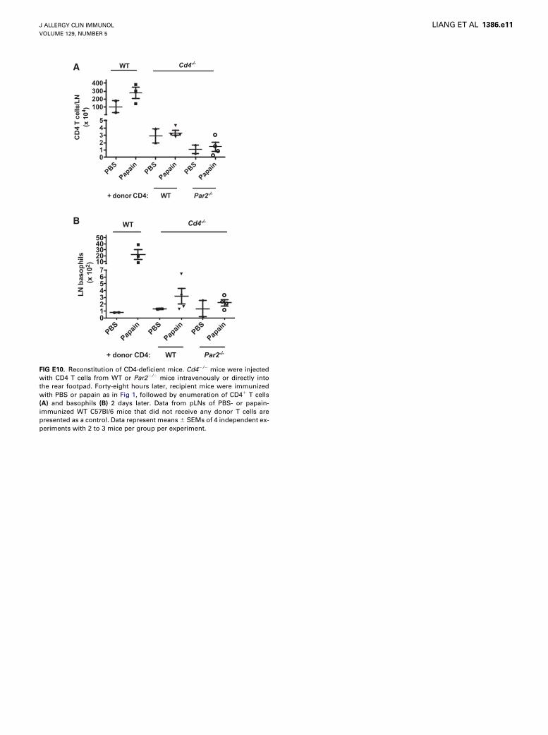
injection. However, we detected a thousand-fold fewer T cells inpLNs of the reconstituted Cd42/2 mice than were present in WTpLNs independent of papain immunization (see Fig E10,A, in thisarticle’s Online Repository at www.jacionline.org). Increasingthe number of donor CD41 T cells did not improve engraftment(data not shown). This limitation precluded meaningful analysisof basophil numbers in pLNs of papain-treated recipients (seeFig E10, B).
Papain induces PAR2-dependent IL-4 production by
naive T cells and basophilsJak3-mediated phosphorylation of STAT5 and STAT6 is inte-
gral for Il4 transcription in T cells.23 Because a Jak3 inhibitorstrongly reduced CCL17 and CCL22 production by naive T cells(Fig 5, E), we hypothesized that papain also induces Il4 expres-sion. Purified naive CD41 T cells from unimmunized mice upre-gulated Il4 mRNA within 3 hours of in vitro stimulation withpapain (Fig 7, A). Naive CD41 T cells from unimmunizedDO11.10/4get/Rag12/2 mice, which should not mount a robustantigen-restricted response to papain, also had increased greenfluorescent protein intensity after papain treatment in vitro, indi-cating IL-4 production (Fig 7, B). This result provoked the hy-pothesis that papain could induce an initial burst of IL-4secretion by naive T cells that might act in an autocrine/paracrinemanner to upregulate chemokine expression and recruit baso-phils. To evaluate the role of IL-4 in papain-evoked T-cell
*P < .05 and ***P < .0001, 1-way ANOVA.D and E, Chem
cells cultured for 20 hours with medium or papain after
oxide) alone, the PAR2 antagonist ENMD-1068 (Fig 5, D
kinases (Fig 5, E). Data are means 6 SEMs of 3 experim
:
responses, we stimulated CD41 T cells from Il42/2micewith pa-pain. Such cells had increased Ccl17/Ccl22 expression after pa-pain treatment that was comparable with that seen in WT cells(see Fig E11 in this article’s Online Repository at www.jacionline.org). Thus IL-4 itself is not required for papain-induced chemokine expression in T cells.Finally, because the receptor mediating papain-induced Il4 ex-
pression in basophils10 was unknown, we determined whetherPAR2 was required for basophil responses to papain. BMBs ex-pressed abundant surface PAR2 (Fig 7,C), andPar2 gene deletionnearly abolished papain-evoked Il4 expression in BMBs (Fig 7,D). These results suggest that PAR2 is the principal papain recep-tor on T cells and basophils and that PAR2 mediates an early cy-tokine/chemokine loop that drives TH2 immune responses topapain in mice.
DISCUSSIONWe have elucidated an innate allergen-recognition pathway in
mice mediated by conventional naive T cells and requiring PAR2,which not only induces basophil recruitment to pLNs throughchemokine production but also provides an immediate source ofIL-4 before induction of the antigen-specific TH2 immune re-sponse. The function or functions of PAR2 in T cells were previ-ously unknown. Although prior work demonstrated that Par22/2
mice had reduced OVA-induced T-cell differentiation and cyto-kine production relative to WT mice, PAR2-activating peptidealone did not stimulate cytokine production by WT murineCD41 T cells.22 We show here that both murine and human naiveT cells express surface PAR2 and respond directly to papain in aPAR2-, p38 mitogen-activated protein kinase–, and Jak3 kinase–dependent manner, and PAR2 appears to be crucial for the im-mune response to papain in vivo.
Published work has also failed to establish the primary cellularreceptor for papain or other protease allergens in immune cells.Human eosinophil degranulation in response to papain requiredthe protease activity of papain but not PAR2.24 By contrast, Kou-zaki et al25 reported that papain-evoked cytokine production byepithelial cells was partially reduced by Par2 small interferingRNA compared with control values, although the effectivenessof small interfering RNA to reduce PAR2 expression was notdemonstrated in this study. Because we detected residual chemo-kine production to papain in the pLNs of Par22/2mice, not all re-sponses to papain might involve PAR2. Given the promiscuity ofother proteases (eg, thrombin) for multiple PARs (PAR1, PAR3,and PAR4) and the ability of papain to induce alterations in mem-brane charge26 or proteolytically cleave other surface receptors onT cells (eg, CD25),27 papain might also induce T-cell activation,basophil activation, or both through other PARs or through PAR-independent mechanisms. Alternate cellular sources of chemo-kines could also compensate for the reduced pLN response to pa-pain in the absence of PAR2 in vivo.In the present study we demonstrated that T cells were required
for CCL17 and CCL22 production and basophil accumulation inpLNs after papain injection. Other leukocytes that have been
okine expression in human peripheral blood CD41 T
pretreatment for 1 hour with vehicle (dimethyl sulf-
), or the inhibitors of p38, JAK3, or c-Jun N-terminal
ents. *P < .05 and **P < .005, 1-way ANOVA.

FIG 7. Papain elicits IL-4 expression in basophils and T cells through PAR2-dependent mechanisms. A, Il4
expression in naive T cells from unimmunized C57Bl/6 mice stimulated with papain was determined by us-
ing qPCR. *P < .05 and **P 5 .002, 1-way ANOVA. B, IL-4 expression (green fluorescent protein [GFP]) in
papain-treated splenocytes from DO11.10/4get/Rag12/2 mice. Data are from a single experiment with 2
mice per group representative of 2 similar experiments. C, PAR2 on BMBs was determined by means of
flow cytometry. Numbers above outlined areas indicate percentages of total live cells. D, Il4 gene expres-
sion in BMBs from C57Bl/6 or Par22/2 mice was determined by means of qPCR. Data represent 2 to 3 exper-
iments with 3 mice per experiment. n.s., Not significant. *P < .05, 1-way ANOVA.
J ALLERGY CLIN IMMUNOL
VOLUME 129, NUMBER 5
LIANG ET AL 1385
previously reported to be reservoirs of these chemokines, includ-ing MCs, macrophages, and DCs, apparently made minor contri-butions to papain-elicited immunity. These results are consistentwith previous work showing that basophil migration to tissuesinfected with Nippostrongylus brasiliensis is absent in Rag22/2
mice.28,29 In contrast, a separate study reported only a modest de-crease in basophil recruitment to draining LNs of T cell–depletedmice (using anti-CD3 antibody) injected with papain plus OVArelative to control values.9 These results suggest that an immuneresponse to papain is distinct from that induced by simultaneousexposure to inert and protease allergens. Generation of mice withconditional deletion of PAR2 in T cells or chimeras of T cell–
deficient mice containing WT or PAR2-deficient BM might fur-ther define the functions of PAR2-expressing T cells in TH2immunity.In agreement with Sokol et al11 andNakagawa et al,30 we found
that decreased pLN basophil numbers in papain-immunizedCcr42/2 mice relative to WT mice led to reduced frequenciesof IL-41 CD4 T cells in these same LNs. Because naive T cellsdo not express CCR4, these results suggest a nonredundant roleof basophils in the antigen-restricted TH2 response to papain.Our work also suggests that quantification of total IL-41 T-cellnumbers (as has been reported in some studies) would not distin-guish differentiated, antigen-specific TH2 cells primed to produce
J ALLERGY CLIN IMMUNOL
MAY 2012
1386 LIANG ET AL
IL-4 from naive CD41 T cells stimulated through PAR2 in the ab-sence of APC-mediated priming. Thus the contribution of baso-phils to TH2 immunity overall is still unclear.
The relevance of this pathway to allergy pathogenesis in humansubjects requires further study.We show that the protease allergenpapain has the capacity to stimulate human and murine naive Tcells and murine basophils through PAR2-dependent mecha-nisms. It is unclear whether human basophils express PAR2 orbecome activated by papain.31,32 It is also unknown whether hu-man MCs respond to papain, although MCs are dispensable forimmunity to papain in mice.6,10 Accordingly, we did not detectPAR2 expression on murine BM-derived mast cells (data notshown), nor did they upregulate Il4 expression in response to pa-pain (see Fig E12 in this article’s Online Repository at www.jacionline.org).
In a previous study naive peripheral blood T cells from allergicasthmatic patients produced CCL17, whereas those from nonal-lergic control subjects did not.33 A recent population study of Ko-rean children demonstrated significant correlation of atopy, serumIgE levels, and eosinophilia with a single nucleotide polymor-phism in Par2, which increased PAR2 expression in PBMCs be-cause of enhanced mRNA stability.34 Our studies provide amolecular mechanism linking these observations, namely that al-lergens could induce pathogenic T-cell responses in atopic sub-jects as a result of increased PAR2 expression. Furtherelucidation of the mechanisms underlying allergen-elicited directprogramming of naive T cells (ie, MHC independent) could leadto the identification of new targets for the treatment of allergiesand helminth infection.
Key messages
d Naive T cells respond to the allergen papain through a Gprotein–coupled receptor, PAR2, to produce IL-4 andchemokines.
d Basophils are recruited to LNs by papain through chemo-kine- and PAR-2–dependent mechanisms and respondwith a burst of IL-4 production.
d PAR2 antagonists might subvert TH2 immunity by reduc-ing the inflammatory response to allergens, such aspapain.
REFERENCES
1. Zhu J, Yamane H, Paul WE. Differentiation of effector CD4 T cell populations (*).
Annu Rev Immunol 2010;28:445-89.
2. Paul WE, Zhu J. How are T(H)2-type immune responses initiated and amplified?
Nat Rev Immunol 2010;10:225-35.
3. Wills-Karp M, Nathan A, Page K, Karp CL. New insights into innate immune
mechanisms underlying allergenicity. Mucosal Immunol 2010;3:104-10.
4. Chapman MD, Pomes A, Breiteneder H, Ferreira F. Nomenclature and structural
biology of allergens. J Allergy Clin Immunol 2007;119:414-20.
5. Stack C, Dalton JP, Robinson MW. The phylogeny, structure and function of trem-
atode cysteine proteases, with particular emphasis on the fasciola hepatica cathep-
sin L family. Adv Exp Med Biol 2011;712:116-35.
6. Chambers L, Brown A, Pritchard DI, Sreedharan S, Brocklehurst K, Kalsheker
NA. Enzymatically active papain preferentially induces an allergic response in
mice. Biochem Biophys Res Commun 1998;253:837-40.
7. Finkelman FD, Urban JF Jr. Cytokines: making the right choice. Parasitol Today
1992;8:311-4.
8. Pulendran B, Tang H, Manicassamy S. Programming dendritic cells to induce T(H)
2 and tolerogenic responses. Nat Immunol 2010;11:647-55.
9. Tang H, Cao W, Kasturi SP, Ravindran R, Nakaya HI, Kundu K, et al. The T helper
type 2 response to cysteine proteases requires dendritic cell-basophil cooperation
via ROS-mediated signaling. Nat Immunol 2010;11:608-17.
10. Sokol CL, Barton GM, Farr AG, Medzhitov R. A mechanism for the initiation of
allergen-induced T helper type 2 responses. Nat Immunol 2008;9:310-8.
11. Sokol CL, Chu NQ, Yu S, Nish SA, Laufer TM, Medzhitov R. Basophils function
as antigen-presenting cells for an allergen-induced T helper type 2 response. Nat
Immunol 2009;10:713-20.
12. Perrigoue JG, Saenz SA, Siracusa MC, Allenspach EJ, Taylor BC, Giacomin PR,
et al. MHC class II-dependent basophil-CD41 T cell interactions promote T(H)2
cytokine-dependent immunity. Nat Immunol 2009;10:697-705.
13. Wada T, Ishiwata K, Koseki H, Ishikura T, Ugajin T, Ohnuma N, et al. Selective
ablation of basophils in mice reveals their nonredundant role in acquired immunity
against ticks. J Clin Invest 2010;120:2867-75.
14. Yoshimoto T, Yasuda K, Tanaka H, Nakahira M, Imai Y, Fujimori Y, et al. Basophils
contribute to T(H)2-IgE responses in vivo via IL-4 production and presentation of
peptide-MHC class II complexes to CD41 T cells. Nat Immunol 2009;10:706-12.
15. Stutte S, Quast T, Gerbitzki N, Savinko T, Novak N, Reifenberger J, et al. Require-
ment of CCL17 for CCR7- and CXCR4-dependent migration of cutaneous den-
dritic cells. Proc Natl Acad Sci U S A 2010;107:8736-41.
16. Alferink J, Lieberam I, Reindl W, Behrens A, Weiss S, Huser N, et al. Compart-
mentalized production of CCL17 in vivo: strong inducibility in peripheral dendritic
cells contrasts selective absence from the spleen. J Exp Med 2003;197:585-99.
17. Dogan RN, Long N, Forde E, Dennis K, Kohm AP, Miller SD, et al. CCL22 reg-
ulates experimental autoimmune encephalomyelitis by controlling inflammatory
macrophage accumulation and effector function. J Leukoc Biol 2011;89:93-104.
18. Heath WR, Carbone FR. Dendritic cell subsets in primary and secondary T cell re-
sponses at body surfaces. Nat Immunol 2009;10:1237-44.
19. Henri S, Guilliams M, Poulin LF, Tamoutounour S, Ardouin L, Dalod M, et al. Dis-
entangling the complexity of the skin dendritic cell network. Immunol Cell Biol
2010;88:366-75.
20. Wirnsberger G, Hebenstreit D, Posselt G, Horejs-Hoeck J, Duschl A. IL-4 induces
expression of TARC/CCL17 via two STAT6 binding sites. Eur J Immunol 2006;36:
1882-91.
21. Steinhoff M, Buddenkotte J, Shpacovitch V, Rattenholl A, Moormann C, Vergnolle
N, et al. Proteinase-activated receptors: transducers of proteinase-mediated signal-
ing in inflammation and immune response. Endocr Rev 2005;26:1-43.
22. Shichijo M, Kondo S, Ishimori M, Watanabe S, Helin H, Yamasaki T, et al. PAR-2
deficient CD41 T cells exhibit downregulation of IL-4 and upregulation of IFN-
gamma after antigen challenge in mice. Allergol Int 2006;55:271-8.
23. Paul WE. What determines Th2 differentiation, in vitro and in vivo? Immunol Cell
Biol 2010;88:236-9.
24. Miike S, Kita H. Human eosinophils are activated by cysteine proteases and release
inflammatory mediators. J Allergy Clin Immunol 2003;111:704-13.
25. Kouzaki H, O’Grady SM, Lawrence CB, Kita H. Proteases induce production of
thymic stromal lymphopoietin by airway epithelial cells through protease-
activated receptor-2. J Immunol 2009;183:1427-34.
26. Mekala DJ, Geiger TL. Functional segregation of the TCR and antigen-MHC com-
plexes on the surface of CTL. J Immunol 2003;171:4089-95.
27. Schulz O, Laing P, Sewell HF, Shakib F. Der p I, a major allergen of the house dust
mite, proteolytically cleaves the low-affinity receptor for human IgE (CD23). Eur J
Immunol 1995;25:3191-4.
28. Min B, Prout M, Hu-Li J, Zhu J, Jankovic D, Morgan ES, et al. Basophils produce
IL-4 and accumulate in tissues after infection with a Th2-inducing parasite. J Exp
Med 2004;200:507-17.
29. Voehringer D, Shinkai K, Locksley RM. Type 2 immunity reflects orchestrated re-
cruitment of cells committed to IL-4 production. Immunity 2004;20:267-77.
30. Nakagawa Y, Takamatsu H, Okuno T, Kang S, Nojima S, Kimura T, et al. Identi-
fication of semaphorin 4B as a negative regulator of basophil-mediated immune re-
sponses. J Immunol 2011;186:2881-8.
31. Zhu W, He SH, Lin ZX, Fu YL. [Histamine release properties of human basophils
in response to various stimuli]. Xi Bao Yu Fen Zi Mian Yi Xue Za Zhi 2005;21:
519-21.
32. Falcone FH, Morroll S, Gibbs BF. Lack of protease activated receptor (PAR) ex-
pression in purified human basophils. Inflamm Res 2005;54(suppl 1):S13-4.
33. Hirata H, Arima M, Cheng G, Honda K, Fukushima F, Yoshida N, et al. Production
of TARC and MDC by naive T cells in asthmatic patients. J Clin Immunol 2003;23:
34-45.
34. Lee JH, Kim KW, Gee HY, Lee J, Lee KH, Park HS, et al. A synonymous variation
in protease-activated receptor-2 is associated with atopy in Korean children.
J Allergy Clin Immunol 2011;128:1326-34, e3.

REFERENCES
E1. Sokol CL, Barton GM, Farr AG, Medzhitov R. A mechanism for the initiation of
allergen-induced T helper type 2 responses. Nat Immunol 2008;9:310-8.
E2. Sokol CL, Chu NQ, Yu S, Nish SA, Laufer TM, Medzhitov R. Basophils function
as antigen-presenting cells for an allergen-induced T helper type 2 response. Nat
Immunol 2009;10:713-20.
E3. Charles N, Watford WT, Ramos HL, Hellman L, Oettgen HC, Gomez G, et al.
Lyn kinase controls basophil GATA-3 transcription factor expression and induc-
tion of Th2 cell differentiation. Immunity 2009;30:533-43.
E4. Kumagai K, Itoh K, Hinuma S, Tada M. Pretreatment of plastic Petri dishes with
fetal calf serum. A simple method for macrophage isolation. J Immunol Methods
1979;29:17-25.
E5. Alkan SS, Akdis AC, Feuerlein D, Gruninger M. Direct measurement of cyto-
kines (IFN-gamma, IL-4, -5, and -6) from organs after antigenic challenge.
Ann N Y Acad Sci 1996;796:82-90.
J ALLERGY CLIN IMMUNOL
VOLUME 129, NUMBER 5
LIANG ET AL 1386.e1
METHODS
ImmunizationMice were immunized subcutaneously in the rear footpad with papain (50
mL of 1 mg/mL solution in PBS) or PBS alone, as previously described.E1,E2
For DC depletion, Cd11c-Dtr mice were depleted of DCs with a single intra-
peritoneal injection of diphtheria toxin (100 ng in 100 mL of PBS) 1 day be-
fore papain immunization.
Antibodies and reagentsFluorescein isothiocyanate (FITC)–anti-mouse FcεRI (MAR-1), allo-
phycocyanin–anti-mouse FcεRI (MAR-1), phycoerythrin (PE)–anti-CD49b
(DX5), FITC–anti-c-kit (2B8), allophycocyanin–anti-c-kit (2B8), allophy-
cocyanin–anti-CD11c (N418), PE–anti-CD11c (N418), FITC–anti-CD11c
(N418), FITC–anti-CD3a (145-2c11), allophycocyanin-eFluo780–anti-
CD3a (17A2), FITC–anti-CD4 (GK1.5), PE–anti-CD4 (GK1.5), allophy-
cocyanin–anti-CD8b, PE-Cy5–anti-CD8b, peridinin-chlorophyll-protein
complex–Cy5.5–anti-CD25 (PC61.5), PE-Cy7–anti-CD44 (1M7), allophy-
cocyanin–anti-CD205 (205yekta), allophycocyanin–anti-CD62 ligand
(MEL-14), PE–anti-IL-4 (11B11), and PE–anti-MHCII (M5/114.15.2),
and anti-human CD45RA were purchased from eBioscience (San Diego,
Calif). Anti-human CD4, PE–anti-CD19 (1D3), FITC–anti-CD8a, allophy-
cocyanin–anti-CCR3 (83103), PE–anti-CCR5 (C34-3448), allophycocya-
nin–anti-CCR6 (140706), anti-CXCR4 (2B11), PE–anti-CXCR5 (2G8),
and Fc Block (2.4G2) were from BD Biosciences (Franklin Lakes, NJ).
FITC–anti-human PAR2 (SAM-11), anti-mouse PAR2 (sc08207), and
allophycocyanin–anti-CCR1 (catalog no. sc-6125) were obtained from
Santa Cruz Biotechnology (Santa Cruz, Calif). PE–anti-CCR4 (2G12), PE–
anti-CCR7 (4B12), and allophycocyanin–anti-CXCR2 (TG11) were from
BioLegend (San Diego, Calif); anti-CCR2 was from Capralogics (Hard-
wick, Mass). Q-Dot 605–anti-human CD3 was from Invitrogen. Murine
anti-PAR2 was conjugated to Alexa Fluor 594 by using the Monoclonal
Antibody Labeling Kit (Molecular Probes, Eugene, Ore). Papain (from
Carica papaya, catalog no. 5125, lot no. D00073805) was from EMD Bio-
sciences (San Diego, Calif), and diphtheria toxin was obtained from Sigma-
Aldrich (St Louis, Mo).
The PAR2 small-molecule antagonist ENMD-1068 (Enzo Life Sci-
ences, Farmingdale, NY) was dissolved in PBS and used at final
concentrations of 1.5 and 5 mmol/L, as indicated. Cells were pretreated
for 1 hour before papain stimulation. Diphtheria toxin (Sigma-Aldrich,
100 mg in 100 mL PBS) was injected intraperitoneally 24 hours before
papain immunization.
Flow cytometryFor detection of basophils and chemokine receptors in naivemice, BMcells
and splenocytes were depleted of CD90.21 T cells and B2201 cells by using
magnetic bead sorting (Miltenyi Biotech). All cells were incubated with mu-
rine Fc Block (2.4G2) and then stained with primary antibodies. Fluorescence
was measured with a FACSCalibur (BD Biosciences) or LSRII (BD Biosci-
ences) flow cytometer and analyzedwith Flowjo software (Tree Star, Inc, Ash-
land, Ore).
Adoptive T-cell transfersSpleens were isolated from age- and sex-matchedWT C57Bl/6 or Par22/2
donors. After red cell lysis, CD41 T cells were isolated from single-cell sus-
pensions bymeans of magnetic bead sorting with the CD41 T cell isolation kit
II, according to the manufacturer’s instructions (Miltenyi Biotec). CD41 cells
(12.53 106) were resuspended in sterile PBS (200 mL) and injected either in-
travenously into the tail vein or subcutaneously into the rear footpads of age-
and sex-matched B6 Cd42/2 recipient mice. Two days after each transfer, the
recipients were either left untreated or injected subcutaneously with 50 mL of
papain (1 mg/mL) or PBS into the rear footpad. Three days later, recipient
mice were evaluated for engraftment and papain responses by means of quan-
tification of CD41 T cells and basophils in spleens and pLNs by using flow
cytometry.
Measurement of papain-specific IgEAntigen-specific IgE levels were measured in serum of human serum
albumin– or papain-immunized mice (5 mice per group), as described
previously.E3
LN cell purifications and in vitro stimulationsB cells, total T cells, or CD41 T cells were purified from pLNs by using
B220, CD90.2, or CD4 microbeads, respectively (Miltenyi Biotec). RNA
was prepared with the RNeasy kit (Qiagen). CD81 T cells were purified
from LN cell preparations depleted of CD41 cells by using CD90.2 microbe-
ads. For in vitro stimulation, naive CD41 T cells were purified from the spleen
by means of negative selection with the naive T cell isolation kit II (Miltenyi
Biotec), followed by selection of CD62L1CD11c2 cells by means of flow cy-
tometry. BM-derived mast cells were obtained from 4- to 6-week-old BM cul-
tures grown in RPMI containing 10% FCS and IL-3 (30 ng/mL) as above. For
macrophage isolation, tissue-culture plates were coated with 2 mL of 100%
heat-inactivated FCS (Life Technologies) overnight at 48C, as previously de-
scribed.E4 Dishes were washed with cold PBS, followed by seeding of splenic
cell suspensions (106 cells/mL). After 4 hours of incubation at 378C, we dec-anted nonadherent cells by means of pipetting and washing with RPMI. Ad-
herent cells were agitated in a solution of 0.2% EDTA and 5% FCS for 2
hours at 48C. Cells were then seeded in RPMI containing 10% FCS (106
cells/mL) in 12-well plates and stimulated with PBS or papain for 20 hours.
Human PBMCs were separated from leukapheresis samples obtained from
theNational Institutes of Health blood bank. NaiveCD41T cells were isolated
by means of magnetic bead sorting with the human naive CD4 T cell isolation
kit (Miltenyi Biotec). cDNAwas generated from 500 ng to 1 mg of total RNA
by using the SuperScript III Reverse Transcriptase Synthesis System (Invitro-
gen).Ccl17 andCcl22 expression was determined by usingCcl17 primer (Ap-
plied Biosystems primer ID Mm00516136_m1) and Ccl22 (Applied
Biosystems primer IDMm00436439_m1) sets with a Taqman RT-PCRmaster
mix (Applied Biosystems). Target gene values were normalized to b-actin
values (primer ID Mm01205647_g1). Human chemokine expression was de-
termined by using Ccl17 (Applied Biosystems, primer ID Hs00171074_m1)
and Ccl22 (Applied Biosystems, primer ID Hs0017080_m1) primers. Target
gene values were normalized to glyceraldehyde-3-phosphate dehydrogenase
(GAPDH) values (Applied Biosystems, primer ID#Hs99999905_m1).
Real-time qPCR and RNA arrayCytokine and chemokine expression in whole pLN RNAwas analyzed by
using the RT2 Profiler Mouse Inflammatory Cytokines and Receptors PCR ar-
ray (catalog no. PAMM-011A; SA Biosciences, Qiagen, Valencia, Calif), ac-
cording to the manufacturer’s instructions.
ELISAHomogenates were prepared from pLNs, as described elsewhere.E5 Naive
CD41 T cells purified from spleens were cultured in RPMI medium supple-
mentedwith 10%FBS for 3 days, followed by supernatant collection. The pro-
tein or supernatant samples were analyzed by using Quantikine ELISA kits
(R&D Systems, Minneapolis, Minn) per the manufacturer’s instructions.

D0 D14 D21 D28
0
10
20
30
40
Pap
ain
-sp
ecif
ic Ig
E
(O
D x
10
3)
HSA
Papain*
*
PBS Papain
Fc RI
CD
49b
0.008 0.105
PBS Papain
CD4
IL
-4
0.05 3.24
FIG E1. Papain induces a TH2 immune response. A and B, Basophils
(FcεRI1CD49b1) in total live c-kit2 population (Fig E1, A) or LN TH2 cell
(CD41IL-41) numbers (Fig E1, B) from pLNs of PBS- or papain-immunized
mice were enumerated by means of flow cytometry 3 or 4 days after papain
immunization, respectively. T cells were restimulated with phorbol 12-
myristate 13-acetate (50 ng/mL) plus ionomycin (1 mmol/L) for 6 hours
in vitro before evaluation. Numbers above outlined areas represent per-
centages of live cells. C, Serum papain-specific IgE levels in human serum
albumin (HSA)–or papain-immunized mice were determined by using
ELISA. *P 5 .02, 2-way ANOVA.
J ALLERGY CLIN IMMUNOL
MAY 2012
1386.e2 LIANG ET AL

FIG E2. Chemokine receptor expression in murine BMBs. A, BMBs were
sorted from BM cultures grown in IL-3 for 7 to 10 days, as described in
theMethods section. Numbers indicate percentages of total live c-kit2 cells.
B and C, BMBs were stained with the indicated chemokine receptor anti-
bodies (open histograms); shaded histograms represent isotype control.
Data are from a single experiment (1 mouse) representative of at least 3 in-
dependent experiments with 1 mouse per experiment.
J ALLERGY CLIN IMMUNOL
VOLUME 129, NUMBER 5
LIANG ET AL 1386.e3

FIG E3. pLN leukocyte populations in naive WT and CCR4-deficient mice. A
and B, Splenic and BM basophils (Fig E3, A) or pLN T (CD31) and B (CD191)
lymphocytes (Fig E3, B) from naive C57Bl/6 WT or CCR4-deficient mice. C,
pLN DCs (MHCIIhiCD11c1) from naive WT or CCR4-deficient mice. Numbers
above outlined areas indicate percentage of total live cells. Data are repre-
sentative of 2 to 3 independent experiments with 3 mice per experiment.
J ALLERGY CLIN IMMUNOL
MAY 2012
1386.e4 LIANG ET AL

FIG E4. Characterization of chemokine expression in pLN cell populations. A, Purity of pLN lymphocyte
populations isolated by means of magnetic bead sorting was determined by using flow cytometry with
the indicated antibodies. Numbers in each quadrant are percentages of total live cells. B, Ccl17 and Ccl22
mRNA expression in B and T cells isolated from pLNs of papain-immunized mice was determined by using
real-time PCR. *P5 .02 and ***P5 .0001, 1-way ANOVA. C, Splenic macrophages were treated with papain
(100 mg/mL) for 20 hours, followed by quantification of Ccl17 and Ccl22 gene expression by using real-time
PCR. Graphs represent means 6 SEMs of 2 to 3 independent experiments with 2 to 3 mice.
J ALLERGY CLIN IMMUNOL
VOLUME 129, NUMBER 5
LIANG ET AL 1386.e5

FIG E5. Role of DCs in the pLN immune response to papain immunization. A,Migratory dermal DCs (mDCs;
CD8a2CD11c1CD2051) from pLNs of WT or Ccr42/2 mice were enumerated 2 days after PBS or papain im-
munization. The graph shows means 6 SEMs of 2 independent experiments with 2 mice per group. SSC,
Side scatter. B, Total pLN DCs (MHCIIhiCD11c1) from Cd11c-Dtr-egfp transgenic mice 2 days after intraper-
itoneal injection with PBS or diphtheria toxin (DT). C and D, Basophils (Fig E5, C) and Ccl17 and Ccl22 gene
expression (Fig E5, D) in pLNs of mice injected with either PBS or diphtheria toxin before papain immuni-
zation. The graph shows means 6 SEMs of 2 independent experiments with 2 to 3 mice per group per ex-
periment. **P 5 .003 and ***P 5 .0006, 1-way ANOVA.
J ALLERGY CLIN IMMUNOL
MAY 2012
1386.e6 LIANG ET AL

FIG E6. Papain induces chemokine gene expression in purified naive T cells. A, Splenic naive T cells
(CD31CD41CD62L1CD11c2) from unimmunized C57/Bl6 WTmice were isolated by sorting. Numbers above
outlined areas indicate percentages of total live cells. B, Purified naive T cells were stimulated with PBS or
the indicated concentrations of papain in vitro for 20 hours, followed by quantification of Ccl17 and Ccl22
gene expression. Data are means 6 SEMs of 2 independent experiments with 2 mice per experiment.
**P 5 .001 and ***P 5 .0005, 1-way ANOVA.
J ALLERGY CLIN IMMUNOL
VOLUME 129, NUMBER 5
LIANG ET AL 1386.e7

FIG E7. PAR2 expression on T cells and effect of PAR2 deficiency on T-cell responses to papain. A and B,
Naive T cells were isolated from spleens of WT C57Bl/6 or Par22/2 mice by means of negative bead selec-
tion. Purity of T-cell fractions (top panels) and PAR2 expression (bottom panels) in naive (CD62L1; Fig E7, A)
and effector/memory (CD62L2; Fig E7, B) CD41 T cells. Numbers above outlined areas indicate percentages
of total live cells. C, Chemotaxis of BMBs from WT or Par22/2 mice to the indicated chemokines was eval-
uated in transwell assays. Data are means 6 SEMs of 2 independent experiments with 2 mice per experi-
ment. D, Splenic CD41 T cells from WT or Par22/2 mice were stimulated with papain (100 mg/mL) for 20
hours in vitro, followed by quantification of Ccl17 and Ccl22 expression by using real-time PCR. E, Splenic
J ALLERGY CLIN IMMUNOL
MAY 2012
1386.e8 LIANG ET AL

CD41 T cells were pretreated with vehicle alone (dimethyl sulfoxide) or the indicated inhibitors for 1 hour,
followed by papain stimulation for 20 hours and evaluation of chemokine expression, as in Fig E7, D. Bar
graphs show means6 SEMs of 2 to 3 independent experiments with 2 to 3 mice per group per experiment.
*P 5 .01 and **P 5 .005, 1-way ANOVA.
:
FIG E8. Leukocyte populations in organs of Cd42/2 mice. A, Absence of CD41 T cells in Cd42/2 mice. Total
splenocytes fromWT C57/Bl6 or Cd42/2 mice were evaluated bymeans of flow cytometry. B, Basophil num-
bers in spleens and freshly isolated BM of naive WT or Cd42/2 mice were quantified by using flow cytom-
etry. C and D, Total DCs (MHCIIhiCD11c1) or migratory dermal DCs (mDCs) in pLNs fromWT or Cd42/2 mice
immunized with PBS or papain. Numbers in each quadrant indicate percentages of total live cells. Bar
graphs show means 6 SEMs of 2 independent experiments with 2 to 3 mice per group per experiment.
J ALLERGY CLIN IMMUNOL
VOLUME 129, NUMBER 5
LIANG ET AL 1386.e9

FIG E9. Responses of T-cell deficient (Tcra2/2) mice to papain. A, B and T cells among total splenocytes of
WT C57Bl/6 or Tcra2/2 mice. Numbers in each quadrant indicate percentages of total live cells. B, Basophil
numbers in spleens and BM of naiveWT or Tcra2/2mice. C and D, Basophils (Fig E9, C) or migratory dermal
DCs (mDCs; Fig E9, D) in pLNs of WT or Tcra2/2 mice 3 days after papain immunization. n.s.,Not significant.
***P5 .0001, 1-way ANOVA. E, Ccl17 and Ccl22 expression in pLNs of WT or Tcra2/2 mice immunized with
PBS or papain determined by using qPCR. ***P 5 .0001, 1-way ANOVA. For flow plots, numbers in each
quadrant indicate percentages of total live cells. Bar graphs show means 6 SEMs of 3 independent exper-
iments with 2 to 3 mice per group per experiment.
J ALLERGY CLIN IMMUNOL
MAY 2012
1386.e10 LIANG ET AL
PBS
Papain
PBS
Papain
PBS
Papain
0
1
2
3
4
5
100
200
300
400
CD
4 T
cells/L
N
(x 1
04)
WT Cd4-/-
WT Par2-/-
+ donor CD4:
PBS
Papain
PBS
Papain
PBS
Papain
0
1
2
3
4
5
6
7
10
20
30
40
50
LN
baso
ph
ils
(x 1
02)
WT Cd4-/-
WT Par2-/-
+ donor CD4:
FIG E10. Reconstitution of CD4-deficient mice. Cd42/2 mice were injected
with CD4 T cells from WT or Par22/2 mice intravenously or directly into
the rear footpad. Forty-eight hours later, recipient mice were immunized
with PBS or papain as in Fig 1, followed by enumeration of CD41 T cells
(A) and basophils (B) 2 days later. Data from pLNs of PBS- or papain-
immunized WT C57Bl/6 mice that did not receive any donor T cells are
presented as a control. Data represent means6 SEMs of 4 independent ex-
periments with 2 to 3 mice per group per experiment.
J ALLERGY CLIN IMMUNOL
VOLUME 129, NUMBER 5
LIANG ET AL 1386.e11

PBS
Papain
0
2
4
6
8
Rela
tiv
e C
cl1
7
exp
ressio
n
PBS
Papain
0
2
4
6
8
10
Rela
tiv
e C
cl2
2
exp
ressio
n
Il4-/-
CD4 T cells
FIG E11. IL-4 is not required for papain-induced chemokine expression in T
cells. Splenic CD41 T cells from WT or Il42/2 mice were stimulated with pa-
pain (100 mg/mL) for 20 hours in vitro, followed by RNA isolation and quan-
tification of Ccl17 and Ccl22 gene expression by using real-time PCR. Data
are means 6 SEMs of 3 independent experiments with 1 mouse per
experiment.
J ALLERGY CLIN IMMUNOL
MAY 2012
1386.e12 LIANG ET AL

PBS
Papain
0.0
0.5
1.0
1.5
Rela
tiv
e Il4
exp
ressio
n
BMMCs
FIG E12. Murine MCs do not upregulate Il4 expression in response to pa-
pain. Bone marrow–derived mast cells (BMMCs) were stimulated with pa-
pain (100 mg/mL) for 20 hours in vitro, followed by RNA isolation and
quantification of Il4 gene expression by using real-time PCR. Data are
means 6 SEMs of 2 independent experiments with cells derived from 2
to 3 mice per experiment.
J ALLERGY CLIN IMMUNOL
VOLUME 129, NUMBER 5
LIANG ET AL 1386.e13
Copyright © 2022 FDOKUMEN