Paracoccidioides brasiliensis Enolase Is a Surface Protein That Binds Plasminogen and Mediates...
11
INFECTION AND IMMUNITY, Sept. 2010, p. 4040–4050 Vol. 78, No. 9 0019-9567/10/$12.00 doi:10.1128/IAI.00221-10 Copyright © 2010, American Society for Microbiology. All Rights Reserved. Paracoccidioides brasiliensis Enolase Is a Surface Protein That Binds Plasminogen and Mediates Interaction of Yeast Forms with Host Cells Sarah Veloso Nogueira, 1 Fernanda L. Fonseca, 2 Marcio L. Rodrigues, 2 Vasanth Mundodi, 3 Erika A. Abi-Chacra, 2 Michael S. Winters, 4 John F. Alderete, 3 and Ce ´lia Maria de Almeida Soares 1 * Laborato ´rio de Biologia Molecular, Instituto de Cie ˆncias Biolo ´gicas, Universidade Federal de Goia ´s, 74001-970 Goia ˆnia, Goia ´s, Brazil 1 ; Laboratorio de Estudos Integrados em Bioquimica Microbiana, Instituto de Microbiologia Professor Paulo de Goes, Universidade Federal do Rio de Janeiro, Rio de Janeiro 21941-590, Brazil 2 ; School of Molecular Biosciences, Washington State University, Pullman, Washington 99163 3 ; and Division of Infectious Diseases, University of Cincinnati College of Medicine, Cincinnati, Ohio 45267 4 Received 5 March 2010/Returned for modification 31 March 2010/Accepted 28 June 2010 Paracoccidioidomycosis (PCM), caused by the dimorphic fungus Paracoccidioides brasiliensis, is a dissemi- nated, systemic disorder that involves the lungs and other organs. The ability of the pathogen to interact with host components, including extracellular matrix (ECM) proteins, is essential to further colonization, invasion, and growth. Previously, enolase (EC 4.2.1.11) was characterized as a fibronectin binding protein in P. brasiliensis. Interaction of surface-bound enolase with plasminogen has been incriminated in tissue invasion for pathogenesis in several pathogens. In this paper, enolase was expressed in Escherichia coli as a recombinant glutathione S-transferase (GST) fusion protein (recombinant P. brasiliensis enolase [rPbEno]). The P. brasil- iensis native enolase (PbEno) was detected at the fungus surface and cytoplasm by immunofluorescence with an anti-rPbEno antibody. Immobilized purified rPbEno bound plasminogen in a specific, concentration- dependent fashion. Both native enolase and rPbEno activated conversion of plasminogen to plasmin through tissue plasminogen activator. The association between PbEno and plasminogen was lysine dependent. In competition experiments, purified rPbEno, in its soluble form, inhibited plasminogen binding to fixed P. brasiliensis, suggesting that this interaction required surface-localized PbEno. Plasminogen-coated P. brasil- iensis yeast cells were capable of degrading purified fibronectin, providing in vitro evidence for the generation of active plasmin on the fungus surface. Exposure of epithelial cells and phagocytes to enolase was associated with an increased expression of surface sites of adhesion. In fact, the association of P. brasiliensis with epithelial cells and phagocytes was increased in the presence of rPbEno. The expression of PbEno was upregulated in yeast cells derived from mouse-infected tissues. These data indicate that surface-associated PbEno may contribute to the pathogenesis of P. brasiliensis. Microbial adhesion to host tissues is the initial event of most infectious process (39). Interaction with extracellular matrix (ECM) proteins has been correlated with the invasive abilities of different organisms (28, 40). ECM underlines epithelial and endothelial cells and surrounds connective tissues, and its ma- jor components are the collagens, laminin, fibronectin, and proteoglycans (52). After adherence, the next step must be to overcome the barriers imposed by epithelial tissues and ECM. The proteolytic activity achieved by subversion of host pro- teases by pathogens, such as plasmin, has been shown to be important during the process of many types of infections (47, 51). Paracoccidioides brasiliensis is the causative agent of para- coccidioidomycosis (PCM), a human systemic mycosis that constitutes a major health problem in South America (44). Clinical manifestations of PCM are related to chronic granu- lomatous reactions with involvement of the lungs and the re- ticuloendothelial system, as well as mucocutaneous areas and other organs (22). In the soil, the fungus grows as saprobic mycelium, resulting in the formation of infectious propagules. After penetrating the host, the fungus differentiates into its yeast form, a fundamental step for the successful establishment of the disease (46). Although P. brasiliensis is not traditionally considered a typ- ical intracellular pathogen, independent studies have demon- strated that P. brasiliensis yeast cells have the capacity to ad- here and invade host cells (4, 24, 31). P. brasiliensis may actively penetrate the mucocutaneous surface and parasitize epithelial cells, thus evading the host defenses and reaching deeper tissues. Fungal ECM-binding adhesins have been characterized in different models, including P. brasiliensis. Vicentini et al. (49) showed specific binding of the protein gp43 to laminin, which is correlated to the adhesiveness of the fungus in vitro as well as to an enhancement of pathogenic potential. We have been systematically searching for new adhesion proteins in P. bra- siliensis that have the potential to play roles in the fungal virulence, and proteins such as P. brasiliensis malate synthase (PbMLS) (34), PbDfg5p (defective for filamentous growth pro- * Corresponding author. Mailing address: Laborato ´rio de Biologia Molecular, Instituto de Cie ˆncias Biolo ´gicas, ICBII, Campus II, Uni- versidade Federal de Goia ´s, 74001-970, Goia ˆnia, Goia ´s, Brazil. Phone and fax: 55-62-35211110. E-mail: [email protected]. Published ahead of print on 6 July 2010. 4040
Transcript of Paracoccidioides brasiliensis Enolase Is a Surface Protein That Binds Plasminogen and Mediates...

INFECTION AND IMMUNITY, Sept. 2010, p. 4040–4050 Vol. 78, No. 90019-9567/10/$12.00 doi:10.1128/IAI.00221-10Copyright © 2010, American Society for Microbiology. All Rights Reserved.
Paracoccidioides brasiliensis Enolase Is a Surface Protein ThatBinds Plasminogen and Mediates Interaction of Yeast
Forms with Host Cells�
Sarah Veloso Nogueira,1 Fernanda L. Fonseca,2 Marcio L. Rodrigues,2 Vasanth Mundodi,3Erika A. Abi-Chacra,2 Michael S. Winters,4 John F. Alderete,3
and Celia Maria de Almeida Soares1*Laboratorio de Biologia Molecular, Instituto de Ciencias Biologicas, Universidade Federal de Goias, 74001-970 Goiania, Goias,
Brazil1; Laboratorio de Estudos Integrados em Bioquimica Microbiana, Instituto de Microbiologia Professor Paulo de Goes,Universidade Federal do Rio de Janeiro, Rio de Janeiro 21941-590, Brazil2; School of Molecular Biosciences,
Washington State University, Pullman, Washington 991633; and Division of Infectious Diseases,University of Cincinnati College of Medicine, Cincinnati, Ohio 452674
Received 5 March 2010/Returned for modification 31 March 2010/Accepted 28 June 2010
Paracoccidioidomycosis (PCM), caused by the dimorphic fungus Paracoccidioides brasiliensis, is a dissemi-nated, systemic disorder that involves the lungs and other organs. The ability of the pathogen to interact withhost components, including extracellular matrix (ECM) proteins, is essential to further colonization, invasion,and growth. Previously, enolase (EC 4.2.1.11) was characterized as a fibronectin binding protein in P.brasiliensis. Interaction of surface-bound enolase with plasminogen has been incriminated in tissue invasion forpathogenesis in several pathogens. In this paper, enolase was expressed in Escherichia coli as a recombinantglutathione S-transferase (GST) fusion protein (recombinant P. brasiliensis enolase [rPbEno]). The P. brasil-iensis native enolase (PbEno) was detected at the fungus surface and cytoplasm by immunofluorescence withan anti-rPbEno antibody. Immobilized purified rPbEno bound plasminogen in a specific, concentration-dependent fashion. Both native enolase and rPbEno activated conversion of plasminogen to plasmin throughtissue plasminogen activator. The association between PbEno and plasminogen was lysine dependent. Incompetition experiments, purified rPbEno, in its soluble form, inhibited plasminogen binding to fixed P.brasiliensis, suggesting that this interaction required surface-localized PbEno. Plasminogen-coated P. brasil-iensis yeast cells were capable of degrading purified fibronectin, providing in vitro evidence for the generationof active plasmin on the fungus surface. Exposure of epithelial cells and phagocytes to enolase was associatedwith an increased expression of surface sites of adhesion. In fact, the association of P. brasiliensis with epithelialcells and phagocytes was increased in the presence of rPbEno. The expression of PbEno was upregulated inyeast cells derived from mouse-infected tissues. These data indicate that surface-associated PbEno maycontribute to the pathogenesis of P. brasiliensis.
Microbial adhesion to host tissues is the initial event of mostinfectious process (39). Interaction with extracellular matrix(ECM) proteins has been correlated with the invasive abilitiesof different organisms (28, 40). ECM underlines epithelial andendothelial cells and surrounds connective tissues, and its ma-jor components are the collagens, laminin, fibronectin, andproteoglycans (52). After adherence, the next step must be toovercome the barriers imposed by epithelial tissues and ECM.The proteolytic activity achieved by subversion of host pro-teases by pathogens, such as plasmin, has been shown to beimportant during the process of many types of infections(47, 51).
Paracoccidioides brasiliensis is the causative agent of para-coccidioidomycosis (PCM), a human systemic mycosis thatconstitutes a major health problem in South America (44).Clinical manifestations of PCM are related to chronic granu-lomatous reactions with involvement of the lungs and the re-
ticuloendothelial system, as well as mucocutaneous areas andother organs (22). In the soil, the fungus grows as saprobicmycelium, resulting in the formation of infectious propagules.After penetrating the host, the fungus differentiates into itsyeast form, a fundamental step for the successful establishmentof the disease (46).
Although P. brasiliensis is not traditionally considered a typ-ical intracellular pathogen, independent studies have demon-strated that P. brasiliensis yeast cells have the capacity to ad-here and invade host cells (4, 24, 31). P. brasiliensis mayactively penetrate the mucocutaneous surface and parasitizeepithelial cells, thus evading the host defenses and reachingdeeper tissues.
Fungal ECM-binding adhesins have been characterized indifferent models, including P. brasiliensis. Vicentini et al. (49)showed specific binding of the protein gp43 to laminin, whichis correlated to the adhesiveness of the fungus in vitro as wellas to an enhancement of pathogenic potential. We have beensystematically searching for new adhesion proteins in P. bra-siliensis that have the potential to play roles in the fungalvirulence, and proteins such as P. brasiliensis malate synthase(PbMLS) (34), PbDfg5p (defective for filamentous growth pro-
* Corresponding author. Mailing address: Laboratorio de BiologiaMolecular, Instituto de Ciencias Biologicas, ICBII, Campus II, Uni-versidade Federal de Goias, 74001-970, Goiania, Goias, Brazil. Phoneand fax: 55-62-35211110. E-mail: [email protected].
� Published ahead of print on 6 July 2010.
4040
tein) (10), triosephosphate isomerase (PbTPI) (41), and glyc-eraldehyde-3-phosphate dehydrogenase (PbGAPDH) (4) werefound to associate with ECM components. In particular, eno-lase from P. brasiliensis (PbEno) is a fibronectin-binding pro-tein, as characterized by affinity ligand assays (17).
The importance of plasminogen in infectious diseases is sup-ported by the fact that many pathogens manifest the ability tobind plaminogen (47, 13). Plasminogen is a single-chain glyco-protein with a molecular mass of 92 kDa. Its protein structurecomprises an N-terminal preactivation peptide, five consecu-tive disulfide-bonded triple-loop kringle domains, and a serine-protease domain containing the catalytic triad (48). The krin-gle domains of plasminogen mediate its attachment to cellssurfaces by binding proteins with accessible carboxyl-terminalor internal lysine residues. The plasminogen system displays aunique role in the host defense by dissolving fibrin clots andserving as an essential component to maintain homeostasis(43). Activation of the fibrinolytic system is dependent on theconversion of plasminogen to the serine-protease plasmin bythe physiological activators urokinase-type plasminogen acti-vator (uPA) and tissue-type plasminogen activator (tPA) (9).Plasmin is involved in fibrinolysis homeostasis and degradationof the extracellular matrix and basement membrane. Themammalian plasminogen-plasmin proteolytic system plays acrucial role in extracellular matrix degradation which is ex-ploited by invasive pathogens, including fungi (25, 47). Mi-crobe-derived plasminogen conversion to plasmin may pro-mote dissemination of the pathogen within the host (1).
Among several proteins, enolase has been found to play amajor role in microbial recruitment of plasminogen (32). Byserving as a key surface receptor for plasminogen recruitment,enolase has been shown to function as mediator of microbialvirulence (6, 15). The potential of P. brasiliensis to recruithuman plasminogen for invasion and virulence has not beenstudied until now. In this study, we demonstrated for the firsttime that P. brasiliensis is capable of recruiting plasminogenand activating the plasminogen fribrinolytic system in a pro-cess, at least in part, mediated by the cell wall-localized eno-lase. Furthermore, recombinant PbEno (rPbEno) promoted anincrease in the adhesion/invasion of P. brasiliensis in in vitromodels of infection, a process that seems to be associated withthe enolase ability of modifying the surface of host cells. Thesedata suggest that PbEno may play a role in mediating the P.brasiliensis recruitment of plasminogen as well as in attachmentand internalization of the fungus to host tissues, potentiallyplaying a role in the establishment of PCM.
MATERIALS AND METHODS
Fungal isolate and growth conditions. Yeast cells were obtained by growing P.brasiliensis isolate 01 (ATCC MYA-826) in Fava-Netto’s medium for 4 days at36°C as described previously (4).
Cloning cDNA containing the complete enolase coding region into an expres-sion vector. The enolase cDNA (GenBank accession number EF558735.1), ob-tained from a library from yeast cells of P. brasiliensis (14), was amplified by PCRusing oligonucleotide sense (5�-GTC GAC ATG GCT ATC ACC AAA ATCCAC G-3�; SalI restriction site underlined) and antisense (5�-GCG GCC GCTTAC ATA TTA ATA GCT GCC C-3�; NotI restriction site underlined) primers.The PCR product was cloned in frame with the glutathione S-transferase (GST)coding region of the pGEX-4T-3 vector (GE Healthcare) to yield the pGEX-4T-3-PbEno construct. The Escherichia coli strain BL21(pLys) competent cellswere transformed with the expression construct.
Expression and characterization of the recombinant enolase. Bacteria trans-formed with the pGEX-4T-3-PbEno construct were grown in LB medium sup-plemented with ampicillin (100 �g/ml) and glucose (20 mM) at 37°C at 200 rpm.Protein expression was induced by the addition of isopropyl-�-D-thiogalacto-pyranoside (IPTG) to a final concentration of 0.1 mM. The GST-PbEno proteinwas affinity purified using glutathione Sepharose 4B (GE Healthcare), and theGST was cleaved by the addition of thrombin (Sigma Aldrich).
Antibody production. The purified rPbEno was used to generate specific rabbitpolyclonal serum. Rabbit preimmune serum was obtained and stored at �20°C.The purified protein was injected into rabbits with Freund’s adjuvant three timesat 2-week intervals. The serum, containing monospecific anti-rPbEno polyclonalantibodies (5.3 �g/�l), was stored at �20°C.
Preparation of P. brasiliensis protein fractions. The P. brasiliensis crude pro-tein extract was obtained by disruption of frozen yeast cells in the presence ofprotease inhibitors 50 �g/ml N-�-p-tosyl-L-lysine chloromethyl ketone (TLCK), 1mM 4-chloromercuribenzoic acid (PCMB), 20 mM leupeptin, 20 mM phenyl-methylsulfonyl fluoride (PMSF), and 5 mM iodoacetamide in homogenizationbuffer (20 mM Tris-HCl, pH 8.8, 2 mM CaCl2). The mixture was centrifuged at12,000 � g at 4°C for 10 min, and the supernatant was used for further analysisof proteins.
Cell wall fractionation was performed basically as described previously (42).The culture filtrate was processed as described previously (33) with modifica-
tions. Yeast cells were harvested from the solid medium and transferred to FavaNetto’s liquid medium. After 1 day of growth at 37°C with gentle agitation, theproteins from the supernatant were precipitated with 10% (wt/vol) trichloroace-tic acid (TCA) during overnight (o/n) incubation at 4°C. The precipitate wascentrifuged for 10 min at 10,000 � g. The pellet was washed twice with acetoneand air dried prior to resuspension in electrophoresis dissolving buffer. Proteinsamples were then subjected to SDS-PAGE. The protein content was quantifiedusing the Bradford assay (8).
Two-dimensional (2D) gel electrophoresis and MALDI-TOF mass spectrom-etry analysis. Samples containing 200 �g of P. brasiliensis yeast protein crudeextract were separated by isoelectric focusing as described by O’Farrell in 1975(37). The second dimension was performed as described by Laemmli in 1970(27). Protein spots were excised from the gel and submitted to reduction, alqui-lation, and in-gel digestion with trypsin (Promega, Madison, WI). The resultingtryptic peptides were extracted and submitted to mass spectrometry (MS) anal-ysis. The protein tryptic fragments were analyzed using a matrix-assisted laserdesorption ionization–time of flight (MALDI-TOF) mass spectrometer (ReflexIV; Bruker Daltonics, Karlsruhe, Germany). The peptide mass list obtained foreach spectrum was searched against the SwissProt database (http://expasy.org/sprot) by using the MASCOT search engine (Matrix Science).
Western blot and ligand binding analyses. Proteins fractionated by gel elec-trophoresis were transferred to nylon membranes. Blots were sequentially incu-bated with the rabbit polyclonal anti-rPbEno antibodies, anti-rabbit immuno-globulin G (IgG) coupled to alkaline phosphatase (Sigma), and developed with5-bromo-4-chloro-3-indolylphosphate–nitroblue tetrazolium (BCIP-NBT). Rab-bit preimmune serum was used as a negative control.
For ligand blot analysis, the membrane was washed three times with 0.1%(vol/vol) Tween 20 in phosphate-buffered saline (PBS) and incubated o/n withhuman plasminogen (hPlg; Sigma) (35 �g/ml) in PBS–1% bovine serum albumin(BSA) (wt/vol). The blot was washed and incubated with mouse anti-humanplasminogen monoclonal antibody (MAb) (1 �g/ml) (R&D Systems) in PBS–1%BSA (wt/vol) for 1 h at room temperature. The membrane was next incubatedwith anti-mouse IgG coupled to alkaline phosphatase (Sigma). The blots weredeveloped with BCIP-NBT.
Immunofluorescence detection of PbEno and plasminogen on the surface ofP. brasiliensis yeast cells. An immunofluorescence assay was performed using amodification of a described procedure (33). Briefly, 2 � 107 yeast cells were fixedwith 4% (vol/vol) paraformaldehyde in PBS for 10 min. The cells were washedtwice in PBS and blocked with BSA for 1 h at room temperature prior toincubation with (i) rabbit anti-rPbEno antibodies or (ii) plasminogen followed byincubation with antiplasminogen monoclonal antibodies. The cells were thenwashed three times with PBS and treated for 1 h at 37°C with affinity-purifiedfluorescein isothiocyanate-conjugated goat anti-rabbit IgG or anti-mouse IgG(Sigma) diluted 1:1,000. Finally, yeast cells were washed twice with PBS andvisualized using the Olympus BX41 microscope at �100 magnification.
Plasminogen binding assay. Cellular assays were performed after coating thewells of multititer plates with fungal cells followed by fixation. Briefly, 1 � 108
yeast cells in PBS were added to the wells and incubated for 1 h, and glutaral-dehyde was added to the wells to a final concentration of 1% (vol/vol) for 10 min.After three washes with PBS, the wells were blocked with 1% (wt/vol) BSA inPBS for 1 h. Different amounts of hPlg (0.05 to 1.0 �g) were added to the wells,
VOL. 78, 2010 FUNCTIONS OF P. BRASILIENSIS ENOLASE 4041

which were incubated for 1 h. Competition experiments were performed by theaddition of increasing concentrations (0.5 �g to 3 �g) of rPbEno for 1 h prior tothe addition of 1 �g of hPlg. Binding was determined by incubation with anti-plasminogen monoclonal antibody. Plates were washed three times with 0.1%Tween 20 (vol/vol) in PBS. Horseradish peroxidase was added to the wells andincubated for 1 h. The absorbance was measured at A405 by using a microplatereader (Bio Tek Instruments Inc., Winooski, VT).
In another set of experiments, wells of multititer plates were coated with 1 �gof rPbEno diluted in carbonate buffer. After blocking and washing as describedabove, different amounts of hPlg (1 �g to 4 �g) were added to the plates.Alternatively, the plates were coated with 1 �g of hPlg diluted in carbonate bufferand incubated o/n at 4°C. A range of concentrations (1 �g to 4 �g) of rPbEnodiluted in 1% BSA (wt/vol) in PBS were added to the hPlg-coated wells, incu-bated for 1 h, and washed with 0.1% (vol/vol) Tween 20 in PBS. Protein-proteininteractions were determined by incubation with anti-rPbEno polyclonal anti-bodies. Competition experiments were performed by the addition of increasingconcentrations (5 mM to 20 mM) of the lysine analogue ε-aminocaproic acid(ε-ACA) (Sigma) to the rPbEno-coated wells. The wells were incubated for 1 hfollowed by the addition of hPlg. Another set of competition experiments in-cluded the addition of specific rPbEno rabbit polyclonal antibodies prior to theaddition of hPlg. All reactions were carried out at 37°C. Binding was determinedby incubation with antiplasminogen monoclonal antibody. Following threewashes, the wells were developed as described above. All final volumes for theenzyme-linked immunosorbent assay (ELISA) reactions were 100 �l.
Plasminogen activation assay. Plasminogen activation was performed by mea-suring the amidolytic activity of the generated plasmin. Wells of multititer plateswere coated with 1 �g of rPbEno or fixed P. brasiliensis and incubated with 1 �ghPlg (Sigma), 3 �g of plasmin substrate (D-valyl-L-lysyl-p-nitroaniline hydrochlo-ride) (Sigma), and 15 ng of tissue plasminogen activator (tPA) (Sigma). Controlexperiments were performed by measuring the generation of plasmin either inthe absence of tPA or in the presence of ε-ACA. Plates were incubated at roomtemperature for 2 h and read at A405.
Degradation of fibrin in jellified matrices. Fibrinolysis was assayed usingpreviously described methods with minor modifications (25). Briefly, 107 P.brasiliensis cells were preincubated with hPlg (50 �g) for 3 h in the presence orabsence of tPA (50 ng) and the serine proteinase inhibitors aprotinin (1 �g) andPMSF (50 mM) in a final volume of 1 ml. Thereafter, the mixtures were washedthree times with PBS to remove free plasminogen. The resulting cell pellets wereplaced in wells of a fibrin substrate matrix gel that contained 1.25% (wt/vol)low-melting-temperature agarose, hPlg (100 �g), and fibrinogen (4 mg; Sigma) ina final volume of 2 ml. Controls consisted of untreated cells (no plasminogenincubation) or incubation systems where no cells were added. The jellified matrixwas incubated in a humidified chamber at 37°C for 12 h. Plasmin activity wasdetected by the observation of clear hydrolysis haloes within the opaque jellified-fibrin-containing matrix.
Influence of enolase on the interaction of P. brasiliensis with host cells. Humantype II alveolar cells (A549 lineage) and murine macrophage-like cells (RAW264.7 [RAW] lineage) were obtained from the American Type Culture Collec-
FIG. 1. Identification of enolase in the P. brasiliensis proteome by two-dimensional gel electrophoresis. (A) Protein staining with Coomassieblue. (B) Reactivity of the P. brasiliensis total extract with rabbit polyclonal antibodies to enolase raised to the recombinant protein. Numbers tothe left of panels A and B refer to the molecular mass of the enolase. At the top is the isoelectric point of the protein. Arrows point to enolase.(C) Peptide mass spectrum generated from tryptic digestion of PbEno. The protein reacting with polyclonal antibodies was removed from the geland submitted to mass spectrometry analysis after trypsin digestion. The black stars indicate molecular mass values, and each peak correspondsto a peptide. Experiments represent three gels from independent protein preparations. BP, base peak.
4042 NOGUEIRA ET AL. INFECT. IMMUN.

tion (ATCC). Cultures were maintained and grown to confluence in 25-cm2
culture flasks containing Dulbecco’s modified Eagle’s medium (DMEM) supple-mented with 10% (vol/vol) fetal bovine serum (FBS) at 37°C with 5% CO2. Toevaluate the effects of rPbEno on the interaction of P. brasiliensis with host cells,RAW and A549 lineages were first exposed to the enzyme and then probed withsuccinylated wheat germ agglutinin (S-WGA). S-WGA has affinity for �1,4-N-acetylglucosamine (GlcNAc) oligomers, which are recognized by the P. brasil-iensis adhesin paracoccin (23). Mammalian cells were placed in a 24-well plate(105 cells/well) and treated with various concentrations of rPbEno (1 and 50�g/ml) for 1 h at 37°C. Controls were exposed to medium alone for the sameamount of time. The cells were detached from plastic surfaces, fixed, and blockedas described previously (3) and then incubated for 30 min at 37°C in 100 �l of a5-�g/ml solution of tetramethyl rhodamine isothiocyanate (TRITC)-labeledS-WGA (EY Laboratories). The cells were washed in PBS and analyzed by flowcytometry as previously described (3). To analyze the effects of rPbEno on theinteraction of P. brasiliensis with host cells, the culture medium of RAW or A549cells was replaced with fresh media for further incubation with P. brasiliensisyeast cells. For flow cytometry experiments, the cell wall of P. brasiliensis wasstained with 0.5 mg/ml fluorescein isothiocyanate (FITC; Sigma) (3, 11) in PBS(25°C) for 10 min (11). Fungal suspensions were prepared in DMEM to generatea ratio of 10 yeasts per host cell. Interactions between fungal and host cellsoccurred at 37°C with 5% CO2 for 18 h. Cells were washed three times with PBSto remove nonadherent yeasts. Fungus-host cell complexes were treated for 10min at 25°C with trypan blue (200 �g/ml) to discriminate between surface-associated and intracellular yeast cells (3, 11). After removal from the plasticsurface with a cell scrapper, the cells were analyzed by flow cytometry as de-scribed previously (3). Control preparations were developed as described aboveby using uninfected cells and nonstained yeast (data not shown). For analysis ofthe morphological aspects of infected cells, the complexes were fixed with para-formaldehyde and stained with 25 �M calcofluor white (Invitrogen, Life Tech-nologies). Control or infected cells were finally observed with an Axioplan 2(Zeiss, Germany) fluorescence microscope, following conditions previously de-scribed (3).
Infection of mice with P. brasiliensis and RNA extraction. Mice were infectedas described previously (16). Female BALB/c mice were infected intraperitone-ally with 1 � 108 yeast cells and intranasally with 5 � 107 yeast cells and killedon the seventh day after infection; livers and spleens were removed from miceinfected intraperitoneally, and lungs were removed from mice infected intrana-sally. One hundred milliliters of this suspension was plated onto brain heartinfusion (BHI) agar (Becton-Dickinson, MD) supplemented with 1% (wt/vol)glucose. After 7 days, total RNA was extracted from the yeast cells (1 � 1010).Control cDNA was prepared by removing P. brasiliensis yeast cells from Fava-Netto cultures and plating to BHI agar as described above.
Quantitative analysis of RNA transcripts by qRT-PCR. Total RNAs weretreated with DNase, and cDNA was prepared using Superscript II reverse trans-criptase (Invitrogen) and oligo(dT)15 primer. Quantitative real-time reversetranscription-PCR (qRT-PCR) analysis was performed on a StepOnePlus real-time PCR system (Applied Biosystems, Foster City, CA) in triplicate. Valueswere averaged from three biological replicates. PCR thermal cycling was per-
formed at 40 cycles of 95°C for 15 s followed by 60°C for 1 min. Ten picomolesof each primer and 40 ng of template cDNA in a total volume of 25 �l SYBRgreen PCR master mix (Applied Biosystems) were used for each experiment. Amelting curve analysis was performed to confirm a single PCR product. The datawere normalized with transcript encoding tubulin amplified in each set of qRT-PCR experiments. A nontemplate control was also included. Relative expressionlevels of the genes of interest were calculated using the standard curve methodfor relative quantification (7).
Statistical analysis. Experiments were performed in triplicate with samples intriplicates. Results are presented as means � standard deviations. Statisticalcomparisons were performed using Student’s t test. Statistical significance wasaccepted for P values of �0.05.
RESULTS
Expression and purification of the P. brasiliensis enolase andproduction of polyclonal antibodies. Previous studies identi-fied PbEno (EC 4.2.1.11) as a fibronectin binding protein. Inthe present work to further investigate the role of PbEno infungus-host interaction, we first expressed the protein in orderto create antibodies specific for PbEno. The cDNA encodingPbEno (GenBank accession number EF558735.1) was clonedinto the expression vector pGEX-4T-3 to obtain the recombi-nant fusion protein GST-PbEno. The fusion protein was affin-ity purified, and the 47-kDa rPbEno was obtained by digestionwith thrombin (data not shown). The purified rPbEno was usedto generate rabbit polyclonal antibodies.
Antibody specificity was evaluated in serological tests usingprotein extracts from cell lysates resolved by 2D gel electro-phoresis. A protein with a pI of 5.67 was recognized by poly-clonal antibodies raised to rPbEno (Fig. 1A and B). Theprotein was analyzed by mass spectrometry (Fig. 1C). Experi-mental masses were searched against data from public genedatabases by using MASCOT. The peptides obtained (Table1)matched PbEno.
Detection of PbEno on fungal surface. In order to deter-mine the cellular distribution of PbEno, we probed differentfractions of fungal cells by Western blot analysis. PbEno wasdetected in total protein extract (Fig. 2A, lane 3), cell wall-enriched fraction (Fig. 2A, lane 4), cytoplasmic fraction(Fig. 2A, lane 5), and the culture filtrate (Fig. 2A, lane 6).Bovine serum albumin (Fig. 2A, lane 1) and rPbEno (Fig.
TABLE 1. Identification of P. brasiliensis enolase by peptide mass fingerprinta
Positionb Identified amino acid sequencec
Mass
Exptl (in-geldigestion)
Theoretical(in silico digestion)
16–32 R.GNPTVEVDVVTETGLHR.A 1,822.9489 1,822.929333–50 R.AIVPSGASTGQHEACELR.D 1,882.9548 1,825.886190–103 K.VDEFLNKLDGTPNK.S 1,589.8751 1,588.8678106–120 K.LGANAILGVSLAIAK.A 1,410.9979 1,410.8678164–184 R.LAFQEFMIVPTAAPSFSEALR.Q 2,325.2178 2,325.1947243–254 K.IALDIASSEFYK.A 1,356.7701 1,356.7045274–285 K.WLTYEQLADLYK.K 1,542.8513 1,542.7838314–331 K.TCDLQVVADDLTVTNPIR.I 2,030.0239 1,973.0008377–393 R.SGETEDVTIADIVVGLR.A 1,773.9489 1,773.9228411–416 K.LNQILR.I 756.4464 756.4726
a Protein scores higher than 76 are significant (P � 0.05). Peptide masses matched with PbEno (GenBank accession number EF558735.1), presenting a score of 113and coverage of 34.25% of the whole deduced sequence.
b Position corresponds to the peptide fragment detected by mass spectrometry.c The sequence within the periods represents the amino acids of the detected fragment. The amino acids outside of the periods represent the amino acids which
remain within the protein following tryptic digestion.
VOL. 78, 2010 FUNCTIONS OF P. BRASILIENSIS ENOLASE 4043

2A, lane 2) were employed as negative and positive controls,respectively. Altogether, these results suggest that PbEno isassociated with the cell wall and is secreted to the extracel-lular space in addition to its expected intracellular distribu-tion in P. brasiliensis.
To further validate PbEno’s association with the fungal sur-face, immunofluorescence was performed. As shown in Fig. 2B(panel 4), rabbit polyclonal antibodies reacted with the surfaceof the organism. Plasminogen antibodies (Fig. 2B, panel 6) alsoreacted with the surface of plasminogen-treated organisms,suggesting an ability of P. brasiliensis to recognize this mole-cule. No fluorescence and immunoreactivity were detectedwhen yeast cells were incubated with the secondary antibodyalone (Fig. 2B, panel 2).
P. brasiliensis and PbEno bind plasminogen. By the immu-nofluorescence assay, we discovered that P. brasiliensis binds tohPlg. To further characterize this phenomenon, P. brasiliensisyeast cells were fixed to the wells of multititer plates andincreasing concentrations of hPlg were added. Figure 3Ashows a dose-dependent pattern of binding of hPlg to fixedfungal cells. The addition of increasing concentrations ofrPbEno decreased hPlg binding to P. brasiliensis in a dose-dependent manner (Fig. 3B). These data further confirmedthat enolase was involved in P. brasiliensis binding to hPlg. Tosupport this supposition, we performed a ligand blot assayusing crude protein extracts, the cell wall-enriched fraction,and rPbEno (Fig. 3C). Different P. brasiliensis proteins, includ-ing enolase, interacted with hPlg. The presence of severalproteins in the ligand blot assay implicated the existence ofother plasminogen binding proteins in P. brasiliensis, as de-scribed for other organisms (15). Therefore, the ability ofrPbEno to bind hPlg was tested in an ELISA. Increasing con-centrations of hPlg bound to immobilized rPbEno in a dose-dependent fashion (Fig. 3D). The same increasing pattern wasobserved when the wells were coated with hPlg and increasingconcentrations of rPbEno were added (Fig. 3E).
Previous work has shown that enolase binds to hPlg throughlysine residues (33, 47). We thus examined if binding ofrPbEno was lysine dependent by using competitive antagonismwith the lysine analog ε-ACA. The results shown in Fig. 3Findicate that lysine residues present on rPbEno may have arole in plasminogen recruitment by P. brasiliensis.
To further validate rPbEno binding to hPlg, competitionexperiments were also performed by the addition of specificrPbEno rabbit polyclonal antibodies to the experimental sys-tem. The presence of enolase-specific antibodies dose-depen-dently decreased hPlg binding to rPbEno (Fig. 3G). In thepresence of preimmune sera, no effects were observed (datanot shown). These data confirmed that enolase specificallybinds hPlg.
Plasminogen activation and fibrinolysis. Once yeast cellsand rPbEno had been seen to bind hPlg, we had expected thatthis interaction could also activate hPlg. For this reason, anELISA was performed to determine the abilities of P. brasil-iensis and rPbEno to produce plasmin from hPlg. In the pres-ence of tPA, rPbEno was able to generate plasmin (Fig. 4A).The addition of ε-ACA inhibited plasmin generation. hPlgactivation was evaluated in assays using fixed fungal cells inthe presence of tPA, confirming that interaction with fixedP. brasiliensis also resulted in hPlg activation (Fig. 4B). Theaddition of increasing concentrations of ε-ACA to the ex-perimental system inhibited plasmin generation in a dose-dependent manner. These results suggest that P. brasiliensisand rPbEno mediate activation of plasminogen to plasmin
FIG. 2. Detection of PbEno and plasminogen binding at the cellsurface of P. brasiliensis. Experiments were performed in triplicate.(A) Western blot analysis of bovine serum albumin (BSA; lane 1),rPbEno (lane 2), P. brasiliensis crude protein extract (lane 3), cellwall-enriched fraction proteins (lane 4), the soluble cytoplasmic frac-tion (lane 5), and secreted proteins (lane 6) blotted onto a nylonmembrane and detected with rabbit polyclonal anti-recombinant eno-lase antibodies. The arrow indicates enolase. (B) Paraformaldehyde-fixed, nonpermeabilized cells were incubated with rPbEno antibodies(panels 3 and 4) or treated with human plasminogen followed byincubation with an antibody raised to this protein (panels 5 and 6).Control systems were obtained with anti-rabbit immunoglobulin G(IgG) coupled to alkaline phosphatase antibody only (panels 1 and 2).Bright-field microscopy is shown in left panels. The same cells areshown under the fluorescence mode in the right panels.
4044 NOGUEIRA ET AL. INFECT. IMMUN.

and that lysine residues are involved in binding and activa-tion of hPlg (Fig. 4B).
Fibrinogen is one of the major substrates of plasminogen/plasmin in vivo, and jellified matrices containing fibrinogenhave been used to examine plasmin activity (1, 25). As dem-onstrated in Fig. 4C, the association of P. brasiliensis withplasminogen and tPA promoted increased fibrinolysis (Fig. 4C,lane 3). Aprotinin (lane 4) and PMSF (lane 5) inhibited pro-
teolysis, indicating the specificity of the reaction. No proteol-ysis was observed either when P. brasiliensis yeast cells wereused alone or in the presence of plasminogen (lanes 1 and 2,respectively). Figure 4C, lane 6, shows a control consisting ofplasminogen and tPA.
Enolase influences the interaction of P. brasiliensis with hostcells. Previous studies had demonstrated that antibodies raisedto a 54-kDa enolase from P. brasiliensis isolate Pb18 abolished
FIG. 3. Plasminogen binding assays. Microtiter plates were coated with fixed P. brasiliensis yeast cells as detailed in Materials and Methods.(A) Plasminogen (0.05 to 1.0 �g) binds to fixed P. brasiliensis in a concentration-dependent manner. (B) In a competition assay, binding ofplasminogen is inhibited by increasing amounts of rPbEno (0.5 to 3.0 �g). (C) Binding of P. brasiliensis proteins to plasminogen. P. brasiliensis crudeprotein extract (lane 1), cell wall-enriched fraction proteins (lane 2), rPbEno (lane 3), and hPlg (lane 4) were sequentially incubated withplasminogen and a mouse monoclonal anti-human plasminogen antibody. The numbers on the left side are molecular size markers. (D) Plas-minogen (1 to 4 �g) binds to rPbEno (1 �g) immobilized on microtiter well plates in a concentration-dependent manner. ELISAs were developedat A405 with antiplasminogen antibody. (E) rPbEno (1 to 4 �g) binds to immobilized plasminogen (1 �g) in a similar fashion. The assay wasdeveloped at A405 with antibodies to rPbEno. (F) Effects of different ε-ACA concentrations (5 to 20 mM) on plasminogen binding. (G) Plasmin-ogen binding to immobilized rPbEno is specifically inhibited by anti-rPbEno. Microtiter plates were coated by overnight incubation with 1 �g ofrPbEno. After blocking, the wells were incubated with decreasing concentrations of rabbit polyclonal rPbEno antibodies. Reactions were developedafter incubation with hPlg, the antiplasminogen antibody followed by secondary antibodies. Panels A, B, D, E, F, and G show the averages of threeindependent experiments performed in triplicates. The error bars indicate the standard deviations from three independent experiments performedin triplicate. *, significantly different from control, at a P value of �0.05.
VOL. 78, 2010 FUNCTIONS OF P. BRASILIENSIS ENOLASE 4045

80% of adhesion to A549 epithelial cells (17). In this work, theparticipation of enolase in the infection of host cells by P.brasiliensis was evaluated by flow cytometry and fluorescencemicroscopy. Exposure of human epithelial cells (Fig. 5A, panela) and murine phagocytes (Fig. 5A, panel b) to rPbEno re-sulted in an expressive increase in their reactivity with WGA,suggesting that the enzyme modifies the surface of host cells topromote an enhanced exposure of GlcNAc residues, which arerecognized by a P. brasiliensis adhesin (23). We therefore askedwhether exposure to the enzyme would make mammalian cellsmore susceptible to infection by P. brasiliensis.
Incubation of FITC-stained P. brasiliensis yeast cells withepithelial or macrophage-like cells under control conditionsresulted in high levels of infection. Approximately 85% of theepithelial cells became fluorescent after interaction with fungi.This index corresponded to almost 90% of infected cells whenphagocytes were used. Pretreatment of the cells with rPbEnocaused an increase in the percentage of infected cells to ap-proximately 97% in both epithelial and macrophage-like sys-tems. More importantly, the intensity of fluorescence in in-
fected cells clearly increased when they were first exposed torPbEno (Fig. 5B, panels a and d). Although this characteristicwas common to both systems of infections, there was a clearerincrease in the dose-response profile of fluorescence in themacrophage system after exposure to the enzyme (Fig. 5B,panels b and e).
To evaluate if P. brasiliensis yeast cells were internalized byepithelial cells, the fungus was treated with trypan blue. Expo-sure to this dye caused an expressive decrease in the levels offluorescence of infected A549 cells, suggesting that fungal cellsadhered to but were not internalized by alveolar epithelialcells. In contrast, the fluorescence levels of infected macro-phages were barely affected by exposure to trypan blue. Thisindicates that internalization of P. brasiliensis by the phago-cytes, and consequent protection against fluorescence quench-ing, occurred efficiently. Results shown in Fig. 5B are repre-sentative of two independent experiments producing the samefluorescence profile in flow cytometry measurements. Statisticswere not included because, although the profiles described inFig. 5B were similar in all experiments, the absolute fluores-
FIG. 4. Plasminogen activation assays. (A) rPbEno (1 �g) generates plasmin from plasminogen in the presence of tPA and in the absence ofε-ACA. (B) P. brasiliensis converts plasminogen into plasmin in the presence of tPA. Various concentrations of ε-ACA (50 mM to 1,000 mM) wereadded to wells containing fixed P. brasiliensis, followed by the addition of plasminogen, and an ELISA was performed as described in Materialsand Methods. The error bars indicate the standard deviations from three independent experiments performed in triplicate. *, significantly differentfrom control, at a P value of �0.05. (C) Fibrinolytic activity of plasminogen-bound P. brasiliensis. Lane 1, P. brasiliensis cells in the absence ofplasminogen; lane 2, P. brasiliensis cells after binding to plasminogen. Lanes 3, 4, and 5 are similar to lane 2, except that they reflect the presenceof tPA, tPA plus aprotinin, and tPA plus PMSF, respectively. Lane 6, controls consisting of plasminogen and tPA.
4046 NOGUEIRA ET AL. INFECT. IMMUN.

cence values may differ considerably in different assays, impair-ing calculation of reliable average values. Data interpretationwas confirmed by fluorescence microscopy (Fig. 5B, panels cand f). In either system, the viability of host cells was notaffected by the fungal infection (data not shown).
Assessment of PbEno by real-time PCR in models of infec-tion. We speculated that if PbEno was required for efficientfungal attachment and invasion of host cells, the upregulationof the gene during infection would be necessary. Relativequantification of gene transcripts was examined by real-timePCR in yeast cells of P. brasiliensis derived from lungs, spleens,and livers of infected mice (Fig. 6). Enolase expression wasupregulated in yeast cells derived from tissues at 7 days posti-noculation.
DISCUSSION
The present study describes characterization of enolase as aplasminogen binding molecule on the surface of P. brasiliensis.The presence of enolase on the surface of cells is not without
precedent. Pitarch et al. (42) analyzed cell wall fractions ofCandida albicans and concluded that enolase can be looselyassociated with the cell surface, as it was released when thecells were treated with SDS. The enzyme was also found to betightly entrapped within the glucan-chitin network, which isconsistent with the identification of enolase as a glucan-asso-ciated integral component of the cell wall of C. albicans (2).The question of how proteins lacking any signal peptide areexported on the cell surface is unresolved. It is clear, however,that fungal cells express many molecules with apparently con-flicting functions (35). The nuclear histone-like protein H2B,for instance, is also found at the cell wall of Histoplasma cap-sulatum, where it functions as a target for protective antibodies(36). In P. brasiliensis, the mitochondrial protein Mdj1p andthe cytosolic enzymes GAPDH and TPI were also character-ized as cell wall components (4, 5, 41). This multiplicity incellular distribution and functions is also common to enolasebecause this protein functions in sugar metabolism but is alsopresent at the cell surface (30) and in secretory vesicles that reachthe extracellular space (45). Enolase has been also described as a
FIG. 5. Exposure of host cells to rPbEno enhances the efficacy of association of P. brasiliensis to host cells. (A) Treatment of A549 epithelialcells (panel a) or RAW phagocytes (panel b) resulted in increased reactivity with WGA, indicating enhanced exposure of GlcNAc residues.(B) Effects of rPbEno on the infection of host cells by P. brasiliensis. Panels a and d demonstrate that P. brasiliensis (Pb) efficiently infects epithelial(A549) and macrophage-like (RAW) cells. Histograms of control cells (noninfected) are shown in red. Exposure of host cells to rPbEno (10 �g/ml,green histogram) results in their increased association with fungi, as determined by the comparison with infection systems prepared in the absenceof enolase (black histograms). Exposure of cells infected with FITC-P. brasiliensis to trypan blue (b and e) resulted in an accentuated reductionof fluorescence levels in A549 cells but not macrophages. The suggestive internalization of P. brasiliensis by macrophages, but not by epithelial cells,was supported by fluorescence microscopy (c and f). In this analysis, yeast fluorescence appears in blue.
VOL. 78, 2010 FUNCTIONS OF P. BRASILIENSIS ENOLASE 4047

component in bacterial cell walls (26), where it mediates theinteraction of Streptococcus pneumoniae with human plasminogen(13). The dual location in the cytosol and on the cell surfaceindicated the pivotal roles of enolase in glycolysis and pathogen-esis, respectively. However, an important and challenging impor-tant issue that needs to be addressed further is the discerning ofthe mechanism of its export to the cell surface.
PbEno has previously been characterized as a 54-kDa fi-bronectin binding protein (17). Differences in molecular massrelated in the previous work could be related to the potentialsites for glycosylation and myristoylation, present in the de-duced sequence of the protein (data not shown). We demon-strated that PbEno was not the only adherence protein but it isinvolved in P. brasiliensis binding to plasminogen. Similarly,enolase is a predominant plasminogen binding and cell wallprotein in C. albicans, Aspergillus fumigatus, and Pneumocystiscarinii (19, 21, 25, 30). Plasminogen is abundant in the circu-lation, and its activation by invasive pathogens could increasethe organism’s potential of tissue invasion. The binding ofplasminogen to mammalian and bacterial cells is mediated byits five kringle domains, which have affinity for lysine (43).Lysine-dependent binding is characteristic of the plasminogen-pathogen interaction (50). We observed that the lysine ana-logue ε-ACA inhibited plasminogen binding to both P. brasil-iensis and rPbEno while it also inhibited activation to plasmin.These data suggest that plasminogen binding to the surface ofP. brasiliensis might involve lysine residues, since the majorityof all of the plasminogen receptor proteins identified havecarboxy-terminal lysine residues (25, 33, 38). With these datataken together, we hypothesized that P. brasiliensis may takeadvantage of the plasminogen-clotting system during invasionof host tissues.
In addition to describing the localization and functional
characterization of PbEno, we described the fibrinolytic poten-tial of P. brasiliensis mediated by the surface-associated eno-lase. Our studies with jellified matrices provided more evi-dence that plasminogen can perform proteolytic activity whilebound to P. brasiliensis. Functional studies to address the sig-nificance of plasminogen binding in the invasiveness of Cryp-tococcus neoformans demonstrated that plasmin-coated organ-isms possess an increased potential to penetrate the ECM invitro (47). There are remarkable studies showing that hostsusceptibility to invasive aspergillosis is strongly influenced bythe plasminogen system and that plasminogen activation onthe surfaces of both A. fumigatus and C. albicans promotesECM invasion (25, 53). In agreement with this finding, Esgleaset al. (20) showed that enolase was important for the adhesionand invasion of brain microvascular endothelial cells by Strep-tococcus suis. Although multiple factors contribute to fungalvirulence, including the expression of extracellular proteases,morphological switching, and adherence, the ability of fungalpathogens to subvert the host plasminogen system suggeststhat plasminogen binding may be an additional mechanismused by fungi to promote dissemination and tissue invasionduring infection (19, 21, 25, 30, 53). The capture of plasmino-gen by adhesins such as enolase and its conversion to plasminhas in fact been described for different pathogens (20).
In the current study, exposure of epithelial cells and phago-cytes to rPbEno enhanced the efficacy of P. brasiliensis associ-ation with host components. Treatment of host cells withenolase caused an increase in exposure of surface N-acetylglu-cosamine. Although the mechanisms connecting exposure toenolase with changes in surface carbohydrates are unclear, thisobservation echoes previous findings showing that animal in-fection with S. pneumoniae, an enolase-producing pathogen(6), results in an increased surface exposure of N-acetylglu-cosamine residues by host tissues (29). Since P. brasiliensis usesN-acetylglucosamine as a surface site of adhesion in host cells(12, 18, 23), we hypothesized that treatment of host cells withenolase could result in increased infectivity.
Yeast cells preferentially adhered to epithelial cells andwere internalized by phagocytes. The mechanisms explaininghow an enzyme could alter surface interactions of a fungalpathogen with host cells are still obscure. Although the mech-anisms by which enolase interferes with steps of the interactionof pathogens with host cells are unknown, it is evident from thecurrent literature that this enzyme may be involved in adhesionto and microbial infection in host elements. Our analysis dem-onstrated that enolase is a surface-secreted protein in P. bra-siliensis as it is in C. neoformans (45). In this way, release of theenzyme to the extracellular space, as demonstrated here, couldsomehow increase the availability of adhesion sites in hostcells. This putative phenomenon would result in higher efficacyof association of fungi with host cells, as currently described inour manuscript.
The results described in this paper expand current knowl-edge on the adhesion and invasion processes of P. brasiliensis.The transcript encoding PbEno was upregulated in yeast cellsderived from tissues from infected mice. Overall, the presentwork is the first study, to our knowledge, to demonstrate theplasminogen binding and activation activity of P. brasiliensisenolase and shows that, similar to its role in other microbes,enolase may contribute to the virulence of P. brasiliensis. In
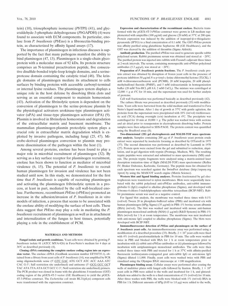
FIG. 6. Analysis of enolase transcripts by quantitative real timeRT-PCR. qRT-PCR plot of PbEno expression levels of transcriptsfrom yeast cells of P. brasiliensis derived from lungs, livers, and spleensof mice after 7 days of infection. Control systems consisted of yeastcells from cultures inoculated in BHI agar. The primers were as fol-lows: sense, 5�-GATTTGCAGGTTGTCGCCGA-3�; antisense, 5�-TGGCTGCCTGGATGGATTCA-3�. The expression values were stan-dardized using the expression values for the constitutive gene encodingthe protein tubulin. The relative quantification (RQ) of the experimentwas performed in triplicate. The error bars indicate the standard de-viations from three independent experiments performed in triplicate.*, significantly different from the control, at a P value of �0.05.
4048 NOGUEIRA ET AL. INFECT. IMMUN.

summary, we have shown that P. brasiliensis can borrow theplasminogen system from the host in a process mediated by thesurface protein enolase.
ACKNOWLEDGMENTS
This work at the Universidade Federal de Goias was supported bygrants from Financiadora de Estudos e Projetos (FINEP 0106121200and 0107055200) and Conselho Nacional de Desenvolvimento Cientí-fico e Tecnologico (CNPq 472947/2007-9 and 558405/2008-8). M.L.R.was supported by grants from the Brazilian agencies FAPERJ andCNPq.
We thank Tereza Cristina Rezende for helpful suggestions.
REFERENCES
1. Agarwal, S., P. Kulshreshtha, D. Bambah Mukku, and R. Bhatnagar. 2008.�-Enolase binds to human plasminogen on the surface of Bacillus anthracis.Biochim. Biophys. Acta 1784:986–994.
2. Angiolella, L., M. Facchin, A. Stringaro, B. Maras, N. Simonetti, and A.Cassone. 1996. Identification of a glucan-associated enolase as a main cellwall protein of Candida albicans and an indirect target of lipopeptide anti-mycotics. J. Infect. Dis. 173:684–690.
3. Barbosa, F. M., F. L. Fonseca, C. Holandino, C. S. Alviano, L. Nimrichter,and M. L. Rodrigues. 2006. Glucuronoxylomannan-mediated interaction ofCryptococcus neoformans with human alveolar cells results in fungal inter-nalization and host cell damage. Microbes Infect. 8:493–502.
4. Barbosa, M. S., S. N. Bao, P. F. Andreotti, F. P. Faria, M. S. Felipe, L. S.Feitosa, M. J. S. Mendes-Giannini, and C. M. A. Soares. 2006. Glyceralde-hyde-3-phosphate dehydrogenase of Paracoccidioides brasiliensis is a cellsurface protein involved in fungal adhesion to extracellular matrix proteinsand interaction with cells. Infect. Immun. 74:382–389.
5. Batista, W. L., A. L. Matsuo, L. Ganiko, T. F. Barros, T. R. Veiga, E.Freymuller, and R. Puccia. 2006. The PbMDJ1 gene belongs to a conservedMDJ1/LON locus in thermodimorphic pathogenic fungi and encodes a heatshock protein that localizes to both the mitochondria and cell wall of Para-coccidioides brasiliensis. Eukaryot. Cell 5:379–390.
6. Bergmann, S., M. Rohde, G. S. Chhatwal, and S. Hammerschmidt. 2001.�-Enolase of Streptococcus pneumoniae is a plasmin(ogen)-binding proteindisplayed on the bacterial cell surface. Mol. Microbiol. 40:1273–1287.
7. Bookout, A. L., C. L. Cumming, D. J. Mangelsdorf, J. M. Pesola, and M. F.Kramer. 2006. High-throughput real-time quantitative reverse transcriptionPCR, p. 15.8.1–15.8.28. In F. M. Ausubel, R. Brent, R. E. Kingston, D. D.Moore, J. G. Seidman, J. A. Smith, and K. Struhl (ed.), Current protocols inmolecular biology. John Wiley & Sons, Hoboken, NJ.
8. Bradford, M. M. 1976. A dye binding assay for protein. Anal. Biochem.72:248–254.
9. Castellino, F. J., and V. A. Ploplis. 2005. Structure and function of theplasminogen/plasmin system. Thromb. Haemost. 93:647–654.
10. Castro, N. D. S., M. S. Barbosa, Z. A. Maia, S. N. Bao, M. S. Felipe, J. M.Santana, M. J. S. Mendes-Giannini, M. Pereira, and C. M. A. Soares. 2008.Characterization of Paracoccidioides brasiliensis PbDfg5p, a cell-wall proteinimplicated in filamentous growth. Yeast 25:141–154.
11. Chaka, W., J. Scharringa, A. F. Verheul, J. Verhoef, A. G. Van Strijp, andI. M. Hoepelman. 1995. Quantitative analysis of phagocytosis and killing ofCryptococcus neoformans by human peripheral blood mononuclear cells byflow cytometry. Clin. Diagn. Lab. Immunol. 2:753–759.
12. Coltri, K. C., A. S. Casabona-Fortunato, M. L. Gennari-Cardoso, C. F.Pinzan, L. P. Ruas, V. S. Mariano, R. Martinez, J. C. Rosa, A. Panunto-Castelo, and M. C. Roque-Barreira. 2006. Paracoccin, a GlcNAc-bindinglectin from Paracoccidioides brasiliensis, binds to laminin and induces TNF-alpha production by macrophages. Microbes Infect. 8:704–713.
13. Cork, A. J., S. Jergic, S. Hammerschmidt, B. Kobe, V. Pancholi, J. L.Benesch, C. V. Robinson, N. E. Dixon, J. A. Aquilina, and M. J. Walker.2009. Defining the structural basis of human plasminogen binding by strep-tococcal surface enolase. J. Biol. Chem. 284:17129–17137.
14. Costa, M., C. L. Borges, A. M. Bailao, G. V. Meirelles, Y. A. Mendonca,S. F. I. M. Dantas, F. P. Faria, M. S. S. Felipe, E. E. W. I. Molinari-Madlum,M. J. S. M. Giannini, R. B. Fiuza, W. S. Martins, M. Pereira, and C. M. A.Soares. 2007. Transcriptome profiling of Paracoccidioides brasiliensis yeast-phase cells recovered from infected mice brings new insights into fungalresponse upon host interaction. Microbiology 153:4194–4207.
15. Crowe, J. D., I. K. Sieywright, G. C. Auld, N. R. Moore, N. A. Gow, and N. A.Booth. 2003. Candida albicans binds human plasminogen: identification ofeight plasminogen-binding proteins. Mol. Microbiol. 47:1637–1651.
16. Dantas, S. F., T. C. Vieira de Rezende, A. M. Bailao, C. P. Taborda, R. S.Santos, K. C. Pacheco, and C. M. A. Soares. 2009. Identification and char-acterization of antigenic proteins potentially expressed during the infectiousprocess of Paracoccidioides brasiliensis. Microbes Infect. 11:895–903.
17. Donofrio, F. C., A. C. Calil, E. T. Miranda, A. M. Almeida, G. Benard, C. P.
Soares, S. V. Nogueira, C. M. A. Soares, and M. J. Mendes-Giannini. 2009.Enolase from Paracoccidioides brasiliensis: isolation and identification as afibronectin-binding protein. J. Med. Microbiol. 58:706–713.
18. dos Reis Almeida, F. B., L. L. de Oliveira, M. Valle de Sousa, M. C. Barreira,and E. S. Hanna. 2010. Paracoccin from Paracoccidioides brasiliensis; puri-fication through affinity with chitin and identification of N-acetyl-beta-D-glucosaminidase activity. Yeast 27:67–76.
19. Eroles, P., M. Sentandreu, M. V. Elorza, and R. Sentandreu. 1997. Thehighly immunogenic enolase and Hsp70p are adventitious Candida albicanscell wall proteins. Microbiology 143:313–320.
20. Esgleas, M., Y. Li, M. A. Handock, J. Harel, J. D. Dubreuil, and M.Gottschalk. 2008. Isolation and characterization of alpha-enolase, a novelfibronectin-binding protein from Streptococcus suis. Microbiology 154:2668–2679.
21. Fox, D., and A. G. Smulian. 2001. Plasminogen-binding activity of enolase inthe opportunistic pathogen Pneumocystis carinii. Med. Mycol. 39:495–507.
22. Franco, M. 1987. Host-parasite relationships in paracoccidioidomycosis.J. Med. Vet. Mycol. 25:5–18.
23. Ganiko, L., R. Puccia, V. S. Mariano, O. A. Sant’Anna, E. Freymuller, M. C.Roque-Barreira, and L. R. Travassos. 2007. Paracoccin, an N-acetyl-gluco-samine-binding lectin of Paracoccidioides brasiliensis, is involved in fungalgrowth. Microbes Infect. 9:695–703.
24. Hanna, S. A., J. L. Monteiro da Silva, and M. J. Mendes-Giannini. 2000.Adherence and intracellular parasitism of Paracoccidioides brasiliensis inVero cells. Microbes Infect. 2:877–884.
25. Jong, A. Y., S. H. M. Chen, M. F. Stins, K. S. Kim, T. L. Tuan, and S. H.Huang. 2003. Binding of Candida albicans enolase to plasmin(ogen) resultsin enhanced invasion of human brain microvascular endothelial cells. J. Med.Microbiol. 52:615–622.
26. Kesimer, M., N. Kilic, R. Mehrotra, D. J. Thornton, and J. K. Sheehan. 2009.Identification of salivary mucin MUC7 binding proteins from Streptococcusgordonii. BMC Microbiol. 9:163.
27. Laemmli, U. K. 1970. Cleavage of structural proteins during the assembly ofthe head of bacteriophage T4. Nature 227:680–685.
28. Lima, O. C., C. C. Figueiredo, J. O. Previato, L. Mendonca-Previato, V.Morandi, and L. M. L. Bezerra. 2001. Involvement of fungal cell wall com-ponents in adhesion of Sporothrix schenckii to human fibronectin. Infect.Immun. 69:6874–6880.
29. Linder, T. E., R. L. Daniels, D. J. Lim, and T. F. DeMaria. 1994. Effect ofintranasal inoculation of Streptococcus pneumoniae on the structure of thesurface carbohydrates of the chinchilla eustachian tube and middle earmucosa. Microb. Pathog. 16:435–441.
30. Lopez-Villar, E., L. Monteoliva, M. R. Larsen, E. Sachon, M. Shabaz, M.Pardo, J. Pla, C. Gil, P. Roepstorff, and C. Nombela. 2006. Genetic andproteomic evidences support the localization of yeast enolase in the cellsurface. Proteomics 6(Suppl. 1):S107–S118.
31. Mendes-Giannini, M. J. S., S. A. Hanna, J. L. da Silva, P. F. Andretti, L. R.Vicentini, G. Bernard, H. L. Lenzi, and C. P. Soares. 2004. Invasion ofepithelial mammalian cells by Paracoccidioides brasiliensis leads to cytoskel-etal rearrangement and apoptosis of the host cell. Microbes Infect. 6:882–891.
32. Miles, L. A., C. M. Dahberg, J. Plescia, J. Felez, K. Kato, and E. F. Plow.1991. Role of cell-surface lysines in plasminogen binding to cells: identifica-tion of alpha-enolase as a candidate plasminogen receptor. Biochemistry30:1682–1691.
33. Mundodi, V., A. S. Kucknoor, and J. F. Alderete. 2008. Immunogenic andplasminogen-binding surface-associated �-enolase of Trichomonas vaginalis.Infect. Immun. 76:523–531.
34. Neto, B. R. D. S., J. F. Silva, M. J. Mendes-Giannini, H. L. Lenzi, C. M. A.Soares, and M. Pereira. 2009. The malate synthase of Paracoccidioides bra-siliensis is a linked surface protein that behaves as an anchorless adhesin.BMC Microbiol. 9:272.
35. Nimrichter, L., M. L. Rodrigues, E. G. Rodrigues, and L. R. Travassos. 2005.The multitude of targets for the immune system and drug therapy in thefungal cell wall. Microbes Infect. 7:789–798.
36. Nosanchuk, J. D., J. N. Steenbergen, L. Shi, G. S. Deepe, Jr., and A. Cas-adevall. 2003. Antibodies to a cell surface histone-like protein protectagainst Histoplasma capsulatum. J. Clin. Invest. 112:1164–1175.
37. O’Farrell, P. H. 1975. High resolution two-dimensional electrophoresis ofproteins. J. Biol. Chem. 250:4007–4021.
38. Pancholi, V., and V. A. Fischetti. 1998. Alpha-enolase, a novel strong plas-min(ogen) binding protein on the surface of pathogenic streptococci. J. Biol.Chem. 273:14503–14515.
39. Patti, J. L., B. L. Allen, M. J. McGavin, and M. Hook. 1994. MSCRAMM-mediated adherence of microorganisms to host tissues. Annu. Rev. Micro-biol. 48:585–617.
40. Penalver, M. C., J. E. O’Connor, J. P. Martinez, and M. L. Gil. 1996. Bindingof human fibronectin to Aspergillus fumigatus conidia. Infect. Immun. 64:1146–1153.
41. Pereira, L. A., S. N. Bao, M. S. Barbosa, J. L. Silva, M. S. Felipe, J. M.Santana, M. J. S. Mendes-Giannini, and C. M. A. Soares. 2007. Analysis of
VOL. 78, 2010 FUNCTIONS OF P. BRASILIENSIS ENOLASE 4049

the Paracoccidioides brasiliensis triosephosphate isomerase suggests the po-tential for adhesin function. FEMS Yeast Res. 7:1381–1388.
42. Pitarch, A., M. Sanchez, C. Nombela, and C. Gil. 2002. Sequential fraction-ation and two-dimensional gel analysis unravels the complexity of the dimor-phic fungus Candida albicans cell wall proteome. Mol. Cell Proteomics1:967–982.
43. Plow, E. F., T. Redlitz, L. A. Miles, and J. L. Hoover-Plow. 1995. The cellbiology of the plasminogen system. FASEB J. 9:939–945.
44. Restrepo, A., J. G. McEwen, and E. Castaneda. 2001. The habitat of Para-coccidioides brasiliensis: how far from solving the riddle? Med. Mycol. 39:233–241.
45. Rodrigues, M. L., E. S. Nakayasu, D. L. Oliveira, L. Nimrichter, J. D.Nosanchuk, I. C. Almeida, and A. Casadevall. 2008. Extracellular vesiclesproduced by Cryptococcus neoformans contain protein components associ-ated with virulence. Eukaryot. Cell 7:58–67.
46. San-Blas, G., G. Nino-Veja, and T. Iturriaga. 2002. Paracoccidioides brasil-iensis and paracoccidioidomycosis: molecular approaches to morphogenesis,diagnosis, epidemiology, taxonomy and genetics. Med. Mycol. 40:225–242.
47. Stie, J., G. Bruni, and D. Fox. 2009. Surface-associated plasminogen binding
of Cryptococcus neoformans promotes extracellular matrix invasion. PLoSOne 3:e5780.
48. Vassalli, J. D., A. P. Sappino, and D. Belin. 1991. The plasminogen activator/plasmin system. J. Clin. Invest. 88:1067–1072.
49. Vicentini, A. P., J. L. Gesztesi, M. F. Franco, W. Souza, J. Z. Moraes, L. R.Travassos, and J. D. Lopes. 1994. Binding of Paracoccidioides brasiliensis tolaminin through surface glycoprotein gp43 leads to enhancement of fungalpathogenesis. Infect. Immun. 4:1465–1469.
50. Vieira, M. L., S. A. Vasconcellos, A. P. Goncales, Z. M. de Morais, and A. L.Nascimento. 2009. Plasminogen acquisition and activation at the surface ofleptospira species lead to fibronectin degradation. Infect. Immun. 77:4092–4101.
51. Walker, M. J., J. D. McArthur, F. Mckay, and M. Ranson. 2005. Is plasmin-ogen deployed as a Streptococcus pyogenes virulence factor? Trends Micro-biol. 13:308–313.
52. Westerlund, B., and T. K. Korhonen. 1993. Bacterial proteins binding to themammalian extracellular matrix. Mol. Microbiol. 4:687–694.
53. Zaas, A. K., G. Liao, J. W. Chien, C. Weinberg, D. Shore, S. S. Giles, K. A.Marr, J. Usuka, L. H. Burch, L. Pereira, J. R. Perfect, G. Peltz, and D. A.Schwartz. 2008. Plasminogen alleles influence susceptibility to invasive as-pergillosis. PLoS Genet. 4:e1000101.
Editor: R. P. Morrison
4050 NOGUEIRA ET AL. INFECT. IMMUN.