A critical role for PfCRT K76T in Plasmodium falciparum verapamil-reversible chloroquine resistance
Upload
johnshopkinsCategory
view
4download
0
BioMed CentralMalaria Journal
ss
Open AcceResearchPlasmodium falciparum enolase: stage-specific expression and sub-cellular localizationIpsita Pal Bhowmick1, Nirbhay Kumar2, Shobhona Sharma1, Isabelle Coppens2 and Gotam K Jarori*1Address: 1Department of Biological Sciences, Tata Institute of Fundamental Research, Homi Bhabha Road, Colaba, Mumbai-400005, India and 2Department of Molecular Microbiology and Immunology, Johns Hopkins Bloomberg School of Public Health, Baltimore, Maryland, USA
Email: Ipsita Pal Bhowmick - [email protected]; Nirbhay Kumar - [email protected]; Shobhona Sharma - [email protected]; Isabelle Coppens - [email protected]; Gotam K Jarori* - [email protected]
* Corresponding author
AbstractBackground: In an earlier study, it was observed that the vaccination with Plasmodium falciparumenolase can confer partial protection against malaria in mice. Evidence has also build up to indicatethat enolases may perform several non-glycolytic functions in pathogens. Investigating the stage-specific expression and sub-cellular localization of a protein may provide insights into itsmoonlighting functions.
Methods: Sub-cellular localization of P. falciparum enolase was examined usingimmunofluorescence assay, immuno-gold electron microscopy and western blotting.
Results: Enolase protein was detected at every stage in parasite life cycle examined. In asexualstages, enolase was predominantly (85–90%) present in soluble fraction, while in sexual stages itwas mostly associated with particulate fraction. Apart from cytosol, enolase was found to beassociated with nucleus, food vacuole, cytoskeleton and plasma membrane.
Conclusion: Diverse localization of enolase suggests that apart from catalyzing the conversion of2-phosphoglycericacid into phosphoenolpyruvate in glycolysis, enolase may be involved in a host ofother biological functions. For instance, enolase localized on the merozoite surface may be involvedin red blood cell invasion; vacuolar enolase may be involved in food vacuole formation and/ordevelopment; nuclear enolase may play a role in transcription.
BackgroundIn recent years, it is being realized that many of the house-keeping metabolic enzymes participate in a host of otherbiological functions inside the cell. It is increasinglybecoming apparent that the ability of a protein to 'Moon-light' i.e. to have multiple and sometimes vastly unrelatedfunctions embedded within one polypeptide chain, is ageneral strategy to enhance the number of protein func-
tions that are encoded by the genome [1]. Many of themetabolic enzymes, specifically the glycolytic ones fromdifferent organisms have diverse functions in addition totheir role in glycolysis. For example, hexokinase2 in yeastis involved in transcriptional regulation [2], glyceralde-hyde 3-phosphate dehydrogenase functions in tubulinbinding, nuclear RNA export, phosphorylation, mem-brane fusion, and transcriptional regulation [3-5], glu-
Published: 30 July 2009
Malaria Journal 2009, 8:179 doi:10.1186/1475-2875-8-179
Received: 10 March 2009Accepted: 30 July 2009
This article is available from: http://www.malariajournal.com/content/8/1/179
© 2009 Pal Bhowmick et al; licensee BioMed Central Ltd. This is an Open Access article distributed under the terms of the Creative Commons Attribution License (http://creativecommons.org/licenses/by/2.0), which permits unrestricted use, distribution, and reproduction in any medium, provided the original work is properly cited.
Page 1 of 16(page number not for citation purposes)

Malaria Journal 2009, 8:179 http://www.malariajournal.com/content/8/1/179
cose-6-phosphate isomerase in cell motility andproliferation [6] and aldolase binds actin and supportprotein trafficking to the plasma membrane [7,8]. Thus,functional moonlighting for many of these house-keepingproteins, seems to be a general phenomenon [9].
Glycolytic enzymes play important roles in Plasmodiumbiology. Intra-erythrocytic stages of Plasmodium falciparumlacks functional TCA cycle and solely rely on glycolysis fortheir energy needs [10-12]. The level of glycolytic flux inparasite infected cells is ~100 fold greater than that ofuninfected red blood cells [13,14] and the activity of someof the glycolytic enzymes (enolase, pyruvatekinase andhexokinase) is greatly up-regulated [15]. In recent years,glycolytic enzymes have also been shown to perform non-glycolytic functions in apicomplexan parasites. In Toxo-plasma and Plasmodium, aldolase has been implicated inhost cell invasion through its interaction with actin andsurface adhesion molecules [7,8,16]. Interestingly, glyco-lytic and non-glycolytic functions of aldolase are mademutually exclusive as adhesins bind at the active siteresulting in loss of catalytic activity. Similarly, glyceralde-hydes 3-phosphate dehydrogenase (GAPDH) has alsobeen shown to perform certain non-glycolytic functionsin P. falciparum [17]. Due to their importance in Plasmo-dium for energy production and other physiological func-tions, glycolytic enzymes have been termed as importanttherapeutic targets and validated in the new large scaleventures for anti-malarials [12,18-21].
Enolase (2-Phospho-D-glycerate hydrolase; EC 4.2.1.11)is one of the three glycolytic enzymes, whose levels arehighly elevated in parasite infected red blood cells (RBC)(about 15-fold) as compared to the uninfected cells [15].Recently, this glycolytic enzyme has also been reported tohave diverse biological functions in different organisms[22-26]. Thus, enolases, which have been well character-ized for their catalytic function in glucose metabolism, areno longer considered to be the house-keeping enzymesonly. Enolase is better described as a multifaceted proteinwith multi-tasking abilities at diverse sub-cellular loca-tions [24]. Recent studies have shown that in many path-ogenic species and in different cell types, enolase ispresent on the cell wall, cell membranes and in the cellnucleus. The unusual location of enolase has beenreported in the apicomplexan parasites viz. Plasmodiumyoelii [27], Toxoplasma gondii [28] and Eimeria tenella [29].In T. gondii, there are two different isozymes (Eno1 andEno2), which have been demonstrated to exhibit stagespecific expression. A comparison of mRNA expression ofglycolytic genes between tachyzoite vs 'in vitro' bradyzoitehas shown that the two enolase genes are the only glyco-lytic genes whose expression is regulated in a stage specificmanner. Eno1 is strongly up-regulated (~1450 fold) inbradyzoite while all other glycolytic transcripts were ele-
vated only by 4- to 8-fold. At protein level, ENO1 is spe-cifically expressed in bradyzoites, while ENO2 expressionis specific to tachyzoite stage. Location of two enolase iso-zymes in the nucleus of actively developing/dividing par-asites has led to the suggestion that these proteins mayplay a role in controlling some nuclear activities duringstage differentiation [28,30]. Among the genes that codefor glycolytic enzymes in T. gondii, silencing of ENO2 hadthe most effect on parasite growth [31]. Such observationsof stage specific isozyme expression, nuclear localizationand growth inhibition on loss of function of this glyco-lytic enzyme suggest interesting nuclear function for thisprotein in T. gondii. Since Plasmodium has only one genefor enolase, it is likely that such non-glycolytic func-tion(s), if mediated, may be embedded in a single enolaseprotein.
Evidence has emerged from the recent experiments thatenolase may have moonlighting functions in Plasmodium.Preliminary immunofluorescence studies on P. yoeliishowed the presence of enolase in parasite nucleus and onmerozoite cell surface. Further evidence for novel surfacefunction(s) for enolase has emerged from the immuniza-tion studies where vaccination with recombinant P. falci-parum enolase (r-Pfen) resulted in partial protectionagainst P. yoelii induced malaria in mice and the detectionof anti-enolase antibodies in human serum samples frommalaria endemic region of India [32]. To examine theinvolvement of a housekeeping protein in diverse cellularfunctions, one can probe its sub-cellular localization andidentify the interactor proteins, as involvement of a pro-tein in multiple functions invariably requires its recruit-ment to different sub-cellular compartments andinteractions with different proteins. These possibilitieshave been examined here using in situ (IFA and IEM) andbiochemical fractionation (sub-cellular fractionation,pull down assays) methods for determining the localiza-tion of enolase in P. falciparum and identify the interactingproteins by co-localization and gel analysis.
MethodsMaterialsMouse monoclonal antibodies against Pfg-27 [33] andmouse anti-Pf HSP70 antibodies [34] used here have beencharacterized earlier. Anti-Pfs-48/45 monoclonal anti-body was obtained from the MR4. Rabbit anti-P. falci-parum aldolase antibody and a preparation of P. yoeliisporozoites were a kind gifts from Prof. Victor Nussenz-weig, Department of Pathology, N.Y. University MedicalCentre, New York, USA, and rabbit anti-Toxoplasma gondiiactin antibody was provided by Prof. David Sibley, Wash-ington university, St. Louis, MO, USA. RPMI media werefrom GIBCO-BRL, NY, USA. Protease inhibitor cocktailwas procured from Roche Applied Science, Indianapolis,IN, USA. Saponin was from Sigma Chemical Co., St Louis,
Page 2 of 16(page number not for citation purposes)

Malaria Journal 2009, 8:179 http://www.malariajournal.com/content/8/1/179
MO, USA. Alexa Fluor 488 conjugated anti-rabbit andanti-mouse IgG and DAPI were from Molecular Probes,NJ, USA and Vectashield-mounting medium was fromVector laboratories, CA, USA
r-Pfen purification and anti enolase antibodiesThe preparation of recombinant 6 × His-tagged P. falci-parum enolase (r-Pfen) and production of rabbit andmouse anti-enolase antibodies were carried out asdescribed earlier [35]. GST tagged r-Pfen was purifiedusing glutathione-sephadex beads. For the in situ localiza-tion studies, the polyclonal anti-r-Pfen antibodies usedhave previously been shown to have high specificityagainst parasite enolase protein and did not cross reactwith homologous host enolases or other parasitic proteinsunder the experimental conditions employed here [32].
Preparation of different stages of the parasitePlasmodium falciparum asexual and gametocyte cultures(3D7 and NF54 isolates) were maintained as describedpreviously [36,37]. The growth rates and proportions ofvarious developmental stages of asexual and sexual para-sites were monitored daily by microscopic examination ofGiemsa-stained blood smears. Culture enriched withgametocytes was prepared and smears were made. Startingwith synchronized asexual parasites grown in suspensionculture as described, gametocytes were prepared by dailymedia changes of static cultures at 37°C.
Indirect immunofluorescence assay (IFA)Immunofluorescence assay was performed at room tem-perature with the air-dried blood smears. Cells were fixedwith 4% formaldehyde in phosphate buffered saline(PBS) for 10 minutes, washed five times, permeabilizedwith 0.25% triton x-100 in PBS for 10 minutes, washed,fixed with 3% BSA-PBS for 45 minutes, incubated for 1hour with the following antibodies combination: (i)mouse anti-enolase antibodies (1:200) with rabbit anti-P.falciparum aldolase (1:100) or (ii) rabbit anti-enolase anti-bodies (1:200) with mouse anti-HSP70 antibody (1:100).All antibody dilutions were made into 1% BSA-PBS. Afterantibody treatment, smears were washed 7–8 times withPBS. These smears were then treated with the secondaryantibodies for 45 minutes (Alexa Fluor 488-conjugatedanti-mouse IgG and Alexa Fluor 568-conjugated anti rabitIgG were used as secondary antibodies (1:500)) andwashed 10–12 times with PBS. Parasite nuclei werestained with DAPI (1 g·ml-1) and mounted with vectash-ield.
Soluble and pellet fraction of asexual and gametocyte stagesTwo separate preparations of erythrocytes, mostly infectedwith mature (stage IV-V) gametocytes and asexual stagesof P. falciparum were harvested using saponin. For asexual
stages, P. falciparum culture was allowed to reach ~5% par-asitaemia, harvested and washed with incomplete RPMIsolution. The gametocytes culture was similarly harvestedand it consisted mostly of stage IV and V parasites withminor contamination (<3%) from mixed asexual stageparasites. Infected erythrocytes from both cultures werethen treated with 0.05% saponin for 10 min at 4°C torelease the parasites from the host erythrocyte membrane[38]. The parasite pellets were then solubilized in NETTbuffer (10 mM Tris-HCl, pH 7.4, 150 mM NaCl, 1 mMEDTA, 0.5% Triton x-100) with cocktail protease inhibitorat 4°C for 10 minutes and centrifuged at 20,000 g for 30minutes. The supernatant and pellets were used for SDS-PAGE and immunoblot analysis. Two dimensional gelelectrophoresis and Western blotting for the visualizationof the P. falciparum enolase variants was performed asdescribed earlier [27].
Cytochalasin-D treatment of parasitesPlasmodium falciparum culture containing wells weretreated with cytochalasin-D dissolved in DMSO at a finalconcentration of 50 M. Briefly, 5l of 2 mM stock ofcytochalasin D in DMSO was mixed with 95 l of com-plete RPMI (100 l of 100 M cytochalasin-D), which wasadded to 100 l of culture to obtain 50 M final concen-tration of the drug. Only DMSO was added to the controlwells. DMSO was kept at < 0.1% (v/v) under the experi-mental conditions. These were then incubated for 4 hoursat 37°C. The cells were spun in the pre-warmed tubes andsupernatant was removed to keep ~50% haematocrit.Thin smears were prepared, air dried and used for indirectimmunofluorescence assay.
Treatment with Triton X-100 prior to fixationDetergent extraction was carried out prior to the fixationof the cells to remove the cytosolic and membrane associ-ated proteins. Plasmodium falciparum-infected blood wassmeared on poly-Lysine coated glass slides, which werethen treated with 1% Triton x-100 for 10 minutes at roomtemperature (20°C). These were then washed three timeswith phosphate buffer saline and used for indirectimmunofluorescence assay. This allowed cytoskeletonassociated enolase to be visualized.
Immuno-gold electron microscopy (IEM)Preparations of P. falciparum-containing red blood cellswere fixed in 4% paraformaldehyde (Electron MicroscopySciences, PA) in 0.25 M HEPES (pH 7.4) for 1 hr at roomtemperature, then in 8% paraformaldehyde in the samebuffer overnight at 4°C. They were infiltrated, frozen andsectioned as previously described [39]. The sections wereimmuno-labeled with mouse anti-r-Pfen antibodies(1:100 in PBS/1% fish skin gelatin), then with anti-mouseIgG antibodies, followed directly by 15 nm protein A-goldparticles (Department of Cell Biology, Medical School,
Page 3 of 16(page number not for citation purposes)

Malaria Journal 2009, 8:179 http://www.malariajournal.com/content/8/1/179
Utrecht University, the Netherlands) before examinationwith a Philips CM120 Electron Microscope (Eindhoven,the Netherlands) under 80 kV.
Interaction of enolase with actin, tubulin and human plasminogenFor detection of direct interaction of enolase with actin (ortubulin), GST tagged-r-Pfen was immobilized on glutath-ione beads and was mixed with G-actin or tubulin. Sam-ples were incubated for 2 hours at 37°C and subjected tocentrifugation. Beads were washed with G-actin buffer (5mM Tris-HCl pH 8.0, 0.2 mM CaCl2, 0.1 mM ATP) ortubulin buffer (80 mM Na-PIPES pH 6.9, 1 mM MgCl2, 1mM EGTA, 1 mM GTP) respectively and analysed on a12% SDS PAGE.
Binding of enolase with plasminogen was investigatedusing ELISA. Wells were coated with 100 l of 100 nMplasminogen (or rabbit muscle pyruvate kinase). Afterover coating with skimmed milk and washing, 100 l of 6× His-tagged r-Pfen in different concentrations was addedand incubated for 4 hours. Mouse anti r-Pfen serum(1:2000) was used in the assay. Remaining steps weresame as described earlier [35].
ResultsExpression levels of enolase at different stages of P. falciparumIFA analysis at different stages in the life cycle of P. falci-parum is shown in Figure 1. Expression levels of enolaseappear to be very similar in most stages except in certainsub-stages of the gametocyte. Figure 1A (a) shows sch-izont and trophozoite stages of the parasite. One of themis a multinucleated schizont stage (marked with ) andthe other two are trophozoites (marked with *). Figure 1A(b) shows a schizont and four ring stage cells (markedwith Ý). Figure 1B shows the distribution of enolase in thegametocyte stages. From the shape of the gametocytes, itappears that three elongated cells represent mature game-tocytes (stage IV/V), whereas the small round, Pfg-27 pos-itive cell may be an early stage II gametocyte (marked with�). Although Pfg-27, a gametocyte marker [40] appears toexpress equally in the various gametocyte sub-stages, themerged image showed that the stage II gametocyte has rel-atively lower levels of expressed enolase. In general, it wasalso observed that the levels of enolase are comparable atthe gametes and the schizont stages (Figure 1C). The mostunusual distribution was observed in sporozoite stage.Figure 1D shows a mosquito derived P. yoelii sporozoitepreparation. Enolase staining in these cells showed apunctuate pattern where particulate enolase appears to bepresent beneath the plasma membrane, which is quitedistinct from the homogenous staining observed for cir-cum sporozoite protein (CSP).
Sub-cellular distribution of enolase in Plasmodium falciparumIn addition to cytosolic localization of enolase, its pres-ence in the nucleus was observed in the ring and tropho-zoite stages (Figure 2). The possibility of spill over ofcytosolic enolase into the nucleus during the processing ofparasite cells could be ruled out by observing the localiza-tion of two other proteins, namely aldolase (Figure 2A)and HSP-70 (Figure 2B). Both these proteins were presentin the cytoplasmic compartment and no nuclear presencewas detected. In a synchronized population of ring stageparasites, wide variation in the relative distribution ofenolase between the nuclear and the cytosolic compart-ments was observed. Certain cells had a faint staining fornuclear enolase, indicating that cytosolic enolase is muchgreater than the nuclear one while others had strongnuclear staining for the enolase (cytosolic << nuclear)(Figure 3).
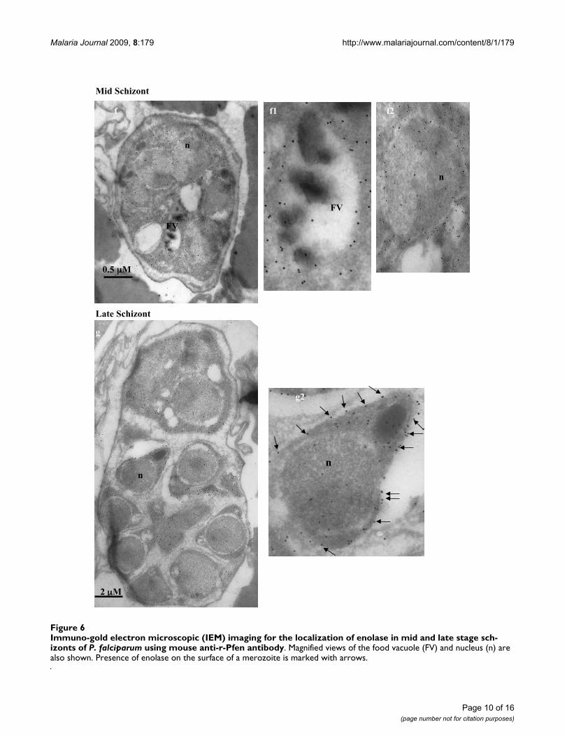
In order to obtain better resolution, the sub-cellular local-ization of enolase in P. falciparum was also examined byimmuno-gold electron microscopy (IEM). Figures 4, 5, 6,7 show the parasite cells in the trophozoite, schizonts andgametocyte stages labeled with anti-r-Pfen. For all theimages presented, a magnified view of food vacuole (FV)and nucleus (n) is shown along side. The nucleus andfood vacuole have significantly higher levels of enolasepresent in early (Figure 4) and mid stage trophozoites(Figure 5) as compared to the late stage trophozoite (Fig-ure 5) and the schizont (Figure 6). Nuclear enolase atgametocyte stage is also low as compared to cytosol (Fig-ure 7). These observations are similar to the pattern ofenolase distribution between nucleus and cytosolobserved using IFAs (Figure 2). It is interesting to note thatin all the stages observed in these IEM images, nuclearenolase exhibited a preferential association with electron-dense heterochromatin region (darker regions in thenucleus). Cell surface localization of enolase wasobserved at the merozoite stage, (Figure 6 g2). Further, inall these images there was no observable association ofPfen with infected host cell cytosol or cell membrane, sug-gesting that Pfen is not secreted into the cytosol or trans-located to the cell membrane of the infected rbcs. Thisobservation is at variance from the earlier reports [41].
Association of enolase with the cytoskeletal elements of the parasiteParasite cell extracts prepared with Triton-X-100 (TX100)as the solubilizing agent, were analyzed on SDS-PAGEand enolase was visualized by western blotting. Figure8(A) presents the distribution of the enolase between thesoluble and particulate fractions in the sexual and asexualstages of the parasite life cycle. While in asexual stages,>80–90% enolase was present in TX100 soluble fractionand only ~10–20% was associated with cytoskeletal com-
Page 4 of 16(page number not for citation purposes)

Malaria Journal 2009, 8:179 http://www.malariajournal.com/content/8/1/179
ponents, in sexual stages the distribution was quite theopposite. Most of the enolase in sexual stages was associ-ated with detergent insoluble fraction. In contrast, aldo-lase was equally distributed between soluble and pelletfractions in sexual stages, while its distribution in asexualstages was more like enolase (soluble >> pellet). Observa-tion of differential distributions of aldolase and enolasein soluble and particulate fractions, ruled out the possibil-ity that such results may arise from incomplete lysis of
cells or cross contamination of particulate fraction withthe soluble proteins.
For the in situ observation of the distribution of enolaseand aldolase in the gametocytes, immunofluorescenceassays were performed on TX100 treated and untreatedcells (Figure 8B(a)). Although both enolase and aldolaseare glycolytic enzymes, they show very different distribu-tion in gametocyte stages as seen from fractionation as
Immunofluorescence assays (IFA) for the localization of enolase at different stages of Plasmodium life cycleFigure 1Immunofluorescence assays (IFA) for the localization of enolase at different stages of Plasmodium life cycle. (A) Asexual stages, (B) Gametocytes, (C) Gamete (mosquito stage) and (D) Sporozoite (mosquito salivary gland stage). Sporozoite preparation was from P. yoelii, while all other stages shown are for P. falciparum. Ring stages (triangle), trophozoite (*), schizont () and stage II gametocyte (diamond). Different fluorescent probes used were, (i) DAPI as a nuclear marker, (ii) mouse anti-r-Pfen (green), (iii) rabbit anti-r-Pfen (red), (iv) mouse anti-Pfg-27 (green) as gametocyte marker, (v) rabbit anti-male gameto-cyte specific -tubulin II antibody (red) and (vi) mouse anti-CSP antibody (green) as sporozoite surface marker. The scale bars shown corresponds to 5 m.
(a) (b)(A)
DAPI
Enolase
(B)
Enolase CSP(D)(C) DAPI α-Tubulin II Enolase
*
*
Pfg-27 Enolase Merged
* *
♦♦ ♦
Page 5 of 16(page number not for citation purposes)

Malaria Journal 2009, 8:179 http://www.malariajournal.com/content/8/1/179
well as IFA studies. Since cytosol has considerable amountof enolase, it was important to ensure that the detergenttreatment did remove cytosolic and the membrane associ-ated enolase. This was evident from the disappearance ofsignals for aldolase (a cytosolic protein) (Figure 8B(a))and a gametocyte membrane protein Pfs48/45 (Figure8B(b)) in the detergent treated preparations. Figure 8Bshows a gametocyte (Figure 8B(c)) and a schizont (Figure8B(d)) stained with DAPI, anti-enolase and anti-actinantibodies, before and after the treatment with TX100.
Co-localization of enolase with actin is observed in thegametocyte stage while it is rather sparse in the schizontstage. Disruption of actin cytoskeleton by treatment withcytochalasin D resulted in disruption of actin co-localiza-tion pattern of enolase in gametocytes (Figure 9A(a)).Such a treatment of ring stage parasites resulted in the lossof translocation of Pfen to the nucleus (Figure 9A(b)).
Observations of co-localization of actin and enolase aswell as the effect of cytochalasin-D on enolase distribu-
Immunofluorescence assays for the localization of enolase, aldolase and HSP-70 in P. falciparum asexual stages (ring, tropho-zoite and schizont)Figure 2Immunofluorescence assays for the localization of enolase, aldolase and HSP-70 in P. falciparum asexual stages (ring, trophozoite and schizont). (A) P. falciparum infected red blood cells were treated with DAPI (blue), mouse anti r-Pfen antibody (green), rabbit anti-P. falciparum aldolase antibody (red). (B) Cells were treated with DAPI, rabbit r-Pfen antibody (red), and mouse anti Pf HSP-70 antibody (green). Overlay panels show the merged of the three images.
(A) DAPI enolase aldolase overlay
Ring stage
Trophozoite stage
Schizont stage
(B) DAPI enolase overlayHSP-70
Ring stage
Schizont stage
Page 6 of 16(page number not for citation purposes)

Malaria Journal 2009, 8:179 http://www.malariajournal.com/content/8/1/179
tion suggested that two proteins may have direct interac-tion. Possibility of such an interaction was investigated byincubating GST tagged-r-Pfen (immobilized on glutath-ione beads) with G-actin or tubulin. Results are shown inFigure 9B. Pull-down assay did not show any interactionwith tubulin, however direct binding of actin with enolasewas observed (Figure 9B, lanes 3 & 5). In the spun-downpreparations of parasite actin filaments, enolase along
with several other proteins has been detected [42]. How-ever, it was not evident from these studies, whether Pfenand actin have direct interaction.
Enolase binds to human plasminogenIn addition to cytosolic and nuclear presence, Plasmodiumspp enolase has been shown to reside on the merozoitesurface (Figure 6) [32]. In certain pathogenic bacteria, cell
Immunofluorescence images showing variation in distribution of enolase between cytosol and nucleus in a population of syn-chronized ring stage parasite cellsFigure 3Immunofluorescence images showing variation in distribution of enolase between cytosol and nucleus in a population of synchronized ring stage parasite cells. There were greater number of cells having more enolase signal arising from nucleus than from cytosol.
Non nuclear
Nuclear ~ cytoplsmic
Nuclear > cytoplsmic
Nuclear >>cytoplsmic
Nuclear>>>cytoplasmic
Nuclear << cytoplsmic
Nuclear << cytoplsmic
DAPI enolase aldolase overlay Bright field
Page 7 of 16(page number not for citation purposes)

Malaria Journal 2009, 8:179 http://www.malariajournal.com/content/8/1/179
Page 8 of 16(page number not for citation purposes)
Immuno-gold electron microscopic (IEM) imaging for the localization of enolase in early trophozoite satge of P. falciparum using mouse anti-r-Pfen antibodyFigure 4Immuno-gold electron microscopic (IEM) imaging for the localization of enolase in early trophozoite satge of P. falciparum using mouse anti-r-Pfen antibody. Magnified views of the food vacuole (FV) and nucleus (n) are also shown. Arrows in food vacuole marks hemozoin associated enolase.
FV
nFV
n
0.2 μm
FV
nn
0.2 μm
n
FV
FV
n
0.2 μm
a a2a1
b b2b1
c c2c1
FV
c1

Malaria Journal 2009, 8:179 http://www.malariajournal.com/content/8/1/179
Page 9 of 16(page number not for citation purposes)
Immuno-gold electron microscopic (IEM) imaging for the localization of enolase in mid and late stage trophozoites of P. falci-parum using mouse anti-r-Pfen antibodyFigure 5Immuno-gold electron microscopic (IEM) imaging for the localization of enolase in mid and late stage tropho-zoites of P. falciparum using mouse anti-r-Pfen antibody. Magnified views of the food vacuole (FV) and nucleus (n) are also shown.
2 μm
FVn
n
FV
Late Trophozoite
FV
n
n
Mid Trophozoite
d
d2
e e1 e2
FV
d1
Malaria Journal 2009, 8:179 http://www.malariajournal.com/content/8/1/179
Page 10 of 16(page number not for citation purposes)
Immuno-gold electron microscopic (IEM) imaging for the localization of enolase in mid and late stage schizonts of P. falciparum using mouse anti-r-Pfen antibodyFigure 6Immuno-gold electron microscopic (IEM) imaging for the localization of enolase in mid and late stage sch-izonts of P. falciparum using mouse anti-r-Pfen antibody. Magnified views of the food vacuole (FV) and nucleus (n) are also shown. Presence of enolase on the surface of a merozoite is marked with arrows.
Mid Schizont
n
n
FV
0.5 M
f
FV
f1 f2
Late Schizont
n
2 M
g
n
g2

Malaria Journal 2009, 8:179 http://www.malariajournal.com/content/8/1/179
surface enolase has been shown to serve as plasminogenreceptor. Through this interaction, these pathogensexploit the fibrinolytic activity of plasmin(ogen) to theiradvantage in tissue invasion [43]. In order to addresswhether Pfen also interacts with plasminogen, an ELISAassay was performed. A concentration dependent bindingof Pfen with human plasminogen was observed in thisassay. However, Pfen did not show any significant bind-ing to rabbit muscle pyruvatekinase, which was used as acontrol (Figure 10).
DiscussionLarge scale stage specific analysis of P. falciparum pro-teome has shown that the enolase is expressed in all stages(Trophozoite, schizont, merozoite, gametocyte) in the lifecycle of the parasite [44,45] and was also associated withthe host cell plasma membrane of the infected red bloodcells (iRBCs) [41]. The results presented here showed thatindeed the Pfen is present in all the asexual and sexualstages of the parasite. However, in the IFA and IEM imagespresented here, there was no detectable Pfen associated
with iRBC membranes or iRBC cytoplasm suggesting thepossibility of cross contamination during biochemicalsample preparations in the earlier studies. The validity ofthe results reported here heavily rely on the specificity ofanti-Pfen antibodies to react only with Plasmodium eno-lase. In an earlier report, several controls were performedto ensure that indeed this was the case. It was shown thatunder the experimental conditions employed here, theseantibodies did not show any cross reactivity towards pro-tein extracts from uninfected RBCs, human & mouse leu-kocytes, and mouse liver in western analysis. Whole cellextracts from P. yoelii and P. falciparum showed a singleband at an expected molecular mass of about 50 kDa [33].The results presented here also indicate that the expressedlevels of enolase protein do not match with the levels oftranscript at various stages of the parasite reported earlier[46,47]. For instance, in sexual stages, gametocytes havenegligible amounts of Pfen transcript and in sporozoites(mosquito salivary gland stage), Pfen transcript is unde-tectable [46-48]. However, the images presented hereshowed good quantities of Pfen protein in both gameto-cytes and sporozoites (Figure 1). These results suggest thepossibility of expressed protein levels of enolase in theparasite being controlled by post-transcriptional regula-tion of translation.
The observed presence of Pfen in multiple sub-compart-ments of the parasite cell may imply multiple physiologi-cal functions for this protein. In the asexual stages, it islargely soluble while in the sexual stages, it is mostly par-ticulate (Figure 8A). It is possible that most of the enolasein asexual stages is recruited for glycolytic function, whilein gametocyte stages, it may have non-glycolytic func-tions. Punctate appearance of Pfen close to the sporozoitesurface (Figure 1D) also indicates additional non-glyco-lytic function for this protein. Plasmodium invades tissuesat sporozoite (liver cells), merozoite (RBCs) and ookinete(mosquito gut wall) stages. Results presented here showedthat the Pfen binds to human plasminogen and is surfacelocalized in merozoite stage. Two signatures of -enolasehave been proposed for such interaction with plasmino-gen, namely the Lysine residues at the C-terminal end andin the central motif 257DLDFKSDDPS [24,49]. The C-ter-minal Lysine residue and the central motif(266DLDFKTPNNDKS in Pfen) are both conserved in P.falciparum enolase [35], and therefore the binding to plas-minogen is expected. The presence of plasminogen recep-tors on cell surface and the non-fibrinolytic functions ofplasminogen have now been documented extensively [50-52]. The nonfibrinolytic roles of plasminogen seem todepend on its ability to activate matrix metalloprotein-ases, which degrade matrix proteins [53]. Such plasmin-dependent pericellular proteolysis may operate whenplasmin(ogen) is tethered to the cell surface through aheterogeneous group of plasminogen receptors, and eno-
Immuno-gold electron microscopic (IEM) imaging for the localization of enolase in gametocyte of P. falciparum using mouse anti-r-Pfen antibodyFigure 7Immuno-gold electron microscopic (IEM) imaging for the localization of enolase in gametocyte of P. fal-ciparum using mouse anti-r-Pfen antibody. Magnified views of the food vacuole (FV) and nucleus (n) are also shown.
n
n
hh1
h2
Page 11 of 16(page number not for citation purposes)

Malaria Journal 2009, 8:179 http://www.malariajournal.com/content/8/1/179
lase is documented to be one such receptor [24,49]. It ispossible that surface localized enolase in Plasmodium zoiteforms may also act as a plasminogen receptor and play arole in tissue invasion processes. This is consistent withthe observation that the anti-Pfen antibodies block mero-zoite invasion into red cells [32].
Glycolytic proteins have been reported to perform diversefunctions at different sub-cellular locations. Plasmodiumaldolase has glycolytic function in cytosol while in associ-ation with acto-myosin complex, it assists the parasite ininvasion and motility functions [8]. In a proteome-wideyeast two hybrid screen La count et al [54], identified glyc-eraldehydes-3-phosphate dehydrogenase (PF14_0598),MSP-9 (PFL1385c), cysteine proteinase (PFB0330c) andformin (PFL0925w) as direct interactors of enolase. Theseauthors also reported HSP-70 (PF11_0351) to be an indi-rect interactor. Observation of direct binding of Pfen andactin as also the extensive association of enolase with theparticulate fraction (especially in the sexual stages)implies a role for enolase in the cytoskeletal organization.In a recent study where protein modifications occurringunder oxidative stress were assessed, Pfen was found toundergo major modifications [55] indicating that it islikely to be a target protein for stress response.
In P. falciparum, presence of relatively high amounts ofnuclear Pfen was observed in the ring and the early tro-
phozoite stages (Figures 2, 3 &4). In IEM images, prefer-ential association of enolase with heterochromatin wasnoted, particularly in early trophozoite (or ring) stages(Figure 4a2 &4b2). Translocation of cytosolic enolase tonucleus, just when the growth phase begins is suggestiveof possible involvement of enolase in transcriptionrelated processes. Possibility of such nuclear function forenolase has been suggested in mammalian [56] and plantcells [57]. The nuclear presence of enolase has also beenreported in closely related apicomplexan Toxoplasma gon-dii and Eimeria tenella. However, a direct correlationbetween translocation of Pfen and transcriptional regula-tion has not yet been demonstrated in parasites [28,29].
Food vacuole in Plasmodium is an acidic proteolytic com-partment central to the metabolism of the parasite [58]. InFigure 4, magnified IEM images of vacuolar region of P.falciparum showed close association of enolase withhaemozoin (marked with arrow heads in Figure 4b1). Inthe growing trophozoite (Figures 4 and 5), there was con-siderable amount of enolase present in the vacuole. How-ever, in the mature trophozoite (Figure 5) and schizontstages (Figure 6), the amounts of enolase associated withFV seemed to decrease (although cytosolic enolase is quiteabundant), suggesting a role in early stages of vacuolardevelopment and/or haemozoin formation. The observedpattern of enolase association with vacuole appeared verysimilar to the proteins which have been implicated in
(A) Comparison of the distribution of enolase, aldolase and actin in soluble and particulate fractions prepared from P. falci-parum cells in sexual and asexual stagesFigure 8(A) Comparison of the distribution of enolase, aldolase and actin in soluble and particulate fractions prepared from P. falciparum cells in sexual and asexual stages. Cells were treated with 0.5% Triton X-100 for 10 minutes at 4°C. Solubilized proteins were removed by centrifugation (10,000 g for 30 minutes). Soluble (S) and particulate (P) fractions were analyzed on 12% SDS-PAGE and the presence of enolase was detected by Western blotting. (B) IFA of P. falciparum gametocyte stages (a, b, c) and asexual schizont stage (d). Cells were fixed either after a treatment with 1% Triton X-100 for 10 minutes (for the removal of cytosolic and membrane proteins) or without Triton treatment. Fixed cells were stained with rabbit or mouse anti-r-Pfen antisera along with (a) rabbit anti-P. falciparum aldolase (red), (b) mouse anti Pfs48/45 (green), (c) rabbit anti-T. gondii actin antibody (red) and (d) schizont (asexual stage) stained with DAPI, anti-r-Pfen and anti-actin antibodies after detergent treatment.
asexual stages
(A) anti-enolase anti-aldolase
S P S P
sexual stages
actinenolase overlay(c)
Triton X-100treated
untreated
5μm
(B)
untreated
Triton X-100treated
aldolaseenolase overlay(a) Pfs-48/45enolase overlay(b)
(d)
overlayenolase actinDAPI
5μm
Page 12 of 16(page number not for citation purposes)

Malaria Journal 2009, 8:179 http://www.malariajournal.com/content/8/1/179
Figure 9 (see legend on next page)
actin overlay bright field
10 μm
enolase
DMSO
enolase overlayDAPI aldolase
Cytochalasin-D
(b)
DMSO
Cytochalasin-D
(a)(A)
(B)
GST-r-Pfen
+ac
tin
bead
+tub
ulin
actin
bead
+ac
tin
tubuli
n
GST-r-Pfen
97
66
43
29
MWM(kDa)
1 2 3 4 5 6 7
GST-r-Pfen
+tub
ulin
Page 13 of 16(page number not for citation purposes)

Malaria Journal 2009, 8:179 http://www.malariajournal.com/content/8/1/179
haem detoxification and vacuole biogenesis [59,60] rais-ing the possibility of enolase being involved in such func-tions. Further support for possible involvement of enolasein haem detoxification arises from the observed associa-tion with ferriprotoporphyrin IX (FPIX), prepared fromchloroquine treated parasites [61]. Other possible vacu-olar function for Pfen can be its involvement in vacuolarfusion and vacuolar protein sorting as observed in yeast[25].
In eukaryotic cells multi-compartment localization of aprotein synthesized in cytoplasm is achieved by compart-ment specific topogenic sequences. Plasmodium falciparumenolase does not have any such signal sequences to be tar-geted to nucleus or cell surface membrane. In Plasmodium,there are no signal sequence(s) known, which ensure tar-geting to food vacuole either. Post-translational modifica-tions and association with other interactors provide
alternative mechanisms for such diverse localization [62].Additional studies are needed to identify protein interac-tors of enolase, post-transcriptional modifications that itundergoes and whether it has any function in nucleus andfood vacuole of the parasite.
ConclusionThe results presented in this paper provide evidence formultiple subcellular localization of enolase and the stagespecific variation in the levels of the expressed enolaseprotein. P. falciparum enolase exhibits great diversity insub-cellular localization with stage specific variation viz-a-viz – a) Presence of enolase on merozoite surface; b)ability of anti-r-Pfen antibodies to interfere with invasionprocess and accord partial protection against malaria; c)nuclear and vacuolar localization and observed shiftbetween soluble to particulate fractions in asexual andsexual stages. These variations are indicative of theinvolvement of P. falciparum enolase in a host of biologi-cal functions. Association of enolase with actin appears tobe important for its translocation to nucleus. An analysisof the nature of interactions of enolase with its interactorproteins and post-translational modifications that itundergoes, may provide insights in to the molecular basisfor the multiple physiological functions that this proteinmight perform in the parasite.
AbbreviationsDAPI: 4',6-Diamidino-2-phenylindole; IFA: immuno flu-orescence assay; IEM: immuno-gold electron microscopy;r-Pfen: recombinant P. falciparum enolase (EC 4.2.1.11);TX100: Triton-X-100.
Competing interestsThe authors declare that they have no competing interests.
Authors' contributionsIPB, SS, GKJ had the idea and designed the study. IPB, IC,NK, GKJ performed all the experiments. IPB, SS, GKJwrote the manuscript. All authors participated in interpre-tation of results, read and approved the final manuscript.
Demonstration of actin association with enolase in P. falciparum: (A) Effect of cytochalasin D (actin depolymerizing drug) on sub-cellular distribution of enolase in (a) gametocyte (sexual stage) and (b) rings (asexual stage)Figure 9 (see previous page)Demonstration of actin association with enolase in P. falciparum: (A) Effect of cytochalasin D (actin depolymer-izing drug) on sub-cellular distribution of enolase in (a) gametocyte (sexual stage) and (b) rings (asexual stage). Cells were treated with 50 M cytochalasin D or DMSO (control) and IFA was performed with anti-r-Pfen antibodies (green) and rabbit anti-T. gondii actin antibody (red). Disruption of actin cytoskeleton led to accumulation of enolase at the two ends of the gametocyte cell (a), whereas in asexual stage translocation of enolase to nucleus was disrupted (b). (B) Direct interaction of GST tagged-r-Pfen with rabbit muscle G-actin and tubulin. GST-r-Pfen was adsorbed on glutathione-sepharose beads and was incubated with G-actin or tubulin for 2 hours at room temperature. Beads were collected by centrifugation and washed with appropriate buffers. The samples were analyzed on 12% SDS-PAGE. Third lane (from left) showed a pull down of G-actin with r-Pfen.
Binding of P. falciparum enolase to human plasminogenFigure 10Binding of P. falciparum enolase to human plasmino-gen. ELISA plates were coated with 100 l of 100 nM plas-minogen (❍-❍) or rabbit muscle pyruvate kinase (●-●). Assay was performed as described in materials and methods. Binding of the r-Pfen to plasminogen is evident from the observed high OD at 405 nm as compared to pyruvatekinase (control).
Pfen (nM)
0 1 2 3 4 5 96 97 98 99 100
OD
40
5n
m
0.0
0.5
1.0
1.5
2.0
2.5
3.0
Page 14 of 16(page number not for citation purposes)

Malaria Journal 2009, 8:179 http://www.malariajournal.com/content/8/1/179
AcknowledgementsWe thank Eiji Nagayasu for help with parasite cultures for IFA, immuno-EM; MR4 for providing Pfs-48/45 Mab, anti-tubulin-II antibody; Dr. Victor Nus-senzweig, for the kind gift of rabbit anti-P. falciparum aldolase antibody and P. yoelii sporozoites and Dr. David Sibley for providing anti-Toxoplasma gon-dii actin antibody. We acknowledge Mr. Arpan Rai of TIFR for preparing goat brain tubulin and help in the GST-enolase pull down experiments. Research on gametocyte biology in the NK lab was supported by NIH RO1-AI46760.
References1. Sriram G, Martinez JA, McCabe ER, Liao JC, Dipple KM: Single-gene
disorders: what role could moonlighting enzymes play? Am JHum Genet 2005, 76:911-924.
2. Niederacher D, Entian KD: Characterization of Hex2 protein, anegative regulatory element necessary for glucose repres-sion in yeast. Eur J Biochem 1991, 200:311-319.
3. Meyer-Siegler K, Mauro DJ, Seal G, Wurzer J, deRiel JK, Sirover MA:A human nuclear uracil DNA glycosylase is the 37-kDa sub-unit of glyceraldehyde-3-phosphate dehydrogenase. Proc NatlAcad Sci USA 1991, 88:8460-8464.
4. Sirover MA: New insights into an old protein: the functionaldiversity of mammalian glyceraldehyde-3-phosphate dehy-drogenase. Biochim Biophys Acta 1999, 1432:159-184.
5. Zheng L, Roeder RG, Luo Y: S phase activation of the histoneH2B promoter by OCA-S, a coactivator complex that con-tains GAPDH as a key component. Cell 2003, 114:255-266.
6. Niinaka Y, Paku S, Haga A, Watanabe H, Raz A: Expression andsecretion of neuroleukin/phosphohexose isomerase/matura-tion factor as autocrine motility factor by tumor cells. CancerRes 1998, 58:2667-2674.
7. Buscaglia CA, Coppens I, Hol WG, Nussenzweig V: Sites of inter-action between aldolase and thrombospondin-related anon-ymous protein in Plasmodium. Mol Biol Cell 2003, 14:4947-4957.
8. Jewett TJ, Sibley LD: Aldolase forms a bridge between cell sur-face adhesins and the actin cytoskeleton in apicomplexanparasites. Mol Cell 2003, 11:885-894.
9. Copley SD: Enzymes with extra talents: moonlighting func-tions and catalytic promiscuity. Curr Opin Chem Biol 2003,7:265-272.
10. Oelshlegel FJ Jr, Sander BJ, Brewer GJ: Pyruvate kinase in malariahost-parasite interaction. Nature 1975, 255:345-347.
11. Lang-Unnasch N, Murphy AD: Metabolic changes of the malariaparasite during the transition from the human to the mos-quito host. Annu Rev Microbiol 1998, 52:561-590.
12. Vivas L, Easton A, Kendrick H, Cameron A, Lavandera JL, Barros D,de las Heras FG, Brady RL, Croft SL: Plasmodium falciparum: stagespecific effects of a selective inhibitor of lactate dehydroge-nase. Exp Parasitol 2005, 111:105-114.
13. Roth E Jr: Plasmodium falciparum carbohydrate metabolism: aconnection between host cell and parasite. Blood Cells 1990,16:453-460.
14. Mehta M, Sonawat HM, Sharma S: Malaria parasite-infectederythrocytes inhibit glucose utilization in uninfected redcells. FEBS Lett 2005, 579:6151-6158.
15. Roth EF Jr, Calvin MC, Max-Audit I, Rosa J, Rosa R: The enzymes ofthe glycolytic pathway in erythrocytes infected with Plasmo-dium falciparum malaria parasites. Blood 1988, 72:1922-1925.
16. Baum J, Richard D, Healer J, Rug M, Krnajski Z, Gilberger TW, GreenJL, Holder AA, Cowman AF: A conserved molecular motordrives cell invasion and gliding motility across malaria lifecycle stages and other apicomplexan parasites. J Biol Chem2006, 281:5197-5208.
17. Daubenberger CA, Tisdale EJ, Curcic M, Diaz D, Silvie O, Mazier D,Eling W, Bohrmann B, Matile H, Pluschke G: The N'-terminaldomain of glyceraldehyde-3-phosphate dehydrogenase ofthe apicomplexan Plasmodium falciparum mediates GTPaseRab2-dependent recruitment to membranes. Biol Chem 2003,384:1227-1237.
18. Robien MA, Bosch J, Buckner FS, Van Voorhis WC, Worthey EA,Myler P, Mehlin C, Boni EE, Kalyuzhniy O, Anderson L, Lauricella A,Gulde S, Luft JR, DeTitta G, Caruthers JM, Hodgson KO, Soltis M,Zucker F, Verlinde CLMJ, Merritt EA, Schoenfeld LW, Hol WGJ:Crystal structure of glyceraldehyde-3-phosphate dehydroge-
nase from Plasmodium falciparum at 2.25 A resolution revealsintriguing extra electron density in the active site. Proteins2006, 62:570-577.
19. Satchell JF, Malby RL, Luo CS, Adisa A, Alpyurek AE, Klonis N, SmithBJ, Tilley L, Colman PM: Structure of glyceraldehyde-3-phos-phate dehydrogenase from Plasmodium falciparum. Acta Crys-tallogr D Biol Crystallogr 2005, 61:1213-1221.
20. Brady RL, Cameron A: Structure-based approaches to thedevelopment of novel anti-malarials. Curr Drug Targets 2004,5:137-149.
21. Parthasarathy S, Ravindra G, Balaram H, Balaram P, Murthy MR:Structure of the Plasmodium falciparum triosephosphate iso-merase-phosphoglycolate complex in two crystal forms:characterization of catalytic loop open and closed conforma-tions in the ligand-bound state. Biochemistry 2002,41:13178-13188.
22. Entelis N, Brandina I, Kamenski P, Krasheninnikov IA, Martin RP,Tarassov I: A glycolytic enzyme, enolase, is recruited as acofactor of tRNA targeting toward mitochondria in Saccha-romyces cerevisiae. Genes Dev 2006, 20:1609-1620.
23. Pancholi V, Fischetti VA: A novel plasminogen/plasmin bindingprotein on the surface of group A streptococci. Adv Exp MedBiol 1997, 418:597-599.
24. Pancholi V: Multifunctional alpha-enolase: its role in diseases.Cell Mol Life Sci 2001, 58:902-920.
25. Decker BL, Wickner WT: Enolase activates homotypic vacuolefusion and protein transport to the vacuole in yeast. J BiolChem 2006, 281:14523-14528.
26. Morita T, Kawamoto H, Mizota T, Inada T, Aiba H: Enolase in theRNA degradosome plays a crucial role in the rapid decay ofglucose transporter mRNA in the response to phosphosugarstress in Escherichia coli. Mol Microbiol 2004, 54:1063-1075.
27. Pal-Bhowmick I, Vora HK, Jarori GK: Sub-cellular localization andpost-translational modifications of enolase in Plasmodiumyoelii suggest moonlighting function(s). Malar J 2007, 6:45.
28. Ferguson DJ, Parmley SF, Tomavo S: Evidence for nuclear locali-sation of two stage-specific isoenzymes of enolase in Toxo-plasma gondii correlates with active parasite replication. Int JParasitol 2002, 32:1399-1410.
29. Labbe M, Peroval M, Bourdieu C, Girard-Misguich F, Pery P: Eimeriatenella enolase and pyruvate kinase: A likely role in glycolysisand in others functions. Int J Parasitol 2006, 36:1443-1452.
30. Fleige T, Fischer K, Ferguson DJ, Gross U, Bohne W: Carbohydratemetabolism in the Toxoplasma gondii apicoplast: localizationof three glycolytic isoenzymes, the single pyruvate dehydro-genase complex, and a plastid phosphate translocator.Eukaryot Cell 2007, 6:984-996.
31. Ananvoranich S, Rayes MA, Riyahi AA, Wang X: RNA Silencing ofglycolysis pathway in Toxoplasma gondii. J Eukryot Microbiol2006, 53(S1):S162-S163.
32. Pal-Bhowmick I, Mehta M, Coppens I, Sharma S, Jarori GK: Protec-tive properties and surface localization of Plasmodium falci-parum enolase. Infect Immun 2007, 75:5500-5508.
33. Wizel B, Kumar N: Identification of a continuous and cross-reacting epitope for Plasmodium falciparum transmission-blocking immunity. Proc Natl Acad Sci USA 1991, 88:9533-9537.
34. Kumar N, Zheng H: Evidence for epitope-specific thymus-inde-pendent response against a repeat sequence in a proteinantigen. Immunology 1998, 94:28-34.
35. Pal-Bhowmick I, Sadagopan K, Vora HK, Sehgal A, Sharma S, JaroriGK: Cloning, over-expression, purification and characteriza-tion of Plasmodium falciparum enolase. Eur J Biochem 2004,271:4845-4854.
36. Trager W, Jensen JB: Human malaria parasites in continuousculture. Science 1976, 193:673-675.
37. Carter R, Miller LH: Evidence for environmental modulation ofgametocytogenesis in Plasmodium falciparum in continuousculture. Bull World Health Organ 1979, 57(Suppl 1):37-52.
38. Hiller NL, Akompong T, Morrow JS, Holder AA, Haldar K: Identifi-cation of a stomatin orthologue in vacuoles induced inhuman erythrocytes by malaria parasites. A role for micro-bial raft proteins in apicomplexan vacuole biogenesis. J BiolChem 2003, 278:48413-48421.
39. Folsch H, Pypaert M, Schu P, Mellman I: Distribution and functionof AP-1 clathrin adaptor complexes in polarized epithelialcells. J Cell Biol 2001, 152:595-606.
Page 15 of 16(page number not for citation purposes)

Malaria Journal 2009, 8:179 http://www.malariajournal.com/content/8/1/179
Publish with BioMed Central and every scientist can read your work free of charge
"BioMed Central will be the most significant development for disseminating the results of biomedical research in our lifetime."
Sir Paul Nurse, Cancer Research UK
Your research papers will be:
available free of charge to the entire biomedical community
peer reviewed and published immediately upon acceptance
cited in PubMed and archived on PubMed Central
yours — you keep the copyright
Submit your manuscript here:http://www.biomedcentral.com/info/publishing_adv.asp
BioMedcentral
40. Alano P, Silvestrini F, Roca L: Structure and polymorphism ofthe upstream region of the pfg27/25 gene, transcriptionallyregulated in gametocytogenesis of Plasmodium falciparum.Mol Biochem Parasitol 1996, 79:207-217.
41. Plasmodium Genome Resource [http://plasmodb.org/plasmshowRecord.do?name=GeneRecordClasses.GeneRecordClass&source_id=PF10_0155&project_id=PlasmoDB]
42. Schmitz S, Grainger M, Howell S, Calder LJ, Gaeb M, Pinder JC,Holder AA, Veigel C: Malaria parasite actin filaments are veryshort. J Mol Biol 2005, 349:113-125.
43. Pancholi V, Fischetti VA: alpha-enolase, a novel strong plas-min(ogen) binding protein on the surface of pathogenicstreptococci. J Biol Chem 1998, 273:14503-14515.
44. Florens L, Washburn MP, Raine JD, Anthony RM, Grainger M, HaynesJD, Moch JK, Muster N, Sacci JB, Tabb DL, Witney AA, Wolters D,Wu Y, Gardner MJ, Holder AA, Sinden RE, Yates JR, Caucci DJ: Aproteomic view of the Plasmodium falciparum life cycle.Nature 2002, 419:520-526.
45. Lasonder E, Ishihama Y, Andersen JS, Vermunt AM, Pain A, SauerweinRW, Eling WM, Hall N, Waters AP, Stunnenberg HG, Mann M: Anal-ysis of the Plasmodium falciparum proteome by high-accuracymass spectrometry. Nature 2002, 419:537-542.
46. Le Roch KG, Johnson JR, Florens L, Zhou Y, Santrosyan A, GraingerM, Yan SF, Williamson KC, Holder AA, Carucci DJ, Yates JR 3rd,Winzeler EA: Global analysis of transcript and protein levelsacross the Plasmodium falciparum life cycle. Genome Res 2004,14:2308-2318.
47. Le Roch KG, Zhou Y, Blair PL, Grainger M, Moch JK, Haynes JD, DeLa Vega P, Holder AA, Batalov S, Carucci DJ, Winzeler EA: Discov-ery of gene function by expression profiling of the malariaparasite life cycle. Science 2003, 301:1503-1508.
48. Young JA, Fivelman QL, Blair PL, de la Vega P, Le Roch KG, Zhou Y,Carucci DJ, Baker DA, Winzeler EA: The Plasmodium falciparumsexual development transcriptome: a microarray analysisusing ontology-based pattern identification. Mol Biochem Para-sitol 2005, 143:67-79.
49. Wygrecka M, Marsh LM, Morty RE, Henneke I, Guenther A, Lohm-eyer J, Markart P, Preissner KT: Enolase-1 promotes plasmino-gen-mediated recruitment of monocytes to the acutelyinflamed lung. Blood 2009, 113:5588-5598.
50. Pancholi V, Fontan P, Jin H: Plasminogen-mediated group Astreptococcal adherence to and pericellular invasion ofhuman pharyngeal cells. Microb Pathog 2003, 35:293-303.
51. Ploplis V, Castellino F: Nonfibrinolytic functions of plasmino-gen. Methods 2000, 21:103-110.
52. Plow EF, Freaney DE, Plescia J, Miles LA: The plasminogen systemand cell surfaces: evidence for plasminogen and urokinasereceptors on the same cell type. J Cell Biol 1986, 103:2411-2420.
53. Gong Y, Hart E, Shchurin A, Hoover-Plow J: Inflammatory macro-phage migration requires MMP-9 activation by plasminogenin mice. J Clin Invest 2008, 118:3012-3024.
54. LaCount DJ, Vignali M, Chettier R, Phansalkar A, Bell R, HesselberthJR, Schoenfeld LW, Ota I, Sahasrabudhe S, Kurschner C, Fields S,Hughes RE: A protein interaction network of the malaria par-asite Plasmodium falciparum. Nature 2005, 438:103-107.
55. Radfar A, Diez A, Bautista JM: Chloroquine mediates specificproteome oxidative damage across the erythrocytic cycle ofresistant Plasmodium falciparum. Free Radic Biol Med 2008,44:2034-2042.
56. Feo S, Arcuri D, Piddini E, Passantino R, Giallongo A: ENO1 geneproduct binds to the c-myc promoter and acts as a transcrip-tional repressor: relationship with Myc promoter-bindingprotein 1 (MBP-1). FEBS Lett 2000, 473:47-52.
57. Lee H, Guo Y, Ohta M, Xiong L, Stevenson B, Zhu JK: LOS2, agenetic locus required for cold-responsive gene transcrip-tion encodes a bi-functional enolase. Embo J 2002,21:2692-2702.
58. Olliaro PL, Goldberg DE: The Plasmodium digestive vacuole:metabolic headquarters and choice drug target. ParasitolToday 1995, 11:294-297.
59. Dluzewski AR, Ling IT, Hopkins JM, Grainger M, Margos G, MitchellGH, Holder AA, Bannister LH: Formation of the food vacuole inPlasmodium falciparum: a potential role for the 19 kDa frag-ment of merozoite surface protein 1 (MSP1(19)). PLoS ONE2008, 3:e3085.
60. Jani D, Nagarkatti R, Beatty W, Angel R, Slebodnick C, Andersen J,Kumar S, Rathore D: HDP-A Novel Heme Detoxification Pro-tein from the Malaria Parasite. PLoS Pathogens 2008, 4:1-15.
61. Famin O, Ginsburg H: The treatment of Plasmodium falciparum-infected erythrocytes with chloroquine leads to accumula-tion of ferriprotoporphyrin IX bound to particular parasiteproteins and to the inhibition of the parasite's 6-phosphoglu-conate dehydrogenase. Parasite 2003, 10:39-50.
62. Danpure CJ: How can the products of a single gene be local-ized to more than one intracellular compartment? Trends CellBiol 1995, 5:230-238.
Page 16 of 16(page number not for citation purposes)
Copyright © 2022 FDOKUMEN