Paracoccin from Paracoccidioides brasiliensis ; purification through affinity with chitin and...
26
A combinatorial genetic library approach to target heterologous glycosylation enzymes to the Endoplasmic Reticulum or the Golgi apparatus of Pichia pastoris Juergen H. Nett 1,* , Terrance, A. Stadheim 1 , Huijuan Li 1 , Piotr Bobrowicz 2 , Stephen R. Hamilton 1 , Robert C. Davidson 1 , Byung-Kwon Choi 1 , Teresa Mitchell 1 , Beata Bobrowicz 1 , Alissa Rittenhour 1 , Stefan Wildt 3 , and Tillman U. Gerngross 4 1 GlycoFi, Inc. a wholly owned subsidiary of Merck & Co., Inc. 21 Lafayette St. Suite 200 Lebanon, NH 03766 4 Thayer School of Engineering and the Department of Biological Sciences, Dartmouth College Hanover, NH 03755 Abstract To humanize the glycosylation pathway in the yeast Pichia pastoris we developed several combinatorial genetic libraries and used them to properly localize active eukaryotic mannosidases and sugar transferases. Here we report the details of the fusion of up to 66 N-terminal targeting sequences of fungal type II membrane proteins to 33 catalytic domains of heterologous glycosylation enzymes. We show that while it is difficult to predict which leader/catalytic domain will result in the desired activity, analysis of the fusion protein libraries allows for the selection of the leader/catalytic domain combinations that function properly. This combinatorial approach together with a high throughput screening protocol has allowed us to humanize the yeast glycosylation pathway to secrete human glycoprotein with complex N-glycosylation. Keywords Pichia pastoris; glycosylation; combinatorial genetic libraries; Golgi targeting Introduction Recent advances in genomics and proteomics have fueled the increasing demand for large quantities of therapeutic proteins. Because the majority of these therapeutically relevant proteins require certain posttranslational modifications, like N-glycosylation, for proper function [Helenius and Aebi, 2001], most glycoproteins of commercial importance are currently expressed in mammalian cell culture. Fungal protein-expression systems are viewed as a potential alternative because of high volumetric productivity [Werten et al., 1999; Durand and Clanet, 1988], low media cost, lack of retroviral contamination and ease of generation of stable cell lines. However the presence of high-mannose type glycans renders glycoproteins derived from fungal expression systems unfit for human applications [Ballou, 1990]. * To whom correspondence should be addressed. [email protected]. 2 Current address: Adimab, Inc. 16 Cavendish Court Lebanon, NH 03766 3 Current address: Merck & Co., Inc. 126E. Lincoln Ave. Rahway, NJ 07065-0900 NIH Public Access Author Manuscript Yeast. Author manuscript; available in PMC 2012 May 24. NIH-PA Author Manuscript NIH-PA Author Manuscript NIH-PA Author Manuscript
Transcript of Paracoccin from Paracoccidioides brasiliensis ; purification through affinity with chitin and...

A combinatorial genetic library approach to target heterologousglycosylation enzymes to the Endoplasmic Reticulum or theGolgi apparatus of Pichia pastoris
Juergen H. Nett1,*, Terrance, A. Stadheim1, Huijuan Li1, Piotr Bobrowicz2, Stephen R.Hamilton1, Robert C. Davidson1, Byung-Kwon Choi1, Teresa Mitchell1, Beata Bobrowicz1,Alissa Rittenhour1, Stefan Wildt3, and Tillman U. Gerngross4
1GlycoFi, Inc. a wholly owned subsidiary of Merck & Co., Inc. 21 Lafayette St. Suite 200 Lebanon,NH 03766
4Thayer School of Engineering and the Department of Biological Sciences, Dartmouth CollegeHanover, NH 03755
AbstractTo humanize the glycosylation pathway in the yeast Pichia pastoris we developed severalcombinatorial genetic libraries and used them to properly localize active eukaryotic mannosidasesand sugar transferases. Here we report the details of the fusion of up to 66 N-terminal targetingsequences of fungal type II membrane proteins to 33 catalytic domains of heterologousglycosylation enzymes. We show that while it is difficult to predict which leader/catalytic domainwill result in the desired activity, analysis of the fusion protein libraries allows for the selection ofthe leader/catalytic domain combinations that function properly. This combinatorial approachtogether with a high throughput screening protocol has allowed us to humanize the yeastglycosylation pathway to secrete human glycoprotein with complex N-glycosylation.
Keywords
Pichia pastoris; glycosylation; combinatorial genetic libraries; Golgi targeting
Introduction
Recent advances in genomics and proteomics have fueled the increasing demand for largequantities of therapeutic proteins. Because the majority of these therapeutically relevantproteins require certain posttranslational modifications, like N-glycosylation, for properfunction [Helenius and Aebi, 2001], most glycoproteins of commercial importance arecurrently expressed in mammalian cell culture. Fungal protein-expression systems areviewed as a potential alternative because of high volumetric productivity [Werten et al.,1999; Durand and Clanet, 1988], low media cost, lack of retroviral contamination and easeof generation of stable cell lines. However the presence of high-mannose type glycansrenders glycoproteins derived from fungal expression systems unfit for human applications[Ballou, 1990].
*To whom correspondence should be addressed. [email protected] address: Adimab, Inc. 16 Cavendish Court Lebanon, NH 037663Current address: Merck & Co., Inc. 126E. Lincoln Ave. Rahway, NJ 07065-0900
NIH Public AccessAuthor ManuscriptYeast. Author manuscript; available in PMC 2012 May 24.
NIH
-PA
Author M
anuscriptN
IH-P
A A
uthor Manuscript
NIH
-PA
Author M
anuscript

Proteins produced in fungal and mammalian systems share the same (mannose)8-(N-acetylglucosamine)2 ((Man)8-(GlcNAc)2) glycan structures leaving the ER, but uponentering the Golgi the glycan processing pathways differ significantly. While themammalian Golgi contains mannosidases and GlcNAc transferases that form the(GlcNAc)2-(Man)3-(GlcNAc)2 precursor for complex mammalian glycans, the yeast Golgi,where this process has been studied extensively [Dean, 1999], contains several mannosyl-and phosphomannosyl transferases that can extend the glycosylation chain to 100 mannoseunits or more.
Early attempts to reengineer the fungal glycosylation pathway have been described withvarying success. Maras and coworkers found that about one third of the N-glycans fromcellobiohydrolase I obtained from Trichoderma reesei can be trimmed to (Man)5-(GlcNAc)2by Aspergillus saitoi α-1,2-mannosidase in vitro [Maras et al., 1999]. Less than 1% of thoseglycans, however, could serve as a productive substrate for GlcNAc-transferase I. Chiba etal. localized an Aspergillus saitoi α-1,2-mannosidase to the ER of Saccharomyces cerevisiaewith modest intracellular mannosidase activity, but reported no activity at all when theyfused the mannosidase to the transmembrane domain of Och1p [Chiba et al., 1998].
Although much is known about the signals that target endogenous proteins in the secretorypathway of S. cerevisiae and other yeasts [Gleeson, 1998], prior to GlycoFi's use of thecombinatorial genetic library disclosed here and in prior publications there was no reliableway of predicting whether a particular heterologously expressed glycosyltransferase ormannosidase in a lower eukaryote will be (1) sufficiently translated, (2) catalytically active,or (3) located to the proper organelle within the secretory pathway.
To overcome these problems we developed a combinatorial genetic library consisting of anarray of different fusion-protein constructs, each containing a fungal cellular targetingsequence fused in frame to a catalytic domain of a heterologous glycosylation enzyme. Wealso developed a 96 well high throughput protein expression protocol that allows us toanalyze large numbers of yeast strains in parallel for their ability to modify N-glycans ofrecombinant reporter proteins.
Whereas small parts of this project have already been described [Choi et al., 2003; Hamiltonet al., 2003], here we provide a previously unpublished comprehensive report of theconstruction and analysis of libraries of several hundred MnsI, GnTI, MnsII, GnTII andGnTIII fusion protein constructs.
Materials and Methods
Strains Plasmids and Reagents
E. coli strain DH5α was used for recombinant DNA work. All yeast strains used in thisstudy were derived from JC308 [Lin Cereghino et al., 2001], which has the phenotypeade1arg4his4ura3 (a gift from James M. Cregg, Keck Graduate Institute, Claremont, CA).Strains PBP1 [Choi et al., 2003], YJN168 [Choi et al., 2003], YSH1 [Hamilton et al., 2003]and RDP27 [Bobrowicz et al., 2004] have been described previously. Strains YJN169 andYJN191 were generated by transformation of PBP1 with plasmids pBC8 and pBC18 thathad been linearized using SalI, and selection on complete synthetic medium lackinghistidine. The fusion vectors for library construction were pJN347 (MnsI), pJN346 (GnTI),pJN348 (MnsII and GnTIII) and pPB124 (GnTII) [Choi et al., 2003; Hamilton et al., 2003].Restriction and modification enzymes were from New England Biolabs. Oligonucleotideswere obtained from the Dartmouth College Core Facility (Hanover, NH) or Integrated DNATechnologies (Coralville, IA). The enzymes N-glycosidase F, mannosidases, andoligosaccharides were obtained from Glyko (San Rafael, CA). DEAE ToyoPearl resin was
Nett et al. Page 2
Yeast. Author manuscript; available in PMC 2012 May 24.
NIH
-PA
Author M
anuscriptN
IH-P
A A
uthor Manuscript
NIH
-PA
Author M
anuscript

from TosoHaas. Metal chelating “HisBind” resin was from Novagen (Madison, WI). 96-well lysate-clearing plates were from Promega (Madison, WI). Protein-binding 96-wellplates were from Millipore (Bedford, MA). 96 well plates for protein expression were fromQiagen (Valencia, Ca). Shakers for 96 well protein expression were model DMS-2500 fromVWR (West Chester, PA) or the High Speed Micro Plate Shaker form Troemner (Thorofare,NJ). Salts and buffering agents were from Sigma (St. Louis, MO). MALDI matrices werefrom Aldrich (Milwaukee, WI).
Construction of targeting domain and catalytic domain libraries
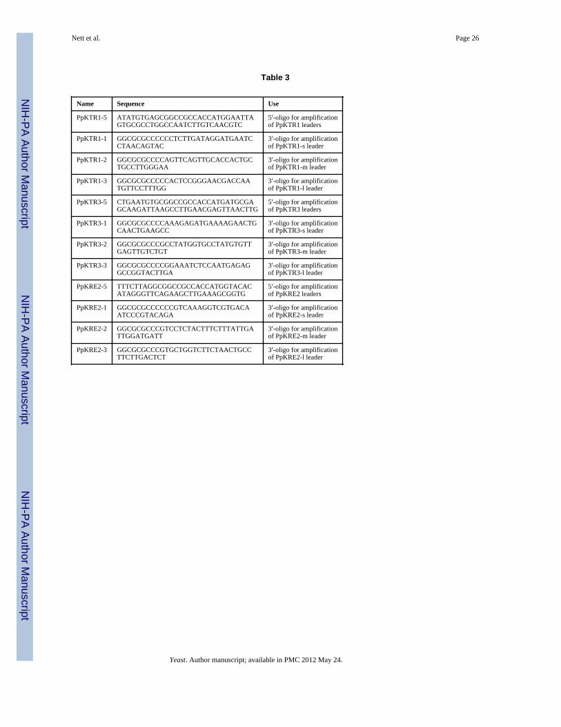
All targeting domains were amplified by PCR using ExTaq (Takara) and the respectivefungal genomic DNA as template, cloned into plasmids pCR2.1 (Invitrogen) or pVM1(pUC19 with additional NotI and AscI sites in the MCS) and sequenced. The 5′-oligointroduced a NotI site and the CACC Kozak consensus sequence just upstream of the nativeATG. In the case of the leaders derived from PpSEC12 or ScSEC12 the codon for the firstamino acid of the targeting domain was changed to ATG. The 3′-oligo introduced an AscIsite and an additional C, which resulted in a fusion linker encoding GlyArgAla. Theoligonucleotides used to amplify targeting domains from PpKTR1, PpKTR3 and PpKRE2are given in Table 3. The sequences of any of the other oligos are available upon request.
After amplification of the targeting domain containing plasmids 36 g of each was digestedwith 50 units AscI for 4 hr. The linearized DNA was then ethanol precipitated, digested with60 units NotI for 15 hr and the resulting inserts were purified by two consecutive rounds ofseparation on agarose gels. For fragments ranging from 87 to 219 nucleotides we used 2 %agarose, for fragments from 240 to 426 nucleotides 1.5 % agarose, and for fragments from435 to 1098 nucleotides 1.0 % agarose. While the bulk of the material was then storedundiluted at −80° C, 1.2 pmole of each was diluted to 100 l, arranged in a 96 well plate andalso stored at −80° C.
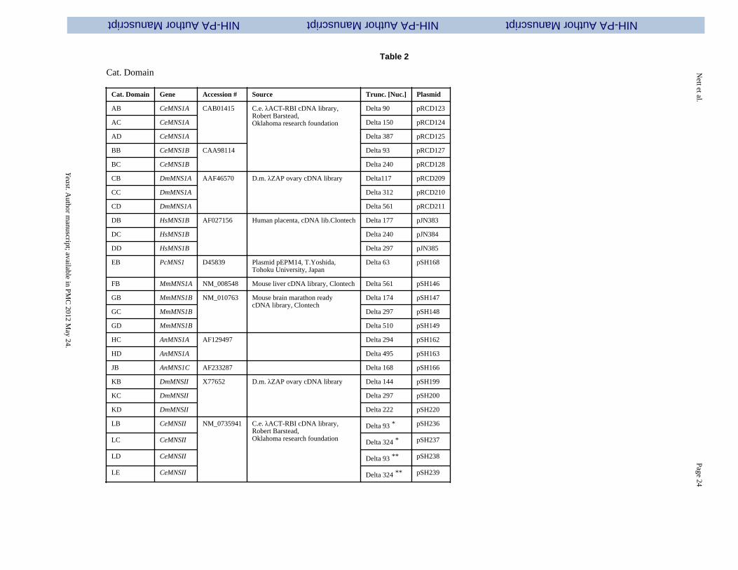
The glycolytic enzyme catalytic domains were amplified using either Ex Taq (Takara) orPfu DNA polymerase (Stratagene) from sources given in Table 2. Generally the full lengthenzymes were amplified first, cloned into pCR2.1 (Invitrogen) and sequenced. Then thetruncated fragments were amplified using those plasmids as template and the 5′oligonucleotides to introduce an AscI site and the 3′ oligos to introduce a PacI site directlyafter the stop codon. The sequences of any of the oligonucleotides are available uponrequest. The fragments were then again cloned into vector pCR2.1 and sequenced. Fromthere they were excised as AscI/PacI fragments and ligated into the respective fusionvectors. The MNSI derived fragments were cloned into pJN347, the GNTI derived fragmentsinto pJN346, the MNSII derived fragments into pJN348, the GNTII derived fragments intopPB124, and the GNTIII derived fragments into pJN348.
High throughput fusion library construction and expression
For construction of the leader/catalytic domain chimeras, 30 g of each glycolytic domaincontaining fusion vector (see Table 2) was digested with 10 units AscI overnight. In themorning another 10 units were added and the incubation was continued for another 1 hr. Thelinearized DNA was Ethanol precipitated and incubated with 50 units NotI and 100 unitsSwaI at RT. After 1 hr the temperature was increased to 37° C and the incubation wascontinued for another 4 hr. Then 20 units calf intestinal alkaline phosphatase were added andthe incubation was continued for another 1 hr. After purification by agarose gel extractionthe DNA was diluted to 3 nM and 2.5 l was loaded into each of the first 69 wells of a 96well PCR plate. Then 2.5 l of a 12 nM solution of the isolated targeting domain NotI/AscIfragments (see above) was added. After addition of 5 l 2x ligation buffer and 0.5 l Quickligase (NEB) the ligation was allowed to proceed for 5 min at RT, and 2 l of the ligation
Nett et al. Page 3
Yeast. Author manuscript; available in PMC 2012 May 24.
NIH
-PA
Author M
anuscriptN
IH-P
A A
uthor Manuscript
NIH
-PA
Author M
anuscript

mix was added to 50 l E. coli strain DH5α made competent according to the method ofHanahan [Hanahan et al., 1991] that were arranged in a 96 well plate. The mixture wasincubated for 20 min on ice, heat shocked at 42° C for 1 min, 200 l SOC were added toeach well of the 96 well plate and the cells were allowed to recover at 37° C for 1 hr. Ofeach transformation mix 200 l were then plated on a single LB Amp plate and incubatedovernight. We routinely obtained 10 to 1000 colonies per plate as compared to 0 to 10colonies on the no insert controls. For each construct two colonies were checked by colonyPCR using GAPScr. (5′-GTCCCTATTTCAATCAATTGAACAAC-3′) as forward primerand a reverse primer that was specific to the individual catalytic domain. A sample gel of thecolony PCR for constructs JB9 – JB32 is given in Fig. 8. Plasmid DNA was isolated frompositive clones and 1 g was digested with 2.5 units AscI for 2 hr to determine whether thesubcloning had resulted in an in frame fusion of both fragments (the case in more than 99 %of all tested clones). In preparation for their transformation into yeast strains the fusionconstructs were then linearized using a unique restriction site in the auxotrophic markerregion of the plasmids (or in the PMA1 promoter in the case of the GNTII containingconstructs). Restriction enzymes used for linearization were SalI for fusions containingcatalytic domains A, D, F, G, M, U and V, EcoNI for fusions containing catalytic domainsC, E, H and J, ApaI for fusion constructs containing K and L, AatII for fusion constructscontaining catalytic domain N, EcoRI for fusion constructs containing catalytic domain Tand XbaI for fusion constructs MA63, MA64 VA63, VA64, VB63, VB64, VC63 and VC64.Fusion constructs UA20 to UA67 were transformed without prior linearization. All leader/catalytic domain chimeras were then introduced into yeast cells via electroporation. MNSIderived fusions were transformed into strain PBP1 and selected on synthetic definedmedium lacking histidine (synthetic defined medium is 1.34% yeast nitrogen base, 2%dextrose, 1.5% agar and 4×10⁻⁵% biotin with amino acids supplemented as appropriate).GNTI containing chimeras were transformed into strains YJN168, YJN169 and YJN191 andselected on synthetic defined medium lacking arginine. MNSII derived fusions weretransformed into strain YSH1 and selected on synthetic defined medium lacking uracil.GNTII containing chimeras were transformed into yeast strain RDP27 and selected on YPDsupplemented with 100 g of the drug Nourseothricin (clonNAT from Hans Knoell Institutefuer Naturstofforschung, Jena, Germany). GNTIII containing fusions were transformed intoYSH1 and selected on synthetic defined medium lacking uracil. Six individual clones ofeach transformation were then analyzed in liquid culture in a high throughput format. To thisend the repatched transformants were grown for 3 days in 0.6 ml buffered glycerol complexmedium (BMGY) consisting of 1% yeast extract, 2% peptone, 100 mM potassiumphosphate buffer, pH 6.0, 1.34% yeast nitrogen base, 4 × 10 −5% biotin, and 1% glycerol indeep well 96 well plates (Flat Bottom Blocks from Qiagen) on a heavy duty orbital shaker.Each starter plate was then used to inoculate 0.6 ml BMGY in 6 identical 96 deep wellplates to an optical density of 1 – 2 at 600 nm and grown overnight. After the cultures hadreached an optical density of 20 – 30 at 600 nm three plates each were combined andwashed once in buffered methanol-complex medium (BMMY) consisting of 1.5% methanolinstead of glycerol in BMGY. Reporter protein expression in the resulting two identicalplates was then induced by growth in 0.6 ml BMMY for 24 h. The two plates were thencombined, the yeast cells removed by centrifugation and 0.8 ml of each supernatant wasused for purification of K3 and glycan analysis.
Reporter protein expression and purification and release of N-linked glycans
The K3 domain reporter protein, under the control of the alcohol oxidase 1 (AOX1)promoter, was used as a model protein and was purified using the 6xHistidine tag asreported previously [Choi et al., 2003]. The glycans were released and separated from theglycoproteins by a modification of a previously reported method [Papac et al., 1998]. Afterthe proteins were reduced and carboxy methylated, and the membranes blocked, the wells
Nett et al. Page 4
Yeast. Author manuscript; available in PMC 2012 May 24.
NIH
-PA
Author M
anuscriptN
IH-P
A A
uthor Manuscript
NIH
-PA
Author M
anuscript
were washed three times with water. The protein was deglycosylated by the addition of 30 lof 10 mM NH4HCO3 pH 8.3 containing one milliunit of N-glycanase (Glyko). After 16hours at 37° C, the solution containing the glycans was removed by centrifugation andevaporated to dryness.
MALDI/time-of-flight (TOF) mass spectrometry
Molecular weights of the glycans were determined by using a Voyager DE PRO linearMALDI-TOF (Applied Biosciences) mass spectrometer with delayed extraction. The driedglycans from each well were dissolved in 15 l of water, and 0.5 l was spotted on stainlesssteel sample plates and mixed with 0.5 l of S-DHB matrix (9 mg/ml of dihydroxybenzoicacid, 1 mg/ml of 5-methoxysalicylic acid in 1:1 water/acetonitrile 0.1% TFA) and allowed todry. Ions were generated by irradiation with a pulsed nitrogen laser (337 nm) with a 4-nspulse time. The instrument was operated in the delayed extraction mode with a 125-ns delayand an accelerating voltage of 20 kV. The grid voltage was 93%, guide wire voltage was0.1%, the internal pressure was less than 5 × 10⁻⁷ torr, and the low mass gate was 875daltons. Spectra were generated from the sum of 100 to 200 laser pulses and acquired with a500 MHz digitizer. (Man)5-(GlcNAc)2 oligosaccharide was used as an external molecularweight standard. All spectra were generated with the instrument in the positive ion mode.
Mannosidase Assays
Fluorescence-labeled (Man)8(GlcNAc)2 (0.5 g) was added to 20 l of supernatant andincubated for 30 hr at room temperature. Following incubation the sample was analyzed byHPLC using an Econosil NH2 4.6 × 250 mm, 5 micron bead, amino-bound silica column(Altech, Avondale, PA). The flow rate was 1.0 ml/min for 40 min and the column wasmaintained to 30°C. After eluting isocratically (68% A:32% B) for 3 min a linear solventgradient (68% A:32% B to 40% A:60% B) was employed over 27 min to elute the glycans(18). Solvent A (acetonitrile) and solvent B (ammonium formate, 50 mM, pH 4.5. Thecolumn was equilibrated with solvent (68% A:32% B) for 20 min between runs.
Results
Cloning of ER and Golgi targeting sequences
Most of the glycosyltransferases in the ER and Golgi of mammals and yeast are type IImembrane proteins [Gleeson, 1998]. Although there is very little sequence similaritybetween these enzymes they all share the same domain structure. A short amino terminalcytoplasmic tail is followed by a transmembrane domain [TMD], a short stem region and alarge carboxy terminal catalytic domain in the lumen of the ER or the Golgi (see Fig. 1). It isgenerally believed that the targeting information resides largely in the TMD and thecytoplasmic tail [Chapman and Munro, 1994; Lussier et al., 1995; Tang et al., 1997;Graham and Krasnov, 1995], however there have been reports that also the lumenal domainscan contain Golgi localization signals [Graham and Krasnov, 1995].
Because we wanted to test a large variety of targeting sequences, we amplified DNAfragments encoding amino terminal regions of varying length of ER and Golgi residing typeII membrane proteins of S. cerevisiae, Pichia pastoris and Kluyveromyces lactis. Theshortest fragments (designated ‘-s’) contained only the cytoplasmic tail and the TMD. Themedium length fragments (designated ‘-m’) also contained parts of, or the entire stemregion. The longest fragments (designated ‘-l’) contained the entire stem region and some,also parts of the catalytic domain (see Fig. 1). We also included fragments of the S.cerevisiae and P. pastoris SEC12 gene, whose gene product Sec12p is a type IItransmembrane protein that acts as a nucleotide exchange factor for the GTP-binding proteinSar1 [Barlowe and Schekman, 1993]. In yeast Sec12p is mainly localized in the ER, but it
Nett et al. Page 5
Yeast. Author manuscript; available in PMC 2012 May 24.
NIH
-PA
Author M
anuscriptN
IH-P
A A
uthor Manuscript
NIH
-PA
Author M
anuscript

has also been shown to cycle between the early Golgi and the ER [Nakano et al.,1988;Boehm, et al., 1994]. Its cytosolic amino terminal catalytic domain is followed by asingle transmembrane domain and the carboxy terminus in the lumen of the ER. In the caseof the S. cerevisiae SEC12 gene, our shortest targeting fragment contained a short stretch ofcytoplasmic domain and the TMD. The medium length fragment contained also a part of theER lumenal domain to serve as a stem region, and the longest fragment contained the TMDand the complete carboxy terminal region. In the case of the P. pastoris SEC12 gene, weonly included the shortest fragment in our studies because the carboxy terminal domainproved to be toxic to bacterial hosts. The other ER targeting domains utilized were aminoterminal fragments of the S. cerevisiae α-glucosidase I [Tang et al., 1997] encoded by GLS1and the S. cerevisiae α-1,2-mannosidase encoded by MNS1.
As targeting domains for the early or cis-Golgi we chose amino terminal fragments of the P.pastoris Och1p, and of the proteins that make up the S. cerevisiae mannan polymerases M-Pol I [Jungman et al., 1999] and M-Pol II, namely Mnn9p, Van1p, Anp1p, Hoc1p, Mnn10pand Mnn11p. Targeting signals for the medial Golgi [Brigance et al., 2000] were derivedfrom S. cerevisiae Kre2p, Ktr1p, Mnn2p, Mnn5p, and Yur1p, from K. lactis Gnt1p and fromP. pastoris proteins with homology to Ktr1p, Ktr3p and Kre2p (unpublished results). Astargeting domains for the late Golgi we included amino terminal regions of S. cerevisiaeMnn1p and Mnn6p. The complete list of targeting domains is given in Table1.
Cloning of heterologous glycosylation enzymes
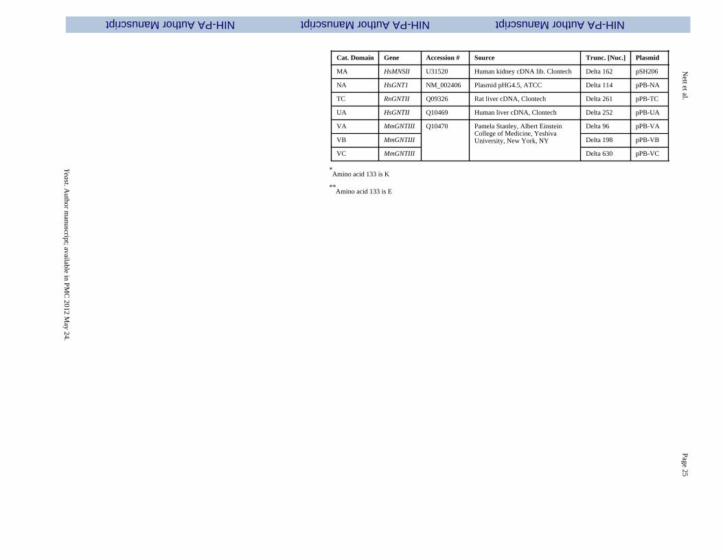
To be able to identify catalytic domains that would function properly in the yeast ER andGolgi we decided to test enzymes with a wide range of pH and temperature optima. Similarto the targeting domains, the glycolytic enzymes were amplified as DNA fragments ofvarying length. The longest fragments contained the enzyme without the TMD. The mediumlength fragments were lacking part of the stem region, and the shortest fragments onlycontained the catalytic domain (see Fig. 1). Some of the mannosidases were selected on thebasis of sequence homology to other known mannosidases and had not been characterizedpreviously. This library of heterologous glycosylation enzymes contained α-1,2-mannosidases (MnsI)) from Caenorhabditis elegans, Drosophila melanogaster, Homosapiens, Penicillium citrinum, Mus musculus and Aspergillus nidulans, -1,2-N-acetylglucosaminyltransferase I (GnTI) from H. sapiens, mannosidases II (MnsII) from D.melanogaster, C. elegans and H. sapiens, -1,2-N-acetylglucosaminyltransferase II (GnTII)from H. sapiens and Rattus norvegicus and -1,4-N-acetylglucosaminyltransferase III(GnTIII) from M. musculus. The complete list of catalytic domains is given in Table 2.
High throughput fusion and expression of leader/catalytic domain chimeras
To facilitate the fusion of targeting sequences to catalytic domains, the targeting domainswere amplified by PCR and cloned into plasmids pCR2.1 (Invitrogen) or pVM1 (a pUC19based plasmid containing a NotI and an AscI site) with NotI sites at the 5′-terminus followedby the Kozak consensus sequence CACC and the start codon. At the 3′ terminus of thetargeting domains the sequence GGGCGCGCC had been added. This introduced an AscIrestriction site and resulted in a fusion linker encoding GlyArgAla. The catalytic domainswere amplified introducing an AscI site at the 5′ end and a PacI site at the 3′ end and clonedinto integration plasmids under the control of the strong PpGAP or PpPMA1 promoters. Theagarose gel purified targeting domain fragments were then, in a high throughput format,ligated in frame into these plasmids to create the fusion libraries. For details see Materialsand Methods. After transformation into bacteria, two colonies per fusion construct wereexamined by colony PCR, which confirmed that more than 95% of all constructs containedthe correct fusions (see Fig. 8). To assess whether the ligation reaction had resulted in in-frame fusions, plasmid DNA was isolated and an AscI restriction digest was performed. This
Nett et al. Page 6
Yeast. Author manuscript; available in PMC 2012 May 24.
NIH
-PA
Author M
anuscriptN
IH-P
A A
uthor Manuscript
NIH
-PA
Author M
anuscript
was based on the fact that only if the AscI site had been recreated a genuine in frame fusionhad occurred. This proved to be true in over 99% of all cases.
Before transformation into yeast strains, the fusion constructs were linearized using anappropriate restriction enzyme that was absent in the leader/catalytic domain chimera butpresent in the integration locus. For details see Materials and Methods. The linearized DNAwas then transformed into yeast strains expressing the Kringle 3 domain of humanplasminogen (K3) by electroporation and six individual clones of each transformation wereanalyzed in liquid culture as described in Materials and Methods.
Analysis of leader/catalytic domain libraries
In the human, Golgi α-1,2-mannosidases (IA-IC) remove mannose from the predominantly(Man)8-(GlcNAc)2 containing glycoproteins that are secreted from the ER to yield the(Man)5(GlcNAc)2 structure, which forms the precursor for complex N-glycans. Wetherefore constructed a library of 19 different α-1,2-mannosidase fragments fused to fungaltargeting domains from the ER and early Golgi (leaders 1 to 32) and transformed them intothe och1 knockout strain PBP1 [Choi et al., 2003] to determine whether this would result intrimming of the fungal glycoforms. Fig. 2 shows that catalytic domains obtained from fungalsources (EB, HC, HD, JB) did not show any activity, whereas the catalytic domains fromputative C. elegans mannosidases showed the highest activity, followed by mannosidasesfrom Drosophila, Mouse and Human. The truncation that removes the N-terminal 129 aminoacids (delta 387) of the putative C.elegans mannosidase IA also showed no activity,suggesting that an essential part of the catalytic domain had been removed. Significantdifferences were also observed in the ability of leader sequences to target fusion proteinswith high mannosidase activity. Generally the longest leader fragments resulted in fusionproteins with lower activity, whereas the fusion proteins containing only the fungal TMDshad the highest activity. Exceptions were the medium sized leaders from ScMNN10 andScMNN11 and the longest leader from ScVAN1, which also showed consistently highactivity. Also all the leaders derived from SEC12 resulted in fusions with very high activity,whereas the leaders derived from PpOCH1 and ScMNN9 typically resulted in fusionproteins with poor apparent mannosidase activity. We also considered the possibility thatsome of the (Man)5(GlcNAc)2 observed was formed outside the cell by mannosidase fusionprotein that had been secreted into the medium. To determine to which extent this was thecase, we examined the supernatants of 39 strains using 2-aminobenzamide-labeled(Man)8(GlcNAc)2 as substrate (see Materials and Methods). Fig. 3 shows that for some ofthe (Man)5(GlcNAc)2- producing strains a high degree of mannosidase activity wasobserved in the culture medium, suggesting that some of the (Man)5(GlcNAc)2 structuresproduced by the strains might have been produced ex vivo. The amount of mannosidaseactivity was independent of the optical density at which protein expression in the culturewas induced and also independent of the amount of methanol that was used for induction(results not shown). The leaders derived from ScMNS1 (#s 4-6) and SEC12 (#s 7-10)typically showed relatively low mannosidase activity in the medium, whereas other leaderstested in the mannosidase library displayed a significant level of leakage into the medium. Itis noteworthy that apparently also the length of the mannosidase domain influences theamount of mannosidase that is secreted into the medium. For example whereas constructsBB4 and BB18 display the maximum amount of activity, constructs BC4 and BC18 thatcontain a shorter mannosidase domain show only background levels of mannosidase activityin the medium.
The second step in the formation of complex N-glycans in the mammalian Golgi is theaddition of N-acetylglucosamine to the 1,3-arm of (Man)5-(GlcNAc)2 to generate GlcNAc-(Man)5-(GlcNAc)2. To replicate this reaction in P. pastoris we fused the soluble domain ofhuman GnTI to leader sequences 1 through 36 of our leader library and transformed the
Nett et al. Page 7
Yeast. Author manuscript; available in PMC 2012 May 24.
NIH
-PA
Author M
anuscriptN
IH-P
A A
uthor Manuscript
NIH
-PA
Author M
anuscript

resulting constructs into three different α-1,2-mannosidase containing yeast strains. YJN168,YJN169 and YJN191 were created from PBP1 by transformation with mannosidase fusionconstructs BC4, BC8 and BC18 respectively. We chose strains that contained mannosidasesthat were targeted to different locations in the secretory pathway, BC4 to the ER, BC8 to theER/early Golgi and BC18 to the early Golgi, because we also wanted to determine the effectof localization of the mannosidase on the formation of GlcNAc-(Man)5-(GlcNAc)2. Asexpected we observed lower amounts of GlcNAc-(Man)5-(GlcNAc)2 when the GnTI fusionlibrary was transformed into strain YJN191 which contained the mannosidase in a latersecretory compartment (see column BC18 in Fig. 4). However the fact that a GnTI chimerathat is targeted to the ER by fusion with the TMD of ScGls1p or ScMns1p still shows someresidual activity in a strain that contains an early Golgi targeted mannosidase is alsoconsistent with the view, that early Golgi enzymes are not stationary in their compartments,but rather undergo continuous recycling through the ER [Todorow et al., 2000]. In our casethis would mean that the early Golgi targeted mannosidase BC18 is recycled to the ERwhere it could form (Man)5-(GlcNAc)2, which in turn would be the substrate for the ERtargeted GnTI fusions NA1 or NA4. An alternative explanation would be that the ERtargeted GnTI is leaking into the early Golgi and is acting on the (Man)5-(GlcNAc)2 that isformed there.
Fig. 4 also shows that, as seen in the mannosidase I fusion library, the short and mediumlength leaders resulted in fusion proteins with higher GnTI activity whereas the long leadersgenerally yielded lower GnTI activity. Again almost all of the SEC12 and VAN1 derivedleaders produced fusions with high activity, whereas only few of the PpOCH1 and ScMNN9derived leaders did.
The third step in the formation of complex N-glycans in the mammalian Golgi is theremoval of the two terminal α-1,3- and α-1,6-linked mannose residues from GlcNAc-(Man)5-(GlcNAc)2 by mannosidase II to form GlcNAc-(Man)3-(GlcNAc)2. To replicate thisreaction in P. pastoris, we transformed a mannosidase II / leader fusion library into theGlcNAc-(Man)5-(GlcNAc)2 producing strain YSH1 [Hamilton et al., 2003], which is anoch1 knockout containing the gene for the Kluyveromyces lactis uridine 5′-diphosphate(UDP)-GlcNAc transporter, the mannosidase fusion FB8 and the GnTI fusion NA15. Asevident from Fig. 5, the human mannosidase II (MA) showed only low levels of activity, asdid D. melanogaster MnsII having N-terminal 48 or 99 amino acid truncations (KB and KC,respectively). However the fusion constructs containing the D. melanogaster mannosidase IIwith the N-terminal 74 amino acids removed (KD) showed ample activity when combinedwith a variety of leader constructs. In the case of the third tested enzyme, the C. elegansmannosidase II, there were only minor differences, but overall the longer catalytic domains(LB, LD) scored slightly higher than the further truncated ones (LC, LE). When combinedwith a highly active mannosidase II catalytic domain the large majority of all leaderconstructs resulted in active fusion constructs, with notable laggards again being leadersderived from PpOCH1, the ScMNN9-m and −l leaders, and leaders derived from ScYUR1.Again it should be noted that despite the GnTI being targeted to the early Golgi, some ER-targeted mannosidase II fusions showed very high activity. This corroborates the resultsobtained with the GnTI fusion library that our fusion proteins apparently are not targeted toexclusively one compartment in the ER or the Golgi, but rather are dispersed in a wide areain the secretory pathway.
The next step in the formation of complex human N-glycans is the addition of anotherGlcNAc to GlcNAc-(Man)3-(GlcNAc)2, a step that is catalyzed by GnTII. We constructed alibrary of fusion constructs of human and rat GnTII with our 66 targeting domains and testedthem in RDP27 [Bobrowicz et al., 2004]. This yeast strain is an och1 knockout containingthe K. lactis (UDP)-GlcNAc transporter, the mannosidase I fusion FB8 and the GnTI fusion
Nett et al. Page 8
Yeast. Author manuscript; available in PMC 2012 May 24.
NIH
-PA
Author M
anuscriptN
IH-P
A A
uthor Manuscript
NIH
-PA
Author M
anuscript
NA15. It also contains a deletion of the P. pastoris homologue of ALG3 [Davidson et al.,2004], an α-1,3-mannosyl-tranferase involved in the dolichol-linked oligosaccharidebiosynthesis. Instead of (Man)8-(GlcNAc)2, knockouts of alg3 transfer the truncated(Man)5-(GlcNAc)2 glycan from dolichol to newly synthesized proteins in the ER. Action ofMnsI and GnTI then leads to the formation of GlcNAc-(Man)3-(GlcNAc)2, which is thesubstrate for GnTII. As shown in Fig. 6 the activity of GnTII / leader fusions appears to bemore dependent on the leader than what had been seen in the cases of MnsI, GnTI or MnsII.The rat GNTII / S.c.MNN2 chimeras TC53, TC54 and TC55 resulted in fusion proteins withfull activity, whereas the human GnTII / leader fusions typically exhibited slightly loweractivity. Other leaders that resulted in fusion proteins with high activity were derived fromScKRE2, PpKTR1, PpKRE2, KlGNTI, ScMNN1 and ScMNN6, however, as opposed to whathad been seen in the cases of the MnsI and GnTI libraries, here the medium length leaderstypically had the highest activity.
A modification of glycosylation that is commonly found on human antibodies is the additionof a -1,4-GlcNAc residue to the (GlcNAc)2-(Man)3-(GlcNAc)2 complex glycan, the socalled bisecting GlcNAc, a reaction catalyzed by N-acetylglucosaminyltransferase III(GnTIII) [Narasimhan, 1982; Bhaumik et al., 1995]. The bisecting GlcNAc has beenimplicated in the modulation of antibody-dependent cellular cytotoxicity (ADCC) [Umanaet al., 1999; Davies et al., 2001; Shinkawa et al., 2003], and might be a potential way togenerate antibodies with optimized effector functions. We fused three different truncationsof mouse GNTIII [Bhaumik et al., 1995] to our 66 targeting domains and transformed thefusion constructs into P. pastoris in an attempt to generate a yeast strain with glycanscontaining bisecting GlcNAc. The recipient yeast strain that was chosen was YSH1 (seeabove), a P. pastoris strain expressing the reporter protein K3 with the glycan structureGlcNAc-(Man)5-(GlcNAc)2. Fig. 7 shows a picture very similar to what was seen in theGnTII fusion library. While some of the leader constructs resulted in fusions with no or onlytrace amounts of activity, some fusion constructs yielded glycans containing a significantamount of bisecting GlcNAc. As seen in the case of GnTII the leaders resulting in thehighest activity were derived from ScMNN2, KlGNT1, ScMNN5 and ScMNN6. The fact thatwe didn't observe complete conversion to the bisecting GlcNAc containing glycan in any ofthe cases might be due to the fact that GlcNAc-(Man)5-(GlcNAc)2 is not the naturalsubstrate for GnTIII, therefore a strain producing a biantennary sugar chain like (GlcNAc)2-(Man)3-(GlcNAc)2 might prove to be a better host for GnTIII.
Discussion
The lack of a suitable mammalian expression system is a significant obstacle to the low-costand safe production of recombinant human glycoproteins for therapeutic applications.Whereas lower eukaryotes such as fungi and yeasts generally offer higher production yieldsand ease of fermentation they also suffer from potential problems associated with the non-human glycosylation pattern that is found on proteins expressed in these organisms. Toovercome these problems we have focused our efforts on engineering commercially relevantyeast and filamentous fungi to produce N-glycans with human-like glycosylation structures.In eukaryotes sugar transferases and glycosidases (e.g. mannosidases) line the inner(luminal) surface of the ER and Golgi to provide a “catalytic” surface that allows for thesequential processing of glycoproteins as they proceed through the secretory pathway. Muchwork has been dedicated to revealing the mechanism by which these glycosylation enzymesare retained and anchored to their respective organelle. Evidence suggests that stem region,transmembrane domain and cytoplasmic tail, individually or in concert, direct enzymes tothe membrane of a specific compartment and thereby localize the associated catalyticdomain to that locus [Gleeson, 1998]. Before our work on optimization of targetingsequences, attempts to engineer the fungal secretory pathway to express functional
Nett et al. Page 9
Yeast. Author manuscript; available in PMC 2012 May 24.
NIH
-PA
Author M
anuscriptN
IH-P
A A
uthor Manuscript
NIH
-PA
Author M
anuscript

heterologous glycosylation enzymes have been described with limited success. Chiba et al.localized an α-1,2-mannosidase of Aspergillus saitoi to the ER of an och1mnn1mnn4 mutantS. cerevisiae strain using the C-terminal motif HDEL as a targeting signal [Chiba et al.,1998]. They were able to show that only 27 % of the N-glycans of the endogenous markerprotein caboxypeptidase Y were trimmed from (Man)8-(GlcNAc)2 to (Man)5-(GlcNAc)2.Kalsner et al. transformed Aspergillus nidulans with the full-length gene for rabbit GNTI[Kalsner et al., 1995]. While they could detect intracellular GnTI activity, they did notobserve any formation of GlcNAc-(Man)5-(GlcNAc)2 despite the fact that Aspergillusnidulans expresses several α1,2-mannosidases [Eades and Hintz, 2000].
In light of these problems, and because prior to our work there was no reliable way ofpredicting whether a particular heterologously expressed glycosyltransferase or mannosidasein a lower eukaryote will be targeted to a specific location in the secretory pathway, wedeveloped a systematic fusion library approach to select for the proper combination oftargeting signal and catalytic domain. Our targeting domain library consisted oftransmembrane domains and adjacent regions of known ER and Golgi residing type IImembrane proteins selected from S. cerevisiae, P. pastoris and K. lactis. For some of theseproteins the regions responsible for proper localization had been characterized previously[Chapman and Munro, 1994; Lussier et al., 1995; Tang et al., 1997; Graham and Krasnov,1995; Sato et al., 1996], but for the majority of the proteins we employed, the targeting hadnot been examined in detail before. To replicate the mammalian glycosylation machinery inP. pastoris we fused these targeting sequences to mannosidases and GlcNAc transferasesfrom different species and tested for their functional expression in P. pastoris strainsproducing the necessary substrates. Because the removal of 1,2-mannose is the first step inthe formation of complex N-glycans we decided to target the α-1,2-mannosidases to an earlycompartment of the secretory pathway, fusing them to leaders 1 through 32. While somemannosidases showed no activity at all, others were able to almost quantitatively remove theα-1,2-mannoses from (Man)8-(GlcNAc)2 to generate (Man)5-(GlcNAc)2 when fused to alarge variety of leader sequences. When we examined the amount of mannosidase that wassecreted into the medium, a phenomenon that had been described previously [Callewaert etal., 2001], it became obvious, that only a small subset of fusion constructs displayed noextracellular mannosidase activity. While the fact that some leader/mannosidase chimerasleak into the medium does not necessarily mean that some or all of their observed activityhappens ex vivo, it is currently not possible to determine where the (Man)5-(GlcNAc)2 isbeing generated in these strains.
The next step in the in the formation of hybrid- and complex-type glycans involves thecorrect localization of GnTI, the enzyme responsible for the formation of GlcNAc-(Man)5-(GlcNAc)2 from (Man)5-(GlcNAc)2. We transformed the GnTI fusion library into threedifferent yeast strains each expressing a different highly active, non-leaking mannosidaseconstruct that was targeted to either the ER (MNS1), the ER/early Golgi (SEC12) or theearly Golgi (VAN1). Whereas a large number of GnTI fusion constructs showed very highactivity when expressed in the first two strains, the same constructs typically showed a lowerlevel of activity in the third. This is consistent with the view that the mannosidase isoptimally targeted to a very early compartment of the secretory pathway to allow GnTI themaximum amount of time to act on the substrate generated by it. On the other hand the factthat a GnTI that is targeted to the ER has any activity at all in a strain with an early Golgitargeted mannosidase also confirms the view that the glycosylation enzymes apparently areable to move within the secretory pathway and are continuously retrieved to their originalcompartments [Todorow et al., 2000; Sato et al., 1996; Harris and Waters, 1996].
The third enzyme in the formation of mammalian complex N-glycans is MnsII, responsiblefor removal of 1,3- and 1,6-mannose from GlcNAc-(Man)5-(GlcNAc)2. Analysis of our
Nett et al. Page 10
Yeast. Author manuscript; available in PMC 2012 May 24.
NIH
-PA
Author M
anuscriptN
IH-P
A A
uthor Manuscript
NIH
-PA
Author M
anuscript
MnsII fusion protein library shows the importance of testing glycosylation enzymes withvarying lengths of stem regions. In the case of MnsII from D. melanogaster the truncationmissing the N-terminal 74 amino acids showed full activity with a number of leaders,whereas the truncations missing the N-terminal 48 or 99 amino acids only had trace amountsof activity.
The next step in complex N-glycan formation is the addition of a second GlcNAc residue toGlcNAc-(Man)3-(GlcNAc)2 by GnTII. To avoid any potential complications arising fromleaking MnsII, we tested the GnTII fusion library in an och1 knockout strain that in additionto expressing MnsI and GnTI leader/fusions was deleted for alg3, an α-1,3-mannosyl-tranferase that is responsible for the first Dol-P-Man-dependent mannosylation step at theluminal side of the ER [Sharma et al., 2001]. In this strain GlcNAc-(Man)3-(GlcNAc)2, thesubstrate for GnTII, is generated without the activity of MnsII. Contrary to what was seenwith the earlier enzymes MnsI, GnTI and MnsII, the activity of the GnTII fusions waslargely dependent on the targeting domain. Leaders derived from ScMNN2 gave rise tofusion proteins with maximum GnTII activity. A similar picture was seen in the fusionlibrary of GnTIII, which is the enzyme that adds the bisecting GlcNAc to (GlcNAc)2-(Man)3-(GlcNAc)2. Here too the leaders derived from ScMNN2 lead to the fusions with thehighest GnTIII activity.
Taken together our results demonstrate that it is almost impossible to predict beforehandprecisely which leader/catalytic domain fusion will exhibit optimal activity and whether theobserved activity actually occurs in vivo. On the other hand we also show that ourcombinatorial genetic library approach and high throughput screening protocol areextremely powerful tools that can be employed to find the exact combinations of leader/catalytic domain fusion proteins that allow for the reengineering of the glycosylationmachinery of commercially important yeast such as P. pastoris to produce human-likeglycan structures. Here we have used these tools to generate yeast strains that attach thecomplex human-like N-glycan (GlcNAc)2-(Man)3-(GlcNAc)2 to an expressed protein, andhave laid the foundation for the complete humanization of the glycosylation machinery ofPichia pastoris.
AcknowledgmentsThe expert technical assistance of Sebastian Rausch, Harry Wischnewski, and Eduard Renfer is gratefullyacknowledged.
We also would like to thank Jim Cregg for the gift of strain JC308.
This research was supported in part by a government grant from the National Institutes of Health (NIH Phase IGrant no. IR43GM66690-1) and a grant from the Department of Commerce, NIST-ATP Cooperative AgreementNumber 70NANB2H3046.
References
Ballou CE. Isolation, characterization, and properties of Saccharomyces cerevisiae mnn mutants withnonconditional protein glycosylation defects. Methods Enzymol. 1990; 185:440–470. [PubMed:2199792]
Barlowe C, Schekman R. SEC12 encodes a guanine nucleotide exchange factor essential for transportvesicle budding from the ER. Nature. 1993; 365:347–349. [PubMed: 8377826]
Bhaumik M, Seldin MF, Stanley P. Cloning and chromosomal mapping of the mouse Mgat3 geneencoding N-acetylglucosaminyltransferase III. Gene. 1995; 164:295–300. [PubMed: 7590346]
Bobrowicz P, Davidson RC, Li H, Potgieter TI, Nett JH, Hamilton SR, Stadheim TA, Miele RG,Bobrowicz B, Mitchell T, Rausch S, Renfer E, Wildt S. Engineering of an artificial glycosylationpathway blocked in core oligosaccharide assembly in the yeast Pichia pastoris: production of
Nett et al. Page 11
Yeast. Author manuscript; available in PMC 2012 May 24.
NIH
-PA
Author M
anuscriptN
IH-P
A A
uthor Manuscript
NIH
-PA
Author M
anuscript

complex humanized glycoproteins with terminal galactose. Glycobiology. 2004; 14:757–766.[PubMed: 15190003]
Boehm J, Ulrich HD, Ossig R, Schmitt HD. Kex2-dependent invertase secretion as a tool to study thetargeting of transmembrane proteins which are involved in ER-Golgi transport in yeast. EMBO J.1994; 13:3696–3710. [PubMed: 8070399]
Brigance WT, Barlowe C, Graham TR. Organization of the yeast Golgi complex into at least fourfunctionally distinct compartments. Mol. Biol. Cell. 2000; 11:171–182. [PubMed: 10637300]
Callewaert N, Laroy W, Cadirgi H, Geysens S, Saelens X, Min Jou W, Contreras R. Use of HDEL-tagged Trichoderma reesei mannosyl oligosaccharide 1,2-alpha-D-mannosidase for N-glycanengineering in Pichia pastoris. FEBS Lett. 2001; 503:173–178. [PubMed: 11513877]
Chapman RE, Munro S. The functioning of the yeast Golgi apparatus requires an ER protein encodedby ANP1, a member of a new family of genes affecting the secretory pathway. EMBO J. 1994;13:4896–4907. [PubMed: 7957057]
Chiba Y, Suzuki M, Yoshida S, Yoshida A, Ikenaga H, Takeuchi M, Jigami, Y, Ichishima E.Production of human compatible high mannose type (Man5GlcNAc2) sugar chains inSaccharomyces cerevisiae. J. Biol. Chem. 1998; 273:26298–26304. [PubMed: 9756858]
Choi BK, Bobrowicz P, Davidson RC, Hamilton SR, Kung DH, Li H, Miele RG, Nett JH, Wildt S,Gerngross TU. Use of combinatorial genetic libraries to humanize N-linked glycosylation in theyeast Pichia pastoris. Proc. Natl. Acad. Sci. 2003; 100:5022–5027. [PubMed: 12702754]
Dean N. Asparagine linked glycosylation in the yeast Golgi. Biochim. Biophys Acta. 1999; 1426:309–322. [PubMed: 9878803]
Davidson RC, Nett JH, Renfer E, Li H, Stadheim TA, Miller BJ, Miele RG, Hamilton SR, Choi BK,Mitchell TI, Wildt S. Functional analysis of the ALG3 gene encoding the Dol-P-Man:Man5GlcNAc2-PP-Dol mannosyltransferase enzyme of P. pastoris. Glycobiology. 2004; 14:399–407. [PubMed: 15033937]
Davies J, Jiang LY, Pan, LZ, LaBarre MJ, Anderson D, Reff M. Expression of GnTIII in arecombinant Anti-CD20 CHO production cell line: Expression of antibodies with alteredglycoforms leads to an increase in ADCC through higher affinity for FcRIII. Biotechnol. Bioeng.2001; 74:288–294. [PubMed: 11410853]
Durand H, Clanet M. Genetic improvement of T. reesei for large scale cellulase production. EnzymeMicrob. Technol. 1988; 10:341–346.
Eades CJ, Hintz WE. Characterization of the class I α-mannosidase gene family in the filamentousfungus Aspergillus nidulans. Gene. 2000; 255:25–34. [PubMed: 10974561]
Gleeson PA. Targeting of proteins to the Golgi apparatus. Histochem. Cell. Biol. 1998; 109:517–532.[PubMed: 9681632]
Graham TR, Krasnov VA. Sorting of yeast α-1,3-mannosyltransferase is mediated by a lumenaldomain interaction, and a transmembrane domain signal that can confer clathrin-dependent Golgilocalization to a secreted protein. Mol. Biol. Cell. 1995; 6:809–824. [PubMed: 7579696]
Hamilton SR, Bobrowicz P, Bobrowicz B, Davidson RC, Li H, Mitchell T, Nett JH, Rausch S,Stadheim TA, Wischnewski H, Wildt S, Gerngross TU. Production of complex humanglycoproteins in yeast. Science. 2003; 301:1244–1246. [PubMed: 12947202]
Hanahan D, Jessee J, Bloom FR. Plasmid transformation of Escherichia coli and other bacteria.Methods Enzymol. 1991; 204:63–113. [PubMed: 1943786]
Harris SL, Waters MG. Localization of a yeast early Golgi mannosyltransferase, Och1p, involvesretrograde transport. J. Cell. Biol. 1996; 132:985–998. [PubMed: 8601597]
Helenius A, Aebi M. Intracellular functions of N-linked glycans. Science. 2001; 291:2364–2369.[PubMed: 11269317]
Jungmann J, Rayner JC, Munro S. The Saccharomyces cerevisiae protein Mnn10p/Bed1p is a subunitof a Golgi mannosyltransferase complex. J. Biol. Chem. 1999; 274:6579–6585. [PubMed:10037752]
Kalsner I, Hintz W, Reid LS, Schachter H. Insertion into Aspergillus nidulans of functional UDP-GlcNAc: alpha 3-D-mannoside beta-1,2-N-acetyglucosaminyl-transferase I, the enzyme catalyzingthe first commited step from oligomannose to hybrid and complex N-glycans. Glycoconj. 1995;12:360–370.
Nett et al. Page 12
Yeast. Author manuscript; available in PMC 2012 May 24.
NIH
-PA
Author M
anuscriptN
IH-P
A A
uthor Manuscript
NIH
-PA
Author M
anuscript
Lin Cereghino GP, Lin Cereghino J, Jay Sunga A, Johnson MA, Lim M, Gleeson MAG, Cregg JM.New selectable marker/auxotrophic host strain combinations for molecular genetic manipulation ofPichia pastoris. Gene. 2001; 263:159–169. [PubMed: 11223254]
Lussier M, Sdicu AM, Ketela T, Bussey H. Localization and targeting of the Saccharomycescerevisiae Kre2p/Mnt1p α1,2-mannosyltransferase to a medial-Golgi compartment. J. Cell Biol.1995; 131:913–927. [PubMed: 7490293]
Maras M, De Bruyn A, Vervecken W, Uusitalo J, Penttilä M, Busson R, Herdewijn P, Contreras R. Invivo synthesis of complex N-glycans by expression of human N-acetylglucosaminyltransferase I inthe filamentous fungus Trichoderma reesei. FEBS Lett. 1999; 452:365–370. [PubMed: 10386623]
Nakano A, Brada D, Schekman R. A membrane glycoprotein Sec12p, required for protein transportfrom the endoplasmic reticulum to the Golgi apparatus in yeast. J. Cell Biol. 1988; 107:851–863.[PubMed: 3047151]
Narasimhan S. Control of glycoprotein synthesis. UDP-GlcNAc: glycopeptide 4-N-acetylglucosaminyltransferase III, an enzyme in hen oviduct which adds GalNAc in 1-4 linkageto the -linked mannose of the trimannosyl core of N-glycosyl oligosaccharides. J. Biol. Chem.1982; 257:10235–10242. [PubMed: 6213618]
Papac DI, Briggs JB, Chin ET, Jones AJ. A high-throughput microscale method to release N-linkedoligosaccharides from glycoproteins for matrix-assisted laser desorption/ionization time-of-flightmass spectrometric analysis. Glycobiology. 1998; 8:445–454. [PubMed: 9597542]
Shinkawa T, Nakamura K, Yamane N, Shoji-Hosaka E, Kanda Y, Sakurada M, Uchida K, Anazawa H,Satoh M, Yamasaki M, Hanai N, Shitara K. The absence of fucose but not the presence ofgalactose or bisecting N-Acetylglucosamine of human IgG1 complex-type oligosaccharides showsthe critical role of enhancing antibody-dependent cellular cytotoxicity. J. Biol. Chem. 2003;278:3466–3473. [PubMed: 12427744]
Sato M, Sato K, Nakano A. Endoplasmic reticulum localization of Sec12p is achieved by twomechanisms: Rer1p-dependent retrieval that requires the transmembrane domain and Rer1p-independent retention that involves the cytoplasmic domain. J. Cell. Biol. 1996; 134:279–293.[PubMed: 8707815]
Sharma CB, Knauer R, Lehle L. Biosynthesis of lipid-linked oligosaccharides in yeast: the ALG3 geneencodes the Dol-P-Man:Man(5)GlcNAc(2)-PP-Dol mannosyltransferase. Biol. Chem. 2001;382:321–328. [PubMed: 11308030]
Tang BL, Low SH, Hong W. Endoplasmatic reticulum retention mediated by the transmembranedomain of type II membrane proteins Sec12p and glucosidase 1. Eur. J. Cell Biol. 1997; 73:98–104. [PubMed: 9208222]
Todorow Z, Spang A, Carmack E, Yates J, Schekman R. Active recycling of yeast Golgimannosyltransferase complexes through the endoplasmic reticulum. Proc. Natl. Acad. Sci. 2000;97:13643–13648. [PubMed: 11095735]
Umana P, Jean-Mairet J, Moudry R, Amstutz H, Bailey JE. Engineered glycoforms of an anti-neuroblastoma IgG1 with optimized antibody-dependent cellular cytotoxic activity. Nat.Biotechnol. 1999; 17:176–180. [PubMed: 10052355]
Werten MW, van den Bosch TJ, Wind RD, Mooibroek H, de Wolf FA. High-yield secretion ofrecombinant gelatins by Pichia pastoris. Yeast. 1999; 15:1087–1096. [PubMed: 10455232]
Nett et al. Page 13
Yeast. Author manuscript; available in PMC 2012 May 24.
NIH
-PA
Author M
anuscriptN
IH-P
A A
uthor Manuscript
NIH
-PA
Author M
anuscript

Fig. 1. Leader and catalytic domain nomenclatureCartoon depicting the structure of type II membrane proteins and the compositional natureof leader/catalytic domain fusion constructs. For details on the sizes of individual leadersand catalytic domains please refer to Tables 1 and 2.
Nett et al. Page 14
Yeast. Author manuscript; available in PMC 2012 May 24.
NIH
-PA
Author M
anuscriptN
IH-P
A A
uthor Manuscript
NIH
-PA
Author M
anuscript
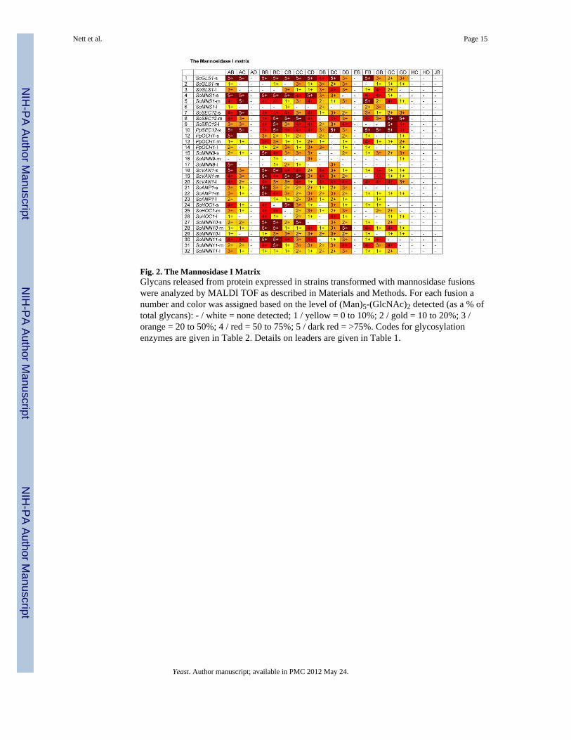
Fig. 2. The Mannosidase I MatrixGlycans released from protein expressed in strains transformed with mannosidase fusionswere analyzed by MALDI TOF as described in Materials and Methods. For each fusion anumber and color was assigned based on the level of (Man)5-(GlcNAc)2 detected (as a % oftotal glycans): - / white = none detected; 1 / yellow = 0 to 10%; 2 / gold = 10 to 20%; 3 /orange = 20 to 50%; 4 / red = 50 to 75%; 5 / dark red = >75%. Codes for glycosylationenzymes are given in Table 2. Details on leaders are given in Table 1.
Nett et al. Page 15
Yeast. Author manuscript; available in PMC 2012 May 24.
NIH
-PA
Author M
anuscriptN
IH-P
A A
uthor Manuscript
NIH
-PA
Author M
anuscript

Fig. 3. Mannosidase leakage assay resultsFluorescence-labeled (Man)8-(GlcNAc)2 was added to culture supernatant, incubated andthen analyzed by HPLC as described in Materials and Methods. For each sample a numberand color was assigned based on the level of (Man)5-(GlcNAc)2 detected (as a % of totalglycans): - / white = none detected; 1 / yellow = 0 to10%; 2 / gold = 10 to 20%; 3 / orange =20 to 50%; 4 / red = 50 to 75%; 5 / dark red = >75%.
Nett et al. Page 16
Yeast. Author manuscript; available in PMC 2012 May 24.
NIH
-PA
Author M
anuscriptN
IH-P
A A
uthor Manuscript
NIH
-PA
Author M
anuscript
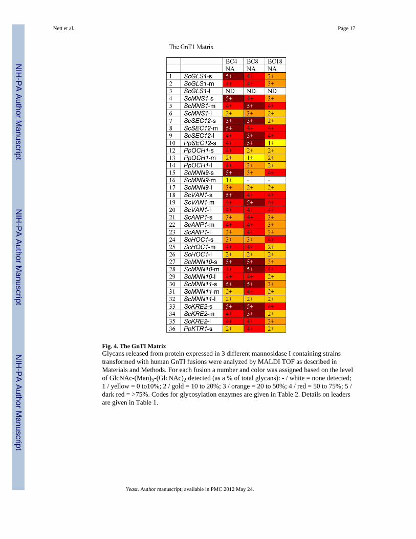
Fig. 4. The GnTI MatrixGlycans released from protein expressed in 3 different mannosidase I containing strainstransformed with human GnTI fusions were analyzed by MALDI TOF as described inMaterials and Methods. For each fusion a number and color was assigned based on the levelof GlcNAc-(Man)5-(GlcNAc)2 detected (as a % of total glycans): - / white = none detected;1 / yellow = 0 to10%; 2 / gold = 10 to 20%; 3 / orange = 20 to 50%; 4 / red = 50 to 75%; 5 /dark red = >75%. Codes for glycosylation enzymes are given in Table 2. Details on leadersare given in Table 1.
Nett et al. Page 17
Yeast. Author manuscript; available in PMC 2012 May 24.
NIH
-PA
Author M
anuscriptN
IH-P
A A
uthor Manuscript
NIH
-PA
Author M
anuscript

Fig. 5. The Mannosidase II MatrixGlycans released from protein expressed in strains transformed with mannosidase II fusionswere analyzed by MALDI TOF as described in Materials and Methods. For each fusion anumber and color was assigned based on the level of GlcNAc-(Man)3-(GlcNAc)2 detected(as a % of total glycans): - / white = none detected; 1 / yellow = 0 to10%; 2 / gold = 10 to20%; 3 / orange = 20 to 50%; 4 / red = 50 to 75%; 5 / dark red = >75%. Codes forglycosylation enzymes are given in Table 2. Details on leaders are given in Table 1.
Nett et al. Page 18
Yeast. Author manuscript; available in PMC 2012 May 24.
NIH
-PA
Author M
anuscriptN
IH-P
A A
uthor Manuscript
NIH
-PA
Author M
anuscript

Fig. 6. The GnTII MatrixGlycans released from protein expressed in strains transformed with GnTII fusions wereanalyzed by MALDI TOF as described in Materials and Methods. For each fusion a numberand color was assigned based on the level of (GlcNAc)2-(Man)3-(GlcNAc)2 detected (as a %of total glycans): - / white = none detected; 1 / yellow = 0 to10%; 2 / gold = 10 to 20%; 3 /orange = 20 to 50%; 4 / red = 50 to 75%; 5 / dark red = >75%. Codes for glycosylationenzymes are given in Table 2. Details on leaders are given in Table 1.
Nett et al. Page 19
Yeast. Author manuscript; available in PMC 2012 May 24.
NIH
-PA
Author M
anuscriptN
IH-P
A A
uthor Manuscript
NIH
-PA
Author M
anuscript
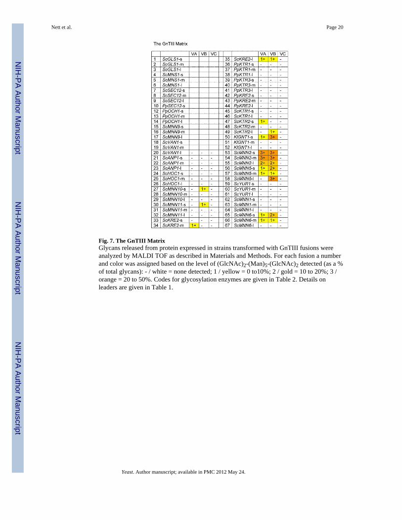
Fig. 7. The GnTIII MatrixGlycans released from protein expressed in strains transformed with GnTIII fusions wereanalyzed by MALDI TOF as described in Materials and Methods. For each fusion a numberand color was assigned based on the level of (GlcNAc)2-(Man)5-(GlcNAc)2 detected (as a %of total glycans): - / white = none detected; 1 / yellow = 0 to10%; 2 / gold = 10 to 20%; 3 /orange = 20 to 50%. Codes for glycosylation enzymes are given in Table 2. Details onleaders are given in Table 1.
Nett et al. Page 20
Yeast. Author manuscript; available in PMC 2012 May 24.
NIH
-PA
Author M
anuscriptN
IH-P
A A
uthor Manuscript
NIH
-PA
Author M
anuscript

Fig. 8. Colony PCR sampleColony PCR screen for formation of fusion constructs JB9 through JB32. Bacterial colonieswere analyzed using oligonucleotides annealing to the GAPDH promoter as forward primerand Aspergillus nidulans MNS1C (construct JB) as reverse primer.
Nett et al. Page 21
Yeast. Author manuscript; available in PMC 2012 May 24.
NIH
-PA
Author M
anuscriptN
IH-P
A A
uthor Manuscript
NIH
-PA
Author M
anuscript

NIH-PA Author ManuscriptNIH-PA Author ManuscriptNIH-PA Author Manuscript
Nett et al.
Page 22
Table 1
Leader# Gene Nucleotides Plasmid Leader# Gene Nucleotides Plasmid
1 ScGLS1-s 1-102 pJN355 35 ScKRE2-l 1-306 pJN279
2 ScGLS1-m 1-255 pJN356 36 PpKTR1-s 1-216 pJN351
3 ScGLS1-l 1-411 pJN373 37 PpKTR1-m 1-336 pJN372
4 ScMNS1-s 1-90 pJN278 38 PpKTR1-l 1-588 pJN352
5 ScMNS1-m 1-246 pJN280 39 PpKTR3-s 1-90 pJN353
6 ScMNS1-l 1-429 pJN282 40 PpKTR3-m 1-255 pJN354
7 ScSEC12-s 988-1140 pJN297 41 PpKTR3-l 1-489 pJN387
8 ScSEC12-m 988-1296 pJN305 42 PpKRE2-s 1-93 pJN366
9 ScSEC12-l 988-1413 pJN291 43 PpKRE2-m 1-252 pJN350
10 PpSEC12-s 1003-1089 pJN328 44 PpKRE2-l 1-450 pJN367
12 PpOCH1-s 1-150 pJN274 45 ScKTR1-s 1-117 pJN301
13 PpOCH1-m 1-240 pJN275 46 ScKTR1-l 1-465 pJN344
14 PpOCH1-l 1-402 pJN276 47 ScKTR2-s 1-120 pJN295
15 ScMNN9-s 1-120 pJN271 48 ScKTR2-m 1-300 pJN288
16 ScMNN9-m 1-273 pJN272 49 ScKTR2-l 1-510 pJN345
17 ScMNN9-l 1-462 pJN342 50 KlGNT1-s 1-93 pJN377
18 ScVAN1-s 1-279 pJN294 51 KlGNT1-m 1-243 pJN378
19 ScVAN1-m 1-294 pJN368 52 KlGNT1-l 1-516 pJN379
20 ScVAN1-l 1-609 pJN357 53 ScMNN2-s 1-108 pJN281
21 ScANP1-s 1-180 pJN302 54 ScMNN2-m 1-291 pJN273
22 ScANP1-m 1-246 pJN303 55 ScMNN2-l 1-450 pJN277
23 ScANP1-l 1-315 pJN304 56 ScMNN5-s 1-105 pJN361
24 ScHOC1-s 1-102 pJN370 57 ScMNN5-m 1-255 pJN362
25 ScHOC1-m 1-252 pJN359 58 ScMNN5-l 1-450 pJN363
26 ScHOC1-l 1-435 pJN360 59 ScYUR1-s 1-108 pJN289
27 ScMNN10-s 1-219 pJN382 60 ScYUR1-m 1-270 pJN290
28 ScMNN10-m 1-363 pJN358 61 ScYUR1-l 1-450 pJN296
29 ScMNN10-l 1-516 pJN369 62 ScMNN1-s 1-126 pJN292
Yeast. A
uthor manuscript; available in P
MC
2012 May 24.

NIH-PA Author ManuscriptNIH-PA Author ManuscriptNIH-PA Author Manuscript
Nett et al.
Page 23
Leader# Gene Nucleotides Plasmid Leader# Gene Nucleotides Plasmid
30 ScMNN11-s 1-156 pJN375 63 ScMNN1-m 1-279 pJN343
31 ScMNN11-m 1-303 pJN376 64 ScMNN1-l 1-459 pJN293
32 ScMNN11-l 1-495 pJN381 65 ScMNN6-s 1-90 pJN364
33 ScKRE2-s 1-174 pJN340 66 ScMNN6-m 1-255 pJN371
34 ScKRE2-m 1-240 pJN380 67 ScMNN6-l 1-480 pJN357
Yeast. A
uthor manuscript; available in P
MC
2012 May 24.
NIH-PA Author ManuscriptNIH-PA Author ManuscriptNIH-PA Author Manuscript
Nett et al.
Page 24
Table 2
Cat. Domain
Cat. Domain Gene Accession # Source Trunc. [Nuc.] Plasmid
AB CeMNS1A CAB01415 C.e. ACT-RBI cDNA library,Robert Barstead,Oklahoma research foundation
Delta 90 pRCD123
AC CeMNS1A Delta 150 pRCD124
AD CeMNS1A Delta 387 pRCD125
BB CeMNS1B CAA98114 Delta 93 pRCD127
BC CeMNS1B Delta 240 pRCD128
CB DmMNS1A AAF46570 D.m. ZAP ovary cDNA library Delta117 pRCD209
CC DmMNS1A Delta 312 pRCD210
CD DmMNS1A Delta 561 pRCD211
DB HsMNS1B AF027156 Human placenta, cDNA lib.Clontech Delta 177 pJN383
DC HsMNS1B Delta 240 pJN384
DD HsMNS1B Delta 297 pJN385
EB PcMNS1 D45839 Plasmid pEPM14, T.Yoshida,Tohoku University, Japan
Delta 63 pSH168
FB MmMNS1A NM_008548 Mouse liver cDNA library, Clontech Delta 561 pSH146
GB MmMNS1B NM_010763 Mouse brain marathon readycDNA library, Clontech
Delta 174 pSH147
GC MmMNS1B Delta 297 pSH148
GD MmMNS1B Delta 510 pSH149
HC AnMNS1A AF129497 Delta 294 pSH162
HD AnMNS1A Delta 495 pSH163
JB AnMNS1C AF233287 Delta 168 pSH166
KB DmMNSII X77652 D.m. ZAP ovary cDNA library Delta 144 pSH199
KC DmMNSII Delta 297 pSH200
KD DmMNSII Delta 222 pSH220
LB CeMNSII NM_0735941 C.e. ACT-RBI cDNA library,Robert Barstead,Oklahoma research foundation
Delta 93 * pSH236
LC CeMNSII Delta 324 * pSH237
LD CeMNSII Delta 93 ** pSH238
LE CeMNSII Delta 324 ** pSH239
Yeast. A
uthor manuscript; available in P
MC
2012 May 24.
NIH-PA Author ManuscriptNIH-PA Author ManuscriptNIH-PA Author Manuscript
Nett et al.
Page 25
Cat. Domain Gene Accession # Source Trunc. [Nuc.] Plasmid
MA HsMNSII U31520 Human kidney cDNA lib. Clontech Delta 162 pSH206
NA HsGNT1 NM_002406 Plasmid pHG4.5, ATCC Delta 114 pPB-NA
TC RnGNTII Q09326 Rat liver cDNA, Clontech Delta 261 pPB-TC
UA HsGNTII Q10469 Human liver cDNA, Clontech Delta 252 pPB-UA
VA MmGNTIII Q10470 Pamela Stanley, Albert EinsteinCollege of Medicine, YeshivaUniversity, New York, NY
Delta 96 pPB-VA
VB MmGNTIII Delta 198 pPB-VB
VC MmGNTIII Delta 630 pPB-VC
*Amino acid 133 is K
**Amino acid 133 is E
Yeast. A
uthor manuscript; available in P
MC
2012 May 24.
NIH
-PA
Author M
anuscriptN
IH-P
A A
uthor Manuscript
NIH
-PA
Author M
anuscript
Nett et al. Page 26
Table 3
Name Sequence Use
PpKTR1-5 ATATGTGAGCGGCCGCCACCATGGAATTAGTGCGCCTGGCCAATCTTGTCAACGTC
5′-oligo for amplificationof PpKTR1 leaders
PpKTR1-1 GGCGCGCCCCCCTCTTGATAGGATGAATCCTAACAGTAC
3′-oligo for amplificationof PpKTR1-s leader
PpKTR1-2 GGCGCGCCCCAGTTCAGTTGCACCACTGCTGCCTTGGGAA
3′-oligo for amplificationof PpKTR1-m leader
PpKTR1-3 GGCGCGCCCCCACTCCGGGAACGACCAATGTTCCTTTGG
3′-oligo for amplificationof PpKTR1-l leader
PpKTR3-5 CTGAATGTGCGGCCGCCACCATGATGCGAGCAAGATTAAGCCTTGAACGAGTTAACTTG
5′-oligo for amplificationof PpKTR3 leaders
PpKTR3-1 GGCGCGCCCCAAAGAGATGAAAAGAACTGCAACTGAAGCC
3′-oligo for amplificationof PpKTR3-s leader
PpKTR3-2 GGCGCGCCCGCCTATGGTGCCTATGTGTTGAGTTGTCTGT
3′-oligo for amplificationof PpKTR3-m leader
PpKTR3-3 GGCGCGCCCCGGAAATCTCCAATGAGAGGCCGGTACTTGA
3′-oligo for amplificationof PpKTR3-l leader
PpKRE2-5 TTTCTTAGGCGGCCGCCACCATGGTACACATAGGGTTCAGAAGCTTGAAAGCGGTG
5′-oligo for amplificationof PpKRE2 leaders
PpKRE2-1 GGCGCGCCCCCCGTCAAAGGTCGTGACAATCCCGTACAGA
3′-oligo for amplificationof PpKRE2-s leader
PpKRE2-2 GGCGCGCCCGTCCTCTACTTTCTTTATTGATTGGATGATT
3′-oligo for amplificationof PpKRE2-m leader
PpKRE2-3 GGCGCGCCCGTGCTGGTCTTCTAACTGCCTTCTTGACTCT
3′-oligo for amplificationof PpKRE2-l leader
Yeast. Author manuscript; available in PMC 2012 May 24.