Isolation, chemical structures and biological activity of the lipo-chitin oligosaccharide nodulation...
12
Plant Molecular Biology 29: 453-464, 1995. © 1995 Kluwer Academic Publishers. Printed in Belgium. 453 Isolation, chemical structures and biological activity of the lipo-chitin oligosaccharide nodulation signals from Rhizobium etli Luis Cfirdenas t, Jimena Dominguez 1, Carmen Quinto 1, Isabel M. L6pez-Lara 2, Ben J. J. Lugtenberg 2, Herman P. Spaink 2'*, Geert Jan Rademaker 3, Johan Haverkamp 3 and Jane E. Thomas-Oates 3 1Departamento de Biologia Molecular de Plantas, Instituto de Biotecnologia, Universidad Nacional Aut6noma de M~xico; 2Institute of Molecular Plant Sciences, Leiden University, Wassenaarseweg 64, 2333 AL Leiden, The Netherlands (* author for correspondence); 3Department of Mass Spectrometry, Bijvoet Center for Biomolecular Research, Utrecht University, Sorbonnelaan 16, 3584 CA Utrecht, The Netherlands Received 14 February 1995; accepted in revised form 9 June 1995 Key words: signal molecules, host specificity, nodulation, flavonoids, plant organogenesis, mass spectrometry Abstract Rhizobium etfi is a microsymbiont of plants of the genus Phaseolus. Using mass spectrometry we have identified the lipo-chitin oligosaccharides (LCOs) that are produced by R. etli strain CE3. They are N-acetylglucosamine pentasaccharides of which the non-reducing residue is N-methylated and N-acylated with cis-vaccenic acid (C18:1) or stearic acid (C18:0) and carries a carbamoyl group at C4. The reducing residue is substituted at the C6 position with O-acetylfucose. Analysis of their biological activity on the host plant Phaseolus vulgaris shows that these LCOs can elicit the formation of nodule primordia which develop to the stage where vascular bundles are formed. The formation of complete nodule structures, including an organized vascular tissue, is never observed. Considering the very close resemblance of the R. etli LCO structures to those of R. loti (I. M. L6pez-Lara, J. D. J. van den Berg, J. E. Thomas Oates, J. Glushka, B. J. J. Lugtenberg, H. P. Spaink, Mol Microbiol 15: 627-638, 1995) we tested the ability ofR. etli strains to nodulate various Lotus species and ofR. loti to nodulate P. vulgaris. The results show that R. etli is indeed able to nodulate Lotus plants. However, several Lotus species are only nodulated when an additional flavonoid independent transcription activator (FITA) nodD gene is provided. Phaseo- lus plants can also be nodulated by R. loti bacteria, but only when the bacteria contain a FITA nodD gene. Apparently, the type of nod gene inducers secreted by the plants is the major basis for the sepa- ration of Phaseolus and Lotus into different cross inoculation groups. Introduction Soil bacteria of the genus Rhizobium are able to establish a symbiosis with specific leguminous plants by forming root nodules in which, after differentiation of the bacteria to bacteroids, at- mospheric nitrogen is fixed. The shape and growth pattern of the nodule that is formed is determined by the plant. Determinate-type nodules develop from primordia in the outer cortex and indeter- minate nodules develop from primordia in the inner cortex [ 7 ]. The specificities of various types
-
Upload
independent -
Category
Documents
-
view
4 -
download
0
Transcript of Isolation, chemical structures and biological activity of the lipo-chitin oligosaccharide nodulation...
Plant Molecular Biology 29: 453-464, 1995. © 1995 Kluwer Academic Publishers. Printed in Belgium. 453
Isolation, chemical structures and biological activity of the lipo-chitin oligosaccharide nodulation signals from Rhizobium etli
Luis Cfirdenas t, Jimena Dominguez 1, Carmen Quinto 1, Isabel M. L6pez-Lara 2, Ben J. J. Lugtenberg 2, Herman P. Spaink 2'*, Geert Jan Rademaker 3, Johan Haverkamp 3 and Jane E. Thomas-Oates 3 1Departamento de Biologia Molecular de Plantas, Instituto de Biotecnologia, Universidad Nacional Aut6noma de M~xico; 2Institute of Molecular Plant Sciences, Leiden University, Wassenaarseweg 64, 2333 AL Leiden, The Netherlands (* author for correspondence); 3Department of Mass Spectrometry, Bijvoet Center for Biomolecular Research, Utrecht University, Sorbonnelaan 16, 3584 CA Utrecht, The Netherlands
Received 14 February 1995; accepted in revised form 9 June 1995
Key words: signal molecules, host specificity, nodulation, flavonoids, plant organogenesis, mass spectrometry
Abstract
Rhizobium etfi is a microsymbiont of plants of the genus Phaseolus. Using mass spectrometry we have identified the lipo-chitin oligosaccharides (LCOs) that are produced by R. etli strain CE3. They are N-acetylglucosamine pentasaccharides of which the non-reducing residue is N-methylated and N-acylated with cis-vaccenic acid (C18:1) or stearic acid (C18:0) and carries a carbamoyl group at C4. The reducing residue is substituted at the C6 position with O-acetylfucose. Analysis of their biological activity on the host plant Phaseolus vulgaris shows that these LCOs can elicit the formation of nodule primordia which develop to the stage where vascular bundles are formed. The formation of complete nodule structures, including an organized vascular tissue, is never observed. Considering the very close resemblance of the R. etli LCO structures to those of R. loti (I. M. L6pez-Lara, J. D. J. van den Berg, J. E. Thomas Oates, J. Glushka, B. J. J. Lugtenberg, H. P. Spaink, Mol Microbiol 15: 627-638, 1995) we tested the ability ofR. etli strains to nodulate various Lotus species and ofR. loti to nodulate P. vulgaris. The results show that R. etli is indeed able to nodulate Lotus plants. However, several Lotus species are only nodulated when an additional flavonoid independent transcription activator (FITA) nodD gene is provided. Phaseo- lus plants can also be nodulated by R. loti bacteria, but only when the bacteria contain a FITA nodD gene. Apparently, the type of nod gene inducers secreted by the plants is the major basis for the sepa- ration of Phaseolus and Lotus into different cross inoculation groups.
Introduction
Soil bacteria of the genus Rhizobium are able to establish a symbiosis with specific leguminous plants by forming root nodules in which, after differentiation of the bacteria to bacteroids, at-
mospheric nitrogen is fixed. The shape and growth pattern of the nodule that is formed is determined by the plant. Determinate-type nodules develop from primordia in the outer cortex and indeter- minate nodules develop from primordia in the inner cortex [ 7 ]. The specificities of various types
454
ofrhizobia for host plants are very different. Some strains such as Rhizobium strain NGR234 [ 12] or strain G R H 2 [ 14, accompanying paper] are able to nodulate a wide range of plant genera, whereas other strains, such as R. etli, only nodulate plant species belonging to a few genera (e.g. Phaseolus, a determinate nodule-forming plant) [ 19].
The Rhizobium nod (nodulation) genes, whose transcription is induced by flavonoids secreted by the plant, are involved in important stages of the nodule formation process and determine host specificity of the bacteria. These important roles of the nod genes are explained by the fact that they are required for the biosynthesis of lipo-chitin oli- gosaccharide (LCO) signal molecules. The LCO molecules from many rhizobial strains have been identified and they all appear to consist of an acylated chitin fragment which can contain strain-specific modifications [3, 9, 18, 20, 22]. Purified LCOs have been shown to induce several responses in the plant which are also induced by rhizobial infection. The responses that can be ob- served at the microscopic level include root hair deformation [10], root hair curling [17], forma- tion of pre-infection threads [30], nodule primor- dia [2, 13, 24] and, in some cases, even complete nodule structures [25, 29].
In this paper we report our identification of the LCOs from R. etli strain CE3 and show that they are able to induce nodule primordia in the roots of the host plant P. vulgaris. Surprisingly, the structures of the LCOs are identical to those pro- duced by various R. loti strains. We show that the barrier to nodulation of the non-host plant Lotus by R. etli is at the level of induction of the nod genes.
Materials and methods
Bacterial strains plasmids and growth conditions
R. etli strain CE3 [31 ] and R. loti strain E1R [ 13] were grown on TY medium (containing 16 g bac- totryptone, 10 g bacto-yeast extract, and 5 g NaC1 per litre). Strains overproducing nod metabolites were constructed by introducing nodD genes from
different rhizobial species, by means of triparental mating and using pRK2013 as helper plasmid [4]. The use of plasmids pMP280 (nodD ofR. loti biovar, viciae) and pMP604 (containing a hybrid nodD which confers flavonoid independent tran- scription activation) has been described previ- ously [13]. Cells to be used for detection and purification of Nod factors were grown at 30 °C to an OD600 of 0.5-0.6 in liquid B - medium [23 ]. Naringenin was added to the cultures when necessary for nod gene induction, to a final con- centration of 1.5/~M. Strains containing plasmids were grown in the presence of 10 #g/ml tetracy- cline.
Detection of Nod metabofites using thin-layer chromatography (TL C) and high-performance liquid chromatography (HPL C)
Bacteria were grown for 22 h in B - medium and then diluted to an OD6o 0 of 0.1. The cells were induced by the addition of naringenin and at the same time 0.5/~Ci of D-[1-14C]-glucosamine (50 mCi/mmol, Amersham) or 0.1/~Ci L-[methyl- ~4C]-methionine (55 mCi/mmol, Amersham) was added, and the culture was incubated at 29 ° C for 18 h. Culture supernatants were extracted with a half volume of water-saturated n-butanol to yield a crude mixture of Nod metabolites. This fraction was evaporated to dryness, resuspended in 20 #1 water-saturated n-butanol, and 1/A of this solu- tion was applied to octadecyl silica TLC plates (Sigma) as described [21]. Plates were developed in acetonitrile/water (1:1, v/v) and dried prior to detection of radiolabelled components using a Molecular Dynamics PhosphoImager equipped with Image Quant software. In order to obtain sufficient quantities of Nod metabolites for the structural determinations, cells were grown under the same conditions, but in the absence of radio- labelled glucosamine in 1 litre flasks. The culture supernatants were extracted with 0.2 volumes water-saturated n-butanol, and the extracts taken to dryness under vacuum. LCOs were redissolved in 20 ml 60 aqueous acetonitrile with vigorous shaking for 18 h, concentrated on an octadecyl
extraction column (J. T. Baker), and purified using reversed-phase HPLC, as described [13]. Quantities of the LCOs were estimated by com- parison of H P L C peak intensities to a standard of the LCO NodRlv-V (C18;4, Ac) [24].
Bioassays
Seeds of P. vulgaris cv. Negro Jamapa were surface-sterilized with absolute ethanol for 1 min, and then with 20~o (v:v) hypochlorite solution for 5 min, followed by three water washes to remove residual hypochlorite. Sterilized seeds were ger- minated aseptically on agar plates (0.5~o). The seedlings were mounted in a curled wire in a tube (20 cm × 3 cm) with the roots in 25 ml of F~hr- aeus medium [5]. The roots were inoculated at a point about 1 cm below the zone of emerging root hairs, with 1/~1 of a solution containing a mixture of LCOs obtained after HPLC purification (the solution was prepared by pooling LCO-contain- ing fractions 38-46 (peak I, Fig. 2), evaporating to dryness and redissolving in 1 ml 25 ~o aqueous methanol). To each plant an amount of LCOs was added which is equivalent to that produced by 1 ml of naringenin-induced culture. The method of application of LCOs does not prevent their diffusion to other places of the root system. Roots were cleared for analysis according to the method of Truchet etal. [28]. Entire root sys- tems, including inoculation sites, were bleached in 25~o (v:v) hypochlorite solution for 20 min, and then stained in 0.01 ~o methylene blue solu- tion for 5 min and, when necessary, destained in 20~o aqueous ethanol solution for 1 h. The fre- quency of nodule initiation was monitored under a stereoscopic microscope using magnifications of 30× and 4 0 x .
Chemical modifications of LCOs
Mild base de-esterification was carried out on 10~o of peak I (Fig. 2), dried under vacuum, by redissolving the LCO in 250 #1 of a 1:1 (v/v) mix- ture of 25~o aqueous ammonia solution and methanol. The reaction was allowed to proceed at ambient temperature for 18 h when the volatile
455
reagents were removed under vacuum. The prod- uct was redissolved in 10/~1 D M S O and 1/zl used for MS analysis. Peracetylation was carried out on 10~ of peak I, which was dried under vacuum and then treated with 250/~1 of a mixture (2:1, v/v) of trifluoroacetic anhydride and glacial ace- tic acid. The reaction proceeded for 20 min at ambient temperature and the reagents were then removed under vacuum. The product was redis- solved in 10/~1 D M S O and 1/~1 used for MS analysis. Permethylation was carried out using 10~ of peak I, which was dried and redissolved in 250 #1 anhydrous dimethyl sulphoxide. One or two pellets of N a O H were rapidly ground in a glass pestle and mortar and added to the sample solution. After 10min at room temperature, 100/~1 methyl iodide was added, and after a fur- ther 10 min, a second 100/~1 aliquot was added. After 20 min, 300/~1 methyl iodide was added and after a final 20 min the reaction was terminated by adding first 1 ml 10 mg/ml sodium thiosulphate solution, followed immediately by 1 ml dichloro- methane. The mixture was shaken thoroughly, and after centrifugation, the aqueous layer was removed and discarded. The organic layer was washed 3 times with 1 ml water, and dried under a stream of nitrogen. In order to retain the car- bamoyl group on permethylation this procedure was modified slightly, so that the first aliquot of methyl iodide was added immediately after the NaOH, instead of waiting for 10 min. All other amounts and times remained exactly the same. Permethylated LCO preparations were redis- solved in 10/~1DMSO and 1/~1 used for FAB-MS analysis. Conversion of the Permethylated LCOs to their partially methylated alditol acetates was achieved as described [11]. Monosaccharide composition analysis was carried out after con- version of 15~o of the LCOs in peak I to their TMS methyl glycosides, as described [21].
Fast-atom bombardment mass spectrometry and collision-induced dissociation tandem mass spec- trometry
Positive-ion fast-atom bombardment mass spec- tra were obtained using MS1 of a JEOL JMS-
456
SX/SX 102A tandem mass spectrometer operated at 10 kV accelerating voltage. The FAB gun was operated at 6 kV accelerating voltage with an emission current of 10 mA and using xenon as the bombarding gas. Spectra were scanned at a speed of 30 s for the full mass range specified by the accelerating voltage used, and were recorded and averaged on a Hewlett Packard HP9000 data system running JEOL Complement software. Collision-induced dissociation mass spectra were recorded with the same machine, with nitrogen as the collision gas in the third-field free-region col- lision cell, at a pressure sufficient to reduce the parent ion to one third of its original intensity. In all experiments mono-thioglycerol was used as the matrix.
Gas chromatography-mass spectrometry (GC-MS)
GC-MS analyses were performed using a Fisons MD800 mass spectrometer fitted with a Fisons GC8060 gas chromatograph, an on-column in-
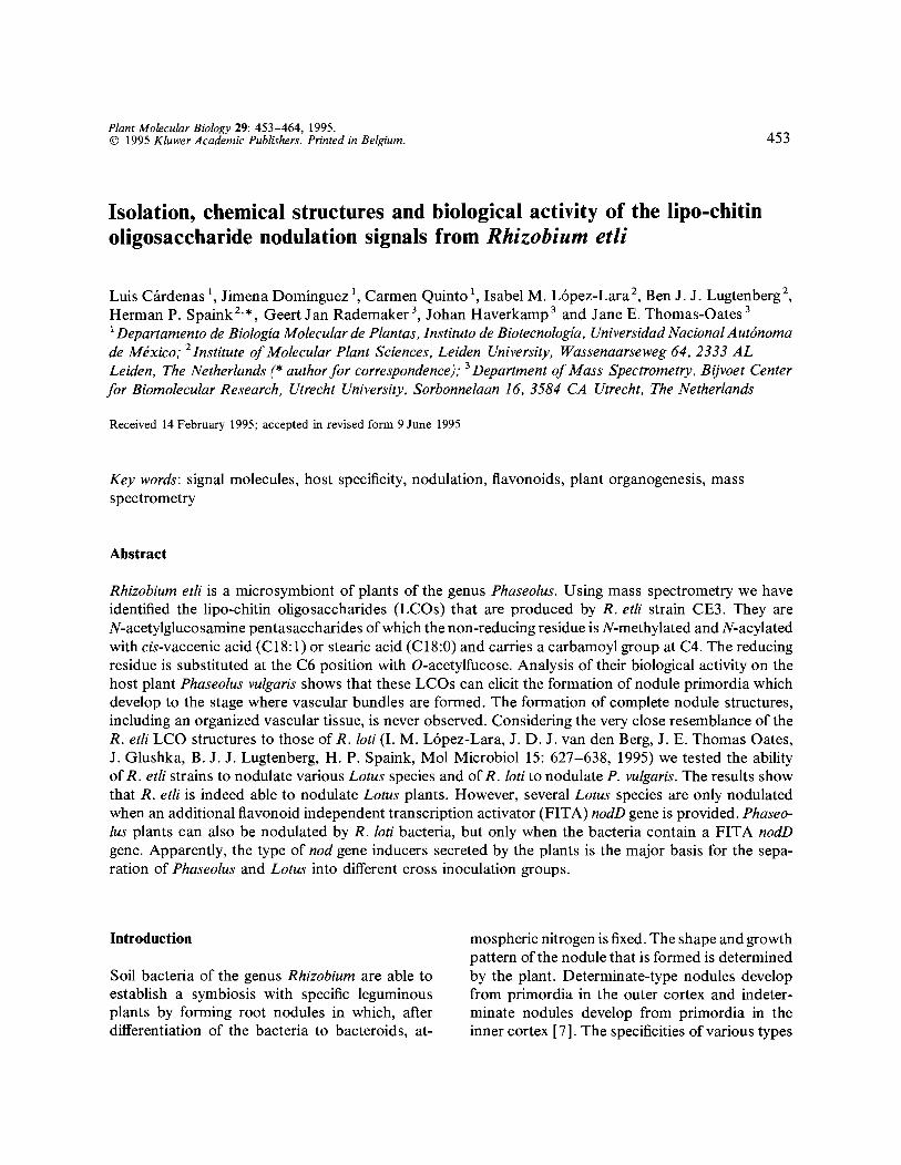
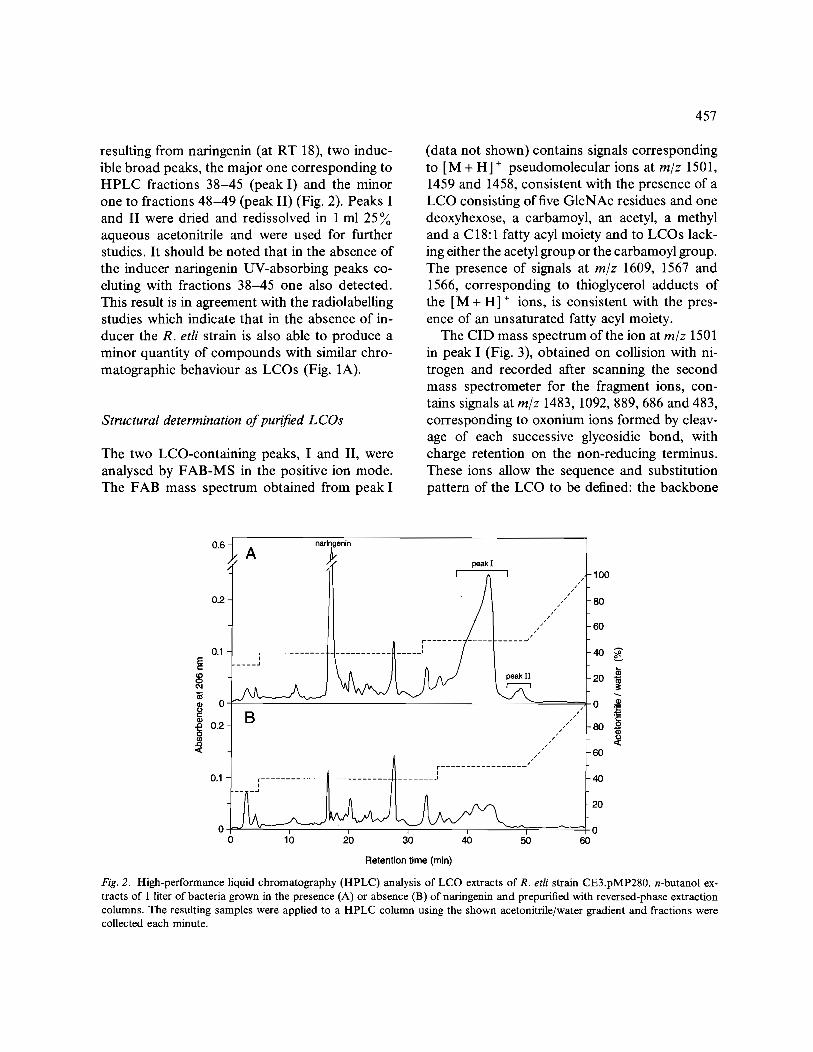
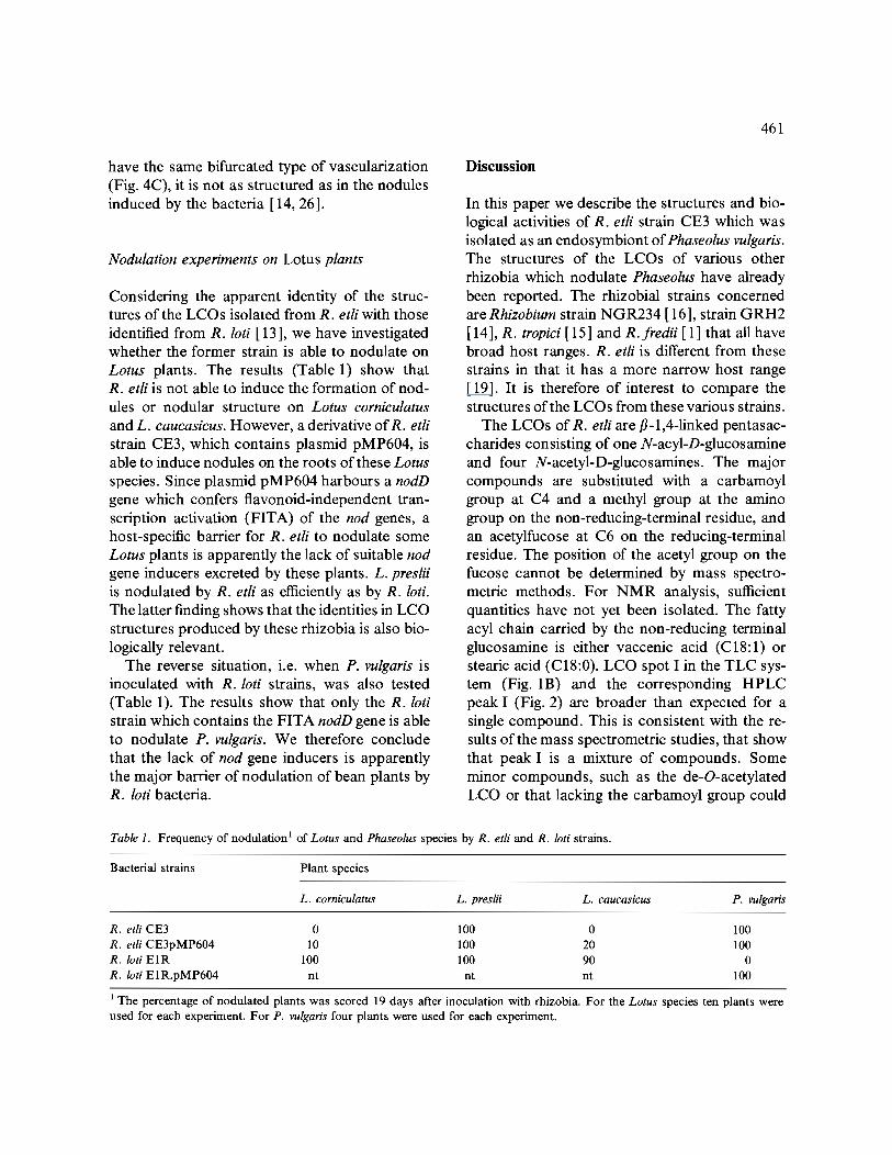
Fig. 1. Thin-layer chromatographic (TLC) analysis of radio- labelled metabolites of R. etli. A. n-butanol extracts of cells grown in the presence of [ 1-14C]-glucosamine were analysed. B. n-butanol extracts of cells grown in the presence of L-[methyl-14C]-methionine were analysed. Lanes: 1, strain CE3 without induction; 2, strain CE3 induced with naringe- nin; 3, strain CE3.pMP280 without induction; 4, strain CE3.pMP280 induced with naringenin.
jector and helium as the carrier gas. Monosac- charide derivatives were separated on a DB-5MS column (0.32 mm x 30 m; J + W Scientific). TMS methyl glycosides were injected directly from so- lution in the TMS reagents (1 #1 injected) and separated using the following temperature pro- gramme: 110 °C for 2min, then ramping at 30 °C/min to 170 °C, holding for 2 min, then ramping at 4 ° C/min to 240 ° C, and holding for 10min. Partially methylated alditol acetates (PMAAS) were injected in solution in dichloro- methane (1/~1 injected) and separated by using the following temperature programme: 50 °C for 2 min, then ramping at 40 °C/min to 130 "C, holding for 2 min, then ramping at 4 ° C/min to 230 °C, and holding for 15 min. Mass spectra were recorded under conditions of electron im- pact in the positive ion mode with an electron energy of 70 eV, and were recorded using linear scanning from m/z 50-500 over 1 s.
Results
Production and purification of LCOs
The production of LCOs was assayed by D-[ 1- 14C]-glucosamine or L-[methyl-14C]-methionine labelling studies as described in Materials and methods. In the presence of the flavonoid narin- genin, R. etli strain CE3 produces radiolabelled metabolites which can be extracted with n-butanol and which behave like LCOs on TLC analysis (Figs. 1A and 1B). However, in the case of the labelling experiment with D- [ 1-14C ]-glucos amine, metabolites that behave similar in the TLC analy- ses were also produced in the absence of narin- genin (Fig. 1A, lane I).
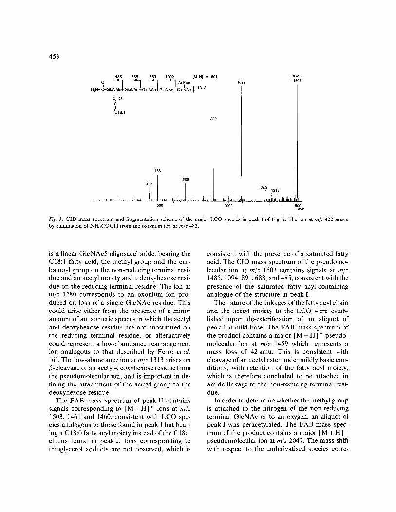
In order to be able to purify sufficient quanti- ties of LCOs to allow chemical analysis we con- structed a series of R. etli LCO-overproducing strains by introducing an additional nodD gene. The best strain is obtained when the nodD gene of R. leguminosarum by. viciae is used (Figs. 1A and 1B, lanes 3 and 4). The HPLC separation of the extracts obtained after the reversed-phase prepurification step yields, in addition to the peak
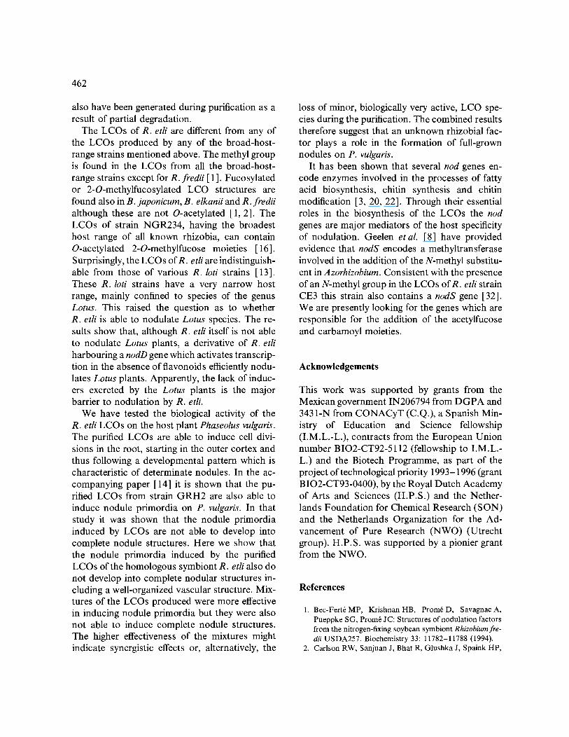
resulting from naringenin (at RT 18), two induc- ible broad peaks, the major one corresponding to HPLC fractions 38-45 (peak I) and the minor one to fractions 48-49 (peak II) (Fig. 2). Peaks I and II were dried and redissolved in 1 ml 25 aqueous acetonitrile and were used for further studies. It should be noted that in the absence of the inducer naringenin UV-absorbing peaks co- eluting with fractions 38-45 one also detected. This result is in agreement with the radiolabelling studies which indicate that in the absence of in- ducer the R. etli strain is also able to produce a minor quantity of compounds with similar chro- matographic behaviour as LCOs (Fig. 1A).
Structural determination of purified LCOs
The two LCO-containing peaks, I and II, were analysed by FAB-MS in the positive ion mode. The FAB mass spectrum obtained from peak I
457
(data not shown) contains signals corresponding to [M + H] ÷ pseudomolecular ions at m/z 1501, 1459 and 1458, consistent with the presence of a LCO consisting of five GlcNAc residues and one deoxyhexose, a carbamoyl, an acetyl, a methyl and a C18:1 fatty acyl moiety and to LCOs lack- ing either the acetyl group or the carbamoyl group. The presence of signals at m/z 1609, 1567 and 1566, corresponding to thioglycerol adducts of the [M + H] ÷ ions, is consistent with the pres- ence of an unsaturated fatty acyl moiety.
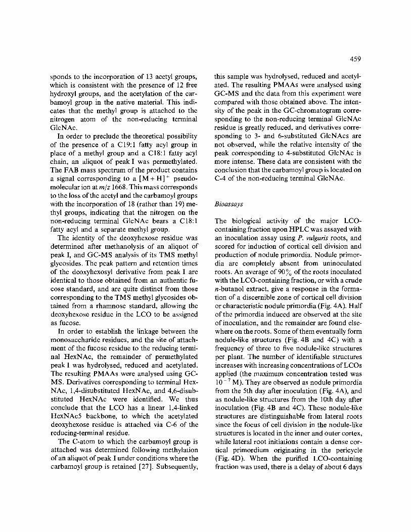
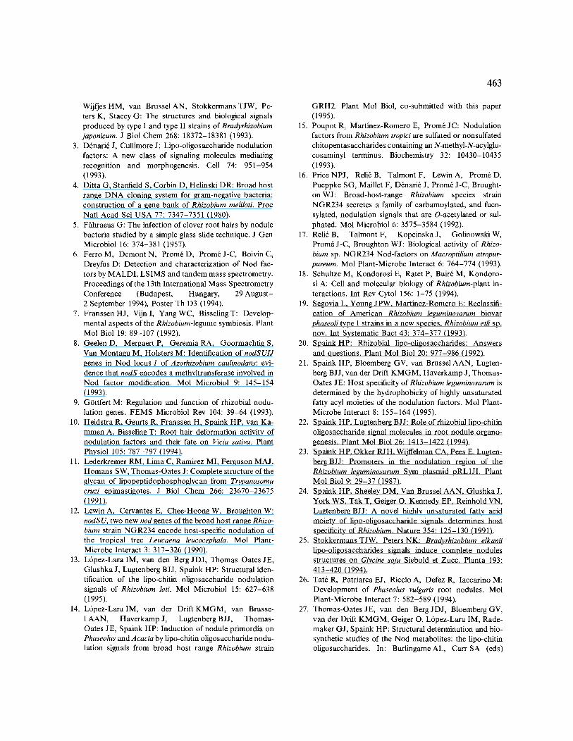
The CID mass spectrum of the ion at m/z 1501 in peak I (Fig. 3), obtained on collision with ni- trogen and recorded after scanning the second mass spectrometer for the fragment ions, con- tains signals at m/z 1483, 1092, 889, 686 and 483, corresponding to oxonium ions formed by cleav- age of each successive glycosidic bond, with charge retention on the non-reducing terminus. These ions allow the sequence and substitution pattern of the LCO to be defined: the backbone
0.6-
0.2
0.1 =E tO 0
0 ¸ e-
.o 0.2-
8 en
A narln~enin
peak I
7/ j . . . . . J/
B
-100 J /4 -
/ - 8 0 / /
/ " - 6 0
-40 ~
-20 ~
/ / _ 0 ~ -80 i j
J
/1" - 6 0
0.1 ~ ~ -40
- 20
0 0 ' '0 do ' ' 0 10 2 40 50 60
Retention time (min)
Fig. 2. High-performance liquid chromatography (HPLC) analysis of LCO extracts of R. etli strain CE3.pMP280. n-butanol ex- tracts of 1 liter of bacteria grown in the presence (A) or absence (B) of naringenin and prepurified with reversed-phase extraction columns. The resulting samples were applied to a HPLC column using the shown acetonitrile/water gradient and fractions were collected each minute.
458
483 686 889 1092 [M+H] + = 1501 0 ~ ~ ~ ~-~ AcFuc
, 2 , - c - G , o . . M o - I -
~=o 18:1
483 l 686
422[ ! 1
, ~,, I m i: ~!t 4 , ,h ,h ~,~L, i ~*,;~1£J, hJ, L I,t,. J~,.~,~,ldJ... ,11 L, l,~,,I U l,J,l, L,/~ ,,I , , 500
889
1092 [M+H] + 1501
, , , , , , , , , , , ,,,,,, _ 1000 1500 m/z
Fig. 3. CID mass spectrum and fragmentation scheme of the major LCO species in peak I of Fig. 2. The ion at m/z 422 arises by elimination of NH2COOH from the oxonium ion at m/z 483.
is a linear GlcNAc5 oligosaccharide, bearing the C18:1 fatty acid, the methyl group and the car- bamoyl group on the non-reducing terminal resi- due and an acetyl moiety and a deoxyhexose resi- due on the reducing terminal residue. The ion at m/z 1280 corresponds to an oxonium ion pro- duced on loss of a single GlcNAc residue. This could arise either from the presence of a minor amount of an isomeric species in which the acetyl and deoxyhexose residue are not substituted on the reducing terminal residue, or alternatively could represent a low-abundance rearrangement ion analogous to that described by Ferro et al. [6]. The low-abundance ion at m/z 1313 arises on /~-cleavage of an acetyl-deoxyhexose residue from the pseudomolecular ion, and is important in de- fining the attachment of the acetyl group to the deoxyhexose residue.
The FAB mass spectrum of peak II contains signals corresponding to [M + H] + ions at m/z 1503, 1461 and 1460, consistent with LCO spe- cies analogous to those found in peak I but bear- ing a C 18:0 fatty acyl moiety instead of the C 18:1 chains found in peak I. Ions corresponding to thioglycerol adducts are not observed, which is
consistent with the presence of a saturated fatty acid. The CID mass spectrum of the pseudomo- lecular ion at m/z 1503 contains signals at m/z 1485, 1094, 891,688, and 485, consistent with the presence of the saturated fatty acyl-containing analogue of the structure in peak I.
The nature of the linkages of the fatty acyl chain and the acetyl moiety to the LCO were estab- lished upon de-esterification of an aliquot of peak I in mild base. The FAB mass spectrum of the product contains a major [M + H] + pseudo- molecular ion at m/z 1459 which represents a mass loss of 42 amu. This is consistent with cleavage of an acetyl ester under mildly basic con- ditions, with retention of the fatty acyl moiety, which is therefore concluded to be attached in amide linkage to the non-reducing terminal resi- due.
In order to determine whether the methyl group is attached to the nitrogen of the non-reducing terminal GlcNAc or to an oxygen, an aliquot of peak I was peracetylated. The FAB mass spec- trum of the product contains a major [M + H] + pseudomolecular ion at m/z 2047. The mass shift with respect to the underivatised species corre-
sponds to the incorporation of 13 acetyl groups, which is consistent with the presence of 12 free hydroxyl groups, and the acetylation of the car- bamoyl group in the native material. This indi- cates that the methyl group is attached to the nitrogen atom of the non-reducing terminal GlcNAc.
In order to preclude the theoretical possibility of the presence of a C19:1 fatty acyl group in place of a methyl group and a C18:1 fatty acyl chain, an aliquot of peak I was permethylated. The FAB mass spectrum of the product contains a signal corresponding to a [M + H] + pseudo- molecular ion at m/z 1668. This mass corresponds to the loss of the acetyl and the carbamoyl groups with the incorporation of 18 (rather than 19) me- thyl groups, indicating that the nitrogen on the non-reducing terminal GlcNAc bears a C18:1 fatty acyl and a separate methyl group.
The identity of the deoxyhexose residue was determined after methanolysis of an aliquot of peak I, and GC-MS analysis of its TMS methyl glycosides. The peak pattern and retention times of the deoxyhexosyl derivative from peak I are identical to those obtained from an authentic fu- cose standard, and are quite distinct from those corresponding to the TMS methyl glycosides ob- tained from a rhamnose standard, allowing the deoxyhexose residue in the LCO to be assigned as fucose.
In order to establish the linkage between the monosaccharide residues, and the site of attach- ment of the fucose residue to the reducing termi- nal HexNAc, the remainder of permethylated peak I was hydrolysed, reduced and acetylated. The resulting PMAAs were analysed using GC- MS. Derivatives corresponding to terminal Hex- NAc, 1,4-disubstituted HexNAc, and 4,6-disub- stituted HexNAc were identified. We thus conclude that the LCO has a linear 1,4-1inked HexNAc5 backbone, to which the acetylated deoxyhexose residue is attached via C-6 of the reducing-terminal residue.
The C-atom to which the carbamoyl group is attached was determined following methylation of an aliquot of peak I under conditions where the carbamoyl group is retained [27]. Subsequently,
459
this sample was hydrolysed, reduced and acetyl- ated. The resulting PMAAs were analysed using GC-MS and the data from this experiment were compared with those obtained above. The inten- sity of the peak in the GC-chromatogram corre- sponding to the non-reducing terminal GIcNAc residue is greatly reduced, and derivatives corre- sponding to 3- and 6-substituted GlcNAcs are not observed, while the relative intensity of the peak corresponding to 4-substituted GIcNAc is more intense. These data are consistent with the conclusion that the carbamoyl group is located on C-4 of the non-reducing terminal GIcNAc.
Bioassays
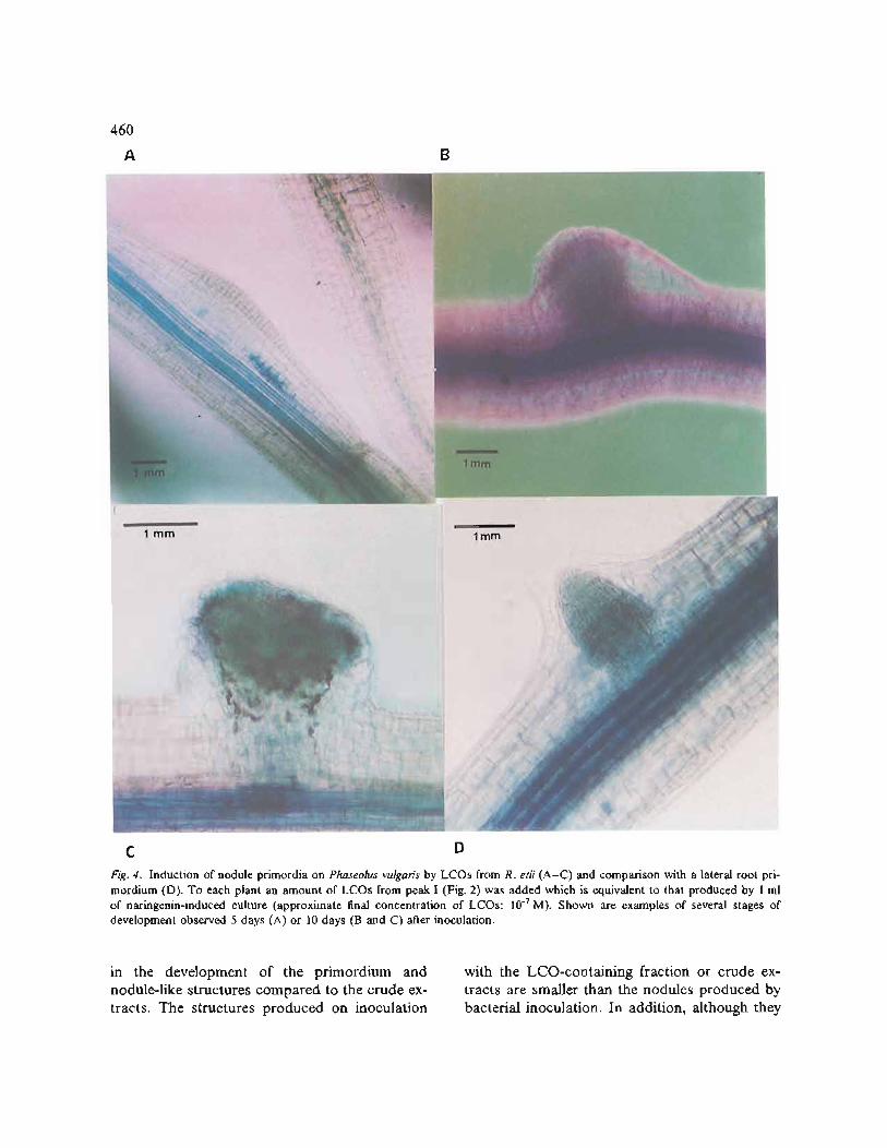
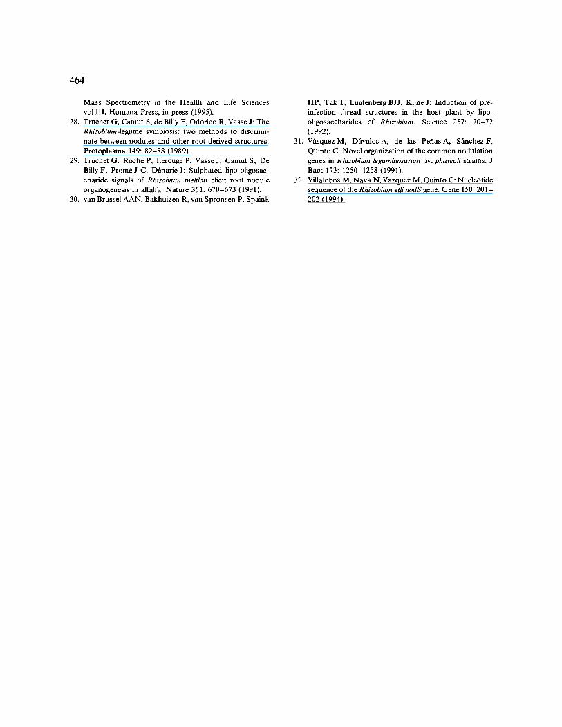
The biological activity of the major LCO- containing fraction upon H P L C was assayed with an inoculation assay using P. vulgaris roots, and scored for induction of cortical cell division and production of nodule primordia. Nodule primor- dia are completely absent from uninoculated roots. An average of 90 ~o of the roots inoculated with the LCO-containing fraction, or with a crude n-butanol extract, give a response in the forma- tion of a discernible zone of cortical cell division or characteristic nodule primordia (Fig. 4A). Half of the primordia induced are observed at the site of inoculation, and the remainder are found else- where on the roots. Some of them eventually form nodule-like structures (Fig. 4B and 4C) with a frequency of three to five nodule-like structures per plant. The number of identifiable structures increases with increasing concentrations of LCOs applied (the maximum concentration tested was 10 - 7 M). They are observed as nodule primordia from the 5th day after inoculation (Fig. 4A), and as nodule-like structures from the 10th day after inoculation (Fig. 4B and 4C). These nodule-like structures are distinguishable from lateral roots since the focus of cell division in the nodule-like structures is located in the inner and outer cortex, while lateral root initiations contain a dense cor- tical primordium originating in the pericycle (Fig. 4D). When the purified LCO-containing fraction was used, there is a delay of about 6 days
460
Fig. 4. Induction of nodule primordia on Phaseolus vulgaris by LCOs from R. etli (A-C) and comparison with a lateral root pri- mordium (D). To each plant an amount of LCOs from peak I (Fig. 2) was added which is equivalent to that produced by 1 ml of naringenin-induced culture (approximate final concentration of LCOs: 10 -7 M). Shown are examples of several stages of development observed 5 days (A) or 10 days (B and C) after inoculation.
in the d e v e l o p m e n t o f the p r i m o r d i u m a n d
n o d u l e - l i k e s t ruc tu re s c o m p a r e d to the c r u d e ex-
t r ac t s . T h e s t ruc tu res p r o d u c e d on i n o c u l a t i o n
wi th the L C O - c o n t a i n i n g f r ac t i on o r c r u d e ex-
t r a c t s a re sma l l e r t h a n the n o d u l e s p r o d u c e d by
b a c t e r i a l i nocu la t i on . I n a d d i t i o n , a l t hough they
461
have the same bifurcated type of vascularization (Fig. 4C), it is not as structured as in the nodules induced by the bacteria [ 14, 26].
Nodulation experiments on Lotus plants
Considering the apparent identity of the struc- tures of the LCOs isolated from R. etli with those identified from R. loti [ 13], we have investigated whether the former strain is able to nodulate on Lotus plants. The results (Table 1) show that R. etli is not able to induce the formation of nod- ules or nodular structure on Lotus corniculatus and L. caucasicus. However, a derivative ofR. etli strain CE3, which contains plasmid pMP604, is able to induce nodules on the roots of these Lotus species. Since plasmid pMP604 harbours a nodD gene which confers flavonoid-independent tran- scription activation (FITA) of the nod genes, a host-specific barrier for R. etli to nodulate some Lotus plants is apparently the lack of suitable nod gene inducers excreted by these plants. L. preslii is nodulated by R. etli as efficiently as by R. loti. The latter finding shows that the identities in LCO structures produced by these rhizobia is also bio- logically relevant.
The reverse situation, i.e. when P. vulgaris is inoculated with R. loti strains, was also tested (Table 1). The results show that only the R. loti strain which contains the FITA nodD gene is able to nodulate P. vulgaris. We therefore conclude that the lack of nod gene inducers is apparently the major barrier of nodulation of bean plants by R. loti bacteria.
Discussion
In this paper we describe the structures and bio- logical activities of R. etli strain CE3 which was isolated as an endosymbiont ofPhaseolus vulgaris. The structures of the LCOs of various other rhizobia which nodulate Phaseolus have already been reported. The rhizobial strains concerned are Rhizobium strain NGR234 [ 16], strain G R H 2 [14], R. tropici [15] and R. fredii [1] that all have broad host ranges. R. etli is different from these strains in that it has a more narrow host range [ 19]. It is therefore of interest to compare the structures of the LCOs from these various strains.
The LCOs ofR. etli are fl-l,4-1inked pentasac- charides consisting of one N-acyl-D-glucosamine and four N-acetyl-D-glucosamines. The major compounds are substituted with a carbamoyl group at C4 and a methyl group at the amino group on the non-reducing-terminal residue, and an acetylfucose at C6 on the reducing-terminal residue. The position of the acetyl group on the fucose cannot be determined by mass spectro- metric methods. For N M R analysis, sufficient quantities have not yet been isolated. The fatty acyl chain carried by the non-reducing terminal glucosamine is either vaccenic acid (C18:1) or stearic acid (C18:0). LCO spot I in the TLC sys- tem (Fig. 1B) and the corresponding H P L C peak I (Fig. 2) are broader than expected for a single compound. This is consistent with the re- sults of the mass spectrometric studies, that show that peak I is a mixture of compounds. Some minor compounds, such as the de-O-acetylated LCO or that lacking the carbamoyl group could
Table 1. Frequency of nodulation I of Lotus and Phaseolus species by R. etli and R. loti strains.
Bacterial strains Plant species
L. corniculatus L. preslii L. caucasicus P. vulgaris
R. etli CE3 0 100 0 100 R. etli CE3pMP604 10 100 20 100 R. loti E1R 100 100 90 0 R. loti E1R.pMP604 nt nt nt 100
1 The percentage of nodulated plants was scored 19 days after inoculation with rhizobia. For the Lotus species ten plants were used for each experiment. For P. vulgaris four plants were used for each experiment.
462
also have been generated during purification as a result of partial degradation.
The LCOs of R. etli are different from any of the LCOs produced by any of the broad-host- range strains mentioned above. The methyl group is found in the LCOs from all the broad-host- range strains except for R. fredii [ 1 ]. Fucosylated or 2-O-methylfucosylated LCO structures are found also in B. japonicum, B. elkanii and R. fredii although these are not O-acetylated [1, 2]. The LCOs of strain NGR234, having the broadest host range of all known rhizobia, can contain O-acetylated 2-O-methylfucose moieties [16]. Surprisingly, the LCOs ofR. etli are indistinguish- able from those of various R. loti strains [13]. These R. loti strains have a very narrow host range, mainly confined to species of the genus Lotus. This raised the question as to whether R. etli is able to nodulate Lotus species. The re- sults show that, although R. etli itself is not able to nodulate Lotus plants, a derivative of R. etli harbouring a nodD gene which activates transcrip- tion in the absence of flavonoids efficiently nodu- lates Lotus plants. Apparently, the lack of induc- ers excreted by the Lotus plants is the major barrier to nodulation by R. etli.
We have tested the biological activity of the R. etli LCOs on the host plant Phaseolus vulgaris. The purified LCOs are able to induce cell divi- sions in the root, starting in the outer cortex and thus following a developmental pattern which is characteristic of determinate nodules. In the ac- companying paper [14] it is shown that the pu- rified LCOs from strain G R H 2 are also able to induce nodule primordia on P. vulgaris. In that study it was shown that the nodule primordia induced by LCOs are not able to develop into complete nodule structures. Here we show that the nodule primordia induced by the purified LCOs of the homologous symbiont R. etli also do not develop into complete nodular structures in- cluding a well-organized vascular structure. Mix- tures of the LCOs produced were more effective in inducing nodule primordia but they were also not able to induce complete nodule structures. The higher effectiveness of the mixtures might indicate synergistic effects or, alternatively, the
loss of minor, biologically very active, LCO spe- cies during the purification. The combined results therefore suggest that an unknown rhizobial fac- tor plays a role in the formation of full-grown nodules on P. vulgaris.
It has been shown that several nod genes en- code enzymes involved in the processes of fatty acid biosynthesis, chitin synthesis and chitin modification [3, 20, 22]. Through their essential roles in the biosynthesis of the LCOs the nod genes are major mediators of the host specificity of nodulation. Geelen etal. [8] have provided evidence that nodS encodes a methyltransferase involved in the addition of the N-methyl substitu- ent in Azorhizobium. Consistent with the presence of an N-methyl group in the LCOs ofR. etli strain CE3 this strain also contains a nodS gene [32]. We are presently looking for the genes which are responsible for the addition of the acetylfucose and carbamoyl moieties.
Acknowledgements
This work was supported by grants from the Mexican government IN206794 from D G P A and 3431-N from CONACyT (C.Q.), a Spanish Min- istry of Education and Science fellowship (I.M.L.-L.), contracts from the European Union number BIO2-CT92-5112 (fellowship to I.M.L.- L.) and the Biotech Programme, as part of the project of technological priority 1993-1996 (grant BIO2-CT93-0400), by the Royal Dutch Academy of Arts and Sciences (H.P.S.) and the Nether- lands Foundation for Chemical Research (SON) and the Netherlands Organization for the Ad- vancement of Pure Research (NWO) (Utrecht group). H.P.S. was supported by a pionier grant from the NWO.
References
1. Bec-Fert6 MP, Krishnan HB, Prom6 D, Savagnac A, Pueppke SG, Prom6 JC: Structures of nodulation factors from the nitrogen-fixing soybean symbiont Rhizobiumfre- dii USDA257. Biochemistry 33:11782-11788 (1994).
2. Carlson RW, Sanjuan J, Bhat R, Glushka J, Spaink HP,
Wijfjes HM, van BrusselAN, Stokkermans TJW, Pe- ters K, Stacey G: The structures and biological signals produced by type I and type II strains of Bradyrhizobium japonicum. J Biol Chem 268:18372-18381 (1993).
3. D6nari+ J, Cullimore J: Lipo-oligosaccharide nodulation factors: A new class of signaling molecules mediating recognition and morphogenesis. Cell 74: 951-954 (1993).
4. Ditta G, Stanfield S, Corbin D, Helinski DR: Broad host range DNA cloning system for gram-negative bacteria: construction of a gene bank of Rhizobium meliloti. Proc Natl Acad Sci USA 77:7347-7351 (1980).
5. F~Jaraeus G: The infection of clover root hairs by nodule bacteria studied by a simple glass slide technique. J Gen Microbiol 16:374-381 (1957).
6. Ferro M, Demont N, Prom6 D, Prom6J-C, Boivin C, Dreyfus D: Detection and characterization of Nod fac- tors by MALDI, LSIMS and tandem mass spectrometry. Proceedings of the 13th International Mass Spectrometry Conference (Budapest, Hungary, 29 August- 2 September 1994), Poster Th D3 (1994).
7. Franssen HJ, Vijn I, YangWC, BisselingT: Develop- mental aspects of the Rhizobium-legume symbiosis. Plant Mol Biol 19:89-107 (1992).
8. Geelen D, Mergaert P, Geremia RA, Goormachtig S, Van Montagu M, Holsters M: Identification of nodSUIJ genes in Nod locus 1 of Azorhizobium caulinodans: evi- dence that nodS encodes a methyltransferase involved in Nod factor modification. Mol Microbiol 9 :145-154 (1993).
9. GOttfert M: Regulation and function of rhizobial nodu- lation genes. FEMS Microbiol Rev 104:39-64 (1993).
10. Heidstra R, Geurts R, Franssen H, Spaink HP, van Ka- mmen A, Bisseling T: Root hair deformation activity of nodulation factors and their fate on Vicia sativa. Plant Physiol 105:787-797 (1994).
11. Lederkremer RM, Lima C, Ramirez MI, Ferguson MAJ, Homans SW, Thomas-Oates J: Complete structure of the glycan of lipopeptidophosphoglycan from Trypanosoma cruzi epimastigotes. J Biol Chem 266:23670-23675 (1991).
12. Lewin A, Cervantes E, Chee-HoongW, Broughton W: nodS U, two new nod genes of the broad host range Rhizo- bium strain NGR234 encode host-specific nodulation of the tropical tree Leucaena leucocephala. Mol Plant- Microbe Interact 3:317-326 (1990).
13. L6pez-Lara IM, van den Berg JDJ, Thomas Oates JE, Glushka J, Lugtenberg B J J, Spaink HP: Structural iden- tification of the lipo-chitin oligosaccharide nodulation signals of Rhizobium loti. Mol Microbiol 15:627-638 (1995).
14. Ldpez-Lara IM, van der Drift KMGM, van Brusse- 1 AAN, Haverkamp J, Lugtenberg B J J, Thomas- Oates JE, Spaink HP: Induction of nodule primordia on Phaseolus and Acacia by lipo-chitin oligosaccharide nodu- lation signals from broad host range Rhizobium strain
463
GRH2. Plant Mol Biol, co-submitted with this paper (1995).
15. Poupot R, Martinez-Romero E, Prom6 JC: Nodulation factors from Rhizobium tropici are sulfated or nonsulfated chitopentasaccharides containing an N-methyl-N-acylglu- cosaminyl terminus. Biochemistry 32: 10430-10435 (1993).
16. Price NPJ, Reli6 B, Talmont F, Lewin A, Prom6 D, Pueppke SG, Maillet F, D6nari6 J, Prom6 J-C, Brought- on WJ: Broad-host-range Rhizobium species strain NGR234 secretes a family of carbamoylated, and fuco- sylated, nodulation signals that are O-acetylated or sul- phated. Mol Microbiol 6:3575-3584 (1992).
17. Reli6 B, Talmont F, Kopcinska J, Golinowski W, Prom6 J-C, Broughton WJ: Biological activity of Rhizo- bium sp. NGR234 Nod-factors on Macroptilium atropur- pureum. Mol Plant-Microbe Interact 6:764-774 (1993).
18. Schultze M, Kondorosi E, Ratet P, Buir6 M, Kondoro- si A: Cell and molecular biology of Rhizobium-plant in- teractions. Int Rev Cytol 156:1-75 (1994).
19. Segovia L, Young JPW, Martinez-Romero E: Reclassifi- cation of American Rhizobium leguminosarum biovar phaseoli type I strains in a new species, Rhizobium etli sp. nov. Int Systematic Bact 43:374-377 (1993).
20. Spaink HP: Rhizobial lipo-oligosaccharides: Answers and questions. Plant Mol Biol 20:977-986 (1992).
21. Spaink HP, Bloemberg GV, van Brussel AAN, Lugten- berg B J J, van der Drift KMGM, Haverkamp J, Thomas- Oates JE: Host specificity of Rhizobium leguminosarum is determined by the hydrophobicity of highly unsaturated fatty acyl moieties of the nodulation factors. Mol Plant- Microbe Interact 8:155-164 (1995).
22. Spaink HP, Lugtenberg B J J: Role of rhizobial lipo-chitin oligosaccharide signal molecules in root nodule organo- genesis. Plant Mol Biol 26:1413-1422 (1994).
23. Spaink HP, Okker RJH, Wijffelman CA, Pees E, Lugten- berg B J J: Promoters in the nodulation region of the Rhizobium leguminosarum Sym plasmid pRL1JI. Plant Mol Biol 9 :29-37 (1987).
24. Spaink HP, Sheeley DM, Van Brussel AAN, Glushka J, York WS, Tak T, Geiger O, Kennedy EP, Reinhold VN, Lugtenberg B J J: A novel highly unsaturated fatty acid moiety of lipo-oligosaccharide signals determines host specificity of Rhizobium. Nature 354:125-130 (1991).
25. Stokkermans TJW, Peters NK: Bradyrhizobium elkanii lipo-oligosaccharides signals induce complete nodules structures on Glycine soja Siebold et Zucc. Planta 193: 413-420 (1994).
26. Tat6 R, Patriarca EJ, Riccio A, Defez R, Iaccarino M: Development of Phaseolus vulgaris root nodules. Mol Plant-Microbe Interact 7:582-589 (1994).
27. Thomas-Oates JE, van den Berg JDJ, Bloemberg GV, van der Drift KMGM, Geiger O, L6pez-Lara IM, Rade- maker GJ, Spaink HP: Structural determination and bio- synthetic studies of the Nod metabolites: the lipo-chitin oligosaccharides. In: BurlingameAL, Carr SA (eds)
464
Mass Spectrometry in the Health and Life Sciences vol III, Humana Press, in press (1995).
28. Truchet G, Camut S, de Billy F, Odorico R, Vasse J: The Rhizobium-legume symbiosis: two methods to discrimi- nate between nodules and other root derived structures. Protoplasma 149:82-88 (1989).
29. Truchet G, Roche P, Lerouge P, Vasse J, Camut S, De Billy F, Prom6J-C, D6nari6 J: Sulphated lipo-oligosac- charide signals of Rhizobium meliloti elicit root nodule organogenesis in alfalfa. Nature 351:670-673 (1991).
30. van Brussel AAN, Bakhuizen R, van Spronsen P, Spaink
31.
32.
HP, Tak T, Lugtenberg BJJ, Kijne J: Induction of pre- infection thread structures in the host plant by lipo- oligosaccharides of Rhizobium. Science 257:70-72 (1992). V~_squez M, D~tvalos A, de las Peflas A, S~achez F, Quinto C: Novel organization of the common nodulation genes in Rhizobium leguminosarum by. phaseoli strains. J Bact 173:1250-1258 (1991). Villalobos M, Nava N, Vazquez M, Quinto C: Nucleotide sequence of the Rhizobium etli nodS gene. Gene 150: 201- 202 (1994).