![[Cool] Gas Chromatography and Lipids](https://static.fdokumen.com/doc/165x107/6325a4b1852a7313b70e98e9/cool-gas-chromatography-and-lipids.jpg)
Negative ion graphitised carbon nano-liquid chromatography/mass spectrometry increases sensitivity...
11
RAPID COMMUNICATIONS IN MASS SPECTROMETRY Rapid Commun. Mass Spectrom. 2004; 18: 2282–2292 Published online in Wiley InterScience (www.interscience.wiley.com). DOI: 10.1002/rcm.1626 Negative ion graphitised carbon nano-liquid chromatography/mass spectrometry increases sensitivity for glycoprotein oligosaccharide analysis Niclas G. Karlsson 1 *, Nicole L. Wilson 1 , Hans-Ju ¨ rgen Wirth 2 , Peter Dawes 2 , Hiren Joshi 1 and Nicolle H. Packer 1 1 Proteome Systems Ltd, locked bag 2073, North Ryde Sydney, New South Wales, 1670, Australia 2 SGE International, 7 Argent Place, Ringwood, Victoria, 3134, Australia Received 17 June 2004; Revised 4 August 2004; Accepted 4 August 2004 Negative ion nano-liquid chromatography/mass spectrometry (nano-LC/MS) and tandem mass spectrometry (nano-LC/MS 2 ), using graphitised carbon as separating medium, were explored for analysing neutral and acidic O-linked and N-linked oligosaccharide alditols. Compared to the sen- sitivity of capillary LC/MS (flow rate of 6 mL/min) coupled with a conventional electrospray ionisa- tion source, the nano-LC/MS (flow rate of 0.6 mL/min) with a nanoflow ion source was shown to increase the sensitivity ten-fold with a detection limit in the low-femtomole range. The absolute signals for the [M–nH] n ions of the oligosaccharides were increased 100-fold, enabling accumula- tion of high-quality fragmentation data in MS 2 mode, in which detection of low abundant sequence ions is necessary for characterisation of highly sialylated N-linked oligosaccharides. Oligosacchar- ides with high numbers of sialic acid residues gave dominant fragments arising from the loss of sialic acid, and less abundant fragments from cleavage of other glycosidic bonds. Enzymatic off- line desialylation of oligosaccharides in the low-femtomole range prior to MS 2 analysis was shown to increase the quality of the spectra. Automated glycofragment mass fingerprinting using the Gly- cosidIQ software confirmed the oligosaccharide sequence for both neutral desialylated as well as sialylated structures. Furthermore, the use of graphitised carbon nano-LC/MS enabled the detection of four sialylated O-linked oligosaccharides on membrane proteins from ovarian tissue (5 mg of total amount of protein). Copyright # 2004 John Wiley & Sons, Ltd. Glycosylation is one of the major post-translational modifica- tions of secreted and membrane-bound proteins. The current understanding of the glycosylation process and its impact on the function of proteins is still in many cases unknown and the relationship between oligosaccharide structure and its biology is not straightforward. Some of the keywords that have emerged as common themes in glycobiology research are interaction and regulation. 1,2 There are examples where interaction is mediated by only a particular oligosaccharide epitope attached to a protein backbone. This, for instance, is the case with the lutropin hormone receptor binding to HSO 4 (-4)GalNAc(b1-4)GlcNAc(b1-2Man(a1-), 3 while, in other cases, the interaction specificity is more forgiving and a range of oligosaccharide structures can fulfil a certain task. This is the case, for example, in the interaction between the selectins and their ligands, where structures such as sialyl Lewis x and sialyl Lewis a or its sulphated analogues are key interaction epitopes, although it is unknown whether the rest of the interacting oligosaccharide is of importance, or whether it is the cooperative interaction between diffuse oli- gosaccharide patches that mediates the interaction. 4,5 Other important interacting oligosaccharide epitopes, such as blood group related epitopes, often manifest themselves on glycoprotein oligosaccharides, 6,7 both from the ABO system and the Lewis system. The belief that structure can relate to function has encouraged researchers to develop techniques for detailed oligosaccharide structural characterisation. These techniques have comprised a mixture of chemical and enzymatic methods in combination with different levels of technical instrumentation such as high-performance liquid chromato- graphy (HPLC), nuclear magnetic resonance (NMR), capil- lary electrophoresis (CE) and gel electrophoresis, and have provided enormous amounts of data. Currently, there are efforts worldwide to assemble these data into databases (e.g. GlycosuiteDB 8 and SweetDB 9 ). Mass spectrometry has emerged as a key instrumentation to enable accurate qualitative glycosylation analysis. The reasons behind its success are the sensitivity of detection and the quality of the data. Both matrix-assisted laser desorption/ ionisation (MALDI)-MS and electrospray ionisation (ESI)-MS have successfully been employed for oligosaccharide analy- sis. 10–12 In combination with on-line separation techniques Copyright # 2004 John Wiley & Sons, Ltd. *Correspondence to: N. G. Karlsson, Proteome Systems Ltd, locked bag 2073, North Ryde Sydney, New South Wales, 1670, Australia. E-mail: [email protected]
Transcript of Negative ion graphitised carbon nano-liquid chromatography/mass spectrometry increases sensitivity...

RAPID COMMUNICATIONS IN MASS SPECTROMETRY
Rapid Commun. Mass Spectrom. 2004; 18: 2282–2292
Published online in Wiley InterScience (www.interscience.wiley.com). DOI: 10.1002/rcm.1626
Negative ion graphitised carbon nano-liquid
chromatography/mass spectrometry increases sensitivity
for glycoprotein oligosaccharide analysis
Niclas G. Karlsson1*, Nicole L. Wilson1, Hans-Jurgen Wirth2, Peter Dawes2,
Hiren Joshi1 and Nicolle H. Packer1
1Proteome Systems Ltd, locked bag 2073, North Ryde Sydney, New South Wales, 1670, Australia2SGE International, 7 Argent Place, Ringwood, Victoria, 3134, Australia
Received 17 June 2004; Revised 4 August 2004; Accepted 4 August 2004
Negative ion nano-liquid chromatography/mass spectrometry (nano-LC/MS) and tandem mass
spectrometry (nano-LC/MS2), using graphitised carbon as separating medium, were explored for
analysing neutral and acidic O-linked and N-linked oligosaccharide alditols. Compared to the sen-
sitivity of capillary LC/MS (flow rate of 6mL/min) coupled with a conventional electrospray ionisa-
tion source, the nano-LC/MS (flow rate of 0.6mL/min) with a nanoflow ion source was shown to
increase the sensitivity ten-fold with a detection limit in the low-femtomole range. The absolute
signals for the [M–nH]n� ions of the oligosaccharides were increased 100-fold, enabling accumula-
tion of high-quality fragmentation data in MS2 mode, in which detection of low abundant sequence
ions is necessary for characterisation of highly sialylated N-linked oligosaccharides. Oligosacchar-
ides with high numbers of sialic acid residues gave dominant fragments arising from the loss of
sialic acid, and less abundant fragments from cleavage of other glycosidic bonds. Enzymatic off-
line desialylation of oligosaccharides in the low-femtomole range prior to MS2 analysis was shown
to increase the quality of the spectra. Automated glycofragment mass fingerprinting using the Gly-
cosidIQ software confirmed the oligosaccharide sequence for both neutral desialylated as well as
sialylated structures. Furthermore, the use of graphitised carbon nano-LC/MS enabled the detection
of four sialylated O-linked oligosaccharides on membrane proteins from ovarian tissue (5mg of
total amount of protein). Copyright # 2004 John Wiley & Sons, Ltd.
Glycosylation is one of the major post-translational modifica-
tions of secreted and membrane-bound proteins. The current
understanding of the glycosylation process and its impact on
the function of proteins is still in many cases unknown and
the relationship between oligosaccharide structure and its
biology is not straightforward. Some of the keywords that
have emerged as common themes in glycobiology research
are interaction and regulation.1,2 There are examples where
interaction is mediated by only a particular oligosaccharide
epitope attached to a protein backbone. This, for instance, is
the case with the lutropin hormone receptor binding to
HSO4(-4)GalNAc(b1-4)GlcNAc(b1-2Man(a1-),3 while, in
other cases, the interaction specificity is more forgiving and
a range of oligosaccharide structures can fulfil a certain
task. This is the case, for example, in the interaction between
the selectins and their ligands, where structures such as sialyl
Lewis x and sialyl Lewis a or its sulphated analogues are key
interaction epitopes, although it is unknown whether the rest
of the interacting oligosaccharide is of importance, or
whether it is the cooperative interaction between diffuse oli-
gosaccharide patches that mediates the interaction.4,5 Other
important interacting oligosaccharide epitopes, such as
blood group related epitopes, often manifest themselves on
glycoprotein oligosaccharides,6,7 both from the ABO system
and the Lewis system.
The belief that structure can relate to function has
encouraged researchers to develop techniques for detailed
oligosaccharide structural characterisation. These techniques
have comprised a mixture of chemical and enzymatic
methods in combination with different levels of technical
instrumentation such as high-performance liquid chromato-
graphy (HPLC), nuclear magnetic resonance (NMR), capil-
lary electrophoresis (CE) and gel electrophoresis, and have
provided enormous amounts of data. Currently, there are
efforts worldwide to assemble these data into databases (e.g.
GlycosuiteDB8 and SweetDB9).
Mass spectrometry has emerged as a key instrumentation
to enable accurate qualitative glycosylation analysis. The
reasons behind its success are the sensitivity of detection and
the quality of the data. Both matrix-assisted laser desorption/
ionisation (MALDI)-MS and electrospray ionisation (ESI)-MS
have successfully been employed for oligosaccharide analy-
sis.10–12 In combination with on-line separation techniques
Copyright # 2004 John Wiley & Sons, Ltd.
*Correspondence to: N. G. Karlsson, Proteome Systems Ltd,locked bag 2073, North Ryde Sydney, New South Wales,1670, Australia.E-mail: [email protected]

such as LC and CE it enables the separation and characterisa-
tion of oligosaccharide isomers.13,14 While normal-phase
chromatography using amine- and amide-type stationary
phases have traditionally been the most popular for separat-
ing oligosaccharides,15 graphitised carbon is gaining popu-
larity due to its ease of use, capacity, and high resolution.16
Miniaturisation of HPLC systems has shown that oligosac-
charide ESI LC/MS analysis can be performed with
sensitivities down to low-femtomole levels in positive ion
mode.17 Negative ion mode provides the additional benefit of
allowing highly sensitive detection of both neutral and
negatively oligosaccharides in the one separation. The likely
increase in sensitivity on going from negative ion capillary
mode LC/MS18–21 to nanoflow LC/MS should allow
oligosaccharide analysis to be performed on even low
abundance proteins.
Two-dimensional sodium dodecyl sulphate/polyacryla-
mide (SDS-PAGE) enables isolation of many glycoproteins in
one experiment. Identification of the glycosylation of these
separated proteins requires not only sensitive detection of
ionised molecules of oligosaccharides and their MS2 frag-
ments, but also intelligent software for oligosaccharide
fragmentation analysis in order to cope with the amount of
data. This report describes the use of negative ion nanoflow
LC/MS of oligosaccharides using graphitised carbon as
stationary phase, and with interpretation of the fragmenta-
tion data by the glycofragment mass fingerprinting software
GlycosidIQ, by matching the ‘in-silico’ fragmented oligosac-
charide structures from the oligosaccharide database Glyco-
suiteDB.22 The sensitivity of the technique is compared to that
of negative ion capillary LC/MS using neutral O-linked
oligosaccharides, and is then evaluated using sialylated
reduced N-linked oligosaccharides. Its applicability for N-
linked and O-linked oligosaccharide analysis was tested
using commercial standards as well as oligosaccharide
mixtures isolated from mucosal surfaces and ovarian tissues.
In order to gain additional information on highly sialylated
oligosaccharide structures, in which the dominating MS2
fragmentation was the loss of sialic acid, an enzymatic
desialylation procedure was developed for sub-picomole
analysis of oligosaccharides of these types prior to analysis by
negative ion LC/MS2.
EXPERIMENTAL
Preparation of ovarian tissueOvarian tissue was collected immediately following surgery
from a patient diagnosed with multiple cancer including
ovarian cancer. The tissues were aliquoted into 2 mL capacity
cryovials and stored at �808C. Tissue samples (approxi-
mately 0.2 mg) were freeze-fractured and ground into a pow-
der. Proteins were extracted by sonication in 2 mL of 100 mM
sodium carbonate using an ultrasonic probe (Branson, Digital
Sonifier, Branson, Danbury, CT, USA) (total of 1 min in 15 s
intervals at 70% amplitude). The samples were then centri-
fuged at 14 000 g for 45 min at 48C. The pellet, enriched with
membrane proteins, was re-extracted once according to the
above protocol before being reduced and alkylated in sample
buffer (7.0 M urea, 2.0 M thiourea, 40 mM Tris base, and 4.0%
(w/v) CHAPS) using 5.0 mM tributylphosphine and 10 mM
acrylamide for approximately 1 h. This membrane fraction
of the tissue was dotblotted onto PVDF PSQ (Millipore,
Bedford, MA, USA) membrane for global oligosaccharide
analysis (50 mL, approximately 5 mg of protein). The protein
concentration in the solution was determined with the Brad-
ford reagent (Sigma-Aldrich, St. Louis, MN, USA) using a
bovine serum albumin standard curve.
Release of oligosaccharidesNeutral O-linked oligosaccharides from rat small intestine
were prepared as described.21 N-Linked oligosaccharides
were released from dotblotted membrane proteins using
PNGase F (Roche, Mannheim, Germany)19 followed by the
release of O-linked oligosaccharide by reductive b-elimina-
tion.18 N-Linked oligosaccharides, either from tissue or as
standards (Dextra, Reading, UK), were converted into
alditols in 20 mL of 20 mM potassium hydroxide/0.5 M
sodium borohydride. (2 h at 508C). The resulting solutions
were neutralised by addition of 1mL of glacial acetic acid,
before being desalted with 25mL of AG50WX8 cation-
exchange resin (BioRad, Hercules, CA, USA) laid on top of
a reverse-phase m-C18 ZipTip (Millipore), and dried by
vacuum centrifugation. Borate was removed by repeated
addition and evaporation of 50mL of 1% acetic acid in
methanol. The N-linked oligosaccharides from tissue sam-
ples were further desalted on home-made micro-desalting
columns (�2mL) containing Carbograph material (Alltech,
Deerfield, IL, USA) laid on top of a reverse-phase m-C18
ZipTip (Millipore).19
Enzymatic desialylation of N-linkedoligosaccharidesA mixture of oligosaccharide alditols from Dextra, described
in Figs. 2(b), 2(d), 2(e) and 2(f) (1–70 fmol of each), was dis-
solved in 18mL of 25 mM sodium phosphate buffer pH 6.0
and 2mL (approximately 10 mU) of Arthrobacter ureafaciens
sialidase (Glyko, Novato, CA, USA) was added and
incubated at 378C for 16 h. Samples were desalted on the
Carbograph micro-desalting columns described above.
Capillary LC/MS and nano-LC/MSof oligosaccharidesOligosaccharide samples (10 mL) were dissolved in water
and injected via a Surveyor autosampler and HPLC system
(Thermo Finnigan, San Jose, CA, USA). Solvent A was
10 mM ammonium bicarbonate and solvent B was 10 mM
ammonium bicarbonate in 80% acetonitrile. Flow rate was
110–120mL/min, split down before the autosampler into
approximately 6mL/min (capillary LC) or 0.5 mL/min
(nano-LC). A 0.5 mm Rep frit (Upchurch Scientific, Oak
Harbor, WA, USA) was in-line with the ProteCol Hypercarb
(5mm particles) column (from SGE, Ringwood, Australia), i.e.,
a 100 mm� 0.3 mm column for capillary LC, or a 100 mm�0.15 mm column for nano-LC. The gradient was developed
from 0–37.5% B over 27.5 min for capillary LC and from
2–30% B for nano-LC. Both columns were then washed
with 100% B for 4–7 min and equilibrated with the starting
ratio of solvents for at least 12 min.
The ion trap mass spectrometer (LCQ-XPþ, Thermo
Finnigan) was tuned with reduced maltoligosaccharides
Copyright # 2004 John Wiley & Sons, Ltd. Rapid Commun. Mass Spectrom. 2004; 18: 2282–2292
Nano-LC/MS of oligosaccharides 2283

(10 mg/mL) infused at 3mL/min in 40% acetonitrile/10 mM
ammonium bicarbonate using the standard ESI source. The
capillary voltage was reduced from 3.5 to 1.1 kV for nano-LC
applications using the nano-ESI source. For nano-LC, the
ProteCol column was connected via a nanoprobe (SGE) to a
distal coated TaperTip emitter (20 mm i.d.; New Objectives,
Woburn, MA, USA). The mass spectrometer was scanned
between m/z 350–2000 followed by a data-dependent MS2
scan of the most abundant signal from the MS1 scan.
Sensitivity testing for sialylated N-linked oligosaccharides
was done using oligosaccharide alditols in the range 1–
70 fmol, using linear regression analysis for evaluation of
data and calculation of detection limit (2� noise estimated
from the zero concentration intercept).
Fragmentation analysis of oligosaccharides was performed
using the GlycosidIQ glycofragment mass fingerprinting
software matching with ‘in-silico’ fragmented oligosacchar-
ides from the GlycosuiteDB8 oligosaccharide database.22 The
precursor ion was searched within �1 Da and the fragments
within �0.7 Da. Structures were evaluated using the seg-
mentation score (number of oligosaccharide sequences that
could be created from the identified glycosidic fragments)
and glycosidic correspondence score (sum of the intensities
of all glycosidic fragments found in the spectrum).22
RESULTS AND DISCUSSION
We have previously described the release and sample pre-
paration of N- and O-linked oligosaccharides released from
glycoproteins blotted onto PVDF membranes, either from
1D or 2D SDS-PAGE.18–21 The sample preparation involved
isolating N-linked oligosaccharides using graphitised carbon
microcolumns after PNGase F treatment of gel-separated gly-
coproteins, and microcolumn anion-exchange desalting after
reductive b-elimination of O-linked oligosaccharides. Both
methods are compatible with negative ion graphitised carbon
LC/MS separation analysis of the released oligosaccharides.
In order to gain increased sensitivity for oligosaccharide
detection and characterisation we have now developed a
robust nanoflow LC/MS system and tested it for its capability
of identifying and characterising both N- and O-linked oligo-
saccharides. In contrast with positive ion mode MS, the tech-
nique has the capacity to detect both highly sialylated
oligosaccharides as well as neutral oligosaccharides in the
same experiment. MS2 fragmentation can be performed
both before and after exoglycosidase treatment (sialidase) at
the low-femtomole level.
Detection of neutral O-linked oligosaccharidesNeutral oligosaccharides released from small intestinal
mucosal proteins from six rats were isolated in 199523 in a
project where identification of acidic oligosaccharides was
the main focus. This sample has since become a standard in
our laboratory for the development and testing of the perfor-
mance and sensitivity of our LC/MS separations. We have
previously shown that negative ion capillary LC/MS can
give sensitivity of detection of individual oligosaccharides
in the pmol range or lower.18 We tested the effect of using
nanoflow LC/MS instead of capillary LC/MS by dropping
the flow rate from around 6–7mL/min down to 0.5–0.6 mL/
min and by changing from the traditional ESI-MS source to a
nano-ESI-MS source. This should, in principle, reduce the
chemical background by at least ten-fold. Using the same
tune file for detecting oligosaccharides by negative ion capil-
lary LC/MS and nano-LC/MS (and lowering the spray vol-
tage from 3.5 to 1.1 kV for nano-LC/MS) showed that, despite
an actual increase in the absolute background noise, the sig-
nal-to-noise ratios for detecting [M–nH]n� ions were
increased by five- to ten-fold for most components (Fig. 1,
Table 1). This indicated that it is the more efficient ion transfer
in the ion source, rather than a lowering of the chemical back-
ground, that contributes to increased sensitivity by nano-LC,
and is thus the main reason behind the increase in sensitivity.
For each mass (MS1) spectrum produced there was also an
MS2 experiment on the most intense precursor ion recorded.
The increase in the absolute ion intensity of the [M–H]�
precursor ion in the chromatogram by at least 100 times
(compare absolute signal in Fig. 1(a) with 1(b)) meant that
high-quality fragment spectra could be produced and
interpreted according to the rules of negative ion fragmenta-
tion of oligosaccharide alditols24 (data not shown). The
higher sensitivity allowed us to identify oligosaccharides pre-
viously unidentified from rat intestine25 such as the [M–H]�
ion at m/z 1098 corresponding to the sequence Fuc(a1-
2)Gal(b1-3)GlcNAc(b1-3)[Gal(b1-u)GlcNAc(b1-6)]GalNAcol,
Figure 1. Comparison of negative ion (a) capillary LC/MS
versus (b) nano-LC/MS analysis of neutral O-linked oligo-
saccharides (5.5 ng) using graphitised carbon chromatogra-
phy (base peak chromatograms). Combined MS1 mass
spectra of the region where oligosaccharides were eluted
are shown as inserts. Retention times and detected
molecular ions relate to Table 1.
2284 N. G. Karlsson et al.
Copyright # 2004 John Wiley & Sons, Ltd. Rapid Commun. Mass Spectrom. 2004; 18: 2282–2292

Table 1. Sensitivity for oligosaccharides analysed by nano- and capillary-LC/MS
[M–H]�
m/z
Retention time (min)
Structure
S/Na
IncreasedsensitivityNanoflow Capillary Nanoflow Capillary
530 78.53 21.79 50 6 9
628 56.07 14.21 28 2 12
733 66.52 16.28 51 3 15
733 72.15 22.15 3 NDb —
733 79.27 23.10 83 11 7
790 66.43 ND 6 ND —
895 72.43 18.76 6 1 5
895 79.55 22.34 5 2 3
936 69.31 17.28 43 7 6
936 73.38 19.25 176 17 10
936 74.97 ND 6 ND —
1041 80.04 22.49 17 ND —
Continues
Nano-LC/MS of oligosaccharides 2285
Copyright # 2004 John Wiley & Sons, Ltd. Rapid Commun. Mass Spectrom. 2004; 18: 2282–2292
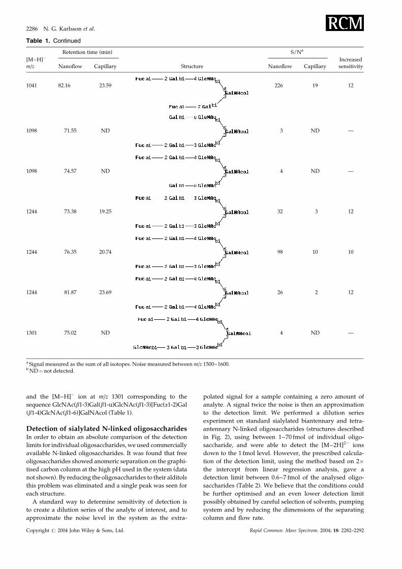
and the [M–H]� ion at m/z 1301 corresponding to the
sequence GlcNAc(b1-3)Gal(b1-u)GlcNAc(b1-3)[Fuc(a1-2)Gal
(b1-4)GlcNAc(b1-6)]GalNAcol (Table 1).
Detection of sialylated N-linked oligosaccharidesIn order to obtain an absolute comparison of the detection
limits for individual oligosaccharides, we used commercially
available N-linked oligosaccharides. It was found that free
oligosaccharides showed anomeric separation on the graphi-
tised carbon column at the high pH used in the system (data
not shown). By reducing the oligosaccharides to their alditols
this problem was eliminated and a single peak was seen for
each structure.
A standard way to determine sensitivity of detection is
to create a dilution series of the analyte of interest, and to
approximate the noise level in the system as the extra-
polated signal for a sample containing a zero amount of
analyte. A signal twice the noise is then an approximation
to the detection limit. We performed a dilution series
experiment on standard sialylated biantennary and tetra-
antennary N-linked oligosaccharides (structures described
in Fig. 2), using between 1–70 fmol of individual oligo-
saccharide, and were able to detect the [M–2H]2� ions
down to the 1 fmol level. However, the prescribed calcula-
tion of the detection limit, using the method based on 2�the intercept from linear regression analysis, gave a
detection limit between 0.6–7 fmol of the analysed oligo-
saccharides (Table 2). We believe that the conditions could
be further optimised and an even lower detection limit
possibly obtained by careful selection of solvents, pumping
system and by reducing the dimensions of the separating
column and flow rate.
Table 1. Continued
[M–H]�
m/z
Retention time (min)
Structure
S/Na
IncreasedsensitivityNanoflow Capillary Nanoflow Capillary
1041 82.16 23.59 226 19 12
1098 71.55 ND 3 ND —
1098 74.57 ND 4 ND —
1244 73.38 19.25 32 3 12
1244 76.35 20.74 98 10 10
1244 81.87 23.69 26 2 12
1301 75.02 ND 4 ND —
a Signal measured as the sum of all isotopes. Noise measured between m/z 1500–1600.b ND¼not detected.
2286 N. G. Karlsson et al.
Copyright # 2004 John Wiley & Sons, Ltd. Rapid Commun. Mass Spectrom. 2004; 18: 2282–2292

Fragmentation analysis of sialylatedN-linked oligosaccharidesAutomated MS2 experiments were performed in each of
the analyses described in this report. This allowed inter-
pretation of fragment spectra for further characterisation of
oligosaccharide structures. The combination of LC with
data-dependent MS2 is a powerful approach to allow charac-
terisation of oligosaccharide isomers in mixtures. Interpreta-
tion of oligosaccharide fragment spectra is usually quite
labour intensive and demands high-level skills to make sense
of the fragmentation pattern. To aid in the interpretation we
used the GlycosidIQ glycofragment mass fingerprinting
software tool (matching MS2 spectra with theoretical
Table 2. Detection limits for sialylated N-linked
oligosaccharide alditols with nano-LC/MS
Monosaccharidecompositiona
[M–2H]2�
m/z
Limit ofdetection(fmol)b
r2 linearregression
value
[5,4,0,1] 966 7 0.96[5,4,1,1] 1039 0.6 0.97[5,4,1.2] 1184 2 0.96[5,4,3,2] 1331 5 0.99[7,6,1,4] 1841 4 0.97
a [Hex, HexNAc, dHex, NeuAc] including reducing end GlcNAcol asHexNAc.b 2�background noise.
Figure 2. Negative ion nano-LC/MS of sialylated N-linked oligosaccharide alditols (20 fmol),
showing base peak chromatogram (a), and selective ion chromatograms of [M–nH]n� ions m/z
966 (b), m/z 1039 (c), m/z 1184 (d), m/z 1331 (e), and m/z 1841 (f). Combined full scan mass
spectra of the corresponding regions where components were detected are inserted.
Nano-LC/MS of oligosaccharides 2287
Copyright # 2004 John Wiley & Sons, Ltd. Rapid Commun. Mass Spectrom. 2004; 18: 2282–2292

spectra generated from oligosaccharides in GlycosuiteDB)
and the GlycoFrag tool (generating theoretical fragments of
submitted oligosaccharides). Figure 3 shows the fragmenta-
tion of the [M–2H]2� ion of the N-linked oligosaccharides
from 20 fmol of sample loaded onto the column. Major frag-
ment ions corresponding to glycosidic fragmentation are
labelled, but additional abundant fragments, including inter-
nal fragments from more than two separate glycosidic and
cross ring cleavages, were also detected. The carboxyl group
introduced by sialic acid was the site for at least one of the
charges of the doubly charged ions detected by MS on all
the N-linked oligosaccharides tested here. In the case of the
tetrasialylated tetraantennary structure there were fewer
charges ([M–2H]2� and [M–3H]3� ions in Fig. 2(f)) on the
molecule than the number of sialic acid residues. Tuning
the mass spectrometric setting in order to introduce more
charges onto the molecule (each sialic acid potentially contri-
buting one negative charge), and thus detecting a quadruply
charged precursor ion, were largely unsuccessful.
The analysis of the fragmentation pattern of all oligosac-
charides indicated that charge-remote fragmentation was
the main mechanism. For singly sialylated structures, the
localisation of the sialic acid at the non-reducing end of the
biantennary structure gave predominantly fragmentation
Figure 3. Negative ion nano-LC/MS2 fragmentation of sialylated N-linked oligosaccharide
alditols (20 fmol) of [M–nH]n� ions m/z 966 (a), m/z 1039 (b), m/z 1184 (c), m/z 1330 (d), and
m/z 1841 (e). Results from the GlycosidIQ software are included on the right-hand side to
illustrate the quality of the spectral matching.
2288 N. G. Karlsson et al.
Copyright # 2004 John Wiley & Sons, Ltd. Rapid Commun. Mass Spectrom. 2004; 18: 2282–2292

patterns directed from the reducing end (Figs. 3(a) and 3(b)).
The second charge of the doubly charged precursor ion was
probably located mostly on the reducing end. This assign-
ment is supported by the fact that the only doubly charged
fragment ions detected contained both the reducing end
GlcNAcol and the sialylated antennary. The GlycosidIQ
glycofragment mass-fingerprinting tool22 gave high confi-
dence to the sequencing of the monosialylated biantennary
structures (Figs. 3(a) and 3(b)). This is shown by a segmenta-
tion score of 1 (non-ambiguity of assigned glycosidic
fragments), and the high proportion of glycosidic fragments
matched (>40% of the fragment intensities corresponding to
glycosidic fragmentation). Sequence ions were mainly
detected as Bi and Ci fragments (Domon and Costello
nomenclature26).
Disialylated structures were also detected as doubly
charged ions in the negative ion mode MS, and it is most
likely that the two sialic acid residues held both the charges. A
consequence of this would be that charge-remote fragmenta-
tion could be directed towards the chemically almost
equivalent sites (the sialic acid residues) (exemplified in
Figs. 3(c) and 3(d)). This resulted in two dominant pathways
for the fragmentation, making the interpretation of the mass
spectra more complicated. The dominant sequence ions in the
two cases presented here are the Yi and Zi ions (Figs. 3(c)
and 3(d)). Since it is less likely that a charge will be localised
on the reducing end of these structures, fewer fragments will
be detected containing the reducing end core. The properties
of negative ion mode fragmentation therefore make manual
interpretation of the spectra difficult. However, this is less of a
problem when using the glycofragment mass-fingerprinting
tool, GlycosidIQ, since its primary scoring is based around
glycosidic fragmentation from the reducing and the non-
reducing end without any preferences.22 Figure 3(c) shows
the fragmentation spectrum of a disialylated core fucosylated
biantennary stucture, with the assignment relying on low
intensity fragment ions in the spectrum. These fragments
would be difficult to detect with the less efficient ion transfer
provided by the capillary LC/MS approach. The biantennary
core fucosylated structure with two terminating sialyl Lewis
x epitopes on each antenna (Fig. 3(d)) displayed a signifi-
cantly more complex spectrum compared to that for the
structure with only sialyl core 2 epitopes on each antenna
(Fig. 3(c)). The increased number of branches made this
structure more prone to cross-ring fragmentation and
generation of internal fragments from more than one
glycosidic cleavage. This latter phenomenon has already
been observed24 for neutral oligosaccharides in negative
mode containing unsialylated Lewis x. The high number of
internal fragments from glycosidic cleavages will still give a
reasonably high number for the glycosidic correspondence
score, but it will become increasingly difficult to predict the
structure from these fragments, resulting in a worse
segmentation score. The segmentation score of 4 shows that
there are three additional possible oligosaccharide sequences
that could be assembled from the assigned glycosidic
fragments in the spectrum.
Oligosaccharides with more sialic acid residues than the
number of charges usually gave intense fragment ions arising
from the loss of each individual sialic acid residue (Fig. 3(e)).
The dominant counter ions on the non-charge-bearing sialic
acid residues were usually found to be protons. The presence
of acidic protons in close proximity to the sialic acid
glycosidic bond is probably the reason for the labile sialic
acid, in this case working as a catalyst for the cleavage of that
bond. We found that inclusion of sodium salts in the mobile
phase stabilised the sialic acid glycosidic bond when a doubly
charged sodiated precursor ion of the tetrasialylated tetra-
antennary oligosaccharide was subjected to fragmentation
(data not shown). However, this approach was abandoned
since the inclusion of the sodium salt also reduced the
sensitivity of oligosaccharide detection. Despite poor frag-
mentation, the tetraantennary tetrasialylated structure gave
low intensity fragment ions that still enabled additional
features of the molecule to be determined in addition to the
fact that it contained sialic acid residues. For instance, the low
intensity ion atm/z 655.1 is the B3 ion of a NeuAc-Gal-GlcNAc
sequence (Fig. 3(e)), and this together with other low intensity
ions were included in the scoring of the correct sequence by
the software. The feature that was observed in this and other
spectra of multisialylated species was that sialic acid is
extremely labile in the MS2 fragmentation mode. This results
in the production of a high number of uncharged fragments
since the sialic acid fragments from the core oligosaccharide
chain. Thus, for sialylated oligosaccharides the total effi-
ciency of generating fragments containing structural infor-
mation is low, and the detection of the low-intensity charge-
bearing fragments is enhanced by the efficient ion transfer of
the precursor ion into the ion trap by nano-LC. A higher
number of precursor ions will then compensate for the
inefficient fragment generation, still enabling efficient oligo-
saccharide characterisation. Despite the high glycosidic
correspondence score of the tetrasialylated structure
described in Fig. 3(d), the assignment of this structure is less
reliable since the sequence was deduced using the low
intensity fragment ions in the presence of pronounced sialic
acid fragmentation.
Fragmentation analysis of desialylatedN-linked oligosaccharidesIn order to gain more information about highly sialylated
structures, several strategies can be used. One includes the
unsatisfactory use of metal ion adducts, such as sodium, to
prevent the catalytic cleavage of sialic acid from the oligosac-
charide chain as described above. An alternative that has
been used for stabilising sialic acid residues is the covalent
derivatisation of the carboxyl group of the sialic acid by
methyl ester or amide formation.27–30 As in all chemical deri-
vatisation there is the probability of loss and incomplete deri-
vatisation. There is also the possibility of enzymatically
removing sialic acid with specific sialidases. This provides
information on the sialic acid linkage but will use more of
the sample, since, in order to determine the degree of sialyla-
tion, each sample has to be run both with and without siali-
dase treatment. The advantage of sensitive mass
spectrometric detection in this case cannot be emphasised
enough.
Four of the standard N-linked oligosaccharide alditols
were treated with A. ureafaciens sialidase and were submitted
to LC/MS2 analysis. We found that neutral N-linked
Nano-LC/MS of oligosaccharides 2289
Copyright # 2004 John Wiley & Sons, Ltd. Rapid Commun. Mass Spectrom. 2004; 18: 2282–2292

oligosaccharide alditols displayed many of the features
previously described for O-linked oligosaccharide alditols
in negative ion mode.24,31 This includes a high proportion of
reducing end Yi and Zi fragments, 0,2Ai fragmentation of 4-
linked GlcNAc, and Zi/Zi fragmentation of 3/4-disubsti-
tuted GlcNAc. Oligosaccharides were desialylated and
detected in LC/MS full scan MS1 mode down to the 20 fmol
level with a signal-to-noise greater than 5. The MS2 spectra of
20 fmol of each oligosaccharide are shown in Fig. 4, together
with the structures predicted by the GlycosidIQ fragmenta-
tion tool. All showed excellent matches to the correct
structures, with segmentation scoring of 1 and high sequence
coverage as measured by the high glycosidic correspondence
score. The quality of the spectra is also characterised by an
even distribution of fragment ions over the mass range.
Identification of oligosaccharides fromovarian tissueGlycosylation has been reported to be altered in cancer pro-
gression.32 Cancers such as colon and breast cancer are
accompanied by an altered O-linked glycosylation. Some of
the most used markers for ovarian cancer are CA 19-9 and
CA 125. CA 19-9 is reported to be analogous to the sialyl
Lewis a epitope, while CA 125 is part of the highly glycosy-
lated mucin MUC16.33 Both of these markers indicate that
glycosylation plays an important role in understanding
Figure 4. Negative ion nano-LC/MS2 fragmentation of sialidase-treated N-linked oligosacchar-
ide alditols (20 fmol) of [M–nH]n� ions m/z 820 (a), m/z 893 (b), m/z 1039 (c), and m/z 1258 (d).
Results from the GlycosidIQ software are included on the right-hand side to illustrate the quality
of the spectral matching.
2290 N. G. Karlsson et al.
Copyright # 2004 John Wiley & Sons, Ltd. Rapid Commun. Mass Spectrom. 2004; 18: 2282–2292

ovarian cancer. A protocol was developed to enrich for ovar-
ian cancer tissue cell membrane proteins to investigate their
glycosylation profiles. PVDF dot blots with ovarian tissue
membrane proteins were treated with PNGase F to remove
the N-linked oligosaccharides followed by reductive b-elim-
ination to release O-linked oligosaccharides.19 When equiva-
lent oligosaccharide samples from this tissue were analysed
by capillary LC/MS, no oligosaccharides could be detected.
By using nano-LC/MS small amounts of N-linked oligosac-
charides were found on the ovarian tissue membrane pro-
teins (data not shown), but the dominant glycosylation was
found to be O-linked. Four main [M–H]� ions with corre-
sponding MS2 spectra were detected (Fig. 5). The fragment
spectra were used to assign the structures as singly and
doubly sialylated Gal(b1-3)GalNAcol and Gal(b1-3)[Gal(b1-)
GlcNAc(b1-6)] GalNAcol. The most dominant structure was
the disialylated structure with the [M–H]� ion at m/z 966
and retention time 67.09 min. The high abundance of this
structure in the sample resulted in observation of a
small amount of in-source desialylation, as indicated by the
in-source formation of the desialylated component m/z 675
apparently coeluting with the m/z 966 component (Figs. 5(b)
and 5(c)). The first component (retention time 60.40 min) cor-
responded to the structure NeuAc(a2-3)Gal(b1-3)GalNAcol
according to glycofragment mass fingerprinting. We have
observed that the extent of in-source fragmentation via loss
of sialic acid increases with the degree of sialylation and
decreases with the molecular mass of the oligosaccharide.
The level of in-source fragmentation of the O-linked disilay-
lated tetrasaccharide was estimated to be <15%, was <5% in
the MS1 spectra of mono- and disilaylated N-linked oligosac-
charides (Figs. 2(d) and 2(e)), but increased to about 20% for
the tetrasialylated N-linked structure (Fig. 2(f)).
Fragmentation of sialylated O-linked oligosaccharides
presented problems similar to those for sialylated N-linked
oligosaccharides. Since the O-linked oligosaccharides are
detected as singly charged species, oligosaccharides with
more than one sialic acid will produce high intensity
fragment ions from the loss of sialic acid. This is illustrated
in Fig. 6 by the disialylated structure with the [M–H]� ion at
m/z 1331, where only low intensity glycosidic fragment ions
were detected after the precursor ion lost both sialic acid
residues. Again, with a less intense precursor ion, these lower
intensity fragments would have been undetected, making the
sequence assignment difficult.
CONCLUSIONS
Analysis of released oligosaccharides by negative ion graphi-
tised carbon nano-LC/MS provides sufficient sensitivity of
detection for analysis of fmol amounts of oligosaccharides.
Together with appropriate sample preparation, as described
previously,19 this technique will provide extensive informa-
tion in cases where only limited amounts of oligosaccharides
are available. Acidic and neutral oligosaccharides can be
detected by a single method. However, sialylation often
makes the assignment of oligosaccharide sequence difficult
by MS2 fragmentation, either producing complex fragmenta-
tion patterns or producing fragmentation that is not conclu-
sive for assigning a full sequence. Problems arising from
complex fragmentations can be overcome by using, for exam-
ple, the GlycosidIQ glycofragment mass-fingerprinting tool
which searches for glycosidic fragmentation, cross ring clea-
vages and internal fragments by matching to the theoretical
fragmentation of structures present in the GlycoSuiteDB
Figure 5. Negative ion nano-LC/MS of O-linked oligosac-
charide alditols released from ovarian tissue membrane
proteins, showing base peak chromatogram (a), and selec-
tive ion chromatograms of [M–H]� ions m/z 675 (b), m/z 966
(c), m/z 1040 (d), and m/z 1331 (e). Included in the figure are
GlycosidIQ matched structures based on MS2 glycofragment
mass fingerprinting.
Figure 6. Negative ion nano-LC/MS2 fragmentation of a
disialylated structure with a [M–H]� ion at m/z 1331 from
ovarian cell membranes.
Nano-LC/MS of oligosaccharides 2291
Copyright # 2004 John Wiley & Sons, Ltd. Rapid Commun. Mass Spectrom. 2004; 18: 2282–2292
database. Desialylation of sialylated structures prior to frag-
mentation analysis was shown to be a method for increasing
the MS2 spectral quality, and a procedure was devised by
which this could be performed at a low-femtomole scale.
Nano-LC/MS and LC/MS2 were used for characterisation
of oligosaccharides from ovarian tissue samples illustrating
the power of the system for investigating biologically inter-
esting questions.
AcknowledgementsWe thank Dr. Rebecca Harcourt for providing us with tissue
samples from Proteome Systems’ Ovarian Cancer Program.
REFERENCES
1. Varki A. Glycobiology 1993; 3: 97.2. Zachara NE, Hart GW. Chem. Rev. 2002; 102: 431.3. Fiete D, Baenziger JU. J. Biol. Chem. 1997; 272: 14629.4. Varki A. Proc. Natl. Acad. Sci. USA 1994; 91: 7390.5. Zak I, Lewandowska E, Gnyp W. Acta Biochim. Pol. 2000; 47:
393.6. Lloyd KO. Glycoconjugate J. 2000; 17: 531.7. Kobata A. Glycoconjugate J. 2000; 17: 443.8. Available: www.glycosuite.com.9. Available: www.dkfz-heidelberg.de/spec2/sweetdb.
10. Harvey DJ. Mass Spectrom. Rev. 1999; 18: 349.11. Reinhold VN, Reinhold BB, Costello CE. Anal. Chem. 1995;
67: 1772.12. Sagi D, Peter-Katalinic J, Conradt HS, Nimtz M. J. Am. Soc.
Mass Spectrom. 2002; 13: 1138.
13. Que AH, Novotny MV. Anal. Bioanal. Chem. 2003; 375:599.
14. Thomsson KA, Karlsson H, Hansson GC. Anal. Chem. 2000;72: 4543.
15. Mechref Y, Novotny MV. Chem. Rev. 2002; 102: 321.16. Davies M, Smith KD, Harbin AM, Hounsell EF. J. Chroma-
togr. 1992; 609: 125.17. Wuhrer M, Koeleman CAM, Deelder AM, Hokke CH. Anal.
Chem. 2004; 76: 833.18. Schulz BL, Packer NH, Karlsson NG. Anal. Chem. 2002; 74:
6088.19. Wilson NL, Schulz BL, Karlsson NG, Packer NH. J. Proteome
Res. 2002; 1: 521.20. Robinson LJ, Karlsson NG, Weiss AS, Packer NH. J. Pro-
teome Res. 2003; 5: 556.21. Holmen JM, Karlsson NG, Abdullah LH, Randell SH,
Sheehan JK, Hansson GC, Davis CW. Am. J. Physiol. LungCell. Mol. Physiol. 2004; in press.
22. Joshi HJ, Harrison MJ, Schulz BL, Cooper CA, Packer NH,Karlsson NG. Proteomics 2004; 4: 1650.
23. Karlsson NG, Olson FJ, Jovall P-A, Andersch Y, Enerback L,Hansson GC. Biochem. J. 2000; 350: 805.
24. Karlsson NG, Schulz BL, Packer NH. J. Am. Soc. MassSpectrom. 2004; 15: 659.
25. Carlstedt I, Herrmann A, Karlsson H, Sheehan J, FranssonLA, Hansson GC. J. Biol. Chem. 1993; 268: 18771.
26. Domon B, Costello CE. Glycoconjugate J. 1988; 5: 397.27. Handa S, Nakamura K. J. Biochem. (Tokyo) 1984; 95: 1323.28. Nakamura K, Handa S. J. Biochem. (Tokyo) 1986; 99: 219.29. Powell AK, Harvey DJ. Rapid Commun. Mass Spectrom. 1996;
10: 1027.30. Karlsson NG, Karlsson H, Hansson GC. Glycoconjate J. 1995;
12: 69.31. Robbe C, Capon C, Coddeville B, Michalski JC. Rapid
Commun. Mass Spectrom. 2004; 18: 412.32. Brockhausen I. Biochim. Biophys. Acta 1999; 1473: 67.33. Yin BW, Dnistrian A, Lloyd KO. Int. J. Cancer 2002; 98: 737.
2292 N. G. Karlsson et al.
Copyright # 2004 John Wiley & Sons, Ltd. Rapid Commun. Mass Spectrom. 2004; 18: 2282–2292