Elucidation of a TRPC6-TRPC5 Channel Cascade That Restricts Endothelial Cell Movement
Structural elucidation of a novel core oligosaccharide backboneof the lipopolysaccharide from the new bacterial species
Agrobacterium larrymoorei
Antonio Molinaro,a,* Cristina De Castro,a,* Rosa Lanzetta,a,* Michelangelo Parrilli,a,*Aida Raio,b Astolfo Zoinac
a Dipartimento di Chimica Organica e Biochimica, Universita di Napoli Federico II, Complesso Universitario Monte Sant’ Angelo, Via Cinthia 4,
I-80126 Napoli, Italyb Istituto per la Protezione delle Piante, CNR, Sezione di Portici, Via Universita 133, I-80055 Portici (NA), Italy
c Dipartimento di Arboricoltura, Botanica e Patologia Vegetale, Universita di Napoli Federico II, Via Universita 100, I-80055 Portici (NA), Italy
Received 29 March 2003; accepted 17 June 2003
Abstract
Agrobacterium larrymoorei is a Gram-negative phytopathogenic bacterium, which produces tumours on Ficus benjamina plants
and differs from other Agrobacteria both genetically and biochemically. The lipopolysaccharide (LPS) plays an important role in the
pathogenesis of Agrobacteria. The present paper is the first report on the molecular primary structure of the core region of an
Agrobacterium LPS. The following structure of the core and lipid A carbohydrate backbone of an R-form LPS of A. larrymoorei
was determined by chemical degradations and 1D and 2D NMR spectroscopy:
All sugars are a-D-pyranoses if not stated otherwise, Kdo is 3-deoxy-D-manno -oct-2-ulosonic acid, Qui3NAcyl is 3,6-dideoxy-3-(3-
hydroxy-2,3-dimethyl-5-oxoprolylamino)glucose, GlcAN and GalAN are amides of GlcA and GalA.
# 2003 Elsevier Ltd. All rights reserved.
Keywords: Core oligosaccharide; R-form lipopolysaccharide; Phytopathogenic bacteria; 3-Hydroxy-2,3-dimethyl-5-oxoprolyl group; Agrobacterium
larrymoorei
1. Introduction
The genus Agrobacterium includes soil-borne Gram-
negative bacteria responsible for the induction of a
neoplastic disease (crown gall) or for abnormal root
proliferation (hairy root) on many different host plants
worldwide. Taxonomy and nomenclature of different
Agrobacterium species is still under discussion and, most
recently, their inclusion in the genus Rhizobium has been
proposed.1
A new bacterial species named Agrobacterium larry-
moorei was first isolated in 1991 in Florida from
tumours developing on Ficus benjamina plants and later
in Italy and The Netherlands.2�4 A. larrymoorei isolates
show an atypical colony morphology different from
other known Agrobacterium species (A . tumefaciens , A.
rhizogenes , A. vitis and A. rubi ). Agrobacterium colonies
are usually 2�/3 mm in diameter, white-glistening,
convex and smooth whereas atypical colonies of A.
larrymoorei strains are 1�/1.5 mm in diameter, flat,
transparent and slow-growing.5 The new bacterium
* Corresponding authors. Tel.: �/39-081-674123; fax: �/39-
081-674393.
E-mail addresses: [email protected] (A. Molinaro),
[email protected] (C. De Castro), [email protected] (R.
Lanzetta), [email protected] (M. Parrilli).
Carbohydrate Research 338 (2003) 2721�/2730
www.elsevier.com/locate/carres
0008-6215/03/$ - see front matter # 2003 Elsevier Ltd. All rights reserved.
doi:10.1016/S0008-6215(03)00316-1

differs from the other Agrobacterium species also in
terms of differential metabolism of carbon substrates
and fatty acid content.3 Moreover, phylogenetic studies
based on rrs gene (i.e., 16S rRNA gene) analysisrevealed that the sequence in the new strains of
Agrobacterium differed enough to consider it as a new
species.3 DNA�/DNA homology studies confirmed this
suggestion since the new putative species showed less
than 60% homology with other Agrobacterium species.2
A. larrymoorei is able to survive and move in the fig
xylem vessels and to induce tumours on aerial parts of
the plants in the presence of wounds; this behaviour israre among pathogenic Agrobacterium species.4 The
event of pathogenesis is modulated by the components
of the external membrane of the bacterium, including
both proteins and lipopolysaccharide (LPS).6,7 In the
last case, the interaction is based on the recognition of
the LPS by particular receptor proteins located on the
plant cell wall. It is possible to saturate these receptors
with an LPS solution leading to the protection of theplant from the bacterial action. Despite the wealth of
information regarding the biological role of the LPS
components, there is only one report on the chemical
structure of the LPS from Agrobacterium ,8 though this
information is the first step toward the comprehension
of the mechanism of pathogenesis.
2. Results
Extraction of freeze-dried bacterial cells of A. larry-
moorei with hot phenol/water gave an LPS fraction in
the water phase. It was further purified by sequential
nuclease and protease treatment and GPC on Sephacryl
HR-400. The purified LPS behaved on SDS-PAGE as a
typical lipooligosaccharide, running to the end of the gel
and giving no ladder-like pattern typical of an O-polysaccharide.
The determination of GlcN and organic phosphate
contents of the LPS gave a molar ratio of �/1:1,
suggesting that only the lipid A moiety is substituted
by phosphate groups in the usual fashion.
Oligosaccharide 1 (Fig. 1) was isolated by GPC after
complete deacylation of the LPS. Analytical high-
performance anion-exchange chromatography(HPAEC) showed the presence of a single peak.
Compositional analysis of 1 revealed D-GlcA, D-Glc,
D-Gal, D-GlcN, Kdo and minor not recognized peaks.
Methylation analysis showed the presence of terminal
GlcA, 3-substituted Glc, 4,6 disubstituted Gal, 6-sub-
stituted GlcN, 4,5-disubstituted and terminal Kdo.
Additionally, a trace amount of a 4-substituted HexA
was found.The structure of 1 was established by 1H and 13C
NMR spectroscopy. Chemical shifts were assigned using
COSY, TOCSY, NOESY, ROESY, 1H,13C HSQC and
HMBC experiments. Anomeric configurations were
established on the basis of the chemical shifts and J1,2
values, which were determined from the DQF-COSY-
spectrum. The data are presented in Table 1.The anomeric region of the 1H NMR spectrum (Fig.
2) contained seven signals (A�/F, in order of decreasing
chemical shift). One of them appeared at 5.81 ppm, i.e.,
roughly in the olefin resonance region. The other six
signals represented two hexosamine, two hexose and two
uronic acid residues. Their identification was achieved
by the complete assignment of all signals and the
determination of the 3JH,H coupling constants. Twohexosamine residues had chemical shifts typical of the
lipid A backbone. Particularly, the anomeric signal A at
5.65 ppm and the proton signals to which it was
correlated in the TOCSY and COSY spectra, were
easily identified as belonging to the GlcN I spin system.
Similarly, the GlcN II spin system with the anomeric
proton D resonance at 4.85 ppm, was recognized. One
hexose spin system, for residue C, with the anomericproton signal at 5.27 ppm possessed the a-galacto
configuration as inferred from the chemical shifts and
the 3J3,4 and 3J4,5 values (3 and 1 Hz, respectively).
Two other anomeric resonances at 4.58 and 4.52 ppm
(E and F, respectively) possessed the b-gluco configura-
tion. The residue E was identified as b-GlcA because in
the TOCSY spectrum correlations within its spin system
ended at H-5 (d, J 10 Hz). The F residue was identifiedas b-glucose from the TOCSY and COSY spectra. The
b-gluco configuration of residues D, E and F was
supported by a NOESY experiment, which showed
intraresidual NOE connectivities from H-1 to H-3 and
H-5.
The characteristic signals of H-3 of two Kdo residues
were present at 1.82 (H-3ax) and 2.17 ppm (H-3eq,
residue G); 1.84 (H-3ax) and 2.23 ppm (H-3eq, residueH). Their a-configuration was established on the basis
of the chemical shift of H-3eq as well as 3JH7,H8a and3JH7,H8b values.9,10
The two remaining resonances in the anomeric region,
B and B?, belonged to the same spin system since they
were correlated in the TOCSY and COSY spectra via
two other proton resonances. As demonstrated below, B
is the anomeric proton of a hex-4-enuronic residue,which resulted from b-elimination in a 4-substituted
uronic acid residue under alkaline conditions.11 Thus,
the B? resonance should belong to H-4 of residue B.
The 13C NMR chemical shifts were assigned by an1H,13C HSQC experiment, using the interpreted 1H
NMR spectrum. Seven signals were identified in the
anomeric region and labelled according to decreasing
chemical shift of the anomeric protons (Table 1). The C-1 and C-2 signals of both Kdo residues G and H were
not detected in this experiment but they were revealed
by a HMBC experiment (see below). Signals shifted to
low-field indicated substitution at O-6 of residues A, C
A. Molinaro et al. / Carbohydrate Research 338 (2003) 2721�/27302722

and D, O-5 (G), O-4 (C and G), O-3 (F), whereas
residues E and H are terminal sugars. The 13C NMR
chemical shifts of the spin system for B confirmed the
previous suggestion of a hex-4-enuronic acid residue.
Moreover, in the 1H,13C HMBC spectrum signals for H-
1, H-4 (B?) and H-3 of residue B correlated to a double-
bond carbon resonance at 149.2 ppm, which was
identified as C-5 of residue B, whereas only proton H-
4 correlated to a carbonyl carbon at 171.2 ppm, which
was recognised as C-6.The NMR data were in accord with methylation data,
and, therefore, the oligosaccharide 1 comprises a lipid A
backbone, two Kdo residues and a small oligosaccharide
following the Kdo residues. This oligosaccharide is built
up of terminal GlcA, 3-subsituted Glc, 4,6-disubstituted
Gal and a terminal hex-4-enuronic residue, which in the
Fig. 1. Structure of oligosaccharide 1 obtained by alkaline treatment from the LPS from A. larrymoorei .
Table 11H and 13C NMR chemical shifts (d , ppm) of sugar residues in oligosaccharide 1 from LPS of A. larrymoorei
Residue H-1/C-1 H-2/C-2 H-3/C-3 H-4/C-4 H-5/C-5 H-6/C-6
A 5.65 3.40 3.89 3.63 4.15 4.28/3.75
GlcN I 91.9 55.8 71.0 71.0 73.6 71.0
B 5.42 3.86 4.34 5.81
D4,5 GalA 100.7 71.6 67.2 108.3 149.2 171.2
C 5.27 3.57 3.93 4.36 4.21 4.19/4.11
Gal 100.5 72.9 72.0 76.8 69.6 68.3
D 4.85 3.03 3.94 3.81 3.71 3.46/3.71
GlcN II 101.0 56.9 71.5 73.8 75.6 63.9
E 4.58 3.38 3.49 3.47 3.84
GlcA 103.3 73.1 73.0 73.3 74.0
F 4.52 3.30 3.52 3.39 3.44 3.72/3.90
Glc 103.6 74.5 81.0 71.1 76.9 61.9
H-3/C-3 H-4/C-4 H-5/C-5 H-6/C-6 H-7/C-7 H-8/C-8
G 1.82/2.17 4.23 4.30 3.73 3.91 3.90/3.67
Kdo I 35.0 69.5 70.2 73.8 70.8 64.6
H 1.84/2.23 4.15 4.30 3.67 3.98 3.88/3.66
Kdo II 35.0 66.5 68.4 74.0 70.5 64.7
Residues are labelled according to decreasing chemical shifts of the anomeric signals. Resonances at 177.0 and 102.0 ppm were
tentatively assigned C-1 and C-2 of two Kdo residues, respectively.
A. Molinaro et al. / Carbohydrate Research 338 (2003) 2721�/2730 2723
initial LPS should bear a substituent at O-4 eliminated
by alkaline treatment.
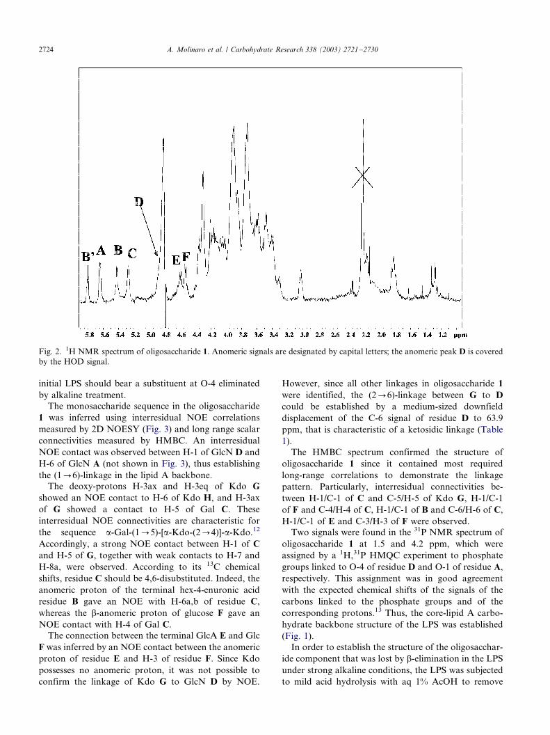
The monosaccharide sequence in the oligosaccharide
1 was inferred using interresidual NOE correlations
measured by 2D NOESY (Fig. 3) and long range scalar
connectivities measured by HMBC. An interresidual
NOE contact was observed between H-1 of GlcN D and
H-6 of GlcN A (not shown in Fig. 3), thus establishing
the (10/6)-linkage in the lipid A backbone.The deoxy-protons H-3ax and H-3eq of Kdo G
showed an NOE contact to H-6 of Kdo H, and H-3ax
of G showed a contact to H-5 of Gal C. These
interresidual NOE connectivities are characteristic for
the sequence a-Gal-(10/5)-[a-Kdo-(20/4)]-a-Kdo.12
Accordingly, a strong NOE contact between H-1 of C
and H-5 of G, together with weak contacts to H-7 and
H-8a, were observed. According to its 13C chemical
shifts, residue C should be 4,6-disubstituted. Indeed, the
anomeric proton of the terminal hex-4-enuronic acid
residue B gave an NOE with H-6a,b of residue C,
whereas the b-anomeric proton of glucose F gave an
NOE contact with H-4 of Gal C.
The connection between the terminal GlcA E and Glc
F was inferred by an NOE contact between the anomeric
proton of residue E and H-3 of residue F. Since Kdo
possesses no anomeric proton, it was not possible to
confirm the linkage of Kdo G to GlcN D by NOE.
However, since all other linkages in oligosaccharide 1
were identified, the (20/6)-linkage between G to D
could be established by a medium-sized downfield
displacement of the C-6 signal of residue D to 63.9
ppm, that is characteristic of a ketosidic linkage (Table
1).
The HMBC spectrum confirmed the structure of
oligosaccharide 1 since it contained most required
long-range correlations to demonstrate the linkage
pattern. Particularly, interresidual connectivities be-
tween H-1/C-1 of C and C-5/H-5 of Kdo G, H-1/C-1
of F and C-4/H-4 of C, H-1/C-1 of B and C-6/H-6 of C,
H-1/C-1 of E and C-3/H-3 of F were observed.
Two signals were found in the 31P NMR spectrum of
oligosaccharide 1 at 1.5 and 4.2 ppm, which were
assigned by a 1H,31P HMQC experiment to phosphate
groups linked to O-4 of residue D and O-1 of residue A,
respectively. This assignment was in good agreement
with the expected chemical shifts of the signals of the
carbons linked to the phosphate groups and of the
corresponding protons.13 Thus, the core-lipid A carbo-
hydrate backbone structure of the LPS was established
(Fig. 1).
In order to establish the structure of the oligosacchar-
ide component that was lost by b-elimination in the LPS
under strong alkaline conditions, the LPS was subjected
to mild acid hydrolysis with aq 1% AcOH to remove
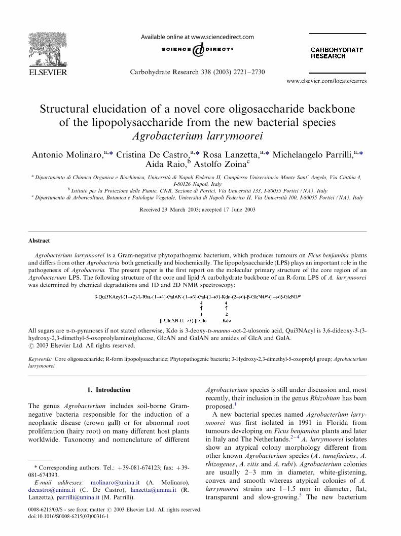
Fig. 2. 1H NMR spectrum of oligosaccharide 1. Anomeric signals are designated by capital letters; the anomeric peak D is covered
by the HOD signal.
A. Molinaro et al. / Carbohydrate Research 338 (2003) 2721�/27302724
terminal Kdo and lipid A. The residual material had no
organic phosphate as indicated by a colorimetric assay
and 31P NMR spectroscopy. Hence, the core region is
free of phosphate.
The oligosaccharide product obtained, 2, was studied
by compositional/methylation analyses and 2D NMR
spectroscopy. Compositional analysis of oligosaccharide
2 showed D-GlcA, D-GalA, D-Glc, D-Gal, L-Rha and D-
Qui3N, the last sugar being detected in a minor amount.
Methylation analysis of oligosaccharide 2 revealed
terminal GlcA, 4-substituted GalA, 2-substituted Rha,
4,6 di-substituted Gal, 3-substituted Glc and small
amounts of 5-substituted Kdo and terminal Qui3N.
Despite the presence of an amino function in oligosac-
charide 2, no acetyl signal was present in the 1H NMR
spectrum. However, in the aliphatic region of the
spectrum there were two singlets at 1.37 and 1.49 ppm
for methyl groups and two doublets at 2.45 and 2.67
ppm for an AB system of a diastereotopic methylene
group (Fig. 4). A large geminal coupling (J 17.2 Hz) in
the AB system was distinctive of a five-membered ring.
Therefore, it was suggested that Qui3N bears an unusual
acyl group at N-3.2D NMR spectroscopic analysis was performed using
COSY, TOCSY, NOESY, HSQC and HMBC experi-
ments (Table 2). Several sets of signals were visible in the
deoxy region of the 1H NMR spectrum, which belonged
to the multiple forms of the Kdo residue at the reducing
end of oligosaccharide 2. Six signals in the anomeric
region were assigned to residues L�/R.
The spin system L was assigned to GalA using
TOCSY and COSY spectra by chemical shifts and3J3,4 and 3J4,5 values (3.6 and 2 Hz, respectively). The
anomeric proton at 5.66 ppm (M) was correlated by the
TOCSY experiment to a methyl signal at 1.21 ppm and,
consequently, the spin system M belongs to rhamnose,
which was confirmed by low 3J1,2 and 3J2,3 values (2 and
3 Hz, respectively). A signal at 5.13 ppm was recognised
as the anomeric proton of galactose N by the 3J3,4 and3J4,5 values.
Two anomeric proton resonances appeared at 4.61
ppm, P deriving from b-glucose and Q from Qui3N, as
evident from the COSY and TOCSY spectra. Particu-
larly, the TOCSY spectrum showed a cross-peak
between the anomeric signal Q and a methyl signal at
1.26 ppm, and all sugar ring 3JH,H values were �/10 Hz.
Finally, residue R was identified as GlcA, as followed
from the chemical shifts and coupling constant values.
The b configuration of residues P�/R was corroborated
by 3J1,2 values of �/8 Hz and NOE contacts of H-1 to
H-3 and H-5.Six signals for anomeric carbons were identified by an
1H,13C HMQC experiment (Table 2). Downfield dis-
placements of carbon resonances were identified for
residue L (C-4), M (C-2), N (C-4 and C-6) and P (C-3),
whereas residues Q and R were recognised as terminal
sugars. Moreover, C-3 of Q was identified as a nitrogen-
Fig. 3. Part of a NOESY spectrum of oligosaccharide 1. Annotations refer to interresidual cross-peaks. Monosaccharides are as
shown in Fig. 2. The capital letters refer to residues as denoted in Table 1.
A. Molinaro et al. / Carbohydrate Research 338 (2003) 2721�/2730 2725

bearing carbon on the basis of its chemical shift of 58.2
ppm. The reducing a-Kdop was tentatively identified
and its C-5 found to have a glycosylation shift. These
data were in agreement with methylation analysis data.
Fig. 4. 1H NMR spectrum and structure of oligosaccharide 2 obtained by mild acid hydrolysis from the LPS from A. larrymoorei .
The significant peaks in the anomeric region are designated by capital letters.
Table 21H and 13C NMR chemical shifts (d , ppm) of sugar residues in oligosaccharide 2 from LPS of A. larrymoorei
Residue H-1/C-1 H-2/C-2 H-3/C-3 H-4/C-4 H-5/C-5 H-6/C-6
L 5.81 4.09 4.00 4.50 4.97
4-GalA 99.9 67.5 71.5 82.5 72.0 175.3
M 5.66 4.10 3.97 3.41 3.77 1.21
2-Rha 99.7 81.8 71.2 73.4 69.2 17.9
N 5.13 3.54 3.86 4.28 4.35 4.09
4,6-Gal 101.0 73.1 71.0 76.4 70.0 68.8
P 4.61 3.29 3.67 3.52 3.49 3.91/3.74
3-Glc 106.0 72.3 81.3 72.4 76.9 61.2
Q 4.61 3.46 3.90 3.23 3.52 1.26
Qui3NAcyl 105.0 72.8 58.2 74.3 74.6 18.3
R 4.34 3.79 3.73 3.91 4.29
GlcA 105.1 69.8 71.2 71.0 75.0 173.6
H-3/C-3 H-4/C-4 H-5/C-5 H-6/C-6 H-7/C-7 H-8/C-8
1.84/2.21 4.13 4.08 3.61 3.98 3.94/3.61
5-Kdo I 35.0 66.1 73.1 72.1 70.1 64.0
Residues are labelled according to decreasing chemical shifts of their anomeric signals. Chemical shifts for the 3-hydroxy-2,3-
dimethyl-5-oxoprolyl group (Acyl): C-1 to C-5 at 176.0, 71.0, 79.9, 44.0 (dH 2.45, 2.67) and 180.0 ppm, respectively; two methyl
groups at dC/dH 18.8/1.49 (CH3-2) and 23.3/1.37 (CH3-3).
A. Molinaro et al. / Carbohydrate Research 338 (2003) 2721�/27302726
Therefore, the main difference between 2 and 1 is the
presence in the former of an additional 2-substituted
rhamnose, a terminal Qui3N and, evidently, the pre-
dicted 4-substituted GalA residue, which was unequi-
vocally identified in 2. The remaining sugar residues,
except for the terminal Kdo and GlcN of lipid A, were
identical in both oligosaccharides.
It was reasonable to arrange the three additional
residues in the oligosaccharide 2 as Q0/2-M0/4-L0/6-
N, and this sequence was confirmed by the ROESY and
HMBC spectra. In the NOESY spectrum (Fig. 5), cross-
peaks between H-1 of GalA L and H-6a,b of Gal N, H-1
of Rha M and H-4 of GalA L, and H-1 of Qui3N Q and
H-2 of Rha M were of particular importance. During
the alkaline treatment, the LPS underwent b-elimination
and lost a Qui3N-(10/2)-Rha disaccharide, leaving a
terminal hex-4-eneuronic acid.
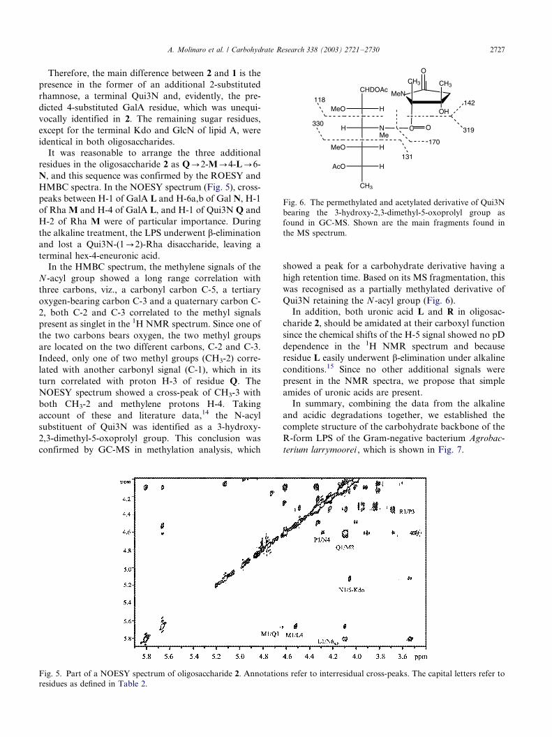
In the HMBC spectrum, the methylene signals of the
N -acyl group showed a long range correlation with
three carbons, viz., a carbonyl carbon C-5, a tertiary
oxygen-bearing carbon C-3 and a quaternary carbon C-
2, both C-2 and C-3 correlated to the methyl signals
present as singlet in the 1H NMR spectrum. Since one of
the two carbons bears oxygen, the two methyl groups
are located on the two different carbons, C-2 and C-3.
Indeed, only one of two methyl groups (CH3-2) corre-
lated with another carbonyl signal (C-1), which in its
turn correlated with proton H-3 of residue Q. The
NOESY spectrum showed a cross-peak of CH3-3 with
both CH3-2 and methylene protons H-4. Taking
account of these and literature data,14 the N-acyl
substituent of Qui3N was identified as a 3-hydroxy-
2,3-dimethyl-5-oxoprolyl group. This conclusion was
confirmed by GC-MS in methylation analysis, which
showed a peak for a carbohydrate derivative having a
high retention time. Based on its MS fragmentation, this
was recognised as a partially methylated derivative of
Qui3N retaining the N -acyl group (Fig. 6).
In addition, both uronic acid L and R in oligosac-
charide 2, should be amidated at their carboxyl function
since the chemical shifts of the H-5 signal showed no pD
dependence in the 1H NMR spectrum and because
residue L easily underwent b-elimination under alkaline
conditions.15 Since no other additional signals were
present in the NMR spectra, we propose that simple
amides of uronic acids are present.
In summary, combining the data from the alkaline
and acidic degradations together, we established the
complete structure of the carbohydrate backbone of the
R-form LPS of the Gram-negative bacterium Agrobac-
terium larrymoorei , which is shown in Fig. 7.
Fig. 5. Part of a NOESY spectrum of oligosaccharide 2. Annotations refer to interresidual cross-peaks. The capital letters refer to
residues as defined in Table 2.
Fig. 6. The permethylated and acetylated derivative of Qui3N
bearing the 3-hydroxy-2,3-dimethyl-5-oxoprolyl group as
found in GC-MS. Shown are the main fragments found in
the MS spectrum.
A. Molinaro et al. / Carbohydrate Research 338 (2003) 2721�/2730 2727

3. Discussion
Depending on the saccharide size, there are two kinds of
lipopolysaccharides, deriving from smooth and rough
bacteria (S-LPS and R-LPS). Both structures are built
up of lipid A and covalently bonded core, and the core
region comprises up to 15 monosaccharides. In S-LPS,
the core region is covalently linked to an O-specific
polysaccharide chain. Both LPS forms are present in
wild-type Gram-negative bacteria, a core-lipid A moiety
being the minimum structural unit required for the life
of bacteria.
The LPS plays a key role in the interaction with, and
defence reactions of, host plants, and, therefore, the
knowledge on the LPS structure is of great relevance.
Despite the wealth of information in the biology of
Agrobacterium genus and in the chemistry and biochem-
istry of the LPS of Gram-negative bacteria, there are no
data available in literature on the chemical structure of
the Agrobacterium LPS, apart from a paper by our
group.8
A. larrymoorei produces only an R-form LPS, a
lipooligosaccharide, and in this work the first core
structure was elucidated for an Agrobacterium species.
From a biochemical point of view, this core is unusual
since it lacks heptose residues and phosphate groups.
Heptoses are absent from the LPS of a few bacteria
studied so far (e.g., in Chlamydia , Branhamella , Acine-
tobacter or Rhizobium ).16,17 It is particularly interesting
that this chemical peculiarity is now found in Agrobac-
terium , a Rhizobium correlated bacterium, confirming
the suggested close taxonomical relationship of these
two genera.
The absence of phosphate groups is more frequent,
and uronic acids are introduced instead to provide a
negative charge.16,17 It is thought that these negatively
charged groups with bivalent ions as counterparts
assemble ionic bridges between LPS molecules, which
contribute to the rigidity and stability of the Gram-
negative cell wall. The LPS of A. larrymoorei is unusual
in this respect since uronic acids are present as urona-
mides and, hence, do not provide any negative charge.
Remarkable is also the finding of an unusual N -acyl
group, which is a derivative pyroglutamic acid. This
group has been identified in the core region only once as
a component of the LPS of Campylobacter coli.18 In
Fig. 7. The complete oligosaccharide backbone of the LPS from A. larrymoorei .
A. Molinaro et al. / Carbohydrate Research 338 (2003) 2721�/27302728

both bacteria, it is linked as amide to Qui3N, which
often bears unusual substituents in the LPS. The same
N -acyl group has also been identified in the O-chain of
Pseudomonas fluorescens14 and Pseudomonas putida (C.de Castro, unpublished data) and similar pyroglutamic
acid derivatives in Vibrio cholerae and Vibrio anguil-
larum .19,20
4. Experimental
4.1. Bacteria and bacterial LPS
Agrobacterium larrymoorei was cultivated as described.5
The LPS was obtained from lyophilised bacteria (3 g) by
the phenol/water extraction method as described.21 Forfurther purification, the LPS fraction was subjected to
sequential nuclease and protease treatment followed by
GPC on Sephacryl HR-400 (Pharmacia) using a column
(1.5�/90 cm) eluted with 50 mM NH4HCO3 at 0.4 mL
min�1. The yield of the purified LPS was 84 mg or 2.8%
of the bacterial dry mass.
4.2. Isolation of oligosaccharides 1 and 2
The LPS (16 mg) was O-de-acylated with anhyd
hydrazine (1 mL) at 37 8C for 30 min, cooled, poured
into ice-cold acetone (20 mL), and allowed to precipi-
tate. The precipitate was centrifuged (3000g , 30 min),
washed twice with ice-cold acetone, dried, dissolved inwater and lyophilised (12 mg, 75% of the LPS mass).
The sample was subsequently N-deacylated with 4 M
KOH as described.15 After desalting using a column
(50�/1.5 cm) of Sephadex G-10 (Pharmacia), the
resulting oligosaccharide fraction (7 mg, 43% of the
LPS mass) was further fractionated by GPC on a
column (50�/3 cm) of TSK HW-40 (E. Merck) in
2:5:250 pyridine�/acetic acid�/water to give oligosacchar-ide 1 (5 mg, 31% of the LPS mass). This fraction
contained only one carbohydrate component as deter-
mined by analytical HPAEC on a CarboPac PA1
column (4�/250 mm) using a linear gradient of sodium
acetate (0.2�/0.9 M) in 0.1 M NaOH over 50 min.
Another portion of the LPS (20 mg) was hydrolysed
in aq 1% AcOH (100 8C, 2 h), and the precipitate (lipid
A) was removed by centrifugation (8000g , 30 min). Thesupernatant was fractionated by GPC on a column
(50�/3 cm) of Sephadex G-50 (Pharmacia). One main
fraction was obtained (11 mg, oligosaccharide 2) as
shown by HPAEC.
4.3. General and analytical methods
The LPS was analysed by discontinuous SDS-PAGE
(12% separating gel) utilizing a miniprotean gel system
(Bio-Rad). A sample (4 mg) was run at constant voltage
(150 V) and stained with silver nitrate.22
Neutral sugars and their absolute configurations, as
well as organic phosphate and Kdo were determined asreported.23�26 For methylation analysis of Kdo and
uronic acids,27 oligosaccharides were N-acetylated with
acetic anhydride (15 mL) in 0.5 M NaOH (150 mL) for 10
min, then carboxyl-methylated with methanolic HCl
(0.1 M, 5 min) and consecutively with diazomethane in
order to improve the solubility in Me2SO. The methy-
lated product28 was hydrolysed with 2 M trifluoroacetic
acid (100 8C, 1 h), carbonyl-reduced with NaBD4,carboxyl-methylated as above, carboxyl-reduced with
NaBD4 (4 8C, 18 h), acetylated and analysed by GLC-
MS. Methylation analysis of the complete core region
was carried out as described above, and the sample was
hydrolysed with 4 M trifluoroacetic acid (100 8C, 4 h),
carbonyl-reduced with NaBD4, carboxyl-methylated,
carboxyl-reduced, acetylated and analysed by GLC-MS.
4.4. NMR spectroscopy
1H and 13C NMR spectra were recorded using a Varian
Inova 500 instrument, and 31P NMR spectra on a
Bruker DRX-400 spectrometer. All experiments were
carried out at 30 8C in D2O solution at pD 14 and 7
(uncorrected values) for oligosaccharides 1 and 2,
respectively. Chemical shifts were measured relative tointernal acetone (dH 2.225, dC 31.5). Aq 85% phosphoric
acid was used as reference (0.00 ppm) for 31P NMR
spectroscopy. Coupling constants were determined on a
first order basis from 2D phase sensitive DQF-
COSY.29,30
Phase-sensitive DQF-COSY was performed using the
standard Varian tndqcosy program (with water suppres-
sion) with an acquisition time 0.258 s and 4096 datapoints in the F2 dimension. The data matrix was zero-
filled in the F1 dimension to give a matrix of 4096�/
2048 points and was resolution enhanced in both
dimensions by a shifted sine-bell function before Fourier
transformation. Similarly, NOE experiments were per-
formed using the Varian standard tnnoesy and tnroesy
programs (both with water suppression) with a mixing
time of 200 ms. The TOCSY experiment was performedusing standard Varian tntocsy program (with water
suppression) with a spinlock time of 80 ms. The hetero-
nuclear experiments were performed using pulse field
gradient programs gHSQC and gHMBC. 1H detected1H,13C gHMBC was optimised for 6 Hz coupling
constant, and 1H detected 1H,31P gHSQC for 8 Hz
coupling constant.
The spectra were assigned using the computer pro-gram PRONTO, which enables a simultaneous display of
different 2D NMR spectra and the individual labelling
of cross-peaks.31
A. Molinaro et al. / Carbohydrate Research 338 (2003) 2721�/2730 2729
Acknowledgements
We thank MIUR, Rome (Progetti di Ricerca di Inter-
esse Nazionale) (to M.P.) and Progetto Giovani Ricer-catori (to C.D.C.) for financial support. NMR
experiments were carried out on a 500 MHz spectro-
meter of Consortium INCA (L488/92, Cluster 11) at
Centro Interdipartimentale Metodologie Chimico Fi-
siche Universita di Napoli. C.D.C. and A.M. thank
Professor Dr Otto Holst for the continuous and fruitful
scientific discussion through the years.
References
1. Young, J. M.; Kuykendall, L. D.; Martinez-Romero, E.;
Kerr, A.; Sawada, H. Int. J. Sys. Evol. Microbiol. 2001,
51 , 89�/103.
2. Bouzar, H.; Jones, J. B. Int. J. Sys. Evol. Microbiol. 2001,
51 , 1023�/1026.
3. Bouzar, H.; Chilton, W. S.; Nesme, X.; Dessaux, Y.;
Vaudequin, V.; Petit, A.; Jones, J. B.; Hodge, N. C. Appl.
Environ. Microbiol. 1995, 61 , 65�/73.
4. Zoina, A.; Raio, A.; Peluso, R. In Proceedings of
‘L’endofitismo di funghi e batteri patogeni in piante arboree
ed arbustive ’; Sassari (Italy), May 20�/21, 2002; pp. 127�/
136.
5. Zoina, A.; Raio, A.; Peluso, R.; Spasiano, A. Plant Pathol.
2001, 50 , 620�/627.
6. Dow, J. M.; von Roepenack, E.; Newman, M.-A. Ann.
Rev. Phytopathol. 2000, 38 , 241�/261.
7. Newman, M.-A.; von Roepenack, E.; Lahaye, A. P.;
Daniels, M. J.; Dow, J. M. Plant J. 2002, 9 , 487�/495.
8. De Castro, C.; De Castro, O.; Molinaro, A.; Parrilli, M.
Eur. J. Biochem. 2002, 269 , 2885�/2888.
9. Birnbaum, G. I.; Roy, R.; Brisson, J. R.; Jennings, H. J.
Carbohydr. Chem. 1987, 6 , 17�/39.
10. Holst, O.; Thomas-Oates, J. E.; Brade, H. Eur. J.
Biochem. 1994, 222 , 183�/194.
11. Bystrova, O. V.; Shashkov, A. S.; Kocharova, N. A.;
Knirel, Y. A.; Lindner, B.; Zahringer, U.; Pier, G. B. Eur.
J. Biochem. 2002, 269 , 2194�/2203.
12. Muller-Loennies, S.; Holst, O.; Brade, H. Eur. J. Biochem.
1994, 224 , 751�/760.
13. Muller-Loennies, S.; Holst, O.; Lindner, B.; Brade, H. Eur.
J. Biochem. 1999, 260 , 235�/249.
14. Zatonsky, G. V.; Kocharova, N. A.; Veremeychenko, S.
P.; Zdorovenko, E. L.; Shashkov, G. M.; Knirel, Y. A.
Carbohydr. Res. 2002, 337 , 2365�/2370.
15. Vinogradov, E. V. Carbohydr. Res. 2002, 337 , 961�/963.
16. Holst, O. In Endotoxin in Health and Disease ; Brade, H.;
Morrsion, D. C.; Opal, S.; Vogel, S., Eds.; Marcel Dekker:
New York, 1999; pp 115�/154.
17. Holst, O. Trends Glycosci. Glycotechnol. 2002, 14 , 87�/103.
18. Aspinall, G. O.; Lynch, C. M.; Pang, H.; Shaver, R. T.;
Moran, A. P. J. Biol. Chem. 1993, 268 , 6263�/6268.
19. Hermansson, K.; Jansson, P.-E.; Holme, T.; Gustavsson,
B. Carbohydr. Res. 1993, 248 , 199�/211.
20. Eguchi, H.; Shunji, K.; Araki, Y. Carbohydr. Res. 1992,
231 , 147�/158.
21. Westphal, O.; Jann, K. Methods Carbohydr. Chem. 1965,
5 , 83�/91.
22. Tsai, C. M.; Frasch, C. E. Anal. Biochem. 1982, 119 , 115�/
119.
23. Vinogradov, E. V.; Holst, O.; Thomas-Oates, J. E.;
Broady, K. W.; Brade, H. Eur. J. Biochem. 1992, 210 ,
491�/498.
24. Kaca, W.; de Jongh-Leuvenink, J.; Zahringer, U.; Brade,
H.; Verhoef, J.; Sinnwell, V. Carbohydr. Res. 1988, 179 ,
289�/299.
25. Holst, O.; Broer, W.; Thomas-Oates, J. E.; Mamat, U.;
Brade, H. Eur. J. Biochem. 1992, 214 , 703�/710.
26. Susskind, M.; Brade, L.; Brade, H.; Holst, O. J. Biol.
Chem. 1998, 273 , 7006�/7017.
27. Molinaro, A.; De Castro, C.; Lanzetta, R.; Evidente, A.;
Parrilli, M.; Holst, O. J. Biol. Chem. 2002, 277 , 10058�/
10063.
28. Ciucanu, I.; Kerek, F. Carbohydr. Res. 1984, 131 , 209�/
217.
29. Piantini, U.; Sørensen, O. W.; Ernst, R. R. J. Am. Chem.
Soc. 1982, 104 , 6800�/6801.
30. Rance, M.; Sørensen, O. W.; Bodenhausen, G.; Wagner,
G.; Ernst, R. R.; Wuthrich, K. Biochem. Biophys. Res.
Commun. 1983, 117 , 479�/485.
31. Kjaer, M.; Andersen, K. V.; Poulsen, F. M. Methods
Enzymol. 1994, 239 , 288�/308.
A. Molinaro et al. / Carbohydrate Research 338 (2003) 2721�/27302730
Copyright © 2022 FDOKUMEN