The Powdery Mildew Effector CSEP0027 Interacts With Barley ...
Loss-of-Function Mutations in Chitin Responsive GenesShow Increased Susceptibility to the Powdery MildewPathogen Erysiphe cichoracearum1[w]
Katrina Ramonell*, Marta Berrocal-Lobo, Serry Koh, Jinrong Wan, Herb Edwards, Gary Stacey,and Shauna Somerville
Department of Biological Sciences, University of Alabama, Tuscaloosa, Alabama 35487–0344 (K.R.);Department of Plant Biology, Carnegie Institution, Stanford, California 94305 (M.B.-L., S.K., S.S.); NationalCenter for Soybean Biotechnology, Divisions of Plant Sciences and Biochemistry, University of Missouri,Columbia, Missouri 65211 (J.W., G.S.); and Department of Biological Sciences, Western Illinois University,Macomb, Illinois 61455–1390 (H.E.)
Chitin is a major component of fungal walls and insect exoskeletons. Plants produce chitinases upon pathogen attack andchito-oligomers induce defense responses in plants, though the exact mechanism behind this response is unknown. Using theATH1 Affymetrix microarrays consisting of about 23,000 genes, we examined the response of Arabidopsis (Arabidopsis thaliana)seedlings to chito-octamers and hydrolyzed chitin after 30 min of treatment. The expression patterns elicited by the chito-octamer and hydrolyzed chitin were similar. Microarray expression profiles for several genes were verified via northernanalysis or quantitative reverse transcription-PCR. We characterized T-DNA insertion mutants for nine chito-oligomerresponsive genes. Three of the mutants were more susceptible to the fungal pathogen, powdery mildew, than wild type asmeasured by conidiophore production. These three mutants included mutants of genes for two disease resistance-like proteinsand a putative E3 ligase. The isolation of loss-of-function mutants with enhanced disease susceptibility provides directevidence that the chito-octamer is an important oligosaccharide elicitor of plant defenses. Also, this study demonstrates thevalue of microarray data for identifying new components of uncharacterized signaling pathways.
Plants in the environment are constantly under siegeby a multitude of disease-causing organisms includ-ing bacteria, fungi, viruses, and nematodes. Plantsmay resist pathogen attack using both preformeddefenses (e.g. antimicrobial compounds) and induc-ible defense responses (for review, see Hammond-Kosack and Jones, 2000; Heath, 2000). Inducibledefenses can be activated upon recognition of generalelicitors such as bacterial flagellin (Gomez-Gomezand Boller, 2002), the polypeptide systemin (Ryanand Pearce, 1998), and multiple host or pathogen cellwall fragments released during pathogen attack(Nurnberger et al., 2004). Recently, oligosaccharideelicitors such as chito-oligomers and oligogalacturo-nides have received renewed attention as importantsignals in plant defense responses. The activation of
defense genes by these elicitors is thought to bereceptor-mediated though little is known about theinitial perception and consequent signaling pathwaysinvolved in plant cells.
Chito-oligosaccharides can be generated from thecell walls of pathogenic fungi by the action of endo-chitinases and were shown to elicit strong defenseresponses in many plant species (Stacey and Shibuya,1997; Shibuya and Minami, 2001). In a previousstudy, we showed that transcript levels for 71 ex-pressed sequence tags, representing 61 genes, werealtered more than 3-fold in chito-oligomer treatedArabidopsis (Arabidopsis thaliana) seedlings, demon-strating the usefulness of Arabidopsis as a modelfor studying chitin signaling in plants (Ramonellet al., 2002). Further experimentation by Zhang et al.(2002) showed that this response was not mediatedby the well-characterized salicylic acid, jasmonic acid,or ethylene-responsive plant defense pathways. Morerecently, Wan et al. (2004) demonstrated that the cel-lular response of Arabidopsis to chitin octamer eli-citation involved activation of a mitogen-activatedprotein kinase cascade. These data indicated that chitinwas inducing defense responses through an unchar-acterized signaling pathway.
In previous experiments, we used a complex mix-ture of chito-oligomers derived from hydrolyzed crab-shell chitin (CSC) to induce defense responses inArabidopsis (Ramonell et al., 2002). However, purified
1 This work was supported by the Chemical Sciences, Geoscien-ces, and Biosciences Division, Office of Basic Energy Sciences, Officeof Science, U.S. Department of Energy (grant no. DE–FG02–02ER15309 to G.S.), by the National Science Foundation (grant no.0114783 to S.S.), by the Carnegie Institution (to S.S. and M.B.-L.), bythe Spanish government (fellowship to M.B.-L.), and by the Univer-sity of Alabama (to K.M.R.).
* Corresponding author; e-mail [email protected]; fax 205–348–1768.
[w] The online version of this article contains Web-only data.Article, publication date, and citation information can be found at
www.plantphysiol.org/cgi/doi/10.1104/pp.105.060947.
This article is published in Plant Physiology Online, Plant Physiology Preview Section, which publishes manuscripts accepted for
publication after they have been edited and the authors have corrected proofs, but before the final, complete issue is published
online. Early posting of articles reduces normal time to publication by several weeks.
Plant Physiology Preview, www.aspb.org � 2005 American Society of Plant Biologists 1 of 10

chitin fragments (degree of polymerization [dp] 5 7–8)induce the synthesis of phytoalexins in suspensioncultured rice (Oryza sativa) cells at nanomolar concen-trations. Yamada et al. (1993) and Zhang et al. (2002)showed that the purified chitin octamer was the mosteffective of the chito-oligomer lengths tested in in-ducing the response of several genes identified asresponsive to CSC (Ramonell et al., 2002). In this work,we compared the gene expression profiles of seedlingstreated with purified chito-octamer (dp 5 8) and a CSCmixture using full-genome ATH1 Affymetrix Gene-Chips. T-DNA insertional mutations in selected chito-oligomer responsive genes were evaluated to deter-mine whether they contribute to disease resistance tothe powdery mildew pathogen, Erysiphe cichoracearum.
RESULTS
Chitin Is Present at the Tips of Growing Hyphaeand in Conidia of E. cichoracearum duringInfection of Arabidopsis
Chitin is one of the major cell wall components inmany fungi. Fungal cell walls change during growthand development and the deposition and subsequentremoval of chitin is particularly important duringseptation (Smits et al., 2001; Adams, 2004). However,the distribution of chitin in E. cichoracearum structuresformed during infections of Arabidopsis was un-known. Cell wall components and chitin were localizedin growing E. cichoracearum using labeled wheat germagglutinin (WGA). WGA contains a group of closelyrelated isolectins that can bind oligosaccharides con-taining terminal GlcNAc or chitobiose and membraneglycoproteins (Peters and Latka, 1986). WGA has beenused in several studies on chitin distribution in fungalwalls (Galun et al., 1976; Tronchin et al., 1981). Laserscanning confocal microscopy visualized WGA-488Alexa conjugates at the growing tips of appressoriaand hyphae (Fig. 1, A and B). WGA staining was alsoobserved in the cell walls of conidia developing onconidiophores (Fig. 1C). As the conidia matured anddetached from the conidiophore, chitin was localizedto both ends of conidia (Fig. 1D). When conidia germi-nated, the region where the appressorial germ tubedeveloped accumulated chitin. Furthermore, althoughmature conidia have chitin at their ends (Fig. 1D),germinated conidia (Fig. 1A) lack detectable chitin inthis position, suggesting temporal and spatial re-gulation of chitin localization. Transmission electronmicroscopy confirmed that WGA-gold colloidal con-jugates were present at the growing tip of appresso-rium (Fig. 1E) and also accumulated in the cell wall ofthe fungal haustorium, a feeding structure that formsin epidermal cells (Fig. 1F). Fungal structures directlyin contact with the plant and likely to be exposed toplant chitinases, such as appressoria and haustoria,show relatively high content of chitin. Therefore, it islikely that chito-oligomers will be generated duringthe course of powdery mildew infections.
Chito-Oligomers Elicit Distinct Gene Expression
Profiles in Arabidopsis
To better understand the responses of Arabidopsisto chito-oligomers, we compared the gene expression
Figure 1. Chitin localization in the powdery mildew, E. cichoracea-rum. Confocal images are presented in A to D and transmissionelectron micrographs in E and F. In A to D, samples were stained with PI(red channel) to highlight fungal structures, although plant structurescan be stainedwith this nonspecific stain, and chitin was localizedwiththe lectin, WGA-Alexa Fluor 488 (green channel). In E and F, WGA-colloidal gold conjugates were used to localize chitin. A, A mergedconfocal micrograph showing chitin localized to the tip of theappressorium (arrow; 1 dpi). Plant guard cells are also partially stainedwith PI in this image. Bar5 12 mm. C, Conidium; Ap, appressorium. B,Chitin localization at the growing tip of hypha (arrow) but not on theelongated hyphal region (3 dpi). Hyp, Hyphae. Bar5 11 mm. C, Chitinlabeling occurs in the developing conidia still attached to conidio-phores (arrows; 7 dpi). Bar 5 31 mm. D, A mature conidium (arrow)detaching from a condiophore shows strong chitin localization at theboth ends. Bar5 16 mm. E, Chitin occurs in the fungal appressorial cellwall but not on the plant cell wall. CW, Plant outer epidermal cell wall;FCW, fungal cell wall. Bar 5 2.15 mm. F, Chitin is found in thehaustorial cell wall (arrows). EHMAT, Extrahaustorial matrix, H,haustorium; Nc, haustorial nucleus. Bar 5 9.21 mm.
Ramonell et al.
2 of 10 Plant Physiology Preview

of seedlings treated with chito-octamer, CSC, or wateras a control using Affymetrix ATH1 full-genomemicroarrays. Of the approximately 23,000 genes rep-resented on the array, 5,012 genes showed alteredexpression according to the statistical analysis ofmicroarrays (SAM) program with a false discoveryrate of #5% (Tusher et al., 2001; Fig. 2A). Clusteranalysis of genes responsive to CSC and the chito-octamer suggest that these two treatments elicit verysimilar responses, especially among the induced genes(Fig. 2A). To narrow the list, genes with changes inexpression of 1.5-fold or more relative to the watercontrol were selected and then compared among thetwo chito-oligomer treatments (Fig. 2B). Full lists of the
selected genes organized by treatment and inductionor repression are provided in supplemental tables(Supplemental Tables I–VI). The chito-octamer uniquelyinduced 68 genes but the CSC induced a larger setof genes (104). In both treatments, more genes weresignificantly repressed than induced. Similar numbersof genes were uniquely repressed by both chito-octamer and CSC treatment (238 and 262 respectively).Thus, the relative proportion of genes uniquely re-pressed by each treatment was larger than the corre-sponding number of genes induced (Fig. 2B, 1 and 2).Though the overall profiles appear relatively similar atfirst glance, there are significant differences in theexpression profiles between the two chitin treatments.
Figure 2. A, Hierarchical cluster ofratio values (relative to the water con-trol treatment) of 5,435 chito-oligomerresponsive genes as determined by theSAM program. Each gene is repre-sented by a single row and eachcolumn represents an individual treat-ment. Magenta represents up-regulatedgenes; green, down-regulated genes;and black, genes with no change inexpression. B, Venn diagrams showingdistribution of the subset of thesegenes showing a $1.5-fold change inexpression in at least one of the twochito-oligomer treatments relative tothe water control. B1, Distribution of1,252 genes identified as expressedat $1.50-fold higher levels in chito-oligomer treated than control samples.B2, Distribution of 1,123 chito-oligomer responsive genes expressedat #0.67 of control samples. 8-mer,Treated with chito-octamer; CSC, trea-ted with CSC; Mock, treated withdH2O. Tables with lists of genes ineach grouping are presented as sup-plemental data (Supplemental TablesI–VI).
Altered Defense Response in Oligosaccharide-Elicitor Mutants
Plant Physiology Preview 3 of 10
This may be due to the effect of different chitin species(e.g. chain lengths) in the CSC or due to differentrelative concentrations, since accurate concentrationmeasurements for a specific oligomer could only bemade for the chito-octamer.
The chito-octamer treatment induced genes forseveral defense-related proteins including pathogen-esis-related protein 5, two WRKY transcription fac-tors, and four disease resistance proteins (three of theToll Interleukin-1 receptor [TIR] or TIR-nucleotide-binding site [NBS]-Leu-rich repeat [LRR] class, andthe other an LRR class protein). The largest class ofgenes repressed in the chito-octamer treatment wereunknown or hypothetical proteins, but several in-teresting genes were repressed 1.5-fold includinggenes encoding ETR2, an LRR transmembrane pro-tein kinase, the plant defensin protein PDF 2.5,a jasmonate-inducible protein, and several zinc-fingerproteins potentially involved in signaling. Treatmentwith chito-octamer was shown previously to elicitstrong defense responses in numerous plant species(Yamada et al., 1993; Zhang et al., 2002). These dataindicate that the chitin octamer is inducing a strongdefense response in Arabidopsis that is consistentwith data previously observed. It is interesting to notethat the chito-octamer induced and repressed severalgenes involved in plant defense. The plant defensinprotein PDF 2.5 was down-regulated, while severaldisease resistance-like proteins and a WRKY tran-scription factor were stimulated. It is unclear whydefense-related genes were repressed by the chitinoctamer. One explanation may be that other signals inaddition to the presence of chitin may be necessary toelicit an appropriate defense response to a specificpathogen. Large numbers of genes were either in-duced or repressed (1,002 and 312, respectively) byboth CSC and the chito-octamer. Many of the genesinduced by CSC and the chito-octamer were defenserelated, which is expected in light of data fromprevious studies (Yamada et al., 1993; Ramonellet al., 2002; Zhang et al., 2002). However, there wasno clear trend in the distribution of genes that wererepressed by both treatments. Clearly, a combinationof chito-oligomer fragments elicits a strong defenseresponse, but a clearer understanding of the corre-sponding repression of genes will require more study.
In the CSC treatment alone, a large number of genescoding for defense-related proteins were induced1.5-fold including numerous disease resistance-likeproteins, WRKY family transcription factors, and anLRR protein kinase. Several cell wall-related tran-scripts including a subunit of cellulose synthase anda xyloglucan endotransglycosylase were also induced.Genes for several transport proteins (ATP-bindingcassette transporter family, integral membrane pro-tein) and some cell wall extensin-related proteins wererepressed 1.5-fold in the CSC treatment, as well asgenes encoding two disease-resistance proteins, RAR1(Required for Mla Resistance 1), an LRR proteinkinase, and a geranyl-geranyl phosphate synthase
involved in the biosynthesis of defensive terpenecompounds. Several classes of signaling molecules,protein kinases, and zinc-finger proteins included bothinduced and repressed genes in the CSC treatment. Inseveral experiments, CSC was shown to induce thestrongest changes in gene expression (Zhang et al.,2002) and this pattern is also repeated in our results.The strength of the response to CSC may be a conse-quence of interactions among multiple lengths ofchito-oligomers causing a stronger response in theplant.
Verification of Microarray Data
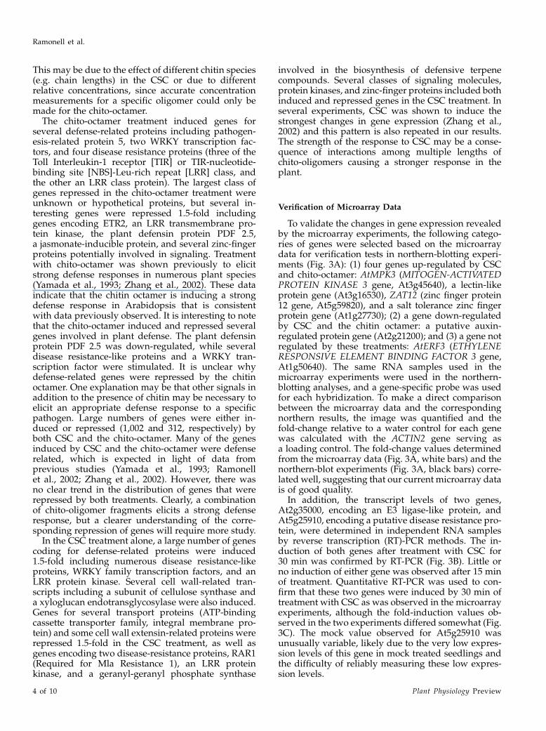
To validate the changes in gene expression revealedby the microarray experiments, the following catego-ries of genes were selected based on the microarraydata for verification tests in northern-blotting experi-ments (Fig. 3A): (1) four genes up-regulated by CSCand chito-octamer: AtMPK3 (MITOGEN-ACTIVATEDPROTEIN KINASE 3 gene, At3g45640), a lectin-likeprotein gene (At3g16530), ZAT12 (zinc finger protein12 gene, At5g59820), and a salt tolerance zinc fingerprotein gene (At1g27730); (2) a gene down-regulatedby CSC and the chitin octamer: a putative auxin-regulated protein gene (At2g21200); and (3) a gene notregulated by these treatments: AtERF3 (ETHYLENERESPONSIVE ELEMENT BINDING FACTOR 3 gene,At1g50640). The same RNA samples used in themicroarray experiments were used in the northern-blotting analyses, and a gene-specific probe was usedfor each hybridization. To make a direct comparisonbetween the microarray data and the correspondingnorthern results, the image was quantified and thefold-change relative to a water control for each genewas calculated with the ACTIN2 gene serving asa loading control. The fold-change values determinedfrom the microarray data (Fig. 3A, white bars) and thenorthern-blot experiments (Fig. 3A, black bars) corre-lated well, suggesting that our current microarray datais of good quality.
In addition, the transcript levels of two genes,At2g35000, encoding an E3 ligase-like protein, andAt5g25910, encoding a putative disease resistance pro-tein, were determined in independent RNA samplesby reverse transcription (RT)-PCR methods. The in-duction of both genes after treatment with CSC for30 min was confirmed by RT-PCR (Fig. 3B). Little orno induction of either gene was observed after 15 minof treatment. Quantitative RT-PCR was used to con-firm that these two genes were induced by 30 min oftreatment with CSC as was observed in the microarrayexperiments, although the fold-induction values ob-served in the two experiments differed somewhat (Fig.3C). The mock value observed for At5g25910 wasunusually variable, likely due to the very low expres-sion levels of this gene in mock treated seedlings andthe difficulty of reliably measuring these low expres-sion levels.
Ramonell et al.
4 of 10 Plant Physiology Preview
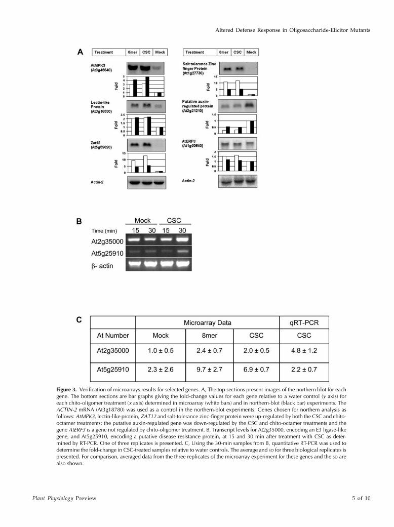
Figure 3. Verification of microarrays results for selected genes. A, The top sections present images of the northern blot for eachgene. The bottom sections are bar graphs giving the fold-change values for each gene relative to a water control (y axis) foreach chito-oligomer treatment (x axis) determined in microarray (white bars) and in northern-blot (black bar) experiments. TheACTIN-2 mRNA (At3g18780) was used as a control in the northern-blot experiments. Genes chosen for northern analysis asfollows: AtMPK3, lectin-like protein, ZAT12 and salt-tolerance zinc-finger protein were up-regulated by both the CSC and chito-octamer treatments; the putative auxin-regulated gene was down-regulated by the CSC and chito-octamer treatments and thegene AtERF3 is a gene not regulated by chito-oligomer treatment. B, Transcript levels for At2g35000, encoding an E3 ligase-likegene, and At5g25910, encoding a putative disease resistance protein, at 15 and 30 min after treatment with CSC as deter-mined by RT-PCR. One of three replicates is presented. C, Using the 30-min samples from B, quantitative RT-PCR was used todetermine the fold-change in CSC-treated samples relative to water controls. The average and SD for three biological replicates ispresented. For comparison, averaged data from the three replicates of the microarray experiment for these genes and the SD arealso shown.
Altered Defense Response in Oligosaccharide-Elicitor Mutants
Plant Physiology Preview 5 of 10
Insertional Mutants in Chito-Oligomer
Responsive Genes
From the microarray data, nine genes (At2g28200,At2g02950, At2g34930, At2g35000, At5g25910, At4g-11830, At2g41940, At4g16820, and At3g57640) wereselected on the basis of their expression profiles forfurther analysis. A search of the insertion sequencedatabase of the Salk Institute Genomic Analysis Lab-oratory (http://signal.salk.edu) was performed toidentify T-DNA insertional mutants with insertionsin the nine genes of interest. All lines were screened toidentify homozygous mutants. One backcross wasperformed on all T-DNA insertional mutants withwild-type Columbia (Col-0).
If we assume that chito-oligomer fragments are oneof the nonspecific signals or pathogen-associated mo-lecular patterns, exchanged between host and patho-gen during fungal and insect attack (Nurnberger et al.,2004) and that plants have evolved a mechanism todetect chito-oligomers and activate defense responses,then one would predict that mutations in chito-oligomer responsive genes would show enhancedsusceptibility to pathogens. This prediction also restson the assumption that a virulent pathogen is not ableto significantly suppress or evade the chito-oligomerelicited defenses. Since the chito-octamer and CSCelicited a number of defense-related genes, one wouldpredict that chito-octamer and CSC responsive genesare more likely to play a role in defense. Nine T-DNAinsertion lines, one for each selected gene, were in-oculated with the fungal pathogen, powdery mildew,and evaluated for disease resistance. To evaluatethe levels of susceptibility to powdery mildew, theinoculated T-DNA lines were compared with pow-dery mildew inoculated Col-0, a susceptible ecotype,Kashmir-1 (Kas-1), a resistant Arabidopsis ecotype,Landsberg erecta (Ler), an ecotype with intermediateresistance to powdery mildew, and two mutant linesimpaired in salicylic acid-mediated plant defense thatwere hyper-susceptible to powdery mildew, the trans-genic plant NahG that contains the bacterial genesalicylate hydrolase and the mutant sid2-1 that lacksa gene involved in salicylic acid biosynthesis (Reuberet al., 1998).
At low-density inoculations, two mutants in genesAt2g35000 and At5g25910 had more severe macro-scopic symptoms compared with Col-0 wild-typeplants (Fig. 4A). In contrast, the other seven insertionmutants exhibited a wild-type (Col-0) disease pheno-type when challenged by low-density inoculum (datanot shown). In addition, there was a clear increase inthe growth of fungal hyphae on both mutants (Fig. 4B)compared to growth on Col-0 wild-type plants, thoughthe fungal growth was not as advanced as that ontransgenic NahG plants (Fig. 4B). Independent SALKinsertion mutants for each of At2g35000 (Salk_066755,Salk_036065, Salk_036066) and At5g25910 (Salk_125444,Salk_107922) showed enhanced disease susceptibility,confirming that the altered disease phenotype can
be attributed to the loss of function of At2g35000 andAt5g25910 and not to a secondary mutation in theSALK lines (Supplemental Fig. 1). To measure diseasedevelopment more precisely, conidiophores per colony(c/c) were counted 4 d postinoculation (dpi) andconidiophores per mm22 (c/mm2) leaf surface werecounted at 5 dpi for each of the mutants (Table I). Threeof the T-DNA insertion lines, At2g34930, At2g35000,and At5g25910, were significantly more susceptible topowdery mildew than Col-0, Ler, and Kas-1 accessions.However, these three mutants were not as susceptibleas transgenic NahG plants, which are impaired in thesalicylic acid defense signaling and are known to behypersusceptible to powdery mildew (Reuber et al.,1998; Table I). The other six T-DNA insertional mutantsdid not show significant changes in disease symptomscompared with Col-0. A pBLAST search on these genes(data not shown) revealed significant numbers offunctionally redundant genes present in the Arabidop-sis genome that could account for the lack of change in
Figure 4. Phenotype of T-DNA mutants. A, Photographs of macro-scopic symptoms of leaves inoculated at low density with E. cichor-acearum. Leaves of insertional mutants in At2g35000 (SALK_066755)and At5g25910 (SALK_054976) had a slightly higher incidence ofmacroscopic symptoms as compared with Col-0. All other T-DNAinsertional mutants had no change in disease phenotype under low-density inoculums (data not shown). All photos 5 dpi. B, Cleared leavesof infected plants stainedwith trypan blue. Trypan blue stains the fungalhyphae and conidiophores on the leaf surface. Highest density of fungalgrowth is seen on insertional mutants of At2g35000 (SALK_066755),At5g25910 (SALK_054976), and transgenic NahG plants. All photo-graphs 5 dpi. Bar 5 0.2 mm.
Ramonell et al.
6 of 10 Plant Physiology Preview
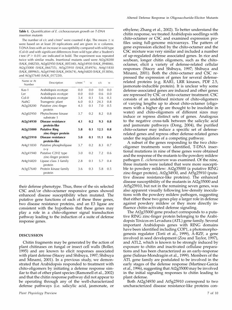
their defense phenotype. Thus, three of the six selectedCSC and/or chito-octamer responsive genes showedenhanced disease susceptibility when mutated. Theputative gene functions of each of these three genes,two disease resistance proteins, and an E3 ligase areconsistent with the hypothesis that these genes mayplay a role in a chito-oligomer signal transductionpathway leading to the induction of a suite of defenseresponse genes.
DISCUSSION
Chitin fragments may be generated by the action ofplant chitinases on fungal or insect cell walls (Boller,1995) and are known to elicit responses associatedwith plant defense (Stacey and Shibuya, 1997; Shibuyaand Minami, 2001). In a previous study, we demon-strated that Arabidopsis responded to treatment withchito-oligomers by initiating a defense response sim-ilar to that of other plant species (Ramonell et al., 2002)and that the chitin response pathway did not appear tobe operating through any of the well-characterizeddefense pathways (i.e. salicylic acid, jasmonate, or
ethylene; Zhang et al., 2002). To better understand thechitin response, we treated Arabidopsis seedlings withchito-octamer or CSC and examined expression pro-files using full-genome microarrays. The pattern ofgene expression elicited by the chito-octamer and theCSC mixture was very similar and included a numberof up-regulated defense associated genes. In rice andsoybean, longer chitin oligomers, such as the chito-octamer, elicit a variety of defense-related cellularresponses (Stacey and Shibuya, 1997; Shibuya andMinami, 2001). Both the chito-octamer and CSC re-pressed the expression of genes for several defense-related proteins (e.g. RAR1, LRR kinases, PDF 2.5,jasmonate-inducible protein). It is unclear why somedefense-associated genes are induced and other genesare repressed by CSC or chito-octamer treatment. CSCconsists of an undefined mixture of chito-oligomersof varying lengths up to about chito-octamer (oligo-mers with a higher dp are thought to be insoluble inwater) and chito-oligomers of different sizes mayinduce or repress distinct sets of genes. Analogousto the negative cross-talk between the salicylic acidand jasmonate pathways (Dong, 2004), the purifiedchito-octamer may induce a specific set of defense-related genes and repress other defense-related genesunder the regulation of a competing pathway.
A subset of the genes responding to the two chito-oligomer treatments were identified, T-DNA inser-tional mutations in nine of these genes were obtainedand the response of the mutants to the powdery mildewpathogen E. cichoracearum was examined. Of the nine,three mutants were isolated that were more suscepti-ble to powdery mildew: At2g35000 (a putative RINGzinc-finger protein), At2g34930, and At5g25910 (puta-tive disease resistance-like proteins). The enhanceddisease susceptibility of the mutants in At2g35000 andAt5g25910, but not in the remaining seven genes, wasalso apparent visually following low-density inocula-tions with the powdery mildew pathogen, suggestingthat either these two genes play a larger role in defenseagainst powdery mildew or they more directly in-fluence chitin-activated defense signaling.
The At2g35000 gene product corresponds to a puta-tive RING zinc-finger protein belonging to the Arabi-dopsis Toxicos en Levadura (ATL) gene family. Severalimportant Arabidopsis genes with RING domainshave been identified including COP1, a photomorpho-genesis regulator (Torii et al., 1999), A-RZF, a geneinvolved in seed development (Zou and Taylor, 1997),and ATL2, which is known to be strongly induced byexposure to chitin and inactivated cellulase prepara-tions and has been characterized as an early-responsegene (Salinas-Mondragon et al., 1999). Members of theATL gene family are postulated to be involved in theearly stages of the defense response (Martinez-Garciaet al., 1996), suggesting that At2g35000 may be involvedin the initial signaling responses to chitin leading toplant defense.
Both At2g34930 and At5g25910 correspond to twouncharacterized disease resistance-like proteins con-
Table I. Quantification of E. cichoracearum growth on T-DNAinsertion mutants
The number of c/c and c/mm2 were counted 4 dpi. The means 6 SE
were based on at least 20 replications and are given in SE columns.T-DNA lines with an increase in susceptibility compared with wild type(Col-0) and with significant differences from wild type after a Student’st test (P . 0.01) are indicated in bold. The experiment was repeatedtwice with similar results. Insertional mutants used were At2g28200(SALK_048250), At2g02950 (SALK_005340), At2g34930 (SALK_094846),At2g35000 (SALK_066755), At5g25910 (SALK_054976), At4g11830(SALK_089965), At2g41940 (SALK_045674), At4g16820 (SALK_013856),and At3g57640 (SALK_017220).
Name or At
NumberDescription c/mm21
SE c/c SE
Kas-1 Arabidopsis ecotype 0.0 0.0 0.0 0.0Ler Arabidopsis ecotype 0.0 0.0 0.6 0.0Col-0 Arabidopsis ecotype 3.4 0.1 5.6 0.3NahG Transgenic plant 6.0 0.3 24.3 0.8At2g28200 Putative zinc-finger
protein4.3 0.1 7.0 0.5
At2g02950 Phytochrome kinasesubstrate 1
3.7 0.2 8.2 0.8
At2g34930 Disease resistanceprotein-like
4.1 0.2 9.3 0.8
At2g35000 Putative Ringzinc-finger protein
5.8 0.1 12.5 0.8
At5g25910 Disease resistanceprotein-like
5.0 0.1 11.1 0.6
At4g11830 Putative phospholipaseD-g
3.7 0.2 8.3 0.7
At2g41940 Putative C2H2 typezinc-finger protein
3.0 0.2 7.2 0.6
At4g16820 Lipase class 3 familyprotein
2.8 0.1 5.7 0.4
At3g57640 Protein kinase familyprotein
3.6 0.3 5.8 0.5
Altered Defense Response in Oligosaccharide-Elicitor Mutants
Plant Physiology Preview 7 of 10
taining LRR regions. The most widely studied LRR-containing genes involved in plant defense are theplant disease resistance (R) proteins (Martin et al.,2003; Nimchuk et al., 2003). R-genes confer resistanceto specific races of pathogens that express the corre-sponding avirulence (Avr) gene (Dixon et al., 1998).More recently, some LRR-receptor-like kinase proteinshave been shown to interact with smaller molecules(e.g. BRI1/brassinosteroid [Kinoshita et al., 2005];FLS2/flg22 peptide [Gomez-Gomez and Boller, 2000]).It is intriguing to speculate that At5g25910 may beacting similarly in chito-oligomer perception whenplants are challenged with fungal pathogens likepowdery mildew.
CONCLUSION
Using genomics techniques, we identified severalgenes that may play a role in the chitin-mediateddefense pathway in Arabidopsis. Our results showedthat T-DNA insertions in three genes, At2g35000 (aputative RING zinc-finger protein), At2g34930, andAt5g25910 (putative disease resistance-like proteins)resulted in plants that were more susceptible topowdery mildew than wild type, indicating that thesegenes may play a role in the defense response ofArabidopsis to powdery mildew. Thus, the generalelicitor, chitin, has been linked to a defense response inplants indicating that it plays a role in plant responsesto fungal pathogens. In addition, this study demon-strates the power of microarray data for identifyingpotential targets for mutation in uncharacterized sig-naling pathways. Additional experiments will now benecessary to elucidate the precise functions of thesegenes in defense responses and chito-oligomer recog-nition in Arabidopsis.
MATERIALS AND METHODS
Plant Growth Conditions and Chitin Treatments
For microarray analysis and northern hybridization, Arabidopsis (Arabi-
dopsis thaliana) L. Col-0 seeds were sterilized and grown hydroponically
according to Zhang et al. (2002). Briefly, Arabidopsis seeds (approximately 30
seeds/10 mL culture) were surface sterilized and allocated in 50-mL Falcon
tubes containing 10 mL of liquid medium (13 Murashige and Skoog basal
salt mixture 1 2% dextrose, pH 5.8). The tubes were incubated with gentle
shaking in a growth chamber with the following settings: constant light at
125 mmol m22 s21 at a temperature of 23�C. Gentle shaking was employed
to minimize the possible induction of genes by touch or physical injury. After
14 d, chitin was added to the medium containing the seedlings at the follow-
ing concentrations: hydrolyzed CSC (Sigma-Aldrich, St. Louis) at a final con-
centration of 100 mg/mL or the purified chito-octamer (dp 5 8) at a final
concentration of 1 mM. The octamer chitin oligomer was a generous gift from
Dr. Naoto Shibuya (National Institute of Agrobiological Resources, Tsukuba,
Japan). The control seedlings were similarly treated with an equivalent
amount of water. Whole seedlings were collected 30 min after treatment,
immediately frozen in liquid N2, and stored at 280�C for later use. Three
independent replicates were conducted.
Seeds of T-DNA insertion lines, Col-0, Kas-1 (Wilson et al., 2001), Ler, the
sid2-1 mutant (Nawrath and Metraux, 1999), and the transgenic NahG line
(Lawton et al., 1995) were obtained from Arabidopsis Biological Resource
Center (ABRC, Ohio State University, Columbus, OH) or from the referenced
sources. T-DNA insertion lines were sequenced and mapped at the SALK
Institute (Alonso et al., 2003; Yamada et al., 2003). For infection studies, seeds
of Col-0, Kas-1, Ler, transgenic NahG plants, and T-DNA insertion mutants
were placed on soil, cold treated for 6 d, and placed into a growth chamber.
Plants were grown on soil for 3 weeks in a growth chamber at 60% (v/v)
humidity at temperatures of 24�C (day) and 22�C (night), with a photoperiod
of 14 h light/10 h dark, and a light intensity of 150 mmol m22 s21 for all
experiments on soil. After 3 weeks, plants were inoculated with the powdery
mildew fungus (see pathogen infections) and placed in a chamber at the same
light and temperature conditions except with 80% humidity.
Chitin treatments of seedlings for the RT-PCR analysis were as follows:
seeds were surface sterilized using 75% ethanol followed by 50% bleach and
a water rinse. Approximately 500 seeds (10 mg) were grown per Erlenmeyer
flask in 125 mL of Murashige Skoog medium (Sigma), at 2 g/L, pH 5.7
supplemented with Gambourg vitamins (Sigma). Erlenmeyer flasks with the
seeds were incubated in the cold for 6 d and then were placed in a shaking
incubator at 150 rpm for 3 weeks under constant illumination (125 mmol m22
s21) at 23�C. After 14 d, hydrolyzed CSC (Sigma) was added to the medium
containing the seedlings at a final concentration of 100 mg/mL. Whole
seedlings were collected 30 min after treatment, immediately frozen in liquid
N2, and stored at 280�C for later use.
Pathogen Infections
Three-week-old Arabidopsis plants were inoculated with the powdery
mildew pathogen, Erysiphe cichoracearum UCSC1 (Adam and Somerville,
1996). Fungal inoculum was prepared and inoculations performed as de-
scribed previously (Wilson et al., 2001). After all inoculations, plants were
placed in a dew chamber in the dark at 100% relative humidity for 1 h and
then were placed into a growth chamber under conditions described pre-
viously.
Light Microscopy and Photography Techniques
To visualize fungal hyphae, infected leaves were cleared in ethanol for 2 h
and then stained with 250 mg/mL trypan blue in a solution of lactic acid,
glycerol, and water (1:1:1) for 15 min and mounted with 60% glycerol solution
(v/v) on microscope slides. Individual colonies were photographed using
a Nikon Eclipse E600 camera and images were analyzed with the Spot Advance
32 program attached to a Leica microscope. The size of the microscopic image
was calculated using a microscopic ruler in order to quantify the numbers of
conidiophores per millimeter squared. A Student’s t test was performed using
Excel (Microsoft, Seattle) to identify ratios that were significantly different from
those in wild-type plants. Both conidiophores/colony and conidiophores per
millimeter squared were measured. Photographs of whole plants and infected
leaves were taken using a Nikon Coolpix E995 digital camera.
Chitin Localization Using Confocal and TransmissionElectron Microscopy
For experiments performed using confocal microscopy, 3-week-old Arabi-
dopsis (Col-0) was infected with E. cichoracearum UCSC1 and cut rosette leaves
were stained with WGA-AlexaFluor488 conjugate (Molecular Probes, Eugene,
OR) at a concentration of 10 mg/mL (w/v) in 13 phosphate-buffered saline
(PBS), pH 7.4, and 0.1% silwet for an hour (up to 24 h) at room temperature.
Leaves were then washed twice in a solution of 13 PBS, pH 7.4, and 0.01%
silwet. The WGA stained and washed leaves were counterstained with
propidium iodide (PI; 10 mg/mL) in the same buffer used for both WGA
stain and wash. PI stained fungal structures and WGA-AlexaFluor488 stained
chitin were visualized simultaneously by Bio-Rad MRC 1024 laser scanning
confocal microscope at 568 nm and 488 nm (excitation by Ar/Kr laser) and
598 nm and 522 nm (emission), respectively (Bio-Rad, Hercules, CA).
Z-series images were acquired by Lasersharp (Bio-Rad) and merged in
Confocal Assistant (version 4.02, Bio-Rad).
For electron micrographs, Arabidopsis leaves infected for 24 h with E.
cichoracearum UCSC1 were cut into small pieces and fixed in 3% glutaralde-
hyde (0.05 M phosphate buffer, pH 6.8) for 1.5 h. Tissue was rinsed in buffer
and post fixed in 1% OsO4 in buffer for 2 h, rinsed in buffer, dehydrated in an
ethanol series, and embedded in Spurr’s medium. Thin sections were cut on a
Reichert ultramicrotome, picked up on gold-gilded grids and floated on 0.01 M
Ramonell et al.
8 of 10 Plant Physiology Preview

potassium phosphate buffered saline (0.15 M NaCl) adjusted to pH 7.4
containing 0.02 mg/mL of Tween 80 (KPBST) for 5 min. Grids were transferred
to KPBST containing WGA-gold (15 nm) complex for 1 h. Control grids were
transferred to KPBST containing WGA-gold complex containing N-acetyl-
D-glucosamine (0.09 M). Grids were rinsed in a steady stream of buffer fol-
lowed by a similar rinse in distilled water. After drying, grids were stained
with uranyl acetate and lead citrate and viewed in a Philips 201 TEM.
Northern Hybridizations
Total RNA was isolated from each sample using Trizol Reagent (Invitrogen,
Carlsbad, CA) according to the manufacturer’s instruction and was further
purified using the Qiagen RNeasy Mini Kit (QIAGEN, Valencia, CA).
For northern hybridizations, 10 mg of total RNA was separated on
a denaturing, formaldehyde, 1% agarose gel and vacuum blotted onto Zeta-
Probe GT blotting membrane (Bio-Rad Laboratories, Hercules, CA). Gene-
specific probes were made with a Prime-a-Gene Labeling System (Promega,
Madison, WI) from the genes listed below. Mitogen-activated protein kinase
3 (AtMPK3) gene (At3g45640), a lectin-like protein gene (At3g16530), and
ACTIN-2 (At3g18780) were from Zhang et al. (2002). The following genes were
amplified from the genomic DNA by their forward and reverse primers: zinc
finger protein (Zat12) gene (At5g59820), LIPOYLTRANSFERASE (At1g04640),
ethylene responsive element binding factor 3 (ERF3) gene (At1g50640), salt
tolerance zinc finger protein gene (At1g27730), and a putative auxin-regulated
protein gene (At2g21210). Primer sequences and probe lengths are given in
Supplemental Table VII. The amplified sequences were cloned into pGEM-T
Easy Vector (Promega, Madison, WI) and confirmed by sequencing. Prehy-
bridization, hybridization, washes, and stripping were conducted according
to the manufacturer’s instructions (Bio-Rad). Images were captured using
FUJIFILM FLA-5000 imaging system (FUJIFILM Medical Systems USA,
Stamford, CT) and quantified using the software FUJIFILM Science Lab 99
Image Gauge (Ver. 3.4; FUJIFILM Medical Systems USA). ACTIN-2 was used
as a loading control. The relative fold change for each gene was determined
using the calculation: (signal intensity of the gene in the treated sample/signal
intensity of ACTIN-2 in the treated sample)/ (signal intensity of the gene in the
control sample/signal intensity of ACTIN-2 in the control sample).
PCR and RT-PCR
Genomic DNA for PCR reactions was extracted using the method de-
scribed by Dellaporta et al. (1983). For RT-PCR, total RNA was isolated from
frozen tissue using the Trizol Reagent (Invitrogen, Carlsbad, CA) according to
the manufacturer’s protocol. Trace amounts of genomic DNA were removed
by digestion with DNAse (DNA-free, Ambion, Austin, TX, or RQ1 DNase,
Promega), first-strand cDNA synthesis was primed with oligo(dT)15 anchor
primer, with the First-Strand Synthesis kit (Amersham-Pharmacia, Rainham,
UK) according to the manufacture’s protocol. An aliquot of 1.5 mL was used as
template for PCR. For nested PCR of T-DNA insertion mutants, 1 mL of 1/50
dilution from the first PCR was used as template. The RT-PCR, PCR, and
nested PCR program consisted of 3 min at 96�C, followed by 40 cycles of 30 s at
94�C, 30 s at 65�C, and 1 min at 72�C. The final extension step consisted of
7 min at 72�C. PCR fragments amplified were visualized on 1.5% agarose gels.
Quantitative RT-PCR
Quantitative RT-PCR experiments were done using the SYBR Green qPCR
kit (Finnzymes, Espoo, Finland) at a final volume of 20 mL with the cycle
protocol recommended by the manufacturer. Samples were run using a DNA
Engine Opticon 2 system with a PTC-200 DNA Engine Cycler and a CFD-3220
Opticon 2 detector (MJ Research, Waltham, MA). Reactions were carried out
using primers at a final concentration of 0.2 mM and 1.5 mL of cDNA as
a template. PCR cycling conditions were composed of an initial step at 95�Cfor 3 min followed by 94�C for 10 s and 62�C for 20 s, 72�C for 24 s before the
first plate reading, and a step of 82�C for 1 s, before the second reading, during
40 cycles. All cDNA samples were assayed in triplicate. Data from the
quantitative RT-PCR was analyzed using Opticon Monitor Analysis software
(version 2.01, MJ Research), and Microsoft Excel (Microsoft). Statistical
analysis of data was done as described by Livak and Schmittgen (2001) using
the 2-DDCT method. For the analysis of At2g35000 and At5g25910 genes,
a Student’s t test was performed to identify induction ratios (CSC treated/
water treated) that were significantly different from a value of 1. Gene-specific
primers were obtained using the Oligo 4.0 program (Scientific & Educational
Software, Cary, NC) and used for RT-PCR and quantitative RT-PCR. PCR
primers were designed for optimal amplification between 62�C to 65�C and to
generate an amplicon of about 300 bp in length. Specific primer sequences
were: ACTIN-2 (At3g18780), forward primer 5#-GTTGGTGATGAAGCA-
CAATCCAAG-3# and reverse primer 5#-CTGGAACAAGACTTCTGGG-
CATCT- 3#; At2g35000, forward primer 5#-GTCGGAAGATTCTTCGGCG-
CATCTCC- 3# and reverse primer 5#-CGACCGGACATTCGTTAATTCAAC- 3#;and At5g25910, forward primer 5#-CCCGCCTTTTCCCAGTACAACG-3# and
reverse primer 5#-GGAAGTGAGCCGTTGAATAGG- 3#.
Characterization of the T-DNA Insertion Lines
The location of the T-DNA insertions within each line was originally
identified at the Salk Institute Genomic Analysis (La Jolla, CA) and confirmed
using the protocols described by Siebert et al. (1995). The SALK insertion lines
used in this study were: SALK_ 048250, SALK_005340, SALK_094846,
SALK_066755, SALK_045674, SALK_017220, SALK_089965, SALK_013856,
SALK_054976, SALK_036066, SALK_125444, SALK_107922, and SALK_036065.
Twelve seeds of each T-DNA insertion line were grown as described pre-
viously; samples from each plant were taken and genomic DNA extraction
was performed as described by Dellaporta et al. (1983). PCR analysis was
performed according to the protocols recommended by SALK Institute
(http://signal.salk.edu/tdna_protocols.html). For the first round of PCR to
confirm the T-DNA insertions, primers were designed to verify the T-DNA
insertion using the SIGnAL iSect Toolbox program (http://signal.salk.edu/
dnaprimers.html). After the primary PCR reaction, 1 mL of 1:50 dilution from
the first PCR was used for a second round of nested PCR. Homozygous
individuals identified from the nested PCR were allowed to self fertilize and
their progeny subjected to an additional round of nested PCR. Individuals,
that passed the second screen, were allowed to self and their progeny were
collected and used for the experiments described in this paper. One backcross
was performed on all T-DNA insertional lines tested. All appropriate seed
stocks will be deposited at the ABRC at Ohio State University. Lists of primers
used in this study are provided in Supplemental Tables VIII, IX, and X.
Microarray Preparation, Hybridization, and
Data Extraction
Total RNA samples were processed according to manufacturer’s protocols
with the following modifications (Affymetrix GeneChip Expression Analy-
sis Technical Manual, Affymetrix, Santa Clara, CA). Single-stranded, then
double-stranded cDNA was synthesized from the poly(A1) mRNA present in
the isolated total RNA (20 mg of total RNA starting material for each sample
reaction) using the SuperScript Double-Stranded cDNA Synthesis kit (Invi-
trogen, Carlsbad, CA) and custom poly (T)-nucleotide primers that contained
a sequence recognized by T7 RNA polymerase. All of the resulting double-
stranded cDNA was used as a template to generate biotin-tagged cRNA from
an in vitro transcription reaction, using the Bio-Array High-Yield RNA
Transcript Labeling kit (Enzo Diagnostics, Farmingdale, NY). Twenty micro-
grams of the resulting biotin-tagged cRNA was fragmented to strands of less
than 100 bases in length following prescribed protocols (Affymetrix GeneChip
Expression Analysis Technical Manual). The fragmented target cRNA (20 mg)
was hybridized at 45�C with rotation at 60 rpm for 16 h (Affymetrix GeneChip
Hybridization Oven 640) to probe sets present on an Affymetrix ATH1
GeneChip array. The GeneChip arrays were washed and then stained
(streptavidin-phycoerythrin) on an Affymetrix Fluidics Station 400, followed
by scanning on a Hewlett-Packard GeneArray scanner (Hewlett-Packard, Palo
Alto, CA). Image analysis and pixel intensity were quantified using Micro-
Array Suite 5.0 software (Affymetrix). Text files were then generated and
exported into GeneSpring 6.0 software (Silicon Genetics, Redwood City, CA)
for normalization and further analysis. Three biological replicates were
performed per treatment.
Microarray Data Analysis
Text files containing raw data were imported into GeneSpring 6.0 (Silicon
Genetics, Redwood City, CA) and were normalized as follows. First, values
below 0.01 were set to 0.01. Each measurement was then divided by the 50th
percentile of all measurements in that sample. Specific samples were then
normalized to one another: samples 1 to 9 (CSC, chito-octamer, and water
treatment, three replicates of each treatment) were normalized against the
Altered Defense Response in Oligosaccharide-Elicitor Mutants
Plant Physiology Preview 9 of 10
median of the control samples (water treatment). Each measurement for each
gene in those specific samples was divided by the median of that gene’s
measurements in the corresponding control samples.
These data on all genes were then extracted and analyzed for significance
using the SAM software (Tusher et al., 2001). Genes determined to be
statistically significant were listed and the resulting information was imported
into GeneSpring 6.0 for further analysis. Genes were grouped by their
biological function via their gene ontology annotation using the gene ontology
annotation search, functional characterization download function on the
Arabidopsis Information Resource Web site (TAIR, www.arabidopsis.org).
Expression data sets have been deposited in the Gene Expression Omnibus
(GSE2538) at the National Center for Biotechnology Information (Edgar et al.,
2002; www.ncbi.nlm.nih.gov/geo/).
ACKNOWLEDGMENTS
We thank D. Alexander (Carnegie Institution) for help with the quanti-
tative RT-PCR reactions and B.-H. Hou and S. Thayer (Carnegie Institution)
for assistance with the microarray experiments. We also thank Dr. Shibuya
(National Institute of Agrobiological Resources, Tsukuba, Japan) for pro-
viding the chito-octamer, and the ABRC for providing seed stocks.
Received February 9, 2005; revised March 17, 2005; accepted March 17, 2005;
published May 27, 2005.
LITERATURE CITED
Adam L, Somerville SC (1996) Genetic characterization of five powdery
mildew disease resistance loci in Arabidopsis thaliana. Plant J 9: 341–356
Adams DJ (2004) Fungal cell wall chitinases and glucanases. Microbiol 150:
2029–2035
Alonso JM, Stepanova AN, Leisse TJ, Kim CJ, Chen H, Shinn P,
Stevenson DK, Zimmerman J, Barajas P, Cheuk R, et al (2003)
Genome-wide insertional mutagenesis of Arabidopsis thaliana. Science
301: 653–657
Boller T (1995) Chemoperception of microbial signals in plant cells. Annu
Rev Plant Physiol 46: 189–214
Dellaporta SL, Wood J, Hicks JB (1983) A plant DNA minipreparation:
version II. Plant Mol Biol Rep 1: 19–20
Dixon MS, Hatzixanthis K, Jones DA, Harrison K, Jones JD (1998) The
tomato Cf-5 disease resistance gene and six homologs show pronounced
allelic variation in leucine-rich repeat copy number. Plant Cell 10:
1915–1925
Dong X (2004) NPR1, all things considered. Curr Opin Plant Biol 7: 547–552
Edgar R, Domrachev M, Lash AE (2002) Gene expression omnibus: NCBI
gene expression and hybridization array data repository. Nucleic Acids
Res 30: 207–210
Galun M, Braun A, Frensdorff A, Galun E (1976) Hyphal walls of isolated
lichen fungi: autoradiographic localization of precursor incorporation
and binding of fluorescein-conjugated lectins. Acta Microbiol 108: 9–16
Gomez-Gomez L, Boller T (2000) FLS2: an LRR receptor-like kinase
involved in the perception of the bacterial elicitor flagellin in Arabi-
dopsis. Mol Cell 6: 1003–1011
Gomez-Gomez L, Boller T (2002) Flagellin perception: a paradigm for
innate immunity. Trends Plant Sci 7: 251–256
Hammond-Kosack K, Jones JDG (2000) Response to plant pathogens. In B
Buchanan, D Gruissem, R Jones, eds, Biochemistry and Molecular
Biology of Plants. American Society of Plant Biologists, Rockville, MD,
pp 1102–1156
Heath MC (2000) Nonhost resistance and nonspecific plant defenses. Curr
Opin Plant Biol 3: 315–319
Kinoshita T, Cano-Delgado AC, Seto H, Hiranuma D, Fujioka S, Yoshida
S, Chory J (2005) Binding of brassinosteroids to the extracellular domain
of plant receptor kinase BRI1. Nature 433: 167–171
Lawton K, Weymann K, Friedrich L, Vernooij B, Uknes S, Ryals J (1995)
Systemic acquired resistance in Arabidopsis required salicylic acid but
not ethylene. Mol Plant Microbe Interact 8: 863–870
Livak KJ, Schmittgen TD (2001) Analysis of relative gene expression data
using real-time quantitative PCR and the 2(-DD C(T)) method. Methods
25: 402–408
Martin GB, Bogdanove AJ, Sessa G (2003) Understanding the functions of
plant disease resistance proteins. Annu Rev Plant Biol 54: 23–61
Martinez-Garcia M, Garciduenas-Pina C, Guzman P (1996) Gene isolation
in Arabidopsis thaliana by conditional overexpression of cDNAs toxic to
Saccharomyces cerevisiae: identification of a novel early response zinc-
finger gene. Mol Gen Genet 252: 587–596
Nawrath C, Metraux JP (1999) Salicylic acid induction-deficient mutants of
Arabidopsis express PR-2 and PR-5 and accumulate high levels of
camalexin after pathogen inoculation. Plant Cell 11: 1393–1404
Nimchuk Z, Eulgem T, Holt BF, Dangl JL (2003) Recognition and response
in the plant immune system. Annu Rev Genet 37: 579–609
Nurnberger T, Brunner F, Kemmerling B, Piater L (2004) Innate immunity
in plants and animals: striking similarities and obvious differences.
Immunol Rev 198: 249–266
Peters W, Latka I (1986) Electron microscopic localization of chitin using
colloidal gold labeled with wheat germ agglutinin. Histochemistry 84:
155–160
Ramonell KM, Zhang B, Ewing RM, Chen Y, Xu D, Stacey G, Somerville S
(2002) Microarray analysis of chitin elicitation in Arabidopsis thaliana.
Mol Plant Pathol 3: 301–311
Reuber TL, Plotnikova JM, Dewdney J, Rogers EE, Wood W, Ausubel FM
(1998) Correlation of defense gene induction defects with powdery
mildew susceptibility in Arabidopsis enhanced disease susceptibility
mutants. Plant J 16: 473–485
Ryan CA, Pearce G (1998) Systemin: a polypeptide signal for plant
defensive genes. Annu Rev Cell Dev Biol 14: 1–17
Salinas-Mondragon RE, Garciduenas-Pina C, Guzman P (1999) Early
elicitor induction in members of a novel multigene family coding for
highly related RING-H2 proteins in Arabidopsis thaliana. Plant Mol Biol
40: 579–590
Shibuya N, Minami E (2001) Oligosaccharide signaling for defense
responses in plants. Physiol Mol Plant Pathol 59: 223–233
Siebert PD, Chenchik A, Kellogg DE, Lukyanov KA (1995) An improved
PCR method for walking in uncloned genomic DNA. Nucleic Acids Res
23: 1087–1088
Smits GJ, van den Ende H, Klis FM (2001) Differential regulation of cell
wall biogenesis during growth and development in yeast. Microbiol 147:
781–794
Stacey G, Shibuya N (1997) Chitin recognition in rice and legumes. Plant
Soil 194: 161–169
Torii KU, Stoop-Myer CD, Okamoto H, Coleman JE, Matsui M, Dong XW
(1999) The RING finger motif of photomorphogenic repressor COP1
specifically interacts with the RING-H2 motif of a novel Arabidopsis
protein. J Biol Chem 274: 27674–27681
Tronchin G, Poulain D, Herbaut J, Biguet J (1981) Localization of
chitin in the cell wall of Candida albicans by means of wheat germ
agglutinin. Fluorescence and ultrastructural studies. Eur J Cell Biol 26:
121–128
Tusher VG, Tibshirani R, Chu G (2001) Significance analysis of micro-
arrays applied to the ionizing radiation response. Proc Natl Acad Sci
USA 98: 5116–5121
Wan J, Zhang S, Stacey G (2004) Activation of a mitogen-activated pro-
tein kinase pathway in Arabidopsis by chitin. Mol Plant Pathol 5:
125–135
Wilson IW, Schiff CL, Hughes DE, Somerville SC (2001) Quantitative trait
loci analysis of powdery mildew disease resistance in the Arabidopsis
thaliana accession Kashmir-1. Genetics 158: 1301–1309
Yamada K, Lim J, Dale JM, Chen H, Shinn P, Palm CJ, Southwick AM, Wu
HC, Kim C, Nguyen M, et al (2003) Empirical analysis of transcriptional
activity in the Arabidopsis genome. Science 302: 842–846
Yamada A, Shibuya N, Kodama O, Akatsuka T (1993) Induction
of phytoalexin formation in suspension-cultured rice cells by
N-acetylchitooligosaccharides. Biosci Biotechnol Biochem 57: 405–409
Zhang B, Ramonell K, Somerville S, Stacey G (2002) Characterization of
early, chitin-induced gene expression in Arabidopsis. Mol Plant-
Microbe Interact 15: 963–970
Zou J, Taylor DC (1997) Cloning and molecular characterization of an
Arabidopsis thaliana RING zinc finger gene expressed preferentially
during seed development. Gene 196: 291–295
Ramonell et al.
10 of 10 Plant Physiology Preview
Copyright © 2022 FDOKUMEN