Targeting of Chitin Synthase 3to Polarized Growth Sites in Yeast Requires Chs5p and Myo2p
16
The Rockefeller University Press, 0021-9525/97/01/95/16 $2.00 The Journal of Cell Biology, Volume 136, Number 1, January 13, 1997 95–110 95 Targeting of Chitin Synthase 3 to Polarized Growth Sites in Yeast Requires Chs5p and Myo2p Beatriz Santos and Michael Snyder Department of Biology, Yale University, New Haven, Connecticut 06520-8103 Abstract. Chitin is an essential structural component of the yeast cell wall whose deposition is regulated throughout the yeast life cycle. The temporal and spa- tial regulation of chitin synthesis was investigated dur- ing vegetative growth and mating of Saccharomyces cerevisiae by localization of the putative catalytic sub- unit of chitin synthase III, Chs3p, and its regulator, Chs5p. Immunolocalization of epitope-tagged Chs3p revealed a novel localization pattern that is cell cycle- dependent. Chs3p is polarized as a diffuse ring at the incipient bud site and at the neck between the mother and bud in small-budded cells; it is not found at the neck in large-budded cells containing a single nucleus. In large-budded cells undergoing cytokinesis, it reap- pears as a ring at the neck. In cells responding to mat- ing pheromone, Chs3p is found throughout the projec- tion. The appearance of Chs3p at cortical sites correlates with times that chitin synthesis is expected to occur. In addition to its localization at the incipient bud site and neck, Chs3p is also found in cytoplasmic patches in cells at different stages of the cell cycle. Epitope-tagged Chs5p also localizes to cytoplasmic patches; these patches contain Kex2p, a late Golgi-associ- ated enzyme. Unlike Chs3p, Chs5p does not accumu- late at the incipient bud site or neck. Nearly all Chs3p patches contain Chs5p, whereas some Chs5p patches lack detectable Chs3p. In the absence of Chs5p, Chs3p localizes in cytoplasmic patches, but it is no longer found at the neck or the incipient bud site, indicating that Chs5p is required for the polarization of Chs3p. Furthermore, Chs5p localization is not affected either by temperature shift or by the myo2-66 mutation, how- ever, Chs3p polarization is affected by temperature shift and myo2-66. We suggest a model in which Chs3p polarization to cortical sites in yeast is dependent on both Chs5p and the actin cytoskeleton/Myo2p. C hitin is an important structural component of many organisms, and in many fungi it helps form the cell walls and septa. In Saccharomyces cerevisiae, chitin is present at very low levels; however, it is essential for cell growth (Shaw et al., 1991). Chitin deposition is spa- tially and temporally regulated throughout the yeast cell cycle and life cycle (for review see Bulawa, 1993; Cid et al., 1995). During vegetative growth, chitin is primarily present in the neck of the bud or in bud scars; smaller amounts of chitin are also detected in the lateral wall. Chitin appears to be deposited at the bud neck in at least two stages. Dur- ing bud formation, a ring of chitin is laid down in the cell wall at the incipient bud site and at the base of an emerg- ing bud. Later, following nuclear separation and cytokine- sis, a thin disk of chitin is synthesized within the chitin ring to form the primary septum between the mother cell and bud. This structure is reinforced by the addition of second- ary septa composed of glucan and mannan. The cells sepa- rate with the help of a chitinase that presumably digests some of the primary septum, leaving most of the chitin in the mother cell as a bud scar. In addition to vegetative growth, chitin synthesis also occurs during mating and sporulation (for review see Bulawa, 1993; Cid et al., 1995). Before mating, cells exposed to mating pheromones form projections; chitin is deposited at the subapical portion of the projection tips. During sporulation, four spore wall layers are formed; one layer is made of chitosan, a de- acetylated derivative of chitin. Three chitin synthase activities (CSI, CSII, and CSIII), have been described so far; each one has a different func- tion. CSI, encoded by the CHS1 gene, acts as a repair en- zyme by replenishing chitin hydrolyzed by the excessive action of chitinase during cell separation (Cabib et al., 1989, 1992). CSII is encoded by the CHS2 gene and is re- sponsible for chitin formation in the central disk of the pri- mary septum (Silverman et al., 1988; Shaw et al., 1991). CSIII activity is required for the formation of the chitin ring at the base of the bud and lateral wall during vegeta- tive growth, and chitin synthesis during mating and sporu- lation (Shaw et al., 1991; Valdivieso et al., 1991). At least three genes are necessary for CSIII activity: CHS3/CAL1/ CSD2/DIT101 (Roncero et al., 1988; Valdivieso et al., 1991; Bulawa, 1992; Pammer et al., 1992; Cabib, 1994), Please address all correspondence to M. Snyder, Department of Biology, P.O.Box 208103, Yale University, New Haven, CT 06520-8103. Tel.: (203) 432-6139. Fax: (203) 432-6161. E-mail: [email protected] on October 17, 2014 jcb.rupress.org Downloaded from Published January 13, 1997
-
Upload
independent -
Category
Documents
-
view
0 -
download
0
Transcript of Targeting of Chitin Synthase 3to Polarized Growth Sites in Yeast Requires Chs5p and Myo2p
The Rockefeller University Press, 0021-9525/97/01/95/16 $2.00The Journal of Cell Biology, Volume 136, Number 1, January 13, 1997 95–110 95
Targeting of Chitin Synthase 3 to Polarized GrowthSites in Yeast Requires Chs5p and Myo2p
Beatriz Santos and Michael Snyder
Department of Biology, Yale University, New Haven, Connecticut 06520-8103
Abstract.
Chitin is an essential structural component of the yeast cell wall whose deposition is regulated throughout the yeast life cycle. The temporal and spa-tial regulation of chitin synthesis was investigated dur-
ing vegetative growth and mating of
Saccharomyces cerevisiae
by localization of the putative catalytic sub-unit of chitin synthase III, Chs3p, and its regulator, Chs5p. Immunolocalization of epitope-tagged Chs3p revealed a novel localization pattern that is cell cycle-dependent. Chs3p is polarized as a diffuse ring at the incipient bud site and at the neck between the mother and bud in small-budded cells; it is not found at the neck in large-budded cells containing a single nucleus. In large-budded cells undergoing cytokinesis, it reap-pears as a ring at the neck. In cells responding to mat-ing pheromone, Chs3p is found throughout the projec-tion. The appearance of Chs3p at cortical sites correlates with times that chitin synthesis is expected to
occur. In addition to its localization at the incipient bud site and neck, Chs3p is also found in cytoplasmic patches in cells at different stages of the cell cycle. Epitope-tagged Chs5p also localizes to cytoplasmic patches; these patches contain Kex2p, a late Golgi-associ-ated enzyme. Unlike Chs3p, Chs5p does not accumu-late at the incipient bud site or neck. Nearly all Chs3p patches contain Chs5p, whereas some Chs5p patches lack detectable Chs3p. In the absence of Chs5p, Chs3p localizes in cytoplasmic patches, but it is no longer found at the neck or the incipient bud site, indicating that Chs5p is required for the polarization of Chs3p. Furthermore, Chs5p localization is not affected either by temperature shift or by the
myo2-66
mutation, how-ever, Chs3p polarization is affected by temperature shift and
myo2-66.
We suggest a model in which Chs3p polarization to cortical sites in yeast is dependent on both Chs5p and the actin cytoskeleton/Myo2p.
C
hitin
is an important structural component of manyorganisms, and in many fungi it helps form thecell walls and septa. In
Saccharomyces cerevisiae
,chitin is present at very low levels; however, it is essentialfor cell growth (Shaw et al., 1991). Chitin deposition is spa-tially and temporally regulated throughout the yeast cellcycle and life cycle (for review see Bulawa, 1993; Cid et al.,1995). During vegetative growth, chitin is primarily presentin the neck of the bud or in bud scars; smaller amounts ofchitin are also detected in the lateral wall. Chitin appearsto be deposited at the bud neck in at least two stages. Dur-ing bud formation, a ring of chitin is laid down in the cellwall at the incipient bud site and at the base of an emerg-ing bud. Later, following nuclear separation and cytokine-sis, a thin disk of chitin is synthesized within the chitin ringto form the primary septum between the mother cell andbud. This structure is reinforced by the addition of second-ary septa composed of glucan and mannan. The cells sepa-rate with the help of a chitinase that presumably digests
some of the primary septum, leaving most of the chitin inthe mother cell as a bud scar. In addition to vegetativegrowth, chitin synthesis also occurs during mating andsporulation (for review see Bulawa, 1993; Cid et al., 1995).Before mating, cells exposed to mating pheromones formprojections; chitin is deposited at the subapical portion ofthe projection tips. During sporulation, four spore walllayers are formed; one layer is made of chitosan, a de-acetylated derivative of chitin.
Three chitin synthase activities (CSI, CSII, and CSIII),have been described so far; each one has a different func-tion. CSI, encoded by the
CHS1
gene, acts as a repair en-zyme by replenishing chitin hydrolyzed by the excessiveaction of chitinase during cell separation (Cabib et al.,1989, 1992). CSII is encoded by the
CHS2
gene and is re-sponsible for chitin formation in the central disk of the pri-mary septum (Silverman et al., 1988; Shaw et al., 1991).CSIII activity is required for the formation of the chitinring at the base of the bud and lateral wall during vegeta-tive growth, and chitin synthesis during mating and sporu-lation (Shaw et al., 1991; Valdivieso et al., 1991). At leastthree genes are necessary for CSIII activity:
CHS3/CAL1/CSD2/DIT101
(Roncero et al., 1988; Valdivieso et al.,1991; Bulawa, 1992; Pammer et al., 1992; Cabib, 1994),
Please address all correspondence to M. Snyder, Department of Biology,P.O.Box 208103, Yale University, New Haven, CT 06520-8103. Tel.: (203)432-6139. Fax: (203) 432-6161. E-mail: [email protected]
on October 17, 2014
jcb.rupress.orgD
ownloaded from
Published January 13, 1997
The Journal of Cell Biology, Volume 136, 1997 96
CHS4/CAL2/CSD4
(Roncero et al., 1988; Bulawa, 1993),and
CHS5/CAL3
(Roncero et al., 1988; Santos, B., M.H.Valdivieso, and A. Durán, unpublished results). Chs3p hassignificant homology with other chitin synthases and isthought to be the catalytic component of this activity.Overexpression of
CHS4
increases CSIII activity three- totenfold indicating that Chs4p might be an activator ofChs3p (Bulawa, 1993). Recently, the
CHS5
gene has beencloned and found to encode a novel protein containing in-ternal repeats and a lysine-rich carboxy terminus (acces-sion number YLR330W; Santos, B., M.H. Valdivieso, andA. Durán, unpublished results). In addition to its role inchitin synthesis, Chs5p is also required for cell fusion dur-ing mating (Santos, B., M.H. Valdivieso, and A. Durán,unpublished results). The precise function of Chs5p andhow it participates in chitin synthesis is not known.
Little is known about the regulation of chitin synthesis,but the existence of different chitin synthases with sepa-rate roles during the yeast life cycle suggests the need forspecific temporal and spatial regulation of each one. In-creased mRNA levels for each chitin synthase gene are de-tected at the particular stage of the cell cycle in which theencoded enzyme is thought to function, suggestive of tran-scriptional regulation (Pammer et al., 1992; Choi et al.,1994
a
). In addition, each of the chitin synthases is presentin the cell as a zymogen that can be activated in vitro byproteolysis indicating that posttranslational regulationalso occurs (Choi et al., 1994
a
,
b
). Spatial regulation re-quires either a mechanism for proper targeting of the ac-tive synthase and/or a strictly localized activation of a ran-domly dispersed enzyme at the site where chitin synthesisis required. Studies of the subcellular localization of CSIand CSII by sucrose gradient sedimentation have shownthese enzymes are present in two different membranepopulations, chitosomes and plasma membrane (Leal-Morales et al., 1994). Several chitin synthases (for exampleChs3p) lack a recognizable signal peptide for secretion,and the delivery of these enzymes to the plasma mem-brane, the location of chitin synthesis, has been suggestedto require a specific transport system. Chitosomes havebeen speculated to be part of such a transport system thatcontrols the spatial regulation of chitin synthesis (Bart-nicki-García, 1990).
Several cytoskeletal elements have been implicated inthe regulation of CSIII and/or activating factors. Spatialcontrol of chitin synthesis may depend in part upon a ringof 10-nm filaments lying underneath the plasma mem-brane in the mother-bud neck (Byers and Goetsch, 1976).This ring is believed to be formed by a family of septinproteins, the products of the
CDC3
,
CDC10
,
CDC11
, and
CDC12
genes; mutations in any of these genes result indiffused deposition of chitin rather than the tight ring inthe neck (Roberts et al., 1983; Longtime et al., 1996). Theactin cytoskeleton and polarity establishment proteins alsoparticipate in the organization of chitin in the cell wall.Mutants defective in actin (Novick and Botstein, 1985),Myo2p, a type V myosin (Johnston et al., 1991), or otherproteins affecting actin function (Amatruda et al., 1990;Haarer et al., 1990; Rodriguez and Paterson, 1990; Am-atruda et al., 1992; Liu and Bretscher, 1992; Donnelly et al.,1993; Haarer et al., 1994) display an altered pattern ofchitin deposition in which chitin is present over the whole
cell surface and is no longer restricted to the neck. There isalso a correlation between actin delocalization and alteredchitin deposition in polarity and bud emergence mutantsincluding
cdc24
(Sloat et al., 1981),
cdc42
(Adams et al.,1990),
cdc43
(Adams et al., 1990),
bem2
(Kim et al., 1994),
rho3
, and
rho4
(Matsui and Toh-e, 1992). How the actincytoskeleton affects chitin distribution is not known. Actinmay affect either the distribution of chitin synthases, theiractivators, or cell wall structures which interact with chitin.
To understand more about how chitin synthesis occursat specific cellular locations, we have used indirect immu-nofluorescence to determine the subcellular localization ofChs3p and its regulator Chs5p in vegetatively growing andpheromone-treated cells. We find that Chs3p is located atsites of polarized cell growth and in cytoplasmic patches;Chs5p is found only in patches. Furthermore, we demon-strate that localization of Chs3p to polarized cortical sitesrequires both Chs5p and Myo2p. Our data suggest a path-way by which Chs5p and the actin cytoskeleton targetChs3p to polarized growth sites in yeast.
Materials and Methods
Yeast Strains, Media, and Microbiological Techniques
Yeast strains used in this study are listed in Table I. Genetic methods andgrowth media were as described in Guthrie and Fink (1991). Yeast trans-formations were by the lithium acetate method of Ito et al. (1983).
Construction of Epitope-tagged Chs5pand Chs3p Strains
A strain containing the
CHS3::3XHA
allele (Y1306) was constructedusing the PCR epitope-tagging method of Schneider et al. (1995). Primers5
9
-GAATTTGAAAGGGAAGATATTCTCAATCGGAAGGAGGAA-AGTGACTCCTTCGTTGCAAGGGAACAAAAGCTGG-3
9
and 5
9
-CAC-ACAACCATATATCAACTTGTAAGTATCACAGTAAAAATATTT-TCATACTGTCTACTATAGGGCGAATTGG-3
9
were used to amplifya region of pMPY-3xHA; the resulting
z
1.5-kb PCR product contains thecomplete
URA3
gene flanked by direct repeats encoding the three copiesof the hemagglutinin HA epitope (Schneider et al., 1995), and is flankedby 57 bp of
CHS3
sequence. This DNA fragment was used to transformY604. For two strains, correct integration occured immediately upstreamof the
CHS3
stop codon as determined by PCR analysis. These strainswere streaked onto 5-fluoro-orotic acid (5-FOA) plates to select cells thathave lost the
URA3
gene through a recombination event between the tworepeated epitope coding regions; this event leaves a single in-frame3XHA-encoding epitope sequence at the 3
9
end of the
CHS3
gene. Properformation of the HA-tagged allele was confirmed by PCR and immuno-blot analyses.
Strain Y1304 containing the
CHS5::3XHA
allele was constructed byPCR similar to the method described above except that primers 5
9
-GGC-AACAGCAACAGTAATAAGAAGAAGAATAAGAAGAATAAGA-AGAAAGGGAAAAAGAAAAGGGAACAAAAGCTGG-3
9
and 5
9
-CGAAAAAATAAACGTGCGTCGTGGAACTCATTGAAGGCATCC-ATTAATCACTATAGGGCGAATTGG-3
9
were used.To construct the
3XHA::CHS5
(Y1303) and
3Xmyc::CHS5
(Y1305)strains, primers 5
9
-CATGTTACGTTTCCGTTTTAGAACCTGGTC-GAGTAGCGAATAATGTCTAGGGAACAAAAGCTGG-3
9
and 5
9
-CGCCAATGAGGCATCCAACTTACCTACTGTTAACAGTACATCA-ACTGACTGTAGGGCGAATTGG-3
9
were used to amplify a region ofpMPY-3xHA or pMPY-3xMYC, respectively (Schneider et al., 1995). Af-ter integration and recombination, the strains contained a single in-frametriple epitope (HA or myc) after the second codon at the 5
9
end of the
CHS5
coding sequence, which was confirmed by PCR and immunoblotanalyses.
Yeast Immunoblot Analysis
Cells were grown in 25 ml of YPDA (rich medium supplemented with ad-
on October 17, 2014
jcb.rupress.orgD
ownloaded from
Published January 13, 1997
Santos and Snyder
Localization of Proteins Involved In Chitin Synthesis
97
enine) to early-log phase (OD
600
5
0.5; 5
3
10
6
cells/ml), washed, and thenlysed using glass beads in 500
m
l of lysing buffer (1 mM DTT, 0.1% NP-40,250 mM NaCl, 5 mM EDTA, 50 mM Tris-HCl, pH 7.5) containing theprotease inhibitors phenyl-methanesulfonyl fluoride (1 mM), leupeptin(1
m
g/ml), pepstatin (1
m
g/ml), SBTI (10
m
g/ml), and TPCK (10
m
g/ml).Lysates were centrifugated 10 min at 14,000
g
to remove unlysed cells. To-tal cell lysates were mixed with an equal volume of twofold concentratedLaemmli sample buffer (Sambrook et al., 1989) and boiled 5 min beforeloading onto 8% polyacrylamide minigels containing SDS. After electro-phoresis, proteins were blotted onto Immobilon-P (Millipore, Bedford,MA), probed with anti-HA antibodies (mAb 12CA5, purchased fromBABCO, Richmond, CA), anti–c-
myc
monoclonal antibodies (9E10; pro-vided by Dr. P. Novick, Yale University, New Haven, CT) or anti-actin anti-bodies (mAb C4, purchased from ICN Biochemicals, Inc., Aurora, OH),and the reactive bands were detected using anti–mouse alkaline phosphatase-conjugated antibodies (Amersham Corp., Arlington Heights, IL) and CDP-Star (Boehringer Mannheim Corp., Indianapolis, IN) detection reagent.
Tunicamycin treatment was performed on 40-ml cultures of strainsY1303 and Y1306 grown in YPDA to mid-log phase. Cells were concen-trated fourfold in fresh YPDA medium, grown for an additional 30 min,and then treated with a final concentration of 10
m
g/ml of tunicamycin(Sigma Chem. Co., St. Louis, MO) at 30
8
C, and 2.5-ml samples were takenat 0, 30, 60 and 90 min. These samples were processed for immunoblotanalysis as described above. For Chs3p, alkaline phosphatase (NBT/BCIP) color detection system was used.
Sucrose Density Gradient Centrifugation
Cell lysates were prepared from strains Y1309 and Y1310 and analyzed asdescribed by Leal-Morales et al. (1994), which optimizes the separation ofchitosomes from other intracellular organelles. Briefly, cells were grownin YPDA to mid-log phase (2
3
10
7
cells/ml), and 3 g of cells (wet weight)were resuspended in 6 ml of 17% sucrose (wt/vol) in 50 mM Tris-HCl, pH7.5, 1 mM EDTA containing the protease inhibitors described above and6 ml of glass beads. Cells were broken by vortexing and the crude extractwas centrifuged at 1,500
g
for 10 min. The supernatant was layered on topof 33 ml of linear sucrose gradient (10–65%, wt/vol) in 50 mM Tris-HCl,1 mM EDTA, pH 7.5. The tubes were centrifuged in an SW28 rotor at25,000 rpm for 20 h at 4
8
C (Beckman, Instrs., Fullerton, CA). 1.25-ml frac-tions were collected from the bottom of the tube using a peristaltic pump
and analyzed by immunoblot analysis as described above. Monoclonalanti-HA or anti-myc antibodies were used for detection of epitope-taggedChs3p and Chs5p, respectively. Rabbit polyclonal antibodies againstPma1p, Anp1p, Kre2p, and carboxypeptidase Y (kindly provided by Dr.C. Slayman, S. Munro, H. Bussey, and P. Novick, respectively) were usedas markers of different cellular compartments. Chs3p, Chs5p, Pma1p, andAnp1p levels were examined in all fractions; Kre2p and Cpy1p levels wereexamined in the odd numbered fractions. Using these conditions theplasma membrane migrates primarily at the bottom of the gradient (Leal-Morales et al., 1994; Fig. 5); the smaller amounts of Pma1p in the lighterfractions may represent material in intermediate compartments. Blotswere scanned using a model 1650 transmittance/reflectance scanning/den-sitometer (BioRad Labs, Hercules, CA).
Indirect Immunofluorescence
Indirect immunofluorescence was performed as outlined by Gehrung andSnyder (1990) and Pringle et al. (1991). Cells were fixed in 3.7% formalde-hyde for 60 min and washed three times with 1.2 M sorbitol, 50 mM potas-sium phosphate buffer, pH 6.8 (solution A), and resuspended in solutionA. Spheroplasts were prepared incubating cells in solution A containing5
m
g/ml Zymolyase 100T, 0.03% glusulase, and 0.2%
b
-mercaptoethanolat 37
8
C for 45 min. Spheroplasts were washed and allowed to settle ontopoly-
l
-lysine–coated slides. They were then washed once with 0.15 MNaCl, 0.05 M sodium phosphate, pH 7.4, 0.1% BSA (PBS/BSA), twicewith PBS/BSA containing 0.1% NP-40 and once with PBS/BSA. For de-tection of HA-tagged proteins, primary antibody incubations were per-formed overnight at 4
8
C using the mouse 12CA5 antibody, and secondaryantibody incubations were performed for 2 h at room temperature usingCY3-conjugated goat anti–mouse antibodies (Jackson Immuno-research,West Grove, PA). Before use, both primary and secondary antibodieswere preabsorbed to whole yeast wild-type cells lacking the epitope tagsand spheroplasts to remove nonspecific antibodies (Burns et al., 1994).Washes before and after the secondary antibody incubations were similarto those described above. Samples were mounted in 70% glycerol, 2%
n
-propyl gallate, and 0.25
m
g/ml HOECHST 33258.For actin and Chs5p double labeling, immunofluorescence was per-
formed as described above, but an additional methanol/acetone fixationwas carried out after cells had settled onto the poly-
l
-lysine–coated slides.Monoclonal anti-actin antibodies (mAb C4) and rabbit anti-HA poly-
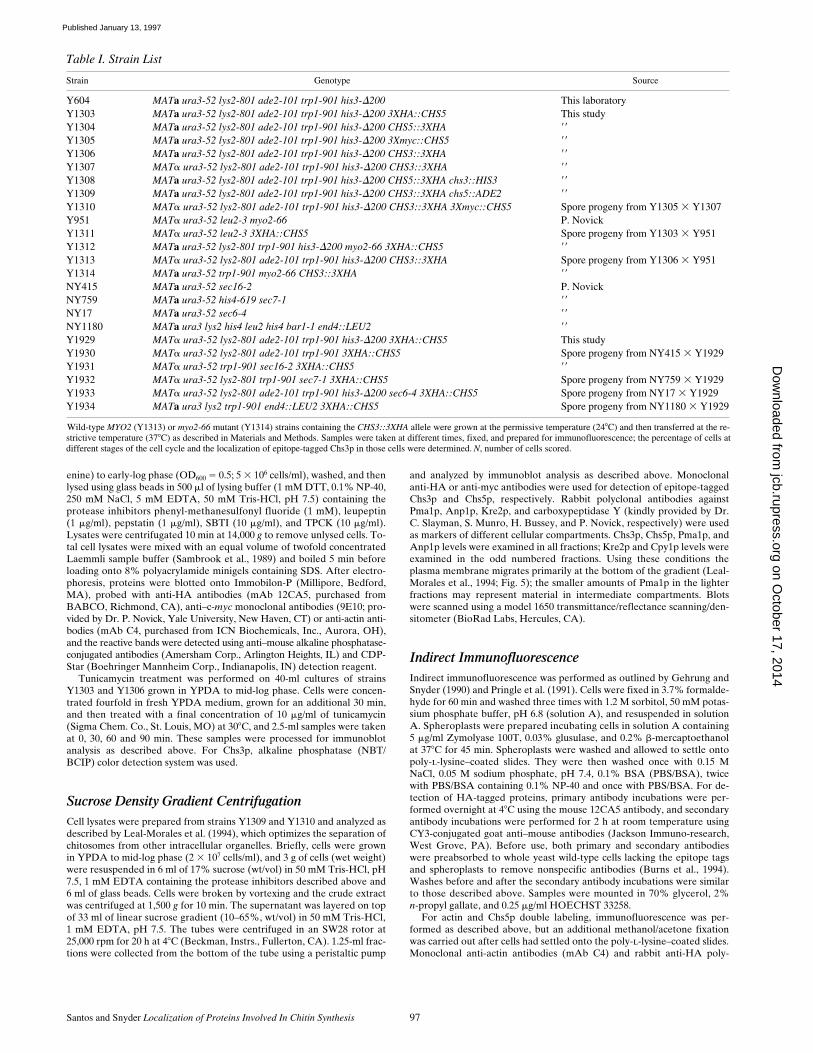
Table I. Strain List
Strain Genotype Source
Y604
MAT
a
ura3-52 lys2-801 ade2-101 trp1-901 his3-
D
200
This laboratoryY1303
MAT
a
ura3-52 lys2-801 ade2-101 trp1-901 his3-
D
200
3XHA::CHS5
This studyY1304
MAT
a
ura3-52 lys2-801 ade2-101 trp1-901 his3-
D
200
CHS5::3XHA
99
Y1305
MAT
a
ura3-52 lys2-801 ade2-101 trp1-901 his3-
D
200
3Xmyc::CHS5
99
Y1306
MAT
a
ura3-52 lys2-801 ade2-101 trp1-901 his3-
D
200
CHS3::3XHA
99
Y1307
MAT
a
ura3-52 lys2-801 ade2-101 trp1-901 his3-
D
200
CHS3::3XHA
99
Y1308
MAT
a
ura3-52 lys2-801 ade2-101 trp1-901 his3-
D
200
CHS5::3XHA chs3::HIS3
99
Y1309
MAT
a
ura3-52 lys2-801 ade2-101 trp1-901 his3-
D
200
CHS3::3XHA chs5::ADE2
99
Y1310
MAT
a
ura3-52 lys2-801 ade2-101 trp1-901 his3-
D
200
CHS3::3XHA 3Xmyc::CHS5
Spore progeny from Y1305
3
Y1307Y951
MAT
a
ura3-52 leu2-3 myo2-66 P. NovickY1311 MATa ura3-52 leu2-3 3XHA::CHS5 Spore progeny from Y1303 3 Y951Y1312 MATa ura3-52 lys2-801 trp1-901 his3-D200 myo2-66 3XHA::CHS5 99
Y1313 MATa ura3-52 lys2-801 ade2-101 trp1-901 his3-D200 CHS3::3XHA Spore progeny from Y1306 3 Y951Y1314 MATa ura3-52 trp1-901 myo2-66 CHS3::3XHA 99
NY415 MATa ura3-52 sec16-2 P. NovickNY759 MATa ura3-52 his4-619 sec7-1 99
NY17 MATa ura3-52 sec6-4 99
NY1180 MATa ura3 lys2 his4 leu2 his4 bar1-1 end4::LEU2 99
Y1929 MATa ura3-52 lys2-801 ade2-101 trp1-901 his3-D200 3XHA::CHS5 This studyY1930 MATa ura3-52 lys2-801 ade2-101 trp1-901 3XHA::CHS5 Spore progeny from NY415 3 Y1929Y1931 MATa ura3-52 trp1-901 sec16-2 3XHA::CHS5 99
Y1932 MATa ura3-52 lys2-801 trp1-901 sec7-1 3XHA::CHS5 Spore progeny from NY759 3 Y1929Y1933 MATa ura3-52 lys2-801 ade2-101 trp1-901 his3-D200 sec6-4 3XHA::CHS5 Spore progeny from NY17 3 Y1929Y1934 MATa ura3 lys2 trp1-901 end4::LEU2 3XHA::CHS5 Spore progeny from NY1180 3 Y1929
Wild-type MYO2 (Y1313) or myo2-66 mutant (Y1314) strains containing the CHS3::3XHA allele were grown at the permissive temperature (248C) and then transferred at the re-strictive temperature (378C) as described in Materials and Methods. Samples were taken at different times, fixed, and prepared for immunofluorescence; the percentage of cells atdifferent stages of the cell cycle and the localization of epitope-tagged Chs3p in those cells were determined. N, number of cells scored.
on October 17, 2014
jcb.rupress.orgD
ownloaded from
Published January 13, 1997
The Journal of Cell Biology, Volume 136, 1997 98
clonal antibodies (BABCO) were used. For detection of primary antibod-ies, CY3-conjugated sheep anti–rabbit antibodies (Sigma) and FITC-con-jugated goat anti–mouse antibodies (Cappel Laboratories, Malvern, PA)were used.
For colocalization experiments of 3Xmyc::Chs5p and Chs3p::3XHA,cells (Y1310) were stained with a mouse anti-myc antibody (9E10) and arabbit anti-HA polyclonal antibody. For double immunofluorescence ex-periments with Golgi-associated proteins, myc-epitope–tagged Chs5p wasdetected as described above and rabbit anti-Anp1p or rabbit anti-Kre2pantibodies were used. For the double immunofluorescence experimentwith Chs5p and Kex2p, strain Y1303 containing HA-epitope–taggedChs5p was transformed with plasmid pBM-KX22 which contains theKEX2 gene under the control of the GAL1 promoter (Redding et al.,1991). Cells were grown in medium containing 2% galactose and immuno-fluorescence was performed as described using monoclonal anti-HA andaffinity-purified rabbit anti-Kex2p antibodies (kindly provided by Dr. R.Fuller, Stanford University, Stanford, CA). For detection of primary anti-bodies, CY3-conjugated goat anti–mouse antibodies and FITC-conju-gated goat anti–rabbit antibodies (Cappel Laboratories) were used.
For immunofluorescence experiments in pheromone-treated cells, cellswere grown in YPDA to early log phase, a-factor (Sigma) was added to afinal concentration of 5 mg/ml, and cells were incubated with shaking at308C for 45 min. Cultures were supplemented with a second addition ofa-factor and incubated with shaking at 308C for another 45 min. Micro-scopic examination of the cultures revealed that after 90 min most of thecells (z90%) were unbudded and had formed shmoos. Cells were fixedwith formaldehyde and processed for indirect immunofluorescence as de-scribed above.
Analysis of temperature-sensitive myo2, sec, and end4D mutant strainswas as follows: myo2-66 mutants were grown in YPDA at room tempera-ture with shaking until mid-log phase. To shift cells to the restrictive tem-perature, an aliquot of the culture was diluted fivefold into prewarmedYPDA at 378C. Samples were taken at 0, 20, 60, and 120 min and fixedwith formaldehyde as described in Lillie and Brown (1994). Control cul-tures of wild-type cells were treated in a similar fashion. In parallel withthese experiments, myo2-66 cells were also diluted and incubated at roomtemperature. For immunolocalization of Chs5p in sec and end4D mutants,strains Y1930, Y1931, Y1932, Y1933, and Y1934 were grown at room tem-perature until mid-log phase and then diluted twofold in prewarmedYPDA at 378C. Samples were taken at 0, 30, 60, 90, and 120 min and fixedwith formaldehyde. Control experiments in which cells were incubated atthe permissive temperature (z248) were also performed. Indirect immuno-fluorescence was performed as described above.
Results
Epitope Tagging of Chs5p
To gain insight into how Chs5p might participate in gener-ating functional CSIII activity, we determined its subcellu-lar distribution. The CHS5 gene was tagged at its genomiclocus with three copies of the hemagglutinin (HA)1 epi-tope coding sequence or three copies of the c-myc epitopecoding sequence using a PCR approach (Schneider et al.,1995; see Materials and Methods). Three strains were pre-pared. The HA epitope coding segment was integrated ei-ther at the NH2-terminal coding region (after the secondcodon) or at the COOH terminus (after the last codon ofthe open reading frame) to generate 3XHA::CHS5 andCHS5::3XHA strains, respectively. The c-myc epitopecoding region was integrated after the second codon togenerate 3Xmyc::CHS5. In each case the resulting epitope-tagged protein is functional. Mutations in the CHS5 genecause reduced chitin levels in the cell wall and resistanceto Calcofluor, a compound that interferes with chitin syn-thesis (Roncero et al., 1988); the 3XHA::CHS5, CHS5::
3XHA, and 3Xmyc::CHS5 strains are each sensitive toCalcofluor similar to wild-type strains indicating that theyhave normal levels of chitin in the cell wall (data notshown).
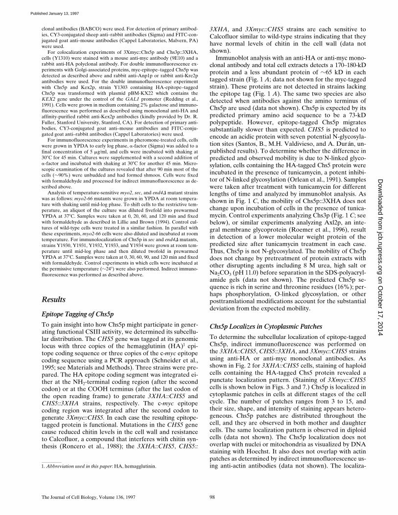
Immunoblot analysis with an anti-HA or anti-myc mono-clonal antibody and total cell extracts detects a 170–180-kDprotein and a less abundant protein of z65 kD in eachtagged strain (Fig. 1 A; data not shown for the myc-taggedstrain). These proteins are not detected in strains lackingthe epitope tag (Fig. 1 A). The same two species are alsodetected when antibodies against the amino terminus ofChs5p are used (data not shown). Chs5p is expected by itspredicted primary amino acid sequence to be a 73-kDpolypeptide. However, epitope-tagged Chs5p migratessubstantially slower than expected. CHS5 is predicted toencode an acidic protein with seven potential N-glycosyla-tion sites (Santos, B., M.H. Valdivieso, and A. Durán, un-published results). To determine whether the difference inpredicted and observed mobility is due to N-linked glyco-sylation, cells containing the HA-tagged Chs5 protein wereincubated in the presence of tunicamycin, a potent inhibi-tor of N-linked glycosylation (Orlean et al., 1991). Sampleswere taken after treatment with tunicamycin for differentlengths of time and analyzed by immunoblot analysis. Asshown in Fig. 1 C, the mobility of Chs5p::3XHA does notchange upon incubation of cells in the presence of tunica-mycin. Control experiments analyzing Chs3p (Fig. 1 C; seebelow), or similar experiments analyzing Axl2p, an inte-gral membrane glycoprotein (Roemer et al., 1996), resultin detection of a lower molecular weight protein of thepredicted size after tunicamycin treatment in each case.Thus, Chs5p is not N-glycosylated. The mobility of Chs5pdoes not change by pretreatment of protein extracts withother disrupting agents including 8 M urea, high salt orNa2CO3 (pH 11.0) before separation in the SDS-polyacryl-amide gels (data not shown). The predicted Chs5p se-quence is rich in serine and threonine residues (16%); per-haps phosphorylation, O-linked glycosylation, or otherposttranslational modifications account for the substantialdeviation from the expected mobility.
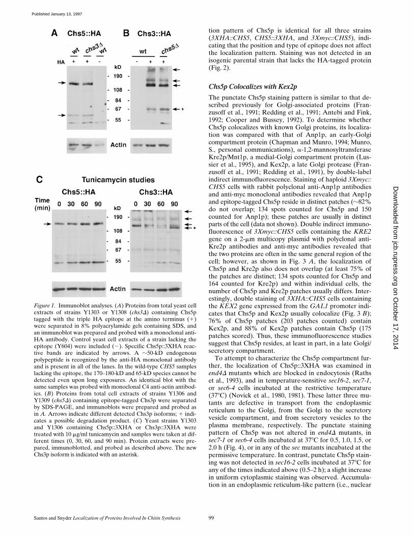
Chs5p Localizes in Cytoplasmic Patches
To determine the subcellular localization of epitope-taggedChs5p, indirect immunofluorescence was performed onthe 3XHA::CHS5, CHS5::3XHA, and 3Xmyc::CHS5 strainsusing anti-HA or anti-myc monoclonal antibodies. Asshown in Fig. 2 for 3XHA::CHS5 cells, staining of haploidcells containing the HA-tagged Chs5 protein revealed apunctate localization pattern. (Staining of 3Xmyc::CHS5cells is shown below in Figs. 3 and 7.) Chs5p is localized incytoplasmic patches in cells at different stages of the cellcycle. The number of patches ranges from 3 to 15, andtheir size, shape, and intensity of staining appears hetero-geneous. Chs5p patches are distributed throughout thecell, and they are observed in both mother and daughtercells. The same localization pattern is observed in diploidcells (data not shown). The Chs5p localization does notoverlap with nuclei or mitochondria as visualized by DNAstaining with Hoechst. It also does not overlap with actinpatches as determined by indirect immunofluorescence us-ing anti-actin antibodies (data not shown). The localiza-1. Abbreviation used in this paper: HA, hemagglutinin.
on October 17, 2014
jcb.rupress.orgD
ownloaded from
Published January 13, 1997
Santos and Snyder Localization of Proteins Involved In Chitin Synthesis 99
tion pattern of Chs5p is identical for all three strains(3XHA::CHS5, CHS5::3XHA, and 3Xmyc::CHS5), indi-cating that the position and type of epitope does not affectthe localization pattern. Staining was not detected in anisogenic parental strain that lacks the HA-tagged protein(Fig. 2).
Chs5p Colocalizes with Kex2p
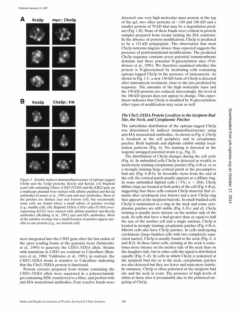
The punctate Chs5p staining pattern is similar to that de-scribed previously for Golgi-associated proteins (Fran-zusoff et al., 1991; Redding et al., 1991; Antebi and Fink,1992; Cooper and Bussey, 1992). To determine whetherChs5p colocalizes with known Golgi proteins, its localiza-tion was compared with that of Anp1p, an early-Golgicompartment protein (Chapman and Munro, 1994; Munro,S., personal communications), a-1,2-mannosyltransferaseKre2p/Mnt1p, a medial-Golgi compartment protein (Lus-sier et al., 1995), and Kex2p, a late Golgi protease (Fran-zusoff et al., 1991; Redding et al., 1991), by double-labelindirect immunofluorescence. Staining of haploid 3Xmyc::CHS5 cells with rabbit polyclonal anti-Anp1p antibodiesand anti-myc monoclonal antibodies revealed that Anp1pand epitope-tagged Chs5p reside in distinct patches (z82%do not overlap; 134 spots counted for Chs5p and 150counted for Anp1p); these patches are usually in distinctparts of the cell (data not shown). Double indirect immuno-fluorescence of 3Xmyc::CHS5 cells containing the KRE2gene on a 2-mm multicopy plasmid with polyclonal anti-Kre2p antibodies and anti-myc antibodies revealed thatthe two proteins are often in the same general region of thecell; however, as shown in Fig. 3 A, the localization ofChs5p and Kre2p also does not overlap (at least 75% ofthe patches are distinct; 134 spots counted for Chs5p and164 counted for Kre2p) and within individual cells, thenumber of Chs5p and Kre2p patches usually differs. Inter-estingly, double staining of 3XHA::CHS5 cells containingthe KEX2 gene expressed from the GAL1 promoter indi-cates that Chs5p and Kex2p usually colocalize (Fig. 3 B);76% of Chs5p patches (203 patches counted) containKex2p, and 88% of Kex2p patches contain Chs5p (175patches scored). Thus, these immunofluorescence studiessuggest that Chs5p resides, at least in part, in a late Golgi/secretory compartment.
To attempt to characterize the Chs5p compartment fur-ther, the localization of Chs5p::3XHA was examined inend4D mutants which are blocked in endocytosis (Rathset al., 1993), and in temperature-sensitive sec16-2, sec7-1,or sec6-4 cells incubated at the restrictive temperature(378C) (Novick et al., 1980, 1981). These latter three mu-tants are defective in transport from the endoplasmicreticulum to the Golgi, from the Golgi to the secretoryvesicle compartment, and from secretory vesicles to theplasma membrane, respectively. The punctate stainingpattern of Chs5p was not altered in end4D mutants, insec7-1 or sec6-4 cells incubated at 378C for 0.5, 1.0, 1.5, or2.0 h (Fig. 4), or in any of the sec mutants incubated at thepermissive temperature. In contrast, punctate Chs5p stain-ing was not detected in sec16-2 cells incubated at 378C forany of the times indicated above (0.5–2 h); a slight increasein uniform cytoplasmic staining was observed. Accumula-tion in an endoplasmic reticulum-like pattern (i.e., nuclear
Figure 1. Immunoblot analyses. (A) Proteins from total yeast cellextracts of strains Y1303 or Y1308 (chs3D) containing Chs5ptagged with the triple HA epitope at the amino terminus (1)were separated in 8% polyacrylamide gels containing SDS, andan immunoblot was prepared and probed with a monoclonal anti-HA antibody. Control yeast cell extracts of a strain lacking theepitope (Y604) were included (2). Specific Chs5p::3XHA reac-tive bands are indicated by arrows. A z50-kD endogenouspolypeptide is recognized by the anti-HA monoclonal antibodyand is present in all of the lanes. In the wild-type CHS5 sampleslacking the epitope, the 170–180-kD and 65-kD species cannot bedetected even upon long exposures. An identical blot with thesame samples was probed with monoclonal C4 anti-actin antibod-ies. (B) Proteins from total cell extracts of strains Y1306 andY1309 (chs5D) containing epitope-tagged Chs3p were separatedby SDS-PAGE, and immunoblots were prepared and probed asin A. Arrows indicate different detected Chs3p isoforms; 1 indi-cates a possible degradation product. (C) Yeast strains Y1303and Y1306 containing Chs5p::3XHA or Chs3p::3XHA weretreated with 10 mg/ml tunicamycin and samples were taken at dif-ferent times (0, 30, 60, and 90 min). Protein extracts were pre-pared, immunoblotted, and probed as described above. The newChs3p isoform is indicated with an asterisk.
on October 17, 2014
jcb.rupress.orgD
ownloaded from
Published January 13, 1997
The Journal of Cell Biology, Volume 136, 1997 100
periphery) was not detected. Staining with anti-Anp1p an-tibodies yielded similar results (not shown). These dataare consistent with the hypothesis that Chs5p lies in a lateGolgi compartment (see Discussion).
Chs5p Is in a Vesicle Compartment
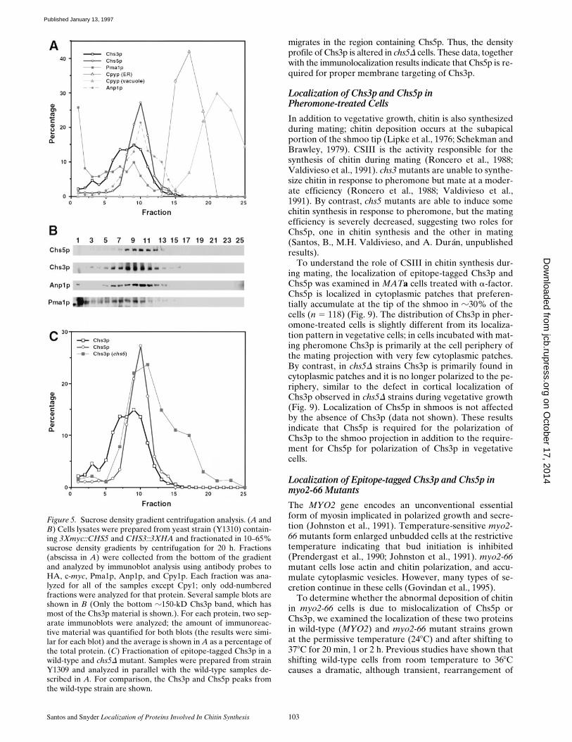
The punctate staining pattern of Chs5p and its colocaliza-tion with Kex2p patches, suggest that Chs5p may be partof a vesicular compartment. To independently test thispossibility, subcellular fractionations were performed us-ing conditions that optimize separation of chitosomes (Leal-Morales et al., 1994). Lysates of 3Xmyc::CHS5 CHS3::3XHA yeast cells were prepared and subjected to sucrosedensity gradient centrifugation. Fractions were collected andanalyzed by immunoblot analysis using antibodies thatrecognize HA, c-myc, Anp1p, Kre2p, Pma1p (a plasmamembrane marker) (Chang and Slayman, 1991; Changand Fink, 1995), and carboxypeptidase Y (Cpy1p; a pro-tein whose three different isoforms are located in the vac-uole, endoplasmic reticulum, or Golgi apparatus; Stevenset al., 1982). As shown in Fig. 5, A and B, all of the detect-
able Chs5p partitions in a vesicular fraction that is similarin density to the Golgi fraction as monitored by Anp1p,Kre2p, and the carboxypeptidase Golgi isoform (data notshown for the latter two proteins, but the results are simi-lar to the Anp1p data). This compartment is distinct fromthe plasma membrane (which primarily migrates at thebottom of the gradient [Fractions 1–3] under these condi-tions; see Materials and Methods), endoplasmic reticulum,and vacuole. Thus, consistent with the immunofluores-cence results, Chs5p is present in a membrane vesicle com-partment.
Chs3p, the Catalytic Component of CSIII,Is a Glycoprotein
Little is known about the regulation or localization ofChs3p, the catalytic subunit of CSIII (Valdivieso et al.,1991). To analyze the subcellular distribution of Chs3pand to test the hypothesis that Chs5p may affect the local-ization of Chs3p, the latter protein was tagged and local-ized. Three copies of the HA epitope coding sequence
Figure 2. Localization of epitope-tagged Chs5p. Haploid yeast cells containing Chs5p tagged at the COOH terminus with triple HA(Y1303) or Chs5p without an epitope tag (Y604) were stained by indirect immunofluorescence with an anti-HA monoclonal antibody(left panels). Hoechst 33258 DNA staining of the same cells is shown in the right panels.
on October 17, 2014
jcb.rupress.orgD
ownloaded from
Published January 13, 1997
Santos and Snyder Localization of Proteins Involved In Chitin Synthesis 101
were integrated into the CHS3 gene after the last codon ofthe open reading frame at the genomic locus (Schneideret al., 1995) to generate the CHS3::3XHA allele. Strainswith mutations in CHS3 are resistant to Calcofluor (Ron-cero et al., 1988; Valdivieso et al., 1991); in contrast, theCHS3::3XHA strain is sensitive to Calcofluor indicatingthat the Chs3::3XHA protein is functional.
Protein extracts prepared from strains containing theCHS3::3XHA allele were separated in a polyacrylamidegel containing SDS, transferred to a filter, and probed withanti-HA monoclonal antibodies. Four reactive bands were
detected: one very high molecular mass protein at the topof the gel, two other proteins of z150 and 180 kD and asmaller protein of 70 kD that may be a degradation prod-uct (Fig. 1 B). None of these bands were evident in proteinsamples prepared from strains lacking the HA construct.In the absence of protein modification, Chs3p is predictedto be a 131-kD polypeptide. The observation that mostChs3p isoforms migrate slower than expected suggests thepresence of posttranslational modifications. The predictedChs3p sequence contains seven potential transmembranedomains and three potential N-glycosylation sites (Val-divieso et al., 1991). We therefore examined whether thisprotein is N-glycosylated by incubating cells containingepitope-tagged Chs3p in the presence of tunicamycin. Asshown in Fig. 1 C, a new 130-kD form of Chs3p is detectedafter tunicamycin treatment, close to the size predicted bysequence. The amounts of the high molecular mass andthe 150-kD proteins are reduced; interestingly, the level ofthe 180-kD species does not appear to change. This exper-iment indicates that Chs3p is modified by N-glycosylation;other types of modification may occur as well.
The Chs3::3XHA Protein Localizes to the Incipient Bud Site, the Neck, and Cytoplasmic Patches
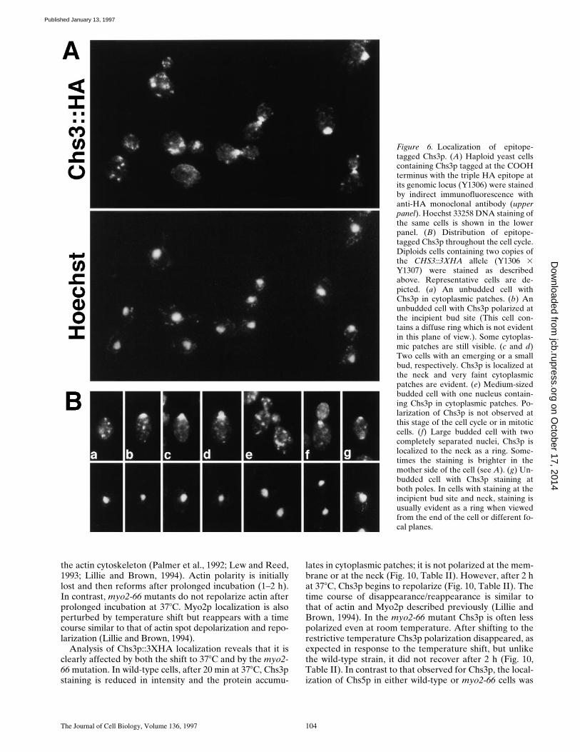
The subcellular distribution of the epitope-tagged Chs3pwas determined by indirect immunofluorescence usinganti-HA monoclonal antibodies. As shown in Fig. 6, Chs3pis localized at the cell periphery and in cytoplasmicpatches. Both haploids and diploids exhibit similar local-ization patterns (Fig. 6). No staining is detected in theisogenic untagged parental strain (e.g., Fig. 2).
The distribution of Chs3p changes during the cell cycle(Fig. 6). In unbudded cells Chs3p is detected as weakly ormoderately staining cytoplasmic patches (Fig. 6 B-a), or asa strongly staining large cortical patch at the presumptivebud site (Fig. 6 B-b). In favorable views from the end ofthe cell, the cortical patch usually appears as a diffuse ring.In some unbudded diploid cells (z3%; n 5 204), Chs3pdiffuse rings are located at both poles of the cell (Fig. 6 B-g),suggesting that these cells contain Chs3p material that re-mains after cytokinesis (see below) and a new Chs3p ringthat appears at the incipient bud site. In small-budded cellsChs3p is maintained as a ring at the neck and some cyto-plasmic patches are still visible (Fig. 6 D-c and d). Chs3pstaining is usually more intense on the mother side of theneck. In cells that have a bud greater than or equal to halfthe size of the mother cell and a single nucleus, Chs3p islocated in strongly staining cytoplasmic patches (Fig. 6 B-e).Mitotic cells also have Chs3p patches. In cells undergoingcytokinesis (large-budded cells with two completely sepa-rated nuclei), Chs3p is usually found at the neck (Fig. 6, Aand B-f). In these latter cells, staining at the neck is some-times more intense on the mother side of the neck than onthe daughter side, but in other cells the signal is distributedequally (Fig. 6 A). In cells in which Chs3p is polarized atthe incipient bud site or at the neck, cytoplasmic patchesare also detected but they are fewer and stain more faintly.In summary, Chs3p is often polarized at the incipient budsite and the neck in yeast. The presence of high levels ofchitin at these sites is presumably due to the polarized tar-geting of Chs3p.
Figure 3. Double indirect immunofluorescence of epitope-taggedChs5p and the Golgi proteins, Kre2p and Kex2p. (A) Haploidyeast cells containing 3Xmyc::CHS5 (Y1305) and the KRE2 gene ona multicopy plasmid were stained with affinity-purified anti-Kre2pantibodies (Lussier et al., 1995) and anti-myc antibodies. Most ofthe patches are distinct (top and bottom cell), but occasionallysome cells are found where a small subset of patches overlap(e.g., middle cell). (B). Haploid 3XHA::CHS5 cells (Y1303) over-expressing KEX2 were stained with affinity-purified anti-Kex2pantibodies (Redding et al., 1991) and anti-HA antibodies. Mostof the patches overlap, but a small fraction of patches appear spe-cific to one protein (e.g., see bottom cell).
on October 17, 2014
jcb.rupress.orgD
ownloaded from
Published January 13, 1997

The Journal of Cell Biology, Volume 136, 1997 102
Colocalization of Epitope-tagged Chs3p and Chs5p in Cytoplasmic Patches
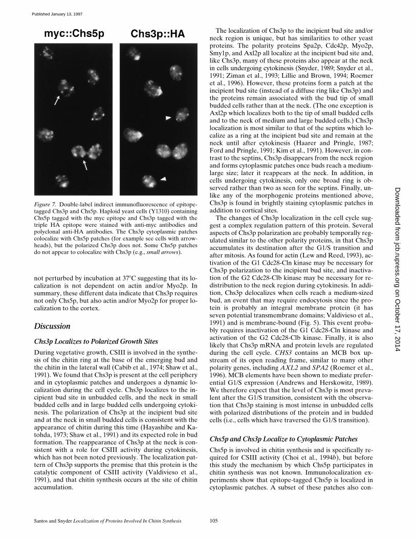
To test whether Chs3p and Chs5p are present in the samecytoplasmic compartment, indirect immunofluorescencewas performed on a strain expressing Chs3p and Chs5pepitope-tagged with HA and myc, respectively. A straincontaining 3Xmyc::CHS5 and CHS3::3XHA was con-structed and stained using anti-myc monoclonal and rabbitpolyclonal anti-HA antibodies. As shown in Fig. 7, thestaining of Chs3p at polarized sites is unique for Chs3p,however, the staining pattern of the Chs5p and Chs3ppatches are very similar. All patches containing Chs3p alsocontain Chs5p and most, but not all (z75%), of Chs5ppatches localize to Chs3p patches (72 cells and 451 Chs5pspots counted). The patches that are unique to Chs5p gen-erally stain more weakly. We do not know whether thepatches in which only Chs5p is found also contain low lev-els of Chs3p that are not detected in these studies or ifthey are truly unique for Chs5p. The colocalization ofChs3p and Chs5p is not due to crossreaction of secondaryantibodies for two reasons: (1) staining of single epitope-tagged strains yields a signal only with the appropriatesecondary antibody and (2) there are regions that areuniquely stained for Chs3p (polarized growth sites) andChs5p (some of the cytoplasmic patches). In summary,these data indicate that Chs3p and Chs5p colocalize inmany cytoplasmic patches. This result raises the possibilitythat Chs5p may be involved in processing or transportingChs3p to the membrane.
Localization of Chs3p Requires Chs5p
To test whether Chs5p participates in the regulation of the
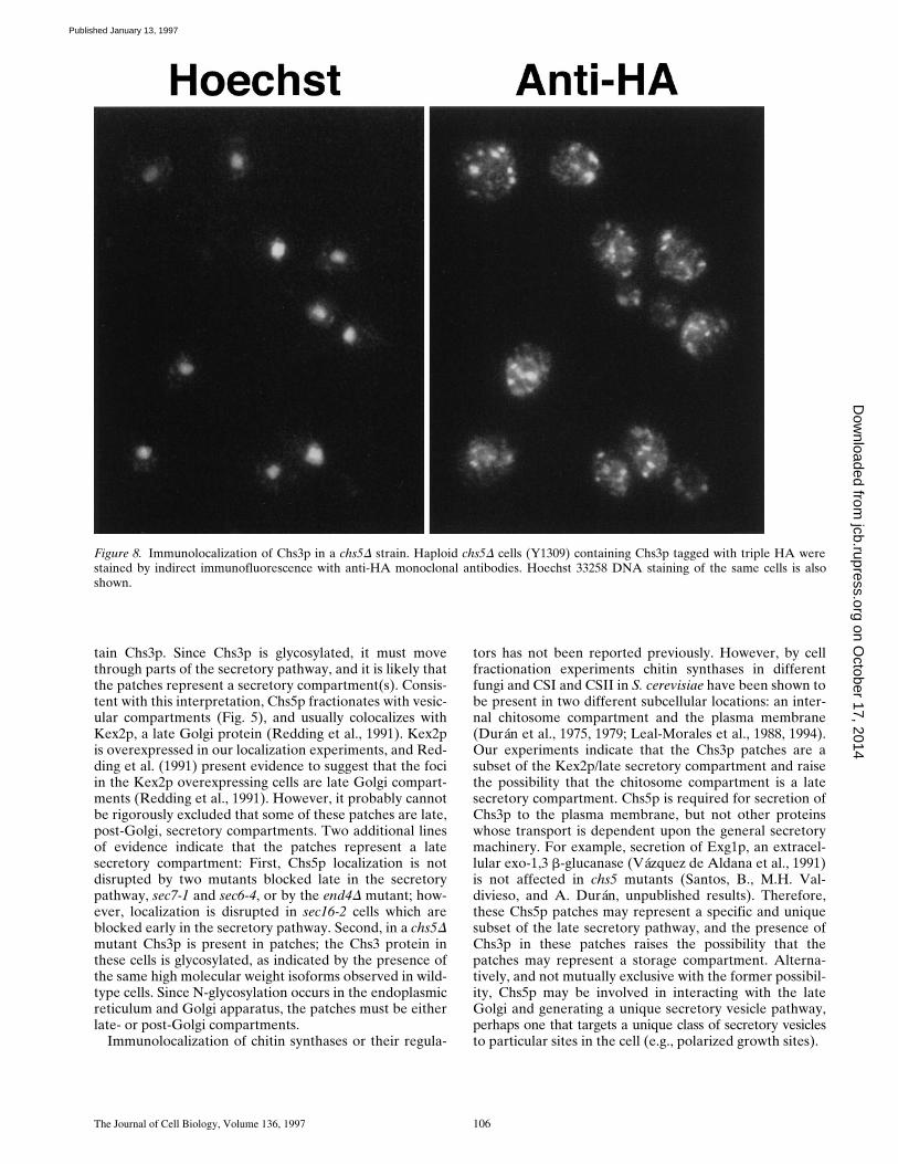
subcellular distribution of Chs3p, localization of Chs3p ina chs5D background was examined. The entire coding re-gion of the CHS5 gene except for the last segment encod-ing the 55 carboxy-terminal amino acids was replaced bythe ADE2 gene, as described (Santos, B., M.H. Valdivieso,and A. Durán, unpublished results). chs5D strains are via-ble, but they have less chitin (z25% of wild-type levels)and lack detectable CSIII activity in vitro (Bulawa, 1993;Choi et al., 1994b). Importantly, in the absence of Chs5p,Chs3p::3XHA localizes to cytoplasmic patches, but it is nolonger found at the incipient bud site or the neck (Fig. 8).Immunoblot analysis shows that both the number andamount of the three high molecular weight Chs3p isoformsis similar in the presence or absence of Chs5p, indicatingthat disruption of CHS5 does not affect either protein lev-els or modification of Chs3p (Fig. 1 B). Localization andprotein levels of Chs5p in a chs3D background were alsoexamined. In the absence of Chs3p, the Chs5p punctate lo-calization pattern (data not shown) and protein levels (Fig.1 A) are not affected. In summary, Chs5p is required forthe polarization of Chs3p to the cortex.
The localization of Chs3p in chs5D cells was also exam-ined using sucrose density gradient centrifugation experi-ments. As shown in Fig. 5 C, in wild-type cells, a large pro-portion of Chs3p::3XHA in our cell lysate preparationsmigrates in the vesicular fractions in a pattern similar tothat of Chs5p; some Chs3p::3XHA appears in a slightlyheavier fraction that could represent a different vesicularcompartment . A small amount of protein is also detected inthe high density region close to the plasma membrane frac-tion at the bottom of the gradient. In chs5D cells, no Chs3p isdetected in the high density fraction, and much of Chs3p isshifted to the lighter density fractions; most Chs3p::3XHA
Figure 4. Localization of Chs5p in sec mutants. Wild-type (WT) and sec mutant yeast strains containing the 3XHA::CHS5 constructwere incubated either at 248C (the permissive temperature for the mutant strains; top panels) or for 2 h at 378C (the restrictive tempera-ture; bottom panels). At the restrictive temperature, Chs5p localized to patches in sec6-4 and sec7-1 cells, which are blocked in Golgi tosecretory vesicle and in vesicle to plasma membrane transport, respectively; this pattern is indistinguishable from wild-type cells. Insec16-2 cells, which are blocked in ER to Golgi transport, patches above background were not evident, but a slight increase in stainingthroughout the cytoplasm is observed. Similar results were observed for cells incubated at 0.5, 1 h, and 1.5 h at 378C.
on October 17, 2014
jcb.rupress.orgD
ownloaded from
Published January 13, 1997
Santos and Snyder Localization of Proteins Involved In Chitin Synthesis 103
migrates in the region containing Chs5p. Thus, the densityprofile of Chs3p is altered in chs5D cells. These data, togetherwith the immunolocalization results indicate that Chs5p is re-quired for proper membrane targeting of Chs3p.
Localization of Chs3p and Chs5p inPheromone-treated Cells
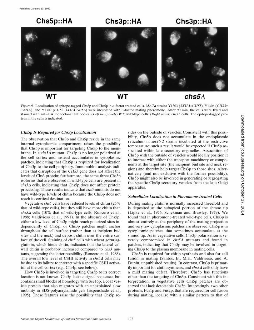
In addition to vegetative growth, chitin is also synthesizedduring mating; chitin deposition occurs at the subapicalportion of the shmoo tip (Lipke et al., 1976; Schekman andBrawley, 1979). CSIII is the activity responsible for thesynthesis of chitin during mating (Roncero et al., 1988;Valdivieso et al., 1991). chs3 mutants are unable to synthe-size chitin in response to pheromone but mate at a moder-ate efficiency (Roncero et al., 1988; Valdivieso et al.,1991). By contrast, chs5 mutants are able to induce somechitin synthesis in response to pheromone, but the matingefficiency is severely decreased, suggesting two roles forChs5p, one in chitin synthesis and the other in mating(Santos, B., M.H. Valdivieso, and A. Durán, unpublishedresults).
To understand the role of CSIII in chitin synthesis dur-ing mating, the localization of epitope-tagged Chs3p andChs5p was examined in MATa cells treated with a-factor.Chs5p is localized in cytoplasmic patches that preferen-tially accumulate at the tip of the shmoo in z30% of thecells (n 5 118) (Fig. 9). The distribution of Chs3p in pher-omone-treated cells is slightly different from its localiza-tion pattern in vegetative cells; in cells incubated with mat-ing pheromone Chs3p is primarily at the cell periphery ofthe mating projection with very few cytoplasmic patches.By contrast, in chs5D strains Chs3p is primarily found incytoplasmic patches and it is no longer polarized to the pe-riphery, similar to the defect in cortical localization ofChs3p observed in chs5D strains during vegetative growth(Fig. 9). Localization of Chs5p in shmoos is not affectedby the absence of Chs3p (data not shown). These resultsindicate that Chs5p is required for the polarization ofChs3p to the shmoo projection in addition to the require-ment for Chs5p for polarization of Chs3p in vegetativecells.
Localization of Epitope-tagged Chs3p and Chs5p in myo2-66 Mutants
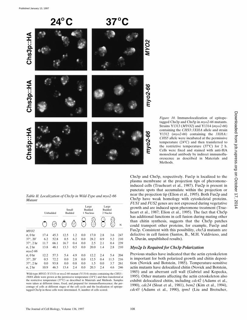
The MYO2 gene encodes an unconventional essentialform of myosin implicated in polarized growth and secre-tion (Johnston et al., 1991). Temperature-sensitive myo2-66 mutants form enlarged unbudded cells at the restrictivetemperature indicating that bud initiation is inhibited(Prendergast et al., 1990; Johnston et al., 1991). myo2-66mutant cells lose actin and chitin polarization, and accu-mulate cytoplasmic vesicles. However, many types of se-cretion continue in these cells (Govindan et al., 1995).
To determine whether the abnormal deposition of chitinin myo2-66 cells is due to mislocalization of Chs5p orChs3p, we examined the localization of these two proteinsin wild-type (MYO2) and myo2-66 mutant strains grownat the permissive temperature (248C) and after shifting to378C for 20 min, 1 or 2 h. Previous studies have shown thatshifting wild-type cells from room temperature to 368Ccauses a dramatic, although transient, rearrangement of
Figure 5. Sucrose density gradient centrifugation analysis. (A andB) Cells lysates were prepared from yeast strain (Y1310) contain-ing 3Xmyc::CHS5 and CHS3::3XHA and fractionated in 10–65%sucrose density gradients by centrifugation for 20 h. Fractions(abscissa in A) were collected from the bottom of the gradientand analyzed by immunoblot analysis using antibody probes toHA, c-myc, Pma1p, Anp1p, and Cpy1p. Each fraction was ana-lyzed for all of the samples except Cpy1; only odd-numberedfractions were analyzed for that protein. Several sample blots areshown in B (Only the bottom z150-kD Chs3p band, which hasmost of the Chs3p material is shown.). For each protein, two sep-arate immunoblots were analyzed; the amount of immunoreac-tive material was quantified for both blots (the results were simi-lar for each blot) and the average is shown in A as a percentage ofthe total protein. (C) Fractionation of epitope-tagged Chs3p in awild-type and chs5D mutant. Samples were prepared from strainY1309 and analyzed in parallel with the wild-type samples de-scribed in A. For comparison, the Chs3p and Chs5p peaks fromthe wild-type strain are shown.
on October 17, 2014
jcb.rupress.orgD
ownloaded from
Published January 13, 1997
The Journal of Cell Biology, Volume 136, 1997 104
the actin cytoskeleton (Palmer et al., 1992; Lew and Reed,1993; Lillie and Brown, 1994). Actin polarity is initiallylost and then reforms after prolonged incubation (1–2 h).In contrast, myo2-66 mutants do not repolarize actin afterprolonged incubation at 378C. Myo2p localization is alsoperturbed by temperature shift but reappears with a timecourse similar to that of actin spot depolarization and repo-larization (Lillie and Brown, 1994).
Analysis of Chs3p::3XHA localization reveals that it isclearly affected by both the shift to 378C and by the myo2-66 mutation. In wild-type cells, after 20 min at 378C, Chs3pstaining is reduced in intensity and the protein accumu-
lates in cytoplasmic patches; it is not polarized at the mem-brane or at the neck (Fig. 10, Table II). However, after 2 hat 378C, Chs3p begins to repolarize (Fig. 10, Table II). Thetime course of disappearance/reappearance is similar tothat of actin and Myo2p described previously (Lillie andBrown, 1994). In the myo2-66 mutant Chs3p is often lesspolarized even at room temperature. After shifting to therestrictive temperature Chs3p polarization disappeared, asexpected in response to the temperature shift, but unlikethe wild-type strain, it did not recover after 2 h (Fig. 10,Table II). In contrast to that observed for Chs3p, the local-ization of Chs5p in either wild-type or myo2-66 cells was
Figure 6. Localization of epitope-tagged Chs3p. (A) Haploid yeast cellscontaining Chs3p tagged at the COOHterminus with the triple HA epitope atits genomic locus (Y1306) were stainedby indirect immunofluorescence withanti-HA monoclonal antibody (upperpanel). Hoechst 33258 DNA staining ofthe same cells is shown in the lowerpanel. (B) Distribution of epitope-tagged Chs3p throughout the cell cycle.Diploids cells containing two copies ofthe CHS3::3XHA allele (Y1306 3Y1307) were stained as describedabove. Representative cells are de-picted. (a) An unbudded cell withChs3p in cytoplasmic patches. (b) Anunbudded cell with Chs3p polarized atthe incipient bud site (This cell con-tains a diffuse ring which is not evidentin this plane of view.). Some cytoplas-mic patches are still visible. (c and d)Two cells with an emerging or a smallbud, respectively. Chs3p is localized atthe neck and very faint cytoplasmicpatches are evident. (e) Medium-sizedbudded cell with one nucleus contain-ing Chs3p in cytoplasmic patches. Po-larization of Chs3p is not observed atthis stage of the cell cycle or in mitoticcells. (f) Large budded cell with twocompletely separated nuclei, Chs3p islocalized to the neck as a ring. Some-times the staining is brighter in themother side of the cell (see A). (g) Un-budded cell with Chs3p staining atboth poles. In cells with staining at theincipient bud site and neck, staining isusually evident as a ring when viewedfrom the end of the cell or different fo-cal planes.
on October 17, 2014
jcb.rupress.orgD
ownloaded from
Published January 13, 1997
Santos and Snyder Localization of Proteins Involved In Chitin Synthesis 105
not perturbed by incubation at 378C suggesting that its lo-calization is not dependent on actin and/or Myo2p. Insummary, these different data indicate that Chs3p requiresnot only Chs5p, but also actin and/or Myo2p for proper lo-calization to the cortex.
Discussion
Chs3p Localizes to Polarized Growth Sites
During vegetative growth, CSIII is involved in the synthe-sis of the chitin ring at the base of the emerging bud andthe chitin in the lateral wall (Cabib et al., 1974; Shaw et al.,1991). We found that Chs3p is present at the cell peripheryand in cytoplasmic patches and undergoes a dynamic lo-calization during the cell cycle. Chs3p localizes to the in-cipient bud site in unbudded cells, and the neck in smallbudded cells and in large budded cells undergoing cytoki-nesis. The polarization of Chs3p at the incipient bud siteand at the neck in small budded cells is consistent with theappearance of chitin during this time (Hayashibe and Ka-tohda, 1973; Shaw et al., 1991) and its expected role in budformation. The reappearance of Chs3p at the neck is con-sistent with a role for CSIII activity during cytokinesis,which has not been noted previously. The localization pat-tern of Chs3p supports the premise that this protein is thecatalytic component of CSIII activity (Valdivieso et al.,1991), and that chitin synthesis occurs at the site of chitinaccumulation.
The localization of Chs3p to the incipient bud site and/orneck region is unique, but has similarities to other yeastproteins. The polarity proteins Spa2p, Cdc42p, Myo2p,Smy1p, and Axl2p all localize at the incipient bud site and,like Chs3p, many of these proteins also appear at the neckin cells undergoing cytokinesis (Snyder, 1989; Snyder et al.,1991; Ziman et al., 1993; Lillie and Brown, 1994; Roemeret al., 1996). However, these proteins form a patch at theincipient bud site (instead of a diffuse ring like Chs3p) andthe proteins remain associated with the bud tip of smallbudded cells rather than at the neck. (The one exception isAxl2p which localizes both to the tip of small budded cellsand to the neck of medium and large budded cells.) Chs3plocalization is most similar to that of the septins which lo-calize as a ring at the incipient bud site and remain at theneck until after cytokinesis (Haarer and Pringle, 1987;Ford and Pringle, 1991; Kim et al., 1991). However, in con-trast to the septins, Chs3p disappears from the neck regionand forms cytoplasmic patches once buds reach a medium-large size; later it reappears at the neck. In addition, incells undergoing cytokinesis, only one broad ring is ob-served rather than two as seen for the septins. Finally, un-like any of the morphogenic proteins mentioned above,Chs3p is found in brightly staining cytoplasmic patches inaddition to cortical sites.
The changes of Chs3p localization in the cell cycle sug-gest a complex regulation pattern of this protein. Severalaspects of Chs3p polarization are probably temporally reg-ulated similar to the other polarity proteins, in that Chs3paccumulates its destination after the G1/S transition andafter mitosis. As found for actin (Lew and Reed, 1993), ac-tivation of the G1 Cdc28-Cln kinase may be necessary forChs3p polarization to the incipient bud site, and inactiva-tion of the G2 Cdc28-Clb kinase may be necessary for re-distribution to the neck region during cytokinesis. In addi-tion, Chs3p delocalizes when cells reach a medium-sizedbud, an event that may require endocytosis since the pro-tein is probably an integral membrane protein (it hasseven potential transmembrane domains; Valdivieso et al.,1991) and is membrane-bound (Fig. 5). This event proba-bly requires inactivation of the G1 Cdc28-Cln kinase andactivation of the G2 Cdc28-Clb kinase. Finally, it is alsolikely that Chs3p mRNA and protein levels are regulatedduring the cell cycle. CHS3 contains an MCB box up-stream of its open reading frame, similar to many otherpolarity genes, including AXL2 and SPA2 (Roemer et al.,1996). MCB elements have been shown to mediate prefer-ential G1/S expression (Andrews and Herskowitz, 1989).We therefore expect that the level of Chs3p is most preva-lent after the G1/S transition, consistent with the observa-tion that Chs3p staining is most intense in unbudded cellswith polarized distributions of the protein and in buddedcells (i.e., cells which have traversed the G1/S transition).
Chs5p and Chs3p Localize to Cytoplasmic Patches
Chs5p is involved in chitin synthesis and is specifically re-quired for CSIII activity (Choi et al., 1994b), but beforethis study the mechanism by which Chs5p participates inchitin synthesis was not known. Immunolocalization ex-periments show that epitope-tagged Chs5p is localized incytoplasmic patches. A subset of these patches also con-
Figure 7. Double-label indirect immunofluorescence of epitope-tagged Chs3p and Chs5p. Haploid yeast cells (Y1310) containingChs5p tagged with the myc epitope and Chs3p tagged with thetriple HA epitope were stained with anti-myc antibodies andpolyclonal anti-HA antibodies. The Chs3p cytoplasmic patchescolocalize with Chs5p patches (for example see cells with arrow-heads), but the polarized Chs3p does not. Some Chs5p patchesdo not appear to colocalize with Chs3p (e.g., small arrows).
on October 17, 2014
jcb.rupress.orgD
ownloaded from
Published January 13, 1997
The Journal of Cell Biology, Volume 136, 1997 106
tain Chs3p. Since Chs3p is glycosylated, it must movethrough parts of the secretory pathway, and it is likely thatthe patches represent a secretory compartment(s). Consis-tent with this interpretation, Chs5p fractionates with vesic-ular compartments (Fig. 5), and usually colocalizes withKex2p, a late Golgi protein (Redding et al., 1991). Kex2pis overexpressed in our localization experiments, and Red-ding et al. (1991) present evidence to suggest that the fociin the Kex2p overexpressing cells are late Golgi compart-ments (Redding et al., 1991). However, it probably cannotbe rigorously excluded that some of these patches are late,post-Golgi, secretory compartments. Two additional linesof evidence indicate that the patches represent a latesecretory compartment: First, Chs5p localization is notdisrupted by two mutants blocked late in the secretorypathway, sec7-1 and sec6-4, or by the end4D mutant; how-ever, localization is disrupted in sec16-2 cells which areblocked early in the secretory pathway. Second, in a chs5Dmutant Chs3p is present in patches; the Chs3 protein inthese cells is glycosylated, as indicated by the presence ofthe same high molecular weight isoforms observed in wild-type cells. Since N-glycosylation occurs in the endoplasmicreticulum and Golgi apparatus, the patches must be eitherlate- or post-Golgi compartments.
Immunolocalization of chitin synthases or their regula-
tors has not been reported previously. However, by cellfractionation experiments chitin synthases in differentfungi and CSI and CSII in S. cerevisiae have been shown tobe present in two different subcellular locations: an inter-nal chitosome compartment and the plasma membrane(Durán et al., 1975, 1979; Leal-Morales et al., 1988, 1994).Our experiments indicate that the Chs3p patches are asubset of the Kex2p/late secretory compartment and raisethe possibility that the chitosome compartment is a latesecretory compartment. Chs5p is required for secretion ofChs3p to the plasma membrane, but not other proteinswhose transport is dependent upon the general secretorymachinery. For example, secretion of Exg1p, an extracel-lular exo-1,3 b-glucanase (Vázquez de Aldana et al., 1991)is not affected in chs5 mutants (Santos, B., M.H. Val-divieso, and A. Durán, unpublished results). Therefore,these Chs5p patches may represent a specific and uniquesubset of the late secretory pathway, and the presence ofChs3p in these patches raises the possibility that thepatches may represent a storage compartment. Alterna-tively, and not mutually exclusive with the former possibil-ity, Chs5p may be involved in interacting with the lateGolgi and generating a unique secretory vesicle pathway,perhaps one that targets a unique class of secretory vesiclesto particular sites in the cell (e.g., polarized growth sites).
Figure 8. Immunolocalization of Chs3p in a chs5D strain. Haploid chs5D cells (Y1309) containing Chs3p tagged with triple HA werestained by indirect immunofluorescence with anti-HA monoclonal antibodies. Hoechst 33258 DNA staining of the same cells is alsoshown.
on October 17, 2014
jcb.rupress.orgD
ownloaded from
Published January 13, 1997
Santos and Snyder Localization of Proteins Involved In Chitin Synthesis 107
Chs5p Is Required for Chs3p Localization
The observation that Chs3p and Chs5p reside in the sameinternal cytoplasmic compartment raises the possibilitythat Chs5p is important for targeting Chs3p to the mem-brane. In a chs5D mutant, Chs3p is no longer polarized atthe cell cortex and instead accumulates in cytoplasmicpatches, indicating that Chs5p is required for localizationof Chs3p to the cell periphery. Immunoblot analysis indi-cates that disruption of the CHS5 gene does not affect thelevels of Chs3 protein; furthermore, the same three Chs3pisoforms that are observed in wild-type cells are present inchs5D cells, indicating that Chs5p does not affect proteinprocessing. These results indicate that chs5 mutants do nothave wild-type levels of chitin because the Chs3p does notreach its cortical destination.
Vegetative chs5 cells have reduced levels of chitin (25%that of wild-type cells), but they still have more chitin thanchs3D cells (10% that of wild-type cells; Roncero et al.,1988; Valdivieso et al., 1991). In the absence of Chs5p,either a low level of Chs3p might reach polarized sites in-dependently of Chs5p, or Chs3p patches might anchorthroughout the cell surface (rather than at incipient budsites and the neck) and deposit chitin over the entire sur-face of the cell. Staining of chs5 cells with wheat germ ag-glutinin, which binds chitin, indicates that the lateral cellwall chitin is probably increased compared to chs3 mu-tants, suggesting the latter possibility (Roncero et al., 1988).The overall low level of CSIII activity in chs5D cells maybe due to its failure to interact with a specific CSIII activa-tor at the cell cortex (e.g., Chs4p; see below).
How Chs5p is involved in targeting Chs3p to its correctlocation is not known. Chs5p lacks a signal sequence, butcontains small blocks of homology with Sec16p, a coat ves-icle protein that also migrates with an unexplained slowmobility in SDS-polyacrylamide gels (Espenshade et al.,1995). These features raise the possibility that Chs5p re-
sides on the outside of vesicles. Consistent with this possi-bility, Chs5p does not accumulate in the endoplasmicreticulum in sec16-2 strains incubated at the restrictivetemperature; such a result would be expected if Chs5p as-sociated within late secretory organelles. Association ofChs5p with the outside of vesicles would ideally position itto interact with either the transport machinery or compo-nents at the target site (the incipient bud site and neck re-gion) and thereby help target Chs3p to those sites. Alter-natively (and not exclusive with the former possibility),Chs5p might also be involved in generating or segregatingthe specific Chs3p secretory vesicles from the late Golgiapparatus.
Subcellular Localization in Pheromone-treated Cells
During mating chitin is normally increased threefold andis deposited at the subapical portion of the shmoo tip(Lipke et al., 1976; Schekman and Brawley, 1979). Wefound that in pheromone-treated wild-type cells, Chs3p isalmost entirely at the periphery of the mating projectionand very few cytoplasmic patches are observed. Chs5p is incytoplasmic patches that sometimes accumulate at theshmoo tip. As in vegetative cells, Chs3p polarization is se-verely compromised in chs5D mutants and found inpatches, indicating that Chs5p may be involved in target-ing Chs3p to the plasma membrane in mating cells.
Chs5p is required for chitin synthesis and also for cellfusion in mating (Santos, B., M.H. Valdivieso, and A.Durán, unpublished results). In contrast, Chs3p is primar-ily important for chitin synthesis, and chs3D cells only havea mild mating defect. Therefore, Chs5p has functionsother than the targeting of Chs3p. Consistent with this in-terpretation, in vegetative cells Chs5p patches are ob-served that lack detectable Chs3p. Interestingly, two otherproteins, Fus1p and Fus2p, that are required for cell fusionduring mating, localize with a similar pattern to that of
Figure 9. Localization of epitope-tagged Chs3p and Chs5p in a-factor treated cells. MATa strains Y1303 (3XHA::CHS5), Y1306 (CHS3::3XHA), and Y1309 (CHS3::3XHA chs5D) were incubated with a-factor mating pheromone. After 90 min, the cells were fixed andstained with anti-HA monoclonal antibodies. (Left two panels) WT, wild-type cells. (Right panel) chs5D cells. The epitope-tagged pro-tein in the cells is indicated.
on October 17, 2014
jcb.rupress.orgD
ownloaded from
Published January 13, 1997
The Journal of Cell Biology, Volume 136, 1997 108
Chs3p and Chs5p, respectively. Fus1p is localized to theplasma membrane at the projection tips of pheromone-induced cells (Trueheart et al., 1987). Fus2p is present inpunctate spots that accumulate within the projection ornear the projection tip (Elion et al., 1995). Both Fus2p andChs5p have weak homology with cytoskeletal proteins.FUS1 and FUS2 genes are not expressed during vegetativegrowth and are induced upon pheromone treatment (True-heart et al., 1987; Elion et al., 1995). The fact that Chs5phas additional functions in cell fusion during mating otherthan chitin synthesis, suggests that the Chs5p patchescould transport other proteins, for example, Fus1p andFus2p. Consistent with this possibility, chs5D mutants aredefective in cell fusion (Santos, B., M.H. Valdivieso, andA. Durán, unpublished results).
Myo2p Is Required for Chs3p Polarization
Previous studies have indicated that the actin cytoskeletonis important for both polarized growth and chitin deposi-tion (Novick and Botstein, 1985). Temperature-sensitiveactin mutants have delocalized chitin (Novick and Botstein,1985) and an aberrant cell wall (Gabriel and Kopecka,1995). Other mutants affecting the actin cytoskeleton alsoexhibit delocalized chitin, including cdc42 (Adams et al.,1990), cdc24 (Sloat et al., 1981), bem2 (Kim et al., 1994),cdc43 (Adams et al., 1990), tpm1 (Liu and Bretscher,
Figure 10. Immunolocalization of epitope-tagged Chs3p and Chs5p in myo2-66 mutants.Strains Y1313 (MYO2) and Y1314 (myo2-66)containing the CHS3::3XHA allele and strainY1312 (myo2-66) containing the 3XHA::CHS5 allele were incubated at the permissivetemperature (248C) and then transferred tothe restrictive temperature (378C) for 2 h.Cells were fixed and stained with anti-HAmonoclonal antibody by indirect immunoflu-orescence as described in Materials andMethods.
Table II. Localization of Chs3p in Wild-Type and myo2-66 Mutant
UnbuddedSmall
Budded
LargeBudded
1 Nucleus
LargeBudded2 Nuclei
N
MYO2rt, 0 hr 17.4 45.3 12.5 1.2 0.0 17.0 2.8 3.6 24737°, 209 6.2 52.8 0.5 6.2 0.0 28.2 0.9 5.2 21037°, 2 hr 11.7 66.1 16.7 0.4 0.0 2.5 2.1 0.4 239rt, 2 hr 13.8 48.1 13.3 0.5 0.0 20.0 1.4 2.8 210myo2-66rt, 0 hr 12.2 57.3 5.4 4.9 0.0 12.2 2.4 5.4 20437°, 209 0.5 72.2 0.0 2.8 0.0 12.5 0.4 11.5 21637°, 2 hr 0.0 93.8 0.0 0.8 0.0 1.5 0.0 3.7 261rt, 2 hr 10.9 46.3 13.4 2.4 0.0 20.3 2.4 4.6 246
Wild-type MYO2 (Y1313) or myo2-66 mutant (Y1314) strains containing the CHS3::3XHA allele were grown at the permissive temperature (24°C) and then transferred atthe restrictive temperature (37°C) as described in Materials and Methods. Sampleswere taken at different times, fixed, and prepared for immunofluorescence; the per-centage of cells at different stages of the cell cycle and the localization of epitope-tagged Chs3p in those cells were determined. N, number of cells scored.
on October 17, 2014
jcb.rupress.orgD
ownloaded from
Published January 13, 1997
Santos and Snyder Localization of Proteins Involved In Chitin Synthesis 109
1992), myo2 (Johnston et al., 1991), and pfy1 (Haarer et al.,1990). Since Chs3p fails to localize properly in myo2-66cells shifted to the restrictive temperature, our results sug-gest that in addition to Chs5p, Chs3p requires Myo2p and/or the actin cytoskeleton for proper polarization. In addi-tion, the delocalized chitin observed in a myo2-66 mutant(Johnston et al., 1991) is most likely due to a lack of polar-ization of Chs3p.
Myo2p has been suggested to be involved in transport-ing a class of secretory vesicles from the mother cell alongactin cables into the bud (Johnston et al., 1991; Lillie andBrown, 1994); myo2ts cells accumulate vesicles at the re-strictive temperature, although the identity of these vesi-cles is unknown (Harsay and Bretscher, 1996). It has beensuggested that the cargo of these vesicles are componentsnecessary for cell wall assembly, such as chitin synthases,chitinase, endoglucanases, or even proteins needed formating (Govindan et al., 1995). Because Chs3p patches ac-cumulate in an myo2-66 mutant and the protein does notlocalize to the incipient bud site or the neck, it is likely thatthe vesicles contain Chs3p and may be the cargo of aMyo2p motor.
An alternative hypothesis for how Myo2p affects chitinsynthesis is that Myo2p may help organize the bud site.myo2-66 mutants arrest as unbudded cells, similar to manybud emergence mutants, and fail to localize many compo-nents that accumulate at the incipient bud site (Johnstonet al., 1991). Components at this site may be important fortargeting CSIII to that site. Septins are deposited at theincipient site and Chs3p has a localization pattern similarto these proteins. Recently, an activator of CSIII, Chs4p,has been shown to directly or indirectly associate with neckfilaments (Longtime et al., 1996). Thus, we speculate thatinteraction of Chs3p with neck filament-associated com-ponents at the incipient bud site (perhaps using Chs5p andChs4p) is important for localization of the activity at that site.
In summary, these results demonstrate that Chs3p is po-larized to bud formation and cytokinesis sites in yeast, andthat Chs5p and Myo2p are required for this targeting. Fur-ther elucidation of the mechanism by which Chs5p helpstarget Chs3p to cortical locations will require additionalstudies.
We thank Angel Durán for his support and encouragement of this workand sharing unpublished results. Howard Bussey, Sean Munro, and Rob-ert Fuller provided anti-Kre2p, anti-Anp1p, and anti-Kex2p antibodies,respectively. Anti-Pma1p and Cpy1p antibodies were from Carolyn Slay-man and Peter Novick, respectively. Peter Novick provided useful adviceand strains. Christine Costigan, Scott Erdman, Kevin Madden, Terry Roe-mer, Laura Vallier, and an anonymous reviewer provided critical com-ments on the manuscript.
This research was supported by grant GM36494 to M. Snyder and apostdoctoral fellowship from Ministerio de Educación y Ciencia, Madrid,Spain to B. Santos.
Received for publication 23 July 1996 and in revised form 16 October 1996.
References
Adams, A.E.M., D.I. Johnson, R.M. Longnecker, B.F. Sloat, and J.R. Pringle.1990. CDC42 and CDC43, two additional genes involved in budding and theestablishment of cell polarity in the yeast Saccharomyces cerevisiae. J. CellBiol. 111:131–142.
Amatruda, J.F., J.F. Cannon, K. Tatchell, C. Hug, and J.A. Cooper. 1990. Dis-ruption of the actin cytoskeleton in yeast capping protein mutants. Nature
(Lond.). 344:352–354.Amatruda, J.F., D.J. Gattermeir, T.S. Karpova, and J.A. Cooper. 1992. Effects
of null mutations and overexpression of capping protein on morphogenesis,actin distribution, and polarized secretion in yeast. J. Cell Biol. 119:1151–1162.
Andrews, B., and I. Herskowitz. 1989. Identification of a DNA binding factorinvolved in cell cycle control of the HO gene. Cell. 57:21–29.
Antebi, A., and G.R. Fink. 1992. The yeast Ca2 1-ATPase homologue, PMR1 isrequired for normal Golgi function and localizes in a novel Golgi-like distri-bution. Mol. Biol. Cell 3:633–654.
Bartnicki-García, S. 1990. Role of vesicles in apical growth and a new mathe-matical model of hyphal morphogenesis. In Tip Growth in Plants and FungalCells. I. B. Heath, editor. Academic Press, Inc., San Diego, CA. pp. 211–232.
Bulawa, C.E. 1992. CSD2, CSD3, and CSD4, genes required for chitin synthesisin Saccharomyces cerevisiae: the CSD2 gene product is related to chitin syn-thases and to developmentally regulated proteins in Rhizobium species andXenopus laevis. Mol. Cell. Biol. 12:1764–1776.
Bulawa, C.E. 1993. Genetics and molecular biology of chitin synthesis in fungi.Ann. Rev. Microbiol. 47:505–534.
Burns, N., B. Grimwade, P.B. Ross-Macdonald, E.-Y. Choi, K. Finberg, G.S.Roeder, and M. Snyder. 1994. Large-scale characterization of gene expres-sion, protein localization and gene disruption in Saccharomyces cerevisiae.Genes Dev. 8:1087–1105.
Cabib, E. 1994. Nomenclature of genes related to chitin synthesis. Yeast NewsLetter. XLIII:58.
Cabib, E., A. Sburlati, B. Bowers, and S.J. Silverman. 1989. Chitin synthase 1,an auxiliary enzyme for chitin synthesis in Saccharomyces cerevisiae. J. CellBiol. 108:1665–1672.
Cabib, E., S.J. Silverman, and J.A. Shaw. 1992. Chitinase and chitin synthase 1:counterbalancing activities in cell separation of Saccharomyces cerevisiae. J.Gen. Microbiol. 138:97–102.
Cabib, E., R. Ulane, and B. Bowers. 1974. A molecular model for morphogene-sis: the primary septum of yeast. Curr. Top. Cell. Reg. 8:1–32.
Chang, A., and C. Slayman. 1991. Maturation of the yeast plasma membrane[H1] ATPase involves phosphorylation during intracellular transport. J.Cell Biol. 115:289–295.
Chang, A., and G.R. Fink. 1995. Targeting of the yeast plasma membrane [H1]ATPase: a novel gene AST1 prevents mislocalization of mutant ATPase tothe vacuole. J. Cell Biol. 128:39–49.
Chapman, R.E., and S. Munro. 1994. The functioning of the Golgi apparatus re-quires an ER protein encoded by ANP1, a member of a new family of genesaffecting the secretory pathway. EMBO (Eur. Mol. Biol. Organ.) J. 13:4896–4907.
Choi, W.J., B. Santos, A. Durán, and E. Cabib. 1994a. Are yeast chitin syn-thases regulated at the transcriptional or the posttranslational levels? Mol.Cell. Biol. 14:7685–7694.
Choi, W.J., A. Sburlati, and E. Cabib. 1994b. Chitin synthase 3 from yeast haszymogenic properties that depend on both the CAL1 and the CAL3 genes.Proc. Natl. Acad. Sci. USA. 91:4727–4730.
Cid, V.J., A. Durán, F. del Rey, M.P. Snyder, C. Nombela, and M. Sánchez.1995. Molecular basis of cell integrity and morphogenesis in Saccharomycescerevisiae. Microbiol. Rev. 59:345–386.
Cooper, A., and H. Bussey. 1992. Yeast Kex1p is a Golgi-associated membraneprotein: deletions in a cytoplasmic targeting domain result in mislocalizationto the vacuolar membrane. J. Cell Biol. 119:1459–1468.
Donnelly, S.F.H., M.J. Pocklington, and E. Orr. 1993. A proline-rich protein,verprolin, involved in cytoskeletal organization and cellular growth in Sac-charomyces cerevisiae. Mol. Microbiol. 10:585–596.
Durán, A., B. Bowers, and E. Cabib. 1975. Chitin synthetase zymogen is at-tached to the plasma membrane. Proc. Natl. Acad. Sci. USA. 72:3952–3955.
Durán, A., E. Cabib, and B. Bowers. 1979. Chitin synthetase distribution on theyeast plasma membrane. Science (Wash. DC). 203:363–365.
Elion, E.A., J. Trueheart, and G.R. Fink. 1995. Fus2 localizes near the site ofcell fusion and is required for both cell fusion and nuclear alignment duringzygote formation. J. Cell Biol. 130:1283–1296.
Espenshade, P., R.E. Gimeno, E. Holzmacher, P. Teung, and C.A. Kaiser. 1995.Yeast SEC16 gene encodes a multidomain vesicle coat protein that interactswith Sec23p. J. Cell Biol. 131:311–324.
Ford, S., and J. Pringle. 1991. Cellular morphogenesis in the Saccharomyces cere-visiae cell cycle: localization of the CDC11 gene product and the timing ofevents at the budding site. Dev. Genet. 12:281–292.
Franzusoff, A., K. Redding, J. Crosby, R.S. Fuller, and R. Schekman. 1991. Lo-calization of components involved in protein transport and processingthrough the yeast Golgi apparatus. J. Cell Biol. 112:27–37.
Gabriel, M., and M. Kopecka. 1995. Disruption of the actin cytoskeleton inbudding yeast results in formation of an aberrant cell wall. Microbiology.141:891–899.
Gehrung, S., and M. Snyder. 1990. The SPA2 gene of Saccharomyces cerevisiaeis important for pheromone-induced morphogenesis and efficient mating. J.Cell Biol. 111:1451–1464.
Govindan, B., R. Bowser, and P. Novick. 1995. The role of Myo2, a yeast classV myosin, in vesicular transport. J. Cell Biol. 128:1055–1068.
Guthrie, C., and G.R. Fink. 1991. Guide to Yeast Genetics and Molecular Biol-ogy. Methods in Enzymology. 194:1–933.
Haarer, B., and J.R. Pringle. 1987. Immunofluorescence localization of the Sac-
on October 17, 2014
jcb.rupress.orgD
ownloaded from
Published January 13, 1997
The Journal of Cell Biology, Volume 136, 1997 110
charomyces cerevisiae CDC12 gene product to the vicinity of the 10 nm fila-ments in the mother-bud neck. Mol. Cell. Biol. 7:3678–3687.
Haarer, B.K., S.H. Lillie, A.E. Adams, V. Magdolen, W. Bandlow, and S.S.Brown. 1990. Purification of profilin from Saccharomyces cerevisiae andanalysis of profilin-deficient cells. J. Cell Biol. 110:105–114.
Haarer, B.K., A. Petzold, S.H. Lillie, and S.S. Brown. 1994. Identification ofMYO4, a second class V myosin gene in yeast. J. Cell Sci. 107:1055–1064.
Harsay, E., and A. Bretscher. 1996. Parallel secretory pathways to the cell sur-face in yeast. J. Cell Biol. 131:297–310.
Hayashibe, M., and S. Katohda. 1973. Initiation of budding and chitin-ring. J.Gen. Appl. Microbiol. 19:23–39.
Ito, H., Y. Fukuda, K. Murata, and A. Kimura. 1983. Transformation of intactyeast cells treated with alkali cations. J. Bacteriol. 153:163–168.
Johnston, G.C., J.A. Prendergast, and R.A. Singer. 1991. The Saccharomycescerevisiae MYO2 gene encodes an essential myosin for vectorial transport ofvesicles. J. Cell Biol. 113:539–551.
Kim, H.B., B.K. Haarer, and J.R. Pringle. 1991. Cellular morphogenesis in theSaccharomyces cerevisiae cell cycle: localization of the CDC3 gene productand the timing of events at the budding site. J. Cell Biol. 112:535–544.
Kim, Y., L. Francisco, G. Chen, E. Marcotte, and C.S.M. Chan. 1994. Control ofcellular morphogenesis by the Ipl2/Bem2 GTPase-activating protein: possi-ble role of protein phosphorylation. J. Cell Biol. 127:1381–1394.
Leal-Morales, C.A., C.E. Bracker, and S. Bartnicki-García. 1988. Localizationof chitin synthetase in cell-free homogenates of Saccharomyces cerevisiae:chitosomes and plasma membrane. Proc. Natl. Acad. Sci. USA. 85:8516–8520.
Leal-Morales, C.A., C.E. Bracker, and S. Bartnicki-García. 1994. Subcellularlocalization, abundance and stability of chitin synthetases 1 and 2 from Sac-charomyces cerevisiae. Microbiolgy. 140:2207–2216.
Lew, D.J., and S.I. Reed. 1993. Morphogenesis in the yeast cell cycle: regulationby Cdc28 and cyclins. J. Cell Biol. 120:1305–1320.
Lillie, S.H., and S.S. Brown. 1994. Immunofluorescence localization of the un-conventional myosin, Myo2p, and the putative kinesin-related protein,Smy1p, to the same regions of polarized growth in Saccharomyces cerevisiae.J. Cell Biol. 125:825–842.
Lipke, P.N., A. Taylor, and C.E. Ballou. 1976. Morphogenic effects of a-factoron Saccharomyces cerevisiae a cells. J. Bacteriol. 127:610–618.
Liu, H., and A. Bretscher. 1992. Characterization of TPM1 disrupted yeast cellsindicates an involvement of tropomyosin in directed vesicular transport. J.Cell Biol. 118:285–299.
Longtime, M.S., D.J. DeMarini, M.L. Valencik, O.S. Al-Awar, H. Fares, C. DeVirgilio, and J.R. Pringle. 1996. The septins: roles in cytokinesis and otherprocesses. Curr. Opin. Cell Biol. 8:106–119.
Lussier, M., A.M. Sdicu, T. Ketela, and H. Bussey. 1995. Localization and tar-geting of the Saccharomyces cerevisiae Kre2p/Mnt1p a1,2-mannosyltrans-ferase to a medial-Golgi compartment. J. Cell Biol. 131:913–927.
Matsui, Y., and A. Toh-e. 1992. Yeast RHO3 and RHO4 ras super-family genesare necessary for bud growth, and their defect is suppressed by a high doseof bud formation genes CDC42 and BEM1. Mol. Cell. Biol. 12:5690–5699.
Novick, P., and D. Botstein. 1985. Phenotypic analysis of temperature-sensitiveyeast actin mutants. Cell. 40:405–416.
Novick, P., S. Ferro, and R. Schekman. 1981. Order of events in the yeast secre-tory pathway. Cell. 25:461–469.
Novick, P., C. Field, and R. Schekman. 1980. Identification of 23 complementa-tion groups required for post-translational events in the yeast secretory path-way. Cell. 21:205–215.
Orlean, P., M.J. Kuranda, and C.F. Albright. 1991. Analysis of glycoproteins in Sac-charomyces cerevisiae. In Guide to Yeast Genetics and Molecular Biology. C.Guthrie and G.R. Fink, editors. Academic Press, San Diego, CA. pp. 682–697.
Palmer, R.E., D.S. Sullivan, T. Huffaker, and D. Koshland. 1992. Role of astralmicrotubules and actin in spindle orientation and migration in the buddingyeast, Saccharomyces cerevisiae. J. Cell Biol. 119:583–593.
Pammer, M., P. Briza, A. Ellinger, T. Schuster, R. Stucka, H. Feldman, and M.Breitenbach. 1992. DIT101 (CSD2, CAL1), a cell cycle-regulated yeast generequired for synthesis of chitin in cell walls and chitosan in spore walls.Yeast. 8:1089–1099.
Prendergast, J.P., L.E. Murray, A.R. Rowley, D.R. Carruthers, R.A. Singer,and G.C. Johnston. 1990. Size selection identifies new genes that regulateSaccharomyces cerevisiae cell proliferation. Genetics. 124:81–90.
Pringle, J., A.E.M. Adams, D.G. Drubin, and B.K. Haarer. 1991. Immunofluo-rescence Methods for Yeast. In Guide to Yeast Genetics and Molecular Bi-ology. C. Guthrie and G.R. Fink, editors. Academic Press, San Diego, CA.pp. 565–601.
Raths, S., J. Rohrer, F. Crausaz, and H. Riezman. 1993. end3 and end4: two mu-tants defective in receptor-mediated and fluid-phase endocytosis in Saccha-romyces cerevisiae. J. Cell Biol. 120:55–65.
Redding, K., C. Holcomb, and R.S. Fuller. 1991. Immunolocalization of Kex2protease identifies a putative late Golgi compartment in the yeast Saccharo-myces cerevisiae. J. Cell Biol. 113:527–538.
Roberts, R.L., B. Bowers, M.L. Slater, and E. Cabib. 1983. Chitin synthesis andlocalization in cell division cycle mutants of Saccharomyces cerevisiae. Mol.Cell Biol. 3:922–930.
Rodriguez, J.R., and B.M. Paterson. 1990. Yeast myosin heavy chain mutant:maintenance of the cell type specific budding pattern and the normal deposi-tion of chitin and cell wall components requires an intact myosin heavy chaingene. Cell Motil. Cytoskeleton. 17:301–308.
Roemer, T., K. Madden, J. Chang, and M. Snyder. 1996. Selection of axialgrowth sites in yeast requires Axl2p, a novel plasma membrane glycoprotein.Genes Dev. 10:777–793.
Roncero, C., M.H. Valdivieso, J.C. Ribas, and A. Durán. 1988. Isolation andcharacterization of Saccharomyces cerevisiae mutants resistant to Calcofluorwhite. J. Bacteriol. 170:1950–1954.
Sambrook, J., E.F. Fritsch, and T. Maniatis. 1989. Molecular Cloning: A Labo-ratory Manual. Cold Spring Harbor Laboratories, Cold Spring Harbor, NY.
Schneider, B.L., W. Seufert, B. Steiner, Q.H. Yang, and A.B. Futcher. 1995.Use of polymerase chain reaction epitope tagging for protein tagging in Sac-charomyces cerevisiae. Yeast. 11:1265–1274.
Shaw, J.A., P.C. Mol, B. Bowers, S.J. Silverman, M.H. Valdivieso, A. Durán,and E. Cabib. 1991. The function of chitin synthases 2 and 3 in the Saccharo-myces cerevisiae cell cycle. J. Cell Biol. 114:111–123.
Schekman, R., and V. Brawley. 1979. Localized deposition of chitin on the yeastcell surface in response to mating pheromone. Proc. Natl. Acad. Sci. USA.76:645–649.
Silverman, S.J., A. Sburlati, M.L. Slater, and E. Cabib. 1988. Chitin synthase 2 isessential for septum formation and cell division in Saccharomyces cerevisiae.Proc. Natl. Acad. Sci. USA. 85:4735–4739.
Sloat, B.F., A. Adams, and J.R. Pringle. 1981. Roles of the CDC24 gene prod-uct in cellular morphogenesis during the Saccharomyces cerevisiae cell cycle.J. Cell Biol. 89:395–405.
Snyder, M. 1989. The SPA2 protein of yeast localizes to sites of cell growth. J.Cell Biol. 108:1419–1429.
Snyder, M., S. Gehrung, and B.D. Page. 1991. Studies concerning the temporaland genetic control of cell polarity in Saccharomyces cerevisiae. J. Cell Biol.114:515–532.
Stevens, T., B. Esmon, and R. Schekman. 1982. Early stages in the yeast secre-tory pathway are required for transport of carboxypeptidase Y to vacuole.Cell. 30:439–448.
Trueheart, J., J.D. Boeke, and G.R. Fink. 1987. Two genes required for cell fu-sion during yeast conjugation: evidence for a pheromone-induced surfaceprotein. Mol. Cell. Biol. 7:2316–2328.
Valdivieso, M.H., P.C. Mol, J.A. Shaw, E. Cabib, and A. Durán. 1991. CAL1, agene required for activity of chitin synthase 3 in Saccharomyces cerevisiae. J.Cell Biol. 114:101–109.
Vázquez de Aldana, C.R., J. Correa, P. San Segundo, A. Bueno, A.R. Nebreda,E. Méndez, and F. del Rey. 1991. Nucleotide sequence of the exo-1,3-B-glu-canase-encoding gene EXG1, of the yeast Saccharomyces cerevisiae. Gene(Amst.). 97:173–182.
Ziman, M., D. Preuss, J. Mulholland, J.M. O’Brien, D. Botstein, and D.I.Johnson. 1993. Subcellular localization of Cdc42p, a Saccharomyces cerevi-siae GTP-binding protein involved in the control of cell polarity. Mol. Biol.Cell. 4:1307–1316.
on October 17, 2014
jcb.rupress.orgD
ownloaded from
Published January 13, 1997