![ChemInform Abstract: Eco-Friendly Synthesis of 1,4Benzodiazepine2,5-diones in the Ionic Liquid [bmim]Br](https://static.fdokumen.com/doc/165x107/6319c85065e4a6af370ff8b8/cheminform-abstract-eco-friendly-synthesis-of-14benzodiazepine25-diones-in-the.jpg)
Chitin and Chitosans: Characteristics, Eco-Friendly Processes ...
30
marine drugs Review Chitin and Chitosans: Characteristics, Eco-Friendly Processes, and Applications in Cosmetic Science Cristina Casadidio , Dolores Vargas Peregrina, Maria Rosa Gigliobianco , Siyuan Deng, Roberta Censi and Piera Di Martino * School of Pharmacy, University of Camerino, 62032 Camerino, Italy; [email protected] (C.C.); [email protected] (D.V.P.); [email protected] (M.R.G.); [email protected] (S.D.); [email protected] (R.C.) * Correspondence: [email protected]; Tel.: +39-0737-402215; Fax: +39-0737-637345 Received: 5 May 2019; Accepted: 19 June 2019; Published: 21 June 2019 Abstract: Huge amounts of chitin and chitosans can be found in the biosphere as important constituents of the exoskeleton of many organisms and as waste by worldwide seafood companies. Presently, politicians, environmentalists, and industrialists encourage the use of these marine polysaccharides as a renewable source developed by alternative eco-friendly processes, especially in the production of regular cosmetics. The aim of this review is to outline the physicochemical and biological properties and the different bioextraction methods of chitin and chitosan sources, focusing on enzymatic deproteinization, bacteria fermentation, and enzymatic deacetylation methods. Thanks to their biodegradability, non-toxicity, biocompatibility, and bioactivity, the applications of these marine polymers are widely used in the contemporary manufacturing of biomedical and pharmaceutical products. In the end, advanced cosmetics based on chitin and chitosans are presented, analyzing different therapeutic aspects regarding skin, hair, nail, and oral care. The innovative formulations described can be considered excellent candidates for the prevention and treatment of several diseases associated with different body anatomical sectors. Keywords: Chitin; chitosan; cosmetics; biodegradability; biomaterials; polysaccharides; green technology; marine cosmetic ingredients; marine green source; marine resources 1. Introduction Global warming, waste disposal, air pollution, and natural resources exhaustion have been recognized as the dominant cause of many environmental disasters. For these problems, politicians, environmentalists, and industrialists are encouraging the use of organic products able to respect our habitat. Presently, organic products are increasing their marketing power and social interest thanks to the high expectations of consumers towards ambient and human health [1,2]. These goods, to be considered “green” or “organic”, must be produced with renewable energy, possess characteristics such as fast degradation, minimize waste production, and be environmentally friendly [3]. “Green Chemistry” and its principles allow the adoption of technologies that fully meet the requirements of cost, safety, and performance. Anastas and Eghbali [4,5] listed the 12 principles of green chemistry, cornerstones for design, development, and industrial assessment of green products, achieving sustainability at the molecular level. Recently, Cervellon et al. [6] outlined a series of indispensable concepts for a product that aims to be classified as "green", especially in the world of cosmetics: Biodegradable: if the goods are disposed of in nature, their degradation process under the action of microorganisms (fungi, bacteria, and molds) will be faster than a conventional product; Biodynamic: more natural and sustainable production processes favor the formation of a product (philosophy of taking/giving back to nature); Mar. Drugs 2019, 17, 369; doi:10.3390/md17060369 www.mdpi.com/journal/marinedrugs
-
Upload
khangminh22 -
Category
Documents
-
view
4 -
download
0
Transcript of Chitin and Chitosans: Characteristics, Eco-Friendly Processes ...
marine drugs
Review
Chitin and Chitosans: Characteristics, Eco-FriendlyProcesses, and Applications in Cosmetic Science
Cristina Casadidio , Dolores Vargas Peregrina, Maria Rosa Gigliobianco , Siyuan Deng,Roberta Censi and Piera Di Martino *
School of Pharmacy, University of Camerino, 62032 Camerino, Italy; [email protected] (C.C.);[email protected] (D.V.P.); [email protected] (M.R.G.); [email protected] (S.D.);[email protected] (R.C.)* Correspondence: [email protected]; Tel.: +39-0737-402215; Fax: +39-0737-637345
Received: 5 May 2019; Accepted: 19 June 2019; Published: 21 June 2019�����������������
Abstract: Huge amounts of chitin and chitosans can be found in the biosphere as important constituentsof the exoskeleton of many organisms and as waste by worldwide seafood companies. Presently,politicians, environmentalists, and industrialists encourage the use of these marine polysaccharidesas a renewable source developed by alternative eco-friendly processes, especially in the production ofregular cosmetics. The aim of this review is to outline the physicochemical and biological propertiesand the different bioextraction methods of chitin and chitosan sources, focusing on enzymaticdeproteinization, bacteria fermentation, and enzymatic deacetylation methods. Thanks to theirbiodegradability, non-toxicity, biocompatibility, and bioactivity, the applications of these marinepolymers are widely used in the contemporary manufacturing of biomedical and pharmaceuticalproducts. In the end, advanced cosmetics based on chitin and chitosans are presented, analyzingdifferent therapeutic aspects regarding skin, hair, nail, and oral care. The innovative formulationsdescribed can be considered excellent candidates for the prevention and treatment of several diseasesassociated with different body anatomical sectors.
Keywords: Chitin; chitosan; cosmetics; biodegradability; biomaterials; polysaccharides; greentechnology; marine cosmetic ingredients; marine green source; marine resources
1. Introduction
Global warming, waste disposal, air pollution, and natural resources exhaustion have beenrecognized as the dominant cause of many environmental disasters. For these problems, politicians,environmentalists, and industrialists are encouraging the use of organic products able to respect ourhabitat. Presently, organic products are increasing their marketing power and social interest thanks to thehigh expectations of consumers towards ambient and human health [1,2]. These goods, to be considered“green” or “organic”, must be produced with renewable energy, possess characteristics such as fastdegradation, minimize waste production, and be environmentally friendly [3]. “Green Chemistry”and its principles allow the adoption of technologies that fully meet the requirements of cost, safety,and performance. Anastas and Eghbali [4,5] listed the 12 principles of green chemistry, cornerstonesfor design, development, and industrial assessment of green products, achieving sustainability at themolecular level. Recently, Cervellon et al. [6] outlined a series of indispensable concepts for a productthat aims to be classified as "green", especially in the world of cosmetics:
Biodegradable: if the goods are disposed of in nature, their degradation process under the actionof microorganisms (fungi, bacteria, and molds) will be faster than a conventional product;
Biodynamic: more natural and sustainable production processes favor the formation of a product(philosophy of taking/giving back to nature);
Mar. Drugs 2019, 17, 369; doi:10.3390/md17060369 www.mdpi.com/journal/marinedrugs

Mar. Drugs 2019, 17, 369 2 of 30
Ecological: the product has respect for the environment by limiting damage, without evaluatingthe mode of production processes;
Natural: found in nature without chemicals or human transformations (it is not certified byorganizations);
Organic: products and raw materials must be grown without the use of pesticides, syntheticfertilizers, genetically modified organisms, or ionizing radiation. They also should come from animalsthat have not taken growth hormones or antibiotics.
A strategy that favors sustainable consumption, minimizing ambient impact, is to raise awarenessof purchasing products based on biopolymers from renewable resources [7,8]. Among biopolymersthat are biodegradable and made from renewable raw materials, chitin and chitosan are widely usedin many sectors such as biomedical, biotechnology, water treatment, food, agriculture, veterinary,and cosmetics [9]. These biopolymers are polymers produced by living organisms such as plants,microorganisms, and animals, and it is no coincidence that chitin and chitosan are considered inrecent publications as polymers of the future, thanks to their innumerable properties and numerousadvantages in their use [3,10–12]. Specifically, in the 21st century, there has been an increase in theproduction and use of regular cosmetics, because they are perceived by the consumers to possessnumerous benefits such as limited environmental incidence, and being beneficial for the skin [13].
In this review, the general properties of chitin and chitosan as natural marine polysaccharideswill be described, highlighting their innovative eco-friendly extraction processes and industrial andbiomedical applications. In the second part, the development of chitin, chitosans, and their derivates ascosmetic products will be investigated considering the definition of cosmetic product by the EuropeanUnion Cosmetic Regulation. Cosmetics are any products, substances, or preparations other than drugsintended to be applied on the external surfaces of the human body (epidermis, hair system, nails,lips, external genital organs) or on teeth and mucous membranes of the mouth for the exclusive orprevalent purpose of cleaning them, perfuming them, modifying their appearance, correcting bodyodors, protecting, or keeping them in good condition [14]. Starting from this regulation, we alsoprovide examples of cosmetics with drug-like substances, up to now without specific legislation,considering them as promising starting points for new cosmetic developments.
2. Chitin and Chitosans: Structure, Properties, and Applications
Biopolymers are used in widespread sectors of applied scientific areas such as biomedical,food, chemical, and industrial fields [15–17]. Among biopolymers derived from natural sources,polysaccharides have gained great attention thanks to their peculiar biomedical and physicochemicalproperties such as biodegradability, biocompatibility, non-toxicity, renewability, and ready availability.They are preferred compared to synthetic polymers for their low price and high presence in naturalliving organisms [11,18]. Polysaccharides can be classified by their nature in acid (carrageenan, alginicacid, hyaluronic acid, chondroitin sulfate), basic (chitin and chitosan, polylisine) or neutral (dextran,agarose, pullulan) [19]. In terms of basic polysaccharides, chitin and its major derivative productchitosan are the most important and abundant marine polymers in the world, and their physicochemicalproperties depend on their origin and extraction method.
2.1. Physical and Chemical Characterization of Chitin and Chitosans
Over 200 years ago, the French botanist Henri Braconnot, thanks to his research on ediblemushrooms, discovered a new polysaccharide that in 1823 took the name of chitin (Figure 1a) [20].Chitin is a natural polysaccharide derived from numerous living organisms and is the secondmost abundant polymerized carbon present in nature, after cellulose. Because of the similarchemical backbone, chitin is often wrongly attributed to cellulose, but instead of a hydroxyl group,chitin presents acetamide groups (CH3CONH2) at C2 position [21]. Its crystalline structure iscomposed of poly (1,4)-linked N-acetyl-2-amino-2-deoxy-β-D-glucose (GlcNAc) with some residues of2-amino-2-deoxy-β-D-glucose and presents white, nitrogenous, inelastic, and hard material features.
Mar. Drugs 2019, 17, 369 3 of 30
The physical and chemical properties of chitin, and chitosan, depend on their different raw materialsand method of preparation. In nature, there are three chitin allomorphs with crystalline forms—α (themost common), β, and γ—which can be characterized by different instruments (X-ray Diffraction (XRD),Solid-State Nuclear Magnetic Resonance (SSNMR) Spectroscopy, Infrared (IR) Spectroscopy, Solid-StateCross-Polarization/Magic-Angle-Spinning (CP–MAS) 13C NMR Spectroscopy, and ThermogravimetricAnalysis (TGA)). The α-chitins are mainly sourced in fungi, yeasts, krill, lobsters, crabs, shrimps, andinsects; β-chitins in squid pens, while γ-chitins in Ptinus beetles and Loligo squids [22]. The principaldifference among these allomorphs mostly depends on their structure: α-chitin shows an orthorhombicunit with chains arranged in antiparallel sheets or stacks; β-chitin has a monoclinic unit with parallelchains; γ-chitin report a unit cell with a random chain trend predominating (two up one down) [23–25].Thanks to its antiparallel microfibril orientation, α-chitin owns strong inter- and intramolecular bondsthat prevent diffusion of small molecules into the crystalline phase and is the preferred chitin allomorphfor the industrial uses. Regarding β-chitin parallel chains, the intramolecular hydrogen bonds haveweak strength and, for this reason, solubility, but also reactivity and the swelling can be managed [26].
Mar. Drugs 2019, 17, x FOR PEER REVIEW 3 of 28
deoxy-β-D-glucose and presents white, nitrogenous, inelastic, and hard material features. The
physical and chemical properties of chitin, and chitosan, depend on their different raw materials and
method of preparation. In nature, there are three chitin allomorphs with crystalline forms—α (the
most common), β, and γ—which can be characterized by different instruments (X-ray Diffraction
(XRD), Solid-State Nuclear Magnetic Resonance (SSNMR) Spectroscopy, Infrared (IR) Spectroscopy,
Solid-State Cross-Polarization/Magic-Angle-Spinning (CP–MAS) 13C NMR Spectroscopy, and
Thermogravimetric Analysis (TGA)). The α-chitins are mainly sourced in fungi, yeasts, krill, lobsters,
crabs, shrimps, and insects; β-chitins in squid pens, while γ-chitins in Ptinus beetles and Loligo squids
[22]. The principal difference among these allomorphs mostly depends on their structure: α-chitin
shows an orthorhombic unit with chains arranged in antiparallel sheets or stacks; β-chitin has a
monoclinic unit with parallel chains; γ-chitin report a unit cell with a random chain trend
predominating (two up one down) [23–25]. Thanks to its antiparallel microfibril orientation, α-chitin
owns strong inter- and intramolecular bonds that prevent diffusion of small molecules into the
crystalline phase and is the preferred chitin allomorph for the industrial uses. Regarding β-chitin
parallel chains, the intramolecular hydrogen bonds have weak strength and, for this reason,
solubility, but also reactivity and the swelling can be managed [26].
In 1859, Rouget observed the mechanism of chitin partial N-deacetylation, which leads to the
formation of its derived compound: chitosan (Figure 1b) [27]. The reaction consists of the protonation
of the chitin amino group at C-2 position of glucosamine. Specifically, when the chitin degree of
deacetylation reaches approximately 50%, chitin conformation becomes soluble in acid water solution
and changes to chitosan cationic structure [24]. Chitosan is a linear heteropolysaccharide consisting
of poly (1,4)-linked 2-amino-β-D-glucose (GlcN), which can be in solid semicrystalline form or in
solution, and represents the progenitor of the numerous chitin families deacetylated to different
degrees [28]. The properties of soluble chitosan refer to the degree of deacetylation, molecular weight,
distribution of remainder acetyl groups, ionic concentration, pH, isolation, and drying conditions.
Chitosan represents a collective name for the N-deacetylated chitin derivates family with different
degrees of deacetylation, and is prepared via three different procedures. One is the (partial)
thermochemical deacetylation of chitin in the solid state under basic conditions (NaOH), the second
is enzymatic hydrolysis in the presence of a chitin deacetylase (chitin + H2O ⇌ chitosan + acetate),
and chitosan can be found also in nature in the structural component of some fungi [29,30].
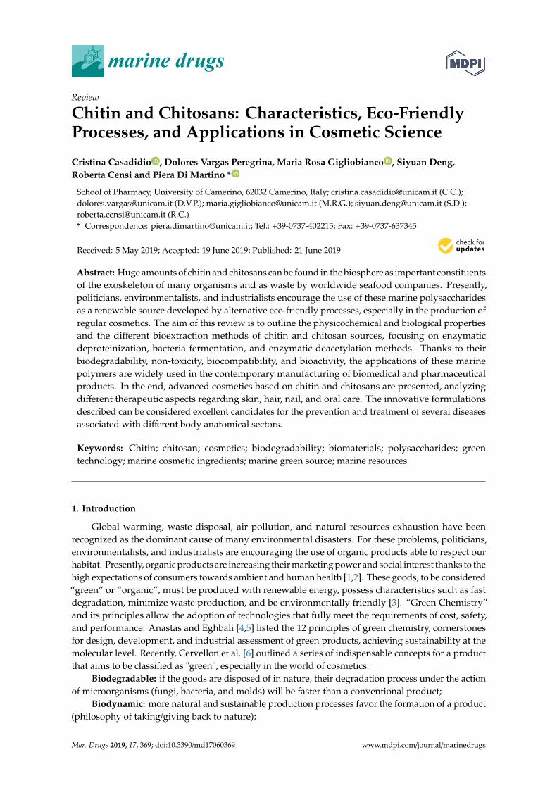
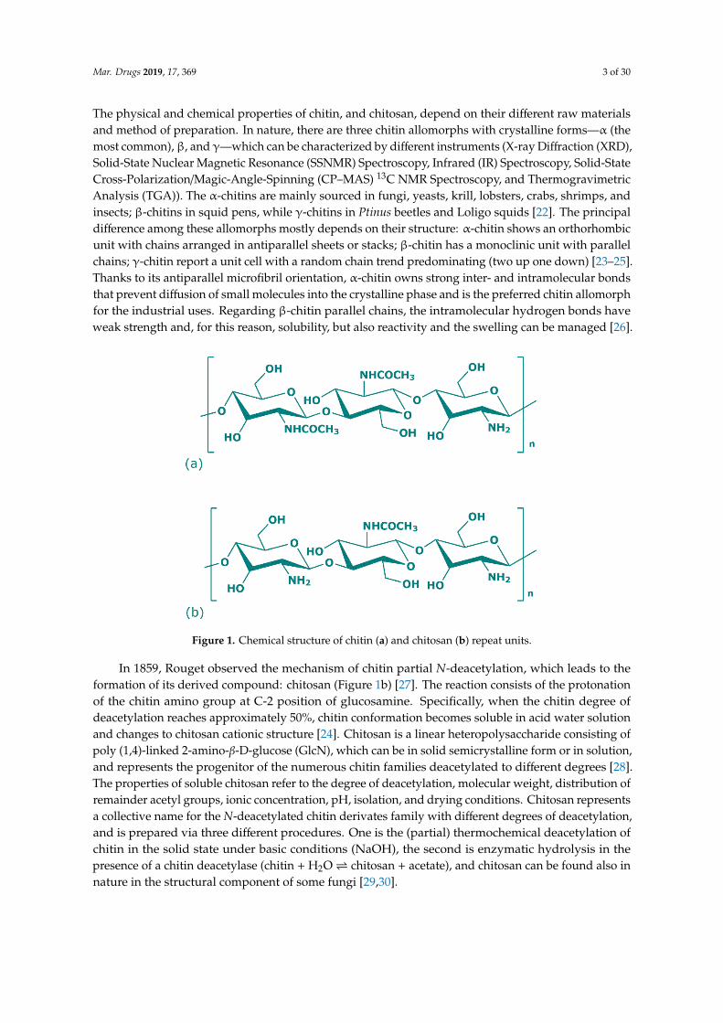
Figure 1. Chemical structure of chitin (a) and chitosan (b) repeat units.
Figure 1. Chemical structure of chitin (a) and chitosan (b) repeat units.
In 1859, Rouget observed the mechanism of chitin partial N-deacetylation, which leads to theformation of its derived compound: chitosan (Figure 1b) [27]. The reaction consists of the protonationof the chitin amino group at C-2 position of glucosamine. Specifically, when the chitin degree ofdeacetylation reaches approximately 50%, chitin conformation becomes soluble in acid water solutionand changes to chitosan cationic structure [24]. Chitosan is a linear heteropolysaccharide consisting ofpoly (1,4)-linked 2-amino-β-D-glucose (GlcN), which can be in solid semicrystalline form or in solution,and represents the progenitor of the numerous chitin families deacetylated to different degrees [28].The properties of soluble chitosan refer to the degree of deacetylation, molecular weight, distribution ofremainder acetyl groups, ionic concentration, pH, isolation, and drying conditions. Chitosan representsa collective name for the N-deacetylated chitin derivates family with different degrees of deacetylation,and is prepared via three different procedures. One is the (partial) thermochemical deacetylation ofchitin in the solid state under basic conditions (NaOH), the second is enzymatic hydrolysis in thepresence of a chitin deacetylase (chitin + H2O chitosan + acetate), and chitosan can be found also innature in the structural component of some fungi [29,30].
Mar. Drugs 2019, 17, 369 4 of 30
2.1.1. Degree of Deacetylation
The degree of deacetylation (DD) is the percentage of glucosamine (C6H13NO5) monomerspresent in the chitin structure. DD produces a huge effect on the solubility of chitin, unlike chitosan,due to its inability to dissolve in aqueous acid solution such as acetic acid. When the DD turnsover 50%, chitin becomes soluble in acid-diluted solution and changes to the chitosan cationicstructure [24]. The determination of DD can be performed by numerous techniques such as Ultraviolet(UV) Spectroscopy, IR Spectroscopy, Proton and Carbon Nuclear Magnetic Resonance Spectroscopy(1H and 13C NMR), SSNMR Spectroscopy, Gel Permeation Chromatography (GPC), Circular Dichroism,Residual Salicylaldehyde Analysis, Titration Methods, Elemental Analysis, High-Performance LiquidChromatography (HPLC), Thermal Analysis, and Mass Spectrometry (MS) [31]. Heidari et al. providedseveral characterizations of DD with Fourier Transform Infrared Spetroscopy (FTIR) following Sabins’slaw [32]:
Absorbance ratio = (A)amide/(A)hydroxyl
(DD) = 97.67 − (26.486 (A1655/A3450))
They obtained for chitin a DD of 50.1% while for three different natural chitosan the DD were73.5%, 82.3%, and 82.5%, respectively [33]. In another work, DD was evaluated with a new FT-Ramanapproach identifying the experimental bands of chitin and chitosan based on density functional theory(DFT) quantum chemical calculations [34]. Dimzon et al. for DD determination used an IR absorbanceratio method improved with the use of partial least squares (PLS). The IR spectral region was settledfrom 1500 to 1800cm−1 and the results obtained were equal to those obtained with potentiometrictitration and better than those obtained with the IR absorbance ratio conventional method [35].
2.1.2. Molecular Weight
The molecular weight (MW) of chitin depends on its origin source and is related to the method ofdecalcification with HCl, achieving the maximal depolymerization. The MW can be determined byHPLC or by viscometry, where the intrinsic viscosity (η) was determined in 0.1 M acid acetic and 0.2 Msodium chloride solution, following the Mark–Houwink equation [29]:
(η) = k·Mα = 1.81 × 10⁻³·M0.93
The weight-average MW of chitin is reported to range from 0.4 to 2.5 × 10⁶ [36,37].Chitosan MW compared to the chitin one is lower, due to the N-deacetylation. Chitosan MW
depends on the degree of polymerization (DP), where oligomers with DP of 8 or less are water-solubleirrespective of deacetylation conditions such as time, temperature, concentration of sodium hydroxide,and pH value [28,31]. The weight-average MW of chitosan is from 1 × 10⁵ to 5 × 10⁵ Da andcan be determined by several methods, including: light-scattering spectrophotometry, GPC, andviscometer [31]. In recent years, size-exclusion chromatography (SEC) has been another approachadopted to better determinate the MW. Kang et al. coupled SEC with multi-angle static laser lightscattering (MALLS) to better improve the characterization of chitosan MW [38]. In another work,Weinhold and Thöming connected SEC instruments with a triple detector array including in-seriesright and low-angle light-scattering (RALS-LALS) and viscometer [39].
2.1.3. Solubility
Chitin shows variations in its solubility according to different sources. The biopolymer isinsoluble in all the usual solvents such as neutral water, salt solutions, and most organic solvents, butpresents solubility with other solvents such as hexafluoroacetone sesquihydrate, hexafluoroisopropanol,chloroalcohols (with sulfuric acid), water mineral solutions, and in a mixture of dimethylacetamide with5% of lithium chloride. Its poor solubility is due to the presence of highly hydrophobic properties and
Mar. Drugs 2019, 17, 369 5 of 30
its extensive semicrystalline structure [36]. Chitin is a polysaccharide with intra- and intermolecularhydrogen bonds, which makes it difficult to dissolve in the previous indicated solvents.
Chitosan is insoluble in neutral water and its solubility is settled with acid solutions such aslactic, acetic, glutamic, and hydrochloric acid solutions (pH up to 6.5), due to the lower number ofN-acetylated groups and due to its primary amino groups (with a pKa of 6.3) which become protonated,leading to a positively charged polymer and giving the characteristics of a strong base. However, whenthe pH reaches the value of 6.0 (and above), the polysaccharide becomes insoluble and precipitatesdue to the deprotonation of the amines. Presently, purified chitosans with high DD are commerciallyavailable in a broad range of MW, both in the form of base and as a salt readily soluble in water withoutthe use of acid solutions [40]. Commonly, the solubility of chitosan decreases when pH raises fromphysiological to basic values, and with increase the ionic strength (salting-out effect) or the MW [24].There are other determining factors that have important effects on chitosan solubility: temperature,average of DD, and DP. The most common solvents for the solubilization of chitosan are: acetic acid (1%with pH close to 4); formic acid (0.2–100%); 1% hydrochloric acid; lactic acid; and diluted nitric acid.Recent research found a neutral chitosan solution with the use of glycerol 2-phospate as solvent [24].On the other hand, chitosan is insoluble in sulfuric and phosphoric acid [29].
2.1.4. Derivatives
Several chitin derivatives are described in the literature, among which chitosan appears to be themain product. Chitosan can be obtained by chitin following two different processes: the enzymatichydrolysis or via chemical deacetylation, as shown in Figure 2. In addition to this, numerous otherchitin products have been identified [24]. Thanks to the availability of the amino group, chitin can beassociated with macromolecules such as proteins, carotenoids, and glucans [28].
Regarding chitosan, its structure has three active groups which can be chemically modified tochange specific properties and activities. These groups are: the primary (C-6) and secondary (C-3)hydroxyl groups at the level of which non-specific reactions occur, and the amino group (C-2), wherespecific reactions can be distinguished. Solubility, and physical and mechanical properties of chitosancan be altered with the chemical modification of these reactive groups, attributing to its new derivativesfurther properties. The main reactions involving the C-3 and C-6 positions are esterification andetherification, while for the amino-C-2 position the quaternization of the amino group is carried out. InC-2, where the aldehyde function reacts with -NH2 by reductive amination, a reaction can be settled inaqueous solution under mild conditions favoring the introduction of different functional groups onchitosan using acrylic reagents in an aqueous medium [31].

Mar. Drugs 2019, 17, 369 6 of 30Mar. Drugs 2019, 17, x FOR PEER REVIEW 6 of 28
Figure 2. Scheme of chitin and chitosan production following chemical methods.
2.2. Chitin and Chitosans Biological Properties
Both chitin and chitosan exhibit various biological properties such as: anticholesterolemic,
wound-healing agents, anticancer, fungistatic, hemostatic, analgesic, antiacid, antiulcer,
immunoadjuvant, etc. [19,21,29,31,40–44]. In cosmetic science, chitin and chitosan have been
investigated as potential excipients and as biological active agents, thanks to their peculiar properties
such as no toxicity, biocompatibility, and biodegradability. This review will discuss four of the main
characteristics and qualities that make these polysaccharides excellent candidates in the formulation
of care products: antimicrobial and antioxidant activities, mucoadhesive and penetration properties.
The indispensable physicochemical parameters of the polysaccharides that promote the biological
effect are summarized in Table 1.
Figure 2. Scheme of chitin and chitosan production following chemical methods.
2.2. Chitin and Chitosans Biological Properties
Both chitin and chitosan exhibit various biological properties such as: anticholesterolemic,wound-healing agents, anticancer, fungistatic, hemostatic, analgesic, antiacid, antiulcer,immunoadjuvant, etc. [19,21,29,31,40–44]. In cosmetic science, chitin and chitosan have beeninvestigated as potential excipients and as biological active agents, thanks to their peculiar propertiessuch as no toxicity, biocompatibility, and biodegradability. This review will discuss four of the maincharacteristics and qualities that make these polysaccharides excellent candidates in the formulation ofcare products: antimicrobial and antioxidant activities, mucoadhesive and penetration properties. Theindispensable physicochemical parameters of the polysaccharides that promote the biological effectare summarized in Table 1.
Mar. Drugs 2019, 17, 369 7 of 30
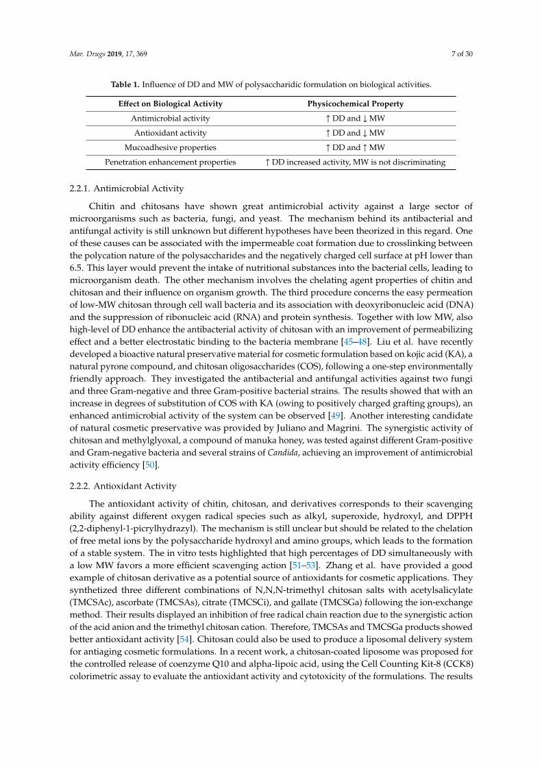
Table 1. Influence of DD and MW of polysaccharidic formulation on biological activities.
Effect on Biological Activity Physicochemical Property
Antimicrobial activity ↑ DD and ↓MW
Antioxidant activity ↑ DD and ↓MW
Mucoadhesive properties ↑ DD and ↑MW
Penetration enhancement properties ↑ DD increased activity, MW is not discriminating
2.2.1. Antimicrobial Activity
Chitin and chitosans have shown great antimicrobial activity against a large sector ofmicroorganisms such as bacteria, fungi, and yeast. The mechanism behind its antibacterial andantifungal activity is still unknown but different hypotheses have been theorized in this regard. Oneof these causes can be associated with the impermeable coat formation due to crosslinking betweenthe polycation nature of the polysaccharides and the negatively charged cell surface at pH lower than6.5. This layer would prevent the intake of nutritional substances into the bacterial cells, leading tomicroorganism death. The other mechanism involves the chelating agent properties of chitin andchitosan and their influence on organism growth. The third procedure concerns the easy permeationof low-MW chitosan through cell wall bacteria and its association with deoxyribonucleic acid (DNA)and the suppression of ribonucleic acid (RNA) and protein synthesis. Together with low MW, alsohigh-level of DD enhance the antibacterial activity of chitosan with an improvement of permeabilizingeffect and a better electrostatic binding to the bacteria membrane [45–48]. Liu et al. have recentlydeveloped a bioactive natural preservative material for cosmetic formulation based on kojic acid (KA), anatural pyrone compound, and chitosan oligosaccharides (COS), following a one-step environmentallyfriendly approach. They investigated the antibacterial and antifungal activities against two fungiand three Gram-negative and three Gram-positive bacterial strains. The results showed that with anincrease in degrees of substitution of COS with KA (owing to positively charged grafting groups), anenhanced antimicrobial activity of the system can be observed [49]. Another interesting candidateof natural cosmetic preservative was provided by Juliano and Magrini. The synergistic activity ofchitosan and methylglyoxal, a compound of manuka honey, was tested against different Gram-positiveand Gram-negative bacteria and several strains of Candida, achieving an improvement of antimicrobialactivity efficiency [50].
2.2.2. Antioxidant Activity
The antioxidant activity of chitin, chitosan, and derivatives corresponds to their scavengingability against different oxygen radical species such as alkyl, superoxide, hydroxyl, and DPPH(2,2-diphenyl-1-picrylhydrazyl). The mechanism is still unclear but should be related to the chelationof free metal ions by the polysaccharide hydroxyl and amino groups, which leads to the formationof a stable system. The in vitro tests highlighted that high percentages of DD simultaneously witha low MW favors a more efficient scavenging action [51–53]. Zhang et al. have provided a goodexample of chitosan derivative as a potential source of antioxidants for cosmetic applications. Theysynthetized three different combinations of N,N,N-trimethyl chitosan salts with acetylsalicylate(TMCSAc), ascorbate (TMCSAs), citrate (TMCSCi), and gallate (TMCSGa) following the ion-exchangemethod. Their results displayed an inhibition of free radical chain reaction due to the synergistic actionof the acid anion and the trimethyl chitosan cation. Therefore, TMCSAs and TMCSGa products showedbetter antioxidant activity [54]. Chitosan could also be used to produce a liposomal delivery systemfor antiaging cosmetic formulations. In a recent work, a chitosan-coated liposome was proposed forthe controlled release of coenzyme Q10 and alpha-lipoic acid, using the Cell Counting Kit-8 (CCK8)colorimetric assay to evaluate the antioxidant activity and cytotoxicity of the formulations. The results
Mar. Drugs 2019, 17, 369 8 of 30
revealed that chitosan-liposome system has low cytotoxicity with an excellent antioxidant activity(clearing reactive oxygen species (ROS) from H2O2) [55].
2.2.3. Mucoadhesive Properties
The main component of mucus is mucin, a glycoprotein rich in negative charges that interactwith the positive ones of chitosan. Previous studies have established that the physical and chemicalcharacteristics of the chitosan favor an improvement of the mucoadhesive properties related prevalentlyto DD and MW. Unlike what happens for antioxidant and antimicrobial activities, in this case there isan improvement of the mucoadhesion when polysaccharides are used with a high degree of DD andhigh MW [56,57]. Pereira et al. designed Aloe vera/vitamin E/chitosan microparticles for burn treatmentapplication that could also be used in the future as a cosmetic proposal. They performed in vitromucoadhesion test demonstrating and confirming the adhesive property of the system correlated withthe presence of chitosan [58].
2.2.4. Penetration Enhancement Properties
The permeation enhancement carried out by chitosan is associated with the opening and destructionof epithelial tight junctions by a decrease in transepithelial electric resistance. The chemical nature ofthe mechanism is based on the electrical interaction between the positive charges of chitosan and thecell membrane, leading to a re-association of the proteins associated with the tight junctions [59–61].In 2016, researchers developed cationic and anionic acrylic nanocapsules with a diameter of 150 nm,embedded into chitosan gel for cosmetic application. The chitosan cationic charged surface allowed adeeper skin penetration of acrylic capsules by induction of the tight junctions opening in the stratumgranulosum, below the stratum corneum [62]. Kojima et al. investigated the distribution of chitosan,using a hair cosmetic ingredient to improve the texture of hair surface, via time-of-flight secondary ionmass spectrometry (TOF-SIMS). They analyzed the penetration of chitosan as a hair conditioning agentand the results showed how cationic chitosan equally incorporated on the hair surface, highlightingan important difference between virgin and bleached hair. The amount of cationic chitosan adsorbedon the virgin hair was lower than the bleached hair, because bleaching leads to a negative chargeenhancement due to cysteic acid group formation [63].
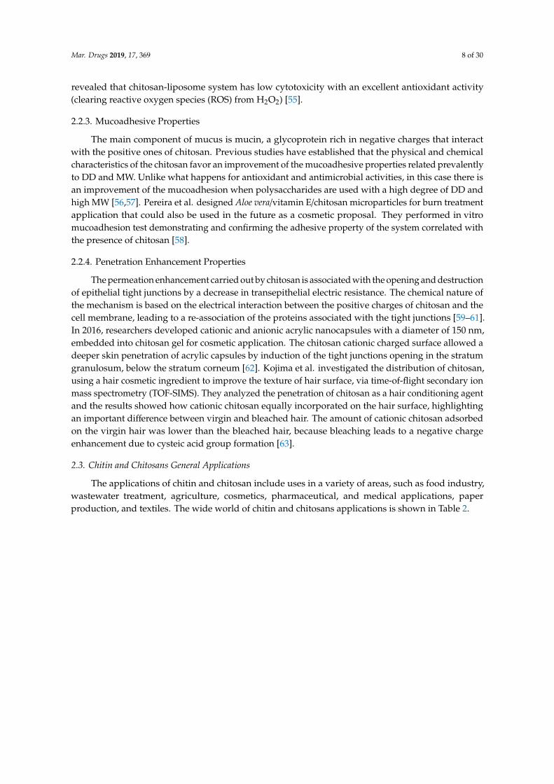
2.3. Chitin and Chitosans General Applications
The applications of chitin and chitosan include uses in a variety of areas, such as food industry,wastewater treatment, agriculture, cosmetics, pharmaceutical, and medical applications, paperproduction, and textiles. The wide world of chitin and chitosans applications is shown in Table 2.
Mar. Drugs 2019, 17, 369 9 of 30
Table 2. Chitin and chitosans applications.
Field Examples Ref.
Indu
stri
alA
pplic
atio
ns
Cosmetics
Biodegradable, biocompatible and nontoxic chitosan microparticles encapsulating jabuticaba peel extract [64]
Modified chitosan microparticles containing rosmarinic acid for skin delivery formulations [65]
Nanoparticles of quaternized cyclodextrin-grafted chitosan associated with hyaluronic acid as promising skinpenetration vehicles [66]
Preventive effect of chitosan oligosaccharide against UV-caused damage in hairless mouse dorsal skin [67]
Periodontal chitosan gels containing moxifloxacin hydrochloride [68]
Fluoride loaded chitosan nanoparticles in the prevention of dental caries [69]
Hydroxyapatite-chitosan sunscreen antibacterial gel for skin health care [70]
Chitosan and surface-deacetylated chitin nanofibrils induced hair growth [71]
Agriculture
Gum arabic/chitosan nanoparticles containing geraniol for pest management [72]
Chitosan natural biopolymer as a growth stimulator of rice yield [73]
Chitosan modified Pt/SiO2 as catalyst for an agricultural synergistic agent [74]
Antifungal chitosan agent used to control Ceratocystis fimbriata plant pathogenic fungus that attacks sweet potato [75]
Eco-friendly chitosan/basalt hydrogel as soil conditioner and booster of plants growth [76]
Food and Nutrition
Food packaging made by chitosan-based films with microparticles of olive pomace [77]
Nisin-loaded chitosan-monomethyl fumaric acid nanoparticles as a direct food additive [78]
Chitosan-TiO2 nanocomposite film as antimicrobial active food packaging [79]
Chitosan as an alternative food preservative to formalin [80]
Fish-purified antioxidant peptide-loaded electrospun chitosan/PVA nanofibrous mat for food biopackagingapplications [81]
Tripolyphosphate and chitosan nanoparticles for encapsulation of C, B9, and B12 vitamins [82]
Starch or chitosan-based matrices carrying thyme extract polyphenols as antioxidant films for food preservation [83]
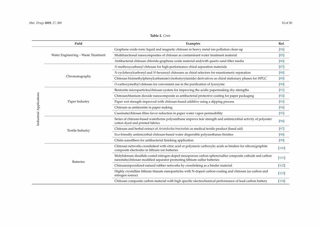
Mar. Drugs 2019, 17, 369 10 of 30
Table 2. Cont.
Field Examples Ref.
Indu
stri
alA
pplic
atio
ns
Water Engineering – Waste Treatment
Graphene oxide-ionic liquid and magnetic chitosan in heavy metal ion pollution clean-up [84]
Multifunctional nanocomposites of chitosan as contaminant water treatment material [85]
Antibacterial chitosan chloride-graphene oxide material and/with quartz sand filter media [86]
Chromatography
N-methoxycarbonyl chitosan for high-performance chiral separation materials [87]
N-cyclohexylcarbonyl and N-hexanoyl chitosans as chiral selectors for enantiomeric separation [88]
Chitosan bis(methylphenylcarbamate)-(isobutyrylamide) derivatives as chiral stationary phases for HPLC [89]
O-carboxymethyl chitosan for convenient use in the purification of lysozyme [90]
Paper Industry
Bentonite microparticles/chitosan system for improving the acidic papermaking dry strengths [91]
Chitosan/titanium dioxide nanocomposite as antibacterial protective coating for paper packaging [92]
Paper wet strength improved with chitosan-based additive using a dipping process [93]
Chitosan as antitermite in paper making [94]
Caseinate/chitosan films favor reduction in paper water vapor permeability [95]
Textile Industry
Series of chitosan-based waterborne polyurethane improve tear strength and antimicrobial activity of polyestercotton dyed and printed fabrics [96]
Chitosan and herbal extract of Aristolochia bracteolate as medical textile product (band aid) [97]
Eco-friendly antimicrobial chitosan-based water dispersible polyurethanes finishes [98]
Chitin nanofibers for antibacterial finishing application [99]
Batteries
Chitosan networks crosslinked with citric acid or polymeric carboxylic acids as binders for silicon/graphitecomposite electrodes in lithium ion batteries [100]
Molybdenum disulfide-coated nitrogen-doped mesoporous carbon sphere/sulfur composite cathode and carbonnanotube/chitosan modified separator promoting lithium sulfur batteries [101]
Chitosan/epoxidized natural rubber networks by crosslinking as a binder material [102]
Highly crystalline lithium titanate nanoparticles with N-doped carbon-coating and chitosan (as carbon andnitrogen source) [103]
Chitosan composite carbon material with high specific electrochemical performance of lead-carbon battery [104]
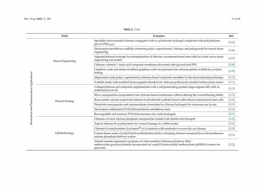
Mar. Drugs 2019, 17, 369 11 of 30
Table 2. Cont.
Field Examples Ref.
Biom
edic
alan
dPh
arm
aceu
tica
lApp
licat
ions
Tissue Engineering
Injectable carboxymethyl chitosan conjugated with α-cyclodextrin hydrogel complexed with poly(ethyleneglycol) (PEG1000) [105]
Electrospun nanofibrous scaffolds containing poly(ε-caprolactone), chitosan, and polypyrrole for neural tissueengineering [106]
Alginate/chitosan hydrogel for transplantation of olfactory ectomesenchymal stem cells for sciatic nerve tissueengineering (rat model) [107]
Chitosan–vitamin C–lactic acid composite membrane decorated with glycerol and PEG [108]
Graphene oxide and amine-modified graphene oxide incorporated into chitosan-gelatin scaffold by covalentlinking [109]
Magnesium oxide-poly(ε-caprolactone)-chitosan-based composite nanofiber by the electrospinning technique [110]
Scaffolds made with modified hydroxyapatite blended into chitosan-grafted-poly (methyl methacrylate) matrix [111]
Wound Healing
Collagen/chitosan gel composite supplemented with a cell-penetrating peptide (oligo-arginine R8) with anantibacterial activity [112]
Silver nanoparticles encapsulation into chitosan-based membranes without altering the wound-healing ability [113]
Rosuvastatin calcium loaded into chitosan hydrochloride scaffolds based with/without mesenchymal stem cells [114]
Phenytoin nanocapsules and nanoemulsions formulated as chitosan hydrogels for cutaneous use in rats [115]
Electrospun antibacterial PVA/Chitosan/Starch nanofibrous mats [116]
Biocompatible and nontoxic PVA/chitosan/nano zinc oxide hydrogels [117]
Ophthalmology
Chitosan-covered calcium phosphate nanoparticles loaded with timolol and lisinopril [118]
Topical chitosan-N-acetylcysteine for corneal damage in a rabbit model [119]
Chitosan-N-acetylcysteine (Lacrimera®) in in patients with moderate to severe dry eye disease [120]
Contact lenses made of poly(2-hydroxyethylmethacrylate) containing chitosan nanoparticles as dexamethasonesodium phosphate delivery system [121]
Timolol maleate imprinted copolymer of carboxymethyl chitosan-g-hydroxy ethylmethacrylate-g-polyacrylamide incorporated on a poly(2-hydroxyethyl methacrylate) p(HEMA) matrix forglaucoma
[122]
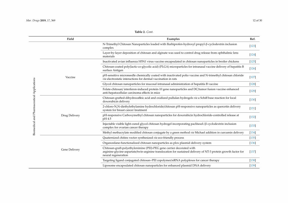
Mar. Drugs 2019, 17, 369 12 of 30
Table 2. Cont.
Field Examples Ref.
Biom
edic
alan
dPh
arm
aceu
tica
lApp
licat
ions
N-Trimethyl Chitosan Nanoparticles loaded with flurbiprofen-hydroxyl propyl-β-cyclodextrin inclusioncomplex [123]
Layer-by-layer deposition of chitosan and alginate was used to control drug release from ophthalmic lensmaterials [124]
Vaccine
Inactivated avian influenza H5N1 virus vaccine encapsulated in chitosan nanoparticles in broiler chickens [125]
Chitosan-coated poly(lactic-co-glycolic acid) (PLGA) microparticles for intranasal vaccine delivery of hepatitis Bsurface Antigen [126]
pH-sensitive microneedle chemically coated with inactivated polio vaccine and N-trimethyl chitosan chloridevia electrostatic interactions for dermal vaccination in rats [127]
Glycol chitosan nanoparticles for mucosal intranasal administration of hepatitis B vaccine [128]
Folate-chitosan/ interferon-induced protein-10 gene nanoparticles and DC/tumor fusion vaccine enhancedanti-hepatocellular carcinoma effects in mice [129]
Drug Delivery
Chitosan-grafted-dihydrocaffeic acid and oxidized pullulan hydrogels via a Schiff base reaction for localdoxorubicin delivery [130]
2-chloro-N,N-diethylethylamine hydrochloride/chitosan pH-responsive nanoparticles as quercetin deliverysystem for breast cancer treatment [131]
pH-responsive Carboxymethyl chitosan nanoparticles for doxorubicin hydrochloride-controlled release atpH 4.5 [132]
Injectable visible light-cured glycol chitosan hydrogel incorporating paclitaxel-/β-cyclodextrin inclusioncomplex for ovarian cancer therapy [133]
Methyl methacrylate modified chitosan conjugate by a green method via Michael addition in curcumin delivery [134]
Gene Delivery
Quaternized chitins vector synthesized via eco-friendly process [135]
Organosilane-functionalized chitosan nanoparticles as plox plasmid delivery system [136]
Chitosan-graft-polyethylenimine (PEI)-PEG gene carrier decorated witharginine-glycine-aspartate/twin-arginine translocation for sustained delivery of NT-3 protein growth factor forneural regeneration
[137]
Targeting ligand conjugated chitosan–PEI copolymer/siRNA polyplexes for cancer therapy [138]
Liposome encapsulated chitosan nanoparticles for enhanced plasmid DNA delivery [139]
Mar. Drugs 2019, 17, 369 13 of 30
3. Extraction of Chitin and Chitosans from Natural Sources
Chitin and chitosan are considered important marine renewable sources due to their highavailability as garbage from the seafood processing industry; chitin availability was estimated tobe approximately over 10 billion tons annually [29,140]. Presently, most producers for commercialpurposes of chitin and chitosan are in Poland, India, Norway, Australia, USA, and Japan [141]. Thereare mainly two chitin extraction methods conducted in the industry—chemical or biological. Bothextraction strategies of chitin consist of two phases—deproteinization with alkaline treatment at hightemperatures, and demineralization with dilute hydrochloric acid. The sequence of these two phasesis interchangeable depending on the source and the proposed use of chitin. The third phase mainlydepends on the starting waste material: if the chitin is extracted from squid pens, a final non-pigmentedwhite powder is obtained. On the other hand, chitin powder isolated from crustacean sources assumesa pale pink color, thus necessitating the bleaching process, which requires the use of hydrogen peroxide,oxalic acid, or potassium permanganate [45,142]. Figure 2 provides a scheme of chitin preparation frommarine shell waste following the chemical process. These synthetic methods are very risky and havemany disadvantages due to high temperature and high concentration of acid and alkali solutions. Inaddition, the production of chitin and chitosan by chemical process has different industrial drawbackssuch as: high energy consumption, long handling times, greater solvent wasted, high environmentalpollution, high production of waste, and difficulty in recovering waste products such as pigment andproteins [143–145]. As an alternative to the chemical process, bioextraction of chitin has been studiedas a newly green ecological process.
3.1. Bioextraction of Chitin
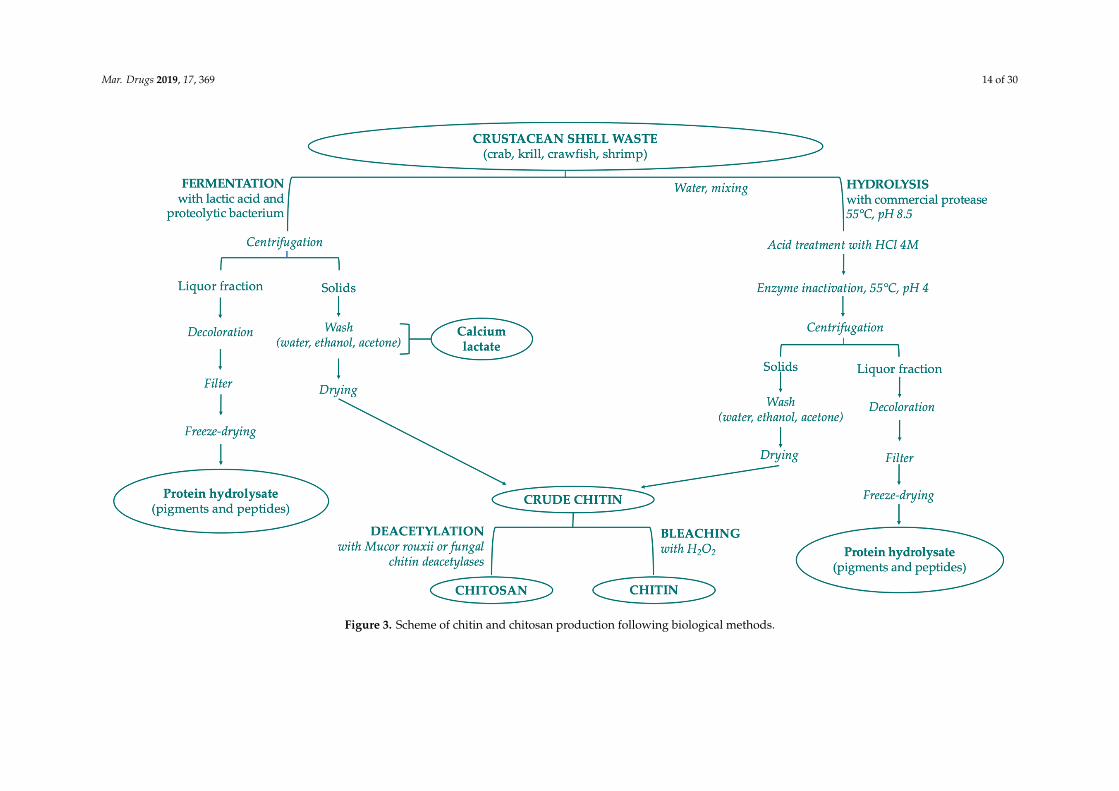
Chitin is a constituent of the organic matrix of different marine organisms, including: arthropodexoskeletons such as crustaceans (crab, shrimp, lobster, krill, crayfish, barnacles) and insects (cockroach,beetle, true fly and worm); mollusk endoskeletons; fungi (Aspergillus niger, Mucor rouxii, Penicillumnotatum); yeasts; algae; cuttlefishes; and squid pen. Crustacean shell is composed of 30–40% proteins,30–50% mineral salts (principally calcium carbonate and phosphate), and 13–42% of chitin withits different chemical structures—α-, β-, γ-form. In minimal percentages, carotenoids (mainlyastaxanthin and its esters) and lipids from visceral or muscular residues can also be found in shellfishwaste [146]. Chitin is extracted from crustacean shell waste with three different steps: demineralization,deproteinization, and bleaching/decoloration. Chemical demineralization and deproteinization presentseveral issues that prevent optimal control of reactions: depolymerization, anomerization, and decreaseof MW by altering the properties of purified chitin. To solve these problems the application ofbioextraction process is preferred, with two different methods: the employment of proteolytic enzymesto digest proteins, or the microorganism-mediated fermentation (Figure 3). The limitations of thesebiological procedures are high cost, lower yield, and the final properties of the products [143,147,148].
Mar. Drugs 2019, 17, 369 14 of 30Mar. Drugs 2019, 17, x FOR PEER REVIEW 13 of 28
Figure 3. Scheme of chitin and chitosan production following biological methods. Figure 3. Scheme of chitin and chitosan production following biological methods.

Mar. Drugs 2019, 17, 369 15 of 30
3.1.1. Chitin Enzymatic Deproteinization
One of the proposed biological alternatives is the use of proteases for deproteinization ofcrustacean shells, mainly deriving from plant, microbial, and animal sources. This method avoidsalkaline treatments and produces, in addition to chitin, protein hydrolysates with nutritional value.Depending on the starting waste, the protease can lead to various deproteinization yields accordingto the conditions tested. The demineralization should be performed first, to increase the tissuepermeability, decrease the presence of potential enzyme inhibitors, and promote the action of theproteolytic enzyme. Chymotrypsin, Papain, Trypsin, Alkalase, Devolvase, Pepsin, and Pancreatinare the major proteolytic enzymes used to extract and separate the protein and chitin residues fromshrimp waste. The final products present more beneficial physicochemical properties compared tothe ones obtained following other methods. Proteases can be purified and extracted commerciallyat high cost and less efficacy, and crudely extracted, mainly derived from bacteria but also fromfish viscera [149,150]. The crude extracted proteases are cheaper and more efficient thanks to thepresence of coexisting proteases. Mhamdi et al. in their study reported the evaluation, characterization,and application of thermostable serine alkaline proteases from actinomycete strain Micromonosporachaiyaphumensis S103 for chitin extraction from shrimp shell (Penaeus kerathurus) waste powder. Thepercentage of deproteinization obtained after 3 hours of hydrolysis at 45 ◦C and pH 8.0 with anenzyme/substrate (E/S) ratio of 20 U/mg reached 93%, one of the best results in the literature comparedto the use of other proteases [151]. In another work, crude digestive alkaline proteases from the visceraof Portunus segnis proved to be very efficient in the production of chitin by deproteinization of bluecrab (P. segnis) and shrimp (P. kerathurus). In this case, the percentage of deproteinization achieved wasnear 85% for blue crab shells and 91% for shrimp shells with an E/S ratio of 5 U/mg of proteins after 3 hincubation at 50 ◦C [152].
3.1.2. Chitin Bacteria Fermentation
Another inexpensive approach for chitin extraction from seafood wastes consists of two differentmethods of fermentation, with and without lactic acid bacteria (LAB). Fermentation can be carriedout by adding selected strains of microorganisms, following one-stage and two-stage fermentation,co-fermentation/subsequent fermentation, or from endogenous microorganisms (auto-fermentation).
Lactic Acid Bacteria Fermentation
LAB fermentation has been studied as an innovative method for enzymatic extraction of chitinand can be combined with chemical treatments reducing the amount of acid and alkali needed [148].The ability of LAB strains is to ferment waste materials and simultaneously produce organic acids insitu (lactic and acetic acids). During fermentation, two fractions are obtained—a liquid fraction rich inproteins, minerals, and pigments and a solid phase containing crude chitin (which can be separatedby filtration and washed with water). The separation into two fractions occurs thanks to the actionof lactic acid that promotes the precipitation of the chitin and the production of calcium lactate afterreaction with calcium carbonate. The lactic acid, obtained by conversion of glucose, at the same timepromotes the lowering of the pH and consequently the activation of proteases. This methodology hasalso been used for the recovery of other products from silage shrimp waste, such carotenoids. The mostused bacterial strains for fermentation are Lactobacillus sp. strain especially L. plantarum, L. paracaseiand L. helveticus. Recently, Castro et al. have extracted and purified chitin from Allopetrolisthespunctatus crabs using Lactobacillus plantarum sp.47, a Gram-positive bacterium isolated from Cohosalmon that produce high lactic acid concentrations. They obtained a 99.6% demineralization, 95.3%deproteinization and 17 mg of lactic acid/g silage choosing optimal fermentation parameters (60 hfermentation, 10% inoculum, 15% sucrose and 85% crab biomass) [153].
Mar. Drugs 2019, 17, 369 16 of 30
Non-Lactic Acid Bacteria Fermentation
For chitin recovery with non-lactic acid bacteria crustacean shell fermentation bacteria andfungi were used as the inoculum source: Pseudomonas sp., Bacillus sp. and Aspergillus sp. In theirresearch, Ghorbel-Bellaaj et al. isolated a protease bacterium identified as Pseudomonas aeruginosa A2. Byevaluation of protease activities and spectral analysis, they showed how chitin extracted by the biologicalmethod was similar to commercial α-chitin. The ability to deproteinize shrimp waste to produce chitinwas also highlighted, overcoming the disadvantages of chemical deproteinization [154]. In anotherwork, the same researchers followed a Plackett–Bhenken design to better improve deproteinizationand demineralization efficiencies of shrimp shells with P. aeruginosa A2. They used these optimizedvariables: shrimp shell concentration (50 g/L), glucose concentration (50 g/L), incubation time (5 days),and inoculum size (0.05 OD), obtaining a deproteinization of 89% and demineralization of 96% [155].
Most commercial bacterial proteases are mainly produced by Bacillus sp., and Hajji et al. haveextracted from the waste of crab shells chitin and fermented-crab supernatants after fermentationusing six different strains of Bacillus. Using specific assays, it has been discovered that fermented-crabsupernatants possess interesting antioxidant and antibacterial properties [156].
Regarding fungi as an inoculum source, three different proteolytic strains of Aspergillus niger,namely 0576, 0307, and 0474, were selected by Teng et al. thanks to their protease activity necessaryto produce chitin. The aim of their study was to obtain two distinct sources of chitin by adding thefermented shrimp shells to mushrooms directly. The proteolytic enzymes released by fungi during thedeproteinization and demineralization of the shrimp shell lead to the release of amino acids, which is asource of nitrogen to promote the growth of fungi [157].
3.2. Enzymatic Deacetylation of Chitin
Chitosan can be derived from chitin by chemical or enzymatic deacetylations. Chemicaldeacetylation is usually preferred because it is cheaper and guarantees suitability for mass production,but, at the same time, presents disadvantages such as energy consumption and increased environmentalpollution due to the alkaline conditions. To overcome these drawbacks, an innovative enzymaticmethod that exploits chitin deacetylases has been explored: enzymatic deacetylation of chitin. Hembachet al., with their research conducted in 2017, have chosen fungal, viral, and bacterial chitin deacetylases,producing 14 possible partially acetylated chitosan tetramers with a defined degree of acetylation anda pattern of acetylation, also showing a purification method [158].
4. Applications in Cosmetics
Among polysaccharides, chitin, chitosan, and their derivatives offer intangible qualities relatedto antiaging, matrix metalloproteinase (MMP) inhibitor, antioxidant, and antifungal properties. Theuse of chitin and chitosans has been suggested in different fields of skin, oral, nail, and hair careapplications, obtaining formulations able to treat diseases related to teeth, hair, nails, and skin.
4.1. Skin Care Applications
Chitin, chitosan, and their derivatives are widely used in cosmetics especially because they exertantioxidant, cleansing, protecting, humectant, and antioxidant functions. Most of the chitin andchitosan cosmetic applications described in the following paragraphs refer mainly to: antiaging andmoisturizing agents, ultraviolet protective compounds, skin cleansing products, and boosting factorsof skin-essential functions such as protection, absorption, thermal regulation, defense, reservation,and synthesis.
4.1.1. Antiaging and Moisturizing Agent
Skin aging is commonly a consequence of the intrinsic aging that occurs with the progression ofyears, but also of extrinsic aging caused by external factors as cigarettes, UV radiation, air pollution, etc.
Mar. Drugs 2019, 17, 369 17 of 30
Dryness, relaxation, roughness, and skin tissue laxity are the main characteristics of skin aging and itsassociation with UV ray exposure represents one of the most documented causes of hyperpigmentationorigin and wrinkles, leading to the phenomenon known as a photoaging [159]. Recently, researchershave shown how chitosan, especially of high MW, possesses film-forming properties that can promotea reduction in cutaneous water loss and an increase in skin elasticity and smoothness, making itinteresting in moisturizing cosmetic applications [160]. Transparency of film is one of the desirablefeatures due to its great impact on the cosmetic fields, especially the ability to obtain a product with nofurther visible changes. Han and Floros calculated the transparency in the following equation [161]:
Transparency = A600/x
where x is the film thickness (mm) and A600 is the absorbance at 600 nm. The greater transparency valueis represented by lower transparency. In 2019, Montenegro and Freier patented a transparent tissuedressing material based on deacetylated native chitosan suitable for different cosmetic applications suchas peelings and face masks [162]. In another work, the skin treated with chitosan film neutralized incitrate buffer (with or without hyaluronic acid) prepared by Libio et al., demonstrated a desquamationof the stratum corneum and a significant increase in the degree of hydration within 10 minutes on amodel of pig skin compared to skin without treatment. These results suggest that the biocompatiblefilm, in the absence of glycerol, promotes a cosmetic effect regarding skin exfoliation, thanks to thebioadhesive properties of chitosan [163].
Morganti et al. in a study conducted in 2013 showed the antiaging activity of a particular cosmeticformulation based on the use of chitin nanofibril-hyaluronan (CN-HA) block copolymeric nanoparticles.These CN-HA block-copolymers should be used with invasive and non-invasive therapies in aestheticmedicine, exploiting drug-delivery properties. In vitro studies have highlighted the ability to easilyencapsulate different active compounds (such as lutein) and in vivo studies have demonstrated theinnovative antiaging properties of these formulations, with encouraging long-lasting results [164–166].As an alternative to the common chemical polymers, Rajashree and Rose investigated the antiagingpower of a gel based on collagen, chitosan, and Aloe vera. Chitosan improved the stability and thecapability to induce local cell proliferation, and in combination with collagen enhanced the skinfibroblast biocompatibility, attachment, and proliferation. The final system was able to increase therejuvenation and regeneration of the skin [167].
4.1.2. Ultraviolet Protective Cosmetics
Chitin and chitosan show adhesive properties (thanks to the electrostatic interactions betweenthe positively charged polysaccharides and negatively charged keratin-based structures [168]), waterresistance, cytocompatibility, and UV absorption (below 400 nm), qualities necessary for the formulationof protective creams. The main radiation characteristics of solar rays are UV, specifically UV-A(320–400 nm), and UV-B (290–320 nm). These radiations cause numerous adverse reactions on the skinsuch as sunburn, skin degeneration, photosensitivity, phototoxicity, photoaging, immunosuppression,and skin cancer. To prevent such diseases caused by excessive exposure of the skin to solar radiation,chitosan-containing sunscreens with substances with strong protective efficacy are used [159,169].Ito et al. used urocanic acid (UCA), the major UV-absorbing chromophore in the skin, to preparenanofibrils of urocanic acid-chitin by UCA hydrolysis. They examined the protective effect of theformulation against UV-B radiation demonstrating in vivo their protective effect but also the ability toinhibit erythema induced by UV-B irradiation and solarization cell generation [170]. In a recent work,solar emulsion based on chitosan nanoparticles (150–500 nm) were prepared with annatto, ultrafilteredannatto, saffron, and ultrafiltered saffron. All formulations were synthesized via ionotropic gelationand showed good preservation and low toxicity, while minimal sun protection was observed withsun protection factor (SPF) values ranging from 2.15 to 4.85. The storage stability was evaluated, and
Mar. Drugs 2019, 17, 369 18 of 30
the final system showed a good storage (regarding pH and viscosity) at room temperature for up to90 days [171].
4.1.3. Skin Cleansing
Cleaning the skin means removing from its surface contaminating foreign substances that areacquired during a simple air exposure or cosmetic product application. Chitosan and its derivatives,thanks to their cationic nature, can be used as positively charged vehicles in the delivery of productsfor personal cleaning. They can indeed exploit the ionic attraction between their charge and the anionicnature of the surface of the skin [172]. A liquid cleansing composition with moisturizing and exfoliatingdual properties was designed and patented by Massaro et al. To date, they have not used chitin as astarting ingredient in the specific cosmetic cleansing but it is listed among the next substances suitablefor the formulation of a new cleansing product [173].
4.2. Nail Care Applications
The nail is a structure produced by the skin and is therefore an appendage of the skin itself.Nail disease, onychosis, has a distinct classification with respect to skin diseases. Onychomycosis, afungal infection of the nail unit, is a common disorder that is currently treated with broad-spectrumantimycotics delivered through topical administration and/or in combination with systemic oral drugs.Most cases of onychomycosis are caused by skin infections or because of a nail trauma (mechanicaltrauma or exposure of chemical agents), altering the natural barrier function of the nail. A topical agentsuch as nail lacquer represents a valid topical formulation to prevent fungal infections compared tocreams and solutions, since it favors a better stay of the formulation at the site of action. Hydroxypropylchitosan (HPCH), a semisynthetic derivative of chitosan, has proved to be a valid candidate for thedelivery of active products to nails, acting as a protective film that preserves nail structure, protectingkeratin, maintaining hydration with a decrease of dystrophy signs in psoriatic nails [174,175]. Tworecent studies conducted by Cantoresi et al. regarding the use of HPCH have shown the efficacy of thetreatment of dystrophy in psoriatic nails by the association of the derivative chitosan with horsetailextract (Equisetum arvense) and methylsulphonyl-methane (DMSO2). During the preliminary study,the efficacy of this formulation was identified and then confirmed by a secondary and randomizedplacebo-controlled double-blind trial. This research covered a period of 24 weeks where the synthesizedproduct proved to have been statistically superior to placebo [174,176]. Ghannoum et al. exemplify theeffects of a HPCH-based nail solution compared to urea and isopropyl alcohol effects on the bovinehoof structure. They used HPCH as a starting material for the composition of film able to preventfungal infection (not to treat it) and to protect keratin and maintain hydration with restructuration ofnails. Unlike the chemicals normally used in cosmetic treatments (isopropyl alcohol or urea), repeatedapplication of the HPCH nail solution can prevent the occurrence of new or recurrent fungal infectionsby increasing hardness, tensile strength, and flexural strength of the hoof sample compared to theuntreated control. HPCH also reduces the crumbling area of the sample after abrasion and penetrationof dermatophyte hyphae [177].
4.3. Hair Care Applications
Hair is the piliferous ends that grow at the level of the skin and are made of solid proteins, in a highpercentage keratin, composed of numerous amino acids among which are lysine and cysteine, but alsofrom melanin, which gives color to hair. Different factors cause damage to hair such as the use of hightemperatures in the drying phase (hairdryer, curling tongs, or hair straighteners), during the coloringphase with contact to aggressive chemical agents, and exposure to UV rays or contact with chlorine.Here, as with previous applications, the use of chitin and chitosan demonstrate peculiar characteristics:electrostatic interaction (with negatively charged hair), hydrophobicity (removing oils and sebumfrom hairs), antibacterial and antifungal activities, interaction with hair keratin creating a transparentand elastic film at the level of the hair surface, favoring and increasing softness and strength of the
Mar. Drugs 2019, 17, 369 19 of 30
hair. Due to these properties, chitin and chitosan are used in cosmetic formulations such as hair tonics,hair colorants, hair sprays, permanent wave agent, rinses, hair gels, etc. As an example of a drugproduct developed for scalp treatment, we report a study of Matos et al., who developed a deliverysystem based on chitosan nanoparticles (about 236 nm) loaded with minoxidil sulfate (MXS-NP) ina 1:1 weight ratio, for targeted release to hair follicles. Chitosan nanoparticles were obtained usinglow-MW chitosan and tripolyphosphate as crosslinked agent. MXS-NP were able to accumulate in thehair follicles and support the release of drugs more than twice compared to the previous microparticlesloaded with MXS alone, maintaining relevant therapeutic concentrations for over 12 hours. Theloading of MXS into chitosan nanoparticles proves to be a promising strategy for the release of drugsto hair follicles, improving the topical treatment of alopecia [178]. Nonetheless, the same strategycould be investigated for the delivery of active natural compounds in cosmetic formulations. In 2017,researchers studied the properties of hair covered with thin films consisting of collagen, chitosan, andhyaluronic acid mixture, evaluating their respective surface and mechanical properties. Chitosan andcollagen were mixed in different volumetric ratios: 25:75, 50:50, and 75:25, while percentages of 1, 2,and 3% of hyaluronic acid were added to the final solution. The film was obtained through solventevaporation method at room temperature. Thin film formulation brings numerous benefits to hairsuch as thickness increase, favorable mechanical properties, better appearance, and conditioning [179].
4.4. Oral Care Applications
When referring to dental care, we mean the organs within the oral cavity such as the teeth and thegum, which is a soft connective tissue that surrounds the teeth and covers the alveolar process. Amongvarious dental diseases, we can include anodontia (genetic disorder of the congenital absence of teeth),dental caries (degenerative disease of tooth tissues), tooth ware (loss of dental substance by meansother than dental caries or dental trauma), periodontal disease (inflammation of dental tissue), andbruxism (rubbing of teeth during sleep). As far as gingiva-related diseases are concerned, gingivitis(an increase in the thickness of the free gum) and periodontitis (an infection involving tooth-supporttissues leading to loss of gingival attachment) are often frequent. Chitosan and its derivatives areused in the treatment of oral problems through the formulation of gels, dentifrices, sprays, chewinggum, mouthwashes and microspheres, to prevent diseases such as oral mucositis, plaque formation,periodontal problems, and bacterial growth control.
4.4.1. Caries Treatment
Dental caries, one of the most common oral health problems worldwide, is a pathological processcaused by the organic acids produced by the dental plaque biofilms present on the enamel surface.The formation of dental caries and enamel surface white spot lesions are dynamic processes associatedwith an imbalance between demineralization and remineralization, which must be treated with aremineralizing agent. He et al. have recently published an anti-cariogenic system able to preventand treat early caries and white lesions and to promote remineralization. A mineral solution ofnanocomplexes of carboxymethyl chitosan/amorphous calcium phosphate (CMC/ACP) was previouslycharacterized and its antibacterial activity was evaluated on enamel-coated blocks of saliva. Theresults shown an inhibition of adherence of Streptococcus mutans and Streptococcus gordonii for 90% and86% respectively, and a reduction of biofilm formation about 45% and 44%. Additionally, CMC/ACPreduced the attachment of Fusobacterium nucleatum (promoter of biofilm development) to streptococcalbiofilm by 75% and acting on both zeta potential of the bacterial suspension and cytochrome c-bindingbacteria [180]. Regarding the formulation of toothpastes, Achmad et al. synthesized a chitosan-baseddentifrice (5%) from white shrimp (Litopenaeusvannamei) able to reduce the number of colonies ofStreptococcus Mutans in the case of early childhood caries. The effectiveness of chitosan toothpaste wasmore efficient than chitosan with 2.5% toothpaste and placebo toothpaste [181].

Mar. Drugs 2019, 17, 369 20 of 30
4.4.2. Erosive Tooth Ware Treatment
The loss of dental substance caused by chemical and mechanical processes that do not involvebacteria belong to erosive tooth ware. In a study published in 2018, researchers tested the preventiveerosive effect of toothpastes in permanent teeth and, for the first time, in deciduous teeth. They notedthat the deciduous teeth had a lower initial superficial microhardness than the permanent teeth. Nosignificant differences were observed between the two types of teeth when fluoride toothpaste (fourdifferent formulations) were used but, in the treatment with placebo dentifrice without fluoride, thedeciduous teeth showed a significantly greater softness compared to the permanent teeth. The presenceof chitosan as a thickener in anti-erosion toothpaste AmF-NaF-SnCl2 allowed a better preventiveeffect only for deciduous teeth while the NaF anti-erosion children’s toothpaste formulation favoredbetter efficacy for both types of teeth [182]. Beltramea et al. evaluated in vitro anti-erosive effects ofphosphorylated chitosan solutions in bovine dentin. The loss of dentin surface, the surface hardnessand the modulus of elasticity were measured by profilometry, nano-hardness, and scanning electronmicroscopy (SEM), demonstrating a preventive and therapeutic action of chitosan in the treatment ofdental erosion [183].
4.4.3. Gingivitis Treatment
An oral disease of lichen planus is the desquamative gingivitis, where the gingiva becomesinflamed and swollen, and takes on a more reddish color. Desquamative gingivitis can be treatedwith administration of topic corticosteroids, such as hydrocortisone sodium succinate, a syntheticwater-soluble derivative of hydrocortisone, with peculiar properties: antivirus, anti-coma, andanti-inflammatory [184]. Last year, Davoudi et al. developed via and environmentally friendlyprocess a chitosan/gelatin/keratin composite containing hydrocortisone sodium succinate as a buccalmucoadhesive patch to treat desquamative gingivitis with pH values suitable for the oral cavity [185].
4.4.4. Periodontitis Treatment
Periodontitis, initiated by bacteria accumulation, consists of the destruction of dental structures(loss of alveolar bone, periodontal ligament tearing) that can lead to the actual loss of teeth [186]. Thedisease, associated with an inflammatory state, can be treated with the administration of statin drugssuch as atorvastatin. To increase the efficacy of the drug and improve its in situ administration, Özdoganet al. have developed a bioadhesive delivery system based on chitosan, for the local administration ofatorvastatin. Good viscosity and bioadhesive properties have been found, favoring an easy applicabilityat the level of the periodontal pocket and a sustained release of the drug. In vitro studies using humangingival fibroblast cells showed that cytokine release decreased with atorvastatin and the presenceof chitosan enhanced anti-inflammatory activity. Following the administration of the system theyfound, compared to the control, a dental bone healing and a decreased level of proinflammatorycytokines such as interleukin-1beta (IL-1β), IL-6; IL-8; IL-10, and anti-inflammatory transforminggrowth factor (TGF) as TGF-β1, TGF-β2 and TGF-β3 [187]. The same research group in another studyevaluated in vivo efficacy on the system previously described, administering chitosan gels with 2%w/v of atorvastatin to rats with periodontitis induced by ligation. The research found no differencebetween the water-soluble and basic chitosan formulations in relation to the anti-inflammatory andbone repair activity [188].
5. Conclusions
The group of green products is wild and huge and among these, green cosmetics are increasingtheir marketing power [189]. This review was mainly focused on the physicochemical and biologicalproperties of chitin and chitosan as marine-based natural polymers, and their potential use as startingmaterial for cosmetics. These polysaccharides and their respective derivatives have gained muchattention in many cosmetic formulations due to their high percentage of nitrogen (6.8%), their
Mar. Drugs 2019, 17, 369 21 of 30
structural characteristics (MW, DD, viscosity, and solubility) and their peculiar biological properties asantibacterial and antioxidant potential agents. One of the major applications of chitin and chitosanis to act as a promising delivery vehicle for active ingredients such as natural compounds, but alsoas a drug-like active ingredient. In addition, these polysaccharides possess positive charges underphysiological conditions that favor the arrangement of a stable system by exploiting the negativelycharged nature of the skin, conferring an electrostatic durable interaction. With this review, we aimedto offer an overview of recent developments regarding the use of chitin and chitosan in cosmeticscience and its applicability in hair, skin, nails, and oral care, also proposing the use of productscontaining drugs (cosmeceutical) as an alternative to the classic cosmetics, hoping for upcomingspecific regulations.
Funding: The authors acknowledge receipt of funding from the European Commission through anH2020-MSCA-ITN-2015 award, as part of the ISPIC project (grant number 675743), an H2020-MSCA-RISE-2016award through the CHARMED project (grant number 734684) and an H2020-MSCA-RISE-2017 award through theCANCER project (grant number 777682).
Conflicts of Interest: The authors confirm that this article content has no conflicts of interest.
References
1. Liobikiene, G.; Mandravickaite, J.; Bernatoniene, J. Theory of planned behavior approach to understand thegreen purchasing behavior in the EU: A cross-cultural study. Ecol. Econ. 2016, 125, 38–46. [CrossRef]
2. Tukker, A.; Cohen, M.J.; Hubacek, K.; Mont, O. Sustainable consumption and production. J. Ind. Ecol. 2010,14, 1–3. [CrossRef]
3. Niaounakis, M. Biopolymers: Applications and Trends; William Andrew: Norwich, NY, USA, 2015.4. Anastas, P.; Eghbali, N. Green chemistry: Principles and practice. Chem. Soc. Rev. 2010, 39, 301–312.
[CrossRef] [PubMed]5. Anastas, P.T.; Warner, J.C. Green chemistry. Frontiers 1998, 640.6. Cervellon, M.-C.; Rinaldi, M.-J.; Wernerfelt, A.-S. How green is green? Consumers’ understanding of green
cosmetics and their certifications’. In Proceedings of the 10th International Marketing Trends Conference,Paris, France, 20–22 January 2011; pp. 20–21.
7. Elliott, R. The taste for green: The possibilities and dynamics of status differentiation through “green”consumption. Poetics 2013, 41, 294–322. [CrossRef]
8. Ritter, A.M.; Borchardt, M.; Vaccaro, G.L.; Pereira, G.M.; Almeida, F. Motivations for promoting theconsumption of green products in an emerging country: Exploring attitudes of Brazilian consumers. J. Clean.Prod. 2015, 106, 507–520. [CrossRef]
9. Kim, S.-K. Marine Cosmeceuticals: Trends and Prospects; CRC Press: Boca Raton, FL, USA, 2016.10. Ravenstijn, J. Bioplastics in consumer electronics. Ind. Biotechnol. 2010, 6, 252–263. [CrossRef]11. Kalia, S.; Avérous, L. Biopolymers: Biomedical and Environmental Applications; John Wiley & Sons: Hoboken,
NJ, USA, 2011; Volume 70.12. Yadav, M.; Goswami, P.; Paritosh, K.; Kumar, M.; Pareek, N.; Vivekanand, V. Seafood waste: A source for
preparation of commercially employable chitin/chitosan materials. Bioresources and Bioprocessing 2019, 6, 8.[CrossRef]
13. Kim, S.; Seock, Y.K. Impacts of health and environmental consciousness on young female consumers’ attitudetowards and purchase of natural beauty products. Int. J. Consum. Stud. 2009, 33, 627–638. [CrossRef]
14. European Commission. Glossary and Acronyms Related to Cosmetics Legislation; Brussels, B., Ed.; EuropeanCommission: Brussels, Belgium, 2015.
15. de Souza, K.V.; Lopes, E.D.O.; Bartiko, D.; Vidal, C.D.S.; de Souza, J.B. Use of biopolymer in pulp and paperindustry wastewater treatment by advanced oxidative process. Sci. For. 2017, 45, 363–372.
16. Casadidio, C.; Butini, M.E.; Trampuz, A.; Di Luca, M.; Censi, R.; Di Martino, P. Daptomycin-loadedbiodegradable thermosensitive hydrogels enhance drug stability and foster bactericidal activity againststaphylococcus aureus. Eur. J. Pharm. Biopharm. 2018, 130, 260–271. [CrossRef] [PubMed]
17. Othman, S.H. Bio-nanocomposite materials for food packaging applications: Types of biopolymer andnano-sized filler. Agric. Agric. Sci. Procedia 2014, 2, 296–303. [CrossRef]
Mar. Drugs 2019, 17, 369 22 of 30
18. Coviello, T.; Matricardi, P.; Marianecci, C.; Alhaique, F. Polysaccharide hydrogels for modified releaseformulations. J. Control. Release 2007, 119, 5–24. [CrossRef] [PubMed]
19. Hoffman, A.S. Hydrogels for biomedical applications. Adv. Drug Deliv. Rev. 2012, 64, 18–23. [CrossRef]20. Muzzarelli, R.A.; Boudrant, J.; Meyer, D.; Manno, N.; DeMarchis, M.; Paoletti, M.G. Current views on
fungal chitin/chitosan, human chitinases, food preservation, glucans, pectins and inulin: A tribute to henribraconnot, precursor of the carbohydrate polymers science, on the chitin bicentennial. Carbohydr. Polym.2012, 87, 995–1012. [CrossRef]
21. Dutta, P.K.; Dutta, J.; Tripathi, V. Chitin and Chitosan: Chemistry, Properties and Applications; CSIR: Delhi, India,2004.
22. Jang, M.K.; Kong, B.G.; Jeong, Y.I.; Lee, C.H.; Nah, J.W. Physicochemical characterization of α-chitin, β-chitin,and γ-chitin separated from natural resources. J. Polym. Sci. Part A Polym. Chem. 2004, 42, 3423–3432.[CrossRef]
23. Aranaz, I.; Mengíbar, M.; Harris, R.; Paños, I.; Miralles, B.; Acosta, N.; Galed, G.; Heras, Á. Functionalcharacterization of chitin and chitosan. Curr. Chem. Biol. 2009, 3, 203–230.
24. Rinaudo, M. Chitin and chitosan: Properties and applications. Prog. Polym. Sci. 2006, 31, 603–632. [CrossRef]25. Van de Velde, K.; Kiekens, P. Structure analysis and degree of substitution of chitin, chitosan and
dibutyrylchitin by ft-ir spectroscopy and solid state 13c nmr. Carbohydr. Polym. 2004, 58, 409–416.[CrossRef]
26. Younes, I.; Rinaudo, M. Chitin and chitosan preparation from marine sources. Structure, properties andapplications. Mar. Drugs 2015, 13, 1133–1174. [CrossRef]
27. Rouget, C. Des substances amylacées dans les tissus des animaux, spécialement des articulés (chitine). Comp.Rend 1859, 48, 792–795.
28. Tharanathan, R.N.; Kittur, F.S. Chitin—the undisputed biomolecule of great potential. Food Sci. Nutr. 2003,43, 61–87. [CrossRef]
29. Zargar, V.; Asghari, M.; Dashti, A. A review on chitin and chitosan polymers: Structure, chemistry, solubility,derivatives, and applications. Chembioeng Rev. 2015, 2, 204–226. [CrossRef]
30. Castro, S.P.M.; Paulín, E.G.L. Is chitosan a new panacea? Areas of application. In The Complex World ofPolysaccharides; InTech: Princeton, NJ, USA, 2012.
31. Lim, S.-H.; Hudson, S.M. Review of chitosan and its derivatives as antimicrobial agents and their uses astextile chemicals. J. Macromol. Sci. Part C Polym. Rev. 2003, 43, 223–269. [CrossRef]
32. Sabnis, S.; Block, L.H. Improved infrared spectroscopic method for the analysis of degree of n-deacetylationof chitosan. Polym. Bull. 1997, 39, 67–71. [CrossRef]
33. Heidari, F.; Razavi, M.; Bahrololoom, M.E.; Tahriri, M.; Rasoulianboroujeni, M.; Koturi, H.; Tayebi, L.Preparation of natural chitosan from shrimp shell with different deacetylation degree. Mater. Res. Innov.2018, 22, 177–181. [CrossRef]
34. Zajac, A.; Hanuza, J.; Wandas, M.; Dyminska, L. Determination of n-acetylation degree in chitosan usingraman spectroscopy. Spectrochim. Acta Part A Mol. Biomol. Spectrosc. 2015, 134, 114–120. [CrossRef]
35. Dimzon, I.K.D.; Knepper, T.P. Degree of deacetylation of chitosan by infrared spectroscopy and partial leastsquares. Int. J. Biol. Macromol. 2015, 72, 939–945. [CrossRef]
36. No, H.K.; Meyers, S.P. Preparation and characterization of chitin and chitosan—A review. J. Aquat. Food Prod.Technol. 1995, 4, 27–52. [CrossRef]
37. Kumar, M.N.R. A review of chitin and chitosan applications. React. Funct. Polym. 2000, 46, 1–27. [CrossRef]38. Kang, Y.; Wu, X.; Ji, X.; Bo, S.; Liu, Y. Strategy to improve the characterization of chitosan by size exclusion
chromatography coupled with multi angle laser light scattering. Carbohydr. Polym. 2018, 202, 99–105.[CrossRef]
39. Weinhold, M.X.; Thöming, J. On conformational analysis of chitosan. Carbohydr. Polym. 2011, 84, 1237–1243.[CrossRef]
40. Prego, C.; Torres, D.; Alonso, M.J. The potential of chitosan for the oral administration of peptides. Expert Opin.Drug Deliv. 2005, 2, 843–854. [CrossRef] [PubMed]
41. Cheba, B.A. Chitin and chitosan: Marine biopolymers with unique properties and versatile applications.Biotechnol. Biochem 2011, 6, 149–153.

Mar. Drugs 2019, 17, 369 23 of 30
42. Helander, I.; Nurmiaho-Lassila, E.-L.; Ahvenainen, R.; Rhoades, J.; Roller, S. Chitosan disrupts the barrierproperties of the outer membrane of gram-negative bacteria. Int. J. Food Microbiol. 2001, 71, 235–244.[CrossRef]
43. Kong, M.; Chen, X.G.; Xing, K.; Park, H.J. Antimicrobial properties of chitosan and mode of action: A state ofthe art review. Int. J. Food Microbiol. 2010, 144, 51–63. [CrossRef] [PubMed]
44. Zhu, Z.; Zheng, T.; Homer, R.J.; Kim, Y.-K.; Chen, N.Y.; Cohn, L.; Hamid, Q.; Elias, J.A. Acidic mammalianchitinase in asthmatic Th2 inflammation and IL-13 pathway activation. Science 2004, 304, 1678–1682.[CrossRef]
45. Synowiecki, J.; Al-Khateeb, N.A. Production, properties, and some new applications of chitin and itsderivatives. Food Sci. Nutr. 2003, 43, 145–171. [CrossRef]
46. Eaton, P.; Fernandes, J.C.; Pereira, E.; Pintado, M.E.; Malcata, F.X. Atomic force microscopy study of theantibacterial effects of chitosans on Escherichia coli and staphylococcus aureus. Ultramicroscopy 2008, 108,1128–1134. [CrossRef]
47. Kurita, K. Chitin and chitosan: Functional biopolymers from marine crustaceans. Mar. Biotechnol. 2006, 8,203. [CrossRef]
48. Gomes, L.P.; Andrade, C.T.; Del Aguila, E.M.; Alexander, C.; Paschoalin, V.M. Assessing the antimicrobialactivity of chitosan nanoparticles by fluorescence-labeling. Int. J. Biotechnol. Bioeng. 2018, 12, 111–117.
49. Liu, X.; Jiang, Q.; Xia, W. One-step procedure for enhancing the antibacterial and antioxidant propertiesof a polysaccharide polymer: Kojic acid grafted onto chitosan. Int. J. Biol. Macromol. 2018, 113, 1125–1133.[CrossRef] [PubMed]
50. Juliano, C.; Magrini, G. Methylglyoxal, the major antibacterial factor in manuka honey: An alternative topreserve natural cosmetics? Cosmetics 2019, 6, 1. [CrossRef]
51. Je, J.-Y.; Park, P.-J.; Kim, S.-K. Free radical scavenging properties of hetero-chitooligosaccharides using an esrspectroscopy. Food Chem. Toxicol. 2004, 42, 381–387. [CrossRef] [PubMed]
52. Park, P.-J.; Je, J.-Y.; Kim, S.-K. Free radical scavenging activity of chitooligosaccharides by electron spinresonance spectrometry. J. Agric. Food Chem. 2003, 51, 4624–4627. [CrossRef] [PubMed]
53. Demetgül, C.; Beyazit, N. Synthesis, characterization and antioxidant activity of chitosan-chromonederivatives. Carbohydr. Polym. 2018, 181, 812–817. [CrossRef]
54. Zhang, J.; Tan, W.; Wang, G.; Yin, X.; Li, Q.; Dong, F.; Guo, Z. Synthesis, characterization, and the antioxidantactivity of N,N,N-trimethyl chitosan salts. Int. J. Biol. Macromol. 2018, 118, 9–14. [CrossRef] [PubMed]
55. Zhao, G.; Hu, C.; Xue, Y. In vitro evaluation of chitosan-coated liposome containing both coenzyme q10 andα-lipoic acid: Cytotoxicity, antioxidant activity, and antimicrobial activity. J. Cosmet. Dermatol. 2018, 17,258–262. [CrossRef]
56. M Ways, T.; Lau, W.; Khutoryanskiy, V. Chitosan and its derivatives for application in mucoadhesive drugdelivery systems. Polymers 2018, 10, 267. [CrossRef]
57. Moreno, J.A.S.; Mendes, A.C.; Stephansen, K.; Engwer, C.; Goycoolea, F.M.; Boisen, A.; Nielsen, L.H.;Chronakis, I.S. Development of electrosprayed mucoadhesive chitosan microparticles. Carbohydr. Polym.2018, 190, 240–247. [CrossRef]
58. Pereira, G.G.; Santos-Oliveira, R.; Albernaz, M.S.; Canema, D.; Weismüller, G.; Barros, E.B.; Magalhães, L.;Lima-Ribeiro, M.H.M.; Pohlmann, A.R.; Guterres, S.S. Microparticles of aloe vera/vitamin e/chitosan:Microscopic, a nuclear imaging and an in vivo test analysis for burn treatment. Eur. J. Pharm. Biopharm. 2014,86, 292–300. [CrossRef] [PubMed]
59. Chaiyasan, W.; Praputbut, S.; Kompella, U.B.; Srinivas, S.P.; Tiyaboonchai, W. Penetration of mucoadhesivechitosan-dextran sulfate nanoparticles into the porcine cornea. Colloids Surf. B Biointerfaces 2017, 149, 288–296.[CrossRef]
60. Smith, J.; Wood, E.; Dornish, M. Effect of chitosan on epithelial cell tight junctions. Pharm. Res. 2004, 21,43–49. [CrossRef]
61. Singla, A.; Chawla, M. Chitosan: Some pharmaceutical and biological aspects-an update. J. Pharm. Pharmacol.2001, 53, 1047–1067. [CrossRef] [PubMed]
62. Contri, R.; Fiel, L.; Alnasif, N.; Pohlmann, A.; Guterres, S.; Schäfer-Korting, M. Skin penetration and dermaltolerability of acrylic nanocapsules: Influence of the surface charge and a chitosan gel used as vehicle. Int. J.Pharm. 2016, 507, 12–20. [CrossRef] [PubMed]
Mar. Drugs 2019, 17, 369 24 of 30
63. Kojima, T.; Kitano, H.; Niwa, M.; Saito, K.; Matsushita, Y.; Fukushima, K. Imaging analysis of cosmeticingredients interacted with human hair using tof-sims. Surf. Interface Anal. 2011, 43, 562–565. [CrossRef]
64. Pimenta, B.C.R.; Quintão, T.D.S.C.; Chaker, J.A.; de Oliveira, P.M.; Gelfuso, G.M.; de OliveiraKarnikowski, M.G.; Gris, E.F. Improving stability of antioxidant compounds from Plinia cauliflora (jabuticaba)fruit peel extract by encapsulation in chitosan microparticles. J. Food Eng. 2018, 238, 195–201.
65. Casanova, F.; Estevinho, B.N.; Santos, L. Preliminary studies of rosmarinic acid microencapsulation withchitosan and modified chitosan for topical delivery. Powder Technol. 2016, 297, 44–49.
66. Sakulwech, S.; Lourith, N.; Ruktanonchai, U.; Kanlayavattanakul, M. Preparation and characterization ofnanoparticles from quaternized cyclodextrin-grafted chitosan associated with hyaluronic acid for cosmetics.Asian J. Pharm. Sci. 2018, 13, 498–504. [CrossRef]
67. Kong, S.-Z.; Li, D.-D.; Luo, H.; Li, W.-J.; Huang, Y.-M.; Li, J.-C.; Hu, Z.; Huang, N.; Guo, M.-H.; Chen, Y.Anti-photoaging effects of chitosan oligosaccharide in ultraviolet-irradiated hairless mouse skin. Exp.Gerontol. 2018, 103, 27–34. [CrossRef]
68. Ganguly, A.; Ian, C.K.; Sheshala, R.; Sahu, P.S.; Al-Waeli, H.; Meka, V.S. Application of diverse naturalpolymers in the design of oral gels for the treatment of periodontal diseases. J. Mater. Sci. Mater. Med. 2017,28, 39. [CrossRef] [PubMed]
69. Nguyen, S.; Escudero, C.; Sediqi, N.; Smistad, G.; Hiorth, M. Fluoride loaded polymeric nanoparticles fordental delivery. Eur. J. Pharm. Sci. 2017, 104, 326–334. [CrossRef] [PubMed]
70. Morsy, R.; Ali, S.S.; El-Shetehy, M. Development of hydroxyapatite-chitosan gel sunscreen combating clinicalmultidrug-resistant bacteria. J. Mol. Struct. 2017, 1143, 251–258. [CrossRef]
71. Azuma, K.; Koizumi, R.; Izawa, H.; Morimoto, M.; Saimoto, H.; Osaki, T.; Ito, N.; Yamashita, M.; Tsuka, T.;Imagawa, T. Hair growth-promoting activities of chitosan and surface-deacetylated chitin nanofibers. Int. J.Biol. Macromol. 2018, 126, 11–17. [CrossRef] [PubMed]
72. de Oliveira, J.L.; Campos, E.V.R.; Pereira, A.E.S.; Nunes, L.E.; da Silva, C.C.; Pasquoto, T.; Lima, R.;Smaniotto, G.; Polanczyk, R.A.; Fraceto, L.F. Geraniol encapsulated in chitosan/gum arabic nanoparticles: Apromising system for pest management in sustainable agriculture. J. Agric. Food Chem. 2018, 66, 5325–5334.[CrossRef] [PubMed]
73. Boonlertnirun, S.; Boonraung, C.; Suvanasara, R. Application of chitosan in rice production. J. Met. Mater.Miner. 2017, 18, 47–52.
74. Xie, H.; Yue, H.; Zhang, W.; Hu, W.; Zhou, X.; Prinsen, P.; Luque, R. A chitosan modified pt/sio2 catalystfor the synthesis of 3-poly (ethylene glycol) propyl ether-heptamethyltrisiloxane applied as agriculturalsynergistic agent. Catal. Commun. 2018, 104, 118–122. [CrossRef]
75. Xing, K.; Xing, Y.; Liu, Y.; Zhang, Y.; Shen, X.; Li, X.; Miao, X.; Feng, Z.; Peng, X.; Qin, S. Fungicidal effect ofchitosan via inducing membrane disturbance against ceratocystis fimbriata. Carbohydr. Polym. 2018, 192,95–103. [CrossRef]
76. Said, M.; Atassi, Y.; Tally, M.; Khatib, H. Environmentally friendly chitosan-g-poly (acrylicacid-co-acrylamide)/ground basalt superabsorbent composite for agricultural applications. J. Polym. Environ.2018, 26, 3937–3948. [CrossRef]
77. de Moraes Crizel, T.; de Oliveira Rios, A.; Alves, V.D.; Bandarra, N.; Moldão-Martins, M.; Flôres, S.H. Activefood packaging prepared with chitosan and olive pomace. Food Hydrocoll. 2018, 74, 139–150. [CrossRef]
78. Khan, I.; Tango, C.N.; Miskeen, S.; Oh, D.-H. Evaluation of nisin-loaded chitosan-monomethyl fumaric acidnanoparticles as a direct food additive. Carbohydr. Polym. 2018, 184, 100–107. [CrossRef] [PubMed]
79. Patinya, S. Fabrication and characterization of chitosan-titanium dioxide nanocomposite film as ethylenescavenging and antimicrobial active food packaging. Food Hydrocoll. 2018, 84, 125–134.
80. Sharif, M.A.; Kumer, A.; Ahmed, M.B.; Paul, S. Chitosan is a new target of chemical replacement to formalinin food preservative. IJCS 2018, 6, 757–760.
81. Hosseini, S.F.; Nahvi, Z.; Zandi, M. Antioxidant peptide-loaded electrospun chitosan/poly (vinyl alcohol)nanofibrous mat intended for food biopackaging purposes. Food Hydrocoll. 2019, 89, 637–648. [CrossRef]
82. Britto, D.D.; Pinola, F.G.; Mattoso, L.H.; Assis, O.B. Analysis of thermal and aqueous suspension stabilities ofchitosan based nanoencapsulated vitamins. Quím. Nova 2016, 39, 1126–1130.
83. Talón, E.; Trifkovic, K.T.; Nedovic, V.A.; Bugarski, B.M.; Vargas, M.; Chiralt, A.; González-Martínez, C.Antioxidant edible films based on chitosan and starch containing polyphenols from thyme extracts. Carbohydr.Polym. 2017, 157, 1153–1161. [CrossRef]
Mar. Drugs 2019, 17, 369 25 of 30
84. Li, L.; Luo, C.; Li, X.; Duan, H.; Wang, X. Preparation of magnetic ionic liquid/chitosan/graphene oxidecomposite and application for water treatment. Int. J. Biol. Macromol. 2014, 66, 172–178. [CrossRef] [PubMed]
85. Morsi, R.E.; Alsabagh, A.M.; Nasr, S.A.; Zaki, M.M. Multifunctional nanocomposites of chitosan, silvernanoparticles, copper nanoparticles and carbon nanotubes for water treatment: Antimicrobial characteristics.Int. J. Biol. Macromol. 2017, 97, 264–269. [CrossRef]
86. Li, X.; Sun, J.; Che, Y.; Lv, Y.; Liu, F. Antibacterial properties of chitosan chloride-graphene oxide compositesmodified quartz sand filter media in water treatment. Int. J. Biol. Macromol. 2019, 121, 760–773. [CrossRef]
87. Tang, S.; Mei, X.; Chen, W.; Huang, S.-H.; Bai, Z.-W. A high-performance chiral selector derived from chitosan(p-methylbenzylurea) for efficient enantiomer separation. Talanta 2018, 185, 42–52. [CrossRef]
88. Tang, S.; Liu, J.-D.; Chen, W.; Huang, S.-H.; Zhang, J.; Bai, Z.-W. Performance comparison of chiral separationmaterials derived from N-cyclohexylcarbonyl and N-hexanoyl chitosans. J. Chromatogr. A 2018, 1532, 112–123.[CrossRef] [PubMed]
89. Tang, S.; Bin, Q.; Chen, W.; Bai, Z.-W.; Huang, S.-H. Chiral stationary phases based on chitosan bis(methylphenylcarbamate)-(isobutyrylamide) for high-performance liquid chromatography. J. Chromatogr. A2016, 1440, 112–122. [CrossRef] [PubMed]
90. Acet, Ö.; Baran, T.; Erdönmez, D.; Aksoy, N.H.; Alacabey, I.; Mentes, A.; Odabasi, M. O-carboxymethylchitosan schiff base complexes as affinity ligands for immobilized metal-ion affinity chromatography oflysozyme. J. Chromatogr. A 2018, 1550, 21–27. [CrossRef] [PubMed]
91. Rahmaninia, M.; Rohi, M.; Hubbe, M.A.; Zabihzadeh, S.M.; Ramezani, O. The performance of chitosan withbentonite microparticles as wet-end additive system for paper reinforcement. Carbohydr. Polym. 2018, 179,328–332. [CrossRef] [PubMed]
92. Tang, Y.; Hu, X.; Zhang, X.; Guo, D.; Zhang, J.; Kong, F. Chitosan/titanium dioxide nanocomposite coatings:Rheological behavior and surface application to cellulosic paper. Carbohydr. Polym. 2016, 151, 752–759.[CrossRef] [PubMed]
93. Chen, Z.; He, Z.; Zhang, L.; Ni, Y. In situ grafting of chitosan onto cellulosic fibers using maleic anhydridefor paper wet strength improvement. BioResources 2018, 13, 4018–4028. [CrossRef]
94. Muryeti, M.; Budimulyani, E. The use of chitosan as antitermite in papermaking. In AIP Conference Proceedings;AIP Publishing: Melville, NY, USA, 2018.
95. Khwaldia, K.; Basta, A.H.; Aloui, H.; El-Saied, H. Chitosan–caseinate bilayer coatings for paper packagingmaterials. Carbohydr. Polym. 2014, 99, 508–516. [CrossRef] [PubMed]
96. Naz, F.; Zuber, M.; Zia, K.M.; Salman, M.; Chakraborty, J.; Nath, I.; Verpoort, F. Synthesis and characterizationof chitosan-based waterborne polyurethane for textile finishes. Carbohydr. Polym. 2018, 200, 54–62. [CrossRef][PubMed]
97. Sumithra, M. Development of medical textile product using chitosan incorporated herbal extract (aristolochiabracteolate). Int. J. Pharm. Life Sci. 2018, 9, 5748–5754.
98. Arshad, N.; Zia, K.M.; Jabeen, F.; Anjum, M.N.; Akram, N.; Zuber, M. Synthesis, characterization of novelchitosan based water dispersible polyurethanes and their potential deployment as antibacterial textile finish.Int. J. Biol. Macromol. 2018, 111, 485–492. [CrossRef] [PubMed]
99. Zou, H.; Lin, B.; Xu, C.; Lin, M.; Zhan, W. Preparation and characterization of individual chitin nanofiberswith high stability from chitin gels by low-intensity ultrasonication for antibacterial finishing. Cellulose 2018,25, 999–1010. [CrossRef]
100. Zhao, X.; Yim, C.-H.; Du, N.; Abu-Lebdeh, Y. Crosslinked chitosan networks as binders for silicon/graphitecomposite electrodes in li-ion batteries. J. Electrochem. Soc. 2018, 165, A1110–A1121. [CrossRef]
101. Jiang, S.; Chen, M.; Wang, X.; Wu, Z.; Zeng, P.; Huang, C.; Wang, Y. MoS2-coated N-doped mesoporous carbonspherical composite cathode and cnts/chitosan modified separator for advanced lithium sulfur batteries.ACS Sustain. Chem. Eng. 2018, 6, 16828–16837. [CrossRef]
102. Lee, S.H.; Lee, J.H.; Nam, D.H.; Cho, M.; Kim, J.; Chanthad, C.; Lee, Y. Epoxidized natural rubber/chitosannetwork binder for silicon anode in lithium-ion battery. ACS Appl. Mater. Interfaces 2018, 10, 16449–16457.[CrossRef] [PubMed]
103. Meng, T.; Zeng, R.; Sun, Z.; Yi, F.; Shu, D.; Li, K.; Li, S.; Zhang, F.; Cheng, H.; He, C. Chitosan-confined synthesisof n-doped and carbon-coated Li4Ti5O12 nanoparticles with enhanced lithium storage for lithium-ion batteries.J. Electrochem. Soc. 2018, 165, A1046–A1053. [CrossRef]

Mar. Drugs 2019, 17, 369 26 of 30
104. Wang, H.; Liu, Z.; Li, H.; Han, G.-C.; Yun, L.; Zhong, H. Electrochemical performance of lead-carbon batterywith chitosan composite carbon/lead negative plate. Int. J. Electrochem. Sci 2018, 13, 136–146. [CrossRef]
105. Saekhor, K.; Udomsinprasert, W.; Honsawek, S.; Tachaboonyakiat, W. Preparation of an injectable modifiedchitosan-based hydrogel approaching for bone tissue engineering. Int. J. Biol. Macromol. 2019, 123, 167–173.[CrossRef] [PubMed]
106. Sadeghi, A.; Moztarzadeh, F.; Mohandesi, J.A. Investigating the effect of chitosan on hydrophilicity andbioactivity of conductive electrospun composite scaffold for neural tissue engineering. Int. J. Biol. Macromol.2019, 121, 625–632. [CrossRef]
107. Salehi, M.; Bagher, Z.; Kamrava, S.K.; Ehterami, A.; Alizadeh, R.; Farhadi, M.; Falah, M.; Komeili, A.Alginate/chitosan hydrogel containing olfactory ectomesenchymal stem cells for sciatic nerve tissueengineering. J. Cell. Physiol. 2019, 234, 15357–15368. [CrossRef]
108. Madni, A.; Khan, R.; Ikram, M.; Naz, S.S.; Khan, T.; Wahid, F. Fabrication and characterization ofchitosan–vitamin c–lactic acid composite membrane for potential skin tissue engineering. Int. J. Polym. Sci.2019, 2019, 4362395. [CrossRef]
109. Olad, A.; Hagh, H.B.K. Graphene oxide and amin-modified graphene oxide incorporated chitosan-gelatinscaffolds as promising materials for tissue engineering. Compos. Part B: Eng. 2019, 162, 692–702. [CrossRef]
110. Rijal, N.P.; Adhikari, U.; Khanal, S.; Pai, D.; Sankar, J.; Bhattarai, N. Magnesium oxide-poly(ε-caprolactone)-chitosan-based composite nanofiber for tissue engineering applications. Mater. Sci.Eng. B 2018, 228, 18–27. [CrossRef]
111. Tithito, T.; Suntornsaratoon, P.; Charoenphandhu, N.; Thongbunchoo, J.; Krishnamra, N.; Tang, I.; Pon-On, W.Fabrication of biocomposite scaffolds made with modified hydroxyapatite inclusion of chitosan-grafted-poly(methyl methacrylate) for bone tissue engineering. Biomed. Mater. (Bristolengland) 2019, 14, 025013. [CrossRef][PubMed]
112. Li, M.; Han, M.; Sun, Y.; Hua, Y.; Chen, G.; Zhang, L. Oligoarginine mediated collagen/chitosan gel compositefor cutaneous wound healing. Int. J. Biol. Macromol. 2019, 122, 1120–1127. [CrossRef] [PubMed]
113. Shao, J.; Wang, B.; Li, J.; Jansen, J.A.; Walboomers, X.F.; Yang, F. Antibacterial effect and wound healingability of silver nanoparticles incorporation into chitosan-based nanofibrous membranes. Mater. Sci. Eng. C2019, 98, 1053–1063. [CrossRef] [PubMed]
114. Maged, A.; Abdelkhalek, A.A.; Mahmoud, A.A.; Salah, S.; Ammar, M.M.; Ghorab, M.M. Mesenchymal stemcells associated with chitosan scaffolds loaded with rosuvastatin to improve wound healing. Eur. J. Pharm.Sci. 2019, 127, 185–198. [CrossRef] [PubMed]
115. Cardoso, A.M.; de Oliveira, E.G.; Coradini, K.; Bruinsmann, F.A.; Aguirre, T.; Lorenzoni, R.; Barcelos, R.C.S.;Roversi, K.; Rossato, D.R.; Pohlmann, A.R. Chitosan hydrogels containing nanoencapsulated phenytoin forcutaneous use: Skin permeation/penetration and efficacy in wound healing. Mater. Sci. Eng. C 2019, 96,205–217. [CrossRef] [PubMed]
116. Adeli, H.; Khorasani, M.T.; Parvazinia, M. Wound dressing based on electrospun pva/chitosan/starchnanofibrous mats: Fabrication, antibacterial and cytocompatibility evaluation and in vitro healing assay. Int.J. Biol. Macromol. 2019, 122, 238–254. [CrossRef]
117. Khorasani, M.T.; Joorabloo, A.; Adeli, H.; Mansoori-Moghadam, Z.; Moghaddam, A. Design and optimizationof process parameters of polyvinyl (alcohol)/chitosan/nano zinc oxide hydrogels as wound healing materials.Carbohydr. Polym. 2019, 207, 542–554. [CrossRef]
118. Nikolskaya, I.; Beznos, O.; Eltsov, A.; Gachok, I.; Chesnokova, N.; Varlamov, V.; Kost, O. The inclusion oftimolol and lisinopril in calcium phosphate particles covered by chitosan: Application in ophthalmology.Mosc. Univ. Chem. Bull. 2018, 73, 85–89. [CrossRef]
119. Fischak, C.; Klaus, R.; Werkmeister, R.M.; Hohenadl, C.; Prinz, M.; Schmetterer, L.; Garhöfer, G. Effect oftopically administered chitosan-n-acetylcysteine on corneal wound healing in a rabbit model. J. Ophthalmol.2017, 2017, 5192924. [CrossRef] [PubMed]
120. Messina, M.; Dua, H.S. Early results on the use of chitosan-n-acetylcysteine (lacrimera®) in the managementof dry eye disease of varied etiology. Int. Ophthalmol. 2018, 39, 693–696. [CrossRef] [PubMed]
121. Behl, G.; Iqbal, J.; O’Reilly, N.J.; McLoughlin, P.; Fitzhenry, L. Synthesis and characterization of poly(2-hydroxyethylmethacrylate) contact lenses containing chitosan nanoparticles as an ocular delivery systemfor dexamethasone sodium phosphate. Pharm. Res. 2016, 33, 1638–1648. [CrossRef] [PubMed]

Mar. Drugs 2019, 17, 369 27 of 30
122. Anirudhan, T.; Nair, A.S.; Parvathy, J. Extended wear therapeutic contact lens fabricated from timololimprinted carboxymethyl chitosan-g-hydroxy ethyl methacrylate-g-poly acrylamide as a onetime medicationfor glaucoma. Eur. J. Pharm. Biopharm. 2016, 109, 61–71. [CrossRef] [PubMed]
123. Shinde, U.A.; Joshi, P.N.; Jain, D.D.; Singh, K. Preparation and evaluation of N-trimethyl chitosan nanoparticlesof flurbiprofen for ocular delivery. Curr. Eye Res. 2019, 44, 1–8. [CrossRef] [PubMed]
124. Silva, D.; Pinto, L.F.; Bozukova, D.; Santos, L.F.; Serro, A.P.; Saramago, B. Chitosan/alginate based multilayersto control drug release from ophthalmic lens. Colloids Surf. B Biointerfaces 2016, 147, 81–89. [CrossRef][PubMed]
125. Mohamed, S.H.; Arafa, A.S.; Mady, W.H.; Fahmy, H.A.; Omer, L.M.; Morsi, R.E. Preparation andimmunological evaluation of inactivated avian influenza virus vaccine encapsulated in chitosan nanoparticles.Biologicals 2018, 51, 46–53. [CrossRef]
126. Li, Z.; Xiong, F.; He, J.; Dai, X.; Wang, G. Surface-functionalized, ph-responsive poly (lactic-co-glycolicacid)-based microparticles for intranasal vaccine delivery: Effect of surface modification with chitosan andmannan. Eur. J. Pharm. Biopharm. 2016, 109, 24–34. [CrossRef]
127. van der Maaden, K.; Sekerdag, E.; Schipper, P.; Kersten, G.; Jiskoot, W.; Bouwstra, J. Layer-by-layer assemblyof inactivated poliovirus and n-trimethyl chitosan on ph-sensitive microneedles for dermal vaccination.Langmuir 2015, 31, 8654–8660. [CrossRef]
128. Pawar, D.; Jaganathan, K. Mucoadhesive glycol chitosan nanoparticles for intranasal delivery of hepatitis bvaccine: Enhancement of mucosal and systemic immune response. Drug Deliv. 2016, 23, 185–194. [CrossRef]
129. Hu, Z.; Chen, J.; Zhou, S.; Yang, N.; Duan, S.; Zhang, Z.; Su, J.; He, J.; Zhang, Z.; Lu, X. Mouse ip-10 genedelivered by folate-modified chitosan nanoparticles and dendritic/tumor cells fusion vaccine effectivelyinhibit the growth of hepatocellular carcinoma in mice. Theranostics 2017, 7, 1942–1952. [CrossRef] [PubMed]
130. Liang, Y.; Zhao, X.; Ma, P.X.; Guo, B.; Du, Y.; Han, X. Ph-responsive injectable hydrogels with mucosaladhesiveness based on chitosan-grafted-dihydrocaffeic acid and oxidized pullulan for localized drug delivery.J. Colloid Interface Sci. 2019, 536, 224–234. [CrossRef] [PubMed]
131. de Oliveira Pedro, R.; Goycoolea, F.M.; Pereira, S.; Schmitt, C.C.; Neumann, M.G. Synergistic effect ofquercetin and ph-responsive deae-chitosan carriers as drug delivery system for breast cancer treatment. Int.J. Biol. Macromol. 2018, 106, 579–586. [CrossRef]
132. Li, T.; Yang, J.; Liu, R.; Yi, Y.; Huang, M.; Wu, Y.; Tu, H.; Zhang, L. Efficient fabrication of reversible ph-inducedcarboxymethyl chitosan nanoparticles for antitumor drug delivery under weakly acidic microenvironment.Int. J. Biol. Macromol. 2019, 126, 68–73. [CrossRef] [PubMed]
133. Hyun, H.; Park, M.H.; Jo, G.; Kim, S.Y.; Chun, H.J.; Yang, D.H. Photo-cured glycol chitosan hydrogel forovarian cancer drug delivery. Mar. Drugs 2019, 17, 41. [CrossRef] [PubMed]
134. Jaiswal, S.; Dutta, P.; Kumar, S.; Koh, J.; Pandey, S. Methyl methacrylate modified chitosan: Synthesis,characterization and application in drug and gene delivery. Carbohydr. Polym. 2019, 211, 109–117. [CrossRef][PubMed]
135. Peng, N.; Ai, Z.; Fang, Z.; Wang, Y.; Xia, Z.; Zhong, Z.; Fan, X.; Ye, Q. Homogeneous synthesis of quaternizedchitin in naoh/urea aqueous solution as a potential gene vector. Carbohydr. Polym. 2016, 150, 180–186.[CrossRef] [PubMed]
136. Kashkouli, K.I.; Torkzadeh-Mahani, M.; Mosaddegh, E. Synthesis and characterization of a novelorganosilane-functionalized chitosan nanocarrier as an efficient gene delivery system: Expression ofgreen fluorescent protein. Int. J. Biol. Macromol. 2019, 125, 143–148. [CrossRef]
137. Wu, D.; Zhang, Y.; Xu, X.; Guo, T.; Xie, D.; Zhu, R.; Chen, S.; Ramakrishna, S.; He, L. Rgd/tat-functionalizedchitosan-graft-pei-peg gene nanovector for sustained delivery of nt-3 for potential application in neuralregeneration. Acta Biomater. 2018, 72, 266–277. [CrossRef]
138. Nam, J.-P.; Nah, J.-W. Target gene delivery from targeting ligand conjugated chitosan–pei copolymer forcancer therapy. Carbohydr. Polym. 2016, 135, 153–161. [CrossRef]
139. Baghdan, E.; Pinnapireddy, S.R.; Strehlow, B.; Engelhardt, K.H.; Schäfer, J.; Bakowsky, U. Lipid coatedchitosan-DNA nanoparticles for enhanced gene delivery. Int. J. Pharm. 2018, 535, 473–479. [CrossRef][PubMed]
140. Yan, N.; Chen, X. Sustainability: Don’t waste seafood waste. Nat. News 2015, 524, 155. [CrossRef] [PubMed]141. Elieh-Ali-Komi, D.; Hamblin, M.R. Chitin and chitosan: Production and application of versatile biomedical
nanomaterials. Int. J. Adv. Res. 2016, 4, 411.
Mar. Drugs 2019, 17, 369 28 of 30
142. Tolaimate, A.; Desbrieres, J.; Rhazi, M.; Alagui, A. Contribution to the preparation of chitins and chitosanswith controlled physico-chemical properties. Polymer 2003, 44, 7939–7952. [CrossRef]
143. Gortari, M.C.; Hours, R.A. Biotechnological processes for chitin recovery out of crustacean waste: Amini-review. Electron. J. Biotechnol. 2013, 16, 14.
144. Cheong, J.Y.; Azwady, A.N.; Rusea, G.; Noormasshela, U.; Shaziera, A.N.; Azleen, A.; Muskhazli, M. Theavailability of astaxanthin from shrimp shell wastes through microbial fermentations, aeromonas hydrophilaand cell disruptions. Int. J. Agric. Biol. 2014, 16, 277–284.
145. Manni, L.; Ghorbel-Bellaaj, O.; Jellouli, K.; Younes, I.; Nasri, M. Extraction and characterization of chitin,chitosan, and protein hydrolysates prepared from shrimp waste by treatment with crude protease frombacillus cereus sv1. Appl. Biochem. Biotechnol. 2010, 162, 345–357. [CrossRef]
146. Hayes, M. Chitin, chitosan and their derivatives from marine rest raw materials: Potential food andpharmaceutical applications. In Marine Bioactive Compounds; Springer: Cham, Switzerland, 2012; pp. 115–128.
147. Nwe, N.; Furuike, T.; Tamura, H. Isolation and characterization of chitin and chitosan from marine origin. InAdvances in Food and Nutrition Research; Elsevier: Amsterdam, The Netherlands, 2014; Volume 72, pp. 1–15.
148. Arbia, W.; Arbia, L.; Adour, L.; Amrane, A. Chitin extraction from crustacean shells using biologicalmethods—A review. Food Technol. Biotechnol. 2013, 51, 12–25.
149. Haddar, A.; Hmidet, N.; Ghorbel-Bellaaj, O.; Fakhfakh-Zouari, N.; Sellami-Kamoun, A.; Nasri, M. Alkalineproteases produced by bacillus licheniformis rp1 grown on shrimp wastes: Application in chitin extraction,chicken feather-degradation and as a dehairing agent. Biotechnol. Bioprocess Eng. 2011, 16, 669. [CrossRef]
150. Sila, A.; Nasri, R.; Bougatef, A.; Nasri, M. Digestive alkaline proteases from the goby (Zosterisessorophiocephalus): Characterization and potential application as detergent additive and in the deproteinizationof shrimp wastes. J. Aquat. Food Prod. Technol. 2012, 21, 118–133. [CrossRef]
151. Mhamdi, S.; Ktari, N.; Hajji, S.; Nasri, M.; Kamoun, A.S. Alkaline proteases from a newly isolatedmicromonospora chaiyaphumensis s103: Characterization and application as a detergent additive and forchitin extraction from shrimp shell waste. Int. J. Biol. Macromol. 2017, 94, 415–422. [CrossRef] [PubMed]
152. Hamdi, M.; Hammami, A.; Hajji, S.; Jridi, M.; Nasri, M.; Nasri, R. Chitin extraction from blue crab (Portunussegnis) and shrimp (Penaeus kerathurus) shells using digestive alkaline proteases from p. Segnis viscera. Int. J.Biol. Macromol. 2017, 101, 455–463. [CrossRef] [PubMed]
153. Castro, R.; Guerrero-Legarreta, I.; Bórquez, R. Chitin extraction from allopetrolisthes punctatus crab usinglactic fermentation. Biotechnol. Rep. 2018, 20, e00287. [CrossRef]
154. Ghorbel-Bellaaj, O.; Jellouli, K.; Younes, I.; Manni, L.; Salem, M.O.; Nasri, M. A solvent-stable metalloproteaseproduced by pseudomonas aeruginosa a2 grown on shrimp shell waste and its application in chitin extraction.Appl. Biochem. Biotechnol. 2011, 164, 410–425. [CrossRef] [PubMed]
155. Ghorbel-Bellaaj, O.; Hmidet, N.; Jellouli, K.; Younes, I.; Maâlej, H.; Hachicha, R.; Nasri, M. Shrimp wastefermentation with pseudomonas aeruginosa a2: Optimization of chitin extraction conditions throughplackett–burman and response surface methodology approaches. Int. J. Biol. Macromol. 2011, 48, 596–602.[CrossRef] [PubMed]
156. Hajji, S.; Ghorbel-Bellaaj, O.; Younes, I.; Jellouli, K.; Nasri, M. Chitin extraction from crab shells by bacillusbacteria. Biological activities of fermented crab supernatants. Int. J. Biol. Macromol. 2015, 79, 167–173.[CrossRef] [PubMed]
157. Teng, W.L.; Khor, E.; Tan, T.K.; Lim, L.Y.; Tan, S.C. Concurrent production of chitin from shrimp shells andfungi. Carbohydr. Res. 2001, 332, 305–316. [CrossRef]
158. Hembach, L.; Cord-Landwehr, S.; Moerschbacher, B.M. Enzymatic production of all fourteen partiallyacetylated chitosan tetramers using different chitin deacetylases acting in forward or reverse mode. Sci. Rep.2017, 7, 17692. [CrossRef]
159. Leonida, M.D.; Kumar, I. Wound healing and skin regeneration. In Bionanomaterials for Skin Regeneration;Springer: Cham, Switzerland, 2016; pp. 17–25.
160. Jimtaisong, A.; Saewan, N. Utilization of carboxymethyl chitosan in cosmetics. Int. J. Cosmet. Sci. 2014, 36,12–21. [CrossRef]
161. Han, J.H.; Floros, J.D. Casting antimicrobial packaging films and measuring their physical properties andantimicrobial activity. J. Plast. Film Sheeting 1997, 13, 287–298. [CrossRef]
162. Montenegro, R.; Freier, T. Chitosan Tissue Dressing. European Patent EP2473201B1, 19 June 2019.
Mar. Drugs 2019, 17, 369 29 of 30
163. Libio, I.C.; Demori, R.; Ferrão, M.F.; Lionzo, M.I.; da Silveira, N.P. Films based on neutralized chitosancitrate as innovative composition for cosmetic application. Mater. Sci. Eng. C 2016, 67, 115–124. [CrossRef][PubMed]
164. Morganti, P.; Palombo, M.; Fabrizi, G.; Guarneri, F.; Slovacchia, F.; Cardillo, A.; Del Ciotto, P.; Carezzi, F.;Morganti, G. New insights on anti. Aging activity of chitin nanofibril. Hyaluronan block copolymersentrapping active ingredients: In vitro and in vivo study. J. Appl. Cosmetol. 2013, 31, 1–29.
165. Morganti, P.; Palombo, P.; Palombo, M.; Fabrizi, G.; Cardillo, A.; Svolacchia, F.; Guevara, L.; Mezzana, P. Aphosphatidylcholine hyaluronic acid chitin–nanofibrils complex for a fast skin remodeling and a rejuvenatinglook. Clin. Cosmet. Investig. Dermatol. 2012, 5, 213–220. [CrossRef] [PubMed]
166. Morganti, P.; Palombo, M.; Tishchenko, G.; Yudin, V.E.; Guarneri, F.; Cardillo, M.; Del Ciotto, P.; Carezzi, F.;Morganti, G.; Fabrizi, G. Chitin-hyaluronan nanoparticles: A multifunctional carrier to deliver anti-agingactive ingredients through the skin. Cosmetics 2014, 1, 140–158. [CrossRef]
167. Rajashree, S.; Rose, C. Studies on an anti-aging formulation prepared using aloe vera blended collagen andchitosan. Int. J. Pharm. Sci. Res. 2018, 9, 582–588.
168. Lehr, C.-M.; Bouwstra, J.A.; Schacht, E.H.; Junginger, H.E. In vitro evaluation of mucoadhesive properties ofchitosan and some other natural polymers. Int. J. Pharm. 1992, 78, 43–48. [CrossRef]
169. Cadet, J.; Douki, T.; Ravanat, J.L. Oxidatively generated damage to cellular DNA by uvb and uva radiation.Photochem. Photobiol. 2015, 91, 140–155. [CrossRef]
170. Ito, I.; Yoneda, T.; Omura, Y.; Osaki, T.; Ifuku, S.; Saimoto, H.; Azuma, K.; Imagawa, T.; Tsuka, T.; Murahata, Y.Protective effect of chitin urocanate nanofibers against ultraviolet radiation. Mar. Drugs 2015, 13, 7463–7475.[CrossRef]
171. Ntohogian, S.; Gavriliadou, V.; Christodoulou, E.; Nanaki, S.; Lykidou, S.; Naidis, P.; Mischopoulou, L.;Barmpalexis, P.; Nikolaidis, N.; Bikiaris, D. Chitosan nanoparticles with encapsulated natural and uf-purifiedannatto and saffron for the preparation of uv protective cosmetic emulsions. Molecules 2018, 23, 2107.[CrossRef]
172. Tunku Mahmud, T.H.; Abdul-Aziz, A.; Muda, R. A review on the potential use of chitosan-based deliverysystem in mild facial cleansing formulation. Int. J. Polym. Mater. Polym. Biomater. 2015, 64, 432–437.[CrossRef]
173. Massaro, M.; Goldberg, J.W.; Subramanyan, K.K.; Johnson, A.W.; Slavtcheff, C.S. Liquid CleansingComposition Having Simultaneous Exfoliating and Moisturizing Properties. U.S. Patent 6,924,256, 8February 2005.
174. Cantoresi, F.; Caserini, M.; Bidoli, A.; Maggio, F.; Marino, R.; Carnevale, C.; Sorgi, P.; Palmieri, R. Randomizedcontrolled trial of a water-soluble nail lacquer based on hydroxypropyl-chitosan (hpch), in the managementof nail psoriasis. Clin. Cosmet. Investig. Dermatol. 2014, 7, 185–190. [PubMed]
175. Baran, R.; Kaoukhov, A. Topical antifungal drugs for the treatment of onychomycosis: An overview ofcurrent strategies for monotherapy and combination therapy. J. Eur. Acad. Dermatol. Venereol. 2005, 19, 21–29.[CrossRef] [PubMed]
176. Cantoresi, F.; Sorgi, P.; Arcese, A.; Bidoli, A.; Bruni, F.; Carnevale, C.; Calvieri, S. Improvement of psoriaticonychodystrophy by a water-soluble nail lacquer. J. Eur. Acad. Dermatol. Venereol. 2009, 23, 832–834.[CrossRef] [PubMed]
177. Ghannoum, M.; Long, L.; Isham, N.; Bulgheroni, A.; Setaro, M.; Caserini, M.; Palmieri, R.; Mailland, F. Abilityof hydroxypropyl chitosan nail lacquer to protect against dermatophyte nail infection. Antimicrob. AgentsChemother. 2015, 59, 1844–1848. [CrossRef] [PubMed]
178. Matos, B.N.; Reis, T.A.; Gratieri, T.; Gelfuso, G.M. Chitosan nanoparticles for targeting and sustainingminoxidil sulphate delivery to hair follicles. Int. J. Biol. Macromol. 2015, 75, 225–229. [CrossRef] [PubMed]
179. Sionkowska, A.; Kaczmarek, B.; Michalska, M.; Lewandowska, K.; Grabska, S. Preparation andcharacterization of collagen/chitosan/hyaluronic acid thin films for application in hair care cosmetics.Pure Appl. Chem. 2017, 89, 1829–1839. [CrossRef]
180. He, J.; Bao, Y.; Li, J.; Qiu, Z.; Liu, Y.; Zhang, X. Nanocomplexes of carboxymethyl chitosan/amorphouscalcium phosphate reduce oral bacteria adherence and biofilm formation on human enamel surface. J. Dent.2019, 80, 15–22. [CrossRef] [PubMed]

Mar. Drugs 2019, 17, 369 30 of 30
181. Achmad, H.; Ramadhany, Y.F. Effectiveness of chitosan tooth paste from white shrimp (litopenaeusvannamei)to reduce number of streptococcus mutans in the case of early childhood caries. J. Int. Dent. Med Res. 2017,10, 358–363.
182. Assunção, C.M.; Lussi, A.; Rodrigues, J.A.; Carvalho, T.S. Efficacy of toothpastes in the prevention of erosivetooth wear in permanent and deciduous teeth. Clin. Oral Investig. 2018, 23, 273–284. [CrossRef] [PubMed]
183. Beltrame, A.P.C.; Suchyta, D.; Alraheam, I.A.; Mohammed, A.; Schoenfisch, M.; Walter, R.; Almeida, I.C.;Souza, L.C.; Miguez, P.A. Effect of phosphorylated chitosan on dentin erosion: An in vitro study. Caries Res.2018, 52, 378–386. [CrossRef]
184. Lam, P.-L.; Lee, K.K.-H.; Wong, R.S.-M.; Cheng, G.Y.M.; Cheng, S.Y.; Yuen, M.C.-W.; Lam, K.-H.; Gambari, R.;Kok, S.H.-L.; Chui, C.-H. Development of hydrocortisone succinic acid/and 5-fluorouracil/chitosanmicrocapsules for oral and topical drug deliveries. Bioorganic Med. Chem. Lett. 2012, 22, 3213–3218.[CrossRef] [PubMed]
185. Davoudi, Z.; Rabiee, M.; Houshmand, B.; Eslahi, N.; Khoshroo, K.; Rasoulianboroujeni, M.; Tahriri, M.;Tayebi, L. Development of chitosan/gelatin/keratin composite containing hydrocortisone sodium succinateas a buccal mucoadhesive patch to treat desquamative gingivitis. Drug Dev. Ind. Pharm. 2018, 44, 40–55.[CrossRef] [PubMed]
186. Goyal, G.; Garg, T.; Rath, G.; Goyal, A.K. Current nanotechnological strategies for an effective delivery ofdrugs in treatment of periodontal disease. Crit. Rev™ Ther. Drug Carr. Syst. 2014, 31, 89–119. [CrossRef]
187. Özdogan, A.I.; Akca, G.; Senel, S. Development and in vitro evaluation of chitosan based system for localdelivery of atorvastatin for treatment of periodontitis. Eur. J. Pharm. Sci. 2018, 124, 208–216. [CrossRef][PubMed]
188. Özdogan, A.I.; Ilarslan, Y.D.; Kösemehmetoglu, K.; Akca, G.; Kutlu, H.B.; Comerdov, E.; Iskit, A.B.; Senel, S.In vivo evaluation of chitosan based local delivery systems for atorvastatin in treatment of periodontitis.Int. J. Pharm. 2018, 550, 470–476. [CrossRef] [PubMed]
189. Liobikiene, G.; Bernatoniene, J. Why determinants of green purchase cannot be treated equally? The case ofgreen cosmetics: Literature review. J. Clean. Prod. 2017, 162, 109–120. [CrossRef]
© 2019 by the authors. Licensee MDPI, Basel, Switzerland. This article is an open accessarticle distributed under the terms and conditions of the Creative Commons Attribution(CC BY) license (http://creativecommons.org/licenses/by/4.0/).