From Allergen Back to Antigen:. a Rational Approach to New Forms of Immunotherapy
Upload
independentCategory
view
0download
0
Cur l 3, a Major Allergen of Curvularia lunata–DerivedShort Synthetic Peptides, Shows Promise forSuccessful Immunotherapy
Vidhu Sharma1, Bhanu Pratap Singh1, and Naveen Arora1
1Allergy and Immunology Section, Institute of Genomics and Integrative Biology (Council of Scientific and Industrial Research), Delhi University
Campus, Delhi, India
Allergens with reduced IgE binding and intact T cell reactivity arerequired for safety and efficacy of immunotherapy (IT). Curvularialunata is an important fungus for respiratory allergic disorders hav-ing cross-reactive and specific allergens. Previously, we have identi-fiedmajor allergens—namely, Cur l 1 (31 kD, serineprotease), Cur l 2(48 kD, enolase), and Cur l 3 (12 kD, cytochrome c)—from this fun-gus. Furthermore, Cur l 3 epitope-peptide, P6, showed immunoge-nicity and higher IgE binding, where cysteine and histidine wereobserved to be vital for IgE binding. Thus, this peptide and threederivatives with reduced IgE binding were selected for analysis inmice. In the present study, the effect of IT was assessed with Cur l3, P6, its derivatives (P6.1–6.3), and P10 in amousemodel of allergy.IT with P6.2 and P10 reduced IgE and IgG1 levels significantly (P ,
0.05), with increase in IgG2a levels as compared to other antigens.Therewas a significant reduction of IL-4 level associatedwith increa-sed IFN-g after IT. Airway inflammation was reduced significantly intermsof eosinophil counts in lung tissueandbronchoalveolar lavagefluid. ITwith P6 andP6.2 induced significantly higher IL-10 secretionthan baseline after 40 days of treatment. Generally, the effect of ITwasmore pronounced after 40 days than after 10 days of treatment.In summary, the modified peptide, P6.2, with reduced IgE binding,but intact immunogenicity, showed promise for successful IT.
Keywords: peptide immunotherapy; mice model; airway hyperreactiv-ity; Cur l 3; Curvularia lunata
The prevalence of allergy and asthma has increased worldwide. Itis an important area of public health concern in industrializednations. Pharmacotherapy has improved in recent years, but givessymptomatic relief with side effects. Immunotherapy (IT) offersprotection against allergen-induced sensitization/symptoms forlong periods, even after the cessation of treatment (1). SpecificIT induces allergen-specific IgG antibody, reduces recruitment ofeosinophils, induces T cell anergy and a shift from T helper type(Th) 2 to Th1 response (1, 2). Recent studies suggest that ITagainst bee venom, house dust mite, and grass pollen was associ-ated with increased synthesis of regulatory T cells inducing IL-10secretion (3).
IT by subcutaneous injections with whole mass extracts caninduce systemic reaction and or asthmatic exacerbations inpatients (4). The purified allergenic protein(s) may cause anadverse reaction due to cross-linking of IgE on mast cells and
basophils, thereby releasing the mediators. Short synthetic pep-tides derived from allergen with reduced or no IgE cross-linkingcapacity, but intact immunodominant T cell regions, havereduced the side effects associated with allergen IT. IT withFel d 1 peptides administered subcutaneously showed improve-ment in nasal and lung symptom scores on exposure to cat aller-gen (5). Clinical studies using short peptides from Fel d 1 andphospholipase A2 have shown promising results, with reductionin Th2 cytokines and late phase reaction on challenge (6–8).
Curvularia lunata (CL) is an important fungal allergen af-flicting sensitization in 7–28% of patients with allergies world-wide (9, 10). A major allergen (Cur l 3, cytochrome c) of CL wascross-reactive among different fungi and grasses (11). Cyto-chrome c is demonstrated as an allergen in ragweed, Kentuckyblue grass, rye grass, Bermuda grass, and dicotyledons, namely,Plantago lanceolata and Echium plantagineum (12–15). Previ-ously, B and T cell epitopes of Cur l 3 (108 aa, AAK67492) weredeciphered using a combination of bioinformatics and experi-mental methods (16). An important peptide, P6, with B andT cell epitope properties, may have potential for IT. Small pep-tides can inhibit IgE cross-linking, and may stimulate T cellsthat have advantage in IT. The present study was aimed at as-sessing the Cur l 3, peptide P6, its three mutants (P6.1, P6.2, andP6.3), and P10 for IT in a mouse model of allergy.
MATERIAL AND METHODS
Animals
Female Balb/c mice (4- to 5-wk old, weighing 20–22 g) were procuredfrom NIN (Hyderabad, India) and housed in the animal care facility ofthe Institute of Genomics and Integrative Biology, Delhi under stan-dard laboratory conditions. The mice were allowed ad libitum access tofood and water, and were maintained on a 12:12-hour light:dark cycle.The temperature and humidity were maintained at 23 6 28C and 55 610%, respectively. Animals were allowed to acclimatize for 1 weekbefore the start of the experiment. The study protocol was approvedby the institutional animal ethics committee.
Chemical Modifications of Peptide P6 and Its Derivative
Peptides for Animal Model Studies
The detailed methodology is provided inthe online supplement. Thepeptides before and after modification were more than 95% pure as de-termined by HPLC (profile not shown).
Experimental Protocol
The IT and sensitization protocol was followed as per Figure 2. Thedetailed methodology is provided in the online supplement.
Immunoglobulin Analysis
Specific IgE, IgG1 and IgG2a were measured in serum samples of all themice against respective antigen by indirect ELISA (11). The detailedmethodology is provided in the online supplement.
(Received in original form February 8, 2011 and in final form June 14, 2011)
This work was supported the NWP 05 task force project of the Council of Scien-
tific and Industrial Research, New Delhi.
Correspondence and requests for reprints should be addressed to Naveen Arora,
Ph.D., Room No. 509, Allergy and Immunology Section, Institute of Genomics
and Integrative Biology (CSIR), Delhi University Campus, Mall Road, Delhi
110007, India. E-Mail: [email protected]
This article has an online supplement, which is accessible from this issue’s table of
contents at www.atsjournals.org
Am J Respir Cell Mol Biol Vol 45. pp 1178–1184, 2011
Originally Published in Press as DOI: 10.1165/rcmb.2011-0048OC on June 9, 2011
Internet address: www.atsjournals.org

Lung Homogenate Preparation and Histopathology
Lung homogenate preparation and histopathology were performed asdetailed in the methodology described in the online supplement.
Eosinophil Peroxidase Activity in Lung Homogenate
Eosinophil peroxidase (EPO) activity was measured in lung homogenatesupernatant. The assay is based on the oxidation of ortho phenylenedi-amine by EPO in the presence of H2O2. Briefly, 50 ml lung homogenate(1:10 vol/vol) was added to wells in duplicate, as was a substrate solutionof 3 mM ortho phenylenediamine, 0.05 M Tris (pH 8.0), 0.1% TritonX-100 (Sigma, St. Louis, MO), and 8.8 mM H2O2 (Sigma). To differen-tiate EPO activity from neutrophil and macrophage myeloperoxidases,activity in the absence or presence of the EPO-specific inhibitor, resor-cinol (1.5 mg/ml; Sigma) was measured. The reaction was stopped after30 minutes by addition of 50 ml of sulfuric acid. The absorbance wasmeasured at 490 nm using an ELISA reader (Molecular Devices, Sun-nyvale, CA).
Splenocyte Culture and Cytokine ELISA
The splenocyte isolation, culture, and cytokine analysis were performedas described in the online supplement.
Statistical Analysis
The data were analyzed using Microsoft Excel 2000 (Microsoft Corp.,Redmond, WA), GraphPad Prism 5.00 and GraphPad InStat (Graph-Pad Software Inc., San Diego, CA). A paired t test was used to deter-mine intragroup statistical differences of means between control andtreated mice. The sample size for each parameter tested was a minimumof five mice. Probability of significance was assigned by nonparametricANOVA using the Kruskal-Wallis test. Each parameter was comparedin all the groups using Dunn’s multiple comparison post test. P , 0.01was considered significant in all cases.
RESULTS
IgE Binding of Modified Derivatives of Peptide P6
Peptide P6 and its derivatives were assessed for IgE binding byELISA. There was maximum reduction in IgE binding (72%)upon chemically modifying sulfhydryl group indicating the rele-vance of cysteine (Cys) residues (Figure 1A). It was followed byderivatives with modified imidazole (67%) and hydroxyl/carboxyl (27%). The derivatives with modification of aminogroup side chains demonstrated minimum reduction in IgE af-finity (15%). The maximum IgE inhibition achieved was 32.8%at 100 mg in Cys-modified P6, followed by histidine (His)-modified P6 (i.e., 37%) (Figure 1B). The chemical modificationof the carboxyl group also had affected hydroxyl groups. Cys-and His-modified peptides showed maximum reduction in IgEbinding. Thus, three derivatives of P6 were chemically synthe-sized, namely, P6.1 (Cys6 modified to Ala), P6.2 (Cys3 and Cys6
each modified to Ala), and P6.3 (His7 modified to Ala). Theamino acid changes in P6 (TRCAQCHTLK) were (in bold):
P6.1, TRCAQAHTLK; P6.2, TRAAQAHTLK; and P6.3,TRCAQCATLK. Peptide P10 (GLFGRKTGSVA) was usedas control.
Serum Antibodies
IT in mice was performed with different antigens, per the pro-tocol described in Figure 2. Specific IgE, IgG1, and IgG2a levelswere estimated by ELISA in sera of mice before and after ITwith various antigens (Figure 3). Absorbance value of preim-mune serum for each antigen was subtracted from the respec-tive group. Mice immunized and challenged with spore-mycelialextract of CL showed high specific IgE levels (mean opticaldensity, 0.598 6 0.05) before IT. Specific IgE levels reducedthreefold as compared to PBS treated group after IT with CLextract. There was a significant reduction (P , 0.001) after 40days of IT (Figure 3) than 10 days of treatment. IT with P6.2reduced IgE levels significantly compared with preimmune
Figure 1. Effect of chemical modifications on P6 peptide.
Side chains of different amino acids of P6 were modifiedusing chemical reagents (trifluoroacetate [TFA] for amino
group side chains, methanol for carboxyl group side
chains, diethylpyrocarbonate for imidazole group, and
Elman’s reagent for sulphydryl groups). IgE binding ofthese variants was measured by (A) ELISA where the chem-
ically modified peptides were coated on the solid phase.
(B) Inhibition of IgE binding of modified peptides was
assessed by ELISA inhibition where these modified pepti-des were incubated with hypersensitive pooled patients’
sera at different concentrations and processed as ELISA.
Figure 2. Experimental protocol. Allergic murine model was created by
sensitizing mice with 10 mg Curvularia extract with 2.5 mg alum intra-
peritoneally on Days 0 and 14. The mice were immunized intranasal onDays 17, 19, and 21. After 10 days, subcutaneous immunotherapy (IT)
with different antigens was performed on Days 31, 33, 35, and 37.
After this, mice from each group were divided into two subgroups:
one subgroup each was intranasally challenged after a gap of 10 daysafter therapy, whereas the other subgroup was challenged 40 days
after therapy. The mice from each subgroup were killed to collect
bronchoalveolar lavage fluid (BALF), lungs, and blood.
Sharma, Singh, and Arora: Engineered Cur l 3 Peptides for Immunotherapy 1179
serum levels (P , 0.001). Cur l 3, P6, P6.1, and P10 reducedspecific IgE levels by twofold after IT as compared with PBS-treated mice.
IT with P6.2 showed a twofold reduction in IgG1 levels after40 days of treatment. IgG2a levels increased significantly in miceafter IT with P6.2 (P , 0.001), Cur l 3, and CL extract (P ,0.01). The P10-treated group also showed a slight increase inIgG2a levels compared with PBS control (Figure 3). PBS-treated mice showed no significant change in IgG2a levels 40days after IT.
Bronchoalveolar Lavage Fluid Analysis
Protein content in bronchoalveolar lavage fluid (BALF) wasmeasured as nonspecific index of edema. Mice sensitized withovalbumin showed the highest protein content in BALF, fol-lowed by the CL-sensitized group. The control group micehad significantly lower levels of protein content in BALF, show-ing the minimal extent of plasma exudation.
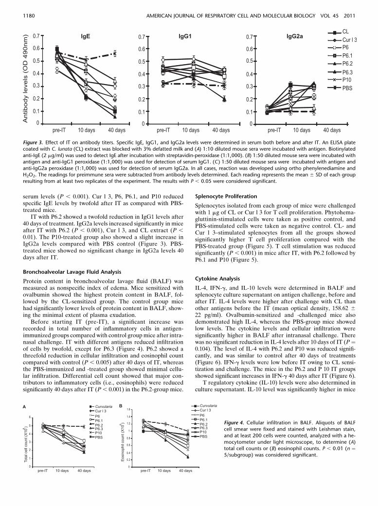
Before starting IT (pre-IT), a significant increase wasrecorded in total number of inflammatory cells in antigen-immunized groups compared with control groupmice after intra-nasal challenge. IT with different antigens reduced infiltrationof cells by twofold, except for P6.3 (Figure 4). P6.2 showed athreefold reduction in cellular infiltration and eosinophil countcompared with control (P , 0.005) after 40 days of IT, whereasthe PBS-immunized and -treated group showed minimal cellu-lar infiltration. Differential cell count showed that major con-tributors to inflammatory cells (i.e., eosinophils) were reducedsignificantly 40 days after IT (P, 0.001) in the P6.2-group mice.
Splenocyte Proliferation
Splenocytes isolated from each group of mice were challengedwith 1 mg of CL or Cur l 3 for T cell proliferation. Phytohema-gluttinin-stimulated cells were taken as positive control, andPBS-stimulated cells were taken as negative control. CL- andCur l 3–stimulated splenocytes from all the groups showedsignificantly higher T cell proliferation compared with thePBS-treated group (Figure 5). T cell stimulation was reducedsignificantly (P , 0.001) in mice after IT, with P6.2 followed byP6.1 and P10 (Figure 5).
Cytokine Analysis
IL-4, IFN-g, and IL-10 levels were determined in BALF andsplenocyte culture supernatant on antigen challenge, before andafter IT. IL-4 levels were higher after challenge with CL thanother antigens before the IT (mean optical density, 158.62 622 pg/ml). Ovalbumin-sensitized and -challenged mice alsodemonstrated high IL-4, whereas the PBS-group mice showedlow levels. The cytokine levels and cellular infiltration weresignificantly higher in BALF after intranasal challenge. Therewas no significant reduction in IL-4 levels after 10 days of IT (P¼0.104). The level of IL-4 with P6.2 and P10 was reduced signifi-cantly, and was similar to control after 40 days of treatments(Figure 6). IFN-g levels were low before IT owing to CL sensi-tization and challenge. The mice in the P6.2 and P 10 IT groupsshowed significant increases in IFN-g 40 days after IT (Figure 6).
T regulatory cytokine (IL-10) levels were also determined inculture supernatant. IL-10 level was significantly higher in mice
Figure 3. Effect of IT on antibody titers. Specific IgE, IgG1, and IgG2a levels were determined in serum both before and after IT. An ELISA plate
coated with C. lunata (CL) extract was blocked with 3% defatted milk and (A) 1:10 diluted mouse sera were incubated with antigen. Biotinylated
anti-IgE (2 mg/ml) was used to detect IgE after incubation with streptavidin-peroxidase (1:1,000). (B) 1:50 diluted mouse sera were incubated withantigen and anti-IgG1 peroxidase (1:1,000) was used for detection of serum IgG1. (C) 1:50 diluted mouse sera were incubated with antigen and
anti-IgG2a peroxidase (1:1,000) was used for detection of serum IgG2a. In all cases, reaction was developed using ortho phenylenediamine and
H2O2. The readings for preimmune sera were subtracted from antibody levels determined. Each reading represents the mean 6 SD of each group
resulting from at least two replicates of the experiment. The results with P , 0.05 were considered significant.
Figure 4. Cellular infiltration in BALF. Aliquots of BALF
cell smear were fixed and stained with Leishman stain,
and at least 200 cells were counted, analyzed with a he-
mocytometer under light microscope, to determine (A)total cell counts or (B) eosinophil counts. P , 0.01 (n ¼5/subgroup) was considered significant.
1180 AMERICAN JOURNAL OF RESPIRATORY CELL AND MOLECULAR BIOLOGY VOL 45 2011

40 days after IT with P6 and P6.2 as compared to baseline (P ,0.001) (Figure 6). Mice receiving IT with P10 also showed in-creased IL-10 level after IT.
Lung Histology
Hematoxylin and eosin–stained lung sections of mice were an-alyzed before and after 10 days (Figure 7A) or 40 days of IT(Figure 7B) after challenge with antigen. There was markeddisruption of alveolar structure and profound cellular infiltra-tion throughout the lung tissue in ovalbumin or CL-sensitized/-challenged mice before IT. CL-sensitized/-challenged mice inthe PBS IT group showed disrupted alveolar structure, withlarge numbers of mucus-secreting cells and goblet cells in thebronchi. PBS-immunized/-challenged (control) mice showednormal lung structure, and had occasional inflammatory cellsin the lung tissue (Figures 7A and 7B). Lung inflammation scorewas reduced significantly for the group treated with P6.2,followed by P10, P6, Cur l 3, CL, P6.1, and P6.3 after 40 daysof IT (Figures 7C and 7D).
The eosinophil peroxidase activity was measured as a markerfor activation of eosinophils. The levels are presented as mean
change in absorbance in BALF and lung homogenate of all mice.EPO activity was markedly increased in BALF and lung ho-mogenate of all antigen-group mice before the IT (Figure7E). Ovalbumin-immunized/-challenged mice demonstrated asignificant enhancement in EPO activity compared with PBS(n ¼ 5; P , 0.02). The mice challenged with CL demonstratedhigh EPO activity before IT. EPO activity in BALF and lunghomogenate of mice was reduced significantly 40 days after ITwith P6.2 as compared to 10 days after IT (Figure 7E). EPOactivity was also reduced after 40 days of IT with CL, Cur l 3,peptides, and P6 derivatives.
DISCUSSION
IT with allergen extracts/purified proteins is associated with ad-verse reactions. Numerous strategies have been developed to re-duce the allergenicity of vaccines, while maintaining the immunemodulation. Such approaches include alteration of the tertiarystructure of the allergen by chemical modification (17), gene/protein engineering (18–20), conjugation of bacterial DNAoligomers to purified allergens (21), and generation of allergenmultimers (22). Synthetic peptides corresponding to T cell epit-opes of the allergen were evaluated in various IT studies (6, 23,24). These peptides were unable to cross-link IgE on mast cells/basophils, and proved effective candidates for IT (7, 25, 26).T cell epitope peptide from Fel d 1 induced IL-10 secretion inhumans and mice, and reduced allergic inflammation (27).C-terminal Cys residues of Asp f 4 allergen were implicatedin IgE binding (28). We have previously identified Cur l 3 asa cross-reactive protein in fungi, grasses, and ragweed (11). Fur-thermore, we have shown that P6 of Cur l 3 has both B andT cell epitopes, and belongs to a conserved region (16). In thepresent study, Cur l 3–derived peptides, P6, its 3 derivatives(P6.1, 6.3, 6.3), and P10, were evaluated for IT in a mousemodel of allergy. In the modified peptides (i.e., P6.1), Cys6
was changed to Ala, P6.2 Cys3, and Cys6, both replaced withAla, whereas, in P6.3, His7 was changed to Ala. Cys- and His-modified peptides showed significant reduction in IgE binding,but the immunogenicity was intact, as revealed by lymphocyteproliferation assay.
Experimental models of allergic asthma were developed inrodents using ovalbumin (29). Allergen extracts/protein fromgrass, mite, and cockroach have been used for development ofanimal models (30–32). Alternaria and Cladosporium sporesinduced airway inflammation in the mouse, and were char-acterized by high numbers of macrophages, neutrophils,
Figure 5. Splenocyte proliferation. Splenocytes isolated from each
group were challenged with CL (1 mg), Cur l 3 (1 mg), or each of the
antigens used for IT. They were cultured for 72 hours in the presence of10% FCS. Cells were washed with RPMI and incubated for 2 hours with
3-(4, 5-dimethylthiazol-2-yl)-2,5-diphenyltetrazolium bromide (MTT;
100 mg/ml). The cells were lysed in acidic isopropanol solution, and
the absorbance read at 570 nm. Phytohemagluttinin (PHA) was used ascontrol for positive stimulation, whereas unstimulated cells gave the
basal stimulation level due to FCS. The bar graph presents mean 6SE values (P , 0.01 [n ¼ 5/subgroup] was considered significant).
Figure 6. Cytokine analysis in BALF supernatant after antigen challenge before and after IT. (A) IL-4, (B) IFN-g, and (C) IL-10 levels were determined
by sandwich ELISA. Capture antibody (1:250 vol/vol) for each cytokine was coated separately on microtitre plates and incubated overnight. After
blocking with 10% FCS, 1:2 vol/vol–diluted BALF samples were added to wells. Biotinylated anti–IL-4, anti–IL-10, or anti–IFN-g were used to detectspecific cytokine levels. The line graph presents mean 6 SE values for each group before and after IT. P , 0.01 (n ¼ 5/subgroup) was considered
significant.
Sharma, Singh, and Arora: Engineered Cur l 3 Peptides for Immunotherapy 1181

Figure 7. Histopathology of lungs and semiquantitative analysis of airway inflammation. Effect of allergen IT on airway inflammation assessed by
lung histopathology after (A) 10 days or (B) 40 days of IT. Lung tissue segments, preserved in formalin, from each mouse were processed forsectioning and embedded in paraffin wax. The sections, cut at 4-um thickness, were stained with hematoxylin and eosin and scanned under
a microscope. (C) Peribronchial and perivascular inflammation were graded on a subjective scale of 0–4, corresponding to mild, moderate,
marked, or severe inflammation, respectively, with an increment of 0.5 if the inflammation fell between two integers. The total lung inflam-mation was defined as the sum of peribronchial and perivascular inflammation scores. The values are expressed as mean SEM for each group of
mice. Total histological score, graded on a scale from 0 to 4: 0, no inflammatory cells; 1,,10%; 2, 10–25%; 3, 25–50%; 4,.50%. For simplicity,
these data are represented as a box and whisker plot (D) for lung inflammation score for pre-IT, 10 and 40 days after IT. (E) Eosinophil peroxidase
levels in BALF of each mouse was determined by transferring 50 ml of 1:10–diluted BALF sample to a microtitre plate containing substratesolution comprised of 3 mM OPD, 0.05 M Tris (pH 8.0), 0.1% Triton X-100, and 8.8 mM H2O2. The graph presents mean change in absorbance
at 490 nm. OD, optical density.
1182 AMERICAN JOURNAL OF RESPIRATORY CELL AND MOLECULAR BIOLOGY VOL 45 2011

eosinophils, and lymphocytes in BALF (33). Gonzalo andcolleagues (34) showed the presence of inflammatory cellsin BALF in ovalbumin-sensitized/-challenged mice. In thepresent study, sensitization/challenge with CL in mice demon-strated significantly increased specific IgE, inflammatory cells(eosinophils), and Th2 cytokine (IL-4). The results were inconcordance with a study on Cladosporium and Alternariasensitization and challenge (33).
Airway hyperreactivity is associated with infiltration ofinflammatory cells, particularly eosinophils (34, 36). Allergenicproteins induced eosinophil activation and release of mediatorsthat caused airway damage and hyperreactivity (37). In thepresent study, cellular infiltration did not change significantlyafter 10 days of IT. However, 40 days after IT, total cell countdecreased significantly in the P6.2 group, followed by P10-, Cur l3–, CL-, P6.1-, and P6.3-treated mice. Eosinophils and EPOactivity were reduced maximally in the P6.2 IT group, followedby P10 signifying therapeutic potential.
Allergen IT decreases Th2-type antibody titers (IgE andIgG1), with an increase in Th1 antibody, IgG2a (8, 37). PeptideIT modulates immune response by inducing T cell anergy,a switch from Th2 to Th1, and induction of regulatory/suppressor T cells (8, 23). In the present study, P6.2 and P10reduced IgE and IgG1 significantly (P , 0.05), with an increasein IgG2a levels, 40 days after IT. P6.2 and P10 showed bettertherapeutic potential, with reduction in IL-4 level substantiallyas compared to P6, P6.1, P6.3, Cur l 3 and CL. This decrease inIL-4 level was associated with increased IFN-g levels, demon-strating modulation of Th2 to Th1 by P6.2 and P10 peptides.These observations suggest the therapeutic potential ofpeptide P6.2 (i.e., modified form of P6). Studies show thatlong-term effects of IT are associated with secretion ofIL-10 T-regulatory cytokine (27, 29, 38, 39). Campbell andcolleagues (27) showed that epitope therapy induced IL-10in a mouse model. It has been demonstrated that anti–IL-10R monoclonal antibody reduced the beneficial effects ofIT (40, 41). In the present study, P6 and P6.2 stimulated highIL-10 secretion after 40 days after IT. However, P10 was notable to induce higher IL-10 levels.
Earlier studies have shown that lung inflammation and infil-tration of cells correlated with antibody titers and cytokines(33–35). In the present study, histopathology showed significantimprovement in the airway inflammation after 40 days of ITwith P6.2 or P10. Janssen and colleagues (29) observed thatovalbumin IT improved allergic inflammation, airway hyperres-ponsiveness, and eosinophilia as compared with its peptide hav-ing both B and T cell epitopes. Here, IT with peptide P6 peptideinduced Th1 response, and proved beneficial in reducing lungand systemic inflammation. Reduction in allergic airway inflam-mation in Cur l 3 and CL extract IT groups after 40 days couldbe due to the presence of multiple T cell epitopes. The presentstudy demonstrated that allergic airway inflammation in themouse model could be reduced effectively by IT, with smallsynthetic peptides having low IgE binding. The effects of ITwere better 40 days after IT than 10 days of IT.
Peptide IT with single T cell epitope results in induction oflinked epitope suppression, whereby cells rendered tolerant toone epitope suppress the function of cells specific for other epit-opes on the same molecule. Previously, mice sensitized withFel d 1, challenged with cat allergen extract, and treated withsingle T cell epitope peptide (Fel d 1 [29–45]) demonstrated sig-nificant reduction in airway inflammation and systemic Th2responses (27, 42). The study also showed that peptide specificT cell population down-regulates an established inflammatoryresponse driven by multiple T cell epitopes. IT results in induc-tion of IL-10 producing “bystander” T cells, apart from the
allergen-specific T cells that result in anergy, and thus reducedT cell–proliferative response. This explanation is in agreementwith the present study, where an increase in IL-10 has been seenin both BALF and culture supernatant of the P6.2-treatedgroup.
In conclusion, CL induced airway inflammation and systemicTh2 response in mice, because it contains a large repertoire ofallergen components. IT in this mouse model with whole extract,Cur l 3, and short synthetic peptides, P6, P6.1, P6.2, P6.3, and P10,proved beneficial in reducing inflammation and modulating Th2to Th1 response. Therapeutic potential of short peptides, P6.2,followed by P10 and P6, was more pronounced compared withother antigens after IT. These short peptides may be tested inhumans for IT.
Author Disclosure: None of the authors has a financial relationship with a com-mercial entity that has an interest in the subject of this manuscript.
Acknowledgments: The authors acknowledge Dr. Pradeep Kumar and Dr. SantoshPasha (Scientist, Institute of Genomics and Integrative Biology) for help in chem-ical modifications of P6 and synthesis of peptides.
References
1. Akdis M, Akdis CA. Therapeutic manipulation of immune tolerance in
allergic disease. Nat Rev Drug Discov 2009;8:645–660.
2. Akdis CA, Blaser K, Akdis M. Mechanisms of allergen-specific immu-
notherapy. Chem Immunol Allergy 2006;91:195–203.
3. Hawrylowicz CM, O’Garra A. Potential role of interleukin-10–secreting reg-
ulatory T cells in allergy and asthma. Nat Rev Immunol 2005;5:271–283.
4. Rolland JM, Gardner LM, O’Hehir RE. Allergen-related approaches to
immunotherapy. Pharmacol Ther 2009;121:273–284.
5. Ali FR, Larche M. Peptide-based immunotherapy: a novel strategy for
allergic disease. Expert Rev Vaccines 2005;4:881–889.
6. Norman PS, Ohman JL Jr., Long AA, Creticos PS, Gefter MA, Shaked
Z, Wood RA, Eggleston PA, Hafner KB, Rao P, et al. Treatment of
cat allergy with T-cell reactive peptides. Am J Respir Crit Care Med
1996;154:1623–1628.
7. von Garnier C, Astori M, Kettner A, Dufour N, Heusser C, Corradin G,
Spertini F. Allergen-derived long peptide immunotherapy down-
regulates specific IgE response and protects from anaphylaxis. Eur J
Immunol 2000;30:1638–1645.
8. Oldfield WL, Kay AB, Larche M. Allergen-derived T cell peptide–induced
late asthmatic reactions precede the induction of antigen-specific hypo-
responsiveness in allergic asthmatic subjects. J Immunol 2001;167:1734–
1739.
9. Gupta R, Singh BP, Sridhara S, Gaur SN, Chaudhary VK, Arora N.
Allergens of Curvularia lunata during cultivation in different media.
J Allergy Clin Immunol 1999;104:857–862.
10. Pumhirum P, Towiwat P, Mahakit P. Aeroallergen sensitivity of Thai
patients with allergic rhinitis. Asian Pac J Allergy Immunol 1997;15:
183–185.
11. Sharma V, Singh BP, Gaur SN, Arora N. Molecular and immunological
characterization of cytochrome c: a potential cross-reactive allergen in
fungi and grasses. Allergy 2008;63:189–197.
12. Ekramoddoullah AK, Kisil FT, Sehon AH. Isolation of allergenically
active cytochrome c from Kentucky blue grass pollen. Int Arch Al-
lergy Appl Immunol 1981;65:367–376.
13. Ekramoddoullah AK, Kisil FT, Sehon AH. Allergenic cross-reactivity of
cytochromes c of Kentucky bluegrass and perennial ryegrass pollens.
Mol Immunol 1982;19:1527–1534.
14. Ekramoddoullah AK, Kisil FT, Bundesen PG, Fischer JM, Rector ES,
Sehon AH. Determinants of ryegrass pollen cytochrome c recognized
by human IgE and murine monoclonal antibodies. Mol Immunol
1984;21:375–382.
15. Matthews PA, Baldo BA, Howden ME. Cytochrome c allergens isolated
from the pollens of the dicotyledons English plantain (Plantago lan-
ceolata) and Paterson’s curse (Echium plantagineum). Mol Immunol
1988;25:63–68.
16. Sharma V, Singh BP, Gaur SN, Pasha S, Arora N. Bioinformatics and
immunologic investigation on B and T cell epitopes of Cur l 3, a major
allergen of Curvularia lunata. J Proteome Res 2009;8:2650–2655.
Sharma, Singh, and Arora: Engineered Cur l 3 Peptides for Immunotherapy 1183

17. Raĭkis BN, Illiutovich NA. Prospects for chemical modification of anti-
gens used in the prevention and treatment of infectious and non-
infectious allergic diseases. Zh Mikrobiol Epidemiol Immunobiol
1981;3:8–13.
18. Ferreira F, Ebner C, Kramer B, Casari G, Briza P, Kungl AJ, Grimm R,
Jahn-Schmid B, Breiteneder H, Kraft D, et al. Modulation of IgE
reactivity of allergens by site-directed mutagenesis: potential use of
hypoallergenic variants for immunotherapy. FASEB J 1998;12:231–242.
19. Vrtala S, Focke M, Kopec J, Verdino P, Hartl A, Sperr WR, Fedorov
AA, Ball T, Almo S, Valent P, et al. Genetic engineering of the major
timothy grass pollen allergen, Phl p 6, to reduce allergenic activity and
preserve immunogenicity. J Immunol 2007;179:1730–1739.
20. Niederberger V, Horak F, Vrtala S, Spitzauer S, Krauth MT, Valent P,
Reisinger J, Pelzmann M, Hayek B, Kronqvist M, et al. Vaccination
with genetically engineered allergens prevents progression of allergic
disease. Proc Natl Acad Sci USA 2004;101:14677–14682.
21. Creticos PS, Lichtenstein LM, Spiegelberg HL, Raz E. Conjugation of
immunostimulatory DNA to the short ragweed allergen Amb a 1
enhances its immunogenicity and reduces its allergenicity. J Allergy
Clin Immunol 2000;106:124–134.
22. Pauli G, Oster JP, Deviller P, Heiss S, Bessot JC, Susani M, Ferreira F,
Kraft D, Valenta R. Skin testing with recombinant allergens rBet v 1
and birch profilin, rBet v 2: diagnostic value for birch pollen and as-
sociated allergies. J Allergy Clin Immunol 1996;97:1100–1109.
23. Muller U, Akdis CA, Fricker M, Akdis M, Blesken T, Bettens F, Blaser
K. Successful immunotherapy with T-cell epitope peptides of bee
venom phospholipase A2 induces specific T-cell anergy in patients
allergic to bee venom. J Allergy Clin Immunol 1998;101:747–754.
24. Fellrath JM, Kettner A, Dufour N, Frigerio C, Schneeberger D,
Leimgruber A. Allergen-specific T-cell tolerance induction with al-
lergen derived long synthetic peptides: results of a phase I trial.
J Allergy Clin Immunol 2003;111:854–861.
25. Smith TR, Alexander C, Kay AB, Larche M, Robinson DS. Cat allergen
peptide immunotherapy reduces CD4 T cell responses to cat allergen
but does not alter suppression by CD4 CD25 T cells: a double-blind
placebo-controlled study. Allergy 2004;59:1097–1101.
26. Larche M. Peptide immunotherapy for allergic diseases. Allergy 2007;62:
325–331.
27. Campbell JD, Buckland KF, McMillan SJ, Kearley J, Oldfield WL, Stern
LJ. Peptide immunotherapy in allergic asthma generates IL-10–
dependent immunological tolerance associated with linked epitope
suppression. J Exp Med 2009;206:1535–1547.
28. Ramachandran H, Banerjee B, Greenberger PA, Kelly KJ, Fink JN,
Kurup VP. Role of C-terminal cysteine residues of Aspergillus
fumigatus allergen Asp f 4 in immunoglobulin E binding. Clin Diagn
Lab Immunol 2004;11:261–265.
29. Janssen EM, van Oosterhout AJM, Nijkamp FP, van Eden W, Wauben
MH. The efficacy of immunotherapy in an experimental murine
model of allergic asthma is related to the strength and site of T cell
activation during immunotherapy. J Immunol 2000;165:7207–7214.
30. Kumar M, Behera AK, Hu J, Lockey RF, Mohapatra SS. IFN-g and
IL-12 plasmid DNAs as vaccine adjuvant in a murine model of
grass allergy. J Allergy Clin Immunol 2001;108:402–408.
31. Sarpong SB, Zhang LY, Kleeberger SR. A novel mouse model of ex-
perimental asthma. Int Arch Allergy Immunol 2003;132:346–354.
32. Srivastava D, Mehta AK, Arora N, Gaur SN, Singh BP. Proteolytically
inactive Per a 10 allergen of Periplanata americana modulates Th2
response and enhances IL-10 in mouse model. J Clin Immunol 2010;
30:426–434.
33. Havaux X, Zeine A, Dits A, Denis O. A new mouse model of lung al-
lergy induced by the spores of Alternaria alternata and Cladosporium
herbarum molds. Clin Exp Immunol 2005;139:179–188.
34. Gonzalo JA, Lloyd CM, Kremer L, Finger E, Martinez-A C, Siegelman
MH. Eosinophil recruitment to the lung in a murine model of allergic
inflammation: the role of T cells, chemokines, and adhesion receptors.
J Clin Invest 1996;98:2332–2345.
35. Tai HY, Tam MF, Chou H, Peng HJ, Su SN, Perng DW, Shen HD. Pen
ch 13 allergen induces secretion of mediators and degradation of
occludin protein of human lung epithelial cells. Allergy 2006;61:382–
388.
36. Eum SY, Haile S, Lefort J, Huerre M, Vargaftig BB. Eosinophil re-
cruitment into the respiratory epithelium following antigenic chal-
lenge in hyper-IgE mice is accompanied by interleukin 5–dependent
bronchial hyperresponsiveness. Proc Natl Acad Sci USA 1995;92:
12290–12294.
37. Larche M, Wraith DC. Peptide based therapeutic vaccines for allergic
and autoimmune diseases. Nat Med 2005;11:S69–S76.
38. Akdis CA, Blesken T, Akdis M, Wuthrich B, Blaser K. Role of inter-
leukin 10 in specific immunotherapy. J Clin Invest 1998;102:98–106.
39. Xystrakis E, Boswell SE, Hawrylowicz CM. T regulatory cells and the
control of allergic disease. Expert Opin Biol Ther 2006;6:121–133.
40. O’Farrell AM, Liu Y, Moore KW, Mui AL. IL-10 inhibits macrophage
activation and proliferation by distinct signaling mechanisms: evi-
dence for Stat3-dependent and -independent pathways. EMBO J
1998;17:1006–1018.
41. Castro AG, Neighbors M, Hurst SD, Zonin F, Silva RA, Murphy E.
Anti–interleukin 10 receptor monoclonal antibody is an adjuvant for
T helper cell type 1 responses to soluble antigen only in the presence
of lipopolysaccharide. J Exp Med 2000;192:1529–1534.
42. Saarne T, Neimert-Andersson T, Gronlund H, Jutel M, Gafvelin G, van
Hage M. Treatment with a Fel d 1 hypoallergen reduces allergic
responses in a mouse model for cat allergy. Allergy 2011;66:255–263.
1184 AMERICAN JOURNAL OF RESPIRATORY CELL AND MOLECULAR BIOLOGY VOL 45 2011
Copyright © 2022 FDOKUMEN