Cloning, expression and sequence homologies of cDNA for human gamma enolase
8
GENOMICS 1, 159-166 (1987) Cloning, Expression, and Sequence Homologies of cDNA for Human Carbonic Anhydrase II HIROSHI MURAKAMI, GREGORY P. MARELICH, JEFFREY H. GRUBS, JOHN W. KYLE, AND WILLIAM S. SLY’ E. A. Daisy Department of Biochemistry, St. Louis University School of Medicine, St. Louis, Missouri 63 104 ReceivedJune 23. 1987 A cDNA clone for human carbonic anhydrase (CA) II was isolated from a kidney XgtlO library. Expres- sion of the cDNA insert in Cos-7 cells produced an immunoprecipitable product and enzymatically ac- tive carbonic anhydrase. The cDNA insert is 1661 bp in length and contains an open reading frame which encodes a 260-amino-acid polypeptide. The deduced amino acid sequence is identical to that reported for human CA II. The protein coding region of this cDNA for human CA II shows 81 and 70% nucleotide iden- tity with cDNAs for CA II from mouse and chick, re- spectively. Even the long 3’-untranslated region of the cDNA for human CA II (703 bp) is 64 and 42% identi- cal to those of CA II from mouse and chick, showing remarkable conservation of the CA II cDNAs in am- niotes. The protein coding region of the human CA II cDNA is 64 and 66% identical with those of human CA I and CA III, which are thought to have arisen from a common precursor by gene duplication. 0 1987 Aoa- demic Press, Inc. INTRODUCI’ION Carbonic anhydrase (CA) (EC 4.2.1.1) catalyzes the reversible hydration of carbon dioxide. Three distinct cytosolic isozymes (CA I, CA II, and CA III) have been identified, and a considerable number of structural and enzymatic studies have been carried out on them (Maren, 1967; Lindskog et aZ., 1971; Carter, 1972; Packer and Sarkanen, 1978; Maren and Sanyal, 1983; Tashian et al., 1983; Hewett-Emmett et al., 1984). They are monomeric zinc metalloenzymes with a mo- lecular weight of approximately 29,000. Each isozyme has characteristic properties and tissue distribution (Tashian et al., 1984; Sanyal, 1984; Sundaram et al., 1986). In addition to these soluble isozymes, membrane- bound carbonic anhydrases in lung and kidney Sequence data from this article have been deposited with the EMBLIGenBank Data Libraries under Accession No. 503037. 1To whom all correspondence should be addressed. (Whitney and Briggle, 1982; Wistrand, 1984; Sanyal et al., 1981; McKinley and Whitney, 1976) and a secre- tory carbonic anhydrase in saliva (Feldstein and Sil- verman, 1984; Murakami and Sly, 1987) have been reported. These isozymes are glycoproteins much larger than the 29,000-D cytosolic isozymes, but their enzymatic and inhibitory properties resemble those of CA II. We are interested in defining the relationships of CA II, a deficiency of which produces the syndrome of osteopetrosis with renal tubular acidosis and cerebral calcification in humans (Sly et al., 1983; Sly et uZ., 1985), to the higher molecular weight carbonic anhy- drases, which are glycoproteins. As a first step toward this end, we purified and characterized a secretory carbonic anhydrase from human saliva. We found that human salivary CA and CA II are related immu- nologically and genetically (Murakami and Sly, 1987). To clarify the relationship of the genes for these iso- zymes, we set out to isolate and compare their cDNAs. In this paper, we report the isolation of the cDNA clone for CA II, show that the cDNA clone specifies active enzyme when expressed in Cos-7 cells, and compare the sequence of the cDNA clone with those of cDNAs for CA II from other species and with cDNA clones for human CA I and CA III, the other cytosolic members of the human CA gene family. MATERIALS AND METHODS [a-32P]dCTP (3000 Ci/mmol) and Trans35S-label (1090 Ci/mmol) were purchased from ICN Radio- chemicals (Irvine, CA). Nick translation kit was ob- tained from Amersham (Arlington Heights, IL). IgG- sorb was from The Enzyme Center (Malden, MA). Screening cDNA Library The human kidney XgtlO cDNA library, which was kindly provided by Dr. Graeme I. Bell at the Chiron Corporation (Bell et uZ., 1986), was screened with a partial human CA II genomic clone (H25-3.8) (Venta 159 0388-7543/81$3.00 Copyright 0 1987 by Academic Press, Inc. All rights of reproduction in any form reserved.
-
Upload
independent -
Category
Documents
-
view
3 -
download
0
Transcript of Cloning, expression and sequence homologies of cDNA for human gamma enolase

GENOMICS 1, 159-166 (1987)
Cloning, Expression, and Sequence Homologies of cDNA for Human Carbonic Anhydrase II
HIROSHI MURAKAMI, GREGORY P. MARELICH, JEFFREY H. GRUBS, JOHN W. KYLE, AND WILLIAM S. SLY’
E. A. Daisy Department of Biochemistry, St. Louis University School of Medicine, St. Louis, Missouri 63 104
ReceivedJune 23. 1987
A cDNA clone for human carbonic anhydrase (CA) II was isolated from a kidney XgtlO library. Expres- sion of the cDNA insert in Cos-7 cells produced an immunoprecipitable product and enzymatically ac- tive carbonic anhydrase. The cDNA insert is 1661 bp in length and contains an open reading frame which encodes a 260-amino-acid polypeptide. The deduced amino acid sequence is identical to that reported for human CA II. The protein coding region of this cDNA for human CA II shows 81 and 70% nucleotide iden- tity with cDNAs for CA II from mouse and chick, re- spectively. Even the long 3’-untranslated region of the cDNA for human CA II (703 bp) is 64 and 42% identi- cal to those of CA II from mouse and chick, showing remarkable conservation of the CA II cDNAs in am- niotes. The protein coding region of the human CA II cDNA is 64 and 66% identical with those of human CA I and CA III, which are thought to have arisen from a common precursor by gene duplication. 0 1987 Aoa-
demic Press, Inc.
INTRODUCI’ION
Carbonic anhydrase (CA) (EC 4.2.1.1) catalyzes the reversible hydration of carbon dioxide. Three distinct cytosolic isozymes (CA I, CA II, and CA III) have been identified, and a considerable number of structural and enzymatic studies have been carried out on them (Maren, 1967; Lindskog et aZ., 1971; Carter, 1972; Packer and Sarkanen, 1978; Maren and Sanyal, 1983; Tashian et al., 1983; Hewett-Emmett et al., 1984). They are monomeric zinc metalloenzymes with a mo- lecular weight of approximately 29,000. Each isozyme has characteristic properties and tissue distribution (Tashian et al., 1984; Sanyal, 1984; Sundaram et al., 1986).
In addition to these soluble isozymes, membrane- bound carbonic anhydrases in lung and kidney
Sequence data from this article have been deposited with the EMBLIGenBank Data Libraries under Accession No. 503037.
1 To whom all correspondence should be addressed.
(Whitney and Briggle, 1982; Wistrand, 1984; Sanyal et al., 1981; McKinley and Whitney, 1976) and a secre- tory carbonic anhydrase in saliva (Feldstein and Sil- verman, 1984; Murakami and Sly, 1987) have been reported. These isozymes are glycoproteins much larger than the 29,000-D cytosolic isozymes, but their enzymatic and inhibitory properties resemble those of CA II.
We are interested in defining the relationships of CA II, a deficiency of which produces the syndrome of osteopetrosis with renal tubular acidosis and cerebral calcification in humans (Sly et al., 1983; Sly et uZ., 1985), to the higher molecular weight carbonic anhy- drases, which are glycoproteins. As a first step toward this end, we purified and characterized a secretory carbonic anhydrase from human saliva. We found that human salivary CA and CA II are related immu- nologically and genetically (Murakami and Sly, 1987). To clarify the relationship of the genes for these iso- zymes, we set out to isolate and compare their cDNAs. In this paper, we report the isolation of the cDNA clone for CA II, show that the cDNA clone specifies active enzyme when expressed in Cos-7 cells, and compare the sequence of the cDNA clone with those of cDNAs for CA II from other species and with cDNA clones for human CA I and CA III, the other cytosolic members of the human CA gene family.
MATERIALS AND METHODS
[a-32P]dCTP (3000 Ci/mmol) and Trans35S-label (1090 Ci/mmol) were purchased from ICN Radio- chemicals (Irvine, CA). Nick translation kit was ob- tained from Amersham (Arlington Heights, IL). IgG- sorb was from The Enzyme Center (Malden, MA).
Screening cDNA Library
The human kidney XgtlO cDNA library, which was kindly provided by Dr. Graeme I. Bell at the Chiron Corporation (Bell et uZ., 1986), was screened with a partial human CA II genomic clone (H25-3.8) (Venta
159 0388-7543/81$3.00 Copyright 0 1987 by Academic Press, Inc.
All rights of reproduction in any form reserved.

160 MURAKAMI ET AL.
1.0 1.i Kb
f
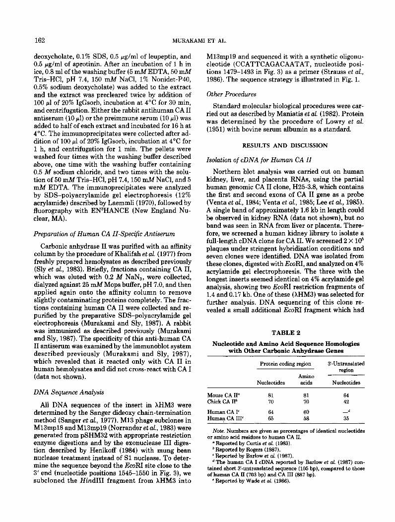
FIG. 1. Restriction map and nucleotide sequencing strategy for the cDNA insert of XHM3. The coding region for human car- bonic anhydrase II is shown by the open rectangle. The sequencing strategy is shown by the arrows below the restriction map. The poly(A) region sequenced with the synthetic oligonucleotide as a primer is shown by the broken arrow.
et al., 1984; Venta et al., 1985; Lee et al., 1985) which contained the first two exons of human CA II gene, with the procedure described by Maniatis et al. (1982). The prehybridizations were carried out for 24 h at 52°C in a solution of 50% formamide, 5X Denhardt’s solution (1X Denhardt’s solution is 0.02% each of Fi- ~011, polyvinylpyrrolidone, and bovine serum albumin fraction V), 5X SSC (1X SSC is 150 mM sodium chlo- ride/15 mM sodium citrate), 0.1% SDS, 0.2 mg/ml denatured salmon sperm DNA, and 50 n&f sodium phosphate buffer, pH 6.5. Hybridizations were per- formed for 16 h at 52°C after addition of the 32P-la- beled probe at approximately 1 X lo6 cpm/ml into the solution of 50% formamide, 1X Denhardt’s solution, 2X SSC, 0.1% SDS, 10% dextran sulfate, 50 pg/ml denatured salmon sperm DNA, and 50 mM sodium phosphate buffer, pH 6.5. [32P]DNA probes were made by nick-translation labeling of the partial geno- mic CA II clone, H25-3.8, with [a-32P]dCTP to a spe- cific activity of approximately 1 X lo8 cpm/pg of DNA, according to the supplier’s instructions. After hybridization, filters were washed twice in 2X SSC/O.l% SDS for 10 min each at room temperature, and twice in 0.1X SSC/O.l% SDS for 10 min each at 60°C.
Expression of Human CA II cDNA in Cos-7 Cells
To express the human CA II cDNA, which was in the EcoRI site of XgtlO, we modified the Cos-7 cell expression vector pJC119 (Sprague et al., 1983). First, to remove the EcoRI site from the vector, pJC119 was digested with EcoRI, and the cohesive ends were filled with DNA polymerase I large fragment (Klenow frag- ment) and religated. Second, to create an EcoRI site at the unique XhoI site, which is downstream of the SV40 late promoter, the plasmid was digested with XhoI, the single-strand overhangs of XhoI site were
TABLE 1
Expression of Carbonic Anhydrase Activity in Transfected Cos-7 Cells
Plasmid
pSHM32 pSHM35 pSHM28
CA activity in transfected Cos-7 cells
(unit/mg of protein)
10.6 0.186 0.136
removed by the treatment of mung bean nuclease, and the blunt-ended plasmid was religated with EcoRI linkers. The excess of linkers was removed by diges- tion with EcoRI. Religation produced pSHM28, which has a unique EcoRI site as a cloning site instead of an XhoI site.
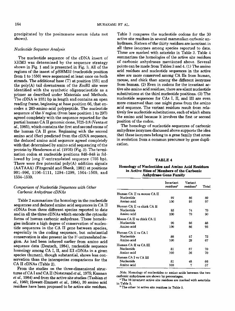
Cos-7 cells (Gluzman, 1981) were transfected using the DEAE-dextran procedure (Lopata et al., 1984) followed by treatment with 100 &f chloroquine (Luthman and Magnusson, 1983). Seventy-six hours posttransfection, cells were harvested, washed with 10 n&f Tris-SO1, pH 7.5, 50 m&f NazS04 at 4”C, then suspended with 0.05% Triton X-100. The carbonic anhydrase activity was measured with the procedure described previously (Murakami and Sly, 1987).
To label the transfected cells, we used the proce- dure described by Oshima et al. (1987), except that the
- 21.5
1 2 3
FIG. 2. Immunoprecipitation with anti-human CA II anti- serum of %4-labeled proteins expressed in transfected Cos-7 cells. Cos-7 cells transfected with the indicated plasmid were labeled with Trans”S-label and analyzed by SDS-polyacrylamide gel electro- phoresis and fluorography as described under Materials and Methods. Lanes 1, 2, and 3 are Cos-7 cells transfected with pSHM32, pSHM35, and pSHM28, respectively. The migration po- sition of purified human CA II is shown by the arrow. The migra- tion positions of molecular weight markers (Bio-Rad) are shown as X10" D on the right.
cDNA FOR CARBONIC ANHYDRASE II FROM HUMAN KIDNEY
cells were labeled for 12 h with 67 &i of Trans35S- The labeled cells were solubilized in 0.2 ml of the label (1090 Ci/mmol) in 1 ml of methionine-free solubilization buffer containing 10 miV Tris-HCl, pH DMEM containing 5% dialyzed fetal bovine serum. 7.4, 66 mM EDTA, 1% Nonidet-P40, 0.4% sodium
10 20 30 40 50 60 GGCGCCCMGCCGCCGCCGCCAGATCGGTGCCGATTCCTGCCCTGCCCCGACCGCCAGCGCGACC
75 65 95 105 115 125 135 ATGTCCCATCACTGGGGGTACGGCAAACACAACGGACCTGAGCACTGGCATMGGACTTCCCCATTGCCMGGGA MetSerHisHisTrpGlyTyrGlyLysHisAsnGlyProGluHisTrpHisLysAspPheProIleAlaLysGly 25
--- __I 150 160 170 180 190 200 210
GAGCGCCAGTCCCCTGTTGACATCGACACTCATACAGCC~GTATGACCCTTCCCTGMGCCCCTGTCTGTTTCC GluArgGlnSerProValAspIleAspThrHisThrAlaLysTyrAspProSerLeuLysP~oLeuSerValSe~ 50
---
225 235 245 255 265 275 265 TATGATCMGCAACTTCCCTGAGGATCCTCMC~TGGTCATGCTTTC~CGTGGAGTTTGATGACTCTCAGGAC TyrAspGlnAlaThrSerLeuArgIleLeuAsnAsnGlyHisAlaPheAsnValGluPheAspAspSerGlnAsp 75
mm.. ------ --- --_
300 310 320 330 340 350 360 ~GCAGTGCTCAAGGGAGGACCCCTGGATGGCACTTACAGATTGATTCAGTTTCACTTTCACTGGGGTTCACTT LysAlaValLeuLysGlyGlyProLeuAspGlyThrTyrArgLeuIleGlnPheHisPheHisTrpGlySe~Leu
..--me- -..- _-- 375 385 395 405 415 425 435
GATCGACAAGGTTCAGAGCATACTGTGGATAAAAAGAAATATGCTGCAGAACTTCACTTGGTTCACTGGAACACC AspGlyGlnGlySerGluHisThrValAspLysLysLysTyrAlaAlaGluLeuHisLeuValHisTrpAsnThr
--..-..- -__ --- --_
450 460 470 480 490 500 510 AAATATGGGGATTTTGGGAGCTGTGCAGCMCCTGATGGACTGGCCGTTCTAGGTATTTTTTTGMGGTTGGC LysTyrGlyAspPheGlyLysAlaValGlnGlnProAspGlyLeuAlaValLeuGlyIlePheLeuLysValGly VW- .,-- --- ---
525 535 545 555 565 575 565 AGCGCTAAACCGGGCCTTCAG~GTTGTTGATGTGCTGGATTCCATT~C~GGGCMGAGTGCTGACTTC SerAlaLysProGlyLeuGlnLysValValAspValLeuAspSerIleLysThrLysGlyLysSerAlaAspPhe
600 610 620 630 640 650 660 ACTMCTTCGATCCTCGTGGCCTCCTTCCTGAATCCCTGGATTACTGGACCTACCCAGGCTCACTGACCACCCCT ThrAsnPheAspProArqGlyLeuLeuProGluSerLeuAspTycTcpThrTyrProGlySerLeuThrThrPro
--- -__ --------- 675 685 695 705 715 725 735
CCTCTTCTGGMTGTGTGACCTGGATTGTGCTCMGGMCCCATCAGCGTCAGCAGCGAGCAGGTGTTG~TTC ProLeuLeuGluCysValThrTrpIleValLeuLysGluProIleSerValSerSerGluGlnValLeuLysPhe -a.. --- ------ ..--
150 760 710 780 790 800 810 CGTAMCTTMCTTCMTGGGGAGGGTGMCCCGMGMCTGATGGTGGACMCTGGCGCCCAGCTCAGCCACTG ArgLysLeuAsnPheAsnGlyGluGlyGluProGluGluLeu~etValAspAsnTrpArgProAiaGlnProLeu
100
125
150
175
200
225
250
825 835 845 655 865 015 005 MGMCAGGC~TCMAGCTTC~TTC~T~GATGGT~~~ATAGT~TGTAT~~~TMTGMTCTTCGGGTG LysAsnArqGlnIleLysAlaSerPheLys l
- I .
900 910 920 930 940 950 960 TTTCCCTTTAGCTMGCACAGATCTACCTTGGTGATTTGGACCCTGGTTGCTTTGTGTCTAGTTTTCTAGACCCT
975 905 995 1005 1015 1025 1035 TCATCTCTTACTTGATAGACTTACTMTGTGAAGACCTGCTGTG
____-__ 1050 1060 1070 1080 1090 1100 1110
GCTGGTTGGTGCTTTGTTTATGGTAGTAGTTTTTCTGTMCACAGMTATAGGATMG~TMGMT~GTAC ------
1125 1135 1145 1155 1165 1175 1185 CTTGACTTTGTTCACAGCATGTAGGGTGATGAGCACTCACMTTGTTGACT~TGCTGCTTTT~CATAGG
1200 1210 1220 1230 1240 1250 1260 AMGTAGMTGGTTGAGTGCAMTCCATAGCACMGATAAATTGTAGTC
1275 1285 1295 1305 1315 1325 1335 ATGATTCTATGTAATGTAAACCAGAAAAAA TAAATGTTCATGATTTCAAGATGTTATATTAAAGAMAACTTTM
1350 1360 1370 1380 1390 1400 1410 ~TTATTATATATTTATAGCAMGTTATCTTATCTT~TATGMTTCTGTTGTMTTTMTGACTTTT~TTACAGA
1425 1435 1445 1455 1465 1475 1485 GATATAMTGMGTATTATCTGTAAAMTTGTTATMTTAGAGTCM
1500 1510 1520 1530 1540 1550 1560 TATATCATMCTTMTAAATATTGTATTTTAGATATATTCTCTMT~TTCAGMTTCT-~O~~(A)
------ -------
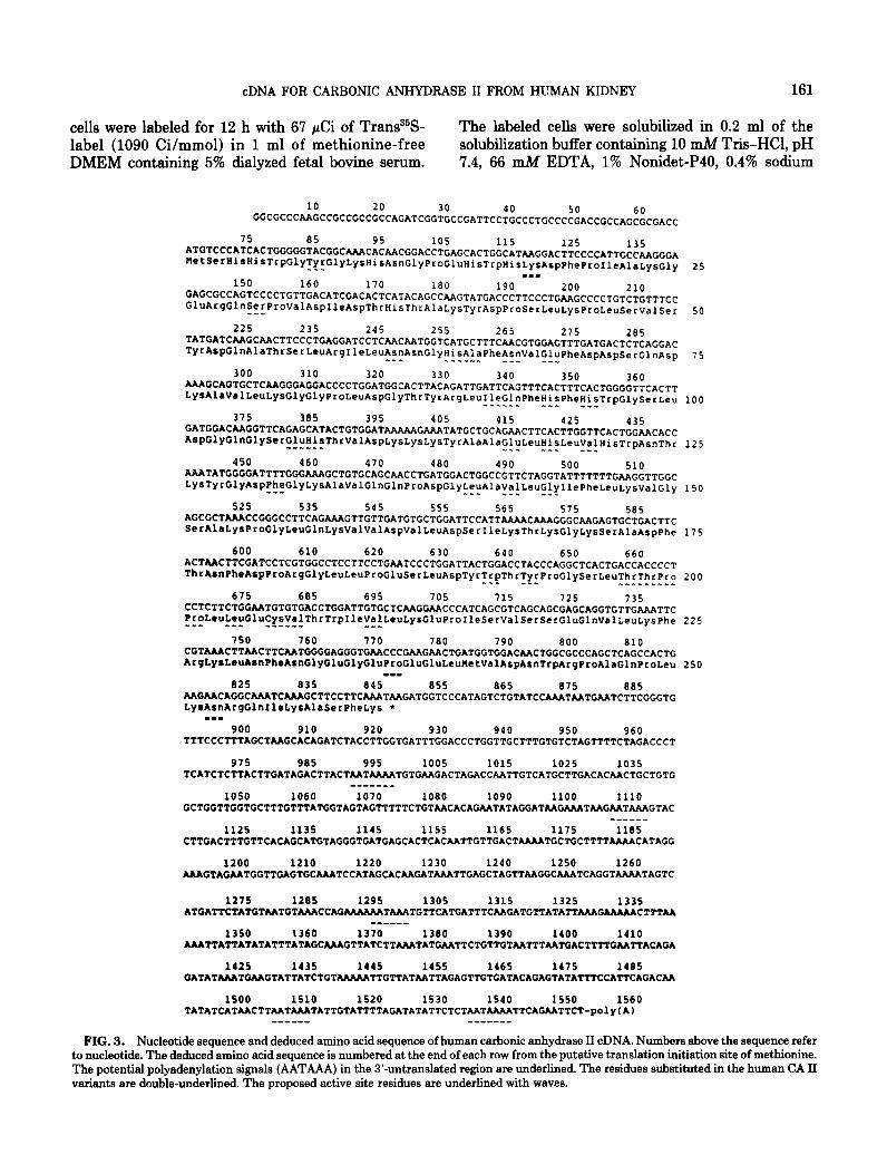
FIG. 3. Nucleotide sequence and deduced amino acid sequence of human carbonic anhydrase II cDNA. Numbers above the sequence refer to nucleotide. The deduced amino acid sequence is numbered at the end of each row from the putative translation initiation site of methionine. The potential polyadenylation signals (AATAAA) in the 3’-untranslated region are underlined. The residues substituted in the human CA II variants are double-underlined. The proposed active site residues are underlined with waves.
162 MURAKAMI ET AL.
deoxycholate, 0.1% SDS, 0.5 pglml of leupeptin, and 0.5 pg/ml of aprotinin. After an incubation of 1 h in ice, 0.8 ml of the washing buffer (5 mM EDTA, 50 n-J4 Tris-HCI, pH 7.4, 150 mM NaCl, 1% Nonidet-P40, 0.5% sodium deoxycholate) was added to the extract and the extract was precleared twice by addition of 100 ~1 of 20% IgGsorb, incubation at 4°C for 30 min, and centrifugation. Either the rabbit antihuman CA II antiserum (10 ~1) or the preimmune serum (10 ~1) was added to half of each extract and incubated for 16 h at 4°C. The immunoprecipitates were collected after ad- dition of 100 gl of 20% IgGsorb, incubation at 4°C for 1 h, and centrifugation for 1 min. The pellets were washed four times with the washing buffer described above, one time with the washing buffer containing 0.5 M sodium chloride, and two times with the solu- tion of 50 mM Tris-HCl, pH 7.4,150 mM NaCl, and 5 mM EDTA. The immunoprecipitates were analyzed by SDS-polyacrylamide gel electrophoresis (12% acrylamide) described by Laemmli (1970), followed by fluorography with EN3HANCE (New England Nu- clear, MA).
Preparation of Human CA II-Specific Antiserum
Carbonic anhydrase II was purified with an affinity column by the procedure of Khalifah et al. (1977) from freshly prepared hemolysates as described previously (Sly et al., 1983). Briefly, fractions containing CA II, which was eluted with 0.2 M NaN3, were collected, dialyzed against 25 mM Mops buffer, pH 7.0, and then applied again onto the affinity column to remove slightly contaminating proteins completely. The frac- tions containing human CA II were collected and re- purified by the preparative SDS-polyacrylamide gel electrophoresis (Murakami and Sly, 1987). A rabbit was immunized as described previously (Murakami and Sly, 1987). The specificity of this anti-human CA II antiserum was examined by the immunoblot system described previously (Murakami and Sly, 1987), which revealed that it reacted only with CA II in human hemolysates and did not cross-react with CA I (data not shown).
DNA Sequence Analysis
All DNA sequences of the insert in XHM3 were determined by the Sanger dideoxy chain-termination method (Sanger et aZ., 1977). Ml3 phage subclones in M13mp18 and M13mp19 (Norrander et al., 1983) were generated from pSHM32 with appropriate restriction enzyme digestions and by the exonuclease III diges- tion described by Henikoff (1984) with mung bean nuclease treatment instead of Sl nuclease. To deter- mine the sequence beyond the EcoRI site close to the 3’ end (nucleotide positions 1545-1550 in Fig. 3), we subcloned the Hind111 fragment from XHM3 into
M13mp19 and sequenced it with a synthetic oligonu- cleotide (CCATTCAGACAATAT, nucleotide posi- tions 1479-1493 in Fig. 3) as a primer (Strauss et aZ., 1986). The sequence strategy is illustrated in Fig. 1.
Other Procedures
Standard molecular biological procedures were car- ried out as described by Maniatis et al. (1982). Protein was determined by the procedure of Lowry et al. (1951) with bovine serum albumin as a standard.
RESULTS AND DISCUSSION
Isolation of cDNA for Human CA II
Northern blot analysis was carried out on human kidney, liver, and placenta RNAs, using the partial human genomic CA II clone, H25-3.8, which contains the first and second exons of CA II gene as a probe (Venta et al., 1984; Venta et al., 1985; Lee et al., 1985). A single band of approximately 1.6 kb in length could be observed in kidney RNA (data not shown), but no band was seen in RNA from liver or placenta. There- fore, we screened a human kidney library to isolate a full-length cDNA clone for CA II. We screened 2 X l@ plaques under stringent hybridization conditions and seven clones were identified. DNA was isolated from these clones, digested with EcoRI, and analyzed on 4% acrylamide gel electrophoresis. The three with the longest inserts seemed identical on 4% acrylamide gel analysis, showing two EcoRI restriction fragments of 1.4 and 0.17 kb. One of these (XHM3) was selected for further analysis. DNA sequencing of this clone re- vealed a small additional EcoRI fragment which had
TABLE 2
Nucleotide and Amino Acid Sequence Homologies with Other Carbonic Anhydrase Genes
Protein coding region 3’-Untranslated region
Amino Nucleotides acids Nucleotides
Mouse CA II” 81 81 64 Chick CA II* IO 70 42
Human CA I’ 64 60 -d Human CA III 65 58 35
Note. Numbers are given as percentages of identical nucleotides or amino acid residues to human CA II.
a Reported by Curtis et al. (1983). * Reported by Rogers (1987). ’ Reported by Barlow et al. (1987). d The human CA I cDNA reported by Barlow et al. (1987) con-
tained short 3’-untranslated sequence (105 bp), compared to those of human CA II (703 bp) and CA III (887 bp).
’ Reported by Wade et al. (1986).

cDNA FOR CARBONIC ANHYDRASE II FROM HUMAN KIDNEY 163
not been detected on gel analysis (see Fig. 1 and Fig. 3).
Expression of Carbonic Anhydrase in Cos-7 Cells
pSHM35, which contains the EcoRI insert in an op- posite orientation, led to enzyme expression following transfection. These results indicated that the clone pSHM32 contains the functionally complete cDNA for CA II.
To confirm that the clone, XHM3, contains the CA We also identified the expressed product of the II cDNA, the nearly full-length EcoRI insert (which cDNA by immunoprecipitation of the transfected contains nucleotides 1 to 1550 but lacks the poly(A) Cos-7 cells with CA II-specific antiserum. Figure 2 tail) was subcloned from XHM3 into the Cos-7 cell shows that pSHM32 expressed a 29,000-D protein expression vector pSHM28 and expressed in Cos cells immunoprecipitated by the anti-CA II antiserum, (Table 1). Plasmid pSHM32, which contains the which migrated at a position identical to that of the EcoRI cDNA insert in the correct orientation, ex- purified human CA II on the gel. However, the protein pressed carbonic anhydrase activity. Neither was not expressed in the cells transfected by pSHM35 pSHM28 (the expression vector without insert) nor or pSHM28. The 29,000-D protein was not immuno-
TABLE 3
Comparison of Nucleotide Sequences of Active Sites in the Carbonic Anhydrase Gene Family
Nucleotide 84 150 246 255 258 264 270 336 339 345 Amino acid 7 29 61 64 65 67 69 91 92 94
Human CA II
Mouse CA II
Chick CA II
Human CA I
Human CA III
Nucleotide Amino acid
TAC 5r*
--T
351 96
TCC Ser*
--G
--G
381 106
GAG Glu*
--A
AAC Asn*
--T
--T
384 107
CAT His* __ C __ C
--c
CAT His -_ C
--c
A-G LYS
414 117
GCT Ala T-C Ser
T-C Ser T-- Ser
A-C Thr
420 119
AAC Asn
C-T His
CGA Arg
426 121
GAG Glu
A-- Aen
-TA Val
453 130
ATT Ile _- C
G-C Val
T-- Phe
CGC Arg
483 140
CAG CAC Gln* His*
--T
--T
489 495 142 144
Human CA II
Mouse CA II
Chick CA II
Human CA I
Human CA III
CAC His*
GAA Glu*
--G
--G
--G
--G
CAC His*
--T
--T
GTT TTT CTG GTT GGT Val Phe Leu Val Gly*
T-- _- - C T- -C -C
-C- C-- T-- Ala Leu
A-C Ile -G --’
Nucleotide 636 642 657 660 663 666 672 678 681 693 Amino acid 191 193 198 199 200 201 203 205 206 210
Human CA II
Mouse CA II Chick CA II
Human CA I
Human CA III
TGG Trp*
TAC 5r*
ACC Thr*
--T
--T
ACC Thr
--T --T
CAT His --G
CCT Pro*
--G --A
--G
CCT Pro*
--A
--C
CTG TGT L?U CYS
-At His
TAT A-- Tyr Ser G-G --C Glu
GTG Val
--A
A-T Ile
GTG Val
--T
A-C Ile C-- Leu
Note. Nucleotides and amino acids different from those of human CA II are shown. Nucleotides and amino acids are numbered according to human CA II in Fig. 3. Amino acid residues which are invariant in all three isozymes among species reported to date are indicated by asterisks.
164 MURAKAMI ET AL.
precipitated by the preimmune serum (data not shown).
Nucleotide Sequence Analysis
The nucleotide sequence of the cDNA insert of XHM3 was determined by the sequence strategy shown in Fig. 1 and is presented in Fig. 3. All of the regions of the insert of pSHM32 (nucleotide position from 1 to 1550) were sequenced at least once on both strands. The additional base (T) at position 1551 and the poly(A) tail downstream of the EcoRI site were identified with the synthetic oligonucleotide as a primer as described under Materials and Methods. The cDNA is 1551 bp in length and contains an open reading frame, beginning at base position 66, that en- codes a 260-amino-acid polypeptide. The nucleotide sequence of the 5’ region (from base position 1 to 297) agreed completely with the sequence reported for the partial human CA II genomic clone, H25-3.8 (Venta et al., 1985), which contains the first and second exons of the human CA II gene. Beginning with the second amino acid (Ser) predicted from the cDNA sequence, the deduced amino acid sequence agreed completely with that determined by amino acid sequencing of the protein by Henderson et al. (1976) (Fig. 3). The termi- nation codon at nucleotide positions 846-848 is fol- lowed by long 3’-untranslated sequence (703 bp). There were five potential poly(A) addition signals (AATAAA) (Fitzgerald and Shenk, 1981) at positions 991-996, 1106-1111, 1294-1299, 1504-1509, and 1534-1539.
Comparison of Nucleotide Sequences with Other Carbonic Anhydrase cDNAs
Table 2 summarizes the homology in the nucleotide sequences and deduced amino acid sequences in CA II cDNAs from three different species reported to date and in all the three cDNAs which encode the cytosolic forms of human carbonic anhydrase. These homolo- gies indicate a high degree of conservation of nucleo- tide sequences in the CA II gene between species, especially in the coding sequence, but substantial conservation is also present in the 3’-untranslated re- gion. As had been inferred earlier from amino acid sequence data (Deutsch, 19&Q, nucleotide sequence homology among CA I, II, and III cDNAs in a given species (human), though substantial, shows less con- servation than the interspecies comparisons for the CA II cDNAs (Table 2).
From the studies on the three-dimensional struc- tures of CA I and CA II (Notstrand et al., 1975; Kannan et al, 1964) and from the active site studies (Tashian et al., 1983; Hewett-Emmett et al., 19&i), 30 amino acid residues have been proposed to be active site residues.
Table 3 compares the nucleotide codons for the 30 active site residues in several mammalian carbonic an- hydrases. Sixteen of the thirty residues are invariant in all three isozymes among species reported to date. These are marked with asterisks in Table 3. Table 4 summarizes the homologies of the active site residues of carbonic anhydrases mentioned above. Several points can be made from Tables 3 and 4. (1) The amino acid residues and nucleotide sequences in the active sites are more conserved among CA 11s from human, mouse, and chick than among the different isozymes from human. (2) Even in codons for the invariant ac- tive site amino acid residues, there are silent nucleotide substitutions at the third nucleotide positions. (3) The nucleotide sequences for CAs I, II, and III are even more conserved than one might guess from the amino acid sequence. The variant residues result from rela- tively few nucleotide substitutions, each of which alters the amino acid because it involves the first or second position of the codon.
The homology of nucleotide sequences of carbonic anhydrase isozymes discussed above supports the idea that these isozymes belong to a gene family that arose in evolution from a common precursor by gene dupli- cation.
TABLE 4
Homology of Nucleotides and Amino Acid Residues in Active Sites of Members of the Carbonic
Anhydrase Gene Family
Invariant Variant residues” residues* Total
Human CA II vs mouse CA II Nucleotide Amino acid
Human CA II vs chick CA II Nucleotide Amino acid
Mouse CA II vs chick CA II Nucleotide Amino acid
Human CA II vs CA I Nucleotide Amino acid
Human CA II vs CA III Nucleotide Amino acid
Human CA I vs CA III Nucleotide Amino acid
92 86 89 100 93 97
83 74 19 100 79 90
90 86 88 100 86 93
88 57 73 166 29 67
81 57 70 100 36 70
81 48 66 100 7 57
Note. Homology of nucleotides or amino acids between the two carbonic anhydrasea are shown by percentages.
’ The 16 invariant active site residues are marked with asterisks in Table 3.
* The other 14 active site residues in Table 3.
cDNA FOR CARBONIC ANHYDRASE II FROM HUMAN KIDNEY 165
TABLE 6 2.
The Variants of Carbonic Anhydrase II in Humans
Nucleotide position 117 771 819 Amino acid position 18 236 252 3.
Human CA II AAG LYS
CA II variant Glu”
Expected nucleotide substitution GAG
’ Reported by Jones et al. (1982). b Reported by Jones and Shaw (1983). c Reported by Lin and Deutsch (1972).
ccc Pro
His*
CAC
AAC Asn
Asp=
GAC
4.
5.
6.
Human CA II Variants 7.
To date, hemolysates from about 50,000 individuals from different human populations have been screened for electrophoretic variants of the CA I and CA II isozymes (Tashian et al., 1983). Three amino acid substitutions have been reported in human CA II (Fig. 3 and Table 5). The determination of the complete nucleotide sequences of cDNA from human CA II allows one to predict the one base substitution in each codon which would cause each CA II variant as shown in Table 5. It should be noted that the substitution from A to G at nucleotide position 117 (amino acid residue 18) would create a Mu111 site (CATG) and a MnII site (GAGG) and that the substitution from A to G at position 819 (residue 252) would remove a Mb011 site (GAAGA) from the CA II cDNA. These restric- tion enzymes might be useful to identify these variant CA II genes by Southern blot analysis of genomic DNAs with the CA II cDNA reported here as a probe.
8.
9.
10.
11.
12.
13.
Note added in proof. Since this work was completed, Montgomery et aZ. reported the nucleotide sequence of human liver carbonic anhydrase II cDNA (MONTGOMERY, J. C., VENTA, P. J., TASHIAN, R. E., AND HEWE’IT-EMMETT, D. (1987). Nucleotide sequence of human liver carbonic anhydrase II cDNA. Nucleic Acids Res. 15: 4687).
14.
15. ACKNOWLEDGMENTS
We thank Dr. Graeme I. Bell at the Cbiron Corporation for pro- viding the human kidney XgtlO cDNA library. We are also grateful to Dr. Richard E. Tashian at the University of Michigan for pro- viding the partial human CA II genomic clone. We also thank Miss Elizabeth Torno for typing this manuscript. This work was sup- ported by Research Grant RR-00036 from the General Clinical Research Center Branch, Division of Research Facilities and Re- sources, National Institutes of Health, support from the National Foundation-March of Dimes, and National Institutes of Health Research Grants GM31988 and ADGM20610.
16.
17.
18.
REFERENCES 19.
1. BARLOW J. H., LOWE, N., EDWARDS, Y. H., AND BUTTER- WORTH, P. H. W. (1987). Human carbonic anhydrase I cDNA. Nucleic Acids Res. 15: 2386.
BELL, G. I., FONG, N. M., STEMPIEN, M. M., WORMSTED, M. A., CAPUT, D., Ku, L., URDEA, M. S., RALL, L. B., AND SAN- CHEZ-PESCADOR, R. (1986). Human epidermal growth factor precursor: cDNA sequence, expression in uitro and gene orga- nization. Nucleic Acids Res. 14: 8427-8446. CARTER, M. J. (1972). Carbonic anhydrase: Isoenzymes, prop- erties, distribution and functional significance. BioZ. Reu. 47: 465-513. CURTIS, P. J., WITHERS, E., DEMUTH, D., WATT, R, VENTA, P. M., AND TASHIAN, R. E. (1983). The nucleotide sequence and derived amino acid sequence of cDNA coding for mouse carbonic anhydrase II. Gene 25: 325-332. DEUTSCH, H. F. (1984). Primary structures and genetic changes in mammalian carbonic anhydrase isozymes. Ann. N. Y. Acad. Sci. 429: 183-194. FELDSTEIN, J. B., AND SILVERMAN, D. N. (1984). Purification and characterization of carbonic anhydrase from the saliva of the rat. J. Biol. Chem. 259: 5447-5453. FITZGERALD, M., AND SHENK, T. (1981). The sequence 5’- AAUAAA-3’ forms part of the recognition site for polyadenyl- ation of late SV40 mRNAs. Cell 24: 251-260. GLUZMAN, Y. (1981). SV40-transformed simian cells support the replication of early SV40 mutants. Cell 23: 175-182. HENDERSON, L. E., HENRIKSSON, D., AND NYMAN, P. 0. (1976). Primary structure of human carbonic anhydrase C. J. Biol. Chem. 251: 5457-5463. HENIKOFF, S. (1984). Unidirectional digestion with exonucle- ase III creates targeted breakpoints for DNA sequencing. Gene 28: 351-359. HEWE=-EMMETT, D., HOPKINS, P. J., TASHIAN, R. E., AND CZELUSNIAK, J. (1984). Origins and molecular evolution of the carbonic anhydrase isozymes. Ann. N.Y. Acad. Sci. 429: 338-358. JONES, G. L., SOFRO, A. S. M., AND SHAW, D. C. (1982). Chem- ical and enzymological characterization of an Indonesian variant of human erythrocyte carbonic anhydrase II, CA II Jogjakarta (17-Lys Glu). Biochem. Genet. 20: 979-1000. JONES, G. L., AND SHAW, D. C. (1983). A chemical and enzy- mological comparison of the common major human erythro- cyte carbonic anhydrase II, its minor component, and a new genetic variant CA II Melbourne (237-Pro His). Hum. Genet. 63: 392-399.
KANNAN, K. K., RAMANADHAM, M., AND JONES, R. A. (1984). Structure, refinement, and function of carbonic anhydrase isozymes: Refinement of human carbonic anhydrase I. Ann. N. Y. Acad. Sci. 429: 49-60. KHALIFAH, R. G., STRADER, D. J., BRYANT, S. H., AND GIB- SON, S. M. (1977). Carbon-13 nuclear magnetic resonance probe of active-site ionizations in human carbonic anhydrase B. Biochemistry 16: 2241-2247.
LAEMMLI, U. K. (1970). Cleavage of structural proteins during the assembly of the head of bacteriophage T4. Nature fLon- don) 227: 680-685.
LEE, B. L., VENTA, P. J., AND TASHIAN, R. E. (1985). DNA polymorphism in the 5’ flanking region of the human carbonic anhydrase II gene on chromosome 8. Hum. Genet. 69: 337-339.
LIN, K. T. D., AND DEUTSCH, H. F. (1972). Human carbonic anhydrase. VIII. Isolation and characterization of a polymor- phic form of a C type isozyme. J. Biol. Chem. 247: 3761-3766. LINDSKOG, S., HENDERSON, L. E., KANNAN, K. K., LILJAS, A., NYMAN, P. O., AND STRONDBERG, B. (1971). Carbonic anhy- drase. In “The Enzymes” (P. D. Boyer, Ed.), Vol. V, pp. 587-665, Academic Press, New York.

20.
21.
22.
23.
24.
25.
26.
21.
28.
29.
30.
31.
32.
33.
34.
35.
MURAKAMI ET ” HL.
LOPATA, M. A., CLEVELAND, D. W., AND SOLLNER-WEBB, B. (1984). High level transient expression of a chloramphenicol acetyl transferase gene by DEAE-dextran mediated DNA transfection coupled with a dimethyl sulfoxide or glycerol shock treatment. Nucleic Acids Res. 12: 5707-5717.
LOWRY, 0. II., ROSEBROUGH, N. J., FARR, A. L., AND RAN- DALL, R. J. (1951). Protein measurement with the Folin phenol reagent. J. Biol. Chem. 193: 265-275. LUTHMAN, H., AND MAGNUSSON, G. (1983). High efficiency polyoma DNA transfection of chloroquine treated cells. Nu- cleic Acids Res. 11: 1295-1308. MANIATIS, T., FRITSCH, E. F., AND SAMBROOK, J. (1982). “Molecular Cloning: A Laboratory Manual,” Cold Spring Harbor Laboratory, Cold Spring Harbor, NY. MAREN, T. H. (1967). Carbonic anhydrase chemistry, physiol- ogy, and inhibition. Physiol. Rev. 47: 595-781. MAREN, T. H., AND SANYAL, G. (1983). The activity of sulfon- amides and anions against the carbonic anhydrases of ani- mals, plants, and bacteria. Annu. Rev. Pharmacol Toxicol. 23: 439-459. MCKINLEY, D. N., AND WHITNEY, P. L. (1976). Particulate carbonic anhydrase in homogenates of human kidney. Bio- chim. Biophys. Acta 446: 780-790. MURAKAMI, H., AND SLY, W. S. (1987). Purification and char- acterization of human salivary carbonic anhydrase. J. Biol. Chem. 262: 1382-1388. NORRANDER, J., KEMPE, T., AND MESSING, J. (1983). Con- struction of improved Ml3 vectors using oligodeoxynucleo- tide-directed mutagenesis. Gene 26: 101-106.
NOTSTFUND, B., VAARA, I., AND KANNAN, K. K. (1975). Struc- tural relationship of human erythrocyte carbonic anhydrases B and C. In “Isozymes” (C. L. Markert, Ed.), Vol. 1, pp. 575-599, Academic Press, Orlando, FL.
OSHIMA, A., KYLE, J. W., MILLER, R. D., HOFFMAN, J. W., POWELL, P. P., GRUBB, J. H., SLY, W. S., TROPAK, M., GUISE, K. S., AND GRAVEL, R. A. (1987). Cloning, sequencing, and expression of cDNA for human /3-glucuronidase. Proc. Natl. Acad. Sci. USA 84: 685-689.
POCKER, Y., AND SARKANEN, S. (1978). Carbonic anhydrase: structure, catalytic versatility, and inhibition. Adv. Enzymol. 47: 149-274.
ROGERS, J. H. (1987). Sequence of carbonic anhydrase II cDNA from chick retina. Eur. J. B&hem. 162: 119-122.
SANGER, F., NICKLEN, S., AND COULSON, A. R. (1977). DNA sequencing with chain-terminating inhibitors. Proc. Natl. Acad. Sci. USA 74: 5463-5467.
SANYAL, G. (1984). Comparative carbon dioxide hydration ki- netics and inhibition of carbonic anhydrase isozymes in verte- brates. Ann. N.Y. Acad. Sci. 429: 165-178.
SANYAL, G., PESSAH, N. I., AND MAREN, T. H. (1981). Kinetics and inhibition of membrane-bound carbonic anhydrase from canine renal cortex. B&him. Biophys. Acta 667: 128-137.
36.
37.
38.
39.
40.
41.
42.
43.
44.
45.
46.
41.
SLY, W. S., HEWETT-EMMETT, D., WHYTE, M. P., Yu, Y.-S. L., AND TASHIAN, R. E. (1983). Carbonic anhydrase II deficiency identified as the primary defect in the autosomal recessive syndrome of osteopetrosis with renal tubular aci- dosis and cerebral calcification. Proc. Natl. Acad. Sci. USA 80: 2752-2756, SLY, W. S., WHYTE, M. P., SUNDARAM, V., TASHIAN, R. E., HEWETT-EMMETT, D., GUIBAUD, P., VAINSEL, M., BALUATE, H. J., GRUSKIN, A., AL-MOSAWI, M., SAKATI, N., AND OHLS- SON, A. (1985). Carbonic anhydrase II deficiency in 12 families with the autosomal recessive syndrome of osteopetrosis with renal tubular acidosis and cerebral calcification. N. Engl. J. Med. 313: 139-145. SPRAGUE, J., CONDRA, J. H., ARNHEITER, H., AND LAZZA- RINNI, R. A. (1983). Expression of a recombinant DNA gene coding for the vesicular stomatitis virus nucleocapsid protein. J. Viral. 45: 773-181. STRAUSS, E. C., KOBORI, J. A., SIU, G., AND HOOD, L. E. (1986). Specific-primer-directed DNA sequencing. Anal. Bio- them. 164:353-360. TASHIAN, R. E., HEWETT-EMMETT, D., DODGSON, S. J., Fos- TER, R. E., II, AND SLY, W. S. (1984). The value of inherited deficiencies of human carbonic anhydrase isoxymes in under- standing their cellular roles. Ann. N.Y. Acad. Sci. 429: 262-275. SUNDARAM, V., RUMBOLO, P., GRUBB, J., STRISCIUGLIO, P., AND SLY, W. S. (1986). Carbonic anhydrase II deficiency: Diagnosis and carrier detection using differential enzyme in- hibition and inactivation. Amer. J. Hum. Genet. 38: 125-136. TASHIAN, R. E., HEWETT-EMME’IT, D., AND GOODMAN, M. (1983). On the evolution and genetics of carbonic anhydrases I, II, and III. In “Isozymes: Current Topics in Biological and Medical Research” (M. C. Rattazzi, J. G. ScandaIios, and G. S. Whitt, Eds.), Vol. 7, pp. 79-100, A. R. Liss, New York. VENTA, P. J., MONTGOMERY, J. C., HEWETT-EMMETT, D., AND TASHIAN, R. E. (1985). Comparison of the 5’ regions of human and mouse carbonic anhydrase II genes and identifi- cation of possible regulatory elements. B&him. Biophys. Acta 826: 195-201. VENTA, P. J., MONTGOMERY, J. C., WIEBAUER, K., HEWE’TT- EMMEW, D., AND TASHIAN, R. E. (1984). Organization of the mouse and human carbonic anhydrase II genes. Ann. N.Y. Acad. Sci. 429: 309-323. WADE, R., GUNNING, P., EDDY, R., SHOWS, T., AND KEDES, L. (1986). Nucleotide sequence, tissue-specific expression, and chromosome location of human carbonic anhydrase III: The human CA III gene is located on the same chromosome as the closely linked CA I and CA II genes. Proc. Natl. Acad. Sci. USA 83: 9571-9575. WHITNEY, P. L., AND BRIGGLE, T. V. (1982). Membrane-as- sociated carbonic anhydrase purified from bovine lung. J. Biol. C&m. 267: 1205612059. WISTRAND, P. J. (1984). Properties of membrane-bound car- bonic anhydrase. Ann. N. Y. Acad. Sci. 429: 195-206.