Plasminogen/plasmin regulates α-enolase expression through the MEK/ERK pathway
7
Plasminogen/plasmin regulates a-enolase expression through the MEK/ERK pathway q Lirla ˆndia P. Sousa a,b , Breno M. Silva a,b , Bruno S.A.F. Brasil a,b , Sarah V. Nogueira a,b , Paulo C.P. Ferreira b , Erna G. Kroon b , Kanefusa Kato c , Cla ´udio A. Bonjardim a,b, * a Grupo de Transduc ¸a ˜o de Sinal, Departamento de Microbiologia, Instituto de Cie ˆncias Biolo ´ gicas, Universidade Federal de Minas Gerais, 31270-901 Belo Horizonte, Minas Gerais, Brazil b Laborato ´rio de Vı ´rus, Departamento de Microbiologia, Instituto de Cie ˆncias Biolo ´ gicas, Universidade Federal de Minas Gerais, 31270-901 Belo Horizonte, Minas Gerais, Brazil c Department of Biochemistry, Institute for Developmental Research, Aichi, Japan Received 22 September 2005 Available online 4 October 2005 Abstract Previously we have demonstrated that both plasminogen (Plg) and plasmin (Pla) regulate the expression of the transcription factors c-FOS and EGR-1 [L.P. De Sousa, B.S. Brasil, B.M. Silva, M.H. Freitas, S.V. Nogueira, P.C. Ferreira, E.G. Kroon, C.A. Bonjardim, Plasminogen/plasmin regulates c-fos and egr-1 expression via the MEK/ERK pathway, Biochem. Biophys. Res. Commun. 329 (2005) 237–245]. Here we show that Plg activates the mitogen-activated protein kinases MEK and ERK which leads to a-enolase (a-ENO) gene expression not only in fibroblasts, but also in peripheral blood mononuclear cells. The a-ENO mRNA accumulation was appar- ent three hours post-Plg treatment and remained elevated out to 28 h, a process that seems to require both de novo protein synthesis and active gene transcription. Pla mimics Plg-stimulated a-ENO expression through its serine protease activity, suggesting that con- version of Plg to active Pla is required. Pharmacological and genetic blockade of MEK caused inhibition of a-ENO mRNA accumu- lation, implicating MEK/ERK as the transduction pathway that leads to a-ENO expression upon Plg stimulation. Furthermore, Plg stimulated DNA binding activity of the transcription factors activator-protein 1 and early growth response gene-1 to their cognate regulatory sequences at a-ENO promoter. Altogether, our data show that Plg/Pla regulates a-ENO expression through the MEK/ ERK pathway. Ó 2005 Elsevier Inc. All rights reserved. Keywords: Plasminogen; Plasmin; a-Enolase; MAPK-ERK1/2; Signal transduction Plasminogen is an abundant zymogen, which exerts its biological activities upon ligand binding to the carboxy-ter- minal lysines of its cell surface receptors via its lysine bind- ing sites [2]. Plg activation with subsequent generation of plasmin (Pla), a serine protease, is mediated by the proteo- lytic activity of urokinase-type Plg activator (uPA) or tissue Plg activator (tPA) [3]. Plg receptors are widely distributed on a variety of cells and their binding by Plg/Pla plays a crucial role to limit the enzymatic activity to the cell surface [2,3]. Among the diverse biological functions associated with the Plg/Pla proteolytic system, beyond the classical dissolu- tion of fibrin deposits [4], other functions have also been as- cribed to these molecules such as cell migration, wound healing, inflammation, metastasis, and signaling events [1,5–8]. The multifunctional protein a-enolase (a-ENO), first characterized as a glycolytic enzyme [9], has also been asso- ciated with the binding of Plg to the cell surface of diverse 0006-291X/$ - see front matter Ó 2005 Elsevier Inc. All rights reserved. doi:10.1016/j.bbrc.2005.09.154 q Abbreviations: Plg, plasminogen; Pla, plasmin; a-ENO, a-enolase; MAPK, mitogen-activated protein kinase; ERK, extracellular-signal regulated kinase; MEK, MAPK/ERK kinase; AP-1, activator protein-1; EGR-1, early growth response-1 gene product. * Corresponding author. Fax: +55 31 3443 6482. E-mail address: [email protected] (C.A. Bonjardim). www.elsevier.com/locate/ybbrc Biochemical and Biophysical Research Communications 337 (2005) 1065–1071 BBRC
Transcript of Plasminogen/plasmin regulates α-enolase expression through the MEK/ERK pathway

www.elsevier.com/locate/ybbrc
Biochemical and Biophysical Research Communications 337 (2005) 1065–1071
BBRC
Plasminogen/plasmin regulates a-enolase expression throughthe MEK/ERK pathwayq
Lirlandia P. Sousa a,b, Breno M. Silva a,b, Bruno S.A.F. Brasil a,b, Sarah V. Nogueira a,b,Paulo C.P. Ferreira b, Erna G. Kroon b, Kanefusa Kato c, Claudio A. Bonjardim a,b,*
a Grupo de Transducao de Sinal, Departamento de Microbiologia, Instituto de Ciencias Biologicas, Universidade Federal de Minas Gerais,
31270-901 Belo Horizonte, Minas Gerais, Brazilb Laboratorio de Vırus, Departamento de Microbiologia, Instituto de Ciencias Biologicas, Universidade Federal de Minas Gerais,
31270-901 Belo Horizonte, Minas Gerais, Brazilc Department of Biochemistry, Institute for Developmental Research, Aichi, Japan
Received 22 September 2005Available online 4 October 2005
Abstract
Previously we have demonstrated that both plasminogen (Plg) and plasmin (Pla) regulate the expression of the transcription factorsc-FOS and EGR-1 [L.P. De Sousa, B.S. Brasil, B.M. Silva, M.H. Freitas, S.V. Nogueira, P.C. Ferreira, E.G. Kroon, C.A. Bonjardim,Plasminogen/plasmin regulates c-fos and egr-1 expression via the MEK/ERK pathway, Biochem. Biophys. Res. Commun. 329 (2005)237–245]. Here we show that Plg activates the mitogen-activated protein kinases MEK and ERK which leads to a-enolase (a-ENO)gene expression not only in fibroblasts, but also in peripheral blood mononuclear cells. The a-ENO mRNA accumulation was appar-ent three hours post-Plg treatment and remained elevated out to 28 h, a process that seems to require both de novo protein synthesisand active gene transcription. Pla mimics Plg-stimulated a-ENO expression through its serine protease activity, suggesting that con-version of Plg to active Pla is required. Pharmacological and genetic blockade of MEK caused inhibition of a-ENO mRNA accumu-lation, implicating MEK/ERK as the transduction pathway that leads to a-ENO expression upon Plg stimulation. Furthermore, Plgstimulated DNA binding activity of the transcription factors activator-protein 1 and early growth response gene-1 to their cognateregulatory sequences at a-ENO promoter. Altogether, our data show that Plg/Pla regulates a-ENO expression through the MEK/ERK pathway.� 2005 Elsevier Inc. All rights reserved.
Keywords: Plasminogen; Plasmin; a-Enolase; MAPK-ERK1/2; Signal transduction
Plasminogen is an abundant zymogen, which exerts itsbiological activities upon ligand binding to the carboxy-ter-minal lysines of its cell surface receptors via its lysine bind-ing sites [2]. Plg activation with subsequent generation ofplasmin (Pla), a serine protease, is mediated by the proteo-lytic activity of urokinase-type Plg activator (uPA) or tissue
0006-291X/$ - see front matter � 2005 Elsevier Inc. All rights reserved.
doi:10.1016/j.bbrc.2005.09.154
q Abbreviations: Plg, plasminogen; Pla, plasmin; a-ENO, a-enolase;MAPK, mitogen-activated protein kinase; ERK, extracellular-signalregulated kinase; MEK, MAPK/ERK kinase; AP-1, activator protein-1;EGR-1, early growth response-1 gene product.* Corresponding author. Fax: +55 31 3443 6482.E-mail address: [email protected] (C.A. Bonjardim).
Plg activator (tPA) [3]. Plg receptors are widely distributedon a variety of cells and their binding by Plg/Pla plays acrucial role to limit the enzymatic activity to the cell surface[2,3].
Among the diverse biological functions associated withthe Plg/Pla proteolytic system, beyond the classical dissolu-tion of fibrin deposits [4], other functions have also been as-cribed to these molecules such as cell migration, woundhealing, inflammation, metastasis, and signaling events[1,5–8].
The multifunctional protein a-enolase (a-ENO), firstcharacterized as a glycolytic enzyme [9], has also been asso-ciated with the binding of Plg to the cell surface of diverse

1066 L.P. Sousa et al. / Biochemical and Biophysical Research Communications 337 (2005) 1065–1071
cell types including monocytes, B and T cells, and neutro-phils [10–12]. Moreover, a-ENO represents up to 70% ofthe Plg profibrinolytic receptors expressed at the surfaceof peripheral blood monocytes [13]. Recently, it was dem-onstrated, by using a monoclonal antibody that specificallyblocked Plg binding to a-ENO (MAb11G1), that 84–90%of leukocytic cell-dependent Plg activation was abrogated,suggesting that a-ENO has a major role in the promotionof Plg activation in several leukocytic cell types [14].
a-ENO is under the transcriptional regulation of a num-ber of stimuli, such as hypoxia, cytokines, and mitogens[15–18]. Here we show that the expression of a-ENO is reg-ulated by Plg/Pla in both fibroblast and peripheral bloodmononuclear cells. In addition, we provide evidence thata-ENO expression is dependent on signals mediated bythe mitogen-activated protein kinases (MAPKs) ERK1/2.
Materials and methods
Cells and reagents. A31 cells (a clone derived from mouse Balb/c 3T3)were cultured in Dulbecco�s modified Eagle�s medium (DMEM) sup-plemented with 10% (v/v) heat-inactivated fetal bovine serum (FBS) andantibiotics in 5% CO2 at 37 �C. Prior to the Plg or Pla treatment, cellswere serum-starved in DMEM supplemented with 1% FBS for 24 h afterreaching 70–80% confluence. Peripheral blood mononuclear cells(PBMCs) from heparinized blood of healthy donors were isolated byFicoll–Paque (Amersham Biosciences, UK) density centrifugation asdescribed [19]. Anti-phospho MAPK ERK1/2 and anti-total ERK1/2 orsecondary anti-rabbit peroxidase conjugate antibodies were purchasedfrom Cell Signaling Technology (Beverly MA, USA). Polyclonal rabbitantibody anti-human a-enolase was as described [20,21]. Anti-EGR-1antibody was from Santa Cruz Biotechnology (Santa Cruz, CA, USA).Anti-FOS antibody was generously provided by Dr. T. Curran (St. JudeChildren�s Research Hospital, Memphis, TE, USA). Anti-b-actin anti-body was from Sigma–Aldrich, Sao Paulo, Brazil. The pharmacologicalinhibitors: PD95098 and genistein, and human Plg were purchased fromCalbiochem (La Jolla, CA, USA). Human Pla, leupeptin, cycloheximide,and actinomycin D were purchased from Sigma–Aldrich, Sao Paulo,Brazil.
Cell treatment. Cells were serum-starved as described above and thenincubated with Plg or Pla, for the times shown. When indicated, cells werepre-incubated for 30 min with the following concentration of inhibitorsprior to and throughout Plg treatment: PD98059 50 lM (MEK), genistein100 lM (tyrosine kinase), cycloheximide 5 lg/ml (translation), and acti-nomycin D 5 lg/ml (transcription). Leupeptin 25 lg/ml, a serine proteaseinhibitor, was pre-incubated with plasmin for 30 min at 37 �C. The drugdoses used throughout the experiments were established based on exper-imental observations, without, nonetheless, cause any harm to the cells,given that no measurable effect on cell viability was verified by Trypanblue dye exclusion. PBMC were harvested, washed, suspended in RPMI1640 without FBS at a concentration of 2 · 106 cells/ml and cultured forthe indicated times, either in the absence or in the presence of Plg (2 lg/ml), and processed for Western or Northern blot analysis.
MEK1 dominant-negative cell lines. To generate cellular clones stablyexpressing MEK1 dominant-negative mutation, A31 cells were transfectedwith 10 lg of plasmid DNA carrying either negative dominance for MEK1or the wild-type MEK1 counterpart [22], by using standard calciumphosphate protocols [23]. Transfectants were ring-cloned after selectionwith G418 (800 lg/ml) (Geneticin, Invitrogen, Sao Paulo, Brazil) for atleast 21 days and then tested for MEK1 unresponsiveness, as evaluated byERK1/2 phosphorylation, after stimulation with epidermal growth factor(50 ng/ml) (Sigma–Aldrich, Sao Paulo, Brazil) or virus infection [24].Selected clones (MEK1-DN) presenting identical responses to the abovestimuli were then used to carry out the experiments.
RNA isolation and northern blotting. A31 cells (5 · 106) were culturedin 75 cm2 tissue culture flasks, starved as above, and then incubated withPlg or Pla for the times shown, or were incubated with the differentinhibitors for 30 min prior to Plg- or Pla-stimulation. At the indicatedtimes, total RNA was isolated as described elsewhere [25] and 15 lg RNAper sample were loaded, electrophoresed on a 1.5% denaturing agarose–formaldehyde gel, transferred onto nylon membrane (Amersham Biosci-ences, UK), UV cross-linked for 5 min, and hybridized with a-ENO probe[15]. Probe was labeled with [a-32P]dCTP (Amersham Biosciences, UK) toa specific activity of 1–5 · 108 cpm/lg DNA, by using multiprime DNAlabelling system from Amersham Biosciences, UK. Hybridizations andwashing procedures were carried out as described [26]. The membraneswere stripped of the probe and re-probed with oligonucleotide for 18SrRNA labeled at the 5 0end with [c-32P]ATP, by the using T4 phage PNK(Promega, USA), which was used as an internal control for RNA loading.
Lysate preparation and western blot analysis. After treatments, the cellswere washed with PBS and lysed on ice in lysis buffer (1% Triton X-100,100 mM Tris/HCl, pH 8.0, 10% glycerol, 5 mM EDTA, 200 mM NaCl,1 mM DTT, 1 mM PMSF, 2.5 lg/ml leupeptin, 5 lg/ml aprotinin, and1 mM sodium orthovanadate). Lysates were scraped and collected intoEppendorf tubes and then were centrifuged at 13,000g for 10 min at 4 �C.Cell lysate samples (30 lg) were separated by electrophoresis on a dena-turing 10% polyacrylamide–SDS gel and transferred onto nitrocellulosemembranes as described [25]. Membranes were blocked overnight at 4 �Cwith phosphate-buffered saline containing 5% (w/v) nonfat dried milk and0.1% Tween 20, washed three times with phosphate-buffered saline con-taining 0.1% Tween 20, and then incubated with specific rabbit polyclonalanti-phospho-ERK1/2 antibody (1:1500), anti-ERK1/2 antibody (1:2000),anti-b-actin antibody (1:3000) or anti-a-enolase antibody (1:20,000) inphosphate-buffered saline containing 5% (w/v) BSA and 0.1% Tween 20.After washing, the membranes were incubated with horseradish peroxi-dase-conjugated secondary anti-rabbit antibody (1:3000). Immunoreactivebands were visualized by using ECL detection system as described inmanufacturer�s instructions (Amersham Biosciences, UK).
Electrophoretic mobility shift assay. A31 cells were cultured and starvedas above and then incubated with Plg for the indicated times, or wereincubated with PD98059 prior to and throughout Plg treatment. Elec-trophoretic mobility shift assay (EMSA) was carried out essentially asdescribed [27]. Nuclear cell extracts were prepared as described [28].Protein concentration was determined by the Bio-Rad assay. Ten micro-grams of protein were pre-incubated with 1.2 ll poly(dI–dC) (5.4 mg/ml)(Amersham Biosciences, UK) at room temperature for 10 min, followedby the addition of a reaction mixture containing 1.25 lg BSA, 0.125 lgEscherichia coli DNA, 0.25 lg yeast tRNA, 2% Ficoll 400, and 0.32 nglabeled probe (8.0 · 104 cpm). The reactions were incubated at roomtemperature for 30 min and then analyzed by 6% polyacrylamide gelelectrophoresis. The 5 0 32P-end-labeled double-stranded probes (only onestrand is shown) corresponding to both the wild-type (WT) and themutated (M) (underlined) a-ENO cis-acting elements (AP-1 or EGR-1)are as follows: WT-AP-1ENO: 5 0-ATTTAGCTGCTGAGTCATGGGGC-30; M-AP-1ENO: 5 0-ATTTAGCTGCTGAGTTGTGGGGC–3 0;WT-EGR-1ENO: 5 0-AGCTGAGGGGGCGTGCCCC-30 and M-EGR-1ENO: 5 0-AGCTGATAGGGCGTGCCCC-30. Competition assays wereperformed with 50-fold molar excess of cold oligonucleotide correspond-ing to c-fos SRE: 5 0-GATGTCCATATTAGGACATC-30. For super-shiftassays, nuclear extracts were incubated with the specific antibody for 1 hat 4 �C and then mixed with the reaction mixture for 30 min at roomtemperature.
Results
Plasminogen stimulates a-ENO expression
Since we knew that Plg exerts its biological effects uponinteraction with its cell surface receptor and by knowingthat a-ENO, in some cells, may function as a Plg receptor,

L.P. Sousa et al. / Biochemical and Biophysical Research Communications 337 (2005) 1065–1071 1067
we asked whether Plg could regulate the expression of a-ENO gene. Here we show that Plg stimulates a-ENOmRNA accumulation with a kinetic of accumulation thatis typical of secondary-response gene. The message wasdetectable within 3 h post-treatment, reached its maximumlevel between 6 and 9 h, remained elevated out to 28 h(Fig. 1A), and declined thereafter to basal levels about40 h (data not shown). In accordance with the mRNAexpression, protein levels of a-ENO were increased byPlg-treatment (Fig. 1B). The a-ENO expression, upon Plgstimulation, was dose-dependent (Fig. 1C) and relied uponboth de novo protein synthesis and active transcription,since cycloheximide and actinomycin D abolished itsmRNA accumulation (Fig. 1D, lanes 4 and 6), respectively.In addition, we show that a-ENO expression is also regu-lated by Plg in peripheral blood mononuclear cells as eval-uated by mRNA and protein levels (Figs. 1E and F),respectively.
Plasminogen-induced a-ENO mRNA expression is MEK
dependent
Our findings also show that the MEK/MAPK signalingpathway and phosphorylation on tyrosine residue play apivotal role in the Plg-stimulated a-ENO expression, sincepre-treatment of the cells with the pharmacological inhibi-tors of MEK (PD98059), or tyrosine kinase (Genistein),drastically reduced the Plg-stimulated a-ENO mRNAaccumulation (Fig. 2A). However, pre-treatment withSB203580, a specific inhibitor of p38 MAPK, did not inter-fere with Plg-induced a-ENO expression (data not shown).
Fig. 1. Plasminogen stimulates a-ENO expression. Quiescent monolayers of Ablot (A, C, and D) or Western blot analysis (B) as described in Materials anduntreated cells; 2–7, cells treated with Plg for the indicated times. (B) Top pancells treated with Plg for the indicated times. Bottom panel: blot was re-proresponse; lanes: 1, untreated cells; 2–5, cells treated with Plg for 6 h at the indicasynthesis and active gene transcription. Cells were left untreated (lane 1); treatactinomycin D (5 lg/ml) for 30 min prior to Plg stimulation for 6 h as shown (Northern blot analysis of PBMC treated with Plg for 15 h. (F) Top panel: WesPBMC cells with Plg for the indicated times. Proteins were fractionated on SDSENO antibody. Bottom panel: blot was probed with b-actin for loading contro(A, C, D, and E): re-probed with 18S rRNA, which was used as an internalexperiments.
In line with the pharmacological inhibition of Plg-stimu-lated a-ENO mRNA accumulation, the same inhibitionwas verified with a cell line expressing MEK1 dominant-negative mutation (Fig. 2B). Plasmin was able to mimicthe effect exerted by Plg on a-ENO mRNA accumulation(Fig. 2C, lanes 1–2) in a dose-dependent manner (datanot shown). This effect is dependent on the Pla serineprotease activity, as accessed by pre-incubation of Plawith leupeptin (a serine protease inhibitor) (Fig. 2C, lane4), suggesting that Plg activation to Pla is required fora-ENO expression.
Plasminogen stimulates ERK1/2 phosphorylation
We then investigated whether the MAPK ERK1/2could be, in fact, associated with the transmission of sig-nals initiated upon Plg binding to the cell surface andleading to a-ENO expression. Indeed, our data show thatPlg stimulation leads to a biphasic ERK1/2 phosphoryla-tion (Fig. 2D). The first and strongest wave of activationoccurs between 10 and 15 min post-stimulation and thendecreases by 30 min (lanes 1–4). The second round ofkinases phosphorylation takes place around 1 h post-Plgtreatment, declines gradually up to 3–4 h (lanes 5–8),and then slightly increases by 6–8 h post-treatment(lanes 9–10). This effect seems to be specific, since MAPKphosphorylation was not affected by pre-incubationwith the PKC inhibitor—bisindolylmaleimide (data notshown). ERK phosphorylation was completely blockedby pre-incubation with the specific MEK inhibitor(PD98059) (data not shown).
31 cells were treated with Plg as indicated and then analyzed by Northernmethods. (A) Top panel: kinetics of a-ENO mRNA expression; lanes: 1,el: kinetics of a-ENO protein accumulation; lanes: 1, untreated cells; 2–6,bed with total ERK1/2 for loading control. (C) Top panel: curve dose–ted dose. (D) Top panel: a-ENO expression relies on both de novo proteined with Plg for 6 h (lane 2) or incubated with cyclohexymide (5 lg/ml) orlanes 3–6). (E,F) Plg induces a-ENO expression in PBMC. (E) Top panel:tern blot analysis performed with cell lysates prepared after treating or not–PAGE gels, transferred onto nitrocellulose, and then probed with anti a-l. The protein positions are indicated in kilodaltons (kDa). Bottom panelcontrol for RNA loading. Data are representative of three independent
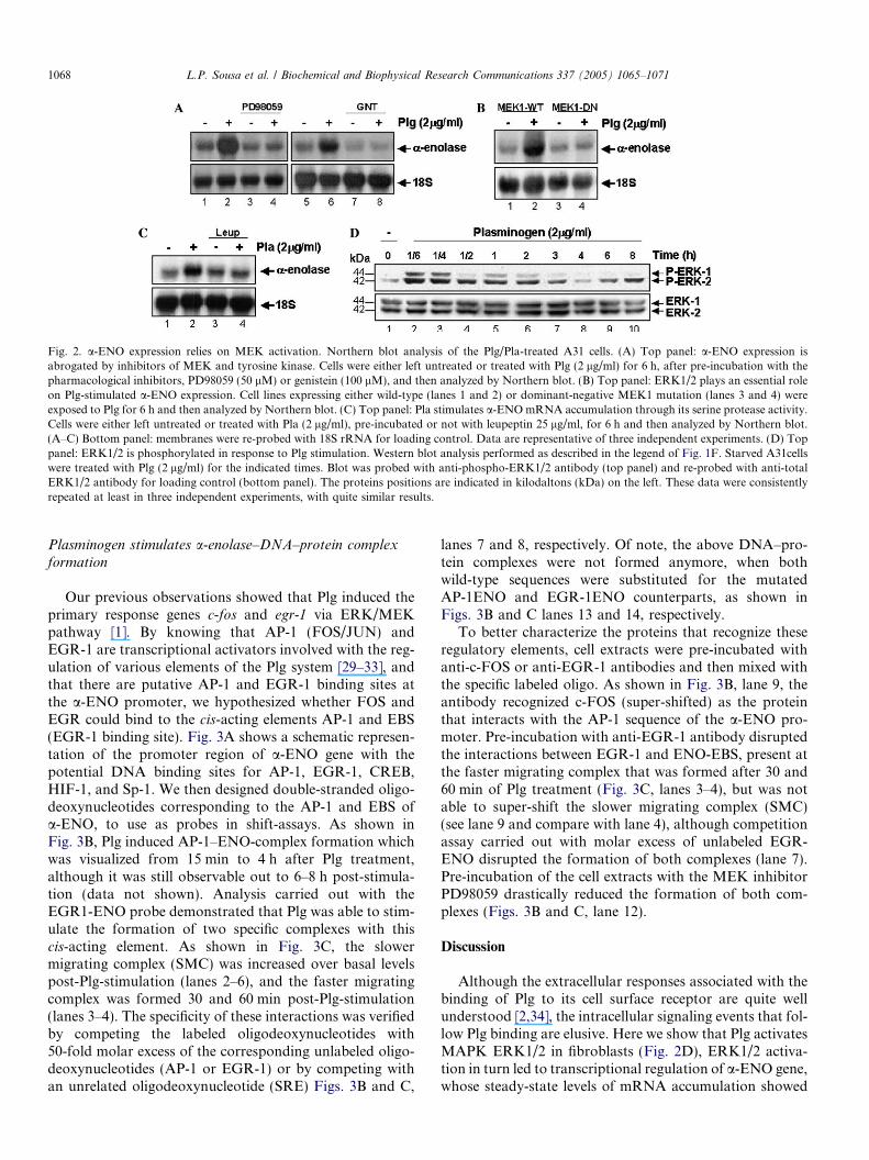
Fig. 2. a-ENO expression relies on MEK activation. Northern blot analysis of the Plg/Pla-treated A31 cells. (A) Top panel: a-ENO expression isabrogated by inhibitors of MEK and tyrosine kinase. Cells were either left untreated or treated with Plg (2 lg/ml) for 6 h, after pre-incubation with thepharmacological inhibitors, PD98059 (50 lM) or genistein (100 lM), and then analyzed by Northern blot. (B) Top panel: ERK1/2 plays an essential roleon Plg-stimulated a-ENO expression. Cell lines expressing either wild-type (lanes 1 and 2) or dominant-negative MEK1 mutation (lanes 3 and 4) wereexposed to Plg for 6 h and then analyzed by Northern blot. (C) Top panel: Pla stimulates a-ENO mRNA accumulation through its serine protease activity.Cells were either left untreated or treated with Pla (2 lg/ml), pre-incubated or not with leupeptin 25 lg/ml, for 6 h and then analyzed by Northern blot.(A–C) Bottom panel: membranes were re-probed with 18S rRNA for loading control. Data are representative of three independent experiments. (D) Toppanel: ERK1/2 is phosphorylated in response to Plg stimulation. Western blot analysis performed as described in the legend of Fig. 1F. Starved A31cellswere treated with Plg (2 lg/ml) for the indicated times. Blot was probed with anti-phospho-ERK1/2 antibody (top panel) and re-probed with anti-totalERK1/2 antibody for loading control (bottom panel). The proteins positions are indicated in kilodaltons (kDa) on the left. These data were consistentlyrepeated at least in three independent experiments, with quite similar results.
1068 L.P. Sousa et al. / Biochemical and Biophysical Research Communications 337 (2005) 1065–1071
Plasminogen stimulates a-enolase–DNA–protein complex
formation
Our previous observations showed that Plg induced theprimary response genes c-fos and egr-1 via ERK/MEKpathway [1]. By knowing that AP-1 (FOS/JUN) andEGR-1 are transcriptional activators involved with the reg-ulation of various elements of the Plg system [29–33], andthat there are putative AP-1 and EGR-1 binding sites atthe a-ENO promoter, we hypothesized whether FOS andEGR could bind to the cis-acting elements AP-1 and EBS(EGR-1 binding site). Fig. 3A shows a schematic represen-tation of the promoter region of a-ENO gene with thepotential DNA binding sites for AP-1, EGR-1, CREB,HIF-1, and Sp-1. We then designed double-stranded oligo-deoxynucleotides corresponding to the AP-1 and EBS ofa-ENO, to use as probes in shift-assays. As shown inFig. 3B, Plg induced AP-1–ENO-complex formation whichwas visualized from 15 min to 4 h after Plg treatment,although it was still observable out to 6–8 h post-stimula-tion (data not shown). Analysis carried out with theEGR1-ENO probe demonstrated that Plg was able to stim-ulate the formation of two specific complexes with thiscis-acting element. As shown in Fig. 3C, the slowermigrating complex (SMC) was increased over basal levelspost-Plg-stimulation (lanes 2–6), and the faster migratingcomplex was formed 30 and 60 min post-Plg-stimulation(lanes 3–4). The specificity of these interactions was verifiedby competing the labeled oligodeoxynucleotides with50-fold molar excess of the corresponding unlabeled oligo-deoxynucleotides (AP-1 or EGR-1) or by competing withan unrelated oligodeoxynucleotide (SRE) Figs. 3B and C,
lanes 7 and 8, respectively. Of note, the above DNA–pro-tein complexes were not formed anymore, when bothwild-type sequences were substituted for the mutatedAP-1ENO and EGR-1ENO counterparts, as shown inFigs. 3B and C lanes 13 and 14, respectively.
To better characterize the proteins that recognize theseregulatory elements, cell extracts were pre-incubated withanti-c-FOS or anti-EGR-1 antibodies and then mixed withthe specific labeled oligo. As shown in Fig. 3B, lane 9, theantibody recognized c-FOS (super-shifted) as the proteinthat interacts with the AP-1 sequence of the a-ENO pro-moter. Pre-incubation with anti-EGR-1 antibody disruptedthe interactions between EGR-1 and ENO-EBS, present atthe faster migrating complex that was formed after 30 and60 min of Plg treatment (Fig. 3C, lanes 3–4), but was notable to super-shift the slower migrating complex (SMC)(see lane 9 and compare with lane 4), although competitionassay carried out with molar excess of unlabeled EGR-ENO disrupted the formation of both complexes (lane 7).Pre-incubation of the cell extracts with the MEK inhibitorPD98059 drastically reduced the formation of both com-plexes (Figs. 3B and C, lane 12).
Discussion
Although the extracellular responses associated with thebinding of Plg to its cell surface receptor are quite wellunderstood [2,34], the intracellular signaling events that fol-low Plg binding are elusive. Here we show that Plg activatesMAPK ERK1/2 in fibroblasts (Fig. 2D), ERK1/2 activa-tion in turn led to transcriptional regulation of a-ENO gene,whose steady-state levels of mRNA accumulation showed

Fig. 3. Analysis of Plg-stimulated DNA-protein complexes formation. EMSA was carried out after treating the starved A31 cells with Plg (2 lg/ml) for theindicated times. Ten micrograms of nuclear protein were incubated with an end-labeled oligodeoxynucleotide probe containing the a-ENO cis-actingelement: AP-1 or EBS. (A) Schematic representation of the 5 0 regulatory sequence of the a-ENO gene. DNA-binding sites for AP-1, EBS (EGR-1 bindingsite), CREB, HIF-1, and Sp-1 are shown. AP-1 (AP-1ENO) and EBS (EGR-1ENO) binding sites are highlighted. Mutated (M) sequences are positionedbelow the wild-type ones. (B,C) Transcriptional complexes formed with AP-1ENO or EGR-1ENO, respectively. DNA–protein complexes formed areindicated. The specificity of the interactions was confirmed by competing the probe with 50-fold molar excess of the indicated cold oligo (lane 7). c-FOSand EGR-1 were super-shifted by specific antibodies (B,C; lane 9). Pre-incubation with PD98059 disrupted the formation of the DNA–protein complex(B,C; lane 12). Mutations mapping at the DNA binding sites of AP-1 or EGR-1 abolished the complexes formation (B,C; lane 14). (C) SMC, slowermigrating complex. These data were repeated in three independent experiments with identical results.
L.P. Sousa et al. / Biochemical and Biophysical Research Communications 337 (2005) 1065–1071 1069
a pattern consistent with a secondary-response gene (Figs.1A andD). By using pharmacological (PD98059) and genet-ic inhibition of MEK (MEK1-DN) we show that MEK/ERK1/2 signaling is necessary for a-ENO mRNA expres-sion (Figs. 2A and B). Plg-stimulated a-ENO message ap-pears to require the simultaneous participation of bothMEK and a tyrosine kinase-sensitive pathway, since pre-in-cubation with genistein also leads to a blockade in the tran-script accumulation (Fig. 2A). Our data also show that thePlg-regulated a-ENO expression is not only restricted tofibroblast cells as it was verified with PBMC as well (Figs.1E and F).
Although plasmin (Pla) and Plg can bind with similaraffinity to diverse blood cells [2], it seems, however, thatthe cellular outcome, may be distinct. For instance, Pla,but not Plg, was able to trigger chemotactic and proinflam-matory responses inmonocytes [35] and expression ofmono-cyte chemoattractant protein-1 (MCP-1) and CD40, inprimarymonocyte, which relied on activation of p38MAPKand JAK/STAT signaling pathways [36], in remarkable con-trast with the ERK1/2 dependence of Plg-stimulated a-ENOexpression. It seems, however, that the requirement of a spe-cific signaling pathway is dependent on the cell line and geneinvolved. Pendurthi et al. [37] demonstrated that Pla stimu-lates the expression of Cyr61 gene via ERK1/2 and tyrosinekinase-dependent pathways in fibroblast, similar to what wehave observed for Plg-stimulated a-ENO expression. In oursystem, Plasmin was also able to stimulate a-ENO mRNAexpression through a serine protease activity-dependent
mechanism (Fig. 2C). This result suggests the requirementof Plg activation to Pla at the cell surface for the initiationof the signaling cascade. This observation was not entirelyunexpected, because Pla also induces c-fos and egr-1 expres-sion, two putative transcriptional activators of a-ENO,through its serine protease activity [1].
Although a-ENO is one of the best characterized pro-teins, little is known about its regulation. It was recentlydemonstrated, by using a MEK-DN approach, thatERK1/2 is involved with the regulation of a-ENO expres-sion in cardiomyocytes submitted to ischemic hypoxia andreoxygenation [38]. Other reports have also demonstratedthat: a-ENO can be regulated by hypoxia on a HIF-1a-dependent manner in Hep3B cells [16]; c-Myc directlytransactivates a-ENO in Rat-1 fibroblasts [39]; enolaseneuron-specific is an EGR-1 target gene in prostate carci-noma cells [40] and a-ENO expression is upregulated in c-
jun transformed rat fibroblast [41]. Although we showedin this report, based on DNA-protein binding experiments,that c-FOS and EGR-1, two downstream targets of MEK,could be involved with a-ENO expression in response to Plgstimulation, our data do not definitively prove that FOSand EGR-1 are, in fact, the transcriptional activators asso-ciated with a-ENO expression upon Plg/Pla stimulation.Further analyses using gene reporter assay with a-enolasepromoter would assist in establishing the transcriptionalactivators that drive the Plg-stimulated a-ENO expression.
Our observation that incubation with anti-EGR-1 anti-body disrupted only the faster migrating complex, but not

1070 L.P. Sousa et al. / Biochemical and Biophysical Research Communications 337 (2005) 1065–1071
the slower migrating one (Fig. 3C, lane 9), could be ex-plained as follows. Even though both complexes have thecapacity to be formed with the same oligodeoxynucleotide,a GC-rich sequence (Fig. 3C, lane 7), we believe that Sp-1might be the protein associated with the SMC, because itcompetes with EGR-1 for binding to the same GC-rich se-quence [42], though this issue remains to be furtherinvestigated.
We have recently demonstrated that interferon a2a1(IFN a2a1) induces a-ENO expression through the MEK/ERK pathway with a similar kinetics [18] to that observedfor Plg. Upon IFN a2a1 stimulation, a-ENO expression onthe surface of PBMCs increases and an enhanced pericellu-lar proteolysis is also observed [18]. In a remarkable con-trast, however, upon Plg stimulation, no significantdifference in a-ENO expression at the surface of PBMCswas verified by FACS analysis (data not shown). In linewith this finding, upon Plg or Pla stimuli no augmentationin pericellular proteolysis either in the A31 cell line or inPBMCs was observed, as accessed by cell surface-generatedplasmin assay (data not shown). Taken together, our datasuggest that even though Plg and IFN control a-ENOexpression, the functioning of a-ENO as Plg receptor mightbe a cytokine-specific regulated process. The role played bya-ENO upon Plg stimulation, however, remains to be fur-ther investigated.
To our best knowledge this is the first report concerningPlg/Pla activation of the MEK/ERK pathway leading tothe transcriptional regulation of the a-ENO gene.
Acknowledgments
The authors are grateful to Angela S. Lopes, Hilda M.V.Gama, Maria A. Souza, Joao R. dos Santos, Bernardete deJesus Martins (in memorium), and Daniela Lemos for theirsecretarial/technical assistance. We also thank Drs. H.A.Armelin and M.C. Sogayar, Department of Biochemistry,University of Sao Paulo, Brazil, for kindly providing uswith the A31 cell line. a-ENO probe was kindly providedby Dr. L.C. Showe (Wistar Institute, Philadelphia, USA).This work was supported by grants of Fundacao de Ampar-o a Pesquisa do Estado de Minas Gerais (FAPEMIG) andConselho Nacional de Desenvolvimento Cientıfico e Tec-nologico (CNPq). L.P.S., B.M.S., and S.V.N. are recipientsof pre-doctoral fellowships either from CNPq or CAPES(Coordenadoria de Aperfeicoamento de Pessoal de NıvelSuperior), Brazilian Ministry of Culture, Science and Tech-nology. B.S.A.F.B. is the recipient of an undergraduate fel-lowship from FAPEMIG. C.A.B., E.G.K., and P.C.P.F.are recipients of research fellowships from CNPq.
References
[1] L.P. De Sousa, B.S. Brasil, B.M. Silva, M.H. Freitas, S.V. Nogueira,P.C. Ferreira, E.G. Kroon, C.A. Bonjardim, Plasminogen/plasminregulates c-fos and egr-1 expression via the MEK/ERK pathway,Biochem. Biophys. Res. Commun. 329 (2005) 237–245.
[2] E.F. Plow, T. Herren, A. Redlitz, L.A. Miles, J.L. Hoover-Plow, Thecell biology of the plasminogen system, FASEB J. 9 (1995) 939–945.
[3] H.R. Lijnen, Elements of the fibrinolytic system, Ann. N.Y. Acad.Sci. 936 (2001) 226–236.
[4] E.F. Plow, D.E. Freaney, J. Plescia, L.A. Miles, The plasminogensystem and cell surfaces: evidence for plasminogen and urokinasereceptors on the same cell type, J. Cell Biol. 103 (1986) 2411–2420.
[5] M. Konakova, F. Hucho, W.D. Schleuning, Downstream targets ofurokinase-type plasminogen-activator-mediated signal transduction,Eur. J. Biochem. 253 (1998) 421–429.
[6] I. Dumler, A. Weis, O.A. Mayboroda, C. Maasch, U. Jerke, H.Haller, D.C. Gulba, The Jak/Stat pathway and urokinase receptorsignaling in human aortic vascular smooth muscle cells, J. Biol.Chem. 273 (1998) 315–321.
[7] D.H. Nguyen, I.M. Hussaini, S.L. Gonias, Binding of urokinase-typeplasminogen activator to its receptor in MCF-7 cells activatesextracellular signal-regulated kinase 1 and 2 which is required forincreased cellular motility, J. Biol. Chem. 273 (1998) 8502–8507.
[8] T. Tarui, M. Majumdar, L.A. Miles, W. Ruf, Y. Takada, Plasmin-induced migration of endothelial cells. A potential target for theanti-angiogenic action of angiostatin, J. Biol. Chem. 277 (2002) 33564–33570.
[9] K. Lohman, O. Meyerhof, Uber die enzymatische Umwandlung vonPhosphoglyzerinsaure in Brenztraubensaure and phophorsaure(Enzymatic transformation of phosphoglyceric acid in pyruvic andphosphoric acid), Biochem. Z 273 (1934) 60–72.
[10] L.A. Miles, C.M. Dahlberg, J. Plescia, J. Felez, K. Kato, E.F. Plow,Role of cell-surface lysines in plasminogen binding to cells: identifi-cation of alpha-enolase as a candidate plasminogen receptor,Biochemistry 30 (1991) 1682–1691.
[11] A. Redlitz, B.J. Fowler, E.F. Plow, L.A. Miles, The role of anenolase-related molecule in plasminogen binding to cells, Eur. J.Biochem. 227 (1995) 407–415.
[12] B. Arza, J. Felez, R. Lopez-Alemany, L.A. Miles, P. Munoz-Canoves,Identification of an epitope of alpha-enolase (a candidate plasmin-ogen receptor) by phage display, Thromb. Haemost. 78 (1997) 1097–1103.
[13] S.B. Hawley, M.A. Green, L.A. Miles, Discriminating between cellsurface and intracellular plasminogen-binding proteins: heterogeneityin profibrinolytic plasminogen-binding proteins on monocytoid cells,Thromb. Haemost. 84 (2000) 882–890.
[14] R. Lopez-Alemany, C. Longstaff, S. Hawley, M. Mirshahi, P.Fabregas, M. Jardi, E. Merton, L.A. Miles, J. Felez, Inhibition ofcell surface mediated plasminogen activation by a monoclonalantibody against alpha-Enolase, Am. J. Hematol. 72 (2003) 234–242.
[15] A. Giallongo, S. Feo, R. Moore, C.M. Croce, L.C. Showe, Molecularcloning and nucleotide sequence of a full-length cDNA for humanalpha enolase, Proc. Natl. Acad. Sci. USA 83 (1986) 6741–6745.
[16] G.L. Semenza, B.H. Jiang, S.W. Leung, R. Passantino, J.P. Concor-det, P. Maire, A. Giallongo, Hypoxia response elements in thealdolase A, enolase 1, and lactate dehydrogenase A gene promoterscontain essential binding sites for hypoxia-inducible factor 1, J. Biol.Chem. 271 (1996) 32529–32537.
[17] M. Scharte, X. Han, D.J. Bertges, M.P. Fink, R.L. Delude, Cytokinesinduce HIF-1 DNA binding and the expression of HIF-1-dependentgenes in cultured rat enterocytes, Am. J. Physiol. Gastrointest. LiverPhysiol. 284 (2003) G373–G384.
[18] L.P. Sousa, B.S. Brasil, M. Silva Bde, S.V. Nogueira, A.A. Andrade,P.C. Ferreira, S.M. Teixeira, K.J. Gollob, E.G. Kroon, K. Kato, C.A.Bonjardim, Characterization of alpha-enolase as an interferon-alpha2 alpha 1 regulated gene, Front. Biosci. 10 (2005) 2534–2547.
[19] J. Felez, C.J. Chanquia, E.G. Levin, L.A. Miles, E.F. Plow, Bindingof tissue plasminogen activator to human monocytes and monocytoidcells, Blood 78 (1991) 2318–2327.
[20] K. Kato, F. Suzuki, R. Semba, Determination of brain enolaseisozymes with an enzyme immunoassay at the level of single neurons,J. Neurochem. 37 (1981) 998–1005.

L.P. Sousa et al. / Biochemical and Biophysical Research Communications 337 (2005) 1065–1071 1071
[21] A. Shimizu, F. Suzuki, K. Kato, Characterization of alpha alpha,beta beta, gamma gamma and alpha gamma human enolase isozymes,and preparation of hybrid enolases (alpha gamma, beta gamma andalpha beta) from homodimeric forms, Biochim. Biophys. Acta 748(1983) 278–284.
[22] A.M. Horgan, P.J. Stork, Examining the mechanism of Erk nucleartranslocation using green fluorescent protein, Exp. Cell. Res. 285(2003) 208–220.
[23] J. Sambrook, E.F. Fritsch, T. Maniats, Molecular Cloning, ALaboratory Manual, second ed., Cold Spring Harbor LaboratoryPress, Cold Spring Harbor, 1989, Books 1 and 2.
[24] A.A. Andrade, P.N. Silva, A.C. Pereira, L.P. De Sousa, P.C. Ferreira,R.T. Gazzinelli, E.G.Kroon, C. Ropert, C.A. Bonjardim, The vacciniavirus-stimulatedmitogen-activated protein kinase (MAPK) pathway isrequired for virus multiplication, Biochem. J. 381 (2004) 437–446.
[25] J.C. de Magalhaes, A.A. Andrade, P.N. Silva, L.P. Sousa, C. Ropert,P.C. Ferreira, E.G. Kroon, R.T. Gazzinelli, C.A. Bonjardim, Amitogenic signal triggered at an early stage of vaccinia virus infection:implication of MEK/ERK and protein kinase A in virus multiplica-tion, J. Biol. Chem. 276 (2001) 38353–38360.
[26] G.M. Church, W. Gilbert, Genomic sequencing, Proc. Natl. Acad.Sci. USA 81 (1984) 1991–1995.
[27] C.A. Bonjardim, A mutant cell line partially responsive to both IFN-alpha and IFN-gamma, Braz. J. Med. Biol. Res. 30 (1997) 41–50.
[28] J.D. Dignam, R.M. Lebovitz, R.G. Roeder, Accurate transcriptioninitiation by RNA polymerase II in a soluble extract from isolatedmammalian nuclei, Nucleic Acids Res. 11 (1983) 1475–1489.
[29] A.E. Dear, M. Costa, R.L. Medcalf, Urokinase-mediated transacti-vation of the plasminogen activator inhibitor type 2 (PAI-2) genepromoter in HT-1080 cells utilises AP-1 binding sites and potentiatesphorbol ester-mediated induction of endogenous PAI-2 mRNA,FEBS Lett. 402 (1997) 265–272.
[30] D. Gavrilov, O. Kenzior, M. Evans, R. Calaluce, W.R. Folk,Expression of urokinase plasminogen activator and receptor inconjunction with the ets family and AP-1 complex transcriptionfactors in high grade prostate cancers, Eur. J. Cancer 37 (2001) 1033–1040.
[31] C. Liu, J. Yao, D. Mercola, E. Adamson, The transcription factorEGR-1 directly transactivates the fibronectin gene and enhancesattachment of human glioblastoma cell line U251, J. Biol. Chem. 275(2000) 20315–20323.
[32] U.R. Pendurthi, L.V. Rao, Suppression of transcription factor Egr-1by curcumin, Thromb. Res. 97 (2000) 179–189.
[33] L.M. Khachigian, V. Lindner, A.J. Williams, T. Collins, Egr-1-induced endothelial gene expression: a common theme in vascularinjury, Science 271 (1996) 1427–1431.
[34] T. Herren, T.A. Burke, M. Jardi, J. Felez, E.F. Plow, Regulation ofplasminogen binding to neutrophils, Blood 97 (2001) 1070–1078.
[35] T. Syrovets, M. Jendrach, A. Rohwedder, A. Schule, T. Simmet,Plasmin-induced expression of cytokines and tissue factor in humanmonocytes involves AP-1 and IKKbeta-mediated NF-kappaB acti-vation, Blood 97 (2001) 3941–3950.
[36] L. Burysek, T. Syrovets, T. Simmet, The serine protease plasmintriggers expression of MCP-1 and CD40 in human primarymonocytes via activation of p38 MAPK and janus kinase (JAK)/STAT signaling pathways, J. Biol. Chem. 277 (2002) 33509–33517.
[37] U.R. Pendurthi, M. Ngyuen, P. Andrade-Gordon, L.C. Petersen,L.V. Rao, Plasmin induces Cyr61 gene expression in fibroblasts viaprotease-activated receptor-1 and p44/42 mitogen-activated proteinkinase-dependent signaling pathway, Arterioscler. Thromb. Vasc.Biol. 22 (2002) 1421–1426.
[38] Y. Mizukami, A. Iwamatsu, T. Aki, M. Kimura, K. Nakamura, T.Nao, T. Okusa, M. Matsuzaki, K. Yoshida, S. Kobayashi, ERK1/2regulates intracellular ATP levels through alpha-enolase expression incardiomyocytes exposed to ischemic hypoxia and reoxygenation, J.Biol. Chem. 279 (2004) 50120–50131.
[39] R.C. Osthus, H. Shim, S. Kim, Q. Li, R. Reddy, M. Mukherjee, Y.Xu, D. Wonsey, L.A. Lee, C.V. Dang, Deregulation of glucosetransporter 1 and glycolytic gene expression by c-Myc, J. Biol. Chem.275 (2000) 21797–21800.
[40] J. Svaren, T. Ehrig, S.A. Abdulkadir, M.U. Ehrengruber, M.A.Watson, J. Milbrandt, EGR1 target genes in prostate carcinoma cellsidentified by microarray analysis, J. Biol. Chem. 275 (2000) 38524–38531.
[41] A.C. Bergman, C. Linder, K. Sakaguchi, M. Sten-Linder, A.A.Alaiya, B. Franzen, M.C. Shoshan, T. Bergman, B. Wiman, G. Auer,E. Appella, H. Jornvall, S. Linder, Increased expression of alpha-enolase in c-jun transformed rat fibroblasts without increasedactivation of plasminogen, FEBS Lett. 417 (1997) 17–20.
[42] C. Liu, A. Calogero, G. Ragona, E. Adamson, D. Mercola, EGR-1,the reluctant suppression factor: EGR-1 is known to function in theregulation of growth, differentiation, and also has significant tumorsuppressor activity and a mechanism involving the induction of TGF-beta1 is postulated to account for this suppressor activity, Crit. Rev.Oncog. 7 (1996) 101–125.