
Aryl hydrocarbon receptor contributes to the MEK/ERK-dependent maintenance of the immature state of...
11
online February 21, 2013 originally published doi:10.1182/blood-2012-07-445106 2013 121: e108-e117 Ana Cuenda, Bárbara Alonso, Angeles Domínguez-Soto, Silvia Sánchez-Ramón and Angel L. Corbí Pedro Maria Fernández-Salguero, Jose Luis Rodríguez-Fernández, Oscar M. Pello, Vicente Andrés, Noemí Aguilera-Montilla, Sonia Chamorro, Concha Nieto, Fátima Sánchez-Cabo, Ana Dopazo, maintenance of the immature state of human dendritic cells Aryl hydrocarbon receptor contributes to the MEK/ERK-dependent http://www.bloodjournal.org/content/121/15/e108.full.html Updated information and services can be found at: (5229 articles) Immunobiology (132 articles) e-Blood Articles on similar topics can be found in the following Blood collections http://www.bloodjournal.org/site/misc/rights.xhtml#repub_requests Information about reproducing this article in parts or in its entirety may be found online at: http://www.bloodjournal.org/site/misc/rights.xhtml#reprints Information about ordering reprints may be found online at: http://www.bloodjournal.org/site/subscriptions/index.xhtml Information about subscriptions and ASH membership may be found online at: Copyright 2011 by The American Society of Hematology; all rights reserved. of Hematology, 2021 L St, NW, Suite 900, Washington DC 20036. Blood (print ISSN 0006-4971, online ISSN 1528-0020), is published weekly by the American Society For personal use only. on October 31, 2014. by guest www.bloodjournal.org From For personal use only. on October 31, 2014. by guest www.bloodjournal.org From
-
Upload
independent -
Category
Documents
-
view
0 -
download
0
Transcript of Aryl hydrocarbon receptor contributes to the MEK/ERK-dependent maintenance of the immature state of...

online February 21, 2013 originally publisheddoi:10.1182/blood-2012-07-445106
2013 121: e108-e117
Ana Cuenda, Bárbara Alonso, Angeles Domínguez-Soto, Silvia Sánchez-Ramón and Angel L. CorbíPedro Maria Fernández-Salguero, Jose Luis Rodríguez-Fernández, Oscar M. Pello, Vicente Andrés, Noemí Aguilera-Montilla, Sonia Chamorro, Concha Nieto, Fátima Sánchez-Cabo, Ana Dopazo, maintenance of the immature state of human dendritic cellsAryl hydrocarbon receptor contributes to the MEK/ERK-dependent
http://www.bloodjournal.org/content/121/15/e108.full.htmlUpdated information and services can be found at:
(5229 articles)Immunobiology (132 articles)e-Blood
Articles on similar topics can be found in the following Blood collections
http://www.bloodjournal.org/site/misc/rights.xhtml#repub_requestsInformation about reproducing this article in parts or in its entirety may be found online at:
http://www.bloodjournal.org/site/misc/rights.xhtml#reprintsInformation about ordering reprints may be found online at:
http://www.bloodjournal.org/site/subscriptions/index.xhtmlInformation about subscriptions and ASH membership may be found online at:
Copyright 2011 by The American Society of Hematology; all rights reserved.of Hematology, 2021 L St, NW, Suite 900, Washington DC 20036.Blood (print ISSN 0006-4971, online ISSN 1528-0020), is published weekly by the American Society
For personal use only.on October 31, 2014. by guest www.bloodjournal.orgFrom For personal use only.on October 31, 2014. by guest www.bloodjournal.orgFrom

e-Blood
IMMUNOBIOLOGY
Aryl hydrocarbon receptor contributes to the MEK/ERK-dependentmaintenance of the immature state of human dendritic cellsNoemı Aguilera-Montilla,1 Sonia Chamorro,1 Concha Nieto,1 Fatima Sanchez-Cabo,2 Ana Dopazo,2
Pedro Maria Fernandez-Salguero,3 Jose Luis Rodrıguez-Fernandez,1 Oscar M. Pello,2 Vicente Andres,2 Ana Cuenda,4
Barbara Alonso,5 Angeles Domınguez-Soto,1 Silvia Sanchez-Ramon,5 and Angel L. Corbı1
1Centro de Investigaciones Biologicas, Consejo Superior de Investigaciones Cientıficas (CSIC), Madrid, Spain; 2Centro Nacional de Investigaciones
Cardiovasculares, Madrid, Spain; 3Universidad de Extremadura, Badajoz, Spain; 4Centro Nacional de Biotecnologıa, CSIC, Madrid, Spain; 5Hospital General
Universitario Gregorio Maranon, Madrid, Spain
Key Points
• Aryl hydrocarbon receptor(AhR) mediates theERK-dependent maintenanceof the immature state ofmonocyte-derived dendriticcells (MDDCs).
• MEK-ERK regulates antigencapture, lymph node homing,and the acquisition ofmaturation-associated genesin MDDCs.
Dendritic cells (DCs) promote tolerance or immunity depending on their maturation
state, which is enhanced or accelerated upon MEK-ERK signaling pathway inhibition.
We have determined the contribution of MEK-ERK activation to the profile of gene
expression of human immature monocyte-derived dendritic cells (MDDCs) and pe-
ripheral blood myeloid DCs. ERK inhibition altered the expression of genes that medi-
ate Chemokine (C-C motif) ligand 19 (CCL19)–directed migration (CCR7) and low-
density lipoprotein (LDL) binding (CD36, SCARB1, OLR1, CXCL16) by immature DCs. In
addition, ERK upregulated CCL2 expression while impairing the expression of DC
maturation markers (RUNX3, ITGB7, IDO1). MEK-ERK–regulated genes exhibited an
overrepresentation of cognate sequences for the aryl hydrocarbon receptor (AhR)
transcription factor, whose transcriptional and DNA-binding activities increased in
MDDCs upon exposure to the MEK1/2 inhibitor U0126. Therefore, the MEK-ERK
signaling pathway regulates antigen capture, lymph node homing, and acquisition of
maturation-associated genes, and its contribution to the maintenance of the immature
state of MDDCs and myeloid DCs is partly dependent on the activity of AhR. Since
pharmacologic modulation of the MEK-ERK signaling pathway has been proposed as a potential therapeutic strategy for cancer,
our findings indicate that ERK inhibitors might influence antitumor responses through regulation of critical DC effector functions.
(Blood. 2013;121(15):e108-e117)
Introduction
Dendritic cells (DCs) are the essential link between innate andadaptive immune responses.1 In the steady-state, tissue-residentimmature DCs capture extracellular material and contribute toperipheral tolerance.1,2 By contrast, upon detection of danger-associated molecular patterns by germ line–encoded pathogen-recognition receptors, immature DCs increase their expressionof major histocompatibility complex (MHC) and costimulatorymolecules and migrate toward peripheral lymph nodes in a chemo-kine receptor 7 (CCR7) –dependent manner.1,2 This maturationevent endows DCs with the ability to promote the generation ofprimary immune responses.1 Thus, DC maturation constitutes anideal target for modulation of immune responses in autoimmunediseases, transplant rejection, or cancer.2
Whereas the critical role of nuclear factor kappa B (NFkB)activation in DC maturation is firmly established, various signalingmechanisms function as modulators of the DC maturation process.3
ERK and p38MAPK signaling pathways have been shown tooppositely contribute to DC maturation.4,5 p38MAPK activation is
a requisite for increased NFkB recruitment to the regulatory regionsof genes whose expression is modulated during DC maturation, andits inhibition reduces costimulatory molecule expression, interleukin-12p70 (IL-12p70) production, and T-cell stimulatory ability.6
Conversely, the MEK-ERK signaling pathway prevents an optimalDCmaturation.4 In response to maturation stimuli, inhibition ofMEK-ERK activation results in higher expression of MHC-II, costimu-latory and adhesion molecules (CD83, CD86, and CD49d),5 higherallostimulatory activity, elevated secretion of IL-12p70 and IL-12p40,5,7 and decreased proinflammatory cytokine production.4
These functions point to a negative regulatory role of theMEK-ERKsignaling pathway on DC maturation.
DC maturation is strongly impaired in patients with tumors, inwhom the low number and the immature state of DCs are responsiblefor cancer-associated immunosuppression.8 Tumor cells, myeloid-derived suppressor cells, and tumor-associated macrophages createa potent immunosuppressive environment that prevents DC matu-ration.8 In this regard, DCs in rapidly growing tumors usually exhibit
Submitted July 20, 2012; accepted February 6, 2013. Prepublished online as
Blood First Edition paper, February 21, 2013; DOI 10.1182/blood-2012-07-
445106.
N.A.-M., S.C., and C. N. contributed equally to this study.
The online version of this article contains a data supplement.
The publication costs of this article were defrayed in part by page charge
payment. Therefore, and solely to indicate this fact, this article is hereby
marked “advertisement” in accordance with 18 USC section 1734.
© 2013 by The American Society of Hematology
e108 BLOOD, 11 APRIL 2013 x VOLUME 121, NUMBER 15
For personal use only.on October 31, 2014. by guest www.bloodjournal.orgFrom

an immature phenotype, and the majority of DCs remain immaturewithin the tumors, whereas mature DCs reside in the surroundingareas.9 DC-based cancer immunotherapy is currently considered ameans to overcome the immunosuppression that takes place in patientswith tumors.10,11 Small-molecule inhibitors of ERK and p38MAPKsignaling pathways are also being evaluated in clinical trials for thetreatment of pathologic disorders.12-14 Therefore, on the basis of theireffects on DC maturation, it is conceivable that pharmacologic MEKinhibitors might diminish the tumor-imposed halt on DC maturation,thus overcoming immunosuppression.
To determine the extent of the contribution of the MEK-ERKsignaling pathway to the maturation state of DCs, we have deter-mined the ERK-dependent gene expression profile in immaturehuman monocyte-derived dendritic cells (MDDCs). Our resultsindicate that the MEK-ERK signaling axis regulates DC functionsthat are critical in the initiation and regulation of immune response,and they provide evidence for the involvement of the aryl hydro-carbon receptor (AhR) transcription factor in the ERK-dependentgene expression profile of immature MDDCs.
Methods
Dendritic cell generation and maturation
Human peripheral blood mononuclear cells were isolated from buffy coatsfrom normal donors over a Lymphoprep (Nycomed Pharma, Oslo, Norway)gradient. Monocytes were purified from peripheral blood mononuclear cellsby magnetic cell sorting using CD14 microbeads (Miltenyi Biotec, BergischGladbach, Germany). Monocytes (.95% CD141 cells) were cultured at0.7 3 106 cells/mL for 5 days in RPMI 1640 supplemented with 10% fetalcalf serum (complete medium) at 37°C in a humidified atmosphere with5% CO2 containing 1000 U/mL granulocyte macrophage colony-stimulatingfactor (GM-CSF) (ImmunoTools; Friesoythe, Germany) and 1000 U/mLIL-4 (ImmunoTools) to generate immature MDDCs, with cytokine additionevery 2 days. Macrophages were generated following the same scheme butusing 10 ng/mL M-CSF (ImmunoTools). For maturation, MDDCs weretreated with 10 ng/mL lipopolysaccharide (LPS; E. coli 055:B5; Sigma).For gene profiling experiments, immature MDDCs were exposed top38MAPK inhibitors SB203580 (13 mM; Enzo Life Sciences, Farmingdale,NY) and BIRB0796 (0.1 mM)15 or MEK inhibitors U0126 (2.5 mM) andPD98059 (40 mM; Calbiochem, Nottingham, UK).16,17 A final dose ofGM-CSF and IL-4 was added to the culture 1 hour after treatment withinhibitors, and cells were collected after 4, 10, or 24 hours. Peripheral bloodCD1c1 DCs were isolated by using the CD1c (BDCA-1)1 Dendritic CellIsolation Kit (Miltenyi Biotec). Chemokine (C-C motif) ligand 2 (CCL2)was measured by using a commercially available enzyme-linked immuno-sorbent assay (BD OptEIATM Human MCP-1 ELISA Set; BD Bioscience).
Cell transfection and reporter gene assays
MDDCs (2 3 106 cells) were nucleofected with 10 mg of a constitutivelyactive (MEK-A) or a double negative (MEK-E) form of MEK, provided byDr Pura Muñoz-Canoves (Universidad Pompeu Fabra, Barcelona, Spain),using the Human Dendritic Cell Nucleofector Kit V (Lonza, Basel,Switzerland). As a control, MDDCs were nucleofected with pCDNA3.1empty vector. After nucleofection, cells were cultured for 24 hours incomplete medium containing GM-CSF and IL-4. In reporter gene assays,MDDCs (2 3 106 cells) or monocytes (107 cells) were nucleofected with975 ng of a xenobiotic responsive element (XRE) –dependent construct(XRE-TATA-luc) and 25 ng of a construct expressing the Renilla luciferasefrom a constitutive promoter (Cignal XRE Reporter Kit; SABiosciences).Where indicated, cells were transfected in parallel with a negative controlTATA-luc construct. After 24 hours, cells were treated for 12 hours withdimethylsulfoxide (DMSO), U0126 (2.5 mM), or tetrachlorodibenzo-p-dioxin
(TCDD; 10 nM) and lysed. Firefly and Renilla luciferase activities weredetermined by using the Dual-Luciferase Reporter Assay System (Promega).HepG2 cells were transfected by using Superfect (Qiagen, Valencia, CA).
Gene expression profiling and quantitative reverse
transcriptase polymerase chain reaction
RNA was isolated from three independent preparations of MDDCs exposedto either DMSO or U0126 for 4, 10, or 24 hours by using AllPrep DNA/RNA/Protein Mini Kit (Qiagen). Labeled RNA was used as a hybridizationprobe on Whole Human Genome Microarrays (Agilent Technologies, PaloAlto, CA). A threshold of 1 was assigned to raw signals and quantiles werenormalized.18 After removing probes with low signals and questionablequality, a moderated t test, as implemented in the Bioconductor limmapackage,19 was used to calculate the P values for each gene comparingconditions. P values were adjusted by using Benjamini-Hochberg methodto account for multiple testing. Adjusted P values smaller than .05 wereconsidered significant. Gene ontology (GO) analysis of microarray resultswas performed by using the Web-based DAVID functional annotation tool(http://david.abcc.ncifcrf.gov/home.jsp). Significantly changed genes werecompared with a background set of the total complement of genes that wererepresented on the microarray. GO terms were considered significant if theyhad a Benjamini-corrected P value lower than .05. All microarray data havebeen deposited in the Gene Expression Omnibus under accession numberGSE39745. For quantitative reverse transcriptase polymerase chain reac-tion, total RNA was retrotranscribed, and complementary DNA was quan-tified by using the Universal ProbeLibrary (Roche Diagnostics). Triplicateassays were performed, and results were normalized according to the ex-pression levels of GAPDH RNA and expressed by using the DDCT methodfor quantization.
Western blot
Affinity-purified rabbit monoclonal antibodies from Cell Signaling Tech-nology (Danvers, MA) were used to detect ERK1/2, phospho-ERK1/2 (Thr202/Tyr204), p38MAPK, and phospho-p38MAPK (Thr180/Tyr182)(D3F9). A polyclonal antibody against AhR (N-19) and a monoclonal anti–glyceraldehyde phosphate dehydrogenase (GAPDH) antibody (6C5) werefrom Santa Cruz Biotechnology (Santa Cruz, CA), and the Ab-11 cocktail(Thermo-Scientific, Fremont, CA) was used to detect THBS1.
Low-density lymphocyte uptake assay
MDDCs from 5 independent donors were treated with DMSO or U0126 for24 hours, washed twice with phosphate-buffered saline, and exposed to1 mg/mL of acetylated low-density lymphocyte (LDL) -PE/Cy5 (acLDL;Invitrogen, Carlsbad, CA) in serum-free RPMI 1640 for 30 to 180 minutesat 37°C. After washing, acLDL uptake was evaluated by flow cytometry.
Chemotaxis assays
Chemotaxis in response to CCL19 was determined in MDDCs previouslyexposed to DMSO or U0126 for 24 hours; 1 3 105 cells were washed,diluted in 100 mL of RPMI 1640 medium containing 0.1% bovine serumalbumin, and seeded in the upper chamber of a 5-mm pore size 24-wellTranswell chamber (Costar; Cambridge, MA). The lower chamber con-tained 600 mL of the same medium supplemented or not with CCL19 (100ng/mL). After 2.5 hours, MDDCs that migrated into the lower chamberwere measured.
Chromatin immunoprecipitation
Chromatin immunoprecipitation assays were performed on MDDCs treatedwith either DMSO or U0126 for 10 hours by using the Magna ChIP GChromatin Immunoprecipitation Kit (Millipore), according to the manu-facturer’s recommendations. For PCR detection of the AhR-binding se-quence within the CYP1B1 gene promoter, the oligonucleotides 59-ATATGACTGGAGCCGACTTTCC-39 and 59- GGCGAACTTTATCGGGTTGA-39 were used, which amplify the 2790/2890 CYP1B1 promoter re-gion. Five micrograms of an anti-AhR RPT9 antibody (Abcam) was used
BLOOD, 11 APRIL 2013 x VOLUME 121, NUMBER 15 ERK CONTRIBUTION TO THE IMMATURE STATE OF MDDC e109
For personal use only.on October 31, 2014. by guest www.bloodjournal.orgFrom
for immunoprecipitation, and an irrelevant immunoglobulin G-1 mousemonoclonal antibody was used as a negative control.
Results
Identification of genes regulated by MEK-ERK1/2 in immature
human MDDCs
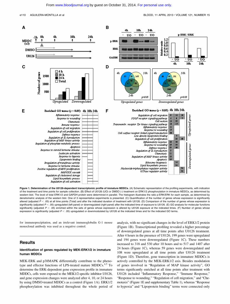
MEK-ERK and p38MAPK differentially contribute to the pheno-type and effector functions of LPS-treated mature MDDCs.4,5 Todetermine the ERK-dependent gene expression profile in immatureMDDCs, cells were exposed to the MEK1/2-specific inhibitor U0126,and gene expression changes were analyzed after 4, 10, or 24 hoursby using DMSO-treated MDDCs as a control (Figure 1A). ERK1/2phosphorylation was inhibited throughout the whole period of
analysis, with no significant changes in the level of ERK1/2 protein(Figure 1B). Transcriptional profiling revealed a higher percentageof downregulated genes at all time points after U0126 treatment.After 4 hours in the presence of U0126, 199 genes were upregulatedand 194 genes were downregulated (Figure 1C). These numbersincreased to 318 and 530 after 10 hours and to 517 and 1407 after24 hours (Figure 1C), whereas 59 genes were downregulated and108 were upregulated at all time points after U0126 treatment(Figure 1D). Therefore, gene transcription in immature MDDCs isactively controlled by the MEK-ERK1/2 axis. Besides modulationof genes involved in “Regulation of MAP kinase activity”, GOterms significantly enriched at all time points after treatment withU0126 included “Inflammatory Response,” “Immune Response,”“Response to wounding,” “Regulation of cell migration,” and “Che-motaxis” (Figure 1E and supplementary Table 1), whereas “Responseto hypoxia” and “Lipoprotein binding” terms were connected only
Figure 1. Determination of the U0126-dependent transcriptomic profile of immature MDDCs. (A) Schematic representation of the profiling experiments, with indication
of the treatment and time points for sample collection. (B) Effect of U0126 (UO) or DMSO (-) treatment on ERK1/2 phosphorylation in immature MDDCs, as determined by
western blot. The level of total ERK1/2 and GAPDH protein were determined in parallel. The histogram illustrates the ratio p-ERK/ERK for each sample, as determined by
densitometric analysis of the western blot. One of 3 representative experiments is presented. (C) Quantification of the number of genes whose expression is significantly
altered (adjusted P , .05) at all time points (Total) and after the indicated duration of treatment with U0126. (D) Comparison of the number of genes whose expression is
significantly (adjusted P , .05) upregulated (left panel) or downregulated (right panel) after the indicated time of exposure to U0126. (E) GO analysis for molecular functions
significantly (adjusted P , .05) enriched within the sets of genes whose expression is altered by U0126 exposure at the indicated times. (F) Number of genes whose
expression is significantly (adjusted P , .05) upregulated or downmodulated by U0126 at the indicated times and for the indicated GO terms.
e110 AGUILERA-MONTILLA et al BLOOD, 11 APRIL 2013 x VOLUME 121, NUMBER 15
For personal use only.on October 31, 2014. by guest www.bloodjournal.orgFrom

to genes whose expression was modified by U0126 after 4 or 10hours (Figure 1E and supplementary Tables 2 and 3). The“Lipoprotein binding” function was enriched among the genesupregulated by U0126 after 10 hours (Figure 1F and supple-mentary Table 3), whereas the “Lipid biosynthesis” process wasexclusively enriched in genes downregulated by U0126 atthe same time point (Figure 1F and supplementary Table 3).Supporting the GO analysis, the list of the genes whose ex-pression was more profoundly affected by U0126 includednumerous genes involved in immune/inflammatory responses,cell migration, and lipoprotein binding (Table 1). Importantly,
U0126 treatment significantly reduced the expression of well-known targets of ERK (FOS, MYC, DUSP6). Therefore, inhibitionof the MEK-ERK signaling axis in immature MDDCs alters theexpression of genes that mediate critical effector functions ofimmature MDDCs as well as functions related to sterol and lipidmetabolism.
Validation of the transcription data and functional predictions
To verify the above predictions, we focused on genes whose ex-pression showed a potent U0126 responsiveness (FOS, DUSP6,
Table 1. Top 25 upregulated and downregulated genes in U0126-treated MDDCs at 4, 10, and 24 hours posttreatment
4 hours 10 hours 24 hours
Gene symbolLog2 foldchange Adjusted p Gene symbol
Log2 foldchange Adjusted p Gene symbol
Log2 foldchange Adjusted P
Upregulated genes PLAT 2.39 1.69 E-07 PLAT 3.39 1.78 E-10 ANGPT2 5.48 0.012988
SHF 1.95 7.79 E-06 PTGER3 3.06 1.44 E-08 RBMXL3 5.09 0.043762
AHRR 1.83 0.011751 ATP1B2 2.71 3.64 E-08 NR4A3 4.85 0.011564
ROR2 1.70 0.014002 TTC9 2.61 2.89 E-12 RALGAPA1 4.38 0.020438
TTC9 1.67 1.50 E-08 SHF 2.50 7.10 E-08 ATP1B2 4.06 2.16 E-10
LRG1 1.64 1.69 E-07 AHRR 2.23 1.53 E-11 A4GALT 3.97 0.000855
ATP1B2 1.59 0.000199 CYP1B1 2.21 1.39 E-09 TMPRSS6 3.71 1.95 E-05
GAD1 1.48 4.16 E-07 GAD1 2.21 2.31 E-10 GSTT1 3.41 7.67 E-10
RASAL1 1.38 0.0001908 SYTL3 2.13 2.36 E-05 TTC9 3.10 7.46 E-13
ZNF395 1.38 1.69 E-07 ROR2 2.06 0.001021 MUC1 2.78 1.96 E-09
RARG 1.36 0.0005075 CD163L1 2.01 5.72 E-05 CD163L1 2.76 2.74 E-06
ARL4C 1.33 0.0491981 CSRP2 1.94 3.14 E-09 XPNPEP2 2.71 0.000513
CXCL2 1.33 0.0020045 RARG 1.85 4.36 E-06 ARL4C 2.64 5.30 E-05
TIPARP 1.24 7.11 E-07 MUC1 1.82 4.68 E-07 PTGER3 2.64 8.52 E-07
PDGFB 1.20 0.0109397 THBS1 1.78 1.05 E-05 SLC26A1 2.62 0.04403
PTGER2 1.17 3.49 E-07 ARL4C 1.77 0.002025 CYP1B1 2.49 9.26 E-10
NINL 1.16 0.0108421 TIPARP 1.69 1.96 E-09 CSRP2 2.46 2.81 E-10
PRDM1 1.14 0.000317 LPAR6 1.64 4.57 E-09 HES1 2.43 2.46 E-05
THBS1 1.14 0.0048596 ANK3 1.61 0.020469 PTP4A3 2.41 1.80 E-05
SYTL3 1.13 0.0417727 RASAL1 1.60 1.27 E-05 ITGB7 2.41 4.05 E-10
CYP1B1 1.10 0.000145 ITGB7 1.59 9.34 E-08 SHF 2.41 6.22 E-07
NCF1 1.08 0.0274784 GSG1 1.59 0.002054 RPS4Y1 2.41 8.68 E-05
XYLT1 1.08 0.0172593 NCF1 1.54 0.000415 CCDC108 2.38 0.031671
MUC1 1.02 0.0020864 PLA2G16 1.52 1.31 E-09 BDH2 2.29 0.009805
LPAR6 1.01 2.03 E-05 ZNF395 1.51 1.36 E-08 EPHB2 2.28 8.15 E-09
Downregulated genes SPRED2 21.50 1.66 E-05 HIVEP3 21.57 1.55 E-03 GATAD1 22.57 3.01 E-08
KMO 21.50 9.46 E-08 ASPHD2 21.57 3.05 E-09 NDP 22.60 1.19 E-08
CDC42EP2 21.54 3.71 E-05 KMO 21.59 1.53 E-07 TDG 22.65 2.81 E-10
CCR2 21.55 3.61 E-04 LEPREL1 21.61 1.00 E-08 MYO1D 22.74 1.77 E-08
ASPHD2 21.56 1.04 E-08 ARAP3 21.61 6.63 E-08 PDLIM7 22.77 5.34 E-10
EGR1 21.56 9.69 E-06 SLAMF8 21.64 8.99 E-08 EBPL 22.77 1.21 E-12
CLDN1 21.57 3.34 E-06 PLA2G4A 21.64 1.76 E-07 MTERFD1 22.81 2.61 E-10
TAGAP 21.61 1.53 E-08 KCNN4 21.66 1.74 E-07 PPIAL4A 22.84 1.61 E-04
PLA2G4A 21.65 3.49 E-07 EXT1 21.70 2.85 E-05 STEAP3 22.92 1.91 E-10
FNIP2 21.69 8.25 E-08 DMPK 21.70 6.05 E-06 EBAG9 22.94 1.34 E-11
KCNN4 21.69 3.23 E-07 ADORA2B 21.72 8.57 E-03 FAM26F 22.96 1.56 E-12
DAB2 21.69 2.95 E-06 CCR2 21.74 3.98 E-02 CHCHD10 23.00 1.81 E-09
CDR2 21.70 3.63 E-04 SLC4A7 21.76 1.81 E-03 TMEM41B 23.02 9.03 E-12
SPRY2 21.71 1.45 E-04 CLDN1 21.81 1.53 E-07 EPCAM 23.03 1.80 E-11
PTX3 21.73 7.12 E-09 ENTPD1 21.86 3.14 E-09 GRHPR 23.08 9.66 E-11
TRIB2 21.81 4.97 E-10 TRIB2 21.95 4.13 E-11 IGSF6 23.16 5.83 E-10
MYC 21.83 2.08 E-08 ZC3H8 21.97 9.34 E-08 CLK1 23.22 6.68 E-10
ADORA2B 21.86 8.93 E-03 PPAP2B 22.04 1.73 E-10 PATZ1 23.39 8.81 E-13
FLT1 21.88 1.69 E-07 FOS 22.20 1.00 E-08 MTFMT 23.41 1.21 E-12
PPAP2B 22.08 4.25 E-10 FLT1 22.32 5.60 E-09 DNM1L 23.43 1.42 E-13
CCL7 22.18 9.23 E-07 IL1R2 22.43 1.00 E-08 PNKD 23.44 2.71 E-14
FOS 22.26 1.62 E-08 EHD1 22.43 2.21 E-12 CCR2 23.61 7.47 E-05
CCL2 22.46 1.79 E-06 PTX3 22.47 3.75 E-12 TMEM167A 23.77 2.79 E-13
EHD1 22.81 1.34 E-13 CCL2 22.61 3.05 E-07 G3BP2 23.83 2.71 E-14
DUSP6 23.80 1.34 E-13 DUSP6 23.05 4.66 E-12 MRPL33 23.88 2.49 E-13
BLOOD, 11 APRIL 2013 x VOLUME 121, NUMBER 15 ERK CONTRIBUTION TO THE IMMATURE STATE OF MDDC e111
For personal use only.on October 31, 2014. by guest www.bloodjournal.orgFrom

THBS1) (Table 1) or that have a large impact on immune responses(IDO1). First, the effect of the structurally unrelated MEK-ERKinhibitor PD98059 on the expression of U0126-responsive geneswas determined. Like U0126, PD98059 reduced the level of ERKactivation (Figure 2A), reduced the mRNA expression level ofFOS and DUSP6 at all time points analyzed, and enhanced that ofIDO1 and THBS1 (Figure 2B) in MDDCs. Second, the expressionof U0126-dependent genes was monitored in MDDCs nucleofectedwith either a constitutively active (MEK-E) or a dominant negative(MEK-A) form of MEK. Overexpression of MEK-E resulted inenhanced phosphorylation of ERK in immature MDDCs and led todownregulation of U0126-upregulated genes (IDO1 and THBS1)and increase of U0126-downregulated genes like DUSP6 (Figure 2C).Altogether, these results validated the data from the global analysisof gene expression and confirmed the relevance of the MEK-ERK
signaling pathway in the maintenance of the gene expressionprofile of immature MDDCs.
Contribution of MEK/ERK to maintenance of the immature state
of MDDCs
LPS triggers a dramatic modulation of the gene expression profileof MDDCs.20 To illustrate the role of the MEK-ERK signalingpathway in the maintenance of the immature MDDC transcriptionalprofile, U0126-dependent gene expression changes were comparedwith those that take place upon LPS-induced MDDC maturation(Figure 3A). U0126 modified the expression of genes whoselevels are highly dependent on the state of maturation ofMDDC, including CCR7, IDO1, RUNX3, ITGB7, CXCL16,SCARB1, OLR1, CCR2, and CXCR421,22 (data not shown). In
Figure 2. Analysis of ERK-dependent gene expres-
sion in immature MDDCs. (A) Effect of U0126 (UO) or
PD98059 (PD) treatment on ERK1/2 phosphorylation in
immature DCs after the indicated time of treatment, as
determined by western blot. The levels of total ERK1/2
and GAPDH protein were determined in parallel. The
histogram illustrates the ratio p-ERK/ERK for each
sample, as determined by densitometric analysis of the
western blot. One of 3 representative experiments is
presented. (B) Relative expression of the indicated
genes in immature MDDCs treated with DMSO (-),
U0126 (UO), or PD98059 (PD) after the indicated time
of treatment. Results are expressed as relative expres-
sion, which indicates the expression of each gene in
each sample relative to its expression in DMSO-treated
immature DCs at every time point (*P , .05). Shown
are the means and standard deviations (SDs) of three
experiments for UO and two experiments for PD-treated
samples. (C) Relative expression of the indicated genes
in immature MDDCs nucleofected with either an empty
vector (-) or expression vector for a constitutively active
(E) or a dominant negative (A) forms of MEK, as
determined by real time quantitative reverse transcrip-
tase polymerase chain reaction 24 hours after nucleo-
fection. Results are expressed as relative expression,
which indicates the expression of each gene relative
to its expression in immature DCs nucleofected with a
constitutively active (E) form of MEK. The graph
represents the mean and standard deviation of two
different experiments. The top panels illustrate the level
of ERK phosphorylation (left) in one representative
experiment, and the ratio p-ERK/ERK after densitomet-
ric analysis of the western blot (right) in each sample.
Figure 3. Effect of U0126 on the expression of DC
maturation-associated genes. (A) Relative expres-
sion of the indicated genes in LPS-treated mature
MDDCs. Results are expressed as relative expression
(log scale), which indicates the expression of each gene
in mature MDDCs relative to its expression in immature
MDDCs. (B) Relative expression of the indicated genes
in immature DCs from four different donors treated with
DMSO (-) or U0126 (UO) for 24 hours. Results are
expressed as relative expression, which indicates the
expression of each gene relative to its expression in
DMSO-treated MDDCs (*P , .05). (C) CCL19-directed
migration of immature DCs exposed to either DMSO (-),
U0126 (UO), or DMSO 1 LPS (LPS) for 24 hours, as
determined in Transwell migration assays on immature
DCs from two independent donors. Results are ex-
pressed as percentage of migrated cells relative to the
input in the presence and absence of CCL19.
e112 AGUILERA-MONTILLA et al BLOOD, 11 APRIL 2013 x VOLUME 121, NUMBER 15
For personal use only.on October 31, 2014. by guest www.bloodjournal.orgFrom

agreement with the profiling results, inhibition of ERK1/2 activationresulted in significantly higher expression of CCR7, IDO1,RUNX3, and ITGB7 (Figure 3B). At the functional level, and aspredicted from their higher expression of CCR7, U0126-treatedimmature MDDCs exhibited a higher chemotactic response towardCCL19 than DMSO-treated MDDCs (Figure 3C). Therefore,inhibition of the phosphorylation state of ERK upregulates genes/functions that constitute a hallmark for MDDC maturation, thuslinking ERK phosphorylation to the maintenance of the MDDCimmature state.
Immature MDDCs display a potent ability for capturing andinternalizing extracellular material.2 The prediction that U0126-regulated genes contribute to lipoprotein binding prompted us toevaluate whether inhibition of ERK phosphorylation influences suchan effector function. U0126-treated cells displayed a significantlyenhanced expression of genes involved in lipoprotein binding (CD36,
OLR1, SCARB1, and CXCL16) (Figure 4A) as well as a significantlyelevated lipoprotein binding ability (Figure 4B). Therefore, the MEK-ERK signaling pathway regulates LDL binding by MDDCs.
Contribution of MEK/ERK to effector functions of MDDCs
in inflammatory responses
The CCL2 chemokine directs monocyte/macrophage recruitmentinto tissues under resting and inflamed conditions.23 Since U0126-regulated genes appeared to be significantly associated with the“Inflammatory Response” GO function, the influence of MEK-ERK activation on CCL2 expression was evaluated. U0126 orPD98059 significantly reduced the levels of CCL2 mRNA inimmature MDDCs at all time points (Figure 4C), and bothinhibitors significantly reduced CCL2 production at early timepoints (Figure 4D). Therefore, the MEK-ERK pathway regulates
Figure 4. Effect of U0126 on the expression of CCL2
and genes involved in LDL binding in immature
MDDCs. (A) Relative expression of the indicated genes
in immature DCs treated with DMSO (-) or U0126
(UO) for 24 hours. Results are expressed as relative
expression, which indicates the expression of each
gene relative to its expression in DMSO-treated MDDCs
(*P , .05). Shown are the means and SDs of three
independent experiments. (B) LDL binding to immature
DCs exposed to either DMSO or U0126 for 24 hours
and exposed to LDL for the indicated periods of time,
as determined by flow cytometry. The lower panel
illustrates the percentage of LDL-positive cells in
each sample (*P , .05). The means and SDs of five
independent experiments are presented. (C) Relative
expression of the CCL2 gene in immature DCs treated
with DMSO (-), U0126 (UO), or PD98059 (PD) after the
indicated time of treatment. The means and SDs of
three independent experiments for UO and two experi-
ments for PD-treated MDDCs are presented. Results
are expressed as relative expression, which indicates
CCL2 expression in each sample relative to its expres-
sion in DMSO-treated MDDCs (*P , .05). (D) Expres-
sion of CCL2 (MCP1) in immature DCs treated with
either DMSO (-), U0126 (UO), or PD98059 (PD) after the
indicated time of treatment, as determined by enzyme-
linked immunosorbent assay. Shown are the means and
SDs of four independent experiments (*P , .05).
Figure 5. Effect of U0126 on gene expression in peripheral blood myeloid DC. Relative expression of the indicated genes in CD1c1 myeloid DCs treated with either
DMSO (-) or U0126 (UO) for 20 hours. Results are expressed as relative expression, which indicates the expression of each gene relative to its expression in DMSO-treated
cells (*P , .05). Shown are the means and SDs of three independent experiments.
BLOOD, 11 APRIL 2013 x VOLUME 121, NUMBER 15 ERK CONTRIBUTION TO THE IMMATURE STATE OF MDDC e113
For personal use only.on October 31, 2014. by guest www.bloodjournal.orgFrom

the release of CCL2, a chemokine constitutively produced byimmature MDDCs.
Contribution of MEK/ERK to the gene expression profile of
peripheral blood myeloid DCs
To determine whether the results observed on MDDCs could beextrapolated to ex vivo isolated cells, peripheral blood CD1c1
myeloid DCs were exposed to U0126 for 20 hours beforeanalyzing the expression of ERK-dependent genes. U0126 down-regulated the expression of the well-established ERK-target geneDUSP6 and, as in MDDCs, enhanced the expression of thematuration-associated ITGB7 gene SCARB1, which codes for theLDL-binding scavenger SRB1, CD36, and the AhR-target genesCYP1B1 and AHRR (Figure 5). In addition, U0126 treatmentdownregulated CCL2 and SERPINB2 gene expression (Figure 5).Therefore, inhibition of MEK/ERK activation in peripheral bloodmyeloid DCs results in gene expression changes that resemblethose observed in U0126-treated MDDCs, because it modulates theexpression of genes associated with DC maturation (ITGB7) orinvolved in the capture of extracellular material (SCARB1, CD36)and myeloid cell recruitment (CCL2).
The p38MAPK inhibitor SB203580 activates ERK and modulates
the expression of ERK-dependent genes in immature MDDCs
Since SB203580 enhances ERK phosphorylation in various cellularsystems,24,25 including MDDCs,5 we determined gene expressionchanges in SB203580-treated MDDCs (supplemental Figure 1A).
SB203580 inhibited p38 phosphorylation at early time points, inagreement with previous reports,26 and enhanced ERK phosphory-lation throughout the whole period of analysis (supplementalFigure 1B). In fact, SB203580 modified the expression of more than2000 genes in MDDCs (supplemental Figure 1C), including well-known p38MAPK targets27,28 and numerous U0126-independentgenes (527 at 4 hours, 1052 at 10 hours, and 1357 at 24 hours)(supplemental Figure 1D). However, a significant number of geneswere oppositely regulated by SB203580 and U0126 (supplementalFigure 1D-E,G), a finding further confirmed at the protein level forRUNX3 and CCL2 (supplemental Figure 1F,H). Since the alternativep38MAPK inhibitor BIRB0796 did not promote ERK phosphory-lation (supplemental Figure 1B) and had no effect on the expressionof several SB203580-responsive genes (supplemental Figure 1E,G),these results suggested that some SB203580-induced gene expres-sion changes in immature MDDCsmay depend, at least partly, on theability of this inhibitor to enhance ERK phosphorylation.
Role of the AhR transcription factor in U0126-dependent gene
expression changes
The search for overrepresented cognate sequences in the proximalregulatory regions of U0126-regulated genes using the JASPARdatabase on Pscan sofware29 revealed a statistically significantenrichment of AhR-binding sequences (supplemental Table 4).The relevance of this prediction was also supported by the highpercentage of known AhR-target genes30,31 within the set ofU0126-upregulated genes in MDDCs (Table 1 and Figure 6A) andex vivo isolated myeloid DCs (Figure 5). To assess whether AhR
Figure 6. Contribution of AhR to the ERK-dependent
gene expression profile in immature DCs. (A) Relative
expression of the indicated genes in immature MDDCs
from five independent donors after treatment with either
DMSO (-), U0126 (UO; 2.5 mM), or TCDD (10 nM) for
24 hours. Results are expressed as relative expression,
which indicates the expression of each gene relative to
its expression in DMSO-treated cells (*P , .05). (B)
Relative expression of the indicated genes in immature
MDDCs treated for 24 hours with U0126 (UO; 2.5
mM), a-NF (1 mM), or both. Results are expressed as
relative expression, which indicates the expression
of each gene relative to its expression in U0126-treated
immature MDDCs (*P , .05). The means and SDs of four
independent experiments are presented. (C) Expression of
THBS1 in immature MDDCs treated for 24 hours with
DMSO (-), U0126 (UO; 2.5 mM), or a-NF (1 mM). The
levels of AhR, phosphorylated ERK, and GAPDH protein
were determined in parallel. (D) Relative expression of
CCR7, RUNX3, IDO1, and ITGB7 genes in immature
MDDCs treated for 24 hours with either DMSO (-) or TCDD
(10 nM). Results are expressed as relative expression,
which indicates the expression of each gene relative to its
expression in DMSO-treated immature MDDCs (*P, .05).
(E) Relative expression of CCR7, RUNX3, IDO1, and
ITGB7 genes in immature MDDCs treated for 24 hours with
either U0126 (UO; 2.5 mM), a-NF (1 mM), or both. Results
are expressed as relative expression, which indicates
the expression of each gene relative to its expression
in U0126-treated immature MDDCs (*P , .05). In (D) and
(E), the means and SDs of four independent experiments
are presented.
e114 AGUILERA-MONTILLA et al BLOOD, 11 APRIL 2013 x VOLUME 121, NUMBER 15
For personal use only.on October 31, 2014. by guest www.bloodjournal.orgFrom

mediates the transcriptional effects of U0126, the influence ofpharmacologic modulators of AhR on U0126-regulated geneexpression was assayed. Like U0126, the AhR ligand TCDD ledto significant enhancement of AhR-target genes in immatureMDDCs (Figure 6A). Furthermore, the U0126-dependent upregu-lation of these genes was inhibited by the AhR antagonist alphanaphthoflavone (a-NF; Figure 6B), an effect confirmed at theprotein level for THBS1 (Figure 6C). The expression of U0126-dependent genes that are related to T-cell stimulatory activityof MDDCs (RUNX3, IDO1) or that are altered upon MDDCmaturation (ITGB7) was also modulated by TCDD (Figure 6D),although only ITGB7 expression was significantly inhibited bya-NF (Figure 6E). Moreover, the U0126-dependent upregulationof CYP1B1 and AHRR was also inhibited by a-NF in monocyte-derived macrophages (supplemental Figure 2A) but was not seen inperipheral blood myeloid DCs (supplemental Figure 2B). Alto-gether, these results strongly suggested the involvement of AhR inthe transcriptional effects of U0126 in immature MDDC andmacrophages.
To establish a link between MEK-ERK inhibition and AhRactivity, the influence of U0126 on the in vivo DNA-binding andthe transcriptional activity of AhR was assessed. DNA-binding ofAhR in vivo was evaluated by chromatin immunoprecipitation oncontrol and U0126-treated cells, using the occupancy of the AhRcognate sequences within the CYP1B1 gene promoter region asreadout. The 2790/–890 CYP1B1 gene promoter region wasreadily amplified in anti-AhR–precipitated DNA from U0126-treated MDDCs, whereas much lower levels of amplification weredetected in anti-AhR–precipitated DNA from control MDDCs(Figure 7A) or after immunoprecipitation with an isotype-matchedantibody (immunoglobulin G, Figure 7A). Therefore, U0126enhances the in vivo occupancy of AhR-binding elements inMDDCs.
Evaluation of the AhR-dependent transcriptional activity wasaccomplished through the use of the AhR-responsive reporterconstruct XRE-TATA-luc, which encodes the firefly luciferase geneunder the control of tandem repeats of the XRE transcriptionalresponse element. In HepG2 human hepatoma cells, the XRE-
TATA-luc construct was responsive to U0126 at two different timepoints, whereas the weak activity of the control TATA-luc reporterconstruct was not affected (Figure 7B). More importantly, theactivity of the AhR-responsive luciferase construct was significantlyenhanced by U0126 in both MDDCs (Figure 7C) and humanmonocytes (Figure 7C). Therefore, inhibition of the MEK-ERKsignaling pathway by U0126 in MDDCs results in enhanced AhRtranscriptional activity and higher occupancy of AhR-bindingelements in vivo, thus establishing AhR as a factor contributingto the maintenance of the immature state of MDDCs.
Discussion
Previous studies have demonstrated the opposite influence of ERKand p38MAPK activation on DC maturation,4 emphasizing thatMEK-ERK signaling pathway blockade accelerates DC maturationand increases proinflammatory cytokine production.5,7,32 We nowreport that the MEK-ERK signaling helps in the maintenance of theimmature state of MDDCs and that AhR mediates some of theERK-dependent transcriptional effects.
The role of ERK in maintaining immature DC homeostasisagrees well with its positive effect on the expression of CCL2, achemokine whose deficiency impairs IL-10 production by macro-phages and DCs,33 as well as with the upregulation of maturation-associated functions/markers upon MEK-ERK inhibition. Of note,U0126 upregulates CCR7 expression and CCL19-directed migra-tion, both of which are maturation-associated properties.1 The U0126-augmented CCL19-dependent migratory response of MDDCsimplies a role for MEK-ERK in DC retention within peripheraltissues and suggests that ERK inhibition would endow DCs witha higher capacity for homing to draining lymph nodes. Since U0126also enhances CXCL16, a chemokine that mediates activatedT-lymphocyte migration,34 MEK-ERK inhibition in DCs wouldenhance the chances for DC–T-cell encounters within the lymphnode. Conversely, inhibition of MEK-ERK increases the expres-sion of genes implicated in LDL binding.35 The capture of ac-LDL
Figure 7. Effect of U0126 on the transcriptional activity of AhR. (A) In vivo occupancy of the CYP1B1 AhR-binding element by AhR. Shown are two independent
chromatin immunoprecipitations on DMSO- and U0126-treated immature MDDCs using a monoclonal antibody specific for AhR (aAhR) or a nonspecific isotype-matched
antibody (IgG). Immunoprecipitated chromatin was analyzed by PCR (30 amplification cycles) using a pair of CYP1B1 promoter-specific primers that amplify the 2790/–890
promoter region. Input DNA lanes represent the PCR analysis performed on DNA isolated from 1% of the starting sonicated lysate. In the right panel, a dashed vertical line has
been inserted to indicate repositioned gel lanes. (B-D) HepG2, MDDCs, or human monocytes were transfected with either the AhR-dependent XRE-TATA-luc construct (B-D)
or a TATA-luc construct (B). Fourteen to 24 hours after transfection, cells were treated with DMSO (-), U0126, or TCDD, and 12 or 18 hours later, cells were lysed and the level
of firefly luciferase was determined. For each individual reporter construct, relative transcriptional activity represents the luciferase activity yielded by each expression vector
relative to the activity measured in DMSO-treated cells. In all cases, firefly luciferase levels were normalized according to the transfection efficiency, as determined through the
use of a cotransfected Renilla luciferase–coding construct. Data represent the means and SDs of eight (HepG2; B), five (MDDCs; C) or three (monocytes; D) independent
experiments.
BLOOD, 11 APRIL 2013 x VOLUME 121, NUMBER 15 ERK CONTRIBUTION TO THE IMMATURE STATE OF MDDC e115
For personal use only.on October 31, 2014. by guest www.bloodjournal.orgFrom

reflects antigen uptake/endocytic ability,36 a defining property ofimmature DCs. However, an increased ability to capture antigensvia receptor-mediated endocytosis has also been demonstrated inmature DCs37 and has been proposed to confer mature DCs withthe ability to capture and present specific antigens and pathogenspreviously recognized by the immune system. Therefore, theelevated antigen-capture ability and the increased CCL19-directedmigration would predict that ERK-defective DCs exhibit anincreased ability to promote effector T-cell responses within thelymph node.
The analysis of the proximal regulatory regions of MEK-ERK–dependent genes revealed a significant enrichment in DNAelements recognized by NFkB, a master regulator of the DCmaturation process,38 Egr1, Klf4, and AhR. The latter is a ligand-activated transcription factor involved in the metabolic response toenvironmental contaminants such as dioxins (e.g., 2,3,7,8-tetra-chlorodibenzo-p-dioxin [TCDD]) and whose deficiency deregulatescell proliferation, differentiation, and immune homeostasis.39-41 Ourresults demonstrate that MEK inhibition increases AhR transcrip-tional activity in MDDCs because (1) the AhR partial antagonista-NF42 impairs U0126-induced gene upregulation; (2) total AhRprotein levels are reduced after U0126 treatment (Figure 6C), inagreement with the fast 26S proteasome-dependent degradation ofactivated AhR;43 (3) U0126 enhances the activity of an AhR-dependent reporter construct in MDDCs; and (4) U0126 treatmentleads to in vivo occupancy of AhR-binding DNA elements. There-fore, AhR mediates, at least in part, the changes in gene expressionthat take place in MDDCs upon exposure to MEK-ERK inhibitors.
Whereas ERK inhibition upregulated AhR-dependent transcrip-tion in in vitro generated monocyte-derived cells (MDDCs andmacrophages), a-NF did not inhibit the U0126-induced upregula-tion of AhR-target genes in peripheral blood CD1c1 DCs. Thecell-type specificity of the ERK–AhR link may reflect the distinctdevelopmental origin (monocyte vs common DC progenitor) ofMDDCs and CD1c1 DCs44 that display opposite immunoregula-tory functions in response to E. coli.45 Additionally, MDDCs andCD1c1 DCs also differ in their cytokine requirements (GM-CSF1IL-4 vs Flt3).44 Thus, although the transcriptional profile of invitro–generated MDDCs is more related to that of CD1c1 bloodDCs than to other primary DC populations,46 the gene expressionsignature of MDDCs is also considerably skewed by IL-4 and,interestingly, it reveals a significantly higher expression of theprototypical AhR-target gene CYP1B1 than other in vitro–generated DC populations.46 Therefore, it is conceivable thatdifferentiation-driving cytokines might determine the level ofexpression or activation of AhR in CD1c1 DCs and MDDCs,thus explaining the cell-context dependency of the ERK–AhRlink that we have observed.
Our results are compatible with previous reports on thecontribution of AhR to immune and inflammatory responses,which are dependent on the activating ligand and the cellularcontext. AhR exhibits anti-inflammatory and immunosuppres-sive roles in several settings;47 AhR agonists have immunosup-pressive effects48 and repress NFkB-responsive genes, and AhRdeficiency enhances proinflammatory cytokine production bymurine macrophages.47 However, AhR ligands can also trans-activate NFkB-responsive genes49 and promote Th17 polariza-tion.47 These dual effects of AhR have also been described forDCs, in which AhR agonists increase MHC-II and CD80 andCD86 levels; impair the LPS-induced secretion of IL-6, tumornecrosis factor alpha, IL-10, and IL-12;47 and negativelyregulate DC immunogenicity.50 Thus, our findings demonstratethat AhR mediates part of the MEK-ERK-dependent maintenanceof the immature state of MDDCs. Since MEK-ERK inhibitors arecurrently tested in cancer clinical trials, our observations maycontribute to the development of novel therapeutic strategies thatconsider the modulation of intratumoral DC maturation.
Acknowledgments
This work was supported by grants from the Ministerio de Cienciae Innovacion (SAF2011-23801), Genoma España (Molecular andCellular Mechanisms in Chronic Inflammatory and AutoimmuneDiseases project), Instituto de Salud Carlos III (Spanish Networkfor the Research in Infectious Diseases REIPI RD06/0008,and AIDS Research Network RIS RD06/0006/1016), andPrograma de Actividades de I 1 D de la Comunidad de Madrid(RAPHYME).
Authorship
Contribution: N.A.-M., S.C., C.N., O.M.P., B.A., and A.D.-S.performed research; F.S.-C. and A.D. performed the statisticalanalysis of microarray data; P.M.F.-S., J.L.R.-F., O.M.P., V.A., A.C.and S.S.-R. provided crucial reagents; N.A.-M., S.C., and A.L.C.designed research and contributed to the preparation of themanuscript; and A.L.C. conceived the study and wrote the paper.
Conflict-of-interest disclosure: The authors declare no compet-ing financial interests.
Correspondence: Dr Angel L. Corbı, Centro de InvestigacionesBiologicas, CSIC, Ramiro de Maeztu, 9, Madrid 28040 Spain;e-mail: [email protected]
References
1. Steinman RM. Decisions about dendritic cells:past, present, and future. Annu Rev Immunol.2012;30:1-22.
2. Steinman RM, Nussenzweig MC. Avoiding horrorautotoxicus: the importance of dendritic cells inperipheral T cell tolerance. Proc Natl Acad SciUSA. 2002;99(1):351-358.
3. Hackstein H, Thomson AW. Dendritic cells:emerging pharmacological targets ofimmunosuppressive drugs. Nat Rev Immunol.2004;4(1):24-34.
4. Nakahara T, Moroi Y, Uchi H, Furue M.Differential role of MAPK signaling in human
dendritic cell maturation and Th1/Th2engagement. J Dermatol Sci. 2006;42(1):1-11.
5. Puig-Kroger A, Relloso M, Fernandez-Capetillo O,Zubiaga A, Silva A, Bernabeu C, Corbı AL.Extracellular signal-regulated protein kinasesignaling pathway negatively regulates thephenotypic and functional maturation ofmonocyte-derived human dendritic cells. Blood.2001;98(7):2175-2182.
6. Saccani S, Pantano S, Natoli G. p38-Dependentmarking of inflammatory genes for increasedNF-kappa B recruitment. Nat Immunol. 2002;3(1):69-75.
7. Yanagawa Y, Iijima N, Iwabuchi K, Onoe K.Activation of extracellular signal-related kinase byTNF-alpha controls the maturation and function ofmurine dendritic cells. J Leukoc Biol. 2002;71(1):125-132.
8. Gabrilovich D. Mechanisms and functionalsignificance of tumour-induced dendritic-celldefects. Nat Rev Immunol. 2004;4(12):941-952.
9. Bell D, Chomarat P, Broyles D, et al. In breastcarcinoma tissue, immature dendritic cells residewithin the tumor, whereas mature dendritic cellsare located in peritumoral areas. J Exp Med.1999;190(10):1417-1426.
e116 AGUILERA-MONTILLA et al BLOOD, 11 APRIL 2013 x VOLUME 121, NUMBER 15
For personal use only.on October 31, 2014. by guest www.bloodjournal.orgFrom
10. Melief CJ. Cancer immunotherapy by dendriticcells. Immunity. 2008;29(3):372-383.
11. Mellman I, Coukos G, Dranoff G. Cancerimmunotherapy comes of age. Nature. 2011;480(7378):480-489.
12. Roberts PJ, Der CJ. Targeting the Raf-MEK-ERKmitogen-activated protein kinase cascade for thetreatment of cancer. Oncogene. 2007;26(22):3291-3310.
13. Cohen P. Targeting protein kinases for thedevelopment of anti-inflammatory drugs. CurrOpin Cell Biol. 2009;21(2):317-324.
14. Gaestel M, Kotlyarov A, Kracht M. Targetinginnate immunity protein kinase signalling ininflammation. Nat Rev Drug Discov. 2009;8(6):480-499.
15. Kuma Y, Sabio G, Bain J, Shpiro N, Marquez R,Cuenda A. BIRB796 inhibits all p38 MAPKisoforms in vitro and in vivo. J Biol Chem. 2005;280(20):19472-19479.
16. Alessi DR, Cuenda A, Cohen P, Dudley DT,Saltiel AR. PD 098059 is a specific inhibitor of theactivation of mitogen-activated protein kinasekinase in vitro and in vivo. J Biol Chem. 1995;270(46):27489-27494.
17. Favata MF, Horiuchi KY, Manos EJ, et al.Identification of a novel inhibitor of mitogen-activated protein kinase kinase. J Biol Chem.1998;273(29):18623-18632.
18. Bolstad BM, Irizarry RA, Astrand M, Speed TP.A comparison of normalization methods for highdensity oligonucleotide array data based onvariance and bias. Bioinformatics. 2003;19(2):185-193.
19. Smyth GK. Linear models and empirical bayesmethods for assessing differential expression inmicroarray experiments. Stat Appl Genet Mol Biol.2004;3:Article3.
20. Huang Q, Liu D, Majewski P, et al. The plasticityof dendritic cell responses to pathogens and theircomponents. Science. 2001;294(5543):870-875.
21. Puig-Kroger A, Sanz-Rodrıguez F, Longo N, et al.Maturation-dependent expression and function ofthe CD49d integrin on monocyte-derived humandendritic cells. J Immunol. 2000;165(8):4338-4345.
22. Fainaru O, Woolf E, Lotem J, et al. Runx3regulates mouse TGF-beta-mediated dendriticcell function and its absence results in airwayinflammation. EMBO J. 2004;23(4):969-979.
23. Kurihara T, Warr G, Loy J, Bravo R. Defects inmacrophage recruitment and host defense in micelacking the CCR2 chemokine receptor. J ExpMed. 1997;186(10):1757-1762.
24. Wang X, Rao J, Studzinski GP. Inhibition of p38MAP kinase activity up-regulates multiple MAPkinase pathways and potentiates 1,25-dihydroxyvitamin D(3)-induced differentiation ofhuman leukemia HL60 cells. Exp Cell Res. 2000;258(2):425-437.
25. Ishii Y, Sakai S, Honma Y. Pyridinyl imidazoleinhibitor SB203580 activates p44/42
mitogen-activated protein kinase and induces thedifferentiation of human myeloid leukemia cells.Leuk Res. 2001;25(9):813-820.
26. Kumar S, Jiang MS, Adams JL, Lee JC.Pyridinylimidazole compound SB 203580 inhibitsthe activity but not the activation of p38 mitogen-activated protein kinase. Biochem Biophys ResCommun. 1999;263(3):825-831.
27. Ferreiro I, Joaquin M, Islam A, et al. Wholegenome analysis of p38 SAPK-mediated geneexpression upon stress. BMC Genomics. 2010;11:144.
28. Wagner EF, Nebreda AR. Signal integrationby JNK and p38 MAPK pathways in cancerdevelopment. Nat Rev Cancer. 2009;9(8):537-549.
29. Zambelli F, Pesole G, Pavesi G. Pscan: findingover-represented transcription factor binding sitemotifs in sequences from co-regulated or co-expressed genes. Nucleic Acids Res. 2009;37(Web Server issue):W247-W252.
30. Frericks M, Temchura VV, Majora M, Stutte S,Esser C. Transcriptional signatures of immunecells in aryl hydrocarbon receptor (AHR)-proficientand AHR-deficient mice. Biol Chem. 2006;387(9):1219-1226.
31. Sparfel L, Pinel-Marie ML, Boize M, Koscielny S,Desmots S, Pery A, Fardel O. Transcriptionalsignature of human macrophages exposed to theenvironmental contaminant benzo(a)pyrene.Toxicol Sci. 2010;114(2):247-259.
32. Agrawal S, Agrawal A, Doughty B, Gerwitz A,Blenis J, Van Dyke T, Pulendran B. Cutting edge:different Toll-like receptor agonists instructdendritic cells to induce distinct Th responses viadifferential modulation of extracellular signal-regulated kinase-mitogen-activated protein kinaseand c-Fos. J Immunol. 2003;171(10):4984-4989.
33. Takada Y, Hisamatsu T, Kamada N, et al.Monocyte chemoattractant protein-1 contributesto gut homeostasis and intestinal inflammationby composition of IL-10-producing regulatorymacrophage subset. J Immunol. 2010;184(5):2671-2676.
34. Matloubian M, David A, Engel S, Ryan JE, CysterJG. A transmembrane CXC chemokine is a ligandfor HIV-coreceptor Bonzo. Nat Immunol. 2000;1(4):298-304.
35. Murphy JE, Tedbury PR, Homer-Vanniasinkam S,Walker JH, Ponnambalam S. Biochemistry andcell biology of mammalian scavenger receptors.Atherosclerosis. 2005;182(1):1-15.
36. Aloulou M, Ben Mkaddem S, Biarnes-Pelicot M,et al. IgG1 and IVIg induce inhibitory ITAMsignaling through FcgRIII controlling inflammatoryresponses. Blood. 2012;119(13):3084-3096.
37. Platt CD, Ma JK, Chalouni C, et al. Maturedendritic cells use endocytic receptors to captureand present antigens. Proc Natl Acad Sci USA.2010;107(9):4287-4292.
38. van de Laar L, van den Bosch A, van der KooijSW, Janssen HL, Coffer PJ, van Kooten C,
Woltman AM. A nonredundant role for canonicalNF-kB in human myeloid dendritic celldevelopment and function. J Immunol. 2010;185(12):7252-7261.
39. Fernandez-Salguero P, Pineau T, Hilbert DM,et al. Immune system impairment and hepaticfibrosis in mice lacking the dioxin-binding Ahreceptor. Science. 1995;268(5211):722-726.
40. Mimura J, Yamashita K, Nakamura K, et al.Loss of teratogenic response to 2,3,7,8-tetrachlorodibenzo-p-dioxin (TCDD) in micelacking the Ah (dioxin) receptor. Genes Cells.1997;2(10):645-654.
41. Schmidt JV, Su GH, Reddy JK, Simon MC,Bradfield CA. Characterization of a murine Ahrnull allele: involvement of the Ah receptor inhepatic growth and development. Proc Natl AcadSci USA. 1996;93(13):6731-6736.
42. Wilhelmsson A, Whitelaw ML, Gustafsson JA,Poellinger L. Agonistic and antagonistic effects ofalpha-naphthoflavone on dioxin receptor function.Role of the basic region helix-loop-helix dioxinreceptor partner factor Arnt. J Biol Chem. 1994;269(29):19028-19033.
43. Chen S, Operana T, Bonzo J, Nguyen N, TukeyRH. ERK kinase inhibition stabilizes the arylhydrocarbon receptor: implications fortranscriptional activation and protein degradation.J Biol Chem. 2005;280(6):4350-4359.
44. Belz GT, Nutt SL. Transcriptional programming ofthe dendritic cell network. Nat Rev Immunol.2012;12(2):101-113.
45. Kassianos AJ, Hardy MY, Ju X, et al. HumanCD1c (BDCA-1)1 myeloid dendritic cells secreteIL-10 and display an immuno-regulatoryphenotype and function in response toEscherichia coli. Eur J Immunol. 2012;42(6):1512-1522.
46. Lundberg K, Albrekt AS, Nelissen I, Santegoets S,de Gruijl TD, Gibbs S, Lindstedt M.Transcriptional profiling of human dendritic cellpopulations and models - unique profiles of in vitrodendritic cells and implications on functionalityand applicability. PLoS ONE. 2013;8(1):e52875.
47. Simones T, Shepherd DM. Consequences of AhRactivation in steady-state dendritic cells. ToxicolSci. 2011;119(2):293-307.
48. Lawrence BP, Denison MS, Novak H, et al.Activation of the aryl hydrocarbon receptor isessential for mediating the anti-inflammatoryeffects of a novel low-molecular-weightcompound. Blood. 2008;112(4):1158-1165.
49. Vogel CF, Sciullo E, Li W, Wong P, Lazennec G,Matsumura F. RelB, a new partner of arylhydrocarbon receptor-mediated transcription. MolEndocrinol. 2007;21(12):2941-2955.
50. Nguyen NT, Kimura A, Nakahama T, et al. Arylhydrocarbon receptor negatively regulatesdendritic cell immunogenicity via a kynurenine-dependent mechanism. Proc Natl Acad Sci USA.2010;107(46):19961-19966.
BLOOD, 11 APRIL 2013 x VOLUME 121, NUMBER 15 ERK CONTRIBUTION TO THE IMMATURE STATE OF MDDC e117
For personal use only.on October 31, 2014. by guest www.bloodjournal.orgFrom


![Antifungal Agents. 11. N -Substituted Derivatives of 1-[(Aryl)(4-aryl-1 H -pyrrol-3-yl)methyl]-1 H -imidazole: Synthesis, Anti Candida Activity, and QSAR Studies](https://static.fdokumen.com/doc/165x107/63341d2c7a687b71aa0889f6/antifungal-agents-11-n-substituted-derivatives-of-1-aryl4-aryl-1-h-pyrrol-3-ylmethyl-1.jpg)
![Problems with a conformation assignment of aryl-substituted resorc[4]arenes](https://static.fdokumen.com/doc/165x107/6324d12685efe380f30661c8/problems-with-a-conformation-assignment-of-aryl-substituted-resorc4arenes.jpg)