Progress in zeolite synthesis promotes advanced applications
Upload
independentCategory
view
5download
0
UNCORRECTED PROOF
SHORT COMMUNICATION
The orphan G protein-coupled receptor GPR55 promotes cancer cell
proliferation via ERK
C Andradas, MM Caffarel1, E Perez-Gomez, M Salazar, M Lorente, G Velasco, M Guzmanand C Sanchez
Department of Biochemistry and Molecular Biology I, School of Biology, Complutense University, Madrid, Spain
GPR55 is an orphan G protein-coupled receptor that maybe engaged by some lipid ligands such as lysophosphati-dylinositol and cannabinoid-type compounds. Very little isknown about its expression pattern and physio-pathologicalrelevance, and its pharmacology and signaling are still rathercontroversial. Here we analyzed the expression and functionof GPR55 in cancer cells. Our data show that human tumorsfrom different origins express GPR55 in an aggressivemanner. Moreover, GPR55 promotes cancer cell prolifera-tion, both in cell cultures and in xenografted mice, throughthe overactivation of the extracellular signal-related kinasecascade. These findings reveal the importance of GPR55 inhuman cancer, and suggest that it could constitute a newbiomarker and therapeutic target in oncology.Oncogene (2010) 0, 000–000. doi:10.1038/onc.2010.402
Keywords: G protein-coupled receptors; GPR55; cancer;cannabinoids
G protein-coupled receptors (GPCRs) constitute thelargest superfamily of cellular receptors. They controlcrucial physiological functions and, consequently, theirdysfunction contributes to many human diseases. Infact, they are—together with enzymes— the mostcommon target of therapeutic drugs (Dorsam andGutkind, 2007). Deorphanizing GPCRs and expandingour knowledge on GPCR-mediated signaling pathwaysis therefore a pivotal strategy to design new diagnosticand therapeutic tools for human pathologies. It has beenrecently proposed that the orphan GPCR GPR55 isengaged and activated by lysophosphatidylinositol(LPI) (Oka et al., 2007). This original observation,made in cells ectopically expressing GPR55, was shortlycorroborated by other reports (Lauckner et al., 2008;Henstridge et al., 2009; Kapur et al., 2009; Oka et al.,2009, 2010; Yin et al., 2009). Moreover, LPI has beenfound to activate GPR55 in cells in which the receptoris endogenously expressed (large dorsal root ganglion
neurons (Lauckner et al., 2008), osteoclasts (Whyteet al., 2009) and lymphoblastoid cells (Oka et al., 2010)),supporting the notion that this phospholipid may be anendogenous GPR55 ligand. Nonetheless, all the func-tions described so far for LPI on GPR55 come fromexperiments in which LPI was exogenously added to thecultured cells, and therefore evidence for the role of thenaturally occurring lipid in more physiological settingsis still missing. It has also been shown that severalcannabinoid-type compounds modulate this receptor(Ryberg et al., 2007; Lauckner et al., 2008; Waldeck-Weiermair et al., 2008; Henstridge et al., 2009, 2010;Kapur et al., 2009; Yin et al., 2009). However, theinconsistencies among the pharmacological resultsobtained so far (some compounds being active in somereports and inactive in others, some being agonists insome studies and antagonists in others, and so on) donot entirely clarify whether GPR55 is an actualcannabinoid receptor (Brown and Robin Hiley, 2009;Ross, 2009). GPR55 mRNA is highly expressed in thebrain, the adrenal glands, parts of the gastrointestinaltract, spleen, tonsils, testes, thymus (Sawzdargo et al.,1999; Ryberg et al., 2007; Oka et al., 2010), large dorsalroot ganglion neurons (Lauckner et al., 2008), osteo-clasts (Whyte et al., 2009), certain microglial cells (Pietret al., 2009), endothelial cells and mesenteric arterialsmooth muscle cells (Daly et al., 2010), but very little isknown about the physiological role of the receptor inthese or other tissues. To date, GPR55 has beenimplicated in the control of pain, specifically in themechanical hyperalgesia induced by inflammatory andneuropathic pain (Staton et al., 2008), and in the controlof bone formation (Whyte et al., 2009). Its widedistribution throughout the body suggests, however,that GPR55 might be involved in many other biologicalfunctions. As some GPCRs have a prominent role incancer cell biology (Dorsam and Gutkind, 2007) and,more specifically, GPR55 has been shown to couple toG12/13 and Gq proteins (Ryberg et al., 2007; Lauckneret al., 2008; Henstridge et al., 2009), which driveoncogenic signaling (Dorsam and Gutkind, 2007;Worzfeld et al., 2008), we sought to analyze thephysio-pathological relevance of this receptor in thecontext of cancer.
We first examined by real-time quantitative PCR theexpression of GPR55 in 24 different human cancer celllines and found that most of them contain detectable
Journal: ONC Disk used Despatch Date: 19/8/2010
Received 22 March 2010; revised 26 July 2010; accepted 28 July 2010
Correspondence: Dr C Sanchez, Department of Biochemistry andMolecular Biology I, School of Biology, Complutense University,C/Jose Antonio Novais, 2, Madrid 28040, Spain.E-mail: [email protected] address: Department of Pathology, University of Cambridge,CB2 1QP, UK.
Oncogene (2010), 1–8& 2010 Macmillan Publishers Limited All rights reserved 0950-9232/10
www.nature.com/onc

UNCORRECTED PROOF
levels of GPR55 mRNA (Supplementary Table 1). Wenext determined GPR55 expression in a series of 38human breast tumors that had been previously char-acterized in terms of their clinico-pathological features(Caffarel et al., 2006). As shown in Figure 1a, higherlevels of this receptor were evident in those tumors withworse prognosis, that is, grade III tumors according toElston–Ellis criteria, which are characterized by poorcell differentiation, little or no tubule formation, andhigh mitotic counts (Elston and Ellis, 1993). We alsoobserved an association between elevated GPR55 levels
and high proliferative indexes (determined by thepercentage of Ki67-positive cells within the tumors)(Figure 1b). No relation between GPR55 expression andother predictive or prognostic markers (estrogen andprogesterone receptors, ErbB2/Her2, p53, tumor size,and presence of metastases) was found (data notshown). To test whether these observations wererestricted to breast cancer, we analyzed GPR55 expres-sion in previously published microarray datasets ofpancreatic tumors (Buchholz et al., 2005) and glioblas-tomas (Freije et al., 2004; Sun et al., 2006). We observed,
NPG_ONC_ONC2010402
p = 0.032p = 0.024
2
3
4
5
0
1
NormalBreast(n=4)
GradesI / II
(n=21)
GradeIII
(n=14)
Rel
ativ
e m
RN
A e
xpre
ssio
n (a
.u.)
Rel
ativ
e m
RN
A e
xpre
ssio
n (a
.u.)
Rel
ativ
e m
RN
A e
xpre
ssio
n (a
.u.)
Rel
ativ
e m
RN
A e
xpre
ssio
n (a
.u.)
Rel
ativ
e m
RN
A e
xpre
ssio
n (a
.u.)
Rel
ativ
e m
RN
A e
xpre
ssio
n (a
.u.)
p = 0.017
p = 0.038
8
12
16
0
4
Ki67-(n=25)
Ki67+(n=9)
p = 0.007
40
20
60
80
100
120
140
0NormalBrain
(n=23)
GradeII
(n=45)
GradeIII
(n=31)
GradeIV
(n=81)
10
5
15
20
25
30
35
0PanIN
1B (n=5)PanIN
2/3 (n=14)
Low GPR55 expressionp = 0.011 p = 0.007
Pro
port
ion
of s
urvi
val
1.0
0.8
0.6
0.4
0.2
0.0
p = 0.015
High GPR55 expression1200
200
0
400
600
800
1000
7000
0
1000
2000
3000
4000
5000
6000
Follow-up time (days) Low Ki67(n=90)
High Ki67(n=90)
Low PCNA(n=90)
High PCNA(n=90)
0 30002500200015001000500
Figure 1 GPR55 is expressed by human tumors in an aggressive mannerQ3 . Analysis of GPR55 expression by real-time quantitativePCR in human breast samples classified according to their histological grade (a) or their Ki67 index (b). RNA was isolated with TrizolReagent (Invitrogen, Carlsbad, CA, USA), including a DNase digestion step, with the Real Star Kit (Durviz, Valencia, Spain), andcDNA was obtained with Transcriptor Reverse Transcriptase (Roche, Basel, Switzerland). Human GPR55 primers were sense50-CTGCCTTGGTTCCACCATA-30and antisense 50-CCAGGATGCAGGTGAGTAAGA-30, and probe was from the UniversalProbe Library (Roche). 18S RNA was used as reference. In (b), the percentage of cells with unequivocal nuclear staining for Ki67 wasscored, and a cut-off of 25% was used for positivity. (c, d) Relative GPR55 mRNA expression in human pancreatic neoplasias (c) andhuman glioblastomas (d). The microarray and clinical data were obtained from the Oncomine database (dataset Buchholz Pancreas)and the GEO database (accession number GSE4290), respectively. (e) Kaplan–Meier survival plot of patients with glioblastomas,grouped as a function of their GPR55 expression (low vs high). Data were obtained from the database in Freije et al. (2004).(f) Relative GPR55 mRNA levels in human glioblastomas, organized according to their Ki67 (left panel) or PCNA (right panel)mRNA expression (low vs high). Data were acquired from the database in Sun et al. (2006). Abbreviations: PanIN 1B, pancreaticintraepithelial neoplasia, papillary grade 1; PanIN 2/3, pancreatic intraepithelial neoplasia grades 2/3.
GPR55 promotes cancer cell proliferationC Andradas et al
2
Oncogene

UNCORRECTED PROOF
as for breast tumors, a link between high GPR55 levelsand the more advanced stages of pancreatic ductaladenocarcinoma progression (Figure 1c) as well ashigher histological grades (III and IV) in glioblastomas(Figure 1d). In addition, we found that high GPR55expression in glioblastomas was associated with de-creased patient overall survival (Figure 1e). Of interest,a relationship between increased GPR55 expression andhigher proliferative rates (defined as higher Ki67 orPCNA mRNA expression) was also observed inglioblastomas (Figure 1f).
We next aimed at determining whether the linkbetween GPR55 levels and tumor proliferative indexesfound in our expression studies was functionallyrelevant. First, we observed that stable overexpressionof 3xHA-GPR55 in HEK293 cells (characterized inSupplementary Figure S1) enhanced their proliferativepotential. Thus, the amount of viable cells 48 h afterseeding, as determined by the colorimetric 3-4,5-dimethylthiazol-2,5-diphenyltetrazolium bromide thia-zol blue (MTT) test, was significantly higher inHEK293-GPR55 cells compared with control HEK293cells (Figure 2a). We confirmed the enhancement ofproliferation upon GPR55 expression by direct countingof cells (Figure 2b) as well as by incorporation of –thymidine (Figure 2c). We subsequently modulated theendogenous levels of GPR55 in breast cancer cells andanalyzed whether cell viability was affected. As shown inFigure 2d (left panel), selective receptor knockdown bysmall interfering RNA significantly reduced the amountof viable cells in the cultures. Moreover, GPR55overexpression increased cell viability (Figure 2d, rightpanel). The same effects were observed in T98G gliomacells (Figure 2e) and pancreatic adenocarcinoma MIAPaCa-2 cells (data not shown).
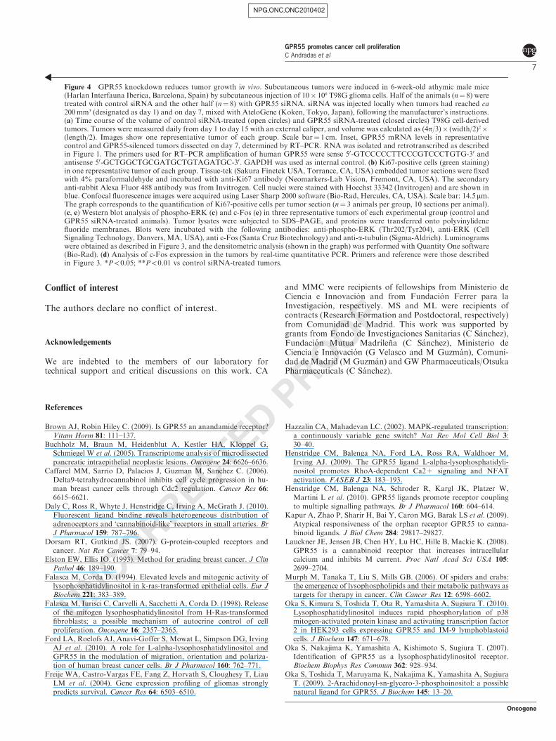
We next analyzed the molecular bases underlying thelink between GPR55 expression and cell proliferation.Stable overexpression of this receptor in HEK293 cells,which was associated with higher proliferative rates(Figures 2a–c), led to enhanced levels of phosphorylated(that is, activated) extracellular signal-related kinase(Q1 ERK) (Figure 3a, left panel). Well-established activa-tors of ERK (the phorbol ester 12-O-tetradecanoyl-phorbol-13-acetate), epidermal growth factor and fetalbovine serum were used as positive controls (Figure 3a,left panel). In contrast, no changes were observed inbasal phospho-Akt levels (Figure 3a, right panel).Similar observations were made when modulating theexpression of endogenous GPR55 in breast cancer cells.Thus, whereas GPR55 silencing decreased ERK activa-tion, ectopic receptor overexpression had the oppositeeffect (Figure 3b, left panel). Again, these effects werenot breast cancer-specific as they were reproduced inglioma cells (Figure 3b, right panel). No significantchanges were observed in phospho-Akt levels whenGPR55 expression was modulated in breast cancer orglioma cells (data not shown). The involvement of theERK cascade in GPR55-mediated enhancement of cellproliferation was further tested by two differentapproaches. First, we observed that GPR55 expressionin HEK293 cells increased the mRNA and protein levels
of c-Fos (Figure 3c), a downstream target of ERKdirectly related to cell proliferation (Hazzalin andMahadevan, 2002). Second, we incubated HEK293 cellswith two different MEK inhibitors (PD98059 andU0126) and found that GPR55-expressing cells de-creased their proliferative potential down to basal levels,whereas that of wild-type HEK293 cells was virtuallyunaffected (Figure 3d). Taken together, these resultssupport that GPR55 signals cell proliferation throughERK.
We next examined the proliferative role of GPR55in vivo. Subcutaneous tumors were generated in nudemice with T98G glioma cells and GPR55 expression wasknocked down by injection of a selective siRNA. Asshown in Figure 4a, the growth of those tumors wassignificantly slower than that of the correspondingcontrol siRNA-treated tumors. Of interest, tumors withlower GPR55 levels showed less Ki67-positive (that is,proliferative) cells (Figure 4b), decreased phospho-ERKlevels (Figure 4c) and reduced c-Fos expression (Figures4d and e). Given that GPR55 is expressed in endothelialcells (Waldeck-Weiermair et al., 2008), we analyzedwhether this antitumoral effect had an antiangiogeniccomponent. Thus, we analyzed the expression of thevascular endothelial marker CD31 in GPR55-silencedtumors and their corresponding controls by immuno-fluorescence, and observed no differences (Supplemen-tary Figure S2).
Our results show that GPR55 is expressed by humantumors from different origin in an aggressive manner Q2. Ithas been previously described that several GPCRs areoverexpressed in human cancers, and that this aberrantexpression participates in cancer progression (Dorsamand Gutkind, 2007). For instance, receptors forchemokines or lysophospholipids such as lysophospha-tidic acid or sphingosine-1-phosphate contribute touncontrolled cell proliferation in many types of cancers(Dorsam and Gutkind, 2007). Although aberrant tumorcell proliferation is sometimes associated with theactivated mutations in GPCRs, there is evidenceshowing that wild-type GPCRs can become tumorigenicwhen exposed to an excess of locally produced agonists(Dorsam and Gutkind, 2007). In this sense, it isinteresting to point out that a possible endogenousligand for GPR55, LPI, is increased in plasma andascites from ovarian cancer patients compared withsamples from healthy controls or patients with non-malignant diseases (Xu et al., 2001). This observation,together with the overexpression of GPR55 in malignanttumors reported herein, is in line with the general ideathat lysophospholipid signaling is altered in cancer.Thus, elevated levels of lysophosphatidic acid andsphingosine-1-phosphate have been found in bodyfluids from cancer patients, together with increasedamounts of their corresponding target GPCRs,enhanced activity of their biosynthetic enzymes anddownregulation of phosphatases that degrade them(Murph et al., 2006). As a further support forthe potential parallelism between lysophosphatidicacid, sphingosine-1-phosphate and LPI, it is worthpointing out that GPR55, as other members of the
NPG_ONC_ONC2010402
GPR55 promotes cancer cell proliferationC Andradas et al
3
Oncogene

UNCORRECTED PROOF
NPG_ONC_ONC2010402
HEK
HEK-GPR55
*
180
220
60
100
140
Cel
l via
bilit
y (%
vs
cont
rol)
Cel
l via
bilit
y (%
vs
cont
rol)
Cel
l via
bilit
y (%
vs
cont
rol)
Cel
l via
bilit
y (%
vs
cont
rol)
Cel
l via
bilit
y (%
vs
cont
rol)
**
**
Num
ber
of c
ells
(%
)
HEK
HEK-GPR55
**
**
*T98G
*
EVSA-T
20
40
60
80
0HEK
Cou
nts
per
min
ute
(x10
-3)
450
400
350
300
250
200
150
100
50
0
100
60
70
80
90
*
140
60
80
100
120
100
60
70
80
90*
150
50
75
100
125
*
**
**
*
1
2
3
4
0
**
GP
R55
exp
ress
ion
(a.u
.)
GP
R55
exp
ress
ion
(a.u
.)
GP
R55
exp
ress
ion
(a.u
.)
GP
R55
exp
ress
ion
(a.u
.)1
0.8
0.6
0.4
0.2
0
1
0.8
0.6
0.4
0.2
0
**
10
7.5
5
2.5
0
0 h 48 h24 h
0 h 48 h24 h HEK-GPR55
a
b
d e
c
Figure 2 GPR55 expression promotes cell proliferation. (a) HEK293 cells stably expressing 3xHA-GPR55 were previously generated(Henstridge et al., 2009). Cell viability was determined in serum-deprived cells by the 3-4,5-dimethylthiazol-2,5-diphenyltetrazoliumbromide thiazol blue (MTT) test (Sigma-Aldrich, St Louis, MO, USA) according to the manufacturer’s instructions. (b) Cells weretrypsinized and counted with trypan blue at the indicated times. (c) Serum-free cells were incubated with 1mCi [3H]-thymidine for 4 h.After this period, precipitation was achieved by 15min incubation with ice-cold 5% trichloroacetic acid. The non-incorporatedthymidine was rinsed twice with chilled 100% ethanol and cells were solubilized and collected in 0.1 N NaOH containing 0.1% sodiumdodecyl sulfate. Radioactivity was measured in a scintillation counter. (d, e) GPR55 expression was modulated in cancer cells (EVSA-Tin d, T98G in e) and cell viability was determined by the MTT test 24 h after 12 h of serum starvation (upper panels). Knockdown(left panels) was achieved by siRNA. The double-stranded RNA duplexes for human GPR55 (ON-TARGETplus SMART pools) werefrom Dharmacon-Thermo Scientific (Lafayette, CO, USA). Sequences were 50-GAAUUCCGCAUGAACAUCAUU-30, 50-GAGAAACAGCUUUAUCGUAUU-30, 50-AAGAACAGGUGGCCCGAUUUU-30 and 50-GCUACUACUUUGUCAUCAAUU-30. The non-targeted control siRNA was from Applied Biosystems-Ambion (Austin, TX, USA) and the sequence was 50-UUCUCCGAACGUGUCACGUtt-30. DharmaFECT 3 (Dharmacon-Thermo Scientific) was used as transfection reagent. For overexpression experiments (rightpanels), cells were transfected with a 3xHA-human recombinant GPR55 plasmid (Henstridge et al., 2009) or the corresponding emptyvector (pcDNA3). Lipofectamine 2000 (Invitrogen) was used as transfection reagent. For both silencing and overexpression approaches,24 h after transfection cells were trypsinized and seeded for MTT experiments. Lower panels represent the relative GPR55 mRNA levels,as determined by real-time quantitative PCR, after GPR55 silencing or overexpression. Results are expressed as mean±s.e.m. *Po0.05;**Po0.01 vs HEK293 cells.
GPR55 promotes cancer cell proliferationC Andradas et al
4
Oncogene

UNCORRECTED PROOF
lysophospholipid GPCR families, exerts its actions, atleast in part, through G12/13 and Gq proteins (Ryberget al., 2007; Lauckner et al., 2008; Henstridge et al.,2009), which signal oncogenic effects (Dorsam andGutkind, 2007; Worzfeld et al., 2008). It is thereforetempting to speculate that LPI, through GPR55, wouldbehave similar to their close relatives lysophosphatidic
acid and sphingosine-1-phosphate in terms of involve-ment in cancer biology. A very recent work proposedthat LPI induces MCF-7 cell migration in a GPR55-mediated manner (Ford et al., 2010). It has also beendescribed that cytosolic phospholipase A2 generatesmitogenic LPI in Ras-transformed epithelial cells (Falascaand Corda, 1994) and Ras-transformed fibroblasts
NPG_ONC_ONC2010402
p-ERK
ERK
Tub Tub
AKT
p-AKT
HEK-GPR55HEK
EVSA-T
p-ERK
ERK
Tub
p-ERK
ERK
Tub
T98G
HEK
HEK-GPR55*
# #10
15
20
0
5
c-F
os m
RN
A e
xpre
ssio
n (a
.u.)
100
110
120
130
80
90
Veh PD98059 U0126
#
Cel
l via
bilit
y (%
vs
cont
rol)
p-ERK
ERK
c-Fos
Tub
Tub
1 23427567
EGF VehVeh FBSTPA
Figure 3 GPR55 signals cell proliferation via ERK. (a) Western blot analysis of phospho-ERK and phospho-Akt in HEK293 cells.Cells were seeded at a density of 10� 103/cm2 and were serum-starved for 12 h before challenge for 10min with vehicle (DMSO) ordifferent control stimuli known to activate ERK (that is, 100 ng/ml epidermal growth factor (EGF), 10 nM TPA or 10% fetal bovineserum (FBS)). Cell lysates were subjected to sodium dodecyl sulfate–polyacrylamide gel electrophoresis, and proteins were transferredonto polyvinylidene fluoride membranes. Blots were incubated with anti-phospho-ERK (Thr202/Tyr204), anti-ERK, anti-phospho-Akt(Ser473), anti-Akt (Cell Signaling Technology, Danvers, MA, USA) and anti-a-tubulin (Tub) (used as loading control, Sigma-Aldrich).Luminograms were obtained with the Amersham Enhanced Chemiluminescence Detection Kit (GE Healthcare, Uppsala, Sweden).Numbers correspond to the densitometric analysis of pERK levels. Data are expressed as relative optical density values. (b) Westernblot analysis of phospho-ERK in cancer cells (EVSA-T, left panel; T98G, right panel) expressing decreased (left histograms) orincreased (right histograms) levels of GPR55. Modulation of GPR55 expression was achieved as described in Figure 2. (c) Analysis ofc-Fos expression by real-time quantitative PCR (graph) and western blot (luminograms). Primers (from Universal Probe Library,Roche) were sense 50-ACTACCACTCACCCGCAGAC-30and antisense 50-CCAGGTCCGTGCAGAAGT-30, and 18S RNA was usedas reference. For protein level analysis, blots were incubated with anti-c-Fos (Santa Cruz Biotechnology, Santa Cruz, CA, USA)and anti-a-tubulin (used as loading control) antibodies. Representative experiments (n¼ 3) are shown. (d) Relative cell viability,as determined by the MTT test, and western blot analysis of phospho-ERK in HEK293 cells incubated with the indicated MEKinhibitors (25 mM PD98059, 5 mM U0126) for 24 h. Results are expressed as mean±s.e.m. *Po0.05 vs HEK293 cells; #Po0.05 vs vehicle(DMSO)-treated cells.
GPR55 promotes cancer cell proliferationC Andradas et al
5
Oncogene

UNCORRECTED PROOF
(Falasca et al., 1998). To determine whether LPI isgenerated in HEK293 cells through this pathway, weinhibited cytosolic phospholipase A2 activity with pyrro-phenone (Seno et al., 2001). This compound blocked theincrease in cell proliferation induced by GPR55 over-expression (Supplementary Figure 3), suggesting thatLPI and/or other potential GPR55 endogenous ligandsare produced by HEK-GPR55 cells.
In summary, our data shed light on two as yetunclear aspects of the GPR55 receptor, that is, itsphysio-pathological role and the signaling pathwayscoupled to it. More importantly, the findings presentedherein reveal not only the possible relevance ofGPR55 in human cancer, but also its potential appli-cation as a new biomarker and therapeutic targetin oncology.
NPG_ONC_ONC2010402
GPR55
Tum
or v
olum
e (m
m3 )
*
**** ** **
***** ** *
**
GAPDH
900
1000
200
300
400
500
600
700
800
100
Time (days)
15
20
25
30
C s
iRN
A
Ki67
0
5
10
Ki6
7-po
sitiv
e ce
lls (
%)
**
Opt
ical
den
sity
(a.
u.)
1.0
1.5
0.5
0
*p-ERK
ERK
Tub
C siRNA GPR55 siRNA
c-Fos
C siRNA GPR55 siRNA*
1.5
1.0
0.5
0c-F
os m
RN
A e
xpre
ssio
n (a
.u.)
**
Tub Opt
ical
den
sity
(a.
u.)
1.2
1.0
0.8
0.6
0.2
0
0.4
1 15141312111098765432
GPR55 promotes cancer cell proliferationC Andradas et al
6
Oncogene
UNCORRECTED PROOF
Conflict of interest
The authors declare no conflict of interest.
Acknowledgements
We are indebted to the members of our laboratory fortechnical support and critical discussions on this work. CA
and MMC were recipients of fellowships from Ministerio deCiencia e Innovacion and from Fundacion Ferrer para laInvestigacion, respectively. MS and ML were recipients ofcontracts (Research Formation and Postdoctoral, respectively)from Comunidad de Madrid. This work was supported bygrants from Fondo de Investigaciones Sanitarias (C Sanchez),Fundacion Mutua Madrilena (C Sanchez), Ministerio deCiencia e Innovacion (G Velasco and M Guzman), Comuni-dad de Madrid (M Guzman) and GW Pharmaceuticals/OtsukaPharmaceuticals (C Sanchez).
References
Brown AJ, Robin Hiley C. (2009). Is GPR55 an anandamide receptor?Vitam Horm 81: 111–137.
Buchholz M, Braun M, Heidenblut A, Kestler HA, Kloppel G,Schmiegel W et al. (2005). Transcriptome analysis of microdissectedpancreatic intraepithelial neoplastic lesions. Oncogene 24: 6626–6636.
Caffarel MM, Sarrio D, Palacios J, Guzman M, Sanchez C. (2006).Delta9-tetrahydrocannabinol inhibits cell cycle progression in hu-man breast cancer cells through Cdc2 regulation. Cancer Res 66:6615–6621.
Daly C, Ross R, Whyte J, Henstridge C, Irving A, McGrath J. (2010).Fluorescent ligand binding reveals heterogeneous distribution ofadrenoceptors and ‘cannabinoid-like’ receptors in small arteries. Br
J Pharmacol 159: 787–796.Dorsam RT, Gutkind JS. (2007). G-protein-coupled receptors and
cancer. Nat Rev Cancer 7: 79–94.Elston EW, Ellis IO. (1993). Method for grading breast cancer. J Clin
Pathol 46: 189–190.Falasca M, Corda D. (1994). Elevated levels and mitogenic activity of
lysophosphatidylinositol in k-ras-transformed epithelial cells. Eur J
Biochem 221: 383–389.Falasca M, Iurisci C, Carvelli A, Sacchetti A, Corda D. (1998). Release
of the mitogen lysophosphatidylinositol from H-Ras-transformedfibroblasts; a possible mechanism of autocrine control of cellproliferation. Oncogene 16: 2357–2365.
Ford LA, Roelofs AJ, Anavi-Goffer S, Mowat L, Simpson DG, IrvingAJ et al. (2010). A role for L-alpha-lysophosphatidylinositol andGPR55 in the modulation of migration, orientation and polariza-tion of human breast cancer cells. Br J Pharmacol 160: 762–771.
Freije WA, Castro-Vargas FE, Fang Z, Horvath S, Cloughesy T, LiauLM et al. (2004). Gene expression profiling of gliomas stronglypredicts survival. Cancer Res 64: 6503–6510.
Hazzalin CA, Mahadevan LC. (2002). MAPK-regulated transcription:a continuously variable gene switch? Nat Rev Mol Cell Biol 3:30–40.
Henstridge CM, Balenga NA, Ford LA, Ross RA, Waldhoer M,Irving AJ. (2009). The GPR55 ligand L-alpha-lysophosphatidyli-nositol promotes RhoA-dependent Ca2+ signaling and NFATactivation. FASEB J 23: 183–193.
Henstridge CM, Balenga NA, Schroder R, Kargl JK, Platzer W,Martini L et al. (2010). GPR55 ligands promote receptor couplingto multiple signalling pathways. Br J Pharmacol 160: 604–614.
Kapur A, Zhao P, Sharir H, Bai Y, Caron MG, Barak LS et al. (2009).Atypical responsiveness of the orphan receptor GPR55 to canna-binoid ligands. J Biol Chem 284: 29817–29827.
Lauckner JE, Jensen JB, Chen HY, Lu HC, Hille B, Mackie K. (2008).GPR55 is a cannabinoid receptor that increases intracellularcalcium and inhibits M current. Proc Natl Acad Sci USA 105:2699–2704.
Murph M, Tanaka T, Liu S, Mills GB. (2006). Of spiders and crabs:the emergence of lysophospholipids and their metabolic pathways astargets for therapy in cancer. Clin Cancer Res 12: 6598–6602.
Oka S, Kimura S, Toshida T, Ota R, Yamashita A, Sugiura T. (2010).Lysophosphatidylinositol induces rapid phosphorylation of p38mitogen-activated protein kinase and activating transcription factor2 in HEK293 cells expressing GPR55 and IM-9 lymphoblastoidcells. J Biochem 147: 671–678.
Oka S, Nakajima K, Yamashita A, Kishimoto S, Sugiura T. (2007).Identification of GPR55 as a lysophosphatidylinositol receptor.Biochem Biophys Res Commun 362: 928–934.
Oka S, Toshida T, Maruyama K, Nakajima K, Yamashita A, SugiuraT. (2009). 2-Arachidonoyl-sn-glycero-3-phosphoinositol: a possiblenatural ligand for GPR55. J Biochem 145: 13–20.
NPG_ONC_ONC2010402
Figure 4 GPR55 knockdown reduces tumor growth in vivo. Subcutaneous tumors were induced in 6-week-old athymic male mice(Harlan Interfauna Iberica, Barcelona, Spain) by subcutaneous injection of 10� 106 T98G glioma cells. Half of the animals (n¼ 8) weretreated with control siRNA and the other half (n¼ 8) with GPR55 siRNA. siRNA was injected locally when tumors had reached ca200mm3 (designated as day 1) and on day 7, mixed with AteloGene (Koken, Tokyo, Japan), following the manufacturer’s instructions.(a) Time course of the volume of control siRNA-treated (open circles) and GPR55 siRNA-treated (closed circles) T98G cell-derivedtumors. Tumors were measured daily from day 1 to day 15 with an external caliper, and volume was calculated as (4p/3)� (width/2)2�(length/2). Images show one representative tumor of each group. Scale bar¼ 1 cm. Inset, GPR55 mRNA levels in representativecontrol and GPR55-silenced tumors dissected on day 7, determined by RT–PCR. RNA was isolated and retrotranscribed as describedin Figure 1. The primers used for RT–PCR amplification of human GPR55 were sense 50-GTCCCCCTTCCCGTCCCTGTG-30 andantisense 50-GCTGGCTGCGATGCTGTAGATGC-30. GAPDH was used as internal control. (b) Ki67-positive cells (green staining)in one representative tumor of each group. Tissue-tek (Sakura Finetek USA, Torrance, CA, USA) embedded tumor sections were fixedwith 4% paraformaldehyde and incubated with anti-Ki67 antibody (Neomarkers-Lab Vision, Fremont, CA, USA). The secondaryanti-rabbit Alexa Fluor 488 antibody was from Invitrogen. Cell nuclei were stained with Hoechst 33342 (Invitrogen) and are shown inblue. Confocal fluorescence images were acquired using Laser Sharp 2000 software (Bio-Rad, Hercules, CA, USA). Scale bar: 14.5mm.The graph corresponds to the quantification of Ki67-positive cells per tumor section (n¼ 3 animals per group, 10 sections per animal).(c, e) Western blot analysis of phospho-ERK (c) and c-Fos (e) in three representative tumors of each experimental group (control andGPR55 siRNA-treated animals). Tumor lysates were subjected to SDS–PAGE, and proteins were transferred onto polyvinylidenefluoride membranes. Blots were incubated with the following antibodies: anti-phospho-ERK (Thr202/Tyr204), anti-ERK (CellSignaling Technology, Danvers, MA, USA), anti c-Fos (Santa Cruz Biotechnology) and anti-a-tubulin (Sigma-Aldrich). Luminogramswere obtained as described in Figure 3, and the densitometric analysis (shown in the graph) was performed with Quantity One software(Bio-Rad). (d) Analysis of c-Fos expression in the tumors by real-time quantitative PCR. Primers and reference were those describedin Figure 3. *Po0.05; **Po0.01 vs control siRNA-treated tumors.
GPR55 promotes cancer cell proliferationC Andradas et al
7
Oncogene

UNCORRECTED PROOF
Pietr M, Kozela E, Levy R, Rimmerman N, Lin YH, Stella N et al.(2009). Differential changes in GPR55 during microglial cellactivation. FEBS Lett 583: 2071–2076.
Ross RA. (2009). The enigmatic pharmacology of GPR55. Trends
Pharmacol Sci 30: 156–163.Ryberg E, Larsson N, Sjogren S, Hjorth S, Hermansson NO, Leonova
J et al. (2007). The orphan receptor GPR55 is a novel cannabinoidreceptor. Br J Pharmacol 152: 1092–1101.
Sawzdargo M, Nguyen T, Lee DK, Lynch KR, Cheng R, Heng HHet al. (1999). Identification and cloning of three novel human Gprotein-coupled receptor genes GPR52, PsiGPR53 and GPR55:GPR55 is extensively expressed in human brain. Brain Res Mol
Brain Res 64: 193–198.Seno K, Okuno T, Nishi K, Murakami Y, Yamada K, Nakamoto S et al.
(2001). Pyrrolidine inhibitors of human cytosolic phospholipase A2.Part 2: synthesis of potent and crystallized 4-triphenylmethylthioderivative ‘pyrrophenone’. Bioorg Med Chem Lett 11: 587–590.
Staton PC, Hatcher JP, Walker DJ, Morrison AD, Shapland EM,Hughes JP et al. (2008). The putative cannabinoid receptor GPR55plays a role in mechanical hyperalgesia associated with inflamma-tory and neuropathic pain. Pain 139: 225–236.
Sun L, Hui AM, Su Q, Vortmeyer A, Kotliarov Y, Pastorino S et al.(2006). Neuronal and glioma-derived stem cell factor inducesangiogenesis within the brain. Cancer Cell 9: 287–300.
Waldeck-Weiermair M, Zoratti C, Osibow K, Balenga N, GoessnitzerE, Waldhoer M et al. (2008). Integrin clustering enables ananda-mide-induced Ca2+ signaling in endothelial cells via GPR55 byprotection against CB1-receptor-triggered repression. J Cell Sci 121:1704–1717.
Whyte LS, Ryberg E, Sims NA, Ridge SA, Mackie K, Greasley PJet al. (2009). The putative cannabinoid receptor GPR55 affectsosteoclast function in vitro and bone mass in vivo. Proc Natl Acad
Sci USA 106: 16511–16516.Worzfeld T, Wettschureck N, Offermanns S. (2008). G(12)/G(13)-
mediated signalling in mammalian physiology and disease. Trends
Pharmacol Sci 29: 582–589.Xu Y, Xiao YJ, Baudhuin LM, Schwartz BM. (2001). The role and
clinical applications of bioactive lysolipids in ovarian cancer. J Soc
Gynecol Investig 8: 1–13.Yin H, Chu A, Li W, Wang B, Shelton F, Otero F et al. (2009). Lipid
G protein-coupled receptor ligand identification using beta-arrestinPathHunter assay. J Biol Chem 284: 12328–12338.
Supplementary Information accompanies the paper on the Oncogene website (http://www.nature.com/onc)
NPG_ONC_ONC2010402
GPR55 promotes cancer cell proliferationC Andradas et al
8
Oncogene
Copyright © 2022 FDOKUMEN