Orphan Nuclear Receptor ERRγ Is a Novel Transcriptional ...
13
Int. J. Mol. Sci. 2020, 21, 7148; doi:10.3390/ijms21197148 www.mdpi.com/journal/ijms Article Orphan Nuclear Receptor ERRγ Is a Novel Transcriptional Regulator of IL-6 Mediated Hepatic BMP6 Gene Expression in Mice Kamalakannan Radhakrishnan 1,† , Yong-Hoon Kim 2,3,† , Yoon Seok Jung 1 , Jina Kim 4 , Don-Kyu Kim 5 , Sung Jin Cho 4,6 , In-Kyu Lee 6,7 , Steven Dooley 8 , Chul-Ho Lee 2,3, * and Hueng-Sik Choi 1, * 1 School of Biological Sciences and Technology, Chonnam National University, Gwangju 61186, Korea; [email protected] (K.R.); [email protected] (Y.S.J.) 2 Laboratory Animal Resource Center, Korea Research Institute of Bioscience and Biotechnology, Daejeon 34141, Korea; [email protected] (Y.-H.K.); [email protected] (C.-H.L.) 3 Department of Functional Genomics, KRIBB School of Bioscience, University of Science and Technology (UST), Daejeon 34141, Korea 4 New Drug Development Center, Daegu Gyeongbuk Medical Innovation Foundation, Daegu 41061, Korea; [email protected] (J.K.); [email protected] (S.J.C.) 5 Department of Integrative Food, Bioscience and Biotechnology, Chonnam National University, Gwangju 61186, Korea; [email protected] 6 Leading-edge Research Center for Drug Discovery and Development for Diabetes and Metabolic Disease, Kyungpook National University Hospital, Daegu 41404, Korea; [email protected] 7 Department of Internal Medicine, School of Medicine, Kyungpook National University, Kyungpook National University Hospital, Daegu 41944, Korea 8 Department of Medicine II, Medical Faculty Mannheim, Heidelberg University, 68167 Mannheim, Germany; [email protected] * Correspondence: [email protected] (C.-H.L.); [email protected] (H.-S.C.); Tel.: +82-42-860-4630 (C.-H.L.); +82-62-530-0506 (H.-S.C.); Fax: +82-42-860-4609 (C.-H.L.); +82-62-530-0506 (H.-S.C.) † These authors contributed equally to this work. Received: 25 August 2020; Accepted: 24 September 2020; Published: 28 September 2020 Abstract: Bone morphogenetic protein 6 (BMP6) is a multifunctional growth factor involved in organ development and homeostasis. BMP6 controls expression of the liver hormone, hepcidin, and thereby plays a crucial role in regulating iron homeostasis. BMP6 gene transcriptional regulation in liver is largely unknown, but would be of great help to externally modulate iron load in pathologic conditions. Here, we describe a detailed molecular mechanism of hepatic BMP6 gene expression by an orphan nuclear receptor, estrogen-related receptor γ (ERRγ), in response to the pro- inflammatory cytokine interleukin 6 (IL-6). Recombinant IL-6 treatment increases hepatic ERRγ and BMP6 expression. Overexpression of ERRγ is sufficient to increase BMP6 gene expression in hepatocytes, suggesting that IL-6 is upstream of ERRγ. In line, knock-down of ERRγ in cell lines or a hepatocyte specific knock-out of ERRγ in mice significantly decreases IL-6 mediated BMP6 expression. Promoter studies show that ERRγ directly binds to the ERR response element (ERRE) in the mouse BMP6 gene promoter and positively regulates BMP6 gene transcription in IL-6 treatment conditions, which is further confirmed by ERRE mutated mBMP6-luciferase reporter assays. Finally, an inverse agonist of ERRγ, GSK5182, markedly inhibits IL-6 induced hepatic BMP6 expression in vitro and in vivo. Taken together, these results reveal a novel molecular mechanism on ERRγ mediated transcriptional regulation of hepatic BMP6 gene expression in response to IL-6. Keywords: estrogen-related receptor γ (ERRγ); bone morphogenetic protein 6 (BMP6); interleukin 6 (IL-6); orphan receptors; transcription; gene expression and regulation; liver
-
Upload
khangminh22 -
Category
Documents
-
view
0 -
download
0
Transcript of Orphan Nuclear Receptor ERRγ Is a Novel Transcriptional ...

Int. J. Mol. Sci. 2020, 21, 7148; doi:10.3390/ijms21197148 www.mdpi.com/journal/ijms
Article
Orphan Nuclear Receptor ERRγ Is a Novel Transcriptional Regulator of IL-6 Mediated Hepatic BMP6 Gene Expression in Mice
Kamalakannan Radhakrishnan 1,†, Yong-Hoon Kim 2,3,†, Yoon Seok Jung 1, Jina Kim 4,
Don-Kyu Kim 5, Sung Jin Cho 4,6, In-Kyu Lee 6,7, Steven Dooley 8, Chul-Ho Lee 2,3,*
and Hueng-Sik Choi 1,*
1 School of Biological Sciences and Technology, Chonnam National University, Gwangju 61186, Korea;
[email protected] (K.R.); [email protected] (Y.S.J.) 2 Laboratory Animal Resource Center, Korea Research Institute of Bioscience and Biotechnology,
Daejeon 34141, Korea; [email protected] (Y.-H.K.); [email protected] (C.-H.L.) 3 Department of Functional Genomics, KRIBB School of Bioscience, University of Science and Technology
(UST), Daejeon 34141, Korea 4 New Drug Development Center, Daegu Gyeongbuk Medical Innovation Foundation, Daegu 41061, Korea;
[email protected] (J.K.); [email protected] (S.J.C.) 5 Department of Integrative Food, Bioscience and Biotechnology, Chonnam National University,
Gwangju 61186, Korea; [email protected] 6 Leading-edge Research Center for Drug Discovery and Development for Diabetes and Metabolic Disease,
Kyungpook National University Hospital, Daegu 41404, Korea; [email protected] 7 Department of Internal Medicine, School of Medicine, Kyungpook National University,
Kyungpook National University Hospital, Daegu 41944, Korea 8 Department of Medicine II, Medical Faculty Mannheim, Heidelberg University,
68167 Mannheim, Germany; [email protected]
* Correspondence: [email protected] (C.-H.L.); [email protected] (H.-S.C.);
Tel.: +82-42-860-4630 (C.-H.L.); +82-62-530-0506 (H.-S.C.); Fax: +82-42-860-4609 (C.-H.L.);
+82-62-530-0506 (H.-S.C.)
† These authors contributed equally to this work.
Received: 25 August 2020; Accepted: 24 September 2020; Published: 28 September 2020
Abstract: Bone morphogenetic protein 6 (BMP6) is a multifunctional growth factor involved in
organ development and homeostasis. BMP6 controls expression of the liver hormone, hepcidin, and
thereby plays a crucial role in regulating iron homeostasis. BMP6 gene transcriptional regulation in
liver is largely unknown, but would be of great help to externally modulate iron load in pathologic
conditions. Here, we describe a detailed molecular mechanism of hepatic BMP6 gene expression by
an orphan nuclear receptor, estrogen-related receptor γ (ERRγ), in response to the pro-
inflammatory cytokine interleukin 6 (IL-6). Recombinant IL-6 treatment increases hepatic ERRγ and
BMP6 expression. Overexpression of ERRγ is sufficient to increase BMP6 gene expression in
hepatocytes, suggesting that IL-6 is upstream of ERRγ. In line, knock-down of ERRγ in cell lines or
a hepatocyte specific knock-out of ERRγ in mice significantly decreases IL-6 mediated BMP6
expression. Promoter studies show that ERRγ directly binds to the ERR response element (ERRE)
in the mouse BMP6 gene promoter and positively regulates BMP6 gene transcription in IL-6
treatment conditions, which is further confirmed by ERRE mutated mBMP6-luciferase reporter
assays. Finally, an inverse agonist of ERRγ, GSK5182, markedly inhibits IL-6 induced hepatic BMP6
expression in vitro and in vivo. Taken together, these results reveal a novel molecular mechanism
on ERRγ mediated transcriptional regulation of hepatic BMP6 gene expression in response to IL-6.
Keywords: estrogen-related receptor γ (ERRγ); bone morphogenetic protein 6 (BMP6); interleukin
6 (IL-6); orphan receptors; transcription; gene expression and regulation; liver

Int. J. Mol. Sci. 2020, 21, 7148 2 of 13
1. Introduction
Bone morphogenetic proteins (BMPs) are secreted-type growth factors belonging to the
transforming growth factor (TGF)-β superfamily, which also includes TGF-βs, activins and growth
differentiation factors [1–3]. The BMP subfamily was initially identified as the key proteins involved
in cartilage induction and bone formation, later studies demonstrated the importance of BMPs in
embryonic development and postnatal tissue remodeling through regulating cell growth,
differentiation, chemotaxis and apoptosis [4–8]. Inside the cell, BMPs are synthesized as precursor
molecule with hydrophobic secretory leader along with propeptide region joined to the mature
region while proteolytic cleavage of precursor protein releases the mature proteins into circulation
[9,10]. Within the BMP family, BMP6 is grouped with BMP5, BMP7 and BMP8 based on the carboxy-
terminal amino acid (aa) sequence of the mature protein. The mature BMP6 is a disulfide-linked
homodimeric protein consists of two 132 aa subunits [11,12]. BMP6 exerts its biological action by
binding to the hetero-oligomeric complexes comprising of BMP type I and type II serine-threonine
kinase receptors on the cell membrane, which leads to phosphorylated activation of downstream
SMAD proteins [13–15]. Hepatic BMP6-SMAD signaling plays an important role in iron homeostasis
through transcriptionally regulating the liver hormone, hepcidin, which mediates the degradation of
iron transporter ferroportin in hepatocytes, macrophages, and enterocytes to control iron entry into
the systemic circulation [16–19].
The estrogen-related receptor γ (ERRγ) along with ERR and ERR constitute the NR3B
subfamily of nuclear receptors. ERRs are orphan nuclear receptors, constitutively active without
binding to endogenous ligands, which plays diverse biological roles in maintenance of homeostasis
and tissue development [20–22]. The first cloned orphan nuclear receptors were ERR and ERR
which are identified through the search of proteins related to estrogen receptor ER [23]. ERRγ was
recognized through yeast two-hybrid screening, which is the last ERR to be characterized in vivo
[24,25]. ERRγ is expressed in several embryonic and adult tissues, especially in tissues with high
energy demand such as the heart, brain, kidney, pancreas, and liver. It regulates target gene
expression by directly binding to extended half-site core sequences referred to as ERR response
element (ERRE) (5′-TCAAGGTCA-3′) as either monomers or dimmers on the regulatory region of the
target genes which are primarily involved in cellular metabolism and energy homeostasis [20]. The
intrinsic transcriptional activity of ERRγ is predominantly regulated through interaction with co-
activator or co-repressor proteins [26,27]. ERRγ plays an important role in glucose, lipids and alcohol
metabolism and its hepatic expression is induced in fasting condition [28–31]. It also involved in iron
homeostasis through regulating the expression of hepatic iron regulating hormone, hepcidin [32].
Although ERRγ is constitutively active, it exhibits limited affinity toward many synthetic ligands,
including 4-hydroxytamoxifen (4-OHT), which act as an inverse agonist by blocking the binding of
ERRγ to response elements on the regulatory regions of target genes [33–35].
How hepatocytic BMP6 gene expression is transcriptionally regulated is currently not well
understood. As we previously reported the interleukin 6 (IL-6) induced hepatic ERRγ gene up-
regulation [32], here we hypothesized that ERRγ may regulate the BMP6 gene expression in
hepatocytes. In this study, we demonstrated a novel regulatory mechanism of ERRγ mediated
hepatic BMP6 gene expression in response to the pro-inflammatory cytokine, IL-6. Importantly,
hepatocyte specific ERRγ knock-out or treatment with an ERRγ specific inverse agonist GSK5182
inhibited IL-6 induced BMP6 gene expression in hepatocytes.
2. Results
2.1. IL-6 Induces Hepatic ERRγ and BMP6 Gene Expression
In a previous study, we showed that the pro-inflammatory cytokine IL-6 induces ERRγ gene
expression in hepatocytes [32]. In the present study, we hypothesized that IL-6 induced hepatic ERRγ
may regulate BMP6 gene expression. IL-6 treatment significantly increased ERRγ and BMP6 mRNA
expression in AML12 and HepG2 cell lines in a time dependent manner (Figure 1A,B). The result was
further confirmed in isolated mouse primary hepatocytes (MPH) treated with IL-6 for 12 h (Figure

Int. J. Mol. Sci. 2020, 21, 7148 3 of 13
1C). To test the effect of IL-6 in vivo, we intraperitoneally injected IL-6 into eight-weeks old C57BL/6J
mice and found both ERRγ and BMP6 mRNA levels were significantly increased (Figure 1D).
Moreover, through immunohistochemical staining we also found that the protein levels of ERRγ and
BMP6 increased in IL-6 treated mouse liver tissues (Figure 1E). These results indicate that IL-6
induces hepatic ERRγ and BMP6 gene expression.
Figure 1. IL-6 promotes hepatic ERRγ and BMP6 gene expression. (A,B), cultured AML12 (A) and
HepG2 (B) cells were treated with IL-6 for indicated time period. The cells were then harvested for
total RNA extraction followed by reverse transcription and quantitative PCR. (C) Quantitative PCR
analysis of total RNA isolated from cultured mouse primary hepatocytes treated with IL-6 for 12 h.
(D) Eight-weeks old C57BL6/J mice were intraperitoneally injected with IL-6 (2.5 µg/kg) and sacrificed
after 1 h. Livers were harvested for total RNA extraction followed by reverse transcription and
quantitative PCR (n = 5 mice for control and n = 4 mice for IL-6). (E) Representative images of ERRγ
and BMP6 immunohistochemistry analysis in control or IL-6 treated mice livers. Data are represented
as mean + SEM. Asterisks indicate that the difference between the groups was significant, as
determined by two-tailed unpaired Student’s t-test. * p < 0.05, ** p < 0.01, *** p < 0.001.
2.2. Overexpression of ERRγ Increases BMP6 Gene Expression in Liver
Since IL-6 increased both ERRγ and BMP6 gene expression, and ERRγ being a transcription
factor, we intended to examine whether ERRγ can regulate hepatic BMP6 gene expression. To test
this hypothesis, we overexpressed ERRγ using an adenoviral ERRγ construct (Ad-ERRγ) in AML12,
HepG2 and MPH. The BMP6 mRNA level was significantly increased in Ad-ERRγ treated cells
compared to controls (Figure 2A–C). This result was verified in C57BL6/J mice by overexpressing
ERRγ in hepatocytes through tail-vein injection of Ad-ERRγ. Hepatic BMP6 mRNA level increased

Int. J. Mol. Sci. 2020, 21, 7148 4 of 13
in Ad-ERRγ treated mice compared to Ad-GFP treated control mice (Figure 2D). The protein
expression of BMP6 in Ad-ERRγ treated mice liver was investigated immunohistochemically and
found increased compared to control mice livers (Figure 2E). These results suggest that
overexpression of ERRγ is sufficient to induce hepatic BMP6 gene expression.
Figure 2. ERRγ increases BMP6 expression in vitro and in vivo. (A–C) Quantitative PCR analysis
showing mRNA levels of ERRγ and BMP6 in AML12 (A), HepG2 (B) and mouse primary hepatocytes
(C), infected with Ad-GFP or Ad-Flag-ERRγ for 48 h. D and E, Either Ad-GFP or Ad-Flag-ERRγ was
injected via tail-vein into mice, which were sacrificed at day 5 after injection (n = 5 mice per group).
Quantitative PCR analysis, showing mRNA levels of ERRγ and BMP6 in mice livers (D).
Representative image of ERRγ and BMP6 immunohistochemistry analysis in mice livers (E). Data are
represented as mean + SEM. Asterisks indicate that the difference between the groups was significant,
as determined by two-tailed unpaired Student’s t-test. * p < 0.05, ** p < 0.01, *** p < 0.001.
2.3. Liver Specific ERRγ Knock-Out Mice Fail to Increase Hepatic BMP6 Gene Expression in Response to
IL-6 Treatment
As the above results show that ERRγ overexpression can increase hepatic BMP6 gene expression
(Figure 2), we wanted to identify whether ERRγ is required for IL-6 mediated BMP6 gene up-
regulation in liver cells. To test this, we knocked-down ERRγ expression in cell lines using adenoviral
shERRγ (Ad-shERRγ). IL-6 treatment increased BMP6 mRNA level, whereas Ad-shERRγ
significantly blunted IL-6 mediated up-regulation of BMP6 mRNA levels in AML12, HepG2 and
MPH (Figure 3A–C). To emphasize the importance of ERRγ in IL-6 mediated hepatic BMP6 gene
regulation in vivo, we generated hepatocyte specific ERRγ knock-out mice (ERRγ-LKO). BMP6
mRNA levels were significantly reduced in ERRγ-LKO mice compared to Wild type (WT) mice in
response to IL-6 treatment (Figure 3D). Moreover, BMP6 protein expression was also markedly

Int. J. Mol. Sci. 2020, 21, 7148 5 of 13
reduced in IL-6 treated ERRγ-LKO mice livers compared to WT mice (Figure 3E). These results imply
that ERRγ is important for IL-6 mediated hepatic BMP6 gene up-regulation.
Figure 3. ERRγ deficiency attenuates IL-6 mediated hepatic BMP6 gene expression. (A–C),
Quantitative PCR analysis, showing mRNA levels of ERRγ and BMP6 in AML12 (A), HepG2 (B) and
mouse primary hepatocytes (C), infected with an adenovirus expressing an unspecific short hairpin
(sh) RNA (Ad-US) or Ad-shERRγ, and treated with IL-6 (20 ng/mL) for 12 h. D and E, Wild type (WT)
or hepatocyte specific ERRγ knock-out (ERRγ LKO) mice were intraperitoneally administered with
IL-6 (2.5 µg/kg) and sacrificed (WT and ERRγ-LKO n = 5 mice; WT + IL-6 and ERRγ-LKO + IL-6 n = 7
mice). Quantitative PCR analysis showing mRNA levels of ERRγ and BMP6 in mice livers (D).
Representative images of immunohistochemistry analysis of ERRγ and BMP6 in mice livers (E). Data
represented as mean + SEM. Asterisks indicate that the difference between the groups was significant,
as determined by One-Way ANOVA with Tukey’s multiple comparisons test. ** p < 0.01, *** p < 0.001.
2.4. ERRγ Activates the BMP6 Gene Promoter
We next examined the potential molecular mechanism underlying ERRγ mediated BMP6 gene
up-regulation in response to IL-6 treatment. To explore the mechanism, we first cloned the mouse
BMP6 gene promoter into a luciferase reporter construct and transiently transfected 293T and AML12
cells. Co-transfection with an ERRγ expression vector or IL-6 treatment significantly increased BMP6
promoter activity in both cell types (Figure 4A,B). A thorough investigation of the mouse BMP6
promoter revealed a putative ERRγ binding motif, ERRE, at 1045 bp upstream of the transcription
start site. To verify the relevance of this putative ERRγ binding site, we constructed an ERRE mutant

Int. J. Mol. Sci. 2020, 21, 7148 6 of 13
mouse BMP6 promoter-luciferase reporter, and found that promoter activation by ERRγ
overexpression was significantly abolished (Figure 4C). Moreover, the observed IL-6 induced 4-fold
increase of BMP6 gene promoter activity in the wild type construct was absent when using the ERRE
mutant promoter construct as readout (Figure 4D). Finally, binding of ERRγ to the endogenous
genomic BMP6 promoter was confirmed by chromatin immuno-precipitation (ChIP) assay, using
ERRγ specific antibody precipitation in AML12 cells that were treated with IL-6 (Figure 4E). To
further validate this result in vivo, we performed comparative ChIP assays in control and IL-6 treated
mouse liver tissues, which showed that ERRγ was recruited to the ERRE region of the BMP6 gene
promoter in response to IL-6 treatment (Figure 4F). In total, our results indicate that ERRγ regulated
BMP6 gene transcription occurs by direct binding and activating the BMP6 gene promoter.
Figure 4. ERRγ activates mouse BMP6 gene promoter activity. (A) 293T cells were transfected with a
mouse BMP6 promoter-luciferase reporter construct (mBMP6-luc) along with pcDNA3.1 empty
vector or pcDNA-ERRγ expression plasmid. (B) AML12 cells were transfected with mBMP6-luc and
stimulated with IL-6 (20 ng/mL) for 12 h. (C) 293T cells were transfected with wild type or ERRE
mutant mBMP6-luc along with the ERRγ expression vector. (D) AML12 cells were transfected with
wild type or ERRE mutant mBMP6-luc and stimulated with IL-6 (20 ng/mL) for 12 h. (E) ChIP assay
showing binding of ERRγ to the BMP6 gene promoter. AML12 cells were treated with IL-6 and soluble
chromatin was immuno-precipitated with an ERRγ antibody. Purified DNA samples were used for
quantitative PCR with primers corresponding to the ERRE region of the mouse BMP6 gene promoter.
(F) In vivo ChIP assay, showing binding of ERRγ to the BMP6 gene promoter in response to IL-6. We
treated C57BL6/J mice with IL-6 and immuno-precipitated soluble chromatin from mice livers with
an ERRγ antibody. Purified DNA was used for quantitative PCR analysis with primers corresponding
to the ERRE region of the mouse BMP6 gene promoter. Data are represented as mean + SEM. Asterisks
Int. J. Mol. Sci. 2020, 21, 7148 7 of 13
indicate that the difference between the groups was significant, as determined by two-tailed unpaired
Student’s t-test. * p < 0.05, ** p < 0.01, *** p < 0.001.
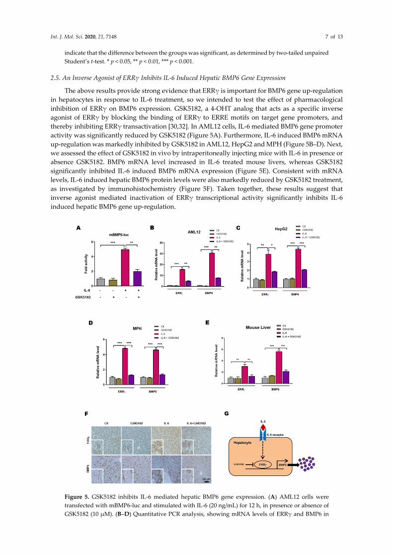
2.5. An Inverse Agonist of ERRγ Inhibits IL-6 Induced Hepatic BMP6 Gene Expression
The above results provide strong evidence that ERRγ is important for BMP6 gene up-regulation
in hepatocytes in response to IL-6 treatment, so we intended to test the effect of pharmacological
inhibition of ERRγ on BMP6 expression. GSK5182, a 4-OHT analog that acts as a specific inverse
agonist of ERRγ by blocking the binding of ERRγ to ERRE motifs on target gene promoters, and
thereby inhibiting ERRγ transactivation [30,32]. In AML12 cells, IL-6 mediated BMP6 gene promoter
activity was significantly reduced by GSK5182 (Figure 5A). Furthermore, IL-6 induced BMP6 mRNA
up-regulation was markedly inhibited by GSK5182 in AML12, HepG2 and MPH (Figure 5B–D). Next,
we assessed the effect of GSK5182 in vivo by intraperitoneally injecting mice with IL-6 in presence or
absence GSK5182. BMP6 mRNA level increased in IL-6 treated mouse livers, whereas GSK5182
significantly inhibited IL-6 induced BMP6 mRNA expression (Figure 5E). Consistent with mRNA
levels, IL-6 induced hepatic BMP6 protein levels were also markedly reduced by GSK5182 treatment,
as investigated by immunohistochemistry (Figure 5F). Taken together, these results suggest that
inverse agonist mediated inactivation of ERRγ transcriptional activity significantly inhibits IL-6
induced hepatic BMP6 gene up-regulation.
Figure 5. GSK5182 inhibits IL-6 mediated hepatic BMP6 gene expression. (A) AML12 cells were
transfected with mBMP6-luc and stimulated with IL-6 (20 ng/mL) for 12 h, in presence or absence of
GSK5182 (10 µM). (B–D) Quantitative PCR analysis, showing mRNA levels of ERRγ and BMP6 in

Int. J. Mol. Sci. 2020, 21, 7148 8 of 13
AML12 (B), HepG2 (C) and mouse primary hepatocytes (D), treated with IL-6 (20 ng/mL) for 12 h, in
presence or absence of GSK5182 (10 µM). E and F, Mice were intraperitoneally injected with IL-6 (2.5
µg/kg), with or without GSK5182 (40 mg/kg) and sacrificed (control and GSK5182 n = 4 mice; IL-6 and
GSK5182+IL-6 n = 6 mice). Quantitative PCR analysis showing mRNA levels of ERRγ and BMP6 in
mice livers (E). Representative image of ERRγ and BMP6 immunohistochemistry analysis in mice
livers (F). (G) A schematic model of IL-6 induced ERRγ mediated BMP6 production from hepatocytes.
Data are represented as mean + SEM. Asterisks indicate that the difference between the groups was
significant, as determined by One-Way ANOVA with Tukey’s multiple comparisons test. * p < 0.05,
** p < 0.01, *** p < 0.001.
3. Discussion
Our study revealed a novel transcriptional regulatory mechanism of BMP6 gene expression in
hepatocytes by an orphan nuclear receptor ERRγ in response to an inflammatory cytokine, IL-6
treatment (Figure 5G). Overexpression of ERRγ is sufficient to induce BMP6 gene expression in
hepatocytes, whereas loss of hepatic ERRγ expression significantly inhibited IL-6 mediated BMP6
up-regulation. Moreover, GSK5182, an inverse agonist of ERRγ showed effective inhibitory action
toward BMP6 gene expression in IL-6 treated mice liver.
Several reports imply the importance of BMP6 in iron homeostasis through endogenously
regulating the expression of hepcidin, a hormone mainly produced by hepatocytes that controls the
entry of iron into plasma [18,19]. However, the exact mechanism underlying the regulation of BMP6
gene expression in liver is largely unknown. In a recent study, the transcription factor nuclear factor
erythroid 2-related factor 2 (Nrf2) was reported to regulate hepatic BMP6 expression in response to
iron by directly binding the BMP6 gene promoter [36]. Here, we show that orphan nuclear receptor
ERRγ transcriptionally regulates hepatic BMP6 expression by directly binding the ERRE motif in the
BMP6 gene promoter, in response to IL-6 treatment. We found a functional ERRE motif AGGTCA in
the BMP6 gene promoter 1045 bp upstream of the transcription start site, and an ERRE mutant BMP6
promoter showed a significantly blunted response to endogenous/exogenous ERRγ mediated
promoter activation. Baseline BMP6 expression was not affected by mutating the ERRE; however,
under inducible conditions, such as in IL-6 treatment, the ERRE mutation significantly blunted BMP6
expression. Thus, ERRγ acts a novel transcriptional regulator of IL-6 mediated hepatic BMP6 gene
expression. Previous reports suggest that IL-6 and BMP signaling synergistically may regulate
hepatic hepcidin gene expression, and that inhibition of BMP signaling attenuates IL-6 induced
hepcidin expression [37]. We now describe ERRγ as mediator of the IL-6 mediated hepatic BMP6
expression, suggesting existence of an autocrine BMP6 signaling loop to up-regulate hepcidin gene
expression in hepatocytes.
The liver consists of different cell types, which are divided into parenchymal and non-
parenchymal cells (NPCs). Parenchymal cells encompass hepatocytes, which is the major cell type in
liver and to a minor extent cholangiocytes. NPCs include hepatic stellate cells, Kupffer cells and liver
sinusoidal epithelial cells. Hepatic BMP6 production was reported from NPCs in several reports
[36,38,39]; however hepatocyte derived BMP6 production was also reported [40,41]. Murine
hepatocytes were reported to produce BMP6 in an in vitro model of cellular lipid accumulation and
hepatocytes were identified as cellular source of hepatic BMP6 in livers of patients with non-alcoholic
fatty liver disease [40]. Chronic dietary iron changes are reported to modulate liver BMP6 expression
in all cell types, including hepatocytes [41]. In the present study, we have confirmed by quantitative
PCR and immunohistochemistry that hepatocytes produce BMP6 in IL-6 treated mice livers through
transcriptional activation by ERRγ. Hepatocyte specific ERRγ knock-out mice significantly inhibited
IL-6 induced hepatic BMP6 expression. Moreover, a small molecule GSK5182 mediated inhibition of
ERRγ transactivation markedly reduced hepatic BMP6 expression in IL-6 treated mice. Taken
together, in livers of IL-6 injected mice, hepatocytes are the source of BMP6, which is transcriptionally
regulated by ERRγ. The proposed IL-6-ERRγ-BMP6 pathway is probably active in inflammatory
conditions when pro-inflammatory cytokines, including IL-6, levels are increased and may up-
regulate hepcidin expression, leading to hypoferremia and anemia, such as in inflammatory anemia.
Int. J. Mol. Sci. 2020, 21, 7148 9 of 13
In conclusion, we report that ERRγ plays an important role in IL-6 mediated BMP6 gene
expression in vivo. GSK5182, an ERRγ inverse agonist significantly inhibits ERRγ-dependent BMP6
gene expression. Therefore, we suggest ERRγ as a novel transcriptional regulator of hepatic BMP6
expression.
4. Materials and Methods
4.1. Animal Experiments
Eight to twelve-week-old male C57BL/6J mice were used for all experiments. C57BL/6J wild type
(WT) mice were obtained from Korea Research Institute of Biosciences and Biotechnology (KRIBB,
Daejeon, Korea). C57BL/6J mice containing floxed ERRγ exon 2 (ERRγf/f) were obtained from
PHENOMIN-iCS, PHENOMIN, the French National Infrastructure in Biology and Health (Illkirch,
France). To produce the liver specific ERRγ knockout line (ERRγ-LKO), ERRγf/f animals were
crossbred with C57BL/6J-Alb-Cre transgenic mice, which express Cre recombinase in hepatocytes
under the control of the albumin promoter (Jackson Laboratories, Bar Harbor, ME, USA). Prior to the
experiments, mice were acclimatized to a 12-h light/dark cycle at 22 ± 2 °C for 2 weeks with unlimited
food and water in a specific pathogen-free facility. WT mice were injected with Ad-GFP or Ad-FLAG-
ERRγ via tail-vein for 5 days. WT and ERRγ-LKO mice were treated with 2.5 µg/kg of recombinant
human IL-6 via intraperitoneal (i.p) injection for indicated time period. Where indicated, GSK5182
(40 mg/kg in 30% PEG400) was administered intraperitoneally (i.p.) into mice. All animal procedures
were approved by the Chonnam National University Animal Care and Use Committee (CNU-
IACUC-YB-2017-40) and by the Institutional Animal Care and Use Committee of KRIBB (KRIBB-
AEC-19128). All animal experiments were performed in accordance with the Guide for the Care and
Use of Laboratory Animals published by the US National Institutes of Health.
4.2. Cell Culture, Transient Transfection and Luciferase Assay
AML12 (mouse immortalized hepatocytes), HepG2 (human hepatoma cells) and 293T (human
embryonic kidney cells) were obtained from the American Type Culture Collection (Manassas, VA,
USA). All cell lines were maintained as described previously [42]. Transient transfections were
performed using Lipofectamine 2000 (Invitrogen, Carlsbad, CA, USA) and FuGENE HD (Promega,
Madison, WA, USA) transfection reagents according to the manufacturer’s instructions. Luciferase
assay carried out as described previously [42]. Briefly, cells were transfected with indicated reporter
plasmids together with expression vectors encoding ERRγ or treated with IL-6. Total cDNA used for
each transfection was adjusted to 1 mg/well by adding an appropriate amount of empty vector and
pCMV–β-gal plasmid was used as an internal control. The luciferase activity was normalized to β-
galactosidase activity.
4.3. Isolation of Primary Hepatocytes
Primary hepatocytes were isolated from C57BL/6J mice by collagenase perfusion [43] and seeded
with Dulbecco’s modified Eagle’s medium (DMEM) (Gibco-BRL, Grand Island, NY, USA)
supplemented with 10% fetal bovine serum (Hyclone, Logan, UT, USA) and antibiotics in a
humidified atmosphere containing 5% CO2 at 37 °C. After attachment, cells are infected with
adenovirus (Ad-GFP, Ad-ERRγ or Ad-shERRγ) or treated with IL-6 for the indicated time period.
4.4. Chemicals and Antibodies
Recombinant human IL-6 is obtained from Prospec Protein Specialists (East Brunswick, NJ, USA,
Cat#cyt-213), and GSK5182 was synthesized as previously described [28,34,35,44]. For
immunohistochemical staining of endogenous ERRγ, rabbit anti-ERRγ serum was generated using a
peptide (404-AGQHMEDPRRAGKMLM-419) from mouse ERRγ helix 9 [45] (AbFrontier/Young in
Frontier, Seoul, Korea). Antibody was affinity purified using the same peptide and tested by Western
blotting. No cross reactivity to ERRα or ERRβ was noted [46]. Mouse monoclonal anti-BMP6 antibody
Int. J. Mol. Sci. 2020, 21, 7148 10 of 13
(Abcam, Cambridge, MA, USA, Clone#morph-6.1, Cat#ab15640) was used for immunohistochemical
staining of BMP6.
4.5. DNA Cloning
Mouse BMP6 gene promoter (−1.5 kb) was PCR-amplified from mouse genomic DNA (Promega)
using the primer: forward, 5′-TCAACGACACCACAAAGAGTTC-3′ and reverse, 5′-
TGCAAGACTTGGTAAATGCTGA-3′ and inserted into the pGL3 basic vector (Promega) using the
NheI and XhoI restriction enzyme sites. ERRE mutant constructs were generated using QuikChange
Lightning Site-Directed mutagenesis kit (Agilent Technologies, Palo Alto, CA, USA, Cat#210518). Ad-
GFP and Ad-FLAG-ERRγ were described previously [32]. The vector expressing ERRγ was described
previously [31].
4.6. Quantitative PCR
Total RNA was isolated from cell lines, mouse primary hepatocytes or mice liver tissues using
the TRIzol reagent (Invitrogen), according to the manufacturer’s instructions. cDNAs generated by
Top Script cDNA synthesis kit (Enzynomics, Daejeon, Korea) were analyzed by the Applied
Biosystems StepOnePlus real-time PCR system (Applied Biosystems) using Power SYBR Green PCR
Master Mix (Applied Biosystems, Foster City, CA, USA). All data were normalized to ribosomal
protein L32 expression. Primers used in this study: Mouse ERRγ, forward 5′-
AAGATCGACACATTGATTCCAGC-3′ and reverse, 5′-CATGGTTGAACTGTAACTCCCAC-3′;
Mouse BMP6, forward 5′-TCAACGACACCACAAAGAGTTC-3′ and reverse, 5′-
TGCAAGACTTGGTAAATGCTGA-3′; and mouse L32, forward 5′-GTGAAGCCCAAGATCGTCAA-
3′ and reverse, 5′-TGTCAATGCCTCTGGGTTTC-3′.
4.7. Chromatin Immunoprecipitation (ChIP) Assay
Formaldehyde cross-linking and chromatin immuno-precipitation (ChIP) analyses were
performed as previously described [29] using SimpleChIP Plus Enzymatic Chromatin IP kit (Cell
Signaling Technology, Danvers, MA, USA, Cat log#25268S) according to manufacturer’s protocol.
Mouse monoclonal anti-ERRγ antibody (Perseus Proteomics, Tokyo, Japan, Clone#H6812, Cat#PP-
H6812-00) was used for immunoprecipitation. The following primer was used for real-time
quantitative PCR analysis of ChIP eluted and Input DNA. Mouse BMP6 ERRE site, forward 5′-
CGGTAACGGCATGGATTAAATAG-3′ and reverse 5′-TGCCTAGCGAGCGTAGGTTTCTA-3′.
4.8. Immunohistochemistry
Liver samples were fixed in 10% neutral buffered formalin, embedded in paraffin, cut into 5 µm-
thick sections. To detect ERRγ and BMP6 protein, liver sections were stained with an anti-ERRγ and
anti-BMP6 antibody and visualized using 3,3′-diaminobenzidine (Vector Lab. Inc., Burlingame, CA,
USA). Images were captured using a light microscope (BX51, Olympus Corporation, Tokyo, Japan).
4.9. Statistical Analysis
Data were analyzed using Prism 8 (GraphPad Software, La Jolla, CA, USA). Data are expressed
as mean + SEM. The two-tailed Student’s t-test was used for statistical analysis between two groups,
whereas statistical significance between multiple treatment groups was determined by analysis of
one-way ANOVA with Tukey’s multiple comparisons test. Differences were considered statistically
significant at p < 0.05.
Author Contributions: K.R., C.-H.L. and H.-S.C. conceived and designed research; K.R. and Y.-H.K. performed
experiments. K.R., H.-S.C., Y.-H.K. and C.-H.L. analyzed the data. S.J.C., I.-K.L., J.K. and Y.S.J. provided
reagents/material/analysis tools. K.R. wrote the original draft manuscript. K.R., D.-K.K., S.D. and H.-S.C.
reviewed and edited the manuscript. All authors have read and agreed to the published version of the
manuscript.

Int. J. Mol. Sci. 2020, 21, 7148 11 of 13
Funding: This work was supported by National Creative Research Initiatives Grant (20110018305) through the
National Research Foundation of Korea (NRF) funded by the Korean government (Ministry of Science and ICT);
Future-based Technology Development Program (BIO Fields) (20100019512) and NRF-2020R1A2C3006952
through the National Research Foundation of Korea (NRF) funded by the Korean government (Ministry of
Science and ICT), and by the KRIBB Research Initiative Program of the Korea; and Korea Health Technology R
& D Project through the Korea Health Industry Development Institute (KHIDI), funded by the Ministry of Health
& Welfare, Korea (Grant Number: HI16C1501).
Acknowledgements: S.D. is supported by The Federal Ministry of Education and Research (BMBF)-Liver System
Medicine (LiSyM) Program. Grant number PTJ-031L0043.
Conflicts of Interest: The authors declare no conflict of interest in regard to this manuscript.
Abbreviations
BMP6 Bone morphogenetic protein 6
ChIP Chromatin immunoprecipitation
ERRγ Estrogen-related receptor γ
ERRE ERR responsive element
IL-6 Interleukin 6
NPCs Non-parenchymal cells
4-OHT 4-hydroxytamoxifen
References
1. Bragdon, B.; Moseychuk, O.; Saldanha, S.; King, D.; Julian, J.; Nohe, A. Bone morphogenetic proteins: A
critical review. Cell. Signal. 2011, 23, 609–620, doi:10.1016/j.cellsig.2010.10.003.
2. Weiss, A.; Attisano, L. The TGFbeta superfamily signaling pathway. Wiley Interdiscip. Rev. Dev. Biol. 2013,
2, 47–63, doi:10.1002/wdev.86.
3. Katagiri, T.; Watabe, T. Bone morphogenetic proteins. Cold Spring Harb. Perspect. Biol. 2016, 8,
doi:10.1101/cshperspect.a021899.
4. Zhao, G.Q. Consequences of knocking out BMP signaling in the mouse. Genesis 2003, 35, 43–56,
doi:10.1002/gene.10167.
5. Sykaras, N.; Opperman, L.A. Bone morphogenetic proteins (BMPs): How do they function and what can
they offer the clinician? J. Oral Sci. 2003, 45, 57–73, doi:10.2334/josnusd.45.57.
6. Simic, P.; Vukicevic, S. Bone morphogenetic proteins in development and homeostasis of kidney. Cytokine
& Growth Factor Rev. 2005, 16, 299–308, doi:10.1016/j.cytogfr.2005.02.010.
7. Oumi, N.; Taniguchi, K.A.; Kanai, A.M.; Yasunaga, M.; Nakanishi, T.; Sato, K. A crucial role of bone
morphogenetic protein signaling in the wound healing response in acute liver injury induced by carbon
tetrachloride. Int. J. Hepatol. 2012, 2012, 476820, doi:10.1155/2012/476820.
8. Wu, M.; Chen, G.; Li, Y.P. TGF-β and BMP signaling in osteoblast, skeletal development, and bone
formation, homeostasis and disease. Bone Res. 2016, 4, 16009, doi:10.1038/boneres.2016.9.
9. Umulis, D.; O’Connor, M.B.; Blair, S.S. The extracellular regulation of bone morphogenetic protein
signaling. Development 2009, 136, 3715–3728, doi:10.1242/dev.031534.
10. Rahman, M.S.; Akhtar, N.; Jamil, H.M.; Banik, R.S.; Asaduzzaman, S.M. TGF-β/BMP signaling and other
molecular events: Regulation of osteoblastogenesis and bone formation. Bone Res. 2015, 3, 15005,
doi:10.1038/boneres.2015.5.
11. Celeste, A.J.; Iannazzi, J.A.; Taylor, R.C.; Hewick, R.M.; Rosen, V.; Wang, E.A.; Wozney, J.M. Identification
of transforming growth factor beta family members present in bone-inductive protein purified from bovine
bone. Proc. Natl. Acad. Sci. USA 1990, 87, 9843–9847, doi:10.1073/pnas.87.24.9843.
12. Gitelman, S.E.; Kobrin, M.S.; Ye, J.Q.; Lopez, A.R.; Lee, A.; Derynck, R. Recombinant Vgr-1/BMP-6-
expressing tumors induce fibrosis and endochondral bone formation in vivo. J. Cell Biol. 1994, 126, 1595–
1609, doi:10.1083/jcb.126.6.1595.
13. Luo, X.; Luo, Z.; Zhang, Z.; Yang, H.; Lai, B.; Yao, Q.; Xiao, L.; Wang, N. Homocysteine upregulates
hepcidin expression through BMP6/SMAD signaling pathway in hepatocytes. Biochem. Biophys. Res.
Commun. 2016, 471, 303–308, doi:10.1016/j.bbrc.2016.02.001.
Int. J. Mol. Sci. 2020, 21, 7148 12 of 13
14. Benn, A.; Hiepen, C.; Osterland, M.; Schutte, C.; Zwijsen, A.; Knaus, P. Role of bone morphogenetic proteins
in sprouting angiogenesis: Differential BMP receptor-dependent signaling pathways balance stalk vs. tip
cell competence. FASEB J. Off. Publ. Fed. Am. Soc. Exp. Biol. 2017, 31, 4720–4733, doi:10.1096/fj.201700193RR.
15. Wang, R.N.; Green, J.; Wang, Z.; Deng, Y.; Qiao, M.; Peabody, M.; Zhang, Q.; Ye, J.; Yan, Z.; Denduluri, S.;
et al. Bone Morphogenetic Protein (BMP) signaling in development and human diseases. Genes Dis. 2014,
1, 87–105, doi:10.1016/j.gendis.2014.07.005.
16. Nemeth, E.; Tuttle, M.S.; Powelson, J.; Vaughn, M.B.; Donovan, A.; Ward, D.M.; Ganz, T.; Kaplan, J.
Hepcidin regulates cellular iron efflux by binding to ferroportin and inducing its internalization. Science
2004, 306, 2090–2093, doi:10.1126/science.1104742.
17. Wang, R.H.; Li, C.; Xu, X.; Zheng, Y.; Xiao, C.; Zerfas, P.; Cooperman, S.; Eckhaus, M.; Rouault, T.; Mishra,
L.; et al. A role of SMAD4 in iron metabolism through the positive regulation of hepcidin expression. Cell
Metab. 2005, 2, 399–409, doi:10.1016/j.cmet.2005.10.010.
18. Babitt, J.L.; Huang, F.W.; Wrighting, D.M.; Xia, Y.; Sidis, Y.; Samad, T.A.; Campagna, J.A.; Chung, R.T.;
Schneyer, A.L.; Woolf, C.J.; et al. Bone morphogenetic protein signaling by hemojuvelin regulates hepcidin
expression. Nat. Genet. 2006, 38, 531–539, doi:10.1038/ng1777.
19. Andriopoulos, B., Jr.; Corradini, E.; Xia, Y.; Faasse, S.A.; Chen, S.; Grgurevic, L.; Knutson, M.D.; Pietrangelo,
A.; Vukicevic, S.; Lin, H.Y.; et al. BMP6 is a key endogenous regulator of hepcidin expression and iron
metabolism. Nat. Genet. 2009, 41, 482–487, doi:10.1038/ng.335.
20. Tremblay, A.M.; Giguere, V. The NR3B subgroup: An ovERRview. Nucl. Recept. Signal. 2007, 5, e009,
doi:10.1621/nrs.05009.
21. Deblois, G.; Giguere, V. Nuclear receptor location analyses in mammalian genomes: From gene regulation
to regulatory networks. Mol. Endocrinol. 2008, 22, 1999–2011, doi:10.1210/me.2007-0546.
22. Eichner, L.J.; Giguere, V. Estrogen related receptors (ERRs): A new dawn in transcriptional control of
mitochondrial gene networks. Mitochondrion 2011, 11, 544–552, doi:10.1016/j.mito.2011.03.121.
23. Giguere, V.; Yang, N.; Segui, P.; Evans, R.M. Identification of a new class of steroid hormone receptors.
Nature 1988, 331, 91–94, doi:10.1038/331091a0.
24. Eudy, J.D.; Yao, S.; Weston, M.D.; Ma-Edmonds, M.; Talmadge, C.B.; Cheng, J.J.; Kimberling, W.J.; Sumegi,
J. Isolation of a gene encoding a novel member of the nuclear receptor superfamily from the critical region
of Usher syndrome type IIa at 1q41. Genomics 1998, 50, 382–384, doi:10.1006/geno.1998.5345.
25. Lui, K.; Huang, Y.; Choi, H.L.; Yu, S.; Wong, K.B.; Chen, S.; Chan, F.L. Molecular cloning and functional
study of rat estrogen receptor-related receptor γ in rat prostatic cells. Prostate 2006, 66, 1600–1619,
doi:10.1002/pros.20429.
26. Xie, Y.B.; Park, J.H.; Kim, D.K.; Hwang, J.H.; Oh, S.; Park, S.B.; Shong, M.; Lee, I.K.; Choi, H.S.
Transcriptional corepressor SMILE recruits SIRT1 to inhibit nuclear receptor estrogen receptor-related
receptor γ transactivation. J. Biol. Chem. 2009, 284, 28762–28774, doi:10.1074/jbc.M109.034165.
27. Misra, J.; Kim, D.K.; Choi, H.S. ERRγ: A junior orphan with a senior role in metabolism. Trends Endocrinol.
Metab. TEM 2017, 28, 261–272, doi:10.1016/j.tem.2016.12.005.
28. Kim, D.K.; Kim, J.R.; Koh, M.; Kim, Y.D.; Lee, J.M.; Chanda, D.; Park, S.B.; Min, J.J.; Lee, C.H.; Park, T.S.; et
al. Estrogen-related receptor γ (ERRγ) is a novel transcriptional regulator of phosphatidic acid
phosphatase, LIPIN1, and inhibits hepatic insulin signaling. J. Biol. Chem. 2011, 286, 38035–38042,
doi:10.1074/jbc.M111.250613.
29. Kim, D.K.; Ryu, D.; Koh, M.; Lee, M.W.; Lim, D.; Kim, M.J.; Kim, Y.H.; Cho, W.J.; Lee, C.H.; Park, S.B.; et
al. Orphan nuclear receptor estrogen-related receptor γ (ERRγ) is key regulator of hepatic gluconeogenesis.
J. Biol. Chem. 2012, 287, 21628–21639, doi:10.1074/jbc.M111.315168.
30. Kim, D.K.; Gang, G.T.; Ryu, D.; Koh, M.; Kim, Y.N.; Kim, S.S.; Park, J.; Kim, Y.H.; Sim, T.; Lee, I.K.; et al.
Inverse agonist of nuclear receptor ERRγ mediates antidiabetic effect through inhibition of hepatic
gluconeogenesis. Diabetes 2013, 62, 3093–3102, doi:10.2337/db12-0946.
31. Kim, D.K.; Kim, Y.H.; Jang, H.H.; Park, J.; Kim, J.R.; Koh, M.; Jeong, W.I.; Koo, S.H.; Park, T.S.; Yun, C.H.;
et al. Estrogen-related receptor γ controls hepatic CB1 receptor-mediated CYP2E1 expression and oxidative
liver injury by alcohol. Gut 2013, 62, 1044–1054, doi:10.1136/gutjnl-2012-303347.
32. Kim, D.K.; Jeong, J.H.; Lee, J.M.; Kim, K.S.; Park, S.H.; Kim, Y.D.; Koh, M.; Shin, M.; Jung, Y.S.; Kim, H.S.;
et al. Inverse agonist of estrogen-related receptor γ controls Salmonella typhimurium infection by
modulating host iron homeostasis. Nat. Med. 2014, 20, 419–424, doi:10.1038/nm.3483.

Int. J. Mol. Sci. 2020, 21, 7148 13 of 13
33. Chao, E.Y.; Collins, J.L.; Gaillard, S.; Miller, A.B.; Wang, L.; Orband-Miller, L.A.; Nolte, R.T.; McDonnell,
D.P.; Willson, T.M.; Zuercher, W.J. Structure-guided synthesis of tamoxifen analogs with improved
selectivity for the orphan ERRγ. Bioorganic Med. Chem. Lett. 2006, 16, 821–824,
doi:10.1016/j.bmcl.2005.11.030.
34. Kim, J.; Woo, S.Y.; Im, C.Y.; Yoo, E.K.; Lee, S.; Kim, H.J.; Hwang, H.J.; Cho, J.H.; Lee, W.S.; Yoon, H.; et al.
Insights of a lead optimization study and biological evaluation of novel 4-hydroxytamoxifen analogs as
estrogen-related receptor γ (ERRγ) inverse agonists. J. Med. Chem. 2016, 59, 10209–10227,
doi:10.1021/acs.jmedchem.6b01204.
35. Kim, J.; Song, J.; Ji, H.D.; Yoo, E.K.; Lee, J.E.; Lee, S.B.; Oh, J.M.; Lee, S.; Hwang, J.S.; Yoon, H.; et al.
Discovery of potent, selective, and orally bioavailable estrogen-related receptor-γ inverse agonists to
restore the sodium iodide symporter function in anaplastic thyroid cancer. J. Med. Chem. 2019, 62, 1837–
1858, doi:10.1021/acs.jmedchem.8b01296.
36. Lim, P.J.; Duarte, T.L.; Arezes, J.; Garcia-Santos, D.; Hamdi, A.; Pasricha, S.R.; Armitage, A.E.; Mehta, H.;
Wideman, S.; Santos, A.G.; et al. Nrf2 controls iron homeostasis in haemochromatosis and thalassaemia via
Bmp6 and hepcidin. Nat. Metab. 2019, 1, 519–531, doi:10.1038/s42255-019-0063-6.
37. Steinbicker, A.U.; Sachidanandan, C.; Vonner, A.J.; Yusuf, R.Z.; Deng, D.Y.; Lai, C.S.; Rauwerdink, K.M.;
Winn, J.C.; Saez, B.; Cook, C.M.; et al. Inhibition of bone morphogenetic protein signaling attenuates anemia
associated with inflammation. Blood 2011, 117, 4915–4923, doi:10.1182/blood-2010-10-313064.
38. Kautz, L.; Besson-Fournier, C.; Meynard, D.; Latour, C.; Roth, M.P.; Coppin, H. Iron overload induces
BMP6 expression in the liver but not in the duodenum. Haematologica 2011, 96, 199–203,
doi:10.3324/haematol.2010.031963.
39. Enns, C.A.; Ahmed, R.; Wang, J.; Ueno, A.; Worthen, C.; Tsukamoto, H.; Zhang, A.S. Increased iron loading
induces Bmp6 expression in the non-parenchymal cells of the liver independent of the BMP-signaling
pathway. PLoS ONE 2013, 8, e60534, doi:10.1371/journal.pone.0060534.
40. Arndt, S.; Wacker, E.; Dorn, C.; Koch, A.; Saugspier, M.; Thasler, W.E.; Hartmann, A.; Bosserhoff, A.K.;
Hellerbrand, C. Enhanced expression of BMP6 inhibits hepatic fibrosis in non-alcoholic fatty liver disease.
Gut 2015, 64, 973–981, doi:10.1136/gutjnl-2014-306968.
41. Rausa, M.; Pagani, A.; Nai, A.; Campanella, A.; Gilberti, M.E.; Apostoli, P.; Camaschella, C.; Silvestri, L.
Bmp6 expression in murine liver non parenchymal cells: A mechanism to control their high iron exporter
activity and protect hepatocytes from iron overload? PLoS ONE 2015, 10, e0122696,
doi:10.1371/journal.pone.0122696.
42. Kim, Y.D.; Park, K.G.; Lee, Y.S.; Park, Y.Y.; Kim, D.K.; Nedumaran, B.; Jang, W.G.; Cho, W.J.; Ha, J.; Lee,
I.K.; et al. Metformin inhibits hepatic gluconeogenesis through AMP-activated protein kinase-dependent
regulation of the orphan nuclear receptor SHP. Diabetes 2008, 57, 306–314, doi:10.2337/db07-0381.
43. Koo, S.H.; Satoh, H.; Herzig, S.; Lee, C.H.; Hedrick, S.; Kulkarni, R.; Evans, R.M.; Olefsky, J.; Montminy, M.
PGC-1 promotes insulin resistance in liver through PPAR-alpha-dependent induction of TRB-3. Nat. Med.
2004, 10, 530–534, doi:10.1038/nm1044.
44. Singh, T.D.; Song, J.; Kim, J.; Chin, J.; Ji, H.D.; Lee, J.-E.; Lee, S.B.; Yoon, H.; Yu, J.H.; Kim, S.K.; et al. A novel
orally active inverse agonist of estrogen-related receptor γ (ERRγ), DN200434, A booster of NIS in
anaplastic thyroid cancer. Clin. Cancer Res. 2019, 25, 5069–5081, doi:10.1158/1078-0432.ccr-18-3007.
45. Alaynick, W.A.; Way, J.M.; Wilson, S.A.; Benson, W.G.; Pei, L.; Downes, M.; Yu, R.; Jonker, J.W.; Holt, J.A.;
Rajpal, D.K.; et al. ERRγ regulates cardiac, gastric, and renal potassium homeostasis. Mol. Endocrinol. 2010,
24, 299–309, doi:10.1210/me.2009-0114.
46. Kim, D.K.; Kim, Y.H.; Hynx, D.; Wang, Y.; Yang, K.J.; Ryu, D.; Kim, K.S.; Yoo, E.K.; Kim, J.S.; Koo, S.H.; et
al. PKB/Akt phosphorylation of ERRγ contributes to insulin-mediated inhibition of hepatic
gluconeogenesis. Diabetologia 2014, 57, 2576–2585, doi:10.1007/s00125-014-3366-x.
© 2020 by the authors. Licensee MDPI, Basel, Switzerland. This article is an open access
article distributed under the terms and conditions of the Creative Commons Attribution
(CC BY) license (http://creativecommons.org/licenses/by/4.0/).