Function of c-mos proto-oncogene product in meiotic maturation in Xenopus oocytes
Upload
independentCategory
view
2download
0
10.1101/gad.615211Access the most recent version at doi: 2011 25: 946-958Genes Dev.
Kei Miyamoto, Vincent Pasque, Jerome Jullien, et al.
by oocytesOct4reprogramming of Nuclear actin polymerization is required for transcriptional
MaterialSupplemental http://genesdev.cshlp.org/content/suppl/2011/04/22/25.9.946.DC1.html
References http://genesdev.cshlp.org/content/25/9/946.full.html#ref-list-1
This article cites 55 articles, 15 of which can be accessed free at:
Open Access Freely available online through the Genes & Development Open Access option.
serviceEmail alerting
click heretop right corner of the article orReceive free email alerts when new articles cite this article - sign up in the box at the
http://genesdev.cshlp.org/subscriptions go to: Genes & DevelopmentTo subscribe to
Copyright © 2011 by Cold Spring Harbor Laboratory Press
Cold Spring Harbor Laboratory Press on May 3, 2011 - Published by genesdev.cshlp.orgDownloaded from

Nuclear actin polymerization is requiredfor transcriptional reprogramming of Oct4by oocytes
Kei Miyamoto, Vincent Pasque, Jerome Jullien, and John B. Gurdon1
Wellcome Trust, Cancer Research UK Gurdon Institute, University of Cambridge, Cambridge CB2 1QN, United Kingdom
Amphibian oocytes can rapidly and efficiently reprogram the transcription of transplanted somatic nuclei. Toexplore the factors and mechanisms involved, we focused on nuclear actin, an especially abundant component ofthe oocyte’s nucleus (the germinal vesicle). The existence and significance of nuclear actin has long been debated.Here, we found that nuclear actin polymerization plays an essential part in the transcriptional reactivation ofthe pluripotency gene Oct4 (also known as Pou5f1). We also found that an actin signaling protein, Toca-1,enhances Oct4 reactivation by regulating nuclear actin polymerization. Toca-1 overexpression has an effect on thechromatin state of transplanted nuclei, including the enhanced binding of nuclear actin to gene regulatory regions.This is the first report showing that naturally stored actin in an oocyte nucleus helps transcriptionalreprogramming in a polymerization-dependent manner.
[Keywords: nuclear actin; transcriptional reprogramming; Xenopus oocyte; Oct4; Toca-1]
Supplemental material is available for this article.
Received November 12, 2010; revised version accepted March 16, 2011.
Differentiated cell states are stable. Once cell types areestablished, they hardly ever change to other cell types innormal development. However, several experimentalapproaches enable differentiated cells or nuclei to gener-ate primitive embryonic cells that can redifferentiate intoevery cell type in a body. These include nuclear transfer(NT) to eggs/oocytes and induced pluripotent stem (iPS)cells (Gurdon and Melton 2008; Jaenisch and Young 2008).Mechanistic insights into these reprogramming processesare still elusive, although it is known that induced pluri-potency can be initiated by defined transcription factors(Takahashi and Yamanaka 2006). Eggs/oocytes reprogramsomatic cells more efficiently than the iPS procedure (Kimet al. 2010; Pasque et al. 2010). However, only a fewreprogramming factors of eggs or oocytes have been iden-tified so far (Tamada et al. 2006; Jullien et al. 2010).
Our aim is to identify reprogramming factors andmechanisms that eggs/oocytes naturally possess. How-ever, reprogramming to pluripotency by eggs or iPS cellsis a complicated process accompanied by many cell di-visions. These often mask direct effects of reprogram-ming factors, and hence a stochastic model for iPS re-programming has been proposed (Hanna et al. 2009;Yamanaka 2009). Numerous cell divisions can also ob-
scure the role of a reprogramming factor (Smith et al.2010). Therefore, a system without cell division is desir-able, especially for the purpose of revealing a mechanisticinsight into reprogramming. One crucial event duringreprogramming is the reactivation of silenced pluripo-tent/embryonic genes such as Oct4 (Boiani et al. 2002;Stadtfeld et al. 2008; Kim et al. 2009). A direct way toinduce the transcriptional activation of Oct4 by oocytefactors is NT into Xenopus laevis oocytes at the germinalvesicle (GV) stage. In this type of NT, no cell division andno new protein synthesis is required before repro-grammed transcripts are detected (Byrne et al. 2003;Gurdon and Melton 2008), and the magnitude of transcrip-tion reaches a level equal to that of a fully transcribed statewhen certain cell types are used (Jullien et al. 2010).
In the current study, we focused on actin in an oocytenucleus (nuclear actin) as a candidate reprogrammingfactor because our preliminary fractionation of repro-gramming-related factors in oocyte extracts identifiedseveral nuclear actin-interacting proteins. Actin is a majorcomponent of the cytoskeleton and plays fundamen-tal roles in many biological processes. However, largeamounts of actin have been also found in a nucleus,including the GV of Xenopus oocytes (Clark and Merriam1977). Recently, several groups have obtained evidencefor actin having a functional role in the nucleus. Thisincludes the finding that nuclear actin is engaged intranscription by all three RNA polymerases (Visa andPercipalle 2010), and the finding that many chromatin
1Corresponding author.E-MAIL [email protected]; FAX 44-1223-334089.Article is online at http://www.genesdev.org/cgi/doi/10.1101/gad.615211.Freely available online through the Genes & Development Open Accessoption.
946 GENES & DEVELOPMENT 25:946–958 � 2011 by Cold Spring Harbor Laboratory Press ISSN 0890-9369/11; www.genesdev.org
Cold Spring Harbor Laboratory Press on May 3, 2011 - Published by genesdev.cshlp.orgDownloaded from
remodeling complexes are associated with actin (Zhaoet al. 1998; Olave et al. 2002; Bettinger et al. 2004). However,it remains unclear whether nuclear actin is involved ingene reactivation, and its involvement in reprogrammingprocesses has never been examined. Here, we found thatnuclear actin polymerization, naturally occurring in theoocyte nucleus, is necessary for transcriptional repro-gramming of Oct4 from transplanted somatic nuclei.
Results
Nuclear filamentous actin is formedin transplanted nuclei
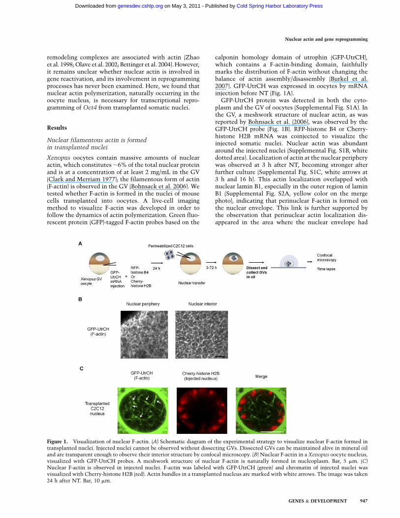
Xenopus oocytes contain massive amounts of nuclearactin, which constitutes ;6% of the total nuclear proteinand is at a concentration of at least 2 mg/mL in the GV(Clark and Merriam 1977); the filamentous form of actin(F-actin) is observed in the GV (Bohnsack et al. 2006). Wetested whether F-actin is formed in the nuclei of mousecells transplanted into oocytes. A live-cell imagingmethod to visualize F-actin was developed in order tofollow the dynamics of actin polymerization. Green fluo-rescent protein (GFP)-tagged F-actin probes based on the
calponin homology domain of utrophin (GFP-UtrCH),which contains a F-actin-binding domain, faithfullymarks the distribution of F-actin without changing thebalance of actin assembly/disassembly (Burkel et al.2007). GFP-UtrCH was expressed in oocytes by mRNAinjection before NT (Fig. 1A).
GFP-UtrCH protein was detected in both the cyto-plasm and the GV of oocytes (Supplemental Fig. S1A). Inthe GV, a meshwork structure of nuclear actin, as wasreported by Bohnsack et al. (2006), was observed by theGFP-UtrCH probe (Fig. 1B). RFP-histone B4 or Cherry-histone H2B mRNA was coinjected to visualize theinjected somatic nuclei. Nuclear actin was abundantaround the injected nuclei (Supplemental Fig. S1B, whitedotted area). Localization of actin at the nuclear peripherywas observed at 3 h after NT, becoming stronger afterfurther culture (Supplemental Fig. S1C, white arrows at3 h and 16 h). This actin localization overlapped withnuclear lamin B1, especially in the outer region of laminB1 (Supplemental Fig. S2A, yellow color on the mergephoto), indicating that perinuclear F-actin is formed onthe nuclear envelope. This link is further supported bythe observation that perinuclear actin localization dis-appeared in the area where the nuclear envelope had
Figure 1. Visualization of nuclear F-actin. (A) Schematic diagram of the experimental strategy to visualize nuclear F-actin formed intransplanted nuclei. Injected nuclei cannot be observed without dissecting GVs. Dissected GVs can be maintained alive in mineral oiland are transparent enough to observe their interior structure by confocal microscopy. (B) Nuclear F-actin in a Xenopus oocyte nucleus,visualized with GFP-UtrCH probes. A meshwork structure of nuclear F-actin is naturally formed in nucleoplasm. Bar, 5 mm. (C)Nuclear F-actin is observed in injected nuclei. F-actin was labeled with GFP-UtrCH (green) and chromatin of injected nuclei wasvisualized with Cherry-histone H2B (red). Actin bundles in a transplanted nucleus are marked with white arrows. The image was taken24 h after NT. Bar, 10 mm.
Nuclear actin and gene reprogramming
GENES & DEVELOPMENT 947
Cold Spring Harbor Laboratory Press on May 3, 2011 - Published by genesdev.cshlp.orgDownloaded from
broken down (Supplemental Fig. S2A, arrows). At 24 hafter NT, filamentous nuclear actin was observed in thetransplanted nuclei (Fig. 1C; Supplemental Fig. S1C, redarrows at 24 h). Actin bundles were formed inside thecircular chromatin (Fig. 1C, arrows), and actin filamentsaround and in the transplanted nuclei were mobile(Supplemental Movie S1).
We next asked whether differences in the donor nuclearstates affected nuclear actin formation. The nuclei ofembryonic stem (ES) cells were injected as representativeof undifferentiated cells. ES nuclei swelled more easilythan C2C12 nuclei and were more extensively dispersed.Slightly more abundant actin filaments were observed inES nuclei than in C2C12 nuclei (Supplemental Fig. S2B).This indicates that nuclei of ES cells, which have thegreatest transcriptional plasticity, exhibit abundant nu-clear actin formation when transplanted to oocytes.
To confirm that GFP-UtrCH faithfully recognizes F-actin,GVs with transplanted nuclei were fixed and stainedwith phalloidin-TRITC, which specifically marks F-actin.Phalloidin staining resulted in a pattern of F-actin forma-tion similar to that visualized by GFP-UtrCH probes(Supplemental Fig. S3A). As a control, F-actin in culturedC2C12 cells before NT was also stained by the samemethod, but such nuclear F-actin formation was notobserved (Supplemental Fig. S3B). In conclusion, F-actinis present in nuclei transplanted into Xenopus oocytes,although it takes ;24 h to see a detectable level of nuclearF-actin. This delay in appearance of nuclear actin maybe because transplanted somatic nuclei have inheriteda mechanism to export actin (Stuven et al. 2003), and/orbecause no active import mechanism for nuclear actinexists in injected nuclei. The strong perinuclear localiza-tion of nuclear actin might reflect this active export(Supplemental Fig. S1C). More interestingly, the timingof nuclear actin appearance is correlated with the timewhen reprogrammed transcripts start to be detected(Halley-Stott et al. 2010).
Nuclear actin polymerization is requiredfor transcriptional reactivation of Oct4
We examined whether polymerized nuclear actin isimportant for transcriptional reactivation by severaldifferent methods (Supplemental Fig. S4A). To inhibitnuclear actin, a monoclonal antibody against b-actin(clone AC15) that recognizes the N-terminal region ofb-actin was injected into the GV with donor C2C12nuclei. Forty-eight hours after NT, Oct4 reactivationwas significantly inhibited (P = 0.0029; fivefold down)by AC15 injection (Fig. 2A). This antibody specificallyand robustly recognizes nuclear b-actin in GVs and showsa stronger inhibition than another anti-bactin antibody(clone AC40) (data not shown). To check the effect ofantibody injection on actin polymerization, nuclear F-actinin the antibody-injected oocytes was visualized by GFP-UtrCH probes. As expected, less F-actin was found in theNT oocytes coinjected with AC15 (Supplemental Fig. S4B).
Several actin mutants that do not polymerize (G13Rand R62D) or that enhance polymerization (G15S and
S14C) have been reported (Posern et al. 2002, 2004). Weexpressed these actin mutants in oocytes by mRNAinjection, and somatic nuclei were subsequently injected(Supplemental Fig. S4A [panel ii],C). The mutant thatenhances polymerization, G15S, significantly enhanced(P = 0.025) Oct4 reactivation by sixfold (Fig. 2B, black bar).The stronger effect of G15S on Oct4 reactivation com-pared with S14C may be explained by the fact that G15Shas a higher affinity to bind actin-associated nucleotidesthan S14C (Posern et al. 2004). In contrast, actin mutantsunable to polymerize did not increase Oct4 reactivation,but R62D rather reduced the reactivation by more thantwofold (Fig. 2B, gray bar). The difference between R62Dand G15S in Oct4 reactivation was 14-fold. These resultsindicate that actin polymerization is important for Oct4gene reactivation.
We next inhibited actin polymerization by the actin-depolymerizing drug cytochalasin B (CB) during incuba-tion of NT oocytes (Supplemental Fig. S4A, panel iii).Treatment with CB greatly reduced polymerized actin inGVs (Supplemental Fig. S4D), although a faint signal ofGFP-UtrCH could still be detected if detection gain wasincreased. CB treatment also reduced Oct4 reactivation(down to 50% 48 h after NT) (Fig. 2C).
Actin is known as an inhibitor of DNaseI. Therefore, itis possible that transplanted nuclei are somewhat de-graded by endogenous DnaseI, and changing actin poly-merization could affect this DNaseI activity. As a result,increased Oct4 transcription would be detected afterincreasing actin polymerization and vice versa. We testedthis possibility by examining the integrity of the DNA ofinjected nuclei using the same quantitative PCR (qPCR)primers as for the transcription assay (Supplemental Fig.S5). During the culture of NT oocytes, no significantDNA degradation was observed (Supplemental Fig. S5A).Furthermore, changing the amount of polymerized actinin the GV (discussed in the next section) did not influenceDNA degradation, at least on the region examined for thetranscriptional reprogramming assay (Supplemental Fig.S5B). These results indicate that altered Oct4 expressionafter changing nuclear actin polymerization is not attrib-utable to altered DNaseI activity in degrading injectedDNA.
Even though evidence that nuclear actin is importantfor Oct4 reactivation has been obtained, we still could notexclude the involvement of cytoplasmic actin-mediatedpathways in gene reactivation. Isolated Xenopus GVs canbe cultured in mineral oil without losing transcriptionalreprogramming activity (Jullien et al. 2010), so that repro-gramming is achieved entirely by oocyte GV components.After isolation of GVs, nuclei were injected with CB orDMSO, a vehicle of CB (Supplemental Fig. S4A, panel iv).We injected 50 mg/mL CB per 10 nL into the GVs, whichdepolymerized almost all actin in GVs (Bohnsack et al.2006). After a 48-h incubation, Oct4 reactivation wasdetected in control samples (Fig. 2D, CB�), but a threefoldreduction was seen when CB was included (Fig. 2D). CBbinds to the barbed end of polymerized actin, inhibitsfurther polymerization, and results in an increased pro-portion of monomeric actin (G-actin). If G-actin binds to
Miyamoto et al.
948 GENES & DEVELOPMENT
Cold Spring Harbor Laboratory Press on May 3, 2011 - Published by genesdev.cshlp.orgDownloaded from

an activator of Oct4 and sequesters it like the transcrip-tional coactivator MAL (Vartiainen et al. 2007), increasedG-actin would cause down-regulation of Oct4. To excludethis possibility, we repeated the oil GV experiment withanother actin-depolymerizing reagent, latrunculin A(LA), which binds to G-actin and inhibits polymerization.This again led to a striking inhibition of transcriptionalreactivation of Oct4 by 17-fold (P = 0.0016) (Fig. 2E). Inconclusion, nuclear actin polymerization is necessary forthe reactivation of the silenced Oct4 gene in oocytes.
An actin signaling protein, Toca-1, regulatesOct4 reactivation
We showed here a requirement of actin polymerizationfor strong Oct4 reactivation by modulating nuclear actinpolymerization. However, one possibility is that Oct4reactivation is inhibited by secondary nonspecific effects,because antibody injection and chemical treatment mayhave a range of pleiotropic effects. To exclude this possi-bility, it is important to find a specific upstream molecule
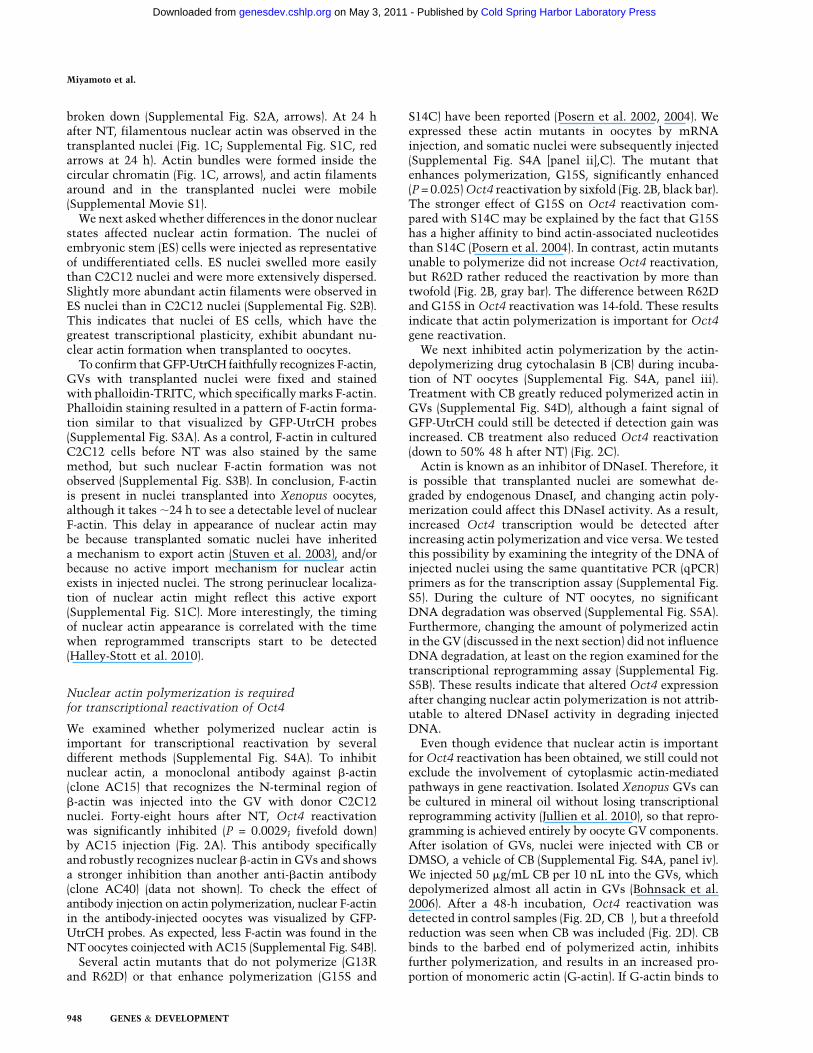
that regulates nuclear actin polymerization and Oct4reactivation. The dynamics of actin polymerization areregulated by actin nucleators and actin signaling proteins.These factors are localized mainly in the cytoplasm, butsome reports suggest that several actin nucleators arepresent in nuclei, although little is known about theirfunction (Percipalle 2009). We tested four such factors—Toca-1 (Ho et al. 2004), N-WASP (Wu et al. 2006), Rac1(Eden et al. 2002), and JMY (Zuchero et al. 2009)—to askwhether they play a role in a reprogramming context.These factors were overexpressed as HA-tagged proteinsin oocytes by mRNA injection before NT (Fig. 3A). Theywere all expressed in GVs, although only a low levelof JMY was detected (Fig. 3B). As a control, differentamounts of these proteins were expressed by injectingvarious amounts of mRNA (1.5, 4.6, or 13.8 ng). Allfactors, except for N-WASP, enhanced Oct4 reactivationin transplanted C2C12 nuclei by up to fourfold, but onlyToca-1 injection with 4.6 and 13.8 ng of mRNA showed astatistically significant and dose-dependent positive effect(P = 0.024 and 0.014, respectively) on Oct4 transcriptional
Figure 2. Nuclear actin polymerization is necessary for transcriptional reprogramming of Oct4. (A) Anti-bactin antibody injection(clone AC15) into GVs inhibits the transcriptional reactivation of Oct4 in NT oocytes. IgG was injected as a control. Relative foldincreases of gene transcripts to control NT were measured by qPCR. Transcription was analyzed 24 h and 48 h after NT. Data representmean 6 SEM; (**) P = 0.0029; F and T-test against control, antibody injection: n = 8 at 48 h and n = 5 at 24 h; control injection: n = 4 at48 h and n = 2 at 24 h. (B) Actin mutant expression influences transcriptional reactivation of Oct4 in NT oocytes. Several human actinmutants (G13R, R62D, G15S, and S14C) and human wild-type actin (wt) were expressed in oocytes, and these oocytes were used for NT.Transcript level is relative to control wild type. Data represent mean 6 SEM; (*) P = 0.025; F and T-test against wild type, n = 6. (C) Theeffect of actin-depolymerizing reagent on Oct4 reactivation in NT oocytes. CB was added to culture medium at a concentration of 5 mg/mL during 48 h of culture. Data represent mean 6 SEM; CB+: n = 10 at 48 h and n = 4 at 24 h; control: n = 11 at 48 h and n = 4 at 24 h. (D)Oct4 reactivation from transplanted nuclei is inhibited by depolymerizing nuclear actin with CB in oil GVs. Nuclei suspended in CB-containing solution (gray bar, CB+) or DMSO-containing solution (black bar, CB�) were injected into oil GVs, and transcriptionalactivation was examined by qRT–PCR analysis. Transcript level is relative to control CB�. Data represent mean 6 SEM; n = 6. (E) Oct4reactivation from transplanted nuclei is inhibited by depolymerizing nuclear actin with LA in oil GVs. Nuclei suspended in LA-containing solution (gray bar, LA+) or DMSO-containing solution (black bar, LA�) were injected into oil GVs. Data represent mean 6
SEM; (**) P = 0.0016; F and T-test against LA�; n = 3.
Nuclear actin and gene reprogramming
GENES & DEVELOPMENT 949
Cold Spring Harbor Laboratory Press on May 3, 2011 - Published by genesdev.cshlp.orgDownloaded from
reactivation (Fig. 3C). Other pluripotency-related genes(Sox2, Klf4, c-myc, Nanog, and Lefty1) and genes highlyexpressed in donor cells (c-Jun and Gapdh) were not
influenced by TOCA-1 overexpression (Fig. 3C), suggest-ing a specific effect of TOCA-1 on Oct4. Remarkably, thispositive effect of TOCA-1 on Oct4 reactivation was
Figure 3. An actin signaling protein, Toca-1, enhances Oct4 reactivation. (A) Schematic diagram of the experimental strategy to assessthe effect of actin nucleators and actin signaling proteins on transcriptional reprogramming from transplanted C2C12 nuclei. (B) Fourfactors tagged with HA are expressed as proteins in both the GV and cytoplasm. These include Xenopus tropicalis JMY (xtJMY), X.
tropicalis TOCA-1 (xtTOCA-1), mouse N-WASP (mN-WASP), and X. laevis RAC1 (xlRAC1). The proteins were detected by Westernblot analysis using anti-HA antibody 24 h after mRNA injection. Actin was used as a loading control. (C) The effect of overexpression ofthe actin polymerization regulator on transcriptional reprogramming. Relative fold increases of gene transcripts to control water-injected samples are shown, measured by qPCR. Data represent mean 6 SEM; (*) P < 0.05; F and T-test against the control waterinjection; n = 4. (D) A mutant form of Toca-1, Toca-1(W518K), does not enhance Oct4 reactivation, while wild-type Toca-1 enhances it.Toca-1(W518K) was similarly overexpressed in oocytes by mRNA injection. Relative fold increases of gene transcripts to control water-injected samples are shown, measured by qPCR. Data represent mean 6 SEM; (*) P = 0.018; F and T-test against the control; Toca1:n = 3; others: n = 4). (E) Reactivation of Oct4, Gdf3, and Eif4e1b genes was enhanced by the overexpression of TOCA-1. Gdf3is a pluripotency gene and Eif4e1b is an oocyte-specific gene. Data represent mean 6 SEM; (**) P < 0.02; (*) P < 0.05; F and T-test againstthe control; n = 5–6).
Miyamoto et al.
950 GENES & DEVELOPMENT
Cold Spring Harbor Laboratory Press on May 3, 2011 - Published by genesdev.cshlp.orgDownloaded from
abrogated by overexpressing the point mutation of TOCA-1(W518K), which is unable to promote actin polymeriza-tion (Fig. 3D; Ho et al. 2004).
Since TOCA-1 has a selective effect on Oct4 reactiva-tion, we investigated other genes whose reactivation isenhanced by TOCA-1 overexpression in the same way asOct4. We checked a list of pluripotency genes that changeepigenetic modifications in the same way as Oct4 dur-ing reprogramming (Mikkelsen et al. 2008). In addition,oocyte-specific genes are known to be reactivated in oursystem (Tamada et al. 2006). Thus, Gdf3 and Eif4e1b wereselected as a pluripotent gene and an oocyte-specific gene,respectively. The effect of TOCA-1 overexpression on thereactivation of these genes was examined. Gdf3 andEif4e1b were expressed very little in C2C12 cells, butwere reactivated after NT to Xenopus GVs (data not
shown). The transcription of these genes and Oct4 wassignificantly enhanced by TOCA-1 (Fig. 3E). The positiveeffect of Toca-1 is not limited to Oct4.
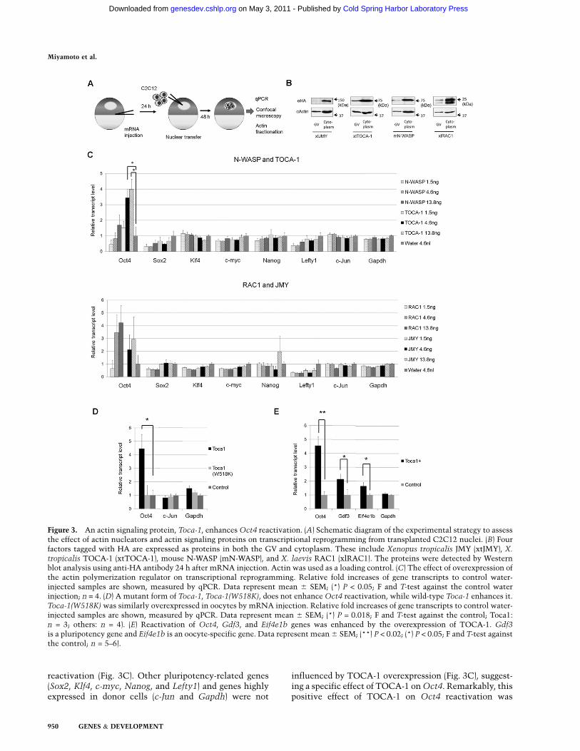
We next examined whether TOCA-1 localizes to trans-planted C2C12 nuclei. Nuclei were transplanted intoToca1-Cherry and GFP-UtrCH mRNA-injected oocytesand observed by the oil GV procedure. TOCA1-CHERRYwas detected in GVs and injected nuclei (Fig. 4A, whitearrow). TOCA-1 was present in the GV and was prefer-entially localized in the area where F-actin is abundantlydetected. Some nuclei in which Toca1-Cherry was notpresent did not show filamentous actin (Fig. 4A, bluearrow). To confirm that endogenous TOCA-1 is present inthe GV, a Western blot was performed. TOCA-1 waspresent in the Xenopus oocyte cytoplasm and also ata weaker level in the GV, which is 20 times smaller in
Figure 4. Toca-1 exists in a transplanted nucleus and enhances nuclear actin polymerization. (A) TOCA1-CHERRY is present ininjected nuclei. Toca1-Cherry and GFP-UtrCH mRNA were injected before NT. The injected nuclei were observed 48 h after NT byconfocal microscopy using the oil GV method. A nucleus in which TOCA1-CHERRY is present shows nuclear F-actin formation (whitearrow). In contrast, nuclear F-actin is not detected in a nucleus without TOCA1-CHERRY (blue arrow). Bar, 20 mm. (B) TOCA1overexpression increases nuclear F-actin formation. Different amounts of TOCA1 were expressed in oocytes by mRNA injection withdifferent concentrations. These injected oocytes were used for NT. The oocytes were subjected to immunofluorescence analysis andphalloidin staining 48 h after NT. Images were obtained from randomly selected areas. Nuclear F-actin amounts were quantified usingthe ImageJ software (Materials and Methods). Statistical differences were measured by F and T-tests. Data represent mean 6 SEM; Toca-11.5 ng: n = 45; Toca-1 4.6 ng and 13.8 ng: n = 55. (C) Actin fractionation experiment indicates that TOCA-1 overexpression increasesthe F-actin population in GVs. TOCA-1 was expressed in oocytes by mRNA injection. GVs were collected from these injected oocytesor noninjected oocytes, and total actin was fractionated to monomeric and polymeric actin (G- and F-actin, respectively). The amountsof G- and F-actin were measured by Western blot analysis using anti-bactin antibody. CB was added to culture medium at a 1 mg/mLconcentration to check that enhanced actin polymerization was diminished with this concentration. F-actin proportion in total actinwas compared among three samples, and fold differences relative to the control noninjected oocytes (Toca� and CB�) are shown. Datarepresent mean 6 SEM; n = 2. (D) Actin fractionation experiment indicates that overexpression of a TOCA-1 mutant, W518K, does notincrease the F-actin population in GVs. F-actin ratio in total actin is relative to control water-injected oocytes. Data represent mean 6
SEM; n = 2.
Nuclear actin and gene reprogramming
GENES & DEVELOPMENT 951
Cold Spring Harbor Laboratory Press on May 3, 2011 - Published by genesdev.cshlp.orgDownloaded from
volume than the cytoplasm (Supplemental Fig. S6A).Immunoprecipitation of TOCA-1 from Xenopus GV ex-tracts confirmed that TOCA-1 is also in the GV, althoughit is not abundant (Supplemental Fig. S6B). Immunofluo-rescence analysis was carried out in order to see locali-zation of endogenous TOCA-1 in transplanted nuclei.Endogenous TOCA-1 was present throughout the GV,including in transplanted nuclei (Supplemental Fig. S6C).
We finally examined the effect of TOCA-1 overexpres-sion on actin polymerization using phalloidin stainingand image analysis (Materials and Methods). Injection of4.6 ng and 13.8 ng of Toca-1 mRNA significantly in-creased nuclear F-actin formation compared with 1.5-nginjection (Fig. 4B), correlating well with Oct4 reactivationdata (Fig. 3C). An actin fractionation experiment alsoshowed that the proportion of nuclear F-actin increasedafter TOCA-1 overexpression (Fig. 4C), and, as expected,the mutant form of TOCA-1(W518K) did not increase therelative proportion of F-actin (Fig. 4D). Our resultsdemonstrate that TOCA-1 enhances nuclear actin poly-merization and Oct4 transcriptional reactivation.
Nuclear actin is important for both continuingtranscription and the transcriptional reprogrammingof Oct4
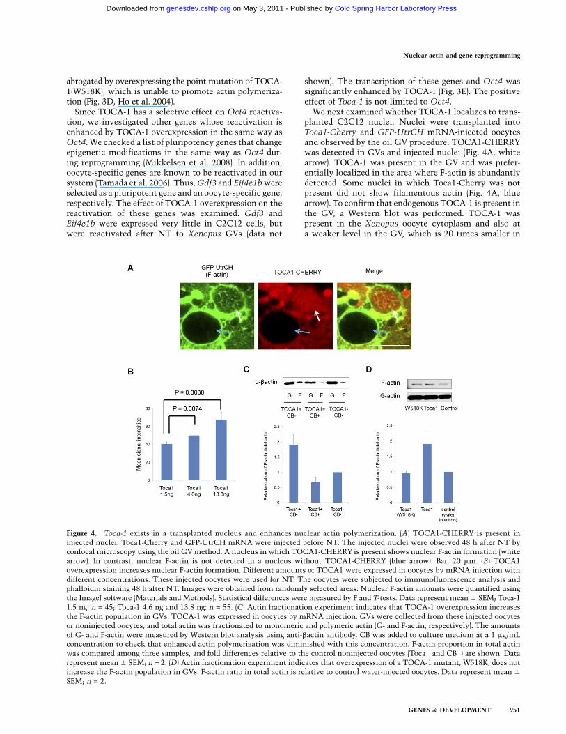
Here we showed a requirement of nuclear actin polymer-ization for Oct4 reactivation and of an upstream mole-cule, Toca-1, for this event. We next asked whethernuclear actin is involved in ongoing transcription ofOct4 as well as in its reprogramming processes. Twoexperiments were designed to test this. First, as depictedin Supplemental Figure S4A (panel i), the anti-bactinantibody was injected into a GV with ES nuclei in whichOct4 was already continuously expressed. Oct4 tran-scription from transplanted ES nuclei was significantlyinhibited (P = 0.0076) by the antibody injection (Fig. 5A).Since transcriptional reprogramming is not required for
continuous Oct4 expression from transplanted ES nuclei,nuclear actin is involved in the ongoing transcription ofOct4. Second, we asked whether TOCA-1 overexpressionenhances continuous Oct4 expression in transplanted ESnuclei. Although TOCA-1 reproducibly enhanced Oct4reactivation in transplanted C2C12 nuclei using the samebatch of oocytes, the continuous Oct4 transcription fromtransplanted ES nuclei was not affected by TOCA-1 (Fig.5B). This result indicates that TOCA-1-mediated actinpolymerization accelerates a reprogramming process, butdoes not affect ongoing transcription. We conclude thatnuclear actin, but not Toca-1, is involved in both ongoingtranscription and reprogramming.
Nuclear actin polymerization andchromatin remodeling
Increased nuclear actin polymerization enhances Oct4reactivation. We asked whether nuclear actin polymeri-zation could affect the chromatin states of transplantednuclei. We carried out coimmunoprecipitation analysis toask whether oocyte nuclear actin was bound to chromatinproteins related to transcription and reprogramming (Zhaoet al. 1998; Bettinger et al. 2004; Hofmann et al. 2004; Dionet al. 2010). This revealed that nuclear actin was bound toRNA polymerase II (Pol II) and the BAF complex (Fig. 6A).Immunofluorescence analysis also showed colocalizationof nuclear actin and elongating Pol II (data not shown).These results suggest that actin in the GV may play a rolein not only transcription, but also chromatin remodeling.
We sought to perform quantitative chromatin immu-noprecipitation (qChIP) analysis in order to see an effectof nuclear actin polymerization on chromatin. However,ChIP using NT oocytes is technically challenging becauseit requires numerous transplanted nuclei. To detect thebinding of nonhistone chromatin proteins like remodelingcomplexes, we used a transgenic ES cell line that pos-sesses ;30 copies of the Oct4 regulatory region driving
Figure 5. Nuclear actin polymerization is impor-tant for both ongoing transcription and transcrip-tional reprogramming of Oct4. (A) Anti-bactinantibody injection into GVs inhibits Oct4 transcrip-tion from transplanted ES nuclei. IgG was injected asa control. Relative fold increases of gene transcriptsto control NT were measured by qPCR. Transcrip-tion was analyzed 48 h after NT. Data representmean 6 SEM; (**) P = 0.0076; F and T-test againstthe control; n = 5. (B) TOCA-1 overexpression doesnot enhance Oct4 transcription from transplantedES nuclei, while it enhances Oct4 transcription fromC2C12 nuclei. Transcript level is relative to controlwater-injected samples. Data represent mean 6
SEM; ES nuclei: n = 4; C2C12 nuclei: n = 3.
Miyamoto et al.
952 GENES & DEVELOPMENT
Cold Spring Harbor Laboratory Press on May 3, 2011 - Published by genesdev.cshlp.orgDownloaded from

Neomycin resistance gene (Neo) expression (Supplemen-tal Fig. S7A). After retinoic acid differentiation, theretinoic acid-treated ES cells (RA-ES cells) showed strongdown-regulation of Neo, indicating a silencing of thetransgenic Oct4 promoter (Supplemental Fig. S7B). Si-lenced Neo expression in RA-ES cells was reactivatedafter NT, and reactivation was enhanced by TOCA-1overexpression (Supplemental Fig. S7C). Furthermore,the enhanced Neo expression was inhibited by adding 1mg/mL CB during incubation of NT oocytes (Supplemen-tal Fig. S7C), and this concentration of CB abolishedincreased actin polymerization by TOCA1 (Fig. 4C). Theseresults indicate that the transgenic Oct4 promoter behaveslike the endogenous Oct4 promoter.
The binding of nuclear actin to the Oct4 upstreamregion was examined by qChIP analysis. AC15 antibodywas used because our preliminary data suggested its pre-cipitation ability was stronger than another anti-bactinantibody (A2103, Sigma) that also recognizes the N-terminalregion, and because it has been used for ChIP analysis
(Ambrosino et al. 2010; Xu et al. 2010). Nuclear actinbinding to the Oct4 regulatory region and gene body wasobserved, and TOCA-1 overexpression enhanced its bind-ing by up to 10-fold (Fig. 6B). The gene body examinedhere is located close to the polyadenylation site of theOct4 gene. These results suggest that TOCA-1 enhancesthe binding of nuclear actin to Oct4. We also examinedPol II and BAF complex binding after TOCA-1 over-expression. Pol II binding to the Oct4 gene was slightlyincreased by TOCA-1 overexpression (Supplemental Fig.S8A), implying that TOCA-1 weakly affects Pol II local-ization in our system. For ChIP analysis of the BAFcomplex, BAF57 antibody was used because it is highlyconserved in the complex among many species (Ho andCrabtree 2010). TOCA-1 overexpression induced an ap-proximately twofold greater BAF57 binding in the Oct4regulatory region (Supplemental Fig. S8B). Together, theeffect of TOCA-1 overexpression on chromatin bindingappeared strongly in nuclear actin, modestly in the BAFcomplex, and weakly in Pol II. In conclusion, TOCA-1overexpression, accompanied by enhanced nuclear actinpolymerization, entails rearrangement of chromatin-binding proteins. These results do not explain all of thepositive effects of TOCA-1 overexpression on Oct4 reac-tivation, but the enhanced access of chromatin factors byTOCA-1 might be one explanation for this positive effect.
Discussion
Nuclear actin was found >30 years ago. Early studies alsoshowed its involvement in the expression of protein-coding genes (Scheer et al. 1984). However, the signifi-cance of these findings has been questioned for a longtime. One of the major reasons for this skepticism is thatthe actin filaments normally observed in the cytoplasmcould not be visualized in the nucleus. Nevertheless,recent studies provided convincing evidence for nuclearactin (McDonald et al. 2006) and its role in nuclearprocesses (Visa and Percipalle 2010). One elegant contri-bution showed that a nuclear actin mesh is formed in thegiant Xenopus oocyte nucleus (Bohnsack et al. 2006). Thisnuclear F-actin scaffold is required to support mechanicalintegrity of the giant nucleus. We extended this viewusing live-cell imaging. The F-actin in transplantednuclei undergoes a global change during reprogramming,and the timing of nuclear actin formation is positivelycorrelated with transcriptional reprogramming.
Nuclear actin is involved in transcription by all threeRNA polymerases (Hofmann et al. 2004; Hu et al. 2004;Philimonenko et al. 2004), and in transcriptional initia-tion after cytokine stimulation (Hofmann et al. 2004) andretinoic acid treatment (Ferrai et al. 2009). Althoughthere is evidence for actin’s involvement in transcription,in vivo evidence is still lacking. In addition, these resultsare accompanied by concerns about contamination andinvolvement of cytoplasmic actin. Considering that bothcytoplasmic and nuclear actin synergistically play animportant role in serum response factor-induced transcrip-tion (Vartiainen et al. 2007), an experimental system thatcan rule out cytoplasmic actin is useful. The Xenopus giant
Figure 6. Association of nuclear actin with chromatin proteinsand chromatin of transplanted nuclei. (A) Coimmunoprecipita-tion analysis in GV lysates to examine binding of nuclear actinto Pol II and the BAF complex. GV proteins were immunopre-cipitated by IgG and antibodies against BAF57, BRG1, BAF53A,and Pol II, and were analyzed by Western blots by probing forb-actin and actin. Five percent input was loaded. (B) TOCA-1overexpression enhances nuclear b-actin binding to the Oct4
gene. ChIP analysis using anti-bactin antibody was performedwith TOCA-1-overexpressed NT oocytes (Toca1+) and normalNT oocytes (Toca1�) 48 h after NT using RA-ES cells. The Oct4
gene regulatory region was expressed as distal enhancer (DE),proximal enhancer (PE), and proximal promoter (PP). As a con-trol, ChIP analysis using nonspecific IgG was carried out withnormal NT oocytes. The relative abundance of precipitatedDNA against total input DNA was measured by qPCR. Datarepresent mean 6 SEM; n = 5.
Nuclear actin and gene reprogramming
GENES & DEVELOPMENT 953
Cold Spring Harbor Laboratory Press on May 3, 2011 - Published by genesdev.cshlp.orgDownloaded from

GV enables us to isolate it in a nonaqueous mediumwithout losing its transcriptional activity (Lund and Paine1990; Jullien et al. 2010). Since reprogramming in an oilGV relies on nuclear contents, the substantial inhibition ofgene reactivation by depolymerizing GV actin in thissystem supports our conclusion.
Cytoplasmic actin filament formation and its properfunction are dependent on actin nucleators (Campelloneand Welch 2010). N-WASP, an actin nucleator, is presentin nuclei and makes a bridge between actin and Pol II(Wu et al. 2006), but its function is largely autoinhibited.Therefore, overexpression of N-WASP itself might nothave any effects on gene reactivation. Toca-1 is identifiedas an upstream regulator of N-WASP to release thisinhibition (Ho et al. 2004), and faithfully enhances Oct4reactivation in our experiments. The prominent effectof Toca-1 on gene reactivation may be explained by itscharacteristic as an upstream molecule in actin polymer-ization signaling (Ho et al. 2004; Fricke et al. 2009). Thesecond strongest effect was induced by Rac1. Consideringthat Rac1 and Toca-1 share the same downstream target(N-WASP and SCAR/WAVE), these actin signaling path-ways may include an important step for Oct4 reactivation.
Nuclear actin and the BAF complex were facilitated intheir binding to Oct4 by Toca-1. This is possibly becauseToca1-mediated actin polymerization near the Oct4 geneincreased an opportunity for the BAF complex to bebound at the area, resulting in an increased chance forBAF to associate with chromatin. This model agrees witha previous study showing that an actin signaling protein,PIP2, allows the BAF complex to associate with polymer-ized actin and chromatin (Zhao et al. 1998; Rando et al.2002). The BAF complex opens chromatin by nucleosomesliding or ejection (Cairns 2009). Importantly, BAF knock-down induces down-regulation of Oct4 (Ho et al. 2009;Kidder et al. 2009). This positive regulator presumablyconverts chromatin into an accessible state for transcrip-tional activators (Singhal et al. 2010), which may allowgeneral transcription factors to act on Oct4 transcription.Indeed, Pol II enhanced its association with the Oct4 genein parallel with these events in our experiment. Toca-1-induced actin polymerization might act upstream in thisseries of reprogramming events.
Our experiments and previous reports show that nu-clear actin is associated with regulatory regions of acti-vating genes and is involved in selective gene activation(Ferrai et al. 2009; Taylor et al. 2010; Xu et al. 2010).Although the reason why only a subset of gene reactiva-tions was affected by actin polymerization is still not clear,one model for how actin selectively binds to specificgenomic sites can be proposed. WASP is supposed to berecruited to DNA by interaction with the transcriptionfactor SP1 (Taylor et al. 2010). Such a recruited actinnucleator can induce actin nucleation at this site, andthus nuclear actin localization occurs at a specific region.Interestingly, the SP1 site on the Oct4 promoter is neces-sary for DNA demethylation and transcription (Simonssonand Gurdon 2004). Recruitment of N-WASP and nuclearactin on the SP1 site might be a necessary step for Oct4transcriptional reprogramming.
We assume that nuclear actin-mediated reprogram-ming is a comparatively late step in the whole transcrip-tional reprogramming process in oocytes. The beginningof reprogramming is characterized by a prompt incorpo-ration of the embryonic linker histone B4 and loss ofsomatic linker histone H1 (Jullien et al. 2010). Subse-quently, gradual swelling of transplanted nuclei proceedsand transcription starts ;24 h after NT. Notably, nuclearactin starts to appear at approximately the same time astranscription starts. As transcription increases towarda peak level after further culture, an active histone mark(histone H3K4 methylation) is added (Murata et al. 2010)and more actin is detected in transplanted nuclei. To-gether with these observations and our present results,nuclear actin is likely to be implicated in the beginning ofthe transcriptional initiation process and enhancement oftranscription. This fits with our results because nuclearactin is associated with the BAF complex and Pol II; thesehave crucial roles in transcriptional initiation and en-hancement. This model is further supported by a recentstudy showing that an actin nucleator binds to H3K4trimethyltransferase RBBP5 and is important for its activ-ity for activating genes (Taylor et al. 2010).
The role of nuclear actin has been elusive. Neverthe-less, many attempts have revealed its novel function inthe nucleus, including transcription and physical genemovement. Here we add a new function of nuclear actinin transcriptional reprogramming. Moreover, our resultsemphasize the significant contribution of nuclear actinpolymerization in transcriptional activation. The impor-tance of G-actin in transcriptional regulation has beenshown clearly in a previous report (Vartiainen et al. 2007).We therefore propose that both nuclear G- and F-actin areinvolved in transcriptional regulation in different ways,and the balance between those two forms of actin may beimportant for regulating transcription. Massive amountsof nuclear actin, which actively change its polymerizedstates during reprogramming, are stored in the oocytenucleus. This helps to explain why an oocyte can repro-gram multiple nuclei efficiently and rapidly.
Materials and methods
NT and mRNA injection
Xenopus oocytes were defolliculated with Liberase (05401127001,Roche). Defolliculated oocytes were kept in modified Barth’ssolution (MBS) at 14°C before use. Mouse cells (ES, RA-ES, andC2C12 cells) were permeabilized with 100 mg/mL Streptolysin O(SLO) for 5–15 min at 37°C. Successful permeabilization (> 95%)was confirmed by Trypan blue staining. Permeabilization wasstopped by the addition of modified Merriam’s medium (20 mMHEPES at pH 7.5, 75 mM KCl, 2 mM MgCl2, 2% PVP, 2 mMb-mercaptoethanol) containing BSA. These permeabilized cellswere suspended in modified Merriam’s medium with 1% BSA toachieve a concentration of 250–300 nuclei per 10 nL, and 10 nL ofthe cell suspension was injected into a Xenopus oocyte nucleus(GV). In every experiment, calibration of the injected cellnumber was performed to deliver a constant number of nucleito each oocyte. Successful injection was confirmed by dissectingthe injected oocytes, since injected nuclei can be observed under
Miyamoto et al.
954 GENES & DEVELOPMENT
Cold Spring Harbor Laboratory Press on May 3, 2011 - Published by genesdev.cshlp.orgDownloaded from
a microscope. The injected oocytes were cultured for up to 3 d at16°C in MBS supplemented with 0.1% BSA and antibiotics.
Different volumes of mRNA, depending on an experiment,were injected into oocytes equatorially. The mRNA injectionwas performed 1 d before NT in order to accumulate the over-expressed proteins in oocytes. Amounts of injected mRNA wereas follows: GFP-UtrCH, 40.8 ng per 40.8 nL; b-actin-R62D/S14C/G15S/G13R/wt-HA, 27.6 ng per 27.6 nL; xtJMY-HA, xtTocal-HA, myc-hToca1(W518K), xtToca-1-Cherry-HA, mN-WASP-HA,and xlRac1-HA, 13.8 ng per 13.8 nL; RFP-histone B4 and Cherry-histone H2B, 4.6 ng per 4.6 nL or 1.5 ng per 4.6 nL.
Oil GV method
NT oocytes cultured in MBS were transferred onto a slide glassand the MBS solution was removed as much as possible. Theoocytes were moved into mineral oil and GVs were dissected (oilGVs). The dissected GVs were transferred onto a slide glass forconfocal microscope analysis. For the NT experiment into oilGV, permeabilized cells (250–300 nuclei per 4.6 nL) were injectedinto the dissected GV. Modified Merriam’s medium containing50 mg/mL CB (Sigma), 5 mM LA (Sigma), or 0.5% DMSO was usedfor cell suspension. The injected GVs were cultured in mineraloil for up to 2 d at 16°C.
Transcriptional reprogramming assay and qRT–PCR
For detecting reprogrammed transcripts from injected mousenuclei, qRT–PCR analysis was performed (Halley-Stott et al.2010). Four whole injected oocytes (equivalent to 1000–1200injected nuclei) or 10 injected oil GVs were pooled as one sample,and RNA was extracted from the samples using Qiagen RNeasycolumns. After RNA extraction, including on-column DNase Idigestion, reverse transcription was performed using SuperScriptIII with gene-specific primers. Real-time PCR was performed asSYBR green assays on an ABI 7300 Real-Time PCR Cycler usingstandard ABI cycling condition. Primers used in this assay arelisted in Supplemental Table S1. Quantification was performedusing ES cell cDNA within the log-linear phase of the amplifica-tion curve. The mean quantity was normalized to mouse Gapdh
transcripts (expressed as Oct4/Gapdh), or a relative transcript levelof every gene was listed.
Cell culture and transfection
Mouse ES cells were grown on gelatin-coated dishes in ES medium(GMEM, 20% ES-grade FBS [Invitrogen], MEM nonessential aminoacids, sodium pyruvate, leukemia inhibitory factor [LIF] [Millipore;ESGRO], b-mercaptoethanol, antibiotics) under feeder-free con-dition. For retinoic acid differentiation, the ES medium waschanged to the differentiation medium (GMEM, 20% FBS[Invitrogen], MEM nonessential amino acids, sodium pyruvate, 1mM retinoic acid, b-mercaptoethanol, antibiotics). C2C12 cellswere maintained in DMEM containing 10% FBS, penicillin, andstreptomycin. Transfection was performed with Lipofectamine2000 (Invitrogen) according to the vendor’s protocol. ES cellswere transfected with Lac repeats–Oct4 regulatory region–Neo,and stably expressing cells were selected using G418. C2C12cells were transiently transfected with the Cherry-laminB1plasmid and used for NT.
Live-cell imaging, immunofluorescence, image analysis
GVs were dissected from injected oocytes in mineral oil. ForF-actin staining on fixed samples, GVs were dissected one by onein GV isolation buffer (20 mM Tris-HCl at pH 7.5, 0.5 mM
MgSO4, 140 mM KCl), immediately transferred into 10% para-formaldehyde containing 1 mM phalloidin-TRITC (Fulka), andincubated for 20–25 min with gentle rotating. After washingwith PBS containing 0.2% Tween20, DNA was stained withDAPI, followed by washing. For TOCA-1 staining, GVs werepermeabilized with PBS containing 0.5% Triton X and blockedwith PBS containing 5% FBS and 0.2% Tween20. Anti-Toca-1antibody (a gift from J. Gallop and M. Kirschner) and anti-phoPolII(S2) (cloneH5:MMS-129R, Covance) were used with1:100 dilution, and anti-rabbit IgG Alexa Fluor 488 and anti-mouse IgM Alexa Flour 633 were used as secondary antibodies(1:200 dilution). Confocal analysis was carried out on a Zeiss 510META confocal LSM microscope equipped with argon (488 nm),HeNe (543 and 637 nm), and MaiTai (740 nm) lasers. For time-lapse experiments, images were taken every 10 min, and thesewere projected using Zeiss LSM 510 software.
Nuclear actin in transplanted nuclei was quantified by ImageJsoftware. F-actin was labeled with phalloidin-TRITC and nucleiwere labeled with DAPI. By using the straight-line selection tool,a line was drawn within a transplanted nucleus whose area couldbe judged by DAPI staining. Strong dot-like staining was avoidedwhen drawing the line. Mean signal intensities in the plottedareas were calculated by the Analyze tool. More than nine NToocytes were used for the analysis, and five images of trans-planted nuclei were selected randomly from each NT oocyte.
qChIP
ChIP analysis was performed as described previously with modi-fications (Astrand et al. 2009; Jullien et al. 2010). NT oocytes thatcontain ;300 mouse nuclei per oocyte were transferred to MBSmedium without BSA, and then a set of seven to 10 oocytes wastransferred into a 1.5-mL tube. After washing with MBS, NToocytes were cross-linked for 10 min at room temperature in1 mL of MBS medium containing 1% formaldehyde, with gentleshaking. Fixation was stopped by rapid washing of oocytes inMBS. The oocytes were ruptured in 280 mL of homogenizationbuffer (50 mM HEPES at pH 7.8, 140 mM NaCl, 1 mM EDTA, 1%Triton X-100, 0.1% sodium deoxycholate, 0.3% SDS, 1 mMDTT, protease inhibitors) by pipetting and kept on ice beforesonication. Sonication was carried out in 1.5-mL tubes for 7 mintwice with a 30-sec on/30-sec off cycle by Bioruptor TWIN(Diagenode). Six-hundred microliters of buffer I (50 mM HEPESat pH 7.8, 140 mM NaCl, 1 mM EDTA, 1% Triton X-100, 0.1%sodium deoxycholate, 1 mM DTT, protease inhibitors) wasadded to the sonicated samples. These solutions from two tubeswere mixed in 2-mL tubes, finally yielding 1800 mL. The sampleswere centrifuged at 13,000 rpm at 4°C. Supernatants weretransferred, and 10% of these supernatants was taken as inputsand mixed with an equal volume of 23 Stop buffer (40 mM Tris-HCl at pH 8.0, 10 mM EDTA, 1% SDS) and 0.3 mg/mL proteinaseK (final concentration). The inputs were incubated overnight at65°C. The rest of the sonicated solution was mixed with anti-body and incubated overnight at 4°C with rotation. The anti-bodies used here were 3 mL of mouse monoclonal anti-bactin(cloneAC15:A1978, Sigma), 2 mL of goat polyclonal anti-BAF57(ab14764, Abcam), 1 mL of mouse monoclonal anti-Pol II[8WG16](ab817, Abcam), 2 mL of rabbit IgG (sc2027, Santa Cruz Bio-technology), and 1 mL of rabbit polyclonal anti-Histone H3(ab1791, Abcam). After antibody reaction, 20 mL of dynabeadsprotein G (Invitorgen) was added and was rotated for another 6 hat 4°C. The bead antibody conjugates were washed in thefollowing order with buffer I, buffer II (50 mM HEPES at pH7.8, 500 mM NaCl, 1 mM EDTA, 1% Triton X-100, 0.1% sodiumdeoxycholate, 1 mM DTT, protease inhibitors), buffer III (20 mMTris-HCl at pH 8.0, 1 mM EDTA, 250 mM LiCl, 0.5% NP40,
Nuclear actin and gene reprogramming
GENES & DEVELOPMENT 955
Cold Spring Harbor Laboratory Press on May 3, 2011 - Published by genesdev.cshlp.orgDownloaded from
0.5% sodium deoxycholate, 1 mM DTT, protease inhibitors), andTE containing 1 mM DTT and protease inhibitors for 30 min at4°C with rotation. After the last washing, 150 mL of 13 Stopbuffer with 0.3 mg/mL proteinase K was added to the beads, andcross-links were reversed by incubation overnight at 65°C. DNAwas isolated from inputs and immunoprecipitates. The DNAwas finally resuspended in 50 mL of elution buffer. Four micro-liters was used for qPCR analysis. When abundant chromatinproteins like core histones were precipitated, 10 NT oocytes(equivalent to 3000 injected nuclei) were enough to detectsignals. However, other cases like nuclear actin required 14–20NT oocytes (equivalent to 4200–6000 injected nuclei). We useda transgenic ES cell line that possesses ;30 copies of the Oct4
regulatory region as a donor cell in order to increase the strengthof the signals. The relative abundance of precipitated DNAagainst total input DNA was measured by qPCR, and this valueis expressed as signal per a single gene copy. In every ChIPanalysis, anti-histone H3 antibody was used as a positive controlto check whether precipitation was carried out successfully.
Coimmunoprecipitation and immunoprecipitation analysis
Ten GVs per coimmunoprecipitation were collected in immu-noprecipitation buffer (20 mM HEPES at pH 7.5, 5 mM KCl,1.5 mM MgCl2, 1 mM EGTA, 150 mM NaCl, 0.1% NP40, 1 mMDTT, 0.2 mM PMSF, protease inhibitor cocktail). The GVs in10 mL of immunoprecipitation buffer were transferred into tubesand disrupted by pipetting up and down. One microliter wastaken as an input, and 1–3 mL of the following antibodies wereadded: mouse monoclonal anti-GFP (11814460001, Roche), rab-bit IgG, goat polyclonal anti-BAF57, rabbit polyclonal anti-BAF53A (ab3882, Abcam), rabbit polyclonal anti-BRG1 (A300-813A, Bethyl Laboratories), and mouse monoclonal anti-PolII[8WG16]. After incubating overnight at 4°C, Dynabeads Pro-tein G was added and incubated for 30 min at room temperature.The beads were washed with immunoprecipitation buffer andbinding proteins were extracted for Western blots. Immunopre-cipitation of Toca-1 from GV extracts was carried out as de-scribed previously (Ho et al. 2004) and the extracts were obtainedfrom 50 GVs.
Nuclear actin fractionation
Thirty GVs were collected in mineral oil in order to minimizeartificial changes in actin polymerization. The GVs were trans-ferred into an ultracentrifuge tube and 500 mL of lysis and F-actinstabilization buffer (50 mM PIPES at pH 6.9, 50 mM NaCl, 5 mMMgCl2, 5 mM EGTA, 5% glycerol, 0.1% NP40, 0.1% TritonX-100, 0.1% Tween20, 0.1% b-mercaptoethanol, 1 mM ATP,protease inhibitor cocktail) was added. GVs were disrupted bypipetting up and down and the lysate was centrifuged at 100,000g
for 1 h at 23°C. The supernatant was transferred immediatelyinto another tube and was used as the G-actin fraction. Thepellet was resuspended in 500 mL of ice-cold water containing100 mM CB and kept on ice for 30 min with some pipetting. Afterthe incubation, samples were sonicated with probe sonicator andused as the F-actin fraction. Total amounts of b-actin or totalactin were measured by the Western blot analysis.
Western blots
Western blots were performed following standard protocols.Anti-mouse, anti-rabbit, or anti-goat IgG Alexa Fluor 680(Invitrogen) was used as a secondary antibody, and bands weredetected using the LI-COR ODYSSEY imaging system. Primaryantibodies used were as follows: rabbit polyclonal anti-Toca1
(a gift from J. Gallop and M. Kirschner), mouse monoclonal anti-b-actin (cloneAC15), rabbit polyclonal anti-actin (A2103, Sigma),rabbit polyclonal anti-Histone H2B (ab1790, Abcam), mousemonoclonal anti-GFP, mouse monoclonal anti-HA (H9658,Sigma), and rabbit polyclonal anti-HA (ab9110, Abcam).
Plasmid construction and RNA synthesis
Actin mutants (Posern et al. 2002, 2004) were subcloned fromoriginal reported vectors into Gateway vectors (pCS2-b-actin-[R62D/S14C/G15S/G13R/wt]-HA). Xenopus tropicalis jmy, X.
tropicalis Toca-1 (fnbp1l), mouse N-WASP (Wasl), and X. laevisrac1 were cloned into pCS2-HA and/or pCS2-Cherry-HA. pCS2-Cherry-hH2B and pCS2-RFP-histoneB4 were produced previ-ously in our laboratory (Jullien et al. 2010). p3216PECMS2(Janicki et al. 2004), pCS2-GFP-UtrCH (Burkel et al. 2007), andpCMV-CuO-lacI-mCherry-laminB1 (Kumaran and Spector 2008)were gifts. For producing Oct4 regulatory region–Neo cell lines,the p3216PECMS2 plasmid was modified. The inducible CMVpromoter was swapped with the murine Oct4 3.6-kb upstreamregion and the CFP reporter was changed to the Neomycin-resistant gene. This plasmid was transfected to ES cells. In vitrosynthesized mRNAs were produced with MEGAscript SP6(AM1330, Ambion).
Statistic analysis
The number of biological replicates are shown as n. In transcrip-tional assays, technical duplicates were performed on all sam-ples and an average of the duplicate was regarded as the value ofone sample. Statistic difference was calculated by F and T-test(P < 0.05). Error bars were represented as standard error.
Acknowledgments
We thank J. Gallop and M. Kirschner for gifts of TOCA-1antibody, Toca-1 mutant plasmids, and N-WASP(VCA) plasmids;R. Treisman for actin mutant plasmids; D. Gorlich for Exportin 6plasmids; W.M. Bement for GFP-UtrCH plasmids; and D. Spectorfor p3216PECMS2 and lamin B1 plasmids. We also thank J.Gallop, D. Gorlich, R. Halley-Stott, and P. Narbonne for valuablecomments on our manuscript. K.M. is supported by the JapanSociety for the Promotion of Science (International ResearchFellowship Program). This research is supported by a grant fromthe Wellcome Trust PhD scholarship (081277) to V.P., and a grantfrom the Wellcome Trust (grant RG54943) to J.J and J.B.G.
References
Ambrosino C, Tarallo R, Bamundo A, Cuomo D, Franci G,Nassa G, Paris O, Ravo M, Giovane A, Zambrano N, et al.2010. Identification of a hormone-regulated dynamic nuclearactin network associated with estrogen receptor a in humanbreast cancer cell nuclei. Mol Cell Proteomics 9: 1352–1367.
Astrand C, Belikov S, Wrange O. 2009. Histone acetylationcharacterizes chromatin presetting by NF1 and Oct1 andenhances glucocorticoid receptor binding to the MMTVpromoter. Exp Cell Res 315: 2604–2615.
Bettinger BT, Gilbert DM, Amberg DC. 2004. Actin up in thenucleus. Nat Rev Mol Cell Biol 5: 410–415.
Bohnsack MT, Stuven T, Kuhn C, Cordes VC, Gorlich D. 2006.A selective block of nuclear actin export stabilizes the giantnuclei of Xenopus oocytes. Nat Cell Biol 8: 257–263.
Boiani M, Eckardt S, Scholer HR, McLaughlin KJ. 2002. Oct4distribution and level in mouse clones: consequences forpluripotency. Genes Dev 16: 1209–1219.
Miyamoto et al.
956 GENES & DEVELOPMENT
Cold Spring Harbor Laboratory Press on May 3, 2011 - Published by genesdev.cshlp.orgDownloaded from
Burkel BM, von Dassow G, Bement WM. 2007. Versatile fluores-cent probes for actin filaments based on the actin-bindingdomain of utrophin. Cell Motil Cytoskeleton 64: 822–832.
Byrne JA, Simonsson S, Western PS, Gurdon JB. 2003. Nuclei ofadult mammalian somatic cells are directly reprogrammedto oct-4 stem cell gene expression by amphibian oocytes.Curr Biol 13: 1206–1213.
Cairns BR. 2009. The logic of chromatin architecture andremodelling at promoters. Nature 461: 193–198.
Campellone KG, Welch MD. 2010. A nucleator arms race:cellular control of actin assembly. Nat Rev Mol Cell Biol
11: 237–251.Clark TG, Merriam RW. 1977. Diffusible and bound actin nuclei
of Xenopus laevis oocytes. Cell 12: 883–891.Dion V, Shimada K, Gasser SM. 2010. Actin-related proteins in
the nucleus: life beyond chromatin remodelers. Curr OpinCell Biol 22: 383–391.
Eden S, Rohatgi R, Podtelejnikov AV, Mann M, Kirschner MW.2002. Mechanism of regulation of WAVE1-induced actinnucleation by Rac1 and Nck. Nature 418: 790–793.
Ferrai C, Naum-Ongania G, Longobardi E, Palazzolo M, DisanzaA, Diaz VM, Crippa MP, Scita G, Blasi F. 2009. Induction ofHoxB transcription by retinoic acid requires actin polymer-ization. Mol Biol Cell 20: 3543–3551.
Fricke R, Gohl C, Dharmalingam E, Grevelhorster A, Zahedi B,Harden N, Kessels M, Qualmann B, Bogdan S. 2009. Dro-
sophila Cip4/Toca-1 integrates membrane trafficking andactin dynamics through WASP and SCAR/WAVE. Curr Biol19: 1429–1437.
Gurdon JB, Melton DA. 2008. Nuclear reprogramming in cells.Science 322: 1811–1815.
Halley-Stott RP, Pasque V, Astrand C, Miyamoto K, Simeoni I,Jullien J, Gurdon JB. 2010. Mammalian nuclear transplanta-tion to germinal vesicle stage Xenopus oocytes—a methodfor quantitative transcriptional reprogramming. Methods 51:56–65.
Hanna J, Saha K, Pando B, van Zon J, Lengner CJ, Creyghton MP,van Oudenaarden A, Jaenisch R. 2009. Direct cell reprogram-ming is a stochastic process amenable to acceleration.Nature 462: 595–601.
Ho L, Crabtree GR. 2010. Chromatin remodelling during de-velopment. Nature 463: 474–484.
Ho HY, Rohatgi R, Lebensohn AM, Le M, Li J, Gygi SP,Kirschner MW. 2004. Toca-1 mediates Cdc42-dependentactin nucleation by activating the N-WASP–WIP complex.Cell 118: 203–216.
Ho L, Ronan JL, Wu J, Staahl BT, Chen L, Kuo A, Lessard J,Nesvizhskii AI, Ranish J, Crabtree GR. 2009. An embryonicstem cell chromatin remodeling complex, esBAF, is essentialfor embryonic stem cell self-renewal and pluripotency. ProcNatl Acad Sci 106: 5181–5186.
Hofmann WA, Stojiljkovic L, Fuchsova B, Vargas GM, MavrommatisE, Philimonenko V, Kysela K, Goodrich JA, Lessard JL, Hope TJ,et al. 2004. Actin is part of pre-initiation complexes and isnecessary for transcription by RNA polymerase II. Nat Cell
Biol 6: 1094–1101.Hu P, Wu S, Hernandez N. 2004. A role for b-actin in RNA
polymerase III transcription. Genes Dev 18: 3010–3015.Jaenisch R, Young R. 2008. Stem cells, the molecular circuitry of
pluripotency and nuclear reprogramming. Cell 132: 567–582.Janicki SM, Tsukamoto T, Salghetti SE, Tansey WP, Sachidanandam
R, Prasanth KV, Ried T, Shav-Tal Y, Bertrand E, Singer RH, et al.2004. From silencing to gene expression: real-time analysis insingle cells. Cell 116: 683–698.
Jullien J, Astrand C, Halley-Stott RP, Garrett N, Gurdon JB.2010. Characterization of somatic cell nuclear reprogram-
ming by oocytes in which a linker histone is required forpluripotency gene reactivation. Proc Natl Acad Sci 107:5483–5488.
Kidder BL, Palmer S, Knott JG. 2009. SWI/SNF-Brg1 regulatesself-renewal and occupies core pluripotency-related genes inembryonic stem cells. Stem Cells 27: 317–328.
Kim JB, Sebastiano V, Wu G, Arauzo-Bravo MJ, Sasse P, GentileL, Ko K, Ruau D, Ehrich M, van den Boom D, et al. 2009.Oct4-induced pluripotency in adult neural stem cells. Cell136: 411–419.
Kim K, Doi A, Wen B, Ng K, Zhao R, Cahan P, Kim J, Aryee MJ,Ji H, Ehrlich LI, et al. 2010. Epigenetic memory in inducedpluripotent stem cells. Nature 467: 285–290.
Kumaran RI, Spector DL. 2008. A genetic locus targeted to thenuclear periphery in living cells maintains its transcriptionalcompetence. J Cell Biol 180: 51–65.
Lund E, Paine PL. 1990. Nonaqueous isolation of transcription-ally active nuclei from Xenopus oocytes. Methods Enzymol
181: 36–43.McDonald D, Carrero G, Andrin C, de Vries G, Hendzel MJ.
2006. Nucleoplasmic b-actin exists in a dynamic equilib-rium between low-mobility polymeric species and rapidlydiffusing populations. J Cell Biol 172: 541–552.
Mikkelsen TS, Hanna J, Zhang X, Ku M, Wernig M, SchorderetP, Bernstein BE, Jaenisch R, Lander ES, Meissner A. 2008.Dissecting direct reprogramming through integrative geno-mic analysis. Nature 454: 49–55.
Murata K, Kouzarides T, Bannister AJ, Gurdon JB. 2010. HistoneH3 lysine 4 methylation is associated with the transcriptionalreprogramming efficiency of somatic nuclei by oocytes. Epi-
genetics Chromatin 3: 4. doi: 10.1186/1756-8935-3-4.Olave IA, Reck-Peterson SL, Crabtree GR. 2002. Nuclear actin
and actin-related proteins in chromatin remodeling. Annu
Rev Biochem 71: 755–781.Pasque V, Miyamoto K, Gurdon JB. 2010. Efficiencies and
mechanisms of nuclear reprogramming. Cold Spring Harb
Symp Quant Biol doi: 101101/sqb.2010.75.002.Percipalle P. 2009. The long journey of actin and actin-associ-
ated proteins from genes to polysomes. Cell Mol Life Sci 66:2151–2165.
Philimonenko VV, Zhao J, Iben S, Dingova H, Kysela K, KahleM, Zentgraf H, Hofmann WA, de Lanerolle P, Hozak P, et al.2004. Nuclear actin and myosin I are required for RNApolymerase I transcription. Nat Cell Biol 6: 1165–1172.
Posern G, Sotiropoulos A, Treisman R. 2002. Mutant actinsdemonstrate a role for unpolymerized actin in control oftranscription by serum response factor. Mol Biol Cell 13:4167–4178.
Posern G, Miralles F, Guettler S, Treisman R. 2004. Mutantactins that stabilise F-actin use distinct mechanisms toactivate the SRF coactivator MAL. EMBO J 23: 3973–3983.
Rando OJ, Zhao K, Janmey P, Crabtree GR. 2002. Phosphatidy-linositol-dependent actin filament binding by the SWI/SNF-like BAF chromatin remodeling complex. Proc Natl Acad Sci
99: 2824–2829.Scheer U, Hinssen H, Franke WW, Jockusch BM. 1984. Micro-
injection of actin-binding proteins and actin antibodiesdemonstrates involvement of nuclear actin in transcriptionof lampbrush chromosomes. Cell 39: 111–122.
Simonsson S, Gurdon J. 2004. DNA demethylation is necessaryfor the epigenetic reprogramming of somatic cell nuclei. NatCell Biol 6: 984–990.
Singhal N, Graumann J, Wu G, Arauzo-Bravo MJ, Han DW,Greber B, Gentile L, Mann M, Scholer HR. 2010. Chromatin-remodeling components of the BAF complex facilitatereprogramming. Cell 141: 943–955.
Nuclear actin and gene reprogramming
GENES & DEVELOPMENT 957
Cold Spring Harbor Laboratory Press on May 3, 2011 - Published by genesdev.cshlp.orgDownloaded from
Smith ZD, Nachman I, Regev A, Meissner A. 2010. Dynamicsingle-cell imaging of direct reprogramming reveals an earlyspecifying event. Nat Biotechnol 28: 521–526.
Stadtfeld M, Maherali N, Breault DT, Hochedlinger K. 2008.Defining molecular cornerstones during fibroblast to iPS cellreprogramming in mouse. Cell Stem Cell 2: 230–240.
Stuven T, Hartmann E, Gorlich D. 2003. Exportin 6: a novelnuclear export receptor that is specific for profilin.actincomplexes. EMBO J 22: 5928–5940.
Takahashi K, Yamanaka S. 2006. Induction of pluripotent stemcells from mouse embryonic and adult fibroblast cultures bydefined factors. Cell 126: 663–676.
Tamada H, Van Thuan N, Reed P, Nelson D, Katoku-Kikyo N,Wudel J, Wakayama T, Kikyo N. 2006. Chromatin decon-densation and nuclear reprogramming by nucleoplasmin.Mol Cell Biol 26: 1259–1271.
Taylor MD, Sadhukhan S, Kottangada P, Ramgopal A, Sarkar K,D’Silva S, Selvakumar A, Candotti F, Vyas YM. 2010.Nuclear role of WASp in the pathogenesis of dysregulatedTH1 immunity in human Wiskott-Aldrich syndrome. SciTransl Med 2: 37–44.
Vartiainen MK, Guettler S, Larijani B, Treisman R. 2007.Nuclear actin regulates dynamic subcellular localizationand activity of the SRF cofactor MAL. Science 316: 1749–1752.
Visa N, Percipalle P. 2010. Nuclear functions of actin. Cold
Spring Harb Perspect Biol 2: a000620. doi: 10.101/cshperspect.a000620.
Wu X, Yoo Y, Okuhama NN, Tucker PW, Liu G, Guan JL. 2006.Regulation of RNA-polymerase-II-dependent transcriptionby N-WASP and its nuclear-binding partners. Nat Cell Biol8: 756–763.
Xu YZ, Thuraisingam T, Morais DA, Rola-Pleszczynski M,Radzioch D. 2010. Nuclear translocation of b-actin is in-volved in transcriptional regulation during macrophage dif-ferentiation of HL-60 cells. Mol Biol Cell 21: 811–820.
Yamanaka S. 2009. Elite and stochastic models for inducedpluripotent stem cell generation. Nature 460: 49–52.
Zhao K, Wang W, Rando OJ, Xue Y, Swiderek K, Kuo A, CrabtreeGR. 1998. Rapid and phosphoinositol-dependent binding ofthe SWI/SNF-like BAF complex to chromatin after T lym-phocyte receptor signaling. Cell 95: 625–636.
Zuchero JB, Coutts AS, Quinlan ME, Thangue NB, Mullins RD.2009. p53-cofactor JMY is a multifunctional actin nucleationfactor. Nat Cell Biol 11: 451–459.
Miyamoto et al.
958 GENES & DEVELOPMENT
Cold Spring Harbor Laboratory Press on May 3, 2011 - Published by genesdev.cshlp.orgDownloaded from
Copyright © 2022 FDOKUMEN