Generation of induced pluripotent stem cells from human cord blood cells with only two factors: Oct4...
10
p u o r G g n i h s i l b u P e r u t a N 0 1 0 2 © natureprotocols / m o c . e r u t a n . w w w / / : p t t h PROTOCOL NATURE PROTOCOLS | VOL.5 NO.4 | 2010 | 811 INTRODUCTION Human induced pluripotent stem cells (iPSC) derived from adult somatic cells could be used in regenerative medicine to repair tissues damaged by disease or injury. Although human iPSC have been generated from various type of somatic cells, such as skin fibroblasts 1 , keratinocytes 2 , neural stem cells (NSCs) 3 , blood cells 4,5 and cord blood-derived endothelial cells (CBEC) 6 , scientists are still debating the best sources from which to derive iPSC. Further studies have shown that the age, origin and cell type used has a profound impact on the reprogramming efficiency as well as the quality of iPSC generation 2,7 . A desirable protocol should aim to not only optimize all these parameters, but also to reduce both the number of factors used and the time required for reprogramming. A key observation in this regard is the recent study by Kim et al. 3 where iPSC were generated from adult NSCs by direct reprogramming with either two transcrip- tion factors (OCT4 and KLF4) or one transcription factor (OCT4) 8 . However, NSCs are usually rare and difficult to access, representing a complicated target for reprogramming. Recently, the possibility of reprogramming blood cells to pluripotency has suggested an alterna- tive source of somatic cells to generate iPSC. In particular, iPSC have been generated from mobilized peripheral blood (mPB) 4 or bone marrow (BM) 5 CD34 + derived cells by ectopic expression of OCT4, SOX2, KLF4 and c-MYC. Although these cells are certainly an acces- sible and widely used source, they may have accumulated genomic alterations as a result of aging or disease, and the isolation of hemat- opoietic stem cells could represent a health risk for the donor 9 . Cord blood (CB) stem cells, currently widely used as a source of hematopoietic stem cells for transplantation, could overcome these problems. The main practical advantages of using CB stem cells are the relative ease of procurement, the absence of risks for the donors and the ability to store fully tested and HLA-typed samples in public banks 10 , available for immediate use. Another relevant consideration is that CB cells are young and are expected to carry minimal somatic mutations 11 . We have demonstrated that CB CD133 + cells can be repro- grammed to pluripotency faster than fibroblasts and keratinocytes by ectopic expression of only two transcription factors (OCT4 and SOX2) 12 . CB CD133 + cells express a subset of pluripotency-associ- ated genes (OCT4, SOX2, NANOG and CRIPTO), albeit at much lower levels than human ES cells (hESCs) (data not shown). On the other hand, the endogenous levels of c-MYC and KLF4 are higher in CB CD133 + cells compared with fibroblasts and keratinocytes ( Fig. 1). The combination of low levels of pluripotency markers with the high levels of KLF4 and c-MYC may allow for enhanced reprogramming of CB CD133 + cells. Cord blood iPS (CBiPS) cells are generated with only two factors offer new possibilities for investigating the molecular mechanisms that underline reprogramming to pluripotency. Furthermore, and from a more practical point of view, CB cells could represent, in the future, an alternative, safer source of iPSC amenable to worldwide banking and distribution. Here, we present a detailed protocol for the derivation of CB CD133 + cells and how to reprogram them to pluripotency by retroviral transfection of OCT4 and SOX2 in 2 weeks without the need for additional chemical compounds. Experimental design Derivation of CB CD133 + cells. Our protocol first focuses on the importance of selecting the correct cell subpopulation, within CB, for reprogramming. CD133 antigen is known as a stem cell marker for hematopoietic stem and progenitor cells. In contrast to CD34, the most widely used marker for hemato- poietic stem cells (HSC) enrichment, CD133 is not found in late progenitors, providing a selection of a more homogeneous population enriched in HSC. CD133 + cells, which represent a small fraction of total nucleated cells in human CB (0.1–0.5%), are positively selected using the standard immunomagnetic separation system. Generation of induced pluripotent stem cells from human cord blood cells with only two factors: Oct4 and Sox2 Alessandra Giorgetti 1,5 , Nuria Montserrat 1,5 , Ignacio Rodriguez-Piza 1 , Carmen Azqueta 2 , Anna Veiga 1 & Juan Carlos Izpisúa Belmonte 1,3,4 1 Center for Regenerative Medicine in Barcelona, Dr. Aiguader, Barcelona, Spain. 2 Banc de Sang i Teixits, Vall d’Hebron, Paseo Vall d’Hebron, Barcelona, Spain. 3 Gene Expression Laboratory, The Salk Institute for Biological Studies, La Jolla, California, USA. 4 Networking Center of Biomedical Research in Bioengineering, Biomaterials and Nanomedicine (CIBER-BBN), Barcelona, Spain. 5 These authors contributed equally to this work. Correspondence should be addressed to J.C.I.B. ([email protected] or [email protected]). Published online 1 April 2010; doi:10.1038/nprot.2010.16 Induced pluripotent stem cells (iPSC) provide an invaluable resource for regenerative medicine as they allow the generation of patient-specific progenitors with potential value for cell therapy. However, in many instances, an off-the-shelf approach is desirable, such as for cell therapy of acute conditions or when the patient’s somatic cells are altered as a consequence of a chronic disease or aging. Cord blood (CB) stem cells appear ideally suited for this purpose as they are young cells expected to carry minimal somatic mutations and possess the immunological immaturity of newborn cells; additionally, several hundred thousand immunotyped CB units are readily available through a worldwide network of CB banks. Here we present a detailed protocol for the derivation of CB stem cells and how they can be reprogrammed to pluripotency by retroviral transduction with only two factors (OCT4 and SOX2) in 2 weeks and without the need for additional chemical compounds.
-
Upload
msbarreiro -
Category
Documents
-
view
2 -
download
0
Transcript of Generation of induced pluripotent stem cells from human cord blood cells with only two factors: Oct4...

p
uor
G g
n ih si l
bu
P eru ta
N 010 2©
nat
ure
pro
toco
ls/
moc. e r
ut an .
ww
w / /:pt t
h
protocol
nature protocols | VOL.5 NO.4 | 2010 | 811
IntroDuctIonHuman induced pluripotent stem cells (iPSC) derived from adult somatic cells could be used in regenerative medicine to repair tissues damaged by disease or injury. Although human iPSC have been generated from various type of somatic cells, such as skin fibroblasts1, keratinocytes2, neural stem cells (NSCs)3, blood cells4,5 and cord blood-derived endothelial cells (CBEC)6, scientists are still debating the best sources from which to derive iPSC. Further studies have shown that the age, origin and cell type used has a profound impact on the reprogramming efficiency as well as the quality of iPSC generation2,7. A desirable protocol should aim to not only optimize all these parameters, but also to reduce both the number of factors used and the time required for reprogramming. A key observation in this regard is the recent study by Kim et al.3 where iPSC were generated from adult NSCs by direct reprogramming with either two transcrip-tion factors (OCT4 and KLF4) or one transcription factor (OCT4)8. However, NSCs are usually rare and difficult to access, representing a complicated target for reprogramming. Recently, the possibility of reprogramming blood cells to pluripotency has suggested an alterna-tive source of somatic cells to generate iPSC. In particular, iPSC have been generated from mobilized peripheral blood (mPB)4 or bone marrow (BM)5 CD34 + derived cells by ectopic expression of OCT4, SOX2, KLF4 and c-MYC. Although these cells are certainly an acces-sible and widely used source, they may have accumulated genomic alterations as a result of aging or disease, and the isolation of hemat-opoietic stem cells could represent a health risk for the donor9.
Cord blood (CB) stem cells, currently widely used as a source of hematopoietic stem cells for transplantation, could overcome these problems. The main practical advantages of using CB stem cells are the relative ease of procurement, the absence of risks for the donors and the ability to store fully tested and HLA-typed samples in public banks10, available for immediate use. Another relevant consideration is that CB cells are young and are expected to carry minimal somatic mutations11.
We have demonstrated that CB CD133 + cells can be repro-grammed to pluripotency faster than fibroblasts and keratinocytes by ectopic expression of only two transcription factors (OCT4 and SOX2)12. CB CD133 + cells express a subset of pluripotency-associ-ated genes (OCT4, SOX2, NANOG and CRIPTO), albeit at much lower levels than human ES cells (hESCs) (data not shown). On the other hand, the endogenous levels of c-MYC and KLF4 are higher in CB CD133 + cells compared with fibroblasts and keratinocytes (Fig. 1). The combination of low levels of pluripotency markers with the high levels of KLF4 and c-MYC may allow for enhanced reprogramming of CB CD133 + cells. Cord blood iPS (CBiPS) cells are generated with only two factors offer new possibilities for investigating the molecular mechanisms that underline reprogramming to pluripotency.
Furthermore, and from a more practical point of view, CB cells could represent, in the future, an alternative, safer source of iPSC amenable to worldwide banking and distribution.
Here, we present a detailed protocol for the derivation of CB CD133 + cells and how to reprogram them to pluripotency by retroviral transfection of OCT4 and SOX2 in 2 weeks without the need for additional chemical compounds.
Experimental designDerivation of CB CD133 + cells. Our protocol first focuses on the importance of selecting the correct cell subpopulation, within CB, for reprogramming. CD133 antigen is known as a stem cell marker for hematopoietic stem and progenitor cells. In contrast to CD34, the most widely used marker for hemato-poietic stem cells (HSC) enrichment, CD133 is not found in late progenitors, providing a selection of a more homogeneous population enriched in HSC. CD133 + cells, which represent a small fraction of total nucleated cells in human CB (0.1–0.5%), are positively selected using the standard immunomagnetic separation system.
Generation of induced pluripotent stem cells from human cord blood cells with only two factors: Oct4 and Sox2Alessandra Giorgetti1,5, Nuria Montserrat1,5, Ignacio Rodriguez-Piza1, Carmen Azqueta2, Anna Veiga1 & Juan Carlos Izpisúa Belmonte1,3,4
1Center for Regenerative Medicine in Barcelona, Dr. Aiguader, Barcelona, Spain. 2Banc de Sang i Teixits, Vall d’Hebron, Paseo Vall d’Hebron, Barcelona, Spain. 3Gene Expression Laboratory, The Salk Institute for Biological Studies, La Jolla, California, USA. 4Networking Center of Biomedical Research in Bioengineering, Biomaterials and Nanomedicine (CIBER-BBN), Barcelona, Spain. 5These authors contributed equally to this work. Correspondence should be addressed to J.C.I.B. ([email protected] or [email protected]).
Published online 1 April 2010; doi:10.1038/nprot.2010.16
Induced pluripotent stem cells (ipsc) provide an invaluable resource for regenerative medicine as they allow the generation of patient-specific progenitors with potential value for cell therapy. However, in many instances, an off-the-shelf approach is desirable, such as for cell therapy of acute conditions or when the patient’s somatic cells are altered as a consequence of a chronic disease or aging. cord blood (cB) stem cells appear ideally suited for this purpose as they are young cells expected to carry minimal somatic mutations and possess the immunological immaturity of newborn cells; additionally, several hundred thousand immunotyped cB units are readily available through a worldwide network of cB banks. Here we present a detailed protocol for the derivation of cB stem cells and how they can be reprogrammed to pluripotency by retroviral transduction with only two factors (oct4 and soX2) in 2 weeks and without the need for additional chemical compounds.

p
uor
G g
n ih si l
bu
P eru ta
N 010 2©
nat
ure
pro
toco
ls/
moc. e r
ut an .
ww
w / /:pt t
h
protocol
812 | VOL.5 NO.4 | 2010 | nature protocols
Retrovirus production. We produce retroviral vectors using the Phoenix Amphotropic packaging cell line. Retroviral vectors based on the Moloney murine leukemia virus (MuLV) have been exten-sively used as the primary tool for gene delivery into hematopoietic cells; however, they have also shown low transduction efficiencies. We could improve the gene transfer of MuLV-based vectors into CD133 + cells using a retronectin-facilitated protocol.
Transduction of CD133 + cells. The standard protocol used for fibroblast reprogramming needs to be modified for the transduc-tion of CD133 + cells, which are in a quiescent state and grow in suspension. First, because the integration and expression of retroviral constructs requires mitotic division of the target cells, it is important to culture the CD133 + cells in vitro in the pres-ence of SCF, Flt3-ligand, TPO and IL-6 cytokines for 24 h. Second, to increase the transduction efficiency, CD133 + cells are seeded over retronectin-coated plates pre-absorbed with the viral par-ticles. CD133 + cells are infected three times every 12 h; on day 3 post-infection, cells are transferred to 6-well plates containing irradiated human fibroblasts (HFF) and hES medium and cultured until iPS colonies appear.
CBiPS cells. As early as 9 d post-transduction with OCT4 and SOX2 (OS), small colonies start to appear, and after 15 d, some of the colonies exhibit a typical hESC morphology.
MaterIalsREAGENTS
Umbilical CB unit providing umbilical CB crItIcal Informed consent must be obtained from the parents. Extraction must conform to national and institutional regulations.Retroviral vectors expressing the reprogramming transgene (see REAGENT SETUP)Phoenix amphotropic 293 cells (ATCC, cat. no. SD 3443)Mitotically inactivated human foreskin fibroblasts (ATCC, cat. no. CRL-2429)Phosphate-buffered saline (PBS) without calcium and magnesium (Invitrogen, cat. no. 2531)EDTA disodium 0.5 M (Sigma-Aldrich, cat. no. E7889)Lympholyte-H (Cederlane, CL5016) ! cautIon Toxic by skin contact; wear gloves and lab coat when handling.CD133 microbead kit (Miltenyi Biotec, cat. no. 130-050-801)DMEM (Invitrogen, cat. no. 11965-092)OPTIMEM (Invitrogen, cat. no. 31985-062)Knockout (KO)-DMEM (Invitrogen, cat. no. 10829-018)Heat-inactivated FBS (Invitrogen, cat. no. 10270-106)KO serum replacement (KOSR; Invitrogen, cat. no. 10828-028)20% human serum albumin solution (HSA) (Instituto Grifols, SA, cat. no. 670612)GlutaMAX (Invitrogen, cat. no. 35050-038)Penicillin–streptomycin (Invitrogen, cat. no. 15140-122)Nonessential amino acid solution (Invitrogen, cat. no. 11140-050)50 mM 2-mercaptoethanol (Invitrogen, cat. no. 31350-010) ! cautIon Toxic by inhalation and skin contact.Recombinant human stem cell factor (SCF, PeproTech, cat. no. 300-07) crItIcal Dilute and store all the growth factors in appropriate aliquots according to the manufacturer’s recommendations at − 20 °C (see REAGENT SETUP).Recombinant human Flt3-ligand (Flt3, PeproTech, cat. no. 300-19) (see REAGENT SETUP)Recombinant human interleukin-6 (IL-6, PeproTech, cat. no. 200-06) (see REAGENT SETUP)Recombinant human thrombopoietin (TPO, PeproTech, cat. no. 300-18) (see REAGENT SETUP)Recombinant human fibroblast growth factor, basic (bFGF, PeproTech, cat. no. 100-18B) (see REAGENT SETUP)Retronectin (Takara, cat. no. T100A)0.05% Trypsin–EDTA (Invitrogen, cat. no. 25300-054)Gelatin 0.1% solution (Millipore, cat. no. ES-006-B)
•
•
••
•
••
•••••••
••••
•
•
•
•
•
•••
DMSO (Sigma, cat. no. D4540) ! cautIon Toxic by inhalation and skin contact.Trypan Blue stain (Invitrogen, cat. no. 15250-061)FuGENE 6 transfection reagent (Roche Applied Science, cat. no. 1181509001)Polybrene (10 mg ml − 1) (Chemicon, cat. no. TR-1003-6)hES medium (see REAGENT SETUP)Freezing medium (see REAGENT SETUP)Mouse IgG1 anti-human CD133–PE (Miltenyi Biotec, cat. no. 130-080-801)Mouse IgG1, k anti-human CD45–APC (Becton Dickinson, cat. no. 555485)
EQUIPMENTClass-II cabinet with aspirator for tissue culture (Bio-II-A, Telstar)Class-II cabinet with aspirator for tissue culture and space for stereomicro-scope (Bio-II-A/G, Telstar)Aspirator tube assembly (Sigma, cat. no. A5177)Stereomicroscope (SZX12, Olympus)Low-end color video camera (TKC1481BEG, JVC)Low-end 8-inch LCD display (TV304, BOMAN)Inverted tissue culture microscope with phase contrast and epifluorescence, with ×5, ×10, ×20 and ×40 objectives (Leica DMIL, Leica)Thermostatized tissue culture centrifuge and swinging rotor with adapters for 15- and 50-ml tubes and microplates (Allegra X-12R centrifuge with SX4750A rotor, Beckman Coulter)Cell-culture incubator set at 37 °C, 5% CO
2 (REVCO, cat. no.
RCO3000D-9-VBC)Microcentrifuge (Eppendorf, cat. no. 5424)Tissue culture dish, 100 mmTissue culture plates, 24 well and 6 wellMini-Macs separator (Miltenyi Biotec, cat. no. 130-090-312)MS column (Miltenyi Biotec, cat. No. 130-041-301)Pre-separation filters (Miltenyi Biotec, cat.no. 130-041-407)Syringe, 2.5 ml (PentaFerte, cat. no. 08L01)Stripper micropipette (Mid Atlantic, cat. no. MXL3-STR)Stripper tips, 150 µmConical tubes, 15 and 50 mlCryovials (Sigma, cat. no. V7634-500EA)Cryo 1 °C freezing container, ‘Mr. Frosty’ (Nalgene, cat. no. 5100-0001)Slide flask (Nunc, cat. no. 170920)Bottle-top filter system, 0.22 µm, 500 ml (Millipore, cat. no. SCGPU05RE)Storage bottle, 500 ml (Corning, cat. no.430282)Filter, Millec-HV PVDF, 0.45 µm (Millipore, cat. no. SLHV033RS)Cell counter or hemocytometerMoflo Cell Sorter (Dako Cytomation)
•
•••••••
••
•••••
•
•
••••••••••••••••••
103
CD133+FibroblastKeratinocyte
Rel
ativ
e ge
ne e
xpre
ssio
n
102
10–1
10–2
10–3
10
Oct4 Sox2 Nanog Cripto cMyc Klf4
1
Figure 1 | Quantitative RT-PCR analysis for pluripotent markers and the two transcription factors c-MYC and KLF4. Comparison of endogenous levels of OCT4, SOX2, NANOG, CRIPTO, c-MYC and KLF4 among cord blood (CB) CD133 + cells, fibroblast and keratinocytes. Transcript levels were normalized to GAPDH levels. Error bars indicate the s.d. generated from triplicates.

p
uor
G g
n ih si l
bu
P eru ta
N 010 2©
nat
ure
pro
toco
ls/
moc. e r
ut an .
ww
w / /:pt t
h
protocol
nature protocols | VOL.5 NO.4 | 2010 | 813
REAGENT SETUPPBS–EDTA buffer Add 2 mM EDTA to 500 ml PBS (vol/vol) and cool the solution at 4 °C before use. Store at 4 °C and use within 2 weeks.iPS cell-freezing medium Mix 10% DMSO (vol/vol) and 90% FBS (vol/vol). Use immediately.Complete DMEM media for 293 Phoenix Amphotropic cells or human fibroblasts High glucose DMEM, 10% FBS, GlutaMAX 200 mM, penicillin–streptomycin (100 U ml − 1, 100 µg ml − 1). To prepare 500 ml of medium, remove 60 ml of DMEM from a new bottle and add 50 ml FBS, 5 ml GlutaMax and 5 ml penicillin–streptomycin. Store at 4 °C in the dark and use within 2–3 weeks.bFGF Spin the lyophilized bFGF vial briefly to bring the contents down. Prepare HSA 0.2% in PBS in a sterile tube (1:50 dilution): 9.8 ml PBS + 0.2 ml HSA. Dissolve bFGF with PBS at 0.2% HSA to obtain a final concentration of 100 µg ml − 1. Make 50 µl aliquots in screw-cap microcentrifuge tubes and store at − 20 °C.
SCF, IL-6, TPO and Flt-3 ligand Centrifuge the lyophilized SCF, IL-6, TPO and Flt-3 ligand vials for 1 min so that all the content will fall down to the bottom. Prepare HSA 0.2% in PBS in a sterile tube (1:50 dilution). Dissolve SCF, IL-6, TPO and Flt-3 ligand with PBS 0.2% HSA to obtain a final concentration of 5 ng ml − 1. Prepare 50 µl aliquots in screw-cap microcentrifuge tubes. Store at − 20 °C.hES cell medium To prepare 500 ml of hES medium, mix 387.5 ml of KO-DMEM with 100 ml of KOSR, 5 ml of Glutamax (1 mM), 5 ml of penicillin–streptomycin, 5 ml of nonessential amino acids (100 µM), 500 µl of 2-mercaptoethanol (100 µM) and 50 µl of bFGF (10 ng ml − 1). Filter the medium with a bottle-top 0.22-µm filter and store at 4 °C (maximum 1 week).
Gelatin-coated culture dishes Add 0.1% gelatin solution to cover the bottom of the dish. Incubate the dish for at least 30 min at 37 °C. Aspirate and leave to dry for at least 10 min in the tissue culture hood. Use immediately.Retroviral vectors pMSCV-based retroviral vectors expressing FLAG-tagged OCT4, SOX2, KLF4 and c-MYC are available from Addgene (e.g. 20072, 20073, 20074 and 20075, respectively).
proceDureumbilical cB collection ● tIMInG ~5 min1| Collect umbilical CB into an integral plastic blood collection bag by venipucture (Fig. 2a). crItIcal step Patients’ informed consents must be obtained before the delivery. crItIcal step The CB should be stored at 4 °C during the transport and before separation. Processing of the CB should be carried out as quickly as possible to prevent the blood hemolysis or coagulum formation. Do not use samples older than 15 h.
Isolation of umbilical cB mononuclear cells using lympholyte-H density gradient ● tIMInG ~2 h2| All subsequent steps should be carried out in a tissue culture hood. Dilute the CB 1:3 with sterile PBS–EDTA in a sterile 500-ml bottle.
3| Layer 35 ml of diluted blood slowly on 15 ml of Lympholyte-H layer (ratio 3:1) in a 50-ml tube (Fig. 2b). crItIcal step Do not mix the tube; make sure the layers are not disturbed.
4| Centrifuge for 25 min at 400g at room temperature (20–22 °C). crItIcal step Acceleration and deceleration should be kept at speeds as low as possible to avoid any disturbance to cells in the white layer (Fig. 2c).
5| Collect the interphase cells (white layer) in a 50-ml tube using a plastic pipette. crItIcal step Some cells might attach to the side of the tube in the form of a pellet; ‘rub’ them gently using the end of the pipette and collect them.
6| Dilute the cell suspension with PBS–EDTA (ratio 1:1) and centrifuge for 5 min at 300g at room temperature.
7| Pipette off the supernatant and pull together the pellets, resuspending the pellets in 50 ml of PBS–EDTA.
8| Count the total number of mononuclear cells using a hematocytometer and asses the viability using Trypan Blue exclusion method (as described in Box 1).
B
a
b c
P
W
RF
Figure 2 | Umbilical cord blood collection and separation. (a) An average of 75 ml of umbilical cord blood is collected into a plastic blood bag by venipucture. (b) Before centrifugation, two distinct layers can be clearly distinguished: the diluted blood (B) over the Lympholyte-H (F). (c) After centrifugation, there should be a well-defined white layer (W) at the interface, red blood cells on the bottom below the Lympholyte (R) and plasma with platelets on the top (P).

p
uor
G g
n ih si l
bu
P eru ta
N 010 2©
nat
ure
pro
toco
ls/
moc. e r
ut an .
ww
w / /:pt t
h
protocol
814 | VOL.5 NO.4 | 2010 | nature protocols
9| Centrifuge the sample for 5 min at 200g at room temperature to eliminate platelets. crItIcal step Activation of platelets may disturb further isolation.? trouBlesHootInG
10| Pipette off the supernatant and resuspend cell pellet in a final volume of 300 µl of PBS–EDTA per 108 total cells. crItIcal step For less than 108 total cells, use 300 µl of PBS–EDTA.
Isolation of cD133 + cells ● tIMInG ~1 h11| Add 100 µl FcR blocking reagent to 108 total cells resuspended in 300 µl of PBS–EDTA.
12| Add 100 µl CD133 microbeads to 108 total cells. crItIcal step Work fast and keep the cells cold using cold solutions to avoid non-specific cell labeling. crItIcal step When working with smaller cell numbers, use 100 µl of CD133 microbeads and FCR blocking. When working with larger cell numbers, scale up all reagent volumes (e.g., for 2 × 10⁸ total cells use twice the volume of all indicated reagents).
13| Mix well and incubate cells for 30 min at 4–8 °C. crItIcal step During incubation, gently shake the tube every few minutes to avoid clumping and cellular adherence.
14| Wash cells by adding up to 50 ml of PBS–EDTA.
15| Centrifuge for 5 min at 300g at room temperature.
16| Pipette off the supernatant.
17| Resuspend cell pellet in 500 µl of PBS–EDTA per 108 total cells and proceed to magnetic separation.
18| Use one MS column up to 107 magnetically labeled cells or up to 2 × 10⁸ total cells. crItIcal step When working with larger cell numbers, divide the cell suspensions using two MS columns.
19| Place the column in the magnetic field of the MACS Separator and place a 15-ml tube under the column. Rinse the column with 500 µl of PBS–EDTA.
20| Apply 500 µl cell suspension onto the column and allow the negative cells to pass through. crItIcal step Avoid forming bubbles/foam that can disturb migration of cells through the column.
21| Wash the column twice with 500 µl PBS–EDTA.
22| Remove the column from separator and place the column on a new collection 15-ml tube.
23| Pipette 1 ml of PBS–EDTA onto the column and firmly flash out the cell fraction magnetically labeled using the plunger.
24| Repeat magnetic separation Steps 19–23. Apply the eluted cells to a new pre-filled column.
BOx 1 | CELL COUNTING The following steps are used to count the total number of mononuclear cells in 10 µl of cell suspension:1. Place 10 µl of mononuclear cell suspension in an appropriate tube containing 90 µl of Trypan Blue and mix gently.2. Place 10 µl of stained cells in a hemocytometer and count the number of alive (unstained) and dead (stained blue) cells.3. Calculate the average number of unstained cells in at least three quadrants and multiply by 104 to find the number of cells per ml. The percentage of viable cells is the number of viable cells divided by the number of total cells.The following steps are used to count the CD133 + selected cells or Phoenix Amphotropic 293 cells in 10 µl of cell suspension:1. Place 10 µl of cell suspension in an appropriate tube and add 10 µl of Trypan Blue and mix gently.2. Place 10 µl of stained cells in a hemocytometer and count the number of alive (unstained) and dead (stained blue) cells.3. Calculate the average number of unstained cells in at least three quadrants and multiply by 103 to find the number of cells per ml.

p
uor
G g
n ih si l
bu
P eru ta
N 010 2©
nat
ure
pro
toco
ls/
moc. e r
ut an .
ww
w / /:pt t
h
protocol
nature protocols | VOL.5 NO.4 | 2010 | 815
25| Count the cells using hematocytometer and asses their viability using Trypan Blue exclusion method (as described in Box 1).
26| Take 5 × 10⁴ cells and carry out the flow cytometry (FACS) analysis to evaluate the efficiency of the magnetic separation (as described in Box 2).
27| Plate the CD133 + cells (50,000 ml − 1) in complete DMEM supplemented with SCF (50 ng ml − 1), Flt-3 ligand (50 ng ml − 1), IL-6 (10 ng ml − 1) and TPO (10 ng ml − 1) in a 6-well plate. Incubate at 37 °C, 5% CO2 for 24 h. crItIcal step As the integration and expression of retroviral constructs require mitotic division of the target cells, it is important to carry out a pre-stimulation step of 24 h to induce the quiescent CD133 + cells to enter in a proliferative status.
retrovirus production ● tIMInG ~1 week28| Defrost a vial of Phoenix Amphotropic 293 cells (for details see Box 3). If using other virus or production approaches, jump to Step 42.! cautIon Use Category 2 (or higher) tissue culture hoods and exercise due caution in the production, storage and use of recombinant retroviral particles.
29| Plate ~2 × 10⁶ cells in complete DMEM medium in 100-mm tissue culture dish and incubate at 37 °C, 5% CO2 for 2 d.
30| When the cells reach 80% confluence (1–2 d), aspirate medium, wash gently with PBS, aspirate and add 3 ml 0.05% Trypsin–EDTA. Incubate for 1 min at 37 °C. Gently tap the tissue culture plate ensuring all cells are in suspension.
31| Add 10 ml complete DMEM medium to the plate, collect cell suspension and transfer to a 50-ml tube.
32| Centrifuge at 200g for 5 min at room temperature.
33| Resuspend pellet in 10 ml complete DMEM medium and count cells (as described in Box 1).
34| Plate 4 × 10⁶ cells in 10 ml final volume in 100-mm culture dishes and place in a 37 °C, 5% CO2 incubator.
35| The next day, co-transfect Phoenix Amphotropic 293 cells with FuGENE–DNA complex according to the manufacturer’s instructions. Briefly, place 0.873 ml of OPTIMEM into separate 1.5-ml tube (one for each of the plasmids to be transfected) and add 27 µl of FuGENE 6 transfection reagent, gently tapping the tube to mix. Next incubate at
BOx 2 | FLOW CYTOMETRY ANALYSIS Flow cytometry analysis provides information about the purity of isolated CD133 + cells.1. To prepare cells for flow analysis, add 10 µl of CD133-PE and 20 µl of CD45–APC in cell suspension (50,000 in 500 µl of PBS), mix well and incubate for 15 min in the dark at room temperature.2. Wash the cells with 2 ml of PBS with 2% human serum albumin (HSA) and centrifuge at 600g for 5 min at room temperature.3. Resuspend the cell pellet in 500 µl of PBS with 2% HSA containing propidium iodide (final concentration 10 µg ml-1) to detect dead cells.4. Analyses: all our analyses were carried out on Moflo Cell Sorter (DakoCytomation) applying Summit software.
BOx 3 | THAWING PHOENIx AMPHOTROPIC 293 CELLS To thaw Phoenix Amphotropic 293 cells, prepare and warm 10 ml of complete DMEM in a 37 °C water bath and proceed as follows:1. Remove a vial of Phoenix Amphotropic 293 from liquid nitrogen tank and place immediately in a 37 °C water bath.2. When the cell suspension is almost fully thawed, remove the vial and spray with ethanol.3. Open the vial and dropwise add 500 µl of complete DMEM medium.4. Using a 5 ml pipette containing 3 ml of complete DMEM medium, aspirate the cell suspension and transfer the cells into a 15-ml tube containing 7 ml of complete DMEM medium.5. Centrifuge at 200g for 5 min at room temperature.6. Pipette off the supernatant and resuspend the cell pellet in 8 ml of complete DMEM medium.7. Plate the cell suspension in a 100-mm tissue culture dish and place in a 37 °C, 5% CO2 incubator.

p
uor
G g
n ih si l
bu
P eru ta
N 010 2©
nat
ure
pro
toco
ls/
moc. e r
ut an .
ww
w / /:pt t
h
protocol
816 | VOL.5 NO.4 | 2010 | nature protocols
room temperature for 5 min. Add 9 µg of pMSCV DNA plasmids dropwise into separate solutions of FuGENE–OPTIMEM and mix by tapping with a finger. Incubate at room temperature for 15 min.
36| Add the FuGENE–DNA solution dropwise onto plate and return to a 37 °C, 5% CO2 incubator overnight. crItIcal step Gently add FuGENE–DNA complex solution dropwise. Phoenix Amphotropic 293 cells get detached easily from the plate if L-polylysine coating is not carried out.
37| Next day, change the media gently (10 ml per plate) and incubate overnight in a 32 °C, 5% CO2 incubator. If desired check transfection efficiency (see Fig. 3). crItIcal step Virus is more stable at 32 °C, resulting in higher infectivity, although 37 °C is acceptable. crItIcal step At this time point, near 100% confluence of cells should be transfected. Monitor transfection efficiency by using preferred GFP reporter plasmid (Fig. 3a,b).
38| Collect 5 ml viral supernatant from every plate 48 h after transfection using plastic pipettes and filter the supernatant through a 0.45-µm PVDF filter to remove any residual cells. crItIcal step Use low protein-binding filters to avoid trapping the virus and reducing the titer. pause poInt Virus can be snap-frozen in cryovials using liquid nitrogen and stored at − 80 °C or in liquid nitrogen for several months. We have observed some loss in infectivity upon freezing.
39| Add fresh 5 ml of complete DMEM to every plate and place in a 37 °C, 5% CO2 incubator. crItIcal step Take care to avoid cells detaching from the tissue culture plates.
40| Add 1 µl (10 mg ml − 1) of polybrene for each ml viral supernatant needed.
41| Every 12 h repeat Steps 38–40 to collect more viral supernatant. Collect twice more. Continue with Step 42 while carrying out these collections.
retroviral transduction of cD133 + cells ● tIMInG ~2d42| Infection day 1: dispense an appropriate volume of retronectin solution (15 µg cm − 2) into each well of a 24-well plate and incubate for 2 h at room temperature. crItIcal step We recommend diluting the retronectin solution in PBS and dispensing 500 µl of diluted retronectin into each well of a 24-well plate.
43| Remove retronectin solution and add 500 µl of PBS containing 2% HSA (vol/vol) into each well for blocking.
44| Incubate the plate for 30 min at room temperature.
45| Remove the PBS–HSA solution and wash once with PBS.
46| Pre-load the viral supernatant derived from Phoenix Amphotropic 293 cells that have been filtered onto retronectin- coated plates. Use equal amounts of each transcription factor to reach a total volume of 1 ml (500 µl for OCT4 and 500 µl for SOX2).
0
a
b100µm
0 100µm
Figure 3 | Phoenix Amphotropic 293 cells post transfection. (a) Representative image of Phoenix Amphotropic 293 cells 48 h after transfection (phase contrast image; scale bar, 100 µm). (b) The transfection efficiency of Phoenix Amphotropic 293 cells after 48 h should be close to 100%. Scale bar, 100 µm.

p
uor
G g
n ih si l
bu
P eru ta
N 010 2©
nat
ure
pro
toco
ls/
moc. e r
ut an .
ww
w / /:pt t
h
protocol
nature protocols | VOL.5 NO.4 | 2010 | 817
47| Set the plate into a centrifuge pre-warmed to 32 °C and centrifuge for 1 h at 2,000g at 32 °C. crItIcal step If the titer of viral vector is high enough, then incubate for 4–6 h in a 32 °C, 5% CO2 incubator. Centrifugation increases infectivity when the viral titer is low.
48| During centrifugation collect the CD133 + cells from Step 27 in a 15-ml tube and count the number of living cells (as describe Box 1).
49| Centrifuge for 5 min at 200g at room temperature.
50| Suspend the pellet cells in complete DMEM medium supplemented with cytokines (SCF, Flt- 3, IL-6 and TPO) at a concentration of 8 × 10⁴ cells per ml.
51| Pipette off the viral supernatant from each well, taking care that the virus bound to the retronectin does not dry out, and wash each well with 1 ml of PBS.
52| Pipette off PBS and immediately add 1 ml of cell suspension into each well.
53| Incubate for 12 h in a 37 °C, 5% CO2 incubator.
54| Infection day 2: repeat Steps 38–40.
55| Remove 500 µl from each well of the 24-well plates containing CD133 + cells and add 500 µl of fresh viral supernatant to infect the cells a second time. crItIcal step It is important to add the cytokine cocktail described in Step 27 (half the amount) to the fresh viral supernatant to keep the cells alive and proliferating (Fig. 4a).
56| Incubate for 12 h in a 37 °C, 5% CO2 incubator.
57| Repeat Steps 55 and 56 to infect the cells a third time and incubate for 24 h in a 37 °C, 5% CO2 incubator. crItIcal step We have noticed that infection efficiency, monitored by a constitutive GFP reporter retrovirus, can vary (10–40%).? trouBlesHootInG
culturing transduced cD133 + cells ● tIMInG ~2 weeks58| Plate the HFF in a gelatin-treated 6-well plate.
59| Collect the cells in a 15-ml tube 1 d after the last infection.
60| Centrifuge for 5 min at 200g at room temperature.
61| Resuspend in 1 ml hES medium and plate the infected CD133 + cells onto a 6-well plate containing HFF feeder and 1 ml of hES medium.
62| After 2 d change the medium daily and maintain in culture until the iPS cell colonies emerge. crItIcal step Because the first week of culture CD133 + cells are still growing in suspension, it is important to
a
b
Figure 4 | Morphology of cord blood (CB) CD133 + cells after infection. (a) CB CD133 + cells growing attached on retronectin during the viral infection could change morphology, losing the round shape. (b) During the first week of reprogramming, CB CD133 + cells grow in suspension and only few of them start to attach to the feeder layer. Scale bar, 100 µm.

p
uor
G g
n ih si l
bu
P eru ta
N 010 2©
nat
ure
pro
toco
ls/
moc. e r
ut an .
ww
w / /:pt t
h
protocol
818 | VOL.5 NO.4 | 2010 | nature protocols
aspirate the medium gently using a 1-ml pipette and to add fresh medium dropwise to prevent the few cells attached to the feeder from coming off (Fig. 4b).
63| After 9 d small colonies start to appear. At 15 d of culture the colonies exhibit typical hESC morphology (Fig. 5a,b).
picking and expanding ips cell colonies ● tIMInG ~9 d64| Day 1: identify colonies of iPS-like morphology and mark them on the bottom of the dish under the inverted microscope.
65| Prepare the required number of 6-well plates with irradiated HFF feeder layers, calculating 1-well per colony to be picked.
66| Day 2: aspirate the medium and add 2 ml of fresh hES medium in each well of the reprogramming dishes and 6-well feeder plates.
67| Place the plate in a 37 °C, 5% CO2 incubator for 20 min.
68| Working on the stereomicroscope placed inside a tissue culture hood, manually pick the single colonies from each well using a stripper micropipette. Transfer colony fragments into a well with an HFF feeder. Incubate in a 37 °C, 5% CO2 incubator. crItIcal step To avoid cross-contamination with colonies originated in the same well, pick each colony with a new micropipette.
69| Day 4: change the medium in each well. Many colony fragments should be attached to the feeder.
70| Days 5–9: change the medium daily. Attached colony fragments will form iPS-like colonies, ready to be passaged by days 8–9.
Freezing of ips cells ● tIMInG ~1 h71| Aspirate the medium and add 2 ml of fresh hES medium in each well.
72| Pick approximately 20 colonies from each well and transfer the cell suspension in a 15-ml tube containing 2 ml pre-warmed hES medium. Centrifuge for 1 min at 200g at room temperature and aspirate the supernatant.
73| Drop-by-drop, add 1 ml of freezing medium and transfer the cells into freezing vial.
74| Keep the vials in a cell-freezing container overnight at − 80 °C and then transfer them into a liquid nitrogen tank the next day.! cautIon Liquid nitrogen is dangerous; use special protective clothing.
● tIMInGStep 1, Umbilical CB collection: ~5 minSteps 2–10, Isolation of umbilical CB mononuclear cells using Lympholyte-H density gradient: ~2 hSteps 11–27, Isolation of CD133 + cells: ~1 h
a
b
Figure 5 | Induced pluripotent stem cells (iPSC) from cord blood CD133 + cells. (a) Morphology of cord blood iPS (CBiPS) cell colonies obtained after the introduction of two transcription factors (OCT4 and SOX2). (b) CBiPS colonies grow as compact and domed colonies that express alkaline phosphatase (AP). Scale bar, 100 µm.
p
uor
G g
n ih si l
bu
P eru ta
N 010 2©
nat
ure
pro
toco
ls/
moc. e r
ut an .
ww
w / /:pt t
h
protocol
nature protocols | VOL.5 NO.4 | 2010 | 819
Steps 28–41, Retrovirus production: ~1 weekSteps 42–57, Retroviral transduction of CD133 + cells: ~2 dSteps 58–63, Culturing transduced CD133 + cells: ~2 weeksSteps 64–70, Picking and expanding iPS cell colonies: ~9 dSteps 71–74, Freezing of iPSC: ~1 h
? trouBlesHootInG Troubleshooting advice can be found in table 1.
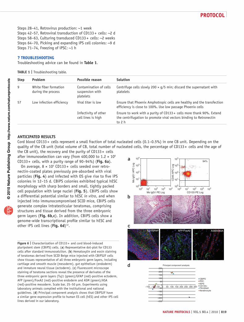
antIcIpateD resultsCord blood CD133 + cells represent a small fraction of total nucleated cells (0.1–0.5%) in one CB unit. Depending on the quality of the CB unit (total volume of CB, total number of nucleated cells, the percentage of CD133 + cells and the age of the CB unit), the recovery and the purity of CD133 + cells after immunoselection can vary (from 400,000 to 1.2 × 10⁶ CD133 + cells, with a purity range of 90–94%) (Fig. 6a).
On average, 8 × 10⁴ CD133 + cells seeded over retro-nectin-coated plates previously pre-absorbed with viral particles (Fig. 4) and infected with OS give rise to five iPS colonies in 12–15 d. CBiPS colonies exhibited typical hESC morphology with sharp borders and small, tightly packed cell population with large nuclei (Fig. 5). CBiPS cells show a differential potential similar to hESC in vitro, and when injected into immunocompromised SCID mice, CBiPS cells generate complex intratesticular teratomas, comprising structures and tissue derived from the three embryonic germ layers (Fig. 6b,c). In addition, CBiPS cells show a genome-wide transcriptional profile similar to hESC and other iPS cell lines (Fig. 6d)12.
taBle 1 | Troubleshooting table.
step problem possible reason solution
9 White fiber formation during the process
Contamination of cells suspension with platelets
Centrifuge cells slowly 200 × g/5 min; discard the supernatant with platelets
57 Low infection efficiency Viral titer is low Ensure that Phoenix Amphotropic cells are healthy and the transfection efficiency is close to 100%. Use low passage Phoenix cells
Infectivity of other cell lines is high
Ensure to work with a purity of CD133 + cells more thank 90%. Extend the centrifugation to promote viral vectors binding to Retronectin to 2 h
250
Principal component analysis
Tuj1/GFAP AFP/FoxA2 ASMA/ASA
–250
200
–200
150
–150 –100 –50
–150
100
250 300 350
CBiPSESCKiPSCD133+FibroblastKeratinocyte
200150100
–100
50
500–50
0
104a
b
c
d
103
Ms
lgG
1 A
PC
Log
CD
45 A
PC
Log
102
101
100
104
103
102
101
100
100 101 102
Ms lgG1 PE Log CD133 PE Log
94%
103 104 100 101 102 103 104
Figure 6 | Characterization of CD133 + and cord blood-induced pluripotent stem (CBiPS) cells. (a) Representative dot-plot for CD133 cells after standard immunoselction. (b) Hematoxylin and eosin staining of teratomas derived from SCID Beige mice injected with CBiPS2F cells show tissues representative of all three embryonic germ layers, including cartilage and smooth muscle (mesoderm), gut epithelium (endoderm) and immature neural tissue (ectoderm). (c) Fluorescent microscope staining of teratoma sections reveal the presence of derivates of the three embryonic germ layers (Tuj1 (green)/GFAP (red)-positive ectoderm, AFP (green)/FoxA2 (red)-positive endoderm and ASM (green)/ASA (red)-positive mesoderm. Scale bar, 25–50 µm. Experiments using laboratory animals complied with the institutional and national guidelines. (d) Principal component analysis shows that CBiPS2F have a similar gene expression profile to human ES cell (hES) and other iPS cell lines derived in our laboratory.

p
uor
G g
n ih si l
bu
P eru ta
N 010 2©
nat
ure
pro
toco
ls/
moc. e r
ut an .
ww
w / /:pt t
h
protocol
820 | VOL.5 NO.4 | 2010 | nature protocols
acknowleDGMents We thank Dr. M. Torrabadella, Director of the Banc de Sang i Teixits, Vall d’Hebron for providing cord blood units. We are grateful to Y. Muñoz Santos for expert assistance with cell culture techniques. N.M. was partially supported by the Juan de la Cierva program. This work was partially supported by grants from MICINN, CIBER, the Fondo de Investigaciones Sanitarias (RETIC-RD06/0010/0016), TERCEL, the G. Harold and Leila Y. Mathers Charitable Foundation and Fundación Cellex.
autHor contrIButIons A.G.: protocol design, isolation of CB CD133 + cells, generation of iPS cell lines and preparation of manuscript; N.M.: protocol design, isolation of CB CD133 + cells, generation of iPS cell lines and preparation of manuscript; I.R.-P.: characterization of iPS cell lines; C.A.: characterization, selection and provision of CB units; A.V.: preparation of manuscript; and J.C.I.B.: preparation of manuscript.
Published online at http://www.natureprotocols.com/. Reprints and permissions information is available online at http://npg.nature.com/ reprintsandpermissions/.
1. Takahashi, K. et al. Induction of pluripotent stem cells from adult human fibroblasts by defined factors. Cell 131, 861–872 (2007).
2. Aasen, T. et al. Efficient and rapid generation of induced pluripotent stem cells from human keratinocytes. Nat. Biotechnol. 26, 1276–1284 (2008).
3. Kim, J.B. et al. Direct reprogramming of human neural stem cells by OCT4. Nature 461, 649–643 (2009).
4. Loh, Y.H. et al. Generation of induced pluripotent stem cells from human blood. Blood 113, 5476–5479 (2009).
5. Ye, Z. et al. Human induced pluripotent stem cells from blood cells of healthy donors and patients with acquired blood disorders. Blood 24, 5473–5480 (2009).
6. Haase, A. et al. Generation of induced pluripotent stem cells from human cord blood. Cell Stem Cell 5, 434–441 (2009).
7. Eminli, S., Utikal, J., Arnold, K., Jaenisch, R. & Hochedlinger, K. Reprogramming of neural progenitor cells into induced pluripotent stem cells in the absence of exogenous Sox2 expression. Stem Cells 26, 2467–2474 (2008).
8. Kim, J.B. et al. Pluripotent stem cells induced from adult neural stem cells by reprogramming with two factors. Nature 454, 646–650 (2008).
9. Anderlini, P. Effects and safety of granulocyte colony-stimulating factor in healthy volunteers. Curr. Opin. Hematol. 16, 35–40 (2009).
10. Gluckman, E. History of cord blood transplantation. Bone Marrow Transplant. 44, 621–626 (2009).
11. Gluckman, E. & Rocha, V. Cord blood transplantation: state of the art. Haematologica 94, 451–454 (2009).
12. Giorgetti, A. et al. Generation of induced pluripotent stem cells from human cord blood using OCT4 and SOX2. Cell Stem Cell 5, 353–357 (2009).