Hypoxia induces CD133 expression in human lung cancer cells by up-regulation of OCT3/4 and SOX2

Developmental Cell
Article
Sox2 in the Dermal Papilla Niche ControlsHair Growth by Fine-Tuning BMP Signalingin Differentiating Hair Shaft ProgenitorsCarlos Clavel,1,2 Laura Grisanti,1,2 Roland Zemla,1,2 Amelie Rezza,1,2 Rita Barros,6 Rachel Sennett,1,2
Amin RezaMazloom,4 Chi-Yeh Chung,1,2 Xiaoqiang Cai,2,3 Chen-Leng Cai,2,3 Larysa Pevny,7 Silvia Nicolis,8 Avi Ma’ayan,4
and Michael Rendl1,2,5,*1Black Family Stem Cell Institute2Department of Developmental and Regenerative Biology3Center for Molecular Cardiology of the Child Health and Development Institute4Department of Pharmacology and Systems Therapeutics5Department of DermatologyMount Sinai School of Medicine, New York, NY 10029, USA6IPATIMUP, University of Porto, 4200-465 Porto, Portugal7Department of Genetics, University of North Carolina, Chapel Hill, NC 27599-7264, USA8Department of Biotechnology and Biosciences, University of Milano-Bicocca, 20126 Milano, Italy*Correspondence: [email protected]
http://dx.doi.org/10.1016/j.devcel.2012.10.013
SUMMARY
How dermal papilla (DP) niche cells regulate hairfollicle progenitors to control hair growth remainsunclear. Using Tbx18Cre to target embryonic DPprecursors, we ablate the transcription factor Sox2early and efficiently, resulting in diminished hair shaftoutgrowth. We find that DP niche expression of Sox2controls the migration speed of differentiating hairshaft progenitors. Transcriptional profiling of Sox2null DPs reveals increased Bmp6 and decreasedBMP inhibitor Sostdc1, a direct Sox2 transcriptionaltarget. Subsequently, we identify upregulated BMPsignaling in knockout hair shaft progenitors anddemonstrate that Bmp6 inhibits cell migration, aneffect that can be attenuated by Sostdc1. A shorterand Sox2-negative hair type lacks Sostdc1 in theDP and shows reduced migration and increasedBMP activity of hair shaft progenitors. Collec-tively, our data identify Sox2 as a key regulator ofhair growth that controls progenitor migration byfine-tuning BMP-mediated mesenchymal-epithelialcrosstalk.
INTRODUCTION
Regulation of stem cell/progenitor functions is essential for
tissue formation, growth, and maintenance throughout life.
In regenerative tissues in particular, the rates of growth and
replenishment through controlling stem cell/progenitor self-
renewal and differentiation are critical for tissue homeostasis
and adaptation to various environmental conditions, but the
mechanisms underlying this important function are poorly under-
stood. In regenerative tissues such as bonemarrow, intestine, or
hair follicles in the skin, signals from the microenvironment,
Developme
called the stem cell niche, instruct stem and progenitor cells in
their function (Moore and Lemischka, 2006; Walker et al., 2009).
In the hair follicle, a dynamic relationship between dermal
papilla (DP) niche cells and epithelial stem cells/progenitors
has been recognized for many years (Driskell et al., 2011; Hardy,
1992; Millar, 2002; Schneider et al., 2009; Sennett and Rendl,
2012; Yang and Cotsarelis, 2010). Mesenchymal-epithelial
signals exchanged between dermal condensates (DP precursor
cells) and stem cells in nascent hair placodes coordinate embry-
onic hair follicle formation (Hardy, 1992; Millar, 2002). Stem cells
are set aside in the upper part of the down-growing follicle
(Nowak et al., 2008), while matrix cell progenitors, direct stem
cell progeny at the base of follicle bulbs, engulf DP precursor
cells to form the DP of mature hair follicles (Millar, 2002;
Schneider et al., 2009). During postnatal hair growth, DP cells
then act as a signaling center to direct the surrounding matrix
cells to proliferate, migrate upward, and differentiate into the
multiple progenitor cell lineages of the outgrowing hair shaft
and its guiding inner root sheath channel (Schneider et al.,
2009; Sennett and Rendl, 2012). This morphogenetic sequence
is repeated in three separate waves during mouse embryonic
hair follicle development, giving rise to guard, awl/auchene,
and zigzag hair follicle types (Figure 1A) (Schlake, 2007). First
wave guard hairs are the longest hairs without kinks and bends,
and are considered to have tactile function, much like whiskers.
Second wave awl hairs are shorter than guard hairs, and
auchene hairs look much like awl hairs, but with a unique identi-
fying bend. Zigzag hairs typically have two kinks in the hair shaft.
After active hair growth, follicles undergo cycles of destruction,
rest, and regrowth to form a new hair follicle in a process of DP
niche/stem cell communication that is reminiscent of embryonic
follicle morphogenesis (Blanpain and Fuchs, 2009; Hsu and
Fuchs, 2012; Sennett and Rendl, 2012).
During embryonic and adult follicle formation, the activation of
signaling pathways such as WNT, tumor growth factor b (TGFb),
and fibroblast growth factor (FGF), and the inhibition of BMP
signaling in follicle stem cells are essential (Hsu and Fuchs,
ntal Cell 23, 981–994, November 13, 2012 ª2012 Elsevier Inc. 981

Figure 1. Sox2 Ablation in DP Precursors Does Not Affect Hair Follicle Formation(A) Schematic of the three major hair follicle formation waves during embryonic morphogenesis.
(B) Sox2 expression analysis (green) in postnatal day P5 skin of Sox2GFP knockin reporter mice. LAMB1 stained the basement membrane (red). Nuclei are
highlighted with DAPI (blue). Asterisk marks autofluorescence of hair shafts. Note GFP is expressed in DPs of first wave guard (1) and second wave awl/auchene
(2) hair follicles. DPs of third wave zigzag follicles (3) do not express Sox2.
(C) High-magnification examples of hair follicle types with Sox2GFP positive and negative DPs.
(D) Quantification of Sox2GFP positive DPs in P5 follicles of all three waves. n = 130 hairs per mouse. Data are mean ± SD from 3 mice.
(E) Targeting of DP precursor cells with Tbx18Cre. Top: Schematic of Tbx18Cre crossed with R26RLacZ reporter line. Bottom: Whole-mount X-Gal stained
Tbx18Cre/R26RLacZ embryo showed robust Cre activity in a hair follicle distribution at E14.5. Histological analyses of sectioned embryowith Cre reporter activity in
DP precursors.
(F) Efficient ablation of SOX2 protein in Tbx18Cre/Sox2fl/fl conditional knockout (cKO) embryo. SOX2 immunofluorescencewas absent in cKOdermal condensates
(arrows) at E14.5. Syndecan-1 (SDC1) labeled dermal condensates and LAMB1 marked the basement membrane.
(G) Immunofluorescence staining for SOX2 at P5. SOX2 is absent in null DPs (arrow) in a Sox2GFP reporter background.
(H) Hematoxylin/eosin staining of Sox2 heterozygous (HET; Tbx18Cre/Sox2GFP/+) and conditional knockout (cKO; Tbx18Cre/Sox2GFP/fl) skins at P5.
(I) Quantification of total hair follicles and of follicles from the three waves at P5. n = 75 hairs per mouse. Data are mean ± SD from 2 mice.
Scale bars, 25 mm (A–G) and 250 mm (H). See also Figures S1 and S2.
Developmental Cell
Sox2 in the DP Niche Regulates Hair Growth
2012; Sennett and Rendl, 2012; Lee and Tumbar, 2012). Simi-
larly, during active hair growth, WNT, FGF, Notch, and BMP
signaling in matrix progenitor cells is important for successful
differentiation into the outgrowing hair shaft (Andl et al., 2004;
DasGupta and Fuchs, 1999; Kobielak et al., 2003; Kulessa
982 Developmental Cell 23, 981–994, November 13, 2012 ª2012 Els
et al., 2000; Lee et al., 2007). While the essential roles of these
signaling pathways have been extensively studied in epithelial
stem cells/progenitors, direct genetic testing of DP niche signals
has been lacking until very recently (Enshell-Seijffers et al., 2010)
due to the long-standing absence of gene ablation tools for the
evier Inc.

Developmental Cell
Sox2 in the DP Niche Regulates Hair Growth
DP. Similarly, the underlying transcriptional control of DP niche
signals and of the specialized niche cell fate that distinguishes
the DP from regular dermal fibroblasts is currently largely
unknown.
The transcription factor Sox2 is a crucial cell fate determinant
in stem cells/progenitors in multiple developmental contexts. It
regulates cell fate decisions in retinal progenitor cells (Taranova
et al., 2006) and neural (Pevny and Nicolis, 2010) and embryonic
stem cells (Boyer et al., 2005), and it is a key factor in pluripo-
tency reprogramming (Takahashi and Yamanaka, 2006).
Recently, Sox2 was shown to play a role in maintaining adult
stem cells in several organ systems (Arnold et al., 2011). In
skin, Sox2 is not expressed in hair follicle stem cells, but is one
of the highest expressed transcription factors in the DP, first
identified in a screen of DP signature genes in growing hair folli-
cles (Rendl et al., 2005). Subsequently, Sox2 expression was
confirmed in embryonic DP precursors and postnatal DPs of
growing follicles (Biernaskie et al., 2009; Driskell et al., 2009;
Tsai et al., 2010), and considered absent during the hair cycle
(Biernaskie et al., 2009). The physiological function of Sox2
during follicle formation and growth is currently unknown.
In this study, we directly test the role of Sox2 in controlling DP
function during follicle formation and growth by ablating Sox2 in
the DP during embryonic hair follicle formation. We utilize
Tbx18Cre as a genetic driver that targets embryonic DP pre-
cursor cells at the earliest stage of hair follicle formation (Grisanti
et al., 2012). In the absence of Sox2, we find a strong reduction
of postnatal hair outgrowth of the three Sox2-positive hair follicle
types and identify slowed migration of differentiating hair shaft
progenitors. We then link reduced migration and slowed hair
shaft production to altered expression of DP-derived BMP regu-
lators Bmp6 and Sostdc1 and to precocious, increased BMP
signaling activity in hair shaft progenitors. Fittingly, a fourth
Sox2-negative hair follicle type naturally displays slower growth,
reduced migration, and increased BMP activity in hair shaft
progenitors. These data reveal an essential role of Sox2 in the
transcriptional control of the mesenchymal niche to orchestrate
hair growth in its interaction with epithelial progenitors.
RESULTS
Sox2 Ablation in Embryonic DP PrecursorsDoes Not Affect Hair Follicle FormationTo determine Sox2 expression throughout hair follicle develop-
ment, we first carefully mapped the Sox2 expression pattern
with Sox2GFP knockin reporter mice that express GFP under
the control of the endogenous Sox2 promoter (Ferri et al.,
2004). At embryonic day E14.5, GFP was strongly expressed in
DP precursor cells of developing guard hair follicles during the
first wave of hair follicle induction (arrow in Figure S1A available
online). At E16.5, DP precursors of second wave follicles of the
awl/auchene hair type also expressed GFP (arrowhead). DP
precursor cells of third wave zigzag follicles at E18.5, however,
did not express Sox2 (asterisk), confirming a previous report
where zigzag DPs lacked Sox2 expression at E18.5 with a
Sox2 transgenic reporter (Driskell et al., 2009). Quantification of
GFP+ DPs confirmed labeling of nearly 100% first and second
wave DPs, while all third wave zigzag DPs lacked Sox2 expres-
sion activity (Figures S1B and S1C).
Developme
This hair type-specific distribution of Sox2 continued during
postnatal hair growth (Figures 1B–D). At P5, first wave guard
hairs are the longest follicles and were identified by distinctly
large DP compartments, while third wave zigzag follicles were
clearly recognized as the shortest hair follicle population with
small DPs (Figures 1B and 1C). Second wave awl/auchene folli-
cles are the second-longest follicles with thin, long DPs (Figures
1B and 1C). Similar to embryonic stages, all DPs of first wave
guard and second wave awl/auchene hair follicles were GFP+,
while zigzag DPs remained negative (Figure 1D) (Driskell et al.,
2009). During the subsequent destruction (catagen) and resting
(telogen) phase of the hair cycle, GFP continued to be expressed
in DPs (Figure S1D) that were identified as Lef1-RFP cell clusters
(Greco et al., 2009; Rendl et al., 2005), although Sox2 was previ-
ously reported absent in the DP during the hair cycle (Biernaskie
et al., 2009). To confirm Sox2 expression in adult DPs, we iden-
tified SOX2 protein in RFP+DPs by immunofluorescence staining
using a SOX2-specific antibody (Figure S1E). Taken together,
these data demonstrate Sox2 expression in DP precursors and
mature DPs in most hair types during morphogenesis and the
hair cycle.
To determine whether Sox2 in the DP niche plays an important
role in hair follicle formation and growth, we next sought to ablate
Sox2 in DP cells as early as possible during development. In an
effort to establish gene targeting of DP precursor cells, we
recently identified in a screen of candidate DP reporters Tbx18
knockin lines (Figures 1E and S1F-S1I) with robust expression
in these cells (Grisanti et al., 2012). X-gal staining in whole-mount
Tbx18LacZ embryos showed Tbx18 expression in skin in a hair
follicle pattern (Figure S1G) and in sections in dermal conden-
sates of DP precursor cells (Figure S1H). No expression was
found in skin prior to E14.5 (data not shown). In other body areas
outside of skin, Tbx18 expression was detected in somites,
limbs, whiskers, meninges, and epicardium (Figure S1I), as
previously described (Cai et al., 2008; Kraus et al., 2001). Next,
in crosses of Tbx18Cre (Cai et al., 2008) with R26RLacZ reporter
mice (Soriano, 1999), we detected Cre activity in developing
hair follicles at E14.5 (Figure 1E, left). Histological analyses of
embryo sections revealed Cre reporter activity in DP precursor
cells already at E14.5 during first wave follicle induction (Fig-
ure 1E, right), suggesting that Tbx18Cre could be used efficiently
to ablate Sox2 in floxed mice. Tbx18Cre is also active in second
and third wave DP precursors at E16.5 and E18.5 (data not
shown). At E16.5 and later developmental stages, Tbx18 expres-
sion and Cre activity also become more widespread throughout
the dermis (data not shown). A detailed characterization of Tbx18
reporter and Cre activities at all developmental stages was
reported recently (Grisanti et al., 2012).
To conditionally knock out Sox2 in DP precursor cells we next
crossed Tbx18Cre with Sox2fl/fl lines, in which the Sox2 coding
sequence is flanked by LoxP sites for Cre-mediated recombina-
tion and gene ablation (Favaro et al., 2009). In addition, we
generated a Sox2 conditional knockout variation, in which one
Sox2 floxed allele was replaced by the Sox2GFP knockin allele,
which allowed for detection of Sox2 promoter activity even after
Sox2 ablation. SOX2 immunofluorescence staining demon-
strated efficient SOX2 ablation in dermal condensates of first
and second wave hair follicles at E14.5 and E16.5 (Figures 1F
and S2A). Costaining for dermal condensate marker SDC1
ntal Cell 23, 981–994, November 13, 2012 ª2012 Elsevier Inc. 983

Developmental Cell
Sox2 in the DP Niche Regulates Hair Growth
(Syndecan-1) (Richardson et al., 2009) confirmed positive
identification of DP precursor cells in the cKO, suggesting that
dermal condensates formed despite efficient Sox2 ablation.
Alkaline phosphatase staining at E18.5 identified DPs in hair
follicles from all three waves (Figure S2B) and hematoxylin/eosin
staining at P0 showed follicles with normal size and distribution
in cKO skins (Figure S2C). Quantification of total hair follicles
(Figure S2D) and hair follicle types per wave (Figure S2E)
confirmed normal formation in embryonic skins with Sox2 cKO
DPs. Similarly, follicle maturation after birth progressed normally
without Sox2. Immunofluorescence staining confirmed absence
of SOX2 protein in postnatal cKO guard hair follicles, which
continued to express GFP from the Sox2GFP reporter allele (Fig-
ure 1G). Follicles from all three waves developedwithout obvious
morphological differences between HET and cKO skins at P5
(Figure 1H). Quantification of hair follicles and of follicle types
per wave was virtually identical in HET and cKO (Figure 1I).
Progression through the adult hair cycle was unaffected as
well. All hair cycle stages, as determined by establishedmorpho-
logical criteria (Muller-Rover et al., 2001), were observed with
similar timing and unaltered follicle morphologies in cKO skins
(Figure S2F). Total hair follicle counts were unchanged (Fig-
ure S2G) and quantification of follicles in catagen, telogen and
anagen stages did not show any changes in cKO skins (Fig-
ure S2H). From these data we conclude that although Sox2 is
efficiently ablated in early dermal condensates, its absence
does not affect initial follicle formation and hair cycle progres-
sion, suggesting that this transcription factor is not required for
these processes.
Sox2 Ablation in the DP Impairs Hair ShaftOutgrowth after BirthWhile follicle morphogenesis and cycling was not affected by
ablation of Sox2 in the DP, we noticed markedly delayed hair
shaft outgrowth. External hair shafts of first wave guard hair folli-
cles normally break through the epidermis and become exter-
nally visible around postnatal day P4–P5. By P8, guard hair
shafts of heterozygous (HET) control pups had grown out
substantially to start forming the hair coat, but in cKO skins
they were strongly growth diminished (Figure 2A, magnification
of insert in Figure 2B). Precise measurement and quantification
of guard hair shaft lengths at P8 and later stages showed a signif-
icant reduction at all time points (Figure 2C). Importantly, the
difference in hair shaft lengths increased over time, but the
relative length of shortened hairs in cKOs compared to HETs re-
mained constant at 65%–70% (Figure 2D). This indicates that the
diminished hair outgrowth in Sox2 cKO guard hair follicles
proceeds at a constantly reduced speed. It further suggests
that it is not the result of a developmentally delayed hair growth
start that would then progress at a normal speed.
Next we tested whether Sox2 expressing second wave awl/
auchene hair shafts were also shorter in cKO pups, and whether
third wave zigzags that do not express Sox2were unaffected, as
predicted based on the differential Sox2 expression in the DP
compartments of these hair types. For this, we shaved off hair
shafts at P20, classified the hair types based on established
length, shape, and medulla size criteria (Schlake, 2007) and
measured their lengths. As shown in Figure 2E, guard and awl/
auchene hairs were considerably shorter inSox2 cKO, but zigzag
984 Developmental Cell 23, 981–994, November 13, 2012 ª2012 Els
lengths were comparable to HET control. Quantification of
shaft lengths confirmed a significant reduction of 20%–55%
(Figure 2F).
To understand the mechanistic underpinnings of the hair shaft
outgrowth reduction, we next determined whether proliferation
of matrix progenitor cells is affected by Sox2 ablation in the
mesenchymal DP compartment. Immunofluorescence stainings
for Ki67, which marks all proliferating cells, did not show any
significant differences between HET and cKO follicles (Fig-
ure 2G). In both, 100% of matrix progenitor cells were Ki67 posi-
tive below the Auber’s line (Figure 2G), which divides the matrix
into a proximal and distal half at the mid-DP level (Vanscott et al.,
1963). Similarly, staining for phospho-Histone H3 (PH3) to iden-
tify cells in mitosis, a short and infrequently detected event
during the cell cycle, was also comparable in HET and cKO
hair bulbs (Figure 2H). To determine whether increased cell
death could possibly cause a loss of matrix cells, we stained
for active caspase-3 that labeled cells undergoing apoptosis.
No significant cell death was detected in cKO follicles and HET
controls (Figure S3A). These data suggest that ablation of Sox2
in DPs does not alter the proliferation rate or cause increased
cell death of surrounding matrix cells at the follicle base.
Next we determined whether aberrant differentiation into the
cell lineages of the hair shaft and inner root sheath (IRS) could
contribute to reduced hair growth in Sox2 ablated follicles.
Immunofluorescence stainings for biochemical markers in P5
HET and cKO guard hair follicles revealed normal Keratin-14
(K14) expression in outer root sheath and matrix progenitor cells
(Figure 2I). Hair shaft cortex and medulla differentiation markers
AE13 and AE15, which label hair keratins and keratohyalin,
respectively, were also unchanged (Figures 2J and 2K). AE15
was further detected in the IRS as expected. Additional nuclear
staining of GATA3 in the companion layer of the IRS was present
in a normal pattern as well (Figure 2L). Similar unchanged stain-
ing for differentiation marker AE13 was already detectable at P0
(Figure S3B). Finally, to evaluate whether Sox2 ablation impacts
normal DPmaturation in developed follicles we analyzed expres-
sion of known DP markers AP, HHIP, and WIF1 (Rendl et al.,
2005), all of which appeared to be present (Figure S3C). Taken
together, these data indicate that Sox2 in the DP is critical for
the normal speed of hair shaft outgrowth in guard, awl, and
auchene follicles by a mechanism other than perturbed prolifer-
ation or differentiation of matrix progenitor cells.
Sox2 Ablation in DP Leads to Delayed ProgenitorMigration into the Differentiating Hair ShaftCompartmentSince neither matrix cell proliferation nor hair shaft differentiation
was affected in cKO hair follicles, we next analyzed whether
altered cell migration from the matrix compartment into the
differentiating hair shaft could possibly explain the delay of hair
shaft production. To label matrix cells and determine their rate
of transition into the hair shaft area, we performed bromodeox-
yuridine (BrdU) pulse-chase experiments. A single BrdU injection
was administered (pulse), and labeled cells were analyzed
between 2 and 54 hr later in harvested skin tissues (chase) (Fig-
ure 3A). In this assay, highly proliferativematrix cells at the follicle
base incorporate BrdU label during the S-phase of the cell cycle
(Figure 3B, left) and are traced as they migrate into the hair shaft
evier Inc.

Figure 2. Sox2 Ablation in the DP Strongly Impairs Hair Shaft Outgrowth
(A) Side view of dorsal skin with outgrowing hair shafts at P8. Hair shafts are shorter in Sox2 cKO skin.
(B) High-magnification view of HET (blue frame) and cKO (red frame) inserts. First wave guard hair shafts are of considerable length by P8 in HET, but are much
shorter in cKO.
(C) Quantification of guard hair shaft lengths. n = 20 guard hairs per mouse. Data are mean ± SD from three mice. **p < 0.01.
(D) Relative guard hair shaft length reduction remains constant at all time points.
(E) Examples of HET and cKO hair shafts from all four hair types at P20. Note that Sox2 expressing guard, awl, and auchene hairs were shorter in cKO. Sox2-
negative zigzag hairs were unchanged.
(F) Quantification of hair shaft lengths of P20 clipped hairs. n = 20 hairs per hair type. Data are mean ± SD from two mice. **p < 0.01.
(G) Immunofluorescence staining of proliferation marker Ki67. Dotted line marks basement membrane. Right: Quantification of Ki67+ cells below Auber’s line
(white). n = 10 guard hairs per mouse. Data are mean ± SD from two mice.
(H) Immunofluorescence staining of mitosis marker phospho-histone H3 (PH3). Right: Quantification of PH3+ cells in guard hair bulbs. n = 20 guard hairs per
mouse. Data are mean ± SD from 2 mice.
(I–L) Immunofluorescence staining of (I) matrix marker K14 and hair shaft differentiation markers (J) AE13 (hair cortex), (K) AE15 (IRS), and (L) GATA3 (inner
root sheath).
Scale bar is 25 mm. See also Figure S3.
Developmental Cell
Sox2 in the DP Niche Regulates Hair Growth
Developmental Cell 23, 981–994, November 13, 2012 ª2012 Elsevier Inc. 985

Figure 3. Sox2 Ablation Impairs Progenitor Migration during Hair Growth
(A) Pulse/chase BrdU labeling to track proliferating andmigrating matrix cells. Timeline of BrdU injection at P5 (pulse) and chase time points for tissue harvest and
BrdU label analysis (red arrows).
(B) Schematic of initial BrdU uptake (2 hr) and of migration of matrix cells toward the hair shaft differentiation areas (36 hr).
(C) BrdU immunofluorescence staining at 2 hr. Proliferation zone ‘‘P’’ is below Auber’s line (white). Note robust BrdU uptake in both Sox2 HET and cKO follicles.
(D) BrdU/AE13 double immunofluorescence staining at 36 hr. Differentiation zone ‘‘D’’: BrdU+ cells in the differentiating hair cortex labeled by AE13 (arrowheads).
Note decreased BrdU+ matrix cells migrating into zone ‘‘D’’ in cKO. Right: Inserts show higher magnification views of zone ‘‘D’’ in HET (blue frame) and cKO
(red frame). Dotted line marks basement membrane. Nuclei are counterstained with DAPI. Scale bars are 25 mm.
(E and F) Quantification of BrdU+ cells in zones ‘‘P’’ and ‘‘D.’’ Note delayed exit of zone ‘‘P’’ and entry into zone ‘‘D’’ of BrdU+ matrix cells in follicles with Sox2
null DPs.
(G) Migration zone ‘‘M’’ between proliferation zone ‘‘P’’ and differentiation zone ‘‘D.’’ Zone ‘‘M’’ is subdivided into proximal zone ‘‘M1’’ and distal zone ‘‘M2’’ at the
level of the distal DP tip. AE13 staining defines the border to differentiation zone ‘‘D.’’
(H and I) Quantification of BrdU+ cells and total cell numbers (as DAPI+ nuclei) in zones ‘‘P,’’ ‘‘M1,’’ and ‘‘M2’’ at 36 hr. Note increased BrdU+ cells (H) and total cell
numbers (I) in zones ‘‘P’’ and ‘‘M1’’ in follicles with Sox2 null DPs.
All box-and-whisker plots: mid-line, median; mean, plus symbol; box, 25th and 75th percentiles; whiskers, 10th and 90th percentiles. N = 10 guard hairs/time point.
Counts are from three (E and F) and two (H and I) independent experiments. **p < 0.01; *p < 0.05.
Developmental Cell
Sox2 in the DP Niche Regulates Hair Growth
area (Figure 3B, right). Already 2 hr after BrdU injection the
majority of matrix cells of Sox2 HET follicles were labeled in
the proliferating Zone ‘‘P’’ below the Auber’s line (Figure 3C).
986 Developmental Cell 23, 981–994, November 13, 2012 ª2012 Els
Importantly, equally rapid labeling occurred in cKO follicles,
again demonstrating comparable matrix cell proliferation. In
both HET and cKO matrix compartments, BrdU labeling peaked
evier Inc.

Developmental Cell
Sox2 in the DP Niche Regulates Hair Growth
at 4 hr after injection (Figure 3E). After 24 hr, the number of
labeled matrix cells started to decrease in both HET and cKO
follicles, as the cells began to move upward toward the hair shaft
(Figure 3E). By 36 hr, depletion of BrdU+ cells in Zone ‘‘P’’
occurred more rapidly in HET follicles (Figures 3D and 3E),
suggesting that matrix cell exit from this area is significantly de-
layed in cKO follicles (Figure 3E). Conversely, at 36 hr, multiple
BrdU+ cells in HET follicles had already entered the differentia-
tion zone ‘‘D,’’ identified as the AE13 labeled cortex of the hair
shaft (Figure 3D, magnification of insert on the right; and Fig-
ure 3F). Only sporadic BrdU/AE13 double-positive cells were
found in cKO follicles. This significant migration delay in cKO
follicles remained detectable up to 48 hr (Figure 3F).
We next had a closer look at the intermediary zone that
connects the proliferating and differentiating zones ‘‘P’’ and
‘‘D’’ (Figure 3G). This migration Zone ‘‘M’’ consists of matrix cells
that transition from the proliferating to the differentiating areas.
Analysis of cKO guard hair follicles revealed increased numbers
of BrdU+ cells in the proximal half between the Auber’s line and
the level of the distal DP tip (Zone ‘‘M1’’)(Figure 3H), but
unchanged BrdU labeled cell numbers in the distal Zone ‘‘M2,’’
compared to control HET follicles. This suggests a backlog of
migrating cells that reaches up to the distal end of the DP.
Indeed, careful analysis of total cell numbers revealed increased
cellularity in zones ‘‘P’’ and ‘‘M1,’’ but not in ‘‘M2’’ (Figure 3I). The
accumulated cells do not appear to reach a level that would
distort the follicle architecture. Analysis of apoptosis by staining
for activated caspase-3 did not show increased cell death
(Figure S3A).
From these data we conclude that the upward movement of
matrix progenitor cells is significantly impaired as they differen-
tiate into the hair shaft cell lineages. This suggests that slowed
hair shaft outgrowth in follicles with Sox2 ablated DPs is caused
by the delayed migration of differentiating matrix cells into the
hair shaft compartment.
Sox2 Ablation Alters DP Signature Gene ExpressionTo gain insights into how ablation of the transcription factor Sox2
in the mesenchymal DP compartment may affect cell migration
and hair growth in the epithelial compartment, we isolated
Sox2 knockout DP cells and determined genome-wide the
potential gene expression changes. For this, hair follicles from
Tbx18Cre/Sox2GFP heterozygous and Tbx18Cre/Sox2GFP/fl condi-
tional knockout P5 skins were freshly isolated and single cells
were stained for integrin-alpha 9 (ITGA9), a marker expressed
by DP cells (Figure 4A) (Rendl et al., 2005; Tsai et al., 2010).
Pure DP cells were then isolated by fluorescence-activated cell
sorting (FACS) as GFP+/ITGA9+ cells, which prevented contam-
ination with GFP-only touch dome cells and ITGA9-only dermal
cells (Figure 4B; and data not shown). To verify high enrichment
of DP cells, we also isolated negative cells as control population
and compared by real-time PCR the expression of several
known DP signature genes (Figure 4C). As expected, signature
gene expression was > 30-fold enriched in GFP+/ITGA9+ cells.
We then proceeded with transcriptional profiling of two inde-
pendently sorted biological replicates using Affymetrix Gene-
Chip Mouse Gene 1.0 ST microarrays. Comparative analysis
between HET and cKO samples and hierarchical clustering
revealed significant >1.25-fold down- and upregulation of 840
Developme
and 223 genes, respectively (Figure 4D).We next grouped down-
regulated and upregulated gene lists into functional categories
based on gene ontology classifications and determined the over-
all enrichment of categories compared to their representation in
the genome. Significantly enriched functional categories of
downregulated genes included protein transport, cell migration,
WNT signaling, mesenchymal cell development, and extracel-
lular matrix (Figure 4E, top). Significantly enriched categories of
upregulated genes included transcriptional regulation, cytoskel-
eton, repressor, and growth (Figure 4E, bottom).
Real-time PCR further validated expression of several down-
regulated and upregulated genes in Sox2 cKO DP cells (Fig-
ure 4F). Absence of any detectable Sox2mRNA levels confirmed
100% gene ablation efficiency. Several regulated genes were
associated with signaling pathways that have been previously
implicated in hair follicle formation and growth, such as Notch1
and Hey1 (Notch signaling), Pbx1, Prdm1/Blimp1, and Sox18
(Figure 4F). Interestingly, among the most strongly regulated
genes were WNT and BMP pathway regulators. Both signaling
pathways are activated and essential during hair shaft differenti-
ation (Andl et al., 2004; DasGupta and Fuchs, 1999; Kobielak
et al., 2003; Kulessa et al., 2000), and recent evidence suggests
a role in DP cells for follicle formation as well (Kishimoto et al.,
2000; Rendl et al., 2008). In Sox2 null DPs, the WNT inhibitors
Sfrp2 and Wif1 were downregulated and upregulated, respec-
tively (Figures 4E and 4F). Sostdc1, an inhibitor of both BMP
and WNT signaling, was also strongly downregulated, while at
the same time Bmp6 was upregulated (Figures 4E and 4F).
From this we conclude that in the absence of Sox2, the level of
WNT signaling could be increased if the differential regulations
of the inhibitors do not cancel each other out. We further
conclude that BMP signaling should be tilted toward increased
activation within the DP and hair epithelium, as both a BMP
inhibitor is decreased and a BMP ligand is increased.
Expression of BMP/WNT Inhibitor Sostdc1 in DP CellsIs Directly Regulated by Sox2
Because the transition of proliferating matrix cells to the differen-
tiating compartment is delayed in follicles with Sox2 null DPs and
BMP signaling has been previously reported to inhibit keratino-
cyte migration during wound healing (Kaiser et al., 1998), we
focused our attention on the decreased expression of BMP/
WNT inhibitor Sostdc1 in Sox2 ablated DP cells. First, we vali-
dated the microarray and real-time PCR regulation by direct
analysis of Sostdc1 expression in HET and cKO skin tissue by
in situ hybridization. In HET hair follicles, Sostdc1 was exclu-
sively expressed in DP cells confirming the DP-specific expres-
sion of this previously identified signature gene (Rendl et al.,
2005). In contrast, Sostdc1 mRNA message was barely detect-
able in Sox2 cKO DPs (Figure 5A). To test whether Sox2 could
regulate Sostdc1 levels, we freshly isolated DP cells by FACS
and lentivirally overexpressed Sox2 (Figure 5B, left). As shown
in Figure 5B (right), Sox2 dramatically upregulated Sostdc1
expression levels in DP cells, suggesting that this gene is
controlled by Sox2.
Next we determined whether Sox2 regulates Sostdc1 as an
immediate target gene by directly binding to regulatory regions
in the Sostdc1 promoter/enhancers. For this, we first identified
potential target binding sites within ± 20kb of the Sostdc1
ntal Cell 23, 981–994, November 13, 2012 ª2012 Elsevier Inc. 987
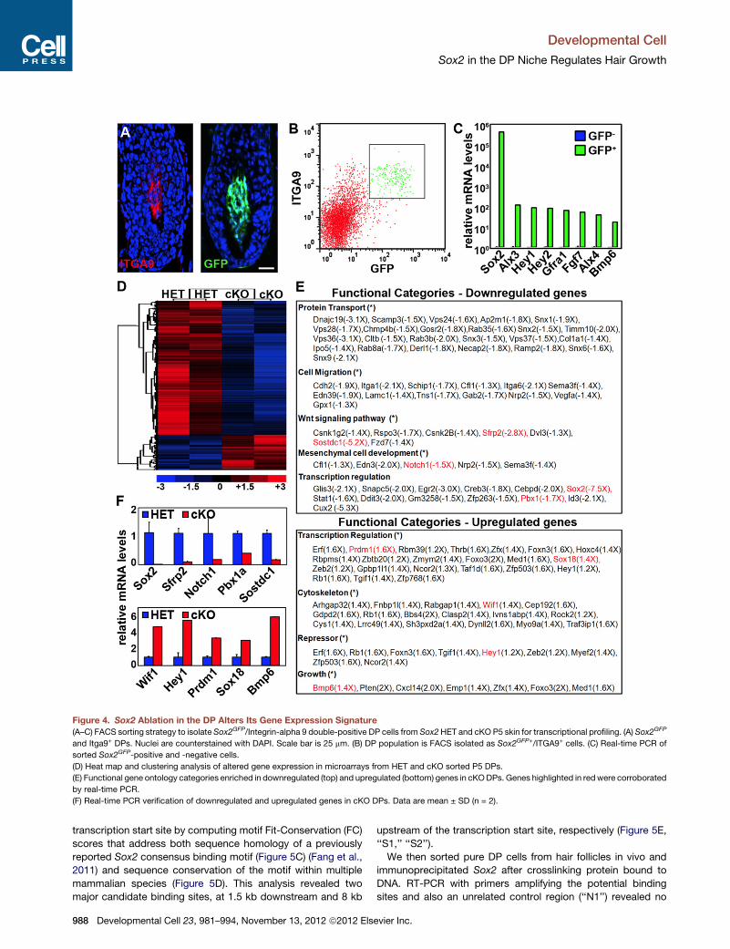
Figure 4. Sox2 Ablation in the DP Alters Its Gene Expression Signature(A–C) FACS sorting strategy to isolate Sox2GFP/Integrin-alpha 9 double-positive DP cells from Sox2HET and cKO P5 skin for transcriptional profiling. (A) Sox2GFP
and Itga9+ DPs. Nuclei are counterstained with DAPI. Scale bar is 25 mm. (B) DP population is FACS isolated as Sox2GFP+/ITGA9+ cells. (C) Real-time PCR of
sorted Sox2GFP-positive and -negative cells.
(D) Heat map and clustering analysis of altered gene expression in microarrays from HET and cKO sorted P5 DPs.
(E) Functional gene ontology categories enriched in downregulated (top) and upregulated (bottom) genes in cKODPs. Genes highlighted in redwere corroborated
by real-time PCR.
(F) Real-time PCR verification of downregulated and upregulated genes in cKO DPs. Data are mean ± SD (n = 2).
Developmental Cell
Sox2 in the DP Niche Regulates Hair Growth
transcription start site by computing motif Fit-Conservation (FC)
scores that address both sequence homology of a previously
reported Sox2 consensus binding motif (Figure 5C) (Fang et al.,
2011) and sequence conservation of the motif within multiple
mammalian species (Figure 5D). This analysis revealed two
major candidate binding sites, at 1.5 kb downstream and 8 kb
988 Developmental Cell 23, 981–994, November 13, 2012 ª2012 Els
upstream of the transcription start site, respectively (Figure 5E,
‘‘S1,’’ ‘‘S2’’).
We then sorted pure DP cells from hair follicles in vivo and
immunoprecipitated Sox2 after crosslinking protein bound to
DNA. RT-PCR with primers amplifying the potential binding
sites and also an unrelated control region (‘‘N1’’) revealed no
evier Inc.

Figure 5. Expression of BMP/WNT Inhibitor
Sostdc1 in DP Cells Is Directly Regulated
by Sox2
(A) In situ hybridization for Sostdc1 mRNA ex-
pression in Sox2 HET and cKO guard hair follicle.
Dotted line marks the basement membrane. Scale
bar is 25 mm.
(B) Real-time PCR for Sox2 (left) and Sostdc1
(right) in isolated DP cells that lentivirally over-
express Sox2. Data are mean ± SD (n = 2).
(C) Predicted SOX2 binding site consensus
sequence.
(D) Heat map of putative SOX2 binding sites in the
Sostdc1 genomic region based on consensus
sequence and species conservation.
(E) Schematic of genomic region displaying two
putative SOX2 binding sites, ‘‘S1’’ and ‘‘S2.’’ ‘‘N1’’
is used as a control site.
(F) ChIP-PCR to detect SOX2 binding to con-
served SOX2 binding sites ‘‘S1,’’ ‘‘S2.’’ ‘‘N1’’ is
a control intronic fragment with no predicted site.
Shown is one of two representative experiments.
(G) ChIP-qPCR analyses to assess relative binding
of SOX2 to ‘‘S1,’’ ‘‘S2’’ sites. Fold enrichments of
immunoprecipitated DNA fragments are com-
pared to control ‘‘N1’’ and presented relative to
IgG. Data are mean ± SD of two independent
experiments.
Developmental Cell
Sox2 in the DP Niche Regulates Hair Growth
bound Sox2 at the S2 and the negative control sites. How-
ever, specific binding of Sox2 was identified at the S1 site
within the first intron of the Sostdc1 gene (Figure 5F). Quantita-
tive real-time PCR of Sox2 binding relative to IgG control
showed a 4003 enrichment at the S1 site (Figure 5G). Taken
together, these chromatin immunoprecipitation and Sostdc1
regulation experiments in freshly isolated DP cells suggest
that Sostdc1 expression is directly regulated by Sox2 and is
therefore strongly decreased in cKO DP cells due to absence
of Sox2.
Precocious and Increased BMP Signaling Activity inDifferentiating Hair Shaft Progenitors in Follicles withSox2 Ablated DPsWe next wondered whether the differential regulation of WNT
inhibitors Sfrp2 and Wif1, and the downregulation of the BMP/
WNT inhibitor Sostdc1 and upregulation of Bmp6 would lead
to increased WNT and BMP signaling within the epithelial
compartment that could cause slowed hair shaft outgrowth.
To determine the level of WNT signaling, we detected by immu-
nofluorescence the nuclear staining of the WNT responsive tran-
Developmental Cell 23, 981–994, N
scription factor b-catenin, which in the
absence of WNT signaling is only detect-
able at adherens junctions at the cell
membrane. In both Sox2 HET and cKO
follicles, nuclear b-catenin was present
in differentiating hair shaft cells at normal
levels (Figure S4A, arrows) (Merrill et al.,
2001), suggesting that WNT signaling
does not directly contribute to altered
migration of differentiating hair shaft line-
ages. It was also detectable in the DP,
but with slightly increased intensity in the cKO (Figure S4A,
open arrowheads).
To detect cells with active BMP signaling, we stained for
SMAD1,5, which becomes phosphorylated downstream of
BMP receptor activation and translocates to the nucleus, where
target genes are transcribed (Massague et al., 2005). During
early postnatal hair growth, active nuclear pSMAD1,5 can be
detected in IRS cells, before expanding into differentiating hair
shaft cortex and medulla at more mature stages (Andl et al.,
2004; Kobielak et al., 2003). As judged by immunofluorescence
staining for pSMAD1,5, BMP signaling was active in HET control
follicles at P5 mostly in IRS cells, located outside the AE13-posi-
tive hair cortex (Figures 6A and S4B, arrowheads). By contrast,
widespread pSMAD1,5 levels were already detectable in hair
cortex and medulla cells within and inside the AE13 expression
domain of cKO guard hair follicles (Figures 6A and S4B, open
arrowheads). Quantification revealed�75% pSMAD1,5-positive
cells in the AE13 domain of cKO, while only �15% were
pSMAD1,5/AE13 double-positive in HET follicles (Figure 6B). In
addition, widespread precocious BMP signaling activity was
already visible in the IRS at P0 in �90% of cKO guard hair
ovember 13, 2012 ª2012 Elsevier Inc. 989
Figure 6. Increased BMP Signaling in Differentiating Hair Shaft Lineages in Sox2 Ablated Guard Hair Follicles
(A) Immunofluorescence for pSMAD1,5 in P5 Sox2GFPHET and cKO guard hair follicles. AE13 highlights differentiating hair cortex (green). GFP signal in the DP is
from Sox2GFP. Normal BMP signaling in IRS (arrowheads). BMP signaling in hair cortex progenitors is upregulated in cKO (open arrowheads). Nuclei are
counterstained with DAPI. Right: blue (HET) and red (cKO) inserts without DAPI.
(B) Quantification of pSMAD1,5-positive cells within the AE13 domain of P5 guard hair follicles.
(C) pSMAD1,5 immunofluorescence at P0. Precocious BMP signaling in IRS (arrowheads) and hair cortex (open arrowheads) in cKO follicles. BMP activity is also
increased in upper DP.
(D) Quantification of hair follicles (HF) with pSMAD1,5-positive IRS/hair cortex progenitors in guard hair follicles at P0.
(E) In situ hybridization for Sostdc1 mRNA expression in Sox2 guard and zigzag hair follicle. Dotted line marks the basement membrane.
(F) In vitro scratch migration assay. BMP6 (500 ng/ml) decreases migration of skin epithelial cells, which is alleviated by SOSTDC1 (300 ng/ml). Right: Quanti-
fication of migrated area. Data are mean ± SEM from two independent experiments. **p < 0.01.
(G) Model of fine-tuning BMP signaling in hair shaft progenitors. Sox2 in the DP niche drives the expression of BMP inhibitor Sostdc1, counteracting BMPs
produced by matrix cells at the base and by the DP to fine-tune BMP signaling in differentiating hair shaft progenitors. In follicles with Sox2 null DPs, the
equilibrium is unbalanced leading to increased BMP signaling and decreased migration during hair shaft differentiation.
All box-and-whisker plots: mid-line, median; mean, plus symbol; box, 25th and 75th percentiles; whiskers, 10th and 90th percentiles. N = 10 guard and zigzag hairs.
Counts are from two independent experiments. **p < 0.01. All scale bars are 25 mm.
See also Figure S4.
Developmental Cell
Sox2 in the DP Niche Regulates Hair Growth
follicles (Figure 6C, arrowheads), compared to only few cells in
�38% of HET guard hairs (Figure 6D). Few AE13-positive hair
cortex (Figure 6C, open arrowheads) and DP cells were also
pSMAD1,5-positive in cKO guard hair follicles. This suggested
that over the course of at least 5 days during early postnatal
hair growth, BMP signaling was precociously activated and
increased in differentiating hair shaft cells in Sox2 cKO follicles.
Reduced Hair Growth Speed and Increased BMPSignaling Activity in Sox2-Negative Zigzag Hair FolliclesWe next wondered about the level of hair shaft cell migration
and BMP signaling activation in zigzag hair follicles, the Sox2-
negative (Figure 1C) and shortest hair type (Figure 2E). First,
we determined whether, as predicted, the percentage of BrdU+
990 Developmental Cell 23, 981–994, November 13, 2012 ª2012 Els
cells entering the differentiation zone ‘‘D’’ is similar in HET and
cKO zigzag hair follicles. Unsurprisingly, the number of traced
cells is not reduced in Sox2 cKO zigzag follicles compared to
HET controls, because this follicle type does not express Sox2
(Figure S4C). Because zigzag follicles are much smaller than
guard hairs and our quantification of labeled cells in zones only
measures relative migration speed in comparably sized follicles,
we established an absolute measurement of migration speed by
determining the total distance that cells traveled from the time
after pulse (2 hr, top level of the DP) to the highest location in
the differentiating compartment at 24 hr (Figure S4D). With this
absolute speed measurement, we determined that migration of
hair shaft progenitors in HET and cKO zigzag follicles was
much slower than that in normal guard hairs, and that cell
evier Inc.
Developmental Cell
Sox2 in the DP Niche Regulates Hair Growth
migration speed in cKO guard hairs is reduced and becomes
more similar to the speed of zigzag follicles (Figure S4E). Impor-
tantly, zigzag follicles by default also express high levels of active
pSMAD1,5 (Figures S4F and S4G, open arrowheads), compa-
rable to Sox2 cKO guard hairs (Figures 6B), linking slower
migration and hair growth with increased BMP signaling activity.
Interestingly, DPs in zigzag follicles also lack Sostdc1 expression
(Figure 6E), suggesting a permissive state for increased BMP
activity in differentiating hair shaft lineages.
BMP Signaling Inhibits Cell Migration, WhichCan Be Attenuated by Sostdc1Because BMP signaling has previously been shown to inhibit cell
migration (Kaiser et al., 1998), we finally tested whether Bmp6
and Sostdc1 could directly affect migration of skin epithelial cells
in vitro to help mechanistically explain slowed movement of
matrix cells from the proliferating into the differentiating
compartments in Sox2 cKO follicles. In a well-established
scratch migration assay, proliferation-inhibited cells migrated
toward the center of the scratch within 24 hr, as expected (Fig-
ure 6F). This was significantly reduced in the presence of
BMP6, but could be partially restored by addition of SOSTDC1,
demonstrating that BMP signaling directly affects cell migration.
It also indicates that the level of BMP signaling activity as the
outcome of positive and negative regulators determines the
extent of migration speed and further suggests that the level of
secreted BMP signaling regulators from the DP niche, controlled
by Sox2, fine-tunes migration of hair shaft progenitors during
hair growth.
DISCUSSION
Although robust expression of Sox2 in DP niche cells during hair
follicle formation and growth was first described years ago, the
function of this transcription factor and of many other genes
has been unexplored largely due to the long-standing absence
of genetic tools to target the DP for gene ablation. Very recently,
two Cre recombinase expressing lines were introduced to study
the function of DP genes (Enshell-Seijffers et al., 2010; Lehman
et al., 2009; Woo et al., 2012). Prx1-Cre broadly targets the
dermis several days before the start of hair morphogenesis,
but is limited to the limbs and focal areas in ventral skin (Logan
et al., 2002) where the timing of hair development and the distri-
bution of hair types is not well understood. CorinCre is active
specifically in DP cells throughout the backskin, where hair
development is well defined, but does not fully ablate genes
before postnatal day P7 (Enshell-Seijffers et al., 2010). Here,
we use Tbx18Cre to target embryonic DP precursors in the back-
skin (Grisanti et al., 2012) to interrogate the functional role of the
DP signature geneSox2 during hair follicle formation and growth.
With our work, we identify a striking reduction of postnatal hair
shaft lengths in Sox2 null guard and awl/auchene hair follicles.
Although hair follicles and shafts did form normally, they grew
out at continuously reduced rates due to delayed migration of
differentiating hair shaft progenitors. Surprisingly, matrix pro-
genitor proliferation was not affected, increased cell death did
not occur and differentiation into IRS and hair shaft cell lineages
was normal as well. This is an unusual finding since direct gene
manipulations in the hair follicle epithelium typically result in
Developme
strongly altered proliferation or differentiation phenotypes
(Alonso et al., 2005; Andl et al., 2004; Ezhkova et al., 2011; Kauf-
man et al., 2003; Kobielak et al., 2003; Kulessa et al., 2000). It
also suggests that signals from the mesenchymal DP niche
under the transcriptional control of Sox2 fine-tune the rate of
epithelial hair shaft outgrowth by affecting hair shaft progenitor
migration rather than regulating the global proliferation and
differentiation programs.
Our comprehensive molecular analysis with microarrays and
real-time PCR in freshly isolated knockout DPs revealed multiple
altered genes that could directly or indirectly cause the observed
hair shaft growth phenotype. Among themost strongly regulated
genes were BMP pathway members that stood out because
the BMP pathway has been shown previously to regulate prolif-
eration,migration and differentiation of hair follicle and epidermal
epithelial cells (Andl et al., 2004; Blessing et al., 1993; Kaiser
et al., 1998; Kobielak et al., 2003; Kulessa et al., 2000). In Sox2
null DPs, we found that the BMP ligand Bmp6 was upregulated
and expression of BMP/WNT inhibitor Sostdc1 was strongly
decreased. This suggested a potential shift toward increased
BMP signaling activity in DP, surrounding matrix cells at the
follicle base and/or differentiating hair shaft lineages due to
loss of Sox2 in the DP. Indeed, we found precocious and
increasedBMP signaling in hair shaft progenitors on their journey
toward becoming terminally differentiated hair shaft and IRS.
Interestingly, we did not observe increased BMP signaling in
highly proliferative matrix cells at the follicle base. This suggests
a gradient model where BMP regulators produced by the DP
locally fine-tune BMP signaling activity at the follicle mid-level,
but not at its proliferative base, to control hair shaft progenitor
migration. In addition, we found increased autocrine BMP
signaling in the upper DP itself, which could indirectly contribute
to regulating hair growth speed through additional downstream
BMP targets in the DP.
Much as proliferation within the hair follicle was unchanged,
we found that increased BMP signaling activity in hair shaft
progenitors did not cause any disturbance of normal hair shaft
differentiation. Conversely, severe hair shaft differentiation
phenotypes were observed in studies of BMP signaling inhibition
by receptor ablation (Andl et al., 2004; Kobielak et al., 2003) or
misexpression of the inhibitor Noggin (Kulessa et al., 2000), sug-
gesting that BMP signaling is required for differentiation, but
increased, precocious signaling interferes with migration rather
than differentiation. In our study, reduced cell migration is the
most likely explanation for slowed matrix cell transition into the
differentiating compartment, which is consistent with previous
reports showing that increased BMP signaling activity can cause
reduced cell migration (Ahmed et al., 2011; Kaiser et al., 1998).
Indeed, our tests of direct effects of BMP6 and SOSTDC1 on
primary skin epithelial cell migration revealed inhibition of migra-
tion by BMP6 that could be alleviated by SOSTDC1. This
supports a model (Figure 6G), in which Sox2 in the DP niche
controls the expression of BMP inhibitor SOSTDC1 and BMP6
to fine-tune epithelial BMP signaling activity for proper timing
of hair shaft progenitor cell migration.
Recent global ablation of Sostdc1 in full knockout mice did not
cause reduced postnatal hair growth phenotypes (Narhi et al.,
2012). Because both downregulated Sostdc1 and upregulated
Bmp6 were observed in Sox2 cKO DPs, the simultaneous
ntal Cell 23, 981–994, November 13, 2012 ª2012 Elsevier Inc. 991

Developmental Cell
Sox2 in the DP Niche Regulates Hair Growth
differential regulation of both BMP inhibitor and ligand may be
essential for precisely fine-tuning levels of BMP signaling activity
to affect hair growth. Increased BMP signaling in the DP of Sox2
mutants may also cause additional downstream effects that
could influence the speed of hair growth. In Sostdc1 null
embryos, hair follicle numbers and placode sizes are increased
(Narhi et al., 2012). At that stage, Sostdc1 is expressed in the
epidermis at the edges of placodes and appears to act as
WNT inhibitor to limit the extent of WNT signaling, without any
evidence of inhibiting BMP signaling. In Sox2 null follicles that
lack Sostdc1 during postnatal hair growth, we did not find
increased or expanded nuclear b-catenin, suggesting that
WNT signaling does not directly contribute to altered hair shaft
progenitor migration. Increased nuclear b-catenin in the DP itself
suggests increased WNT signaling in the DP, which could indi-
rectly affect hair growth speed through WNT targets in the DP
(Enshell-Seijffers et al., 2010).
Our observation that shorter Sox2-negative zigzag hair
follicles exhibit molecular and cell migration features similar to
Sox2 cKO guard hairs reveals the physiologic significance of
the connection between Sox2 regulation of BMP ligand/inhibitor
in the DP and BMP pathway activation and migration speed in
hair shaft progenitors. By default, BMP signaling activity is
increased and migration speed is decreased in zigzag hair folli-
cles, much like in guard hair follicles after ablation of Sox2, likely
contributing to slower hair growth in this hair type. However, it
should be noted that these two hair follicle types were at different
developmental stages in our analysis, as zigzag follicles develop
several days after guard hairs. As global BMP signaling progres-
sively increases in postnatal back skin (Andl et al., 2004), it is
possible that additional Sox2-independent factors drive the
observed BMP signaling and migration differences between
guard and zigzag hair follicles. As Sox2 cKO guard hairs grow
slower but otherwise retain all other size, form, and shape
features without becoming more ‘‘zigzag like,’’ Sox2 likely does
not constitute a master transcriptional regulator of overall hair
type fate, but appears to be a bona fide key regulator of hair
growth speed. In summary, our findings demonstrate that
Sox2, as a direct transcriptional regulator in the DP, controls
niche signals that act on surrounding hair shaft progenitors in
the mesenchymal-epithelial crosstalk that orchestrates the
speed of hair follicle growth.
EXPERIMENTAL PROCEDURES
Mice
Sox2fl/fl and Sox2GFP/+ knockin and Lef1-RFP transgenic lines were previously
described (Ellis et al., 2004; Favaro et al., 2009; Rendl et al., 2005). Tbx18LacZ/+
was previously described (Cai et al., 2008). The previously published
Tbx18Cre/+ knockin line (Cai et al., 2008) was modified to remove neomycin
through genetic crossing with a FLP deleter line. R26RLacZ reporter
mice (Soriano, 1999) were obtained from Jackson Laboratories (Bar Harbor,
ME). For 5-bromo-20-deoxyuridine (BrdU) pulse experiments, mice were in-
jected intraperitoneally with 50 mg/g BrdU (Sigma-Aldrich). All animal experi-
ments were performed in accordance with the guidelines and approval of the
Institutional Animal Care and Use Committee at Mount Sinai School of
Medicine.
Histology, Immunofluorescence, and In Situ Hybridization
Histology, immunofluorescence, and in situ hybridization were carried out
as described previously (Rendl et al., 2005; Grisanti et al., 2012). Details of
992 Developmental Cell 23, 981–994, November 13, 2012 ª2012 Els
antibodies and probes are provided in Supplemental Experimental
Procedures.
Chromatin Immunoprecipitation
Chromatin immunoprecipitation was performed according to manufacturer’s
instructions using the EZ-Magna ChIP kit (Millipore) and sorted DP cell
extracts. Cells were fixed in 1% formaldehyde, lysed, and chromatin was soni-
cated to 200 base pair fragments. Chromatin was immunoprecipitated with
SOX2 antibodies (Millipore) or control IgG. RT-PCR and real-time PCR were
performed with the LightCycler system and DNA master SYBR Green I
reagents (Roche). Relative binding was calculated with the 2�DDCT method.
Binding site specific primer sequences are provided in Supplemental Experi-
ment Procedures.
In Vitro Scratch Migration Assay
Primary early passage keratinocytes were cultured in 6-well plates until
reaching confluence, starved in serum-free basal medium for 20 hr, followed
by 10 mg/ml mitomycin C for 2 hr. A straight scratch was made with a pipet
tip, followed by culture with 500 ng/ml BMP6 and/or 300 ng/ml SOSTDC1
(R&D Biosystems). Phase-contrast images of 10 nonoverlapping fields in trip-
licate scratch wounds were taken per time point and relative areas covered by
migrating cells were quantified with ImageJ.
Cell Isolation, Real-Time PCR, and Microarray Analysis
Cell isolation, real-time PCR, and microarray analysis were carried out as
described previously (Rendl et al., 2005). See Supplemental Experimental
Procedures for detailed methods and primer sequences.
Statistics
A two-tailed Student’s t test was used to calculate statistical significance.
SUPPLEMENTAL INFORMATION
Supplemental Information includes four figures and Supplemental Experi-
mental Procedures and can be found with this article online at http://dx.doi.
org/10.1016/j.devcel.2012.10.013.
ACKNOWLEDGMENTS
We thank Valerie Horsley, Hoang Nguyen, Robert Krauss, Phil Soriano, and
Tudorita Tumbar for insightful discussions and valuable comments on the
manuscript and Elena Ezkhova and Jisheng Zhang for support with keratino-
cyte cultures. We also thank the personnel of the Mount Sinai Flow Cytometry
Core Facility and NYUGenomics Core for excellent cell sorting andmicroarray
service. This work was supported by a grant to A.M. from NIH/NIGMS (R01-
GM098316) and to M.R. from NIH/NIAMS (R01-AR059143).
Received: July 25, 2012
Revised: September 25, 2012
Accepted: October 16, 2012
Published online: November 12, 2012
REFERENCES
Ahmed, M.I., Mardaryev, A.N., Lewis, C.J., Sharov, A.A., and Botchkareva,
N.V. (2011). MicroRNA-21 is an important downstream component of BMP
signalling in epidermal keratinocytes. J. Cell Sci. 124, 3399–3404.
Alonso, L., Okada, H., Pasolli, H.A., Wakeham, A., You-Ten, A.I., Mak, T.W.,
and Fuchs, E. (2005). Sgk3 links growth factor signaling to maintenance of
progenitor cells in the hair follicle. J. Cell Biol. 170, 559–570.
Andl, T., Ahn, K., Kairo, A., Chu, E.Y., Wine-Lee, L., Reddy, S.T., Croft, N.J.,
Cebra-Thomas, J.A., Metzger, D., Chambon, P., et al. (2004). Epithelial
Bmpr1a regulates differentiation and proliferation in postnatal hair follicles
and is essential for tooth development. Development 131, 2257–2268.
Arnold, K., Sarkar, A., Yram, M.A., Polo, J.M., Bronson, R., Sengupta, S.,
Seandel, M., Geijsen, N., and Hochedlinger, K. (2011). Sox2(+) adult stem
evier Inc.

Developmental Cell
Sox2 in the DP Niche Regulates Hair Growth
and progenitor cells are important for tissue regeneration and survival of mice.
Cell Stem Cell 9, 317–329.
Biernaskie, J., Paris, M., Morozova, O., Fagan, B.M., Marra, M., Pevny, L., and
Miller, F.D. (2009). SKPs derive from hair follicle precursors and exhibit
properties of adult dermal stem cells. Cell Stem Cell 5, 610–623.
Blanpain, C., and Fuchs, E. (2009). Epidermal homeostasis: a balancing act of
stem cells in the skin. Nat. Rev. Mol. Cell Biol. 10, 207–217.
Blessing, M., Nanney, L.B., King, L.E., Jones, C.M., and Hogan, B.L. (1993).
Transgenic mice as a model to study the role of TGF-beta-related molecules
in hair follicles. Genes Dev. 7, 204–215.
Boyer, L.A., Lee, T.I., Cole, M.F., Johnstone, S.E., Levine, S.S., Zucker, J.P.,
Guenther, M.G., Kumar, R.M., Murray, H.L., Jenner, R.G., et al. (2005). Core
transcriptional regulatory circuitry in human embryonic stem cells. Cell 122,
947–956.
Cai, C.L., Martin, J.C., Sun, Y., Cui, L., Wang, L., Ouyang, K., Yang, L., Bu, L.,
Liang, X., Zhang, X., et al. (2008). A myocardial lineage derives from Tbx18
epicardial cells. Nature 454, 104–108.
DasGupta, R., and Fuchs, E. (1999). Multiple roles for activated LEF/TCF
transcription complexes during hair follicle development and differentiation.
Development 126, 4557–4568.
Driskell, R.R., Giangreco, A., Jensen, K.B., Mulder, K.W., and Watt, F.M.
(2009). Sox2-positive dermal papilla cells specify hair follicle type in mamma-
lian epidermis. Development 136, 2815–2823.
Driskell, R.R., Clavel, C., Rendl, M., and Watt, F.M. (2011). Hair follicle dermal
papilla cells at a glance. J. Cell Sci. 124, 1179–1182.
Ellis, P., Fagan, B.M., Magness, S.T., Hutton, S., Taranova, O., Hayashi, S.,
McMahon, A., Rao, M., and Pevny, L. (2004). SOX2, a persistent marker for
multipotential neural stem cells derived from embryonic stem cells, the embryo
or the adult. Dev. Neurosci. 26, 148–165.
Enshell-Seijffers, D., Lindon, C., Kashiwagi, M., and Morgan, B.A. (2010).
beta-catenin activity in the dermal papilla regulatesmorphogenesis and regen-
eration of hair. Dev. Cell 18, 633–642.
Ezhkova, E., Lien, W.H., Stokes, N., Pasolli, H.A., Silva, J.M., and Fuchs, E.
(2011). EZH1 and EZH2 cogovern histone H3K27 trimethylation and are essen-
tial for hair follicle homeostasis and wound repair. Genes Dev. 25, 485–498.
Fang, X., Yoon, J.G., Li, L., Yu, W., Shao, J., Hua, D., Zheng, S., Hood, L.,
Goodlett, D.R., Foltz, G., and Lin, B. (2011). The SOX2 response program in
glioblastoma multiforme: an integrated ChIP-seq, expression microarray,
and microRNA analysis. BMC Genomics 12, 11.
Favaro, R., Valotta, M., Ferri, A.L., Latorre, E., Mariani, J., Giachino, C., Lancini,
C., Tosetti, V., Ottolenghi, S., Taylor, V., and Nicolis, S.K. (2009). Hippocampal
development and neural stem cell maintenance require Sox2-dependent regu-
lation of Shh. Nat. Neurosci. 12, 1248–1256.
Ferri, A.L., Cavallaro, M., Braida, D., Di Cristofano, A., Canta, A., Vezzani, A.,
Ottolenghi, S., Pandolfi, P.P., Sala, M., DeBiasi, S., and Nicolis, S.K. (2004).
Sox2 deficiency causes neurodegeneration and impaired neurogenesis in
the adult mouse brain. Development 131, 3805–3819.
Greco, V., Chen, T., Rendl, M., Schober, M., Pasolli, H.A., Stokes, N., Dela
Cruz-Racelis, J., and Fuchs, E. (2009). A two-step mechanism for stem cell
activation during hair regeneration. Cell Stem Cell 4, 155–169.
Grisanti, L., Clavel, C., Cai, X., Rezza, A., Tsai, S.Y., Sennett, R., Mumau, M.,
Cai, C.L., and Rendl, M. (2012). Tbx18 targets dermal condensates for
labeling, isolation, and gene ablation during embryonic hair follicle formation.
J Invest Dermatol., in press. Published online September 20, 2012. http://dx.
doi.org/10.1038/jid.2012.329.
Hardy, M.H. (1992). The secret life of the hair follicle. Trends Genet. 8, 55–61.
Hsu, Y.C., and Fuchs, E. (2012). A family business: stem cell progeny join the
niche to regulate homeostasis. Nat. Rev. Mol. Cell Biol. 13, 103–114.
Kaiser, S., Schirmacher, P., Philipp, A., Protschka, M., Moll, I., Nicol, K., and
Blessing, M. (1998). Induction of bone morphogenetic protein-6 in skin
wounds. Delayed reepitheliazation and scar formation in BMP-6 overexpress-
ing transgenic mice. J. Invest. Dermatol. 111, 1145–1152.
Developme
Kaufman, C.K., Zhou, P., Pasolli, H.A., Rendl, M., Bolotin, D., Lim, K.C., Dai, X.,
Alegre, M.L., and Fuchs, E. (2003). GATA-3: an unexpected regulator of cell
lineage determination in skin. Genes Dev. 17, 2108–2122.
Kishimoto, J., Burgeson, R.E., and Morgan, B.A. (2000). Wnt signaling
maintains the hair-inducing activity of the dermal papilla. Genes Dev. 14,
1181–1185.
Kobielak, K., Pasolli, H.A., Alonso, L., Polak, L., and Fuchs, E. (2003). Defining
BMP functions in the hair follicle by conditional ablation of BMP receptor IA.
J. Cell Biol. 163, 609–623.
Kraus, F., Haenig, B., and Kispert, A. (2001). Cloning and expression analysis
of the mouse T-box gene Tbx18. Mech. Dev. 100, 83–86.
Kulessa, H., Turk, G., and Hogan, B.L.M. (2000). Inhibition of Bmp signaling
affects growth and differentiation in the anagen hair follicle. EMBO J. 19,
6664–6674.
Lee, J., Basak, J.M., Demehri, S., and Kopan, R. (2007). Bi-compartmental
communication contributes to the opposite proliferative behavior of
Notch1-deficient hair follicle and epidermal keratinocytes. Development 134,
2795–2806.
Lee, J., and Tumbar, T. (2012). Hairy tale of signaling in hair follicle develop-
ment and cycling. Semin. Cell Dev. Biol, in press. Published online August
21, 2012. http://dx.doi.org/10.1016/j.semcdb.2012.08.003.
Lehman, J.M., Laag, E., Michaud, E.J., and Yoder, B.K. (2009). An essential
role for dermal primary cilia in hair follicle morphogenesis. J. Invest.
Dermatol. 129, 438–448.
Logan, M., Martin, J.F., Nagy, A., Lobe, C., Olson, E.N., and Tabin, C.J. (2002).
Expression of Cre Recombinase in the developing mouse limb bud driven by
a Prxl enhancer. Genesis 33, 77–80.
Massague, J., Seoane, J., and Wotton, D. (2005). Smad transcription factors.
Genes Dev. 19, 2783–2810.
Merrill, B.J., Gat, U., DasGupta, R., and Fuchs, E. (2001). Tcf3 and Lef1
regulate lineage differentiation of multipotent stem cells in skin. Genes Dev.
15, 1688–1705.
Millar, S.E. (2002). Molecular mechanisms regulating hair follicle development.
J. Invest. Dermatol. 118, 216–225.
Moore, K.A., and Lemischka, I.R. (2006). Stem cells and their niches. Science
311, 1880–1885.
Muller-Rover, S., Handjiski, B., van der Veen, C., Eichmuller, S., Foitzik, K.,
McKay, I.A., Stenn, K.S., and Paus, R. (2001). A comprehensive guide for
the accurate classification of murine hair follicles in distinct hair cycle stages.
J. Invest. Dermatol. 117, 3–15.
Narhi, K., Tummers, M., Ahtiainen, L., Itoh, N., Thesleff, I., and Mikkola, M.L.
(2012). Sostdc1 defines the size and number of skin appendage placodes.
Dev. Biol. 364, 149–161.
Nowak, J.A., Polak, L., Pasolli, H.A., and Fuchs, E. (2008). Hair follicle stem
cells are specified and function in early skin morphogenesis. Cell Stem Cell
3, 33–43.
Pevny, L.H., and Nicolis, S.K. (2010). Sox2 roles in neural stem cells. Int. J.
Biochem. Cell Biol. 42, 421–424.
Rendl, M., Lewis, L., and Fuchs, E. (2005). Molecular dissection of mesen-
chymal-epithelial interactions in the hair follicle. PLoS Biol. 3, e331.
Rendl, M., Polak, L., and Fuchs, E. (2008). BMP signaling in dermal papilla cells
is required for their hair follicle-inductive properties. Genes Dev. 22, 543–557.
Richardson, G.D., Fantauzzo, K.A., Bazzi, H., Maatta, A., and Jahoda, C.A.
(2009). Dynamic expression of Syndecan-1 during hair follicle morphogenesis.
Gene Expr. Patterns 9, 454–460.
Schlake, T. (2007). Determination of hair structure and shape. Semin. Cell Dev.
Biol. 18, 267–273.
Schneider, M.R., Schmidt-Ullrich, R., and Paus, R. (2009). The hair follicle as
a dynamic miniorgan. Curr. Biol. 19, R132–R142.
Sennett, R., and Rendl, M. (2012). Mesenchymal-epithelial interactions during
hair follicle morphogenesis and cycling. Semin. Cell Dev. Biol, in press.
Published online August 31, 2012. http://dx.doi.org/10.1016/j.semcdb.2012.
08.011.
ntal Cell 23, 981–994, November 13, 2012 ª2012 Elsevier Inc. 993

Developmental Cell
Sox2 in the DP Niche Regulates Hair Growth
Soriano, P. (1999). Generalized lacZ expression with the ROSA26 Cre reporter
strain. Nat. Genet. 21, 70–71.
Takahashi, K., and Yamanaka, S. (2006). Induction of pluripotent stem cells
from mouse embryonic and adult fibroblast cultures by defined factors. Cell
126, 663–676.
Taranova, O.V., Magness, S.T., Fagan, B.M., Wu, Y., Surzenko, N., Hutton,
S.R., and Pevny, L.H. (2006). SOX2 is a dose-dependent regulator of retinal
neural progenitor competence. Genes Dev. 20, 1187–1202.
Tsai, S.Y., Clavel, C., Kim, S., Ang, Y.S., Grisanti, L., Lee, D.F., Kelley, K., and
Rendl, M. (2010). Oct4 and klf4 reprogram dermal papilla cells into induced
pluripotent stem cells. Stem Cells 28, 221–228.
994 Developmental Cell 23, 981–994, November 13, 2012 ª2012 Els
Vanscott, E.J., Ekel, T.M., and Auerbach, R. (1963). Determinants of Rate and
Kinetics of Cell Division in Scalp Hair. J. Invest. Dermatol. 41, 269–273.
Walker, M.R., Patel, K.K., and Stappenbeck, T.S. (2009). The stem cell niche.
J. Pathol. 217, 169–180.
Woo, W.M., Zhen, H.H., and Oro, A.E. (2012). Shh maintains dermal papilla
identity and hair morphogenesis via a Noggin-Shh regulatory loop. Genes
Dev. 26, 1235–1246.
Yang, C.C., and Cotsarelis, G. (2010). Review of hair follicle dermal cells.
J. Dermatol. Sci. 57, 2–11.
evier Inc.
Copyright © 2022 FDOKUMEN