Identifying Defects in Aerospace Composite Sandwich Panels ...
Upload
khangminh22Category
view
0download
0
Clemastine rescues myelination defectsand promotes functional recovery in hypoxicbrain injury
Bruce A. C. Cree,1,* Jianqin Niu,2,3,* Kimberly K. Hoi,2 Chao Zhao,4 Scott D. Caganap,1
Roland G. Henry,1 Dang Q. Dao,5 Daniel R. Zollinger,2 Feng Mei,3 Yun-An A. Shen,1
Robin J. M. Franklin,4 Erik M. Ullian,5 Lan Xiao3, Jonah R. Chan1 and Stephen P. J. Fancy1,2,6,7
*These authors contributed equally to this work.
Hypoxia can injure brain white matter tracts, comprised of axons and myelinating oligodendrocytes, leading to cerebral palsy in
neonates and delayed post-hypoxic leukoencephalopathy (DPHL) in adults. In these conditions, white matter injury can be
followed by myelin regeneration, but myelination often fails and is a significant contributor to fixed demyelinated lesions, with
ensuing permanent neurological injury. Non-myelinating oligodendrocyte precursor cells are often found in lesions in plentiful
numbers, but fail to mature, suggesting oligodendrocyte precursor cell differentiation arrest as a critical contributor to failed
myelination in hypoxia. We report a case of an adult patient who developed the rare condition DPHL and made a nearly complete
recovery in the setting of treatment with clemastine, a widely available antihistamine that in preclinical models promotes oligo-
dendrocyte precursor cell differentiation. This suggested possible therapeutic benefit in the more clinically prevalent hypoxic injury
of newborns, and we demonstrate in murine neonatal hypoxic injury that clemastine dramatically promotes oligodendrocyte
precursor cell differentiation, myelination, and improves functional recovery. We show that its effect in hypoxia is oligodendroglial
specific via an effect on the M1 muscarinic receptor on oligodendrocyte precursor cells. We propose clemastine as a potential
therapy for hypoxic brain injuries associated with white matter injury and oligodendrocyte precursor cell maturation arrest.
1 UCSF Weill Institute for Neurosciences, Department of Neurology, University of California at San Francisco, San Francisco, CA94158, USA
2 Department of Pediatrics, University of California at San Francisco, San Francisco, CA 94158, USA3 Department of Histology and Embryology, Collaborative Innovation Center for Brain Research, Third Military Medical
University, Chongqing 400038, China4 Wellcome Trust-Medical Research Council Cambridge Stem Cell Institute, University of Cambridge, Cambridge CB2 0AH, UK5 Department of Ophthalmology, University of California at San Francisco, San Francisco, CA 94158, USA6 Division of Neonatology, University of California at San Francisco, San Francisco, CA 94158, USA7 Newborn Brain Research Institute, University of California at San Francisco, San Francisco, CA 94158, USA
Correspondence to: Stephen P.J. Fancy
University of California at San Francisco,
675, Nelson Rising Lane,
Sandler NS260,
San Francisco,
CA 94158, USA
E-mail: [email protected]
Keywords: oligodendrocyte; hypoxia; myelination; cerebral palsy; clemastine
doi:10.1093/brain/awx312 BRAIN 2018: 141; 85–98 | 85
Received June 16, 2017. Revised October 5, 2017. Accepted October 14, 2017. Advance Access publication December 13, 2017
� The Author (2017). Published by Oxford University Press on behalf of the Guarantors of Brain. All rights reserved.
For Permissions, please email: [email protected]
Dow
nloaded from https://academ
ic.oup.com/brain/article/141/1/85/4735219 by guest on 12 July 2022

Abbreviations: CAP = compound action potential; cKO = conditional knockout; DPHL = delayed post-hypoxic leukoencepha-lopathy; OPC = oligodendrocyte precursor cell
IntroductionApproximately 63 000 very low birth weight (41500 g)
(Martin et al., 2008) infants are born in the USA each
year, representing 1.5% of all live births. The significance
of brain injury in this large group is highlighted by the
occurrence of cognitive or behavioural deficits in 25–
50%, with major motor deficits (cerebral palsy) occurring
in 5–10% (Woodward et al., 2005; Bayless and Stevenson,
2007; Platt et al., 2007; Allin et al., 2008; Kobaly et al.,
2008; Larroque et al., 2008; Volpe et al., 2009). Recent
improvements in neonatal critical care led to a marked in-
crease in survival (50–70%) of extremely low birth weight
(41000 g) premature infants; however, permanent disabil-
ity exceeds 50% in these children contributing significantly
to their burden of care (Marlow et al., 2005, 2007; Wood
et al., 2005; Wolke et al., 2008; Volpe et al., 2009). Post-
hypoxic white matter injury is commonly encountered in
premature infants. In humans, premyelinating oligodendro-
cytes are most abundant at the gestational age of 23–32
weeks and are thought to be selectively vulnerable to hyp-
oxic injury in the neonatal brain (Back et al., 2002). White
matter injury is the most common ischaemic brain injury in
premature infants, and comprises focal injury to white
matter tracts as well as diffuse gliotic lesions (Khwaja
and Volpe, 2008). Such diffuse and focal injuries, collect-
ively known as white matter injury in the newborn brain,
are the most reliable prognostic indicators of development
of severe cerebral palsy and cognitive disability in prema-
ture infants (Woodward et al., 2006).
In these neonatal injuries, oligodendrocyte precursor cells
(OPCs) are recruited to areas of injury and can regenerate
damaged myelin sheaths in a process termed remyelination.
However, failure of remyelination often occurs and contrib-
utes significantly to ongoing neurological dysfunction,
axonal loss and disease progression. Although oligodendro-
cytes are thought to be cellular targets of excitotoxic
damage in newborn brain injuries (Karadottir et al.,
2005; Khwaja and Volpe, 2008), evidence indicates that
failure of myelination is a significant contributor to fixed
demyelinated lesions (Billiards et al., 2008; Segovia et al.,
2008; Fancy et al., 2011, 2014; Buser et al., 2012; Verney
et al., 2012). Non-myelinating OPCs are often found in
neonatal white matter injury lesions in plentiful numbers,
but fail to differentiate (Billiards et al., 2008; Segovia et al.,
2008; Fancy et al., 2011, 2014; Buser et al., 2012). Thus,
failure in myelin generation seems to be due to both early
premyelinating oligodendrocyte degeneration and a later
phase of persistent premyelinating oligodendrocyte
maturation arrest. This maturation block has profound
implications for our approach to white matter injury
pathogenesis, raising questions about the nature of the
differentiation block, and suggesting that promotion of
OPC differentiation through therapeutic manipulation
might yield improved clinical outcomes.
In an effort to identify small molecules that promote
OPC differentiation and myelination, we reported a novel
in vitro high-throughput screening platform using micropil-
lar arrays (Mei et al., 2014). Upon screening 1000 bio-
active molecules, we identified a cluster of antimuscarinic
compounds that significantly enhanced oligodendrocyte
differentiation. The most efficacious of these was clemas-
tine, a widely available first-generation antihistamine with a
favourable safety profile, used primarily for symptomatic
treatment of seasonal allergies. Clemastine significantly pro-
moted differentiation of purified OPCs cultured in isolation
as well as myelination when OPCs were co-cultured with
purified dorsal root ganglion neurons. Clemastine also
promoted remyelination in an in vivo focal gliotoxic
model of demyelination (Mei et al., 2014), as well as in
cuprizone-induced demyelination (Li et al., 2015), and has
been shown to enhance myelination in the prefrontal cortex
of socially isolated mice (Liu et al., 2016). Here we report a
new application for clemastine in the treatment of hypoxic
brain injury.
Our interest in clemastine for the treatment of hypoxia
stemmed from a case of an adult patient who developed
delayed post hypoxic leukoencephalopathy (DPHL), a rare
adult condition (with no murine model) that occurs follow-
ing a period of prolonged cerebral hypoxia. Most DPHL
patients experience irreversible neurological injury due to
demyelination with subsequent failed remyelination. In con-
trast, our patient made a nearly complete recovery in the
setting of treatment with clemastine. The remarkable
clinical and radiographic recovery following DPHL in the
setting of treatment with clemastine, and its benign side
effect profile, suggested potential therapeutic benefit in the
much more clinically prevalent hypoxic white matter injury
seen in premature newborns. Using a murine model of
neonatal hypoxia, we show that clemastine significantly
promotes OPC differentiation and myelination, ultrastruc-
tural myelin production, and improves functional recovery,
via an oligodendroglial specific effect on the M1 muscarinic
receptor on OPCs.
Materials and methods
DPHL patient
The clemastine-treated DPHL patient provided written in-formed consent to describe our observations. The hospitalethics committee approved off-label treatment with clemastine.
86 | BRAIN 2018: 141; 85–98 B. A. C. Cree et al.
Dow
nloaded from https://academ
ic.oup.com/brain/article/141/1/85/4735219 by guest on 12 July 2022

Murine neonatal hypoxia and clem-astine treatment
Mice pups with their parents were subjected to chronic sublethalhypoxia starting at postnatal Day 3, receiving chronic neonatalhypoxaemia [10% fraction of inspired oxygen (FiO2)]. Micepups exposed to hypoxia were either treated daily with clemas-tine by oral gavage (10 mg per kg body weight) or vehicle salinecontrol. The clemastine dose used in this study was the same asprevious studies (Mei et al., 2014, Li et al., 2015, Liu et al.,2016). Hypoxic mice pups were compared to normoxic miceand clemastine-treated littermates at postnatal Days 10, 13 or14. At least four mice per group (for each of the three groupsnormoxia/hypoxia/hypoxia with clemastine) per time point wereused for all experiments. Saline and clemastine-treated animalsexposed to hypoxia were from the same litters. Multiple CNSregions at these times, including optic nerve, corpus callosum,striatal white matter, cerebellar white matter, and spinal cordwhite matter were assessed. All animal husbandry and proced-ures were performed according to UCSF guidelines underIACUC approved protocols.
In situ hybridization
The technique has been described previously (Fancy et al.,2011). Briefly, DIG-labelled antisense probes, including Plpand Mag were used to target mature oligodendrocytes, andPdgfra was used to target OPCs. The targeted mRNA-express-ing cells were visualized as a dark purple deposition with NBT/BCIP–alkaline phosphatase combination. Cell counting wasperformed under a 10� objective lens for each sample usingan Image-Pro Plus software.
Immunohistochemistry
The method has been described previously (Fancy et al., 2011).Primary antibodies were to the following proteins: PDGFRa (1:200, rat anti-mouse, 558774, BD Biosciences), MBP (1: 500, ratanti-mouse, MCA409S, Serotec), Olig2 (1: 2000, rabbit anti-mouse, from C.D. Stiles, Harvard), MOG (1: 500, mouse,MAB5680, Chemicon), NF (1: 1000, mouse anti-mouse,MAB5266, Chemicon), NF (1: 1000, rabbit anti-mouse,N4142, Sigma), Caspr (1: 400, rabbit anti-mouse, from E.Peles, Weizmann Institute), AnkG (1: 150, mouse anti-mouse,75-146, Antibodies Inc), SMI-32 (1: 1000, mouse, NE1023,Millipore), Ki67 (1:200, rabbit, ab16667, Abcam). MBP-posi-tive areas and total MBP fluorescent signal intensities were mea-sured in the fixed area of striatum using Image-Pro Plussoftware, and relative expression changes among the threegroups were normalized to the normoxia control group.
Western blot
Western blot was used to measure the quantity of MBP proteinin the striatum of hypoxic versus clemastine-treated hypoxicanimals (four animals in each group) at postnatal Day 10. Todissect striatum tissue from whole brain, striatum was rapidlysliced and picked out using coronal brain matrices at intervalsof 1 mm. Protein samples were homogenized and extractedusing RIPA lysis buffer, and the lysates containing 60 mgprotein separated on 12.5% SDS-PAGE gels, transferred to
nitrocellulose membranes and probed with antibody againstMBP (1: 2000, rat anti-mouse, MCA409S, Serotec). b-actin(1: 2000, mouse anti-mouse, NB600-501, Novus Biologicals)was used as the loading control. Quantification of band inten-sity was analysed using Image-Pro Plus software.
Electron microscopy and g-ratioanalysis
As previously described (Fancy et al., 2011), mice were anaes-thetized and perfused transcardially with 4% glutaraldehydeand brain samples were post-fixed overnight. Samples werestained with osmium tetroxide overnight and dehydrated in aseries of ethanol dehydration treatments. Embedding was per-formed in TAAB resin. Sections were cut at 1-mm intervals andstained with toluidine blue for light microscopy analysis.Axons were then examined using electron microscopy, andg-ratios were calculated as the diameter of the axon dividedby the diameter of the axon and the surrounding myelinsheath. G-ratio statistics included myelinated and unmyeli-nated axons, and combined all observations from eachanimal, comparing means between animals.
Compound action potentialmeasurement
Compound action potential (CAP) recordings were performedas previously described (Shen et al., 2014). In brief, optic nerveswere dissected and placed in oxygenated recording solution atroom temperature. Each end of the nerve was drawn into acustom made suction electrode. A stimulus was applied at oneend of the nerve and the CAP was recorded at the oppositeend. The latency of the peak of the CAP and the length of thenerve was used to calculate conduction velocity.
Statistical analyses
Statistical significance between groups was determined withGraphPad Prism 5 software. Statistical analyses were per-formed by one-way analysis of variance (ANOVA) followedby Tukey’s post hoc test. A probability of P50.05 was con-sidered statistically significant.
Results
Clemastine treatment in humanDPHL case
White matter injury and demyelination following necrosis
of vulnerable oligodendrocytes is thought to underlie the
pathophysiology of DPHL, a rare adult condition occurring
following a period of prolonged cerebral hypo-oxygen-
ation. DPHL is characterized by a hypoxic insult leading
to coma with complete initial recovery followed by progres-
sive neurologic deficits days to weeks after the initial event
(Shillito et al., 1936; Plum et al., 1962; Choi, 1983). There
are no recognized therapies for DPHL, no animal model of
the condition, and most patients experience irreversible
Clemastine in hypoxic brain injury BRAIN 2018: 141; 85–98 | 87
Dow
nloaded from https://academ
ic.oup.com/brain/article/141/1/85/4735219 by guest on 12 July 2022

neurological injury. Most cases are due to carbon monoxide
poisoning (Shillito et al., 1936; Plum et al., 1962; Choi,
1983) or drug overdose (Weinberger et al., 1994;
Gottfried et al., 1997) leading to respiratory arrest, al-
though DPHL was also reported following cardiac arrest,
surgery, anaesthesia, asphyxia, gas poisoning and shock
(Hori et al., 1991; Heckmann et al., 1998; Shprecher and
Mehta, 2010). A sustained deterioration phase following
the relapse reflects widespread demyelination and necrosis
of oligodendrocytes in arterial border zones from the
hypoxic-ischaemic insult with subsequent failure of remye-
lination (Grinker 1926; Plum et al., 1962; Ginsberg, 1979).
We report a case of a 45-year-old male with a history of
obstructive sleep apnoea, major depression and anabolic
steroid use for bodybuilding who was found unresponsive
and cyanotic at home. An extensive workup revealed non-
ischaemic biventricular dilated cardiomyopathy. The acute
respiratory failure and cardiogenic shock were thought to
be secondary to anabolic steroid use and obstructive sleep
apnoea. His cardiac function began to improve with sup-
portive care, and he was discharged after 8 days with
intact cognitive status. Three weeks later the patient de-
veloped rapidly progressive cognitive decline with profound
short-term memory loss, impaired executive function, and
paucity of speech, psychomotor retardation, urinary incon-
tinence and gait impairment. His exam showed a flat affect,
orientation only to self, perseveration, apraxia, impaired
verbal recall, left-right confusion and bilateral frontal release
signs. His Montreal Cognitive Assessment (MoCA) score
was 6/30. Gait was wide-based, ataxic, and he was able to
walk only a few steps requiring one- or two-person assist-
ance. An extensive diagnostic evaluation for infectious, auto-
immune, and toxic-metabolic aetiology was unrevealing.
Brain MRI performed 1 month after the initial hypoxic
event was normal (Fig. 1A). A repeat brain MRI 2 months
after the hypoxic event showed extensive, diffuse increased
signal throughout the hemispheres (Fig. 1B). The globus
pallidus showed changes consistent with sequelae of global
Figure 1 Findings in human DPHL case treated with clemastine. Axial FLAIR (fluid attenuated inversion recovery) at 1 (A), 2 (B), 6
(C), and 12 (D) months after injury in a human case of DPHL, showing MRI white matter abnormalities on FLAIR and diffusion weighted images,
subtle at 1 month and striking at 2 months, parallel to patient’s worsening clinical course; white matter signal changes showed a periventricular/
deep white matter distribution with involvement of the corpus callosum (particularly splenium), with sparing of U-fibres. The patient was started
on clemastine treatment at 2 months. At 6 months, MRI abnormalities in the white matter were partially normalized, showing fuzzy signal changes,
unchanged at 12 months follow-up. The long-term follow-up (6 and 12 months) also showed a progressive global volume loss with consequent
prominent sulci and lateral ventricles. (E–G) Globus pallidi MRI findings in DPHL. Brain MRI showed E and inset symmetrical T1 shortening,
(F) T2/FLAIR hypointensity with (G) corresponding areas of susceptibility artefacts on T2*-weighted images within the globi pallidi. CT scan was
unremarkable for calcification within the basal ganglia (not depicted). The findings remained unchanged from presentation throughout the course
and—in the context of the patient’s history—likely represent chronic haemorrhage and necrosis secondary to the initial hypoxic damage.
(H) Single voxel spectroscopy, acquired 2 months after the hypoxic event showed elevated peaks of choline (Cho) and lactate (Lac) and low
N-acetylaspartate (NAA) within the deep white matter, consistent with myelinopathy. Spectroscopy of white matter was acquired with short
(not shown) and long echo times each (echo time 27 ms and 288 ms, respectively, repetition time 1500 ms, field strength 3 T).
88 | BRAIN 2018: 141; 85–98 B. A. C. Cree et al.
Dow
nloaded from https://academ
ic.oup.com/brain/article/141/1/85/4735219 by guest on 12 July 2022

ischaemia (Fig. 1E–G) and magnetic resonance spectroscopy
showed elevated choline, decreased N-acetyl aspartate and a
lactic acid peak—changes consistent with diffuse myelinopa-
thy (Fig. 1H). DPHL was diagnosed based on the history of
hypoxic coma with initial complete recovery followed by
rapidly progressive cognitive decline as well as the charac-
teristic changes on MRI (Lee and Lyketsos, 2001; Molloy
et al., 2006; Zamora et al., 2015) and magnetic resonance
spectroscopy (Gottfried et al., 1997). Based on our preclin-
ical experience with clemastine, we reasoned that this pa-
tient’s recovery might be favourably influenced through
inducing oligodendrocyte maturation with this putative
remyelinating medication. Given the severity of the patient’s
neurological deficits and likely poor recovery, clemastine
fumarate (5.36 mg twice daily, a dose based on preclinical
studies) (Mei et al., 2014) was started as an innovative,
empiric, off-label therapy. Treatment began 2 months after
the hypoxic event and was continued for 10 months.
The patient was seen in follow-up 5 months after his hyp-
oxic event. His cognitive function had markedly improved
and he was able to return to work (in information technol-
ogy), although he endorsed persistent impairment in short-
term memory and attention, as well as lack of motivation
and depression. His performance on the MoCA improved to
30/30 and the remainder of his neurological exam was
normal. Follow-up brain MRI at 6 and 12 months following
the hypoxic event showed improvement and nearly complete
resolution of the diffuse white matter hyperintensity (Fig. 1C
and D). Taken together these findings are highly consistent
with global hypoxia, followed by DPHL with striking
radiographic recovery by 12 months after the acute injury.
The clinical and radiographic recovery following DPHL in
the setting of treatment with clemastine suggested that this
therapy might promote white matter repair in hypoxic brain
injuries. Moreover, the benign side-effect profile of this medi-
cation suggested possible application to the more common
white matter injury seen in premature newborns.
Clemastine promotes oligodendro-cyte precursor cell differentiationin neonatal hypoxia
To investigate whether clemastine might be a useful treat-
ment following hypoxic brain injury in neonates we made
use of a model where mice are subjected to chronic sub-
lethal hypoxia during the early postnatal period (10% FiO2
between postnatal Days 3–10). This early postnatal period
in mice is a period of rapid brain development, where
OPCs and premyelinating oligodendrocytes are most abun-
dant, mimicking human gestational ages of 23–32 weeks
when premature infants are exposed to hypoxia in the
extrauterine environment. Moreover, this murine model
replicates the significant OPC maturation delay that is a
hallmark of human neonatal injury, without leading to
widespread oligodendroglial death, making it a useful
model for testing OPC differentiation-promoting therapies.
Non-myelinating OPCs are often found in human neo-
natal white matter injury lesions in plentiful numbers, but
show a persistent maturation arrest with subsequent deficits
in myelination (Billiards et al., 2008; Segovia et al., 2008;
Fancy et al., 2011, 2014; Buser et al., 2012). Similarly, we
found significant delays in expression of myelin proteins
such as MBP in corpus callosum (Fig. 2A and B) and stri-
atal white matter (Fig. 2C and D) of neonatal mice exposed
to chronic hypoxia when compared to normoxic controls.
Neurofilament (NF) staining (Fig. 2E and F) (and lack of
SMI-32 + axonal spheroids, Supplementary Fig. 1) suggests
that reductions in MBP expression in hypoxic mice are not
due to loss of axonal targets for myelin wrapping.
Treatment of neonatal mice during hypoxia with 10 mg/
kg clemastine once daily by oral gavage between postnatal
Days 3–10 leads to a significant increase in MBP
(P = 1.75 � 10�4) (Fig. 2A, B and Supplementary Fig. 2A)
and myelin oligodendrocyte glycoprotein (MOG)
(Supplementary Fig. 2B) protein expression detected by
immunohistochemistry in corpus callosum, and in MBP
protein detected by immunohistochemistry (P = 7.79
� 10�4) (Fig. 2C and D) and western blot analysis
(Supplementary Fig. 2C and D) in striatal white matter
compared to untreated hypoxic littermates. Post-treatment
levels of MBP in hypoxic mice were not significantly dif-
ferent from normoxic controls in corpus callosum and stri-
atum (Fig. 2B and D), suggesting a rescue of MBP
expression defects by clemastine during chronic hypoxia.
Neonatal hypoxia leads to similar effects on cerebellar
white matter, with significant (�5-fold) (Fig. 2G and H)
reductions in expression of MBP protein. As in corpus cal-
losum and striatum (Fig. 2E and F), neurofilament staining
suggests that the reduced MBP is not due to loss of axonal
targets for myelination (Fig. 2I and J, and Supplementary
Fig. 1). Daily treatment with oral clemastine during
hypoxia leads to significant (�3-fold) increases in MBP
in the cerebellar foliae compared to untreated hypoxic
littermates (Fig. 2G and H).
OPCs in neonatal white matter injury lesions demon-
strate a persistent maturation arrest, appearing blocked in
their ability to differentiate into mature oligodendrocytes
(Billiards et al., 2008; Segovia et al., 2008; Buser et al.,
2012). We found that oral administration of clemastine in
murine neonatal hypoxia leads to significant increases in
the numbers of differentiating OPCs expressing the markers
proteolipid protein (Plp) and myelin associated glycopro-
tein (Mag) mRNA in corpus callosum (Fig. 3A and B)
and striatum (Fig. 3C and D) compared to untreated hyp-
oxic littermates. These alterations in differentiation kinetics
occur in the absence of changes in OPC numbers, which
appear unaltered with either hypoxia or clemastine treat-
ment compared to normoxic controls (Fig. 3E, F and
Supplementary Fig. 3). Additionally, we observe no appre-
ciable Caspase 6 + or Tunel + cell death in either hypoxic
animals or normoxic controls (Supplementary Fig. 4), sug-
gesting that the increases in numbers of Plp + and Mag +
cells are the result of clemastine promoting OPC
Clemastine in hypoxic brain injury BRAIN 2018: 141; 85–98 | 89
Dow
nloaded from https://academ
ic.oup.com/brain/article/141/1/85/4735219 by guest on 12 July 2022

Figure 2 Clemastine promotes myelin protein expression in neonatal hypoxia. (A) MBP protein expression in the (A) forebrain,
(C) striatum white matter and (G) cerebellum of postnatal Day 10 (P10) normoxic mice (‘Normoxia’), versus those exposed to neonatal hypoxia
(‘Hypoxia’), versus hypoxic littermates treated with clemastine (‘Hypoxia + Clemastine’), quantified, in (B) corpus callosum, (D) striatum, and
(H) cerebellum as total MBP positive area (‘Area’) and total MBP fluorescent signal intensities (‘Intensity Sum’) measured using Image-Pro Plus
software (with relative expression among the three groups normalized to the normoxia group). (E and I) Neurofilament (NF) and MBP staining in
the three groups in (E) postnatal Day 10 striatum and (I) cerebellum suggests no axonal loss under chronic hypoxic conditions, with quantification
in (F) striatum and (J) cerebellum as total neurofilament area (fold change relative to normoxic group). For all graphs, *P5 0.05, **P5 0.001.
90 | BRAIN 2018: 141; 85–98 B. A. C. Cree et al.
Dow
nloaded from https://academ
ic.oup.com/brain/article/141/1/85/4735219 by guest on 12 July 2022
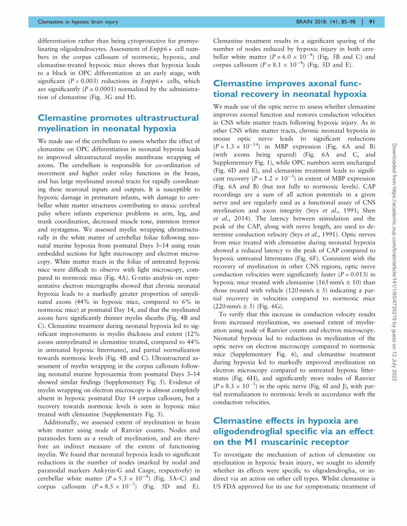
differentiation rather than being cytoprotective for premye-
linating oligodendrocytes. Assessment of Enpp6 + cell num-
bers in the corpus callosum of normoxic, hypoxic, and
clemastine-treated hypoxic mice shows that hypoxia leads
to a block in OPC differentiation at an early stage, with
significant (P = 0.003) reductions in Enpp6 + cells, which
are significantly (P = 0.0001) normalized by the administra-
tion of clemastine (Fig. 3G and H).
Clemastine promotes ultrastructuralmyelination in neonatal hypoxia
We made use of the cerebellum to assess whether the effect of
clemastine on OPC differentiation in neonatal hypoxia leads
to improved ultrastructural myelin membrane wrapping of
axons. The cerebellum is responsible for co-ordination of
movement and higher order relay functions in the brain,
and has large myelinated axonal tracts for rapidly coordinat-
ing these neuronal inputs and outputs. It is susceptible to
hypoxic damage in premature infants, with damage to cere-
bellar white matter structures contributing to ataxic cerebral
palsy where infants experience problems in arm, leg, and
trunk coordination, decreased muscle tone, intention tremor
and nystagmus. We assessed myelin wrapping ultrastructu-
rally in the white matter of cerebellar foliae following neo-
natal murine hypoxia from postnatal Days 3–14 using resin
embedded sections for light microscopy and electron micros-
copy. White matter tracts in the foliae of untreated hypoxic
mice were difficult to observe with light microscopy, com-
pared to normoxic mice (Fig. 4A). G-ratio analysis on repre-
sentative electron micrographs showed that chronic neonatal
hypoxia leads to a markedly greater proportion of umyeli-
nated axons (44% in hypoxic mice, compared to 6% in
normoxic mice) at postnatal Day 14, and that the myelinated
axons have significantly thinner myelin sheaths (Fig. 4B and
C). Clemastine treatment during neonatal hypoxia led to sig-
nificant improvements in myelin thickness and extent (12%
axons unmyelinated in clemastine treated, compared to 44%
in untreated hypoxic littermates), and partial normalization
towards normoxic levels (Fig. 4B and C). Ultrastructural as-
sessment of myelin wrapping in the corpus callosum follow-
ing neonatal murine hypoxaemia from postnatal Days 3–14
showed similar findings (Supplementary Fig. 5). Evidence of
myelin wrapping on electron microscopy is almost completely
absent in hypoxic postnatal Day 14 corpus callosum, but a
recovery towards normoxic levels is seen in hypoxic mice
treated with clemastine (Supplementary Fig. 5).
Additionally, we assessed extent of myelination in brain
white matter using node of Ranvier counts. Nodes and
paranodes form as a result of myelination, and are there-
fore an indirect measure of the extent of functioning
myelin. We found that neonatal hypoxia leads to significant
reductions in the number of nodes (marked by nodal and
paranodal markers Ankyrin-G and Caspr, respectively) in
cerebellar white matter (P = 5.3 � 10�4) (Fig. 5A–C) and
corpus callosum (P = 8.5 � 10�7) (Fig. 5D and E).
Clemastine treatment results in a significant sparing of the
number of nodes reduced by hypoxic injury in both cere-
bellar white matter (P = 6.0 � 10�4) (Fig. 5B and C) and
corpus callosum (P = 8.1 � 10�4) (Fig. 5D and E).
Clemastine improves axonal func-tional recovery in neonatal hypoxia
We made use of the optic nerve to assess whether clemastine
improves axonal function and restores conduction velocities
in CNS white matter tracts following hypoxic injury. As in
other CNS white matter tracts, chronic neonatal hypoxia in
mouse optic nerve leads to significant reductions
(P = 1.3 � 10�14) in MBP expression (Fig. 6A and B)
(with axons being spared) (Fig. 6A and C, and
Supplementary Fig. 1), while OPC numbers seem unchanged
(Fig. 6D and E), and clemastine treatment leads to signifi-
cant recovery (P = 1.2 � 10�5) in extent of MBP expression
(Fig. 6A and B) (but not fully to normoxic levels). CAP
recordings are a sum of all action potentials in a given
nerve and are regularly used as a functional assay of CNS
myelination and axon integrity (Stys et al., 1991; Shen
et al., 2014). The latency between stimulation and the
peak of the CAP, along with nerve length, are used to de-
termine conduction velocity (Stys et al., 1991). Optic nerves
from mice treated with clemastine during neonatal hypoxia
showed a reduced latency to the peak of CAP compared to
hypoxic untreated littermates (Fig. 6F). Consistent with the
recovery of myelination in other CNS regions, optic nerve
conduction velocities were significantly faster (P = 0.013) in
hypoxic mice treated with clemastine (165 mm/s � 10) than
those treated with vehicle (120 mm/s � 5) indicating a par-
tial recovery in velocities compared to normoxic mice
(220 mm/s � 5) (Fig. 6G).
To verify that this increase in conduction velocity results
from increased myelination, we assessed extent of myelin-
ation using node of Ranvier counts and electron microscopy.
Neonatal hypoxia led to reductions in myelination of the
optic nerve on electron microscopy compared to normoxic
mice (Supplementary Fig. 6), and clemastine treatment
during hypoxia led to markedly improved myelination on
electron microscopy compared to untreated hypoxic litter-
mates (Fig. 6H), and significantly more nodes of Ranvier
(P = 8.3 � 10�3) in the optic nerve (Fig. 6I and J), with par-
tial normalization to normoxic levels in accordance with the
conduction velocities.
Clemastine effects in hypoxia areoligodendroglial specific via an effecton the M1 muscarinic receptor
To investigate the mechanism of action of clemastine on
myelination in hypoxic brain injury, we sought to identify
whether its effects were specific to oligodendroglia, or in-
direct via an action on other cell types. Whilst clemastine is
US FDA approved for its use for symptomatic treatment of
Clemastine in hypoxic brain injury BRAIN 2018: 141; 85–98 | 91
Dow
nloaded from https://academ
ic.oup.com/brain/article/141/1/85/4735219 by guest on 12 July 2022
Figure 3 Clemastine promotes OPC differentiation in neonatal hypoxia. (A) Numbers of differentiating OPCs expressing mRNA by in
situ hybridization for the markers Plp (PLP) and Mag (MAG) in corpus callosum (A and B) and striatum (C and D) of hypoxic neonatal mice at
postnatal Day 10 compared to hypoxic littermates treated with clemastine (‘Hypoxia + Clemastine’). (E and F) Numbers of undifferentiated
OPCs expressing mRNA by in situ hybridization for the marker Pdgfra (PDGFRa) in striatum white matter (E) and counts (F) in striatum and
corpus callosum (CC) of postnatal Day 10 normoxic mice versus hypoxic and hypoxic treated with clemastine. (G) Early differentiating OPCs
expressing mRNA by in situ hybridization for the marker Enpp6 in corpus callosum (G) and counts (H) in postnatal Day 10 normoxic mice versus
hypoxic and hypoxic treated with clemastine. For all graphs, *P5 0.05, **P5 0.001.
92 | BRAIN 2018: 141; 85–98 B. A. C. Cree et al.
Dow
nloaded from https://academ
ic.oup.com/brain/article/141/1/85/4735219 by guest on 12 July 2022

A
B
C
Figure 4 Clemastine promotes ultrastructural myelination on electron microscopy in neonatal hypoxia. (A) Light microscopy of
the white matter of cerebellar foliae at postnatal Day 14 in resin embedded sections stained with toluidine blue from normoxic mice, hypoxic mice
(Hypoxia P3-P14), and hypoxic littermates treated with clemastine (Hypoxia and Clemastine treatment P3-P14). (B) Representative electron
microscopy images from cerebellar fimbriae of the three groups at postnatal Day 14. (C) Quantification of myelin sheath thickness and the
proportion of myelinated and unmyelinated axons in normoxic (blue square), hypoxic (red triangle) and clemastine-treated hypoxic (black filled
circle) mice at postnatal Day 14 by g-ratio analysis. The scatterplot displays g-ratios of individual axons as a function of axonal diameter. All g-ratios
were analysed from transmission electron microscopy images.
Clemastine in hypoxic brain injury BRAIN 2018: 141; 85–98 | 93
Dow
nloaded from https://academ
ic.oup.com/brain/article/141/1/85/4735219 by guest on 12 July 2022

seasonal allergies as an antihistamine through the histamine
receptor 1 (Hrh1), its effect on OPC maturation is
mediated by antimuscarinic action at the M1 muscarinic
acetylcholine receptor (Chrm1) (Mei et al., 2016). We gen-
erated Chrm1 conditional knockout (cKO) mice, which
have the M1 muscarinic receptor specifically deleted from
OPCs, by crossing the Chrm1 floxed line with a Cnp-cre
line. The resulting Chrm1-cKO (Cnp-cre; Chrm1 fl/fl) mice
were exposed to chronic neonatal hypoxia between post-
natal Days 3–10 as above, and compared to wild-type lit-
termates. Treatment of wild-type mice during hypoxia with
10 mg/kg clemastine once daily by oral gavage between
postnatal Days 3–10 leads to significant increases in MBP
(P = 0.006) (Fig. 7A and E) protein and Mag mRNA
(P = 1.06 � 10�9) (Fig. 7C and G) expression in striatal
white matter compared to untreated wild-type hypoxic lit-
termates. However, the effect of clemastine on both MBP
protein expression and Mag mRNA expression was lost in
Chrm1-cKO mice (Fig. 7A, C, E and G), with no significant
difference found between clemastine-treated Chrm1-cKO
and untreated wild-type or Chrm1-cKO mice (Fig. 7A, C,
E and G). Similar results were found in the corpus callosum
(Supplementary Fig. 7). Whilst hypoxic Chrm1-cKOs do
show an �30% increase in MBP and MAG expression in
the corpus callosum compared to hypoxic wild-type mice
(Supplementary Fig. 7D and E), this is not significantly
different, and suggests that Chrm1-cKOs do not phenocopy
the protective effects of the drug to the same extent in the
setting of hypoxia as has been previously shown (Mei et al.,2016). No significant differences were found in neurofila-
ment staining (Fig. 7B and F) or OPC numbers (Fig. 7D
and H) between any of the groups. Together these results
indicate that the effects of clemastine on myelination and
OPC differentiation in hypoxic brain injury are specific to
Figure 5 Clemastine increases node of Ranvier formation in neonatal hypoxia. (A) Magnified region of a cerebellar fimbria at
postnatal Day 10 from neonatal hypoxic mice and hypoxic littermates treated with clemastine, showing expression of MBP and the paranodal
marker Caspr, indicating nodes of Ranvier. (B–E) Node of Ranvier formation in the three groups in postnatal Day 10 cerebellar white matter (B)
and corpus callosum (D) using markers Ankyrin-G (nodal marker) and Caspr (paranodal marker), with quantification in cerebellum (C) and
corpus callosum (E) showing density of nodes (nodes were counted only if Ank-G/Caspr double positive). ‘Normoxia’ and
‘Hypoxia + Clemastine’ groups were not significantly different. For all graphs, *P5 0.05, **P5 0.001.
94 | BRAIN 2018: 141; 85–98 B. A. C. Cree et al.
Dow
nloaded from https://academ
ic.oup.com/brain/article/141/1/85/4735219 by guest on 12 July 2022

Figure 6 Clemastine improves myelination and CNS conduction velocities in neonatal hypoxic optic nerve. (A) MBP expression
(top) and neurofilament (NF) staining (bottom) in the optic nerve of postnatal Day 13 normoxic mice, versus those exposed to neonatal hypoxia
(postnatal Days 3–13), versus hypoxic littermates treated with clemastine, with quantification of (B) total MBP positive area and (C) neurofi-
lament area measured using Image-Pro Plus software (relative expression among the three groups normalized to the normoxia group).
(D) Undifferentiated OPCs in the optic nerves stained for PDGFRa with quantification of OPC density in (E). (F) Representative traces from
postnatal Day 13 optic nerve CAP recordings from nerves of equal length of hypoxic (black) and clemastine-treated hypoxic (red) mice. Traces
from normoxic mice were excluded because they were longer than nerves from hypoxic mice. (G) Conduction velocities of postnatal Day 13
optic nerves as measured by suction electrodes (n = 8 nerves per group, one-way ANOVA followed by Bonferroni’s correction). Data are shown
in box-and-whisker plots (maximum, 75th percentile, median, 25th percentile and minimum). (H) Representative electron microscopy images
from optic nerves of the three groups at postnatal Day 14. (I) Node of Ranvier formation in the three groups in postnatal Day 13 optic nerve using
markers Ankyrin-G (nodal marker) and Caspr (paranodal marker), with quantification in J showing density of nodes (nodes counted only if Ank-G/
Caspr double positive). For all graphs, *P5 0.05, **P5 0.001.
Clemastine in hypoxic brain injury BRAIN 2018: 141; 85–98 | 95
Dow
nloaded from https://academ
ic.oup.com/brain/article/141/1/85/4735219 by guest on 12 July 2022

Figure 7 Clemastine function in hypoxia is oligodendroglial specific via an effect on the M1 muscarinic receptor. Chronic
neonatal hypoxemia (10% FiO2) between postnatal Days 3–10 was performed in conditional Chrm1 knock-out mice (Chrm1-cKO mice are Cnp-
cre; Chrm1 fl/fl) and their wild-type littermates [cre negative mice were considered as wild-type (WT)]. (A) MBP protein expression in the brain
striatum white matter of postnatal Day 10 hypoxic wild-type mice, versus hypoxic wild-type mice treated with clemastine (WT + Clemastine),
versus hypoxic Chrm1 knock-out littermates (Chrm1-cKO), versus hypoxic Chrm1 knock-out mice treated with clemastine (Chrm1-
cKO + Clemastine). (B) Neurofilament (NF) and MBP staining in these groups. (C) Differentiation marker Mag mRNA and (D) OPC marker
PDGFRa protein expression in striatum in the same groups. (E) Quantification in striatum of total MBP positive area (‘Area’) and total MBP
fluorescent signal intensities (‘Intensity Sum’) measured using Image-Pro Plus software (with relative expression among the four groups nor-
malized to the wild-type hypoxia group). (F) Total neurofilament area fold change in striatum in the four groups normalized to wild-type hypoxia
group. (G) Numbers of Mag mRNA-positive cells in striatum among the four groups (normalized to the wild-type hypoxia group). (H) Numbers
of PDGFRa positive OPCs in striatum in the four groups (normalized to the wild-type hypoxia group). For all graphs, *P5 0.05, **P5 0.001.
96 | BRAIN 2018: 141; 85–98 B. A. C. Cree et al.
Dow
nloaded from https://academ
ic.oup.com/brain/article/141/1/85/4735219 by guest on 12 July 2022

oligodendroglial lineage via an effect on the M1 muscarinic
receptor in OPCs.
DiscussionOther than supportive care, there are currently no proven
treatments for hypoxic white matter injury in either the
neonatal or adult setting. Evidence suggests that OPC dif-
ferentiation arrest is a critical contributor to failed myelin
generation during hypoxia, leading to progressive neuro-
logical injury, and suggesting that promotion of OPC dif-
ferentiation through therapeutic manipulation could yield
improved clinical outcomes. Evidence suggests that inhibit-
ing the muscarinic pathway in oligodendrocyte precursors
promotes their differentiation (Abiraman et al., 2015; Mei
et al., 2016), and that Chrm1 on OPCs may be the specific
target for this activity (Mei et al., 2016), prompting us to
assess anti-muscarinic therapy for hypoxic brain injury
involving white matter injury and OPC maturation arrest.
The mechanisms of disease in DPHL are far less well
understood than in neonatal hypoxic injury, due to the
very rare nature of the condition in humans and lack of
reflective animal models, but failure of myelin generation
following a demyelinating insult is thought to underlie the
pathophysiology. We present a case of severe DPHL who
markedly improved in the setting of off-label use of clem-
astine. Because spontaneous recovery has been reported in
some cases of DPHL (Min, 1986; Lee and Lyketsos, 2001)
it is unclear how this patient would have improved without
clemastine treatment. Nevertheless, because of the profound
neurological deficits he experienced 2 months after the hyp-
oxic event, his prognosis at that time was considered to be
very poor based on a prior report of patients with delayed
akinetic-mutism following carbon monoxide poisoning (Lee
and Marsden, 1994). That the patient recovered to return
to intellectually demanding work was sufficiently unusual
to promote our interest in studying clemastine treatment for
hypoxic injury. Using a murine neonatal model, we present
data showing that clemastine dramatically promotes myelin
generation during global hypoxic injury. These experimen-
tal studies provide biological plausibility that improvement
in our case of adult DPHL could be clemastine-mediated.
Proof that clemastine is beneficial in DPHL will require
study through randomized controlled trials, which will be
challenging to accomplish given the rarity of this disorder.
Our murine studies in neonatal hypoxia have broader
implication: 1.5% of all live births in the USA each year
are very low birth weight infants and recent improvements
in neonatal critical care have led to a marked increase
in survival (50–70%) of extremely low birth weight
(41000 g) premature infants. Other than therapeutic hypo-
thermia there are no treatments for neonatal hypoxic injury
and there is a significant healthcare burden from disability
related to cerebral palsy (Glass and Rowitch, 2016). White
matter injury is the most reliable prognostic indicator of de-
velopment of severe cerebral palsy and cognitive disability
in premature infants (Woodward et al., 2006). A significant
contributor to failure of myelin generation in these infants
is persistent OPC maturation arrest. Our murine model
replicates the significant OPC maturation delay that is
a hallmark of the human hypoxic neonatal injury,
making it an ideal model for testing OPC differentiation
promoting therapies. We found that clemastine dramatic-
ally promotes OPC differentiation, myelination, and
improves functional recovery in neonatal hypoxic injury.
Moreover, we show that its effects in hypoxia are oligo-
dendroglial-specific via an effect on the M1 muscarinic
receptor on OPCs. Further work will be required to
assess clemastine effects in human neonatal injury. Given
the excellent safety profile of clemastine in adults, and the
results from our animal studies, we propose clemastine as
a potential treatment to promote myelin recovery in
premature infants.
AcknowledgementsWe would like to thank J. Josh Lawrence (Texas Tech
University) and Susumu Tonegawa (MIT) for the Chrm1
floxed mice.
FundingThis work was supported by a March of Dimes Foundation
Grant No. 6-FY17-551 (to S.P.J.F.). Support from Mead
Johnson and Company LLC (A121425) (J.R.C. and
S.P.J.F.). S.P.J.F. is a Harry Weaver Neuroscience Scholar
of the National Multiple Sclerosis Society.
Supplementary materialSupplementary material is available at Brain online.
ReferencesAbiraman K, Pol SU, O’Bara MA, Chen GD, Khaku ZM, Wang J,
et al. Anti-muscarinic adjunct therapy accelerates functional human
oligodendrocyte repair. J Neuroscience 2015; 35: 3676–88.
Allin M, Walshe M, Fern A, Nosart C, Cuddy M, Rifkin L, et al.
Cognitive maturation in preterm and term born adolescents.
J Neurol Neurosurg Psychiatry 2008; 79: 381–6.
Back SA, Han BH, Luo NL, Chricton CA, Xanthoudakis S, Tam J,
et al. Selective vulnerability of late oligodendrocyte progenitors to
hypoxia-ischemia. J Neurosci 2002; 22: 455–63.
Bayless S, Stevenson J. Executive functions in school-age children born
very prematurely. Early Hum Dev 2007; 83: 247–54.Billiards SS, Haynes RL, Folkerth RD, Borenstein NS, Trachtenberg
FL, Rowitch DH, et al. Myelin abnormalities without oligodendro-
cyte loss in periventricular leukomalacia. Brain Pathol 2008; 18:
153–63.
Buser JR, Maire J, Riddle A, Gong X, Nguyen T, Nelson K, et al.
Arrested preoligodendrocyte maturation contributes to myelination
failure in premature infants. Ann Neurol 2012; 71: 93–109.
Clemastine in hypoxic brain injury BRAIN 2018: 141; 85–98 | 97
Dow
nloaded from https://academ
ic.oup.com/brain/article/141/1/85/4735219 by guest on 12 July 2022

Choi IS. Delayed neurologic sequelae in carbon monoxide intoxica-
tion. Arch Neurol 1983; 40: 433–5.
Fancy SP, Harrington EP, Yuen TJ, Silbreis JC, Zhao C, Baranzini SE,
et al. Axin2 as regulatory and therapeutic target in newborn brain
injury and remyelination. Nat Neurosci 2011; 14: 1009–16.
Fancy SP, Harrington EP, Baranzini SE, Silbreis JC, Shiow LR, Yuen
TJ, et al. Parallel states of pathological Wnt signaling in neonatal
brain injury and colon cancer. Nat Neurosci 2014; 17: 506–12.
Ginsberg MD. Delayed neurological deterioration following hypoxia.
Adv Neurol 1979; 26: 21–44.Glass HC, Rowitch DH. The role of the neurointensive care nursery
for neonatal encephalopathy. Clin Perinatol 2016; 43: 547–57.
Gottfried JA, Mayer SA, Shungu DC, Chang Y, Duyn JH. Delayed
posthypoxic demyelination. Association with arylsulfatase A defi-
ciency and lactic acidosis on proton MR spectroscopy. Neurology
1997; 49: 1400–4.Grinker R. Parkinsonism following carbon monoxide poisoning.
J Nerv Ment Dis 1926; 64: 18–28.
Heckmann JG, Erbguth F, Neundorfer B. Delayed postanoxic demye-
lination registry. Neurology 1998; 51: 1235–6.
Hori A, Hirose G, Kataoka S, Tsukada K, Furui K, Tonami H.
Delayed postanoxic encephalopathy after strangulation. Serial neu-
roradiological and neurochemical studies. Arch Neurol 1991; 48:
871–4.
Karadottir R, Cavelier P, Bergersen LH, Attwell D. NMDA receptors
are expressed in oligodendrocytes and activated in ischaemia.
Nature 2005; 438: 1162–6.
Khwaja O, Volpe JJ. Pathogenesis of cerebral white matter injury of
prematurity. Arch Dis Child Fetal Neonatal 2008; 93: F153–61.
Kobaly K, Schluchter M, Minich N, Friedman H, Taylor HG, Wilson-
Costello D, et al. Outcomes of extremely low birth weight (51 kg)
and extremely low gestational age (528 weeks) infants with bronch-
opulmonary dysplasia: effects of practice changes in 2000 to 2003.
Pediatrics 2008; 121: 73–81.
Larroque B, Ancel PY, Marret S, Marchand L, Andre M, Arnaud C,
et al. Neurodevelopmental disabilities and special care of 5-year-old
children born before 33 weeks of gestation (the EPIPAGE study): a
longitudinal cohort study. Lancet 2008; 371: 813–30.
Lee HB, Lyketsos CG. Delayed post-hypoxic leukoencephalopathy.
Psychosomatics 2001; 42: 530–3.
Lee MS, Marsden CD. Neurological sequelae following carbon mon-
oxide poisoning clinical course and outcome according to the clin-
ical types and brain computed tomography scan findings. Mov
Disord 1994; 9: 550–8.
Li Z, He Y, Fan S, Sun B. Clemastine rescues behavioral changes and
enhances remyelination in the cuprizone mouse model of demyelin-
ation. Neurosci Bull 2015; 31: 617–25.
Liu J, Dupree JL, Gacias M, Frawley R, Sikder T, Naik P, et al.
Clemastine enhances myelination in the prefrontal cortex and res-
cues behavioral changes in socially isolated mice. J Neurosci 2016;
36: 957–62.
Marlow N, Wolke D, Bracewell MA, Samara M; EPICure Study
Group. Neurologic and developmental disability at six years of
age after extremely preterm birth. N engl J Med 2005; 352: 9–19.
Marlow N, Hennessy EM, Bracewell MA, Wolke D; EPICure Study
Group. Motor and executive function at 6 years of age after
extremely preterm birth. Pediatrics 2007; 120: 793–804.
Martin JA, Kung HC, Mathews TJ, Hoyert DL, Strobino DM, Guyer
B, et al. Annual summary of vital statistics: 2006. Pediatrics 2008;
121: 788–801.
Mei F, Fancy SP, Shen YA, Niu J, Zhao C, Presley B, et al. Micropillararrays as a high-throughput screening platform for therapeutics in
multiple sclerosis. Nat Med 2014; 20: 954–60.
Mei F, Lehmann-Horn K, Shen YA, Rankin KA, Stebbins KJ, Lorrain
DS, et al. Accelerated remyelination during inflammatory demyelin-ation prevents axonal loss and improves functional recovery. Elife
2016; 5: e18246.
Min SK. A brain syndrome associated with delayed neuropsychiatric
sequelae following acute carbon monoxide intoxication. ActaPsychiatr Scand 1986; 73: 80–6.
Molloy S, Soh C, Williams TL. Reversible delayed posthypoxic leu-
koencephalopathy. AJNR Am J Neuroradiol 2006; 27: 1763–5.Platt MJ, Cans C, Johnson A, Surman G, Topp M, Torrioli MG, et al.
Trends in cerebral palsy among infants of very low birthweight
(51500 g) or born prematurely (532 weeks) in 16 European cen-
tres: a database study. Lancet 2007; 369: 43–50.Plum F, Posner, JB, Hain RF. Delayed neurological deterioration after
anoxia. Arch Intern Med 1962; 110: 18–25.
Segovia KN, McCLure M, Moravec M, Luo NL, Wan Y, Gong X,
et al. Arrested oligodendrocyte lineage maturation in chronic peri-natal white matter injury. Ann Neurol 2008; 63: 520–30.
Shen YA, Chen Y, Dao DQ, Mayoral SR, Wu L, Meijer D, et al.
Phosphorylation of LKB1/Par-4 establishes Schwann cell polar-
ity to initiate and control myelin extent. Nat Commun 2014; 5:4991.
Shillito FH, Drinker CK, Shaughnessy TJ. The problem of nervous and
mental sequelae in carbon monoxide poisoning. JAMA 1936; 106:669–74.
Shprecher D, Mehta L. The syndrome of delayed post-hypoxic leu-
koencephalopathy. Neurorehabilitation 2010; 26: 65–72.
Stys PK, Ransom BR, Waxman SG. Compound action potential ofnerve recorded by suction electrode: a theoretical and experimental
analysis. Brain Res 1991; 546: 18–32.
Weinberger LM, Schmidley JW, Schafer IA, Raghavan S. Delayed
postanoxic demyelination and arylsulfatase-A pseudodeficiency.Neurology 1994; 44: 152–4.
Wolke D, Samara M, Bracewell M, Marlow N, EPICure Study Group.
Specific language difficulties and school achievement in chil-dren born at 25 weeks of gestation or less. J Pediatr 2008; 152:
256–62.
Verney C, Pogledic I, Biran V, Adle-Biassette H, Fallet-Bianco C,
Gressens P. Microglial reaction in axonal crossroads is a hallmarkof noncystic periventricular white matter injury in very preterm in-
fants. J Neuropathol Exp Neurol 2012; 71: 251–64.
Volpe JJ. Brain injury in premature infants: a complex amalgam of
destructive and developmental disturbances. Lancet Neurol 2009; 8:110–24.
Wood NS, Costeloe K, Gibson AT, Hennessy EM, Marlow N,
Wilkinson A, et al. The EPICure study: associations and antecedentsof neurological and developmental disability at 30 months of age
following extremely preterm birth. Arch Dis Child Fetal Neonatal
2005; 90: F134–40.
Woodward LJ, Edgin JO, Thompson D, Inder TE. Object workingmemory deficits predicted by early brain injury and development
in the preterm infant. Brain 2005; 128(Pt 11): 2578–87.
Woodward LJ, Anderson PJ, Austin NC, Howard K, Inder TE.
Neonatal MRI to predict neurodevelopmental outcomes in preterminfants. N Engl J Med 2006; 355: 685–94.
Zamora CA, Nauen D, Hynecek R,Ilica AT, Izbudak I, Sair HI, et al.
Delayed posthypoxic leukoencephalopathy: a case series and review
of the literature. Brain Behav 2015; 5: e00364.
98 | BRAIN 2018: 141; 85–98 B. A. C. Cree et al.
Dow
nloaded from https://academ
ic.oup.com/brain/article/141/1/85/4735219 by guest on 12 July 2022
Copyright © 2022 FDOKUMEN