High dose M-CSF partially rescues the Dap12?/? osteoclast phenotype
13
Journal of Cellular Biochemistry 90:871–883 (2003) FAST TRACKS High Dose M-CSF Partially Rescues the Dap12/ Osteoclast Phenotype Roberta Faccio, 1,2 Wei Zou, 1 Graziana Colaianni, 1,2 Steven L. Teitelbaum, 1 and F. Patrick Ross 1 * 1 Washington University School of Medicine, St. Louis, Missouri 63110 2 Department of Human Anatomy, School of Medicine, Bari, Italy 70100 Abstract Osteoclasts are macrophage derived cells and as such are subject to regulation by molecules impacting other members of the immune system. Dap12 is an adaptor protein expressed by NK cells and B and T lymphocytes. Dap12 also mediates maturation of myeloid cells and is expressed by osteoclasts which are dysfunctional in its absence. We find Dap12/ osteoclast precursors fail to differentiate, in vitro, and the abnormality is partially rescued by high dose M-CSF. The relative paucity of osteoclast number, even in presence of high dose cytokine, is attended by dampened proliferation of precursor cells and their failure to normally migrate towards the osteoclast-recognized matrix protein, osteopontin. Furthermore, Dap12/ osteoclasts generated in high dose M-CSF fail to normally organize their cytoskeleton. The incapacity of Dap12 null cells to undergo normal osteoclast differentiation is not due to blunted stimulation of major RANK ligand (RANKL) or M-CSF induced signaling pathways. On the other hand, when plated on osteopontin, Dap12/ pre-osteoclasts do not activate the tyrosine kinase, Syk, which normally binds to the adaptor protein and transmits downstream signals. Attesting to the importance of the Dap12/Syk complex, Syk deficient macrophages do not undergo normal osteoclastogenesis. Furthermore, the same cells plated onto osteopontin, adhere poorly and fail to phosphorylate c-Src or Pyk2, two kinases central to organization of the osteoclast cytoskeleton. J. Cell. Biochem. 90: 871 – 883, 2003. ß 2003 Wiley-Liss, Inc. Key words: Dap12; M-CSF; Syk; osteoclasts; bone Skeletal remodeling is an ever-occurring process in man which pivots on the activity and recruitment of the unique bone resorb- ing cell, the osteoclast [Chambers, 2000]. The osteoclast originates by hematopoietic pre- cursors of the monocyte/macrophage family migrating to the bone environment, where, in the presence of the cytokines RANK ligand (RANKL) and M-CSF, they multinucleate and assume the unique osteoclast phenotype thus acquiring the capacity to degrade mineralized matrix [Teitelbaum, 2000; Boyle et al., 2003]. RANK signaling, activated by its ligand RANKL which is expressed on stromal cells and osteoblasts [Suda et al., 1999], is mediated by a series of protein kinases including c-Src, c-Jun N terminal kinase (JNK), p38, extracel- lular signal related kinase (ERK), phosphoino- sitol-3-kinase (PI-3K), and those activating NF- kB [Darnay et al., 1998; Galibert et al., 1998; Matsumoto et al., 2000; Lee et al., 2002]. M- CSF, which via its receptor, c-Fms, stimulates many of the same pathways, promotes prolif- eration of osteoclast precursors and survival of the mature resorptive cell [Tanaka et al., 1993; Woo et al., 2002]. Together, therefore, RANKL and M-CSF induce expression of genes, such as those encoding tartrate-resistant acid phospha- tase (TRAP), cathepsin K (CATK), calcitonin receptor, and b3 integrin, which characterize the mature osteoclast and its committed pre- cursors [Kudo et al., 2002; Faccio et al., 2003b]. Once the resorptive cell is in contact with bone, a series of matrix-derived signals, mediat- ed largely through the avb3 integrin, prompt ß 2003 Wiley-Liss, Inc. Roberta Faccio and Wei Zou contributed equally to this project. Grant sponsor: National Institutes of Health (to F.P.R.); Grant numbers: AR48812, AR46852; Grant sponsor: Clin- ical Nutrition Research Unit (to S.L.T.); Grant numbers: AR48853, AR46523, AR32788, DK-56341. *Correspondence to: F. Patrick Ross, PhD, Department of Pathology and Immunology, Campus Box 8118, 660 South Euclid Avenue, St. Louis, MO 63110. E-mail: [email protected] Received 13 August 2003; Accepted 18 August 2003 DOI 10.1002/jcb.10694
-
Upload
independent -
Category
Documents
-
view
6 -
download
0
Transcript of High dose M-CSF partially rescues the Dap12?/? osteoclast phenotype

Journal of Cellular Biochemistry 90:871–883 (2003)
FASTTRACKS
High Dose M-CSF Partially Rescuesthe Dap12�/� Osteoclast Phenotype
Roberta Faccio,1,2 Wei Zou,1 Graziana Colaianni,1,2 Steven L. Teitelbaum,1 and F. Patrick Ross1*1Washington University School of Medicine, St. Louis, Missouri 631102Department of Human Anatomy, School of Medicine, Bari, Italy 70100
Abstract Osteoclasts are macrophage derived cells and as such are subject to regulation by molecules impactingothermembers of the immune system.Dap12 is anadaptor protein expressedbyNKcells andBandT lymphocytes.Dap12also mediates maturation of myeloid cells and is expressed by osteoclasts which are dysfunctional in its absence.We findDap12�/� osteoclast precursors fail to differentiate, in vitro, and the abnormality is partially rescued by high doseM-CSF.The relative paucity of osteoclast number, even in presence of high dose cytokine, is attended by dampened proliferationof precursor cells and their failure to normally migrate towards the osteoclast-recognized matrix protein, osteopontin.Furthermore, Dap12�/� osteoclasts generated in high dose M-CSF fail to normally organize their cytoskeleton. Theincapacity of Dap12 null cells to undergo normal osteoclast differentiation is not due to blunted stimulation of majorRANK ligand (RANKL) orM-CSF induced signaling pathways.On the other hand,whenplatedonosteopontin,Dap12�/�pre-osteoclasts do not activate the tyrosine kinase, Syk, which normally binds to the adaptor protein and transmitsdownstream signals. Attesting to the importance of the Dap12/Syk complex, Syk deficient macrophages do not undergonormal osteoclastogenesis. Furthermore, the same cells plated onto osteopontin, adhere poorly and fail to phosphorylatec-Src or Pyk2, two kinases central to organization of the osteoclast cytoskeleton. J. Cell. Biochem. 90: 871–883, 2003.� 2003 Wiley-Liss, Inc.
Key words: Dap12; M-CSF; Syk; osteoclasts; bone
Skeletal remodeling is an ever-occurringprocess in man which pivots on the activityand recruitment of the unique bone resorb-ing cell, the osteoclast [Chambers, 2000]. Theosteoclast originates by hematopoietic pre-cursors of the monocyte/macrophage familymigrating to the bone environment, where, inthe presence of the cytokines RANK ligand(RANKL) and M-CSF, they multinucleate andassume the unique osteoclast phenotype thus
acquiring the capacity to degrade mineralizedmatrix [Teitelbaum, 2000; Boyle et al., 2003].
RANK signaling, activated by its ligandRANKL which is expressed on stromal cellsand osteoblasts [Suda et al., 1999], is mediatedby a series of protein kinases including c-Src,c-Jun N terminal kinase (JNK), p38, extracel-lular signal related kinase (ERK), phosphoino-sitol-3-kinase (PI-3K), and those activating NF-kB [Darnay et al., 1998; Galibert et al., 1998;Matsumoto et al., 2000; Lee et al., 2002]. M-CSF, which via its receptor, c-Fms, stimulatesmany of the same pathways, promotes prolif-eration of osteoclast precursors and survival ofthe mature resorptive cell [Tanaka et al., 1993;Woo et al., 2002]. Together, therefore, RANKLand M-CSF induce expression of genes, such asthose encoding tartrate-resistant acid phospha-tase (TRAP), cathepsin K (CATK), calcitoninreceptor, and b3 integrin, which characterizethe mature osteoclast and its committed pre-cursors [Kudo et al., 2002; Faccio et al., 2003b].
Once the resorptive cell is in contact withbone, a series of matrix-derived signals, mediat-ed largely through the avb3 integrin, prompt
� 2003 Wiley-Liss, Inc.
Roberta Faccio and Wei Zou contributed equally to thisproject.
Grant sponsor: National Institutes of Health (to F.P.R.);Grant numbers: AR48812, AR46852; Grant sponsor: Clin-ical Nutrition Research Unit (to S.L.T.); Grant numbers:AR48853, AR46523, AR32788, DK-56341.
*Correspondence to: F. Patrick Ross, PhD, Department ofPathology and Immunology, Campus Box 8118, 660 SouthEuclid Avenue, St. Louis, MO 63110.E-mail: [email protected]
Received 13 August 2003; Accepted 18 August 2003
DOI 10.1002/jcb.10694

the osteoclast to reorganize its cytoskeleton andassume a unique polarized morphological andfunctional phenotype [McHugh et al., 2000;Faccio et al., 2003a]. Among the most dramaticof these polarized features is formation of thecell’s ruffled membrane which is encompassedby a ‘‘sealing zone’’ or ‘‘actin ring’’ [Teti et al.,1991]. This circular structure, which servesto isolate the osteoclast resorptive micro-environment from the general extracellularspace, is characterized by the presence of denseF-actin bundles associated with several cytos-keletal proteins and transmembrane receptors[Akisaka et al., 2001].
The fact that osteoclasts are derived frommacrophages, cells which are fundamental toimmune recognition, has led to a series of ex-periments which link the immune system toosteoclast recruitment and function. For exam-ple, T-lymphocycte-produced cytokines, includ-ing RANKL and TNFa, appear central to theenhanced osteoclastogenesis responsible for thebone loss attending menopause and the peri-articular bone erosions of rheumatoid arthritis[Cenci et al., 2000; Weitzmann et al., 2000;Romas et al., 2002]. In this context, the processof antigen presentation, itself, is also a funda-mental event in pathological osteoclastogenesis[Jenkins et al., 2002].
These and other insights gained into themeans by which osteoclast precursors differ-entiate and how the mature polykaryon resorbsbone have led to the identification of a numberof new anti-osteoporosis therapeutic targetsincluding CATK, c-Src, and the avb3 integrin,thus encouraging the exploration of other candi-dates [Wilder, 2002; Zaidi et al., 2003]. Amongthe most promising of such potential targets areintraosteoclastic signaling molecules which alsofunction in the immune system [Paloneva et al.,2002; Kaifu et al., 2003].
Much of the information in hand regardingthe molecular mechanisms of osteoclast for-mation and function is derivative of studiesperformed on human disease or geneticallymanipulated animals, particularly those withosteopetrosis [Marks, 1989; McLean and Olsen,2001]. Presently, at least 24 genes or loci, theproducts of which positively or negatively reg-ulate osteoclastogenesis and osteoclast functionhave been identified [Boyle et al., 2003; Teitel-baum and Ross, 2003]. Some such genes impactformation and/or survival of osteoclast precur-sors. Others mediate either the ability of these
precursors to differentiate or the capacity ofthe mature osteoclast to resorb bone.
Dap12 is a transmembrane adapter moleculeexpressed in a variety of cells of the immunesystem [Tomasello et al., 1998; Lanier andBakker, 2000]. In myeloid cells, Dap12 pairswith surface residing receptors including trig-gering receptor expressed on myeloid cells(TREMs) [Colonna, 2003]. The cytoplasmicdomain of Dap12 contains the immunoreceptortyrosine-based activation ITAM motif, whichfunctions as a docking site for tyrosine kinases,including Syk [McVicar et al., 1998]. Interest-ingly, deletion of the Dap12 gene, in man,results in Nasu–Hakola disease (NHD), whichincludes skeletal abnormalities in its phenotype[Kondo et al., 2002; Kaifu et al., 2003]. Further-more, Dap12 is expressed by osteoclasts and, asevidenced by the development of osteopetrosisin mice lacking the protein, is essential fornormal osteoclast function [Kondo et al., 2002;Kaifu et al., 2003].
In this exercise, we turned to the molecularpathogenesis of Dap12 deficient osteopetrosis.We find that the failure of Dap12�/� myeloidcells to generate osteoclasts can be rescuedsubstantially, but incompletely, by increasingambient M-CSF. Regardless of cytokine concen-tration, however, the resorptive capacity andcytoskeleton of Dap12 deficient osteoclasts arederanged. Attesting to the importance of Dap12associated signaling molecules in osteoclastfunction, bone marrow macrophages (BMMs)Syk lacking to differentiate into mature osteo-clasts, and exhibit abnormal cytoskeletal func-tion associated with dampened c-Src and Pyk2phosphorylation.
MATERIALS AND METHODS
In Vitro Generation of OCsand Bone Resorption Assay
BMMs Dap12þ/þ and Dap12�/� [Bakkeret al., 2000], or Sykþ/� and Syk�/� [Mocsaiet al., 2002] kindly provided by Dr. C.A. Lowell(Department of Laboratory Medicine, Univer-sity of California, San Francisco, CA), wereisolated as previously described [Faccio et al.,2003b] and cultured for 3 days in a-MEM sup-plemented with 10% FCS and 1:10 CMG14–12culture supernatant [Takeshita et al., 2000],which contained the equivalent of 100 ng/mlof recombinant M-CSF. A total of 5� 104 cellswere cultured in a-MEM containing 10% heat
872 Faccio et al.

inactivated FBS with 100 ng/ml RANKL[McHugh et al., 2000] and increasing concen-trations (from 10 to 100 ng/ml) of mouse re-combinant M-CSF (R&D Systems, Inc.,Minneapolis, MN) in 96-well tissue cultureplates. Cells were fixed and stained for TRAPactivity after 5 days in culture, using a com-mercial kit (Sigma 387-A, St. Louis, MO). Boneresorption was performed by culturing BMMsexposed to RANKL and M-CSF for 5 days ontodentine. Cells were fixed and stained withFITC–phalloidin to visualize F-actin to detectthe actin ring organization or removed by brieftreatment with 2 N NaOH. Resorption pits werevisualized by hematoxylin staining (Sigma).
Proliferation Assay
BMMs from Dap12þ/þ and Dap12�/� micewere cultured in the presence of increasingconcentrations of M-CSF with or withoutRANKL (100 ng/ml). After 2 days, viable cellnumber was calculated using the MTT (3-[4,5dimethylthiazol-2-yl]-2,5-diphenyltetrazoliumbromide; Sigma) method or Cell ProliferationELISA system (Amersham Pharmacia Biotech,Piscataway, NJ). Briefly, 10 ml MTT (5 mg/ml)were added in each well, containing 100 ml cul-ture media, and incubated at 378C for 4 h. Thereaction was terminated with 150 ml isopropa-nol/0.04 N HCl and MTT absorbance deter-mined at the optical density of 570 nm. Six-wellswere used for each variable and each experi-ment was repeated twice. For the ELISA assay,BrdU was added to each well to a final concen-tration of 10 mM. Cells were incubated at 378Cfor additional 2 h and BrdU incorporation wasdetected following manufacturer’s instruction.
Immunoblot
BMMs from Dap12þ/þ and Dap12�/� micewere cultured for 3 days in the presence of100 ng/ml RANKL and 10 or 100 ng/ml purifiedM-CSF, starved for 2 h and stimulated for theindicated times with 100 ng/ml RANKL or forc-Fms signaling, 10 and 100 ng/ml purified M-CSF. Pre-osteoclasts were lifted with trypsin/EDTA, re-suspended in serum-free medium andplated on 5 mg/ml OPN-coated dishes for theindicated times. Cells were washed twice withice-cold PBS and lysed in the buffer containing10 mM Tris (pH 7.4), 150 mM NaCl, 1 mMEDTA, 0.2% sodium deoxycolate, 1% NP 40,1 mM NaF, 2 mM Na3VO4, and 1� proteaseinhibitor cocktail (Sigma). Forty micrograms of
cell lysates were boiled in the presence ofSDS sample buffer for 5 min and subjected toelectrophoresis on 8% SDS–PAGE. Polyclonalanti-ERK1/2 or anti-phospho-ERK1/2 MAPKpolyclonal antibodies (Cell Signaling Techno-logy, Inc., Beverly, MA) were used for ERK-MAPK immunoblot. IkBa polyclonal antibodywas purchased from Santa Cruz Biotechnology(Santa Cruz, CA) and phospho-IkBa, phosphop38, and p-JNK antibodies were purchasedfrom Cell Signaling Technology. Anti-c-Fosand -Syk were obtained from Santa Cruz Bio-technology, Inc. Phospho-Src antibody was pur-chased from Cell Signaling and phospho-Pyk2from Biosource International (Camarillo, CA).
RNA Preparation and Reverse-TranscriptionPolymerase Chain Reaction (RT-PCR) Analyses
Total RNA from cultured cells was isolatedby the guanidine/phenol method. For RT-PCRanalysis, cDNAs were synthesized from 1 mgof total RNA using reverse transcriptase andoligo dT primers in a volume of 20 ml, and thereaction mixture was finally adjusted to 100 mlwith TE buffer for PCR analysis. PCR wasperformed with 1 ml of cDNA reaction mixtureby using Platinum Pfx polymerase (Invitrogen,Carlsbad, CA) and appropriate primers in avolume of 50 ml. The following primers forCATK, 50-GGAAGAAGACTCACCAGAAGC-30
and 30-GCTATATAGCCGCCTCCACAG-50; formatrix metalloproteinase-9 (MMP-9), 50-CCT-GTGTGTTCCCGTTCATCT-30 and 30-CGC-TGGAATGATCTAAGCCCA-50; for calcitoninreceptor, 50-CATTCCTGTTACTTGGTTGGC-30
and 30-AGCAATCGACAAGGAGTGAC-50; andfor GAPDH, 50-ACTTTGTCAAGCTCATTT-CC-30 and 30-TGCAGCGAACTTTATTGATG-50
were used.The samples were transferred to a program-
mable thermal cycler (Hybaid US, Franklin,MA) that had been preheated to 958C andincubated for 21–40 PCR cycles. Each cycleconsisted of a denaturation step at 958C for1 min, an annealing step at 608C for 1 min, andan extension step at 728C for 1 min. Ten micro-liters aliquots of PCR products were separatedby electrophoresis on a 1.5% agarose gel.
Adhesion and Migration Assay
Dap12þ/þ or Dap12�/� pre-osteoclasts werelifted with Trypsin–EDTA. Adhesion assaywas performed on coverslips coated with 5 mg/ml human osteopontin. A total of 5� 104
Dap12�/� Function in Osteoclasts 873

pre-osteoclasts diluted in a-MEMþ 0.5% BSA,with or without 100 ng/ml M-CSF, were addedto each well. Following 1 h of incubation at 378C,cells were washed and TRAP stained. Migrationassay was performed using transwell filters,8 mm pore size (Costar, Cambridge, MA), where-in the lower side of the membrane was coatedwith the same concentration of osteopontin for2 h at room temperature. In some experiments,100 ng/ml M-CSF was added in the lowercompartment of the transwell as chemoattrac-tant. Cells attached to the top surface of themembrane were removed with cotton swabs.Cells that had migrated to the lower side wereviewed at 300� magnification, and the numberof cells per field determined. Results representthe averages from 15 fields�SE of a represen-tative experiment.
RESULTS
M-CSF Partially RescuesDap12�/� Osteoclasts
Attesting to failed osteoclast function in vivo,Dap12 deficient mice develop progressive osteo-petrosis [Kaifu et al., 2003]. Furthermore,Dap12�/� osteoclast differentiation and func-
tion are attenuated in vitro. Because we haveshown that a similar osteoclast phenotype,namely that of the b3 integrin-deleted mouse,is rescued by high dose M-CSF [Faccio et al.,2003b], we asked if the cytokine has salutaryeffects on cells lacking Dap12. When cultured inthe presence of RANKL, low dose M-CSF (10 ng/ml) induces TRAP expression by Dap12�/�BMMs and many cells become binucleated(Fig. 1). In contrast, increasing the concentra-tion of the cytokine to 100 ng/ml yields manylarge, TRAP-expressing mutant polykaryons.On the other hand, although Dap12�/� osteo-clasts generated in high dose M-CSF approx-imate their wild type counterpart in size, theirmorphology is not completely normalized norare they as numerous.
To determine if the relative paucity ofDap12�/� osteoclasts reflects blunted precur-sor division, we measured the proliferative rateof early and committed osteoclast progenitors,namely BMMs and mononuclear TRAP expres-sing pre-osteoclasts, respectively, in the pre-sence of increasing doses of M-CSF (Fig. 2).Consistent with the failure of Dap12�/� cellsto generate normal numbers of osteoclastseven in the presence of high dose M-CSF, the
Fig. 1. M-CSFpartially rescuesosteoclastogenesis ofDap12�/�cells.Wild type (WT) andDap12�/� osteoclasts were generatedfrom bonemarrowmacrophages (BMMs) cultured for 5 day withRANK ligand (RANKL) (100 ng/ml) and low (10 ng/ml) or high(100 ng/ml) dose M-CSF. Images represent tartrate-resistant acidphosphatase (TRAP) stained cultures of osteoclasts at 100� or
200� magnification. Within 5 days WT cells form numerouslarge multinucleated cells independent of M-CSF concentration.Although less pronounced,manyDap12�/� cells become large,multinucleated, and TRAP positive only when cultured in thepresence of high dose M-CSF.
874 Faccio et al.

proliferative response of Dap12�/� precursorsto M-CSF is less particularly as the concentra-tion of the cytokine increases.
Dap12�/� Osteoclasts Fail to NormallyOrganize Their Cytoskeleton or Resorb Bone
Since Dap12�/� osteoclasts generated inhigh dose M-CSF differ morphologically fromtheir wild type counterparts we asked if theyalso differ functionally. To this end, we gener-ated Dap12�/� osteoclasts on dentine slices, inhigh dose M-CSF, and stained them with FITC–phalloidin. Documenting that the abnormalshape of these polykaryons is reflective of theircytoskeletal organization, confocal microscopyreveals they are incapable of actin ring forma-tion as the cytoskeletal protein forms numerousclusters distributed peripherally in the mutantcells (Fig. 3A). In keeping with their dys-functional cytoskeleton, Dap12�/� osteoclasts,established in parallel cultures, are incapable ofdegrading mineralized matrix as evidenced by acomplete absence of dentin resorptive lacunae(Fig. 3B). Therefore, high dose M-CSF promotesformation of multinucleated Dap12�/� osteo-clasts but, unlike its impact on b3 integrin
deleted cells [Faccio et al., 2003a], is incapable ofnormalizing their cytoskeleton.
RANK and c-Fms Signal Normallyin Dap12�/� Cells
The fact that M-CSF only partially rescuesthe phenotype of Dap12�/� osteoclasts raisedthe possibility that the cytokine, even in abund-ance, is incapable of inducing terminal differ-entiation of the cells. To determine if such is thecase, we cultured BMMs, with time, in RANKLand low or high dose M-CSF and measuredthree markers of osteoclast differentiation byRT-PCR. As seen in Figure 4, CATK, MMP-9,and calcitonin receptor mRNA levels are dimin-ished in day two and four Dap12�/� osteoclas-togenic cultures maintained in low M-CSF, butthe same markers are completely normalized inhigh concentration of the cytokine. These dataare consistent with the posture that Dap12�/�osteoclast precursors fail to fully differentiate inRANKL plus low dose M-CSF because they donot normally transmit intracellular signalsinduced by these cytokines. To test this hypoth-esis, we first assessed intracellular signalingpathways induced by RANKL and found that
Fig. 2. Defective proliferation of Dap12�/� cells. WT and Dap12�/� BMMs or pre-osteoclasts werecultured for 3 days in the presence of increasing concentrations of M-CSF after which proliferation wasevaluated by BrdU incorporation or MTT assay. The proliferative response of Dap12�/� cells is diminishedparticularly as the concentration of cytokine increases.
Dap12�/� Function in Osteoclasts 875

three such events, namely activation of p38 andAKT, as well as phosphorylation and degrada-tion of IkBa, are indistinguishable from normalin Dap12�/� BMMs, indicating that Dap12is not required for RANK signaling (Fig. 5A).Furthermore, despite the inability of low con-centrations of the cytokine to induce osteoclastformation, Dap12�/� pre-osteoclasts normallyactivate the M-CSF responsive signalingmolecules, ERK and c-Fos, regardless of con-centration of the cytokine (Fig. 5B,C). There-fore, and again in contrast to b3 integrindeficient osteoclasts [Faccio et al., 2003b], theability of M-CSF to partially rescue the Dap12phenotype is not dependent on ERK and c-Fosactivation.
High Dose M-CSF Is Requiredfor Spreading and Migration
Cytoskeletal organization of osteoclasts isdependent upon recognition of extracellularmatrix. Thus, we assessed the capacity ofDap12�/� pre-osteoclasts, in the presence orabsence of M-CSF, to adhere and migrate to
osteopontin, an extracellular matrix protein re-cognized by the osteoclast integrin, avb3. Bothwild type and Dap12�/� pre-osteoclasts attachto the matrix protein within 30 min (Fig. 6A). Onthe other hand, whereas wild type cells spreadon osteopontin within this time frame, thoselacking Dap12 do not. Importantly, the spread-ing defect of the mutant osteoclast precursors isrescued by high dose M-CSF. Similarly, migra-tion to OPN is decreased two-fold in cells lack-ing DAP12 (Fig. 6B). Adding high dose M-CSFas chemoattractant to the well approximatelydoubles the migratory capacity of both wild typeand Dap12 deficient pre-osteoclasts but fails torescue the phenotype of the mutant cells. Thus,consistent with the partial rescue of Dap12�/�osteoclastogenesis by high dose M-CSF, thecytokine corrects some but not all of the mutantcell’s capacity to recognize extracellular matrix.
Syk Is Required for Cell Spreadingand Osteoclast Differentiation
The protein tyrosine kinase Syk binds tothe phosphorylated ITAM domain of Dap12,
Fig. 3. Dap12�/� osteoclasts fail to organize their cytoskele-ton or resorbmineralizedmatrix.WT andDap12�/� osteoclastswere generated on dentine slices in the presence of RANKL andhigh dose M-CSF. After 5 days, the cells were fixed and stainedwith FITC–phalloidin to visualize actin organization (A) or
removed to identify bone resorption pits (B). While WTosteoclasts form numerous well defined actin rings (A, arrows)and resorptive pits (B, arrows), Dap12�/� osteoclasts exhibit adisorganized cytoskeleton (A) and fail to resorb bone (B).
876 Faccio et al.

undergoes its own phosphorylation and med-iates Dap12 dependent intracellular events[Lanier and Bakker, 2000]. Thus, to furtherexplore Dap12 downstream signals required forefficient cell spreading and in turn osteoclasto-genesis, we analyzed Syk function. Since Syk isphosphorylated in response to integrin engage-ment [Obergfell et al., 2002] we asked if thetyrosine kinase is activated during cell adhesionto an avb3 integrin substrate. Figure 7A showsthat within 30 min of adhesion to osteopontin,wild type pre-osteoclasts induce phosphoryla-tion whereas their Dap12 deficient counter-parts fail to do so. With this information in hand,we turned to the impact of Syk on osteoclastdifferentiation. Syk�/� BMMs generated bytransplantation of fetal liver stem cells intolethally irradiated Sykþ/� mice [Mocsai et al.,2002], fail to differentiate into mature osteo-clasts in the presence of the two cytokinesregardless of the dose of M-CSF (Fig. 7B).Attachment of Syk�/� pre-osteoclasts to OPNis delayed and, similar to Dap12�/� cells, thoseSyk�/� pre-osteoclasts that adhere to thematrix protein fail to spread properly (Fig. 8A).In keeping with this observation, phospho-rylation of c-Src and Pyk2, two signalingmolecules which are central to organization
of the osteoclast cytoskeleton, is defective inOPN-adherent, Syk deficient cells (Fig. 8B).Thus, Syk participates in osteoclast differentia-tion and cytoskeletal organization.
DISCUSSION
DAP12 is a membrane-associated proteinfirst identified as an adaptor molecule for arange of activating receptors found on cells ofthe lymphoid and myeloid lineage [Lanier andBakker, 2000]. While its small external domaindoes not appear to bind ligands, two distinctivemotifs provide the molecular basis for its act-ivity. The transmembrane region contains apositively charged residue that facilitates non-covalent interactions with receptors. ITAMwithin the intracellular tail is phosphorylatedfollowing interaction between DAP12 and one ofits cognate receptors, resulting to recruitmentto the site of the src homology-2 (SH2) domainsof ZAP-70 or Syk, tyrosine kinases that activatedownstream signals.
Molecules that bind to and hence stimulatesignal transduction through DAP12 are pri-marily on B or T cells. To examine the role of theadaptor in immunity two groups generated micelacking a functional protein. In one instance the
Fig. 4. High dose M-CSF induces expression of osteoclastspecific mRNAs by Dap12�/� cells. WT and Dap12�/� BMMswere cultured for 4 days with RANKL and 10 ng/ml (L) or 100 ng/ml (H) M-CSF. The indicated osteoclastogenic markers wereanalyzed by PCR. Untreated BMMs (day 0) do not express
osteoclast markers. After 2 days in low or high M-CSF, WT cellsexpress cathepsin K (CATK), matrix metalloproteinase-9 (MMP-9), and calcitonin receptor. At each day, expression of thesemarkers by Dap12�/� cells is diminished in low M-CSF butnormalized in high M-CSF.
Dap12�/� Function in Osteoclasts 877

gene was targeted for deletion [Bakker et al.,2000], while the second approach yielded aknock-in mouse in which the ITAM motif wasrendered inactive [Tomasello et al., 2000]. Inboth circumstances, the resulting animals aredeficient in their innate immune response, aresult consistent with the known functions ofDAP12 and its activating receptors on lymphoidcells. Subsequent studies have uncovered de-fects in a number of cellular-based immunefunctions [Wu et al., 2000; Lucas et al., 2002;Sjolin et al., 2002].
In addition to its expression by immune cellsDAP12 is also found on macrophages [Lanierand Bakker, 2000], where it modulates mono-cyte differentiation [Aoki et al., 2000]. Theactivating receptors in these circumstances
are incompletely defined, but include myeloid-DAP12 associating lectin-1 (MDL-1), signalregulatory protein 1 beta (SIRPb), and severalmembers of the TREMs.
A potential role of DAP12 and TREMs inskeletal biology was first suggested by NHD.The major manifestations of this rare disorder,found in Japanese and Finnish populations, area combination of demyelination, leading to earlypre-senile dementia and the presence of lipid-filled bone cysts. The Finnish patients have alarge deletion in the DAP12 gene while a muta-tion within the coding region characterizes theJapanese cohort [Paloneva et al., 2000]. In bothinstances the net result is DAP12 inactivation.Interestingly, a third mouse lacking DAP12exhibits defects in osteoclast function resultingin osteopetrosis [Kaifu et al., 2003]. More recentstudies reveal further heterogeneity in NHD inthat a number of affected patients have normalDAP12 expression, but functionally relevantmutations in TREM2 [Paloneva et al., 2002;Cella et al., 2003]. The inability of the cellsderived from TREM2 deficient patients to dif-ferentiate into osteoclasts suggests TREM2is upstream of Dap12 in the osteoclastogenicprocess.
As part of their studies, Kaifu and co-workerscultured Dap12�/� bone marrow cells with M-CSF and RANKL but were unable to generatelarge numbers of osteoclasts. The few cellsexpressing TRAP contain only a small numberof nuclei and fail to spread. These findings, plusmild osteopetrosis, are similar to the results ofsimilar experiments involving b3 integrin defi-cient mice [McHugh et al., 2000]. This observa-tion, plus the fact that much of the phenotypeof b3 null osteoclasts is rescued by increasingthe level of M-CSF in the culture medium[Faccio et al., 2003b] prompted us to ask ifthe same occurs in the absence of Dap12.We find this to be so and extended the study toexamine selected markers of osteoclast differ-entiation induced by high dose M-CSF. Con-firming our morphological observations, mRNAlevels of CATK, MMP-9, and the calcitoninreceptor are all enhanced with time in Dap12null pre-osteoclasts, by the presence of addi-tional M-CSF throughout the period of osteo-clast differentiation.
We next asked if Dap12�/� osteoclasts sogenerated are functional. As part of their capa-city to resorb mineralized matrix, osteoclastsform a resorptive organelle at the bone–cell
Fig. 5. RANKL and M-CSF signaling is normal in Dap12�/�pre-osteoclasts. BMMs stimulated with RANKL (A) and pre-osteoclasts exposed to low (B) or high dose M-CSF (C) for up to 1h, were subjected to immunoblot to detect activation of theindicated proteins. Phosphorylation of p-38, AKT, and IkBa inresponse to RANKL, and IkBa degradation and re-synthesisoccurs equally inWT andDap12�/�BMMs. Phosphorylation ofERK and activation of c-Fos in pre-osteoclasts stimulated withlow or high dose M-CSF is similar in WT or Dap12�/� pre-osteoclasts. b-Actin serves as loading control.
878 Faccio et al.
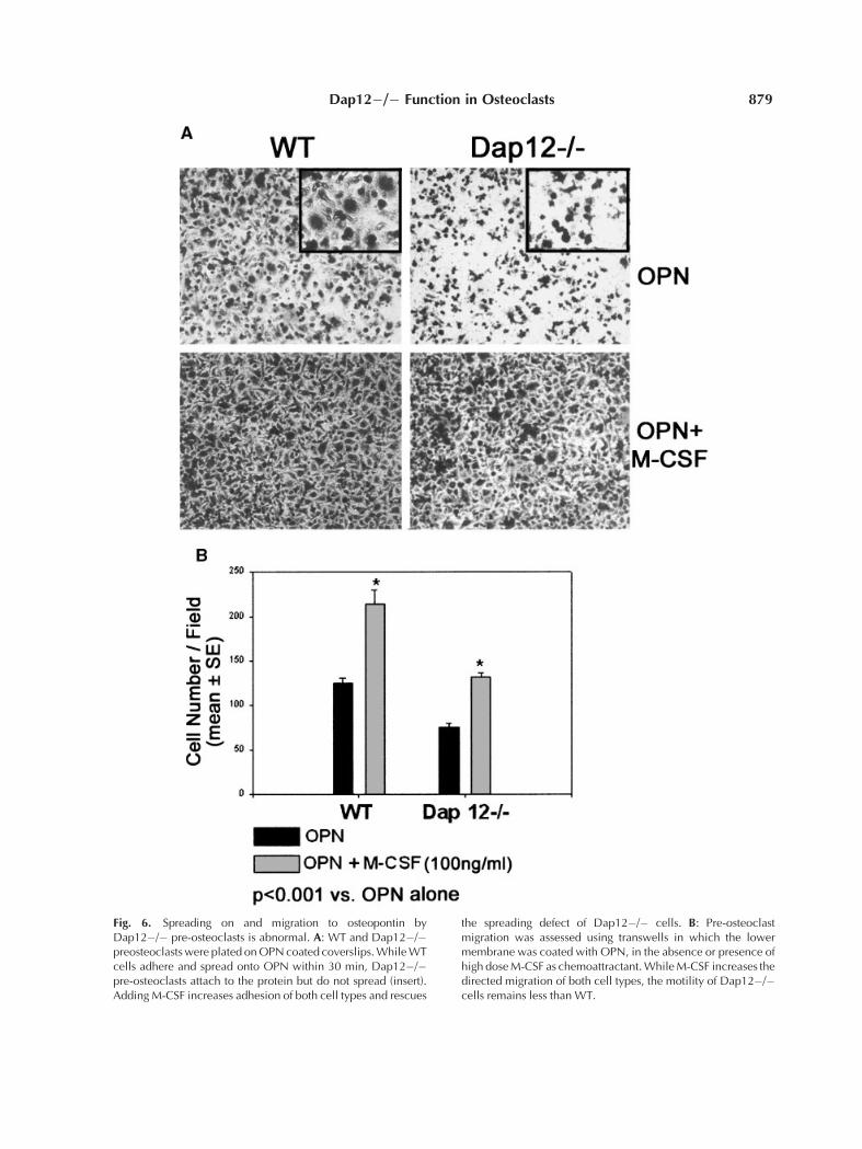
Fig. 6. Spreading on and migration to osteopontin byDap12�/� pre-osteoclasts is abnormal. A: WT and Dap12�/�preosteoclastswere plated onOPNcoated coverslips.WhileWTcells adhere and spread onto OPN within 30 min, Dap12�/�pre-osteoclasts attach to the protein but do not spread (insert).AddingM-CSF increases adhesion of both cell types and rescues
the spreading defect of Dap12�/� cells. B: Pre-osteoclastmigration was assessed using transwells in which the lowermembrane was coated with OPN, in the absence or presence ofhigh doseM-CSF as chemoattractant.WhileM-CSF increases thedirected migration of both cell types, the motility of Dap12�/�cells remains less than WT.
Dap12�/� Function in Osteoclasts 879

interface, characterized by a ring of corticalactin. Cells lacking DAP12 fail to generate thismarker of osteoclast polarization and this defectis not rescued by exposure to an increased con-centration of M-CSF. Interestingly, the samehigh level of M-CSF restores the polarizingcapacity of b3�/� osteoclasts but again, in thiscircumstance, the cells remain incapable ofresorption [Faccio et al., 2003b].
M-CSF, in addition to functioning as anosteoclast differentiation factor, also regulatesthe proliferation, spreading, and migration ofboth early precursors, in the form of spleen- orbone marrow-derived macrophages, as well ascells committed to the osteoclast lineage byexposure to RANKL [Stanley et al., 1997; Fenget al., 2002; Faccio et al., 2003b]. Because M-CSF does not completely normalize Dap12�/�osteoclast number we reasoned the residualdefect may reflect dampened precursor prolif-
eration in response to an abundance of thecytokine. In fact, such is the case.
While increased concentrations of M-CSFmarkedly increases generation of large osteo-clastic cells from Dap12�/� progenitors, themutant polykaryons are incapable of actin ringformation. Thus, while M-CSF has a posi-tive effect on Dap12�/� osteoclastogenesis, aprofound defect in cytoskeletal organizationpersists.
Similar to Dap12, the avb3 integrin regulatesthe osteoclast cytoskeleton [Faccio et al.,2003a]. Furthermore, both Dap12 and avb3interact with c-Cbl, a key osteoclastogenicsignaling molecule, also known to mediate cellspreading [McVicar et al., 1998; Sanjay et al.,2001]. These observations prompted us to ask iflike b3�/� pre-osteoclasts, those derived fromDap12 deficient mice, fail to spread on the avb3ligand, osteopontin. While wild type cells arefully spread within 30 min even in the absence ofM-CSF, such does not occur in those lackingDap12, despite their capacity to attach to thematrix protein. Addition of M-CSF rescuesthe spreading defect, suggesting that signalsemanating from DAP12 enhance the capacityof avb3 to ligate osteopontin. Haptotaxis andchemotaxis assays, using osteopontin as thecoating ligand and M-CSF as the chemoattrac-tant, reveal that once again cells lacking DAP12are deficient in both parameters and can bepartially rescued by the presence of M-CSF.Since attachment and spreading are both avb3dependent and cross talk exits between c-Fmsand avb3 [Faccio et al., 2003b], our findingsreveal a complex interaction between the tworeceptors and Dap12.
To identify potential effectors of DAP12, weexamined the activation of Syk, a tyrosinekinase interacting with and functioning down-stream of the adaptor protein in myeloid cells[Lanier and Bakker, 2000]. In our first studies,we used wild type and DAP12 null pre-osteo-clasts that we allowed to adhere to osteopontin,and then assessed the level of Syk phosphoryla-tion. Only cells expressing DAP12 are capable ofactivating Syk. To confirm this observation, weused pre-osteoclasts generated from macro-phages lacking Syk. In this instance, we treatedthe cells with RANKL and both low and highdose M-CSF. Irrespective of M-CSF concen-tration, absence of Syk precludes formation offully mature, spread osteoclasts. Furthermore,absence of the enzyme blocks matrix-induced
Fig. 7. Syk regulates intracellular signaling and osteoclastformation. A: WT and Dap12�/� pre-osteoclasts were main-tained in suspension (S) or plated onOPN (O) for 30min and Sykphosphorylation was analyzed by immunoblot. Cell adhesionincreases Syk phosphorylation inWTbut not in Dap12�/� cells.B: Sykþ/� and Syk�/� BMMs were maintained in the presenceof RANKL and low or high doseM-CSF. Sykþ/� osteoclasts formwithin 5 days in both conditions. In contrast, Syk�/� cells,independent of M-CSF concentrations, form TRAP-expressingcells with few nuclei, which fail to spread normally.
880 Faccio et al.

activation of c-Src and Pyk2, two tyrosinekinases essential to avb3 function in these cells[McVicar et al., 1998; Sanjay et al., 2001]. Thesemorphological findings suggest that, as in otherinstances in myeloid differentiation, Syk ap-pears to be downstream of DAP12. Thesestudies led us to examine the intracellularsignals that follow ligation of RANK andc-Fms, the receptors for RANKL and M-CSF,respectively. Despite the fact that Dap12�/�BMMs fail to become mature bone-resorbingosteoclasts, these cells normally activate majorsignals that follow binding of RANKL to RANK,
including those involving NF-kB and Akt[Darnay et al., 1998; Galibert et al., 1998;Matsumoto et al., 2000; Ross, 2000; Lee et al.,2002]. Similarly, M-CSF dependent phosphor-ylation of ERKs and induction of their distaltarget c-Fos is intact, an observation thatdiffers from that seen in the b3 null mice,where neither signal is activated by the cyto-kine [Kudo et al., 2002; Faccio et al., 2003b].
Finally, an important distinction exists be-tween the numbers of osteoclasts present in vivoin DAP12 null mice and the capacity to generatethe cells in vitro [Kaifu et al., 2003]. Thus,
Fig. 8. Syk�/� pre-osteoclasts adhere poorly and fail toactivate Src and Pyk2. A: Sykþ/� and Syk�/� pre OCs wereplated on OPN coated dishes. While Sykþ/� cells spread ontoOPNwithin 30min, Syk�/� pre-osteoclasts do not spread on theprotein.B: Sykþ/� and Syk�/� pre-osteoclasts weremaintained
in suspension (S) or plated on OPN (O) for 30 min and Src andPyk2 phosphorylation was analyzed by immunoblot. Celladhesion increases Src and Pyk2 phosphorylation in Sykþ/�but not in Syk�/� cells. b-Actin serves as loading control.
Dap12�/� Function in Osteoclasts 881

histomorphometry reveals that the mild osteo-petrosis of these animals is accompanied by nodeficiency of osteoclasts. This in vivo findingstands in contrast to in vitro data and ourcurrent observations, in which few matureosteoclasts are generated. One possible expla-nation lies in our studies of mice lacking NF-kBinducing kinase (NIK), where once again dis-parity exits between in vivo osteoclast numberand in the capacity to generate these cellsin vitro [Novack et al., 2003]. The fact that thein vitro phenotype can be rescued by TGFb, aprotein found in high concentration in bonematrix, suggests that complex mechanisms arein play in intact animals. Precisely what addi-tional factors permit generation of normalnumbers of albeit dysfunctional osteoclasts inDap12 mice, is an issue which may provideimportant insights into the mechanisms ofosteoclast recruitment and function.
ACKNOWLEDGMENTS
We thank Dr. C.A. Lowell (University ofCalifornia, San Francisco, CA) for provid-ing BMMs Dap12þ/þ, Dap�/�, Sykþ/�, andSyk�/�.
REFERENCES
Akisaka T, Yoshida H, Inoue S, Shimizu K. 2001. Organi-zation of cytoskeletal F-actin, G-actin, and gelsolin in theadhesion structures in cultured osteoclast. J Bone MinerRes 16:1248–1255.
Aoki N, Kimura S, Takiyama Y, Atsuta Y, Abe A, Sato K,Katagiri M. 2000. The role of the DAP12 signal in mousemyeloid differentiation. J Immunol 165:3790–3796.
Bakker AB, Hoek RM, Cerwenka A, Blom B, Lucian L,McNeil T, Murray R, Phillips LH, Sedgwick JD, LanierLL. 2000. DAP12-deficient mice fail to develop autoim-munity due to impaired antigen priming. Immunity 13:345–353.
Boyle WJ, Simonet WS, Lacey DL. 2003. Osteoclats differ-entiation and activation. Nature 15:337–342.
Cella M, Buonsanti C, Strader C, Kondo T, Salmaggi A,Colonna M. 2003. Impaired differentiation of osteo-clasts in TREM-2-deficient individuals. J Exp Med 198:645–651.
Cenci S, Weitzmann MN, Roggia C, Namba N, Novack D,Pacifici R. 2000. Estrogen deficiency induces bone loss byenhancing T cell production of TNFa. J Clin Invest 106:1229–1237.
Chambers TJ. 2000. Regulation of the differentiation andfunction of osteoclasts. J Pathol 192:4–13.
Colonna M. 2003. TREMs in the immune system andbeyond. Nat Rev Immunol 3:445–453.
Darnay BG, Haridas V, Ni J, Moore PA, Aggarwal BB.1998. Characterization of the intracellular domain ofreceptor activator of NF-kappaB (RANK). Interactionwith tumor necrosis factor receptor-associated factors
and activation of NF-kappab and c-Jun N-terminalkinase. J Biol Chem 273:20551–20555.
Faccio R, Novack DV, Zallone A, Ross FP, Teitelbaum SL.2003a. Dynamic changes in the osteoclast cytoskeleton inresponse to growth factors and cell attachment arecontrolled by {beta}3 integrin. J Cell Biol 162:499–509.
Faccio R, Zallone A, Ross FP, Teitelbaum SL. 2003b. c-Fmsand the avb3 integrin collaborate during osteoclastdifferentiation. J Clin Invest 111:749–758.
Feng X, Takeshita S, Namba N, Wei S, Teitelbaum SL, RossFP. 2002. Tyrosines 559 and 807 in the cytoplasmic tail ofthe m-csf receptor play distinct roles in osteoclast differ-entiation and function. Endocrinology 143:4868–4874.
Galibert L, Tometsko ME, Anderson DM, Cosman D,Dougall WC. 1998. The involvement of multiple tumornecrosis factor receptor (TNFR)-associated factors in thesignaling mechanisms of receptor activator of NF-kappaB, a member of the TNFR superfamily. J BiolChem 273:34120–34127.
Jenkins JK, Hardy KJ, McMurray RW. 2002. The patho-genesis of rheumatoid arthritis: A guide to therapy. AmJ Med Sci 323:171–180.
Kaifu T, Nakahara J, Inui M, Mishima K, Momiyama T,Kaji M, Sugahara A, Koito H, Ujike-Asai A, Nakamura A,Kanazawa K, Tan-Takeuchi K, Iwasaki K, YokoyamaWM, Kudo A, Fujiwara M, Asou H, Takai T. 2003. Osteo-petrosis and thalamic hypomyelinosis with synapticdegeneration in DAP12-deficient mice. J Clin Invest111:323–332.
Kondo T, Takahashi K, Kohara N, Takahashi Y, Hayashi S,Takahashi H, Matsuo H, Yamazaki M, Inoue K,Miyamoto K, Yamamura T. 2002. Heterogeneity of pre-senile dementia with bone cysts (Nasu–Hakola disease):Three genetic forms. Neurology 59:1105–1107.
Kudo O, Sabokbar A, Pocock A, Itonaga I, Athanasou NA.2002. Isolation of human osteoclasts formed in vitro:Hormonal effects on the bone-resorbing activity ofhuman osteoclasts. Calcif Tissue Int 71:539–546.
Lanier LL, Bakker ABH. 2000. The ITAM-bearing trans-membrane adaptor DAP12 in lymphoid and myeloid cellfunction. Immunol Today 21:611–614.
Lee SE, Woo KM, Kim SY, Kim H-M, Kwack K, Lee ZH,Kim H-H. 2002. The phosphatidylinositol 3-kinase, p38,and extracellular signal-regulated kinase pathways areinvolved in osteoclast differentiation. Bone 30:71–77.
Lucas M, Daniel L, Tomasello E, Guia S, Horschowski N,Aoki N, Figarella-Branger D, Gomez S, Vivier E. 2002.Massive inflammatory syndrome and lymphocytic im-munodeficiency in KARAP/DAP12-transgenic mice. EurJ Immunol 32:2653–2663.
Marks SC, Jr. 1989. Osteoclast biology: Lessons from mam-malian mutations. Am J Med Genet 34:43–54.
Matsumoto M, Sudo T, Saito T, Osada A, Tsujimoto M.2000. Involvement of p38 mitogen-activated proteinkinase signaling pathway in osteoclastogenesis mediatedby receptor activator of NF-kappa B ligand (RANKL).J Biol Chem 275:31155–31161.
McHugh KP, Hodivala-Dilke K, Zheng MH, Namba N, LamJ, Novack D, Feng X, Ross FP, Hynes RO, Teitelbaum SL.2000. Mice lacking b3 integrins are osteosclerotic becauseof dysfunctional osteoclasts. J Clin Invest 105:433–440.
McLean W, Olsen BR. 2001. Mouse models of abnormalskeletal development and homeostasis. Trends Genet 17:S38–S43.
882 Faccio et al.

McVicar DW, Taylor LS, Gosselin P, Willette-Brown J,Mikhael AI, Geahlen RL, Nakamura MC, Linnemeyer P,Seaman WE, Anderson SK, Ortaldo JR, Mason LH. 1998.DAP12-mediated signal transduction in natural killercells. A dominant role for the Syk protein-tyrosinekinase. J Biol Chem 273:32934–32942.
Mocsai A, Zhou M, Meng F, Tybulewicz VL, Lowell CA.2002. Syk is required for integrin signaling in neutro-phils. Immunity 16:547–548.
Novack DV, Yin L, Hagen-Stapleton A, Schreiber RD,Goeddel DV, Ross FP, Teitelbaum SL. 2003. The IkBfunction of NF-kB2 p100 controls stimulated osteoclas-togenesis. J Exp Med 198:771–781.
Obergfell A, Eto K, Mocsai A, Buensuceso C, Moores SL,Brugge JS, Lowell CA, Shattil SJ. 2002. Coordinate in-teractions of Csk, Src, and Syk kinases with {alpha}IIb-{beta}3 initiate integrin signaling to the cytoskeleton.J Cell Biol 157:265–275.
Paloneva J, Kestila M, Wu J, Salminen A, Bohling T,Ruotsalainen V, Hakola P, Bakker AB, Phillips JH,Pekkarinen P, Lanier LL, Timonen T, Peltonen L. 2000.Loss-of-function mutations in TYROBP (DAP12) resultin a presenile dementia with bone cysts. Nat Genet 25:356–361.
Paloneva J, Manninen T, Christman G, Hovanes K,Mandelin J, Adolfsson R, Bianchin M, Bird T,Miranda R, Salmaggi A, Tranebjaerg L, Konttinen Y,Peltonen L. 2002. Mutations in two genes encodingdifferent subunits of a receptor signaling complex resultin an identical disease phenotype. Am J Hum Genet 71:656–662.
Romas E, Gillespie MT, Martin TJ. 2002. Involvementof receptor activator of NFkappaB ligand and tumornecrosis factor-alpha in bone destruction in rheumatoidarthritis. Bone 30:340–346.
Ross FP. 2000. RANKing the importance of measles virus inPaget’s disease. J Clin Invest 105:555–558.
Sanjay A, Houghton A, Neff L, Didomenico E, Bardelay C,Antoine E, Levy J, Gailit J, Bowtell D, Horne WC, BaronR. 2001. Cbl associates with Pyk2 and Src to regulate Srckinase activity, avb3 integrin-mediated signaling, celladhesion, and osteoclast motility. J Cell Biol 152:181–196.
Sjolin H, Tomasello E, Mousavi-Jazi M, Bartolazzi A, KarreK, Vivier E, Cerboni C. 2002. Pivotal role of KARAP/DAP12 adaptor molecule in the natural killer cell-mediated resistance to murine cytomegalovirus infection.J Exp Med 195:825–834.
Stanley ER, Berg KL, Einstein DB, Lee PSW, Pixley FJ,Wang Y, Yeung Y-G. 1997. Biology and action of colony-stimulating factor-1. Mol Reprod Dev 46:4–10.
Suda T, Takahashi N, Udagawa N, Jimi E, Gillespie MT,Martin TJ. 1999. Modulation of osteoclast differentiationand function by the new members of the tumor necrosisfactor receptor and ligand families. Endocr Rev 20:345–357.
Takeshita S, Kaji K, Kudo A. 2000. Identification andcharacterization of the new osteoclast progenitor withmacrophage phenotypes being able to differentiate intomature osteoclasts. J Bone Miner Res 15:1477–1488.
Tanaka S, Takahashi N, Udagawa N, Tamura T, Akatsu T,Stanley ER, Kurokawa T, Suda T. 1993. Macrophagecolony-stimulating factor is indispensable for both pro-liferation and differentiation of osteoclast progenitors.J Clin Invest 91:257–263.
Teitelbaum SL. 2000. Bone resorption by osteoclasts.Science 289:1504–1508.
Teitelbaum SL, Ross FP. 2003. Genetic regulation of osteo-clast development and function. Nat Rev Genet 4:638–649.
Teti A, Marchisio PC, Zallone AZ. 1991. Clear zone in osteo-clast function: Role of podosomes in regulation of bone-resorbing activity. Am J Physiol 261:C1–C7.
Tomasello E, Olcese L, Vely F, Geourgeon C, Blery M,Moqrich A, Gautheret D, Djabali M, Mattei M-G, VivierE. 1998. Gene structure, expression pattern, and biolo-gical activity of mouse killer cell activating receptor-associated protein (KARAP)/DAP-12. J Biol Chem 273:34115–34119.
Tomasello E, Desmoulins P-O, Chemin K, Guia S, CremerH, Ortaldo J, Love P, Kaiserlian D, Vivier E. 2000.Combined natural killer cell and dendritic cell functionaldeficiency in KARAP/DAP12 loss-of-function mutantmice. Immunity 13:355–364.
Weitzmann MN, Cenci S, Rifas L, Brown C, Pacifici R.2000. IL-7 stimulates osteoclast formation by upregulat-ing the T-cell production of soluble osteoclastogeniccytokines. Blood 96:1873–1878.
Wilder RL. 2002. Integrin avb3 as a target for treatment ofrheumatoid arthritis and related rheumatic diseases.Ann Rheum Dis 61:ii96–ii99.
Woo KM, Kim HM, Ko JS. 2002. Macrophage colony-stimulating factor promotes the survival of osteoclastprecursors by up-regulating Bcl-XL. Exp Mol Med 34:340–346.
Wu J, Cherwinski H, Spies T, Phillips JH, Lanier LL. 2000.DAP10 and DAP12 form distinct, but functionally co-operative, receptor complexes in natural killer cells.J Exp Med 192:1059–1068.
Zaidi M, Blair HC, Moonga BS, Abe E, Huang CL-H. 2003.Osteoclastogenesis, bone resorption, and osteoclast-based therapeutics. J Bone Miner Res 18:599–609.
Dap12�/� Function in Osteoclasts 883