Interleukin (IL)-6 Induction of Osteoclast Differentiation Depends on IL6 Receptors Expressed on...
8
Interleukin (IL)-6 Induction of Osteoclast Differentiation Depends on IL-6 Receptors Expressed on Osteoblastic Cells But Not on Osteoclast Progenitors By Nobuyuki Udagawa,*~ Naoyuki Takahashi,* Takenobu Katagiri,* Tatsuya Tamura,* Seiki Wada, l: David M. Findlay,:[: T.John Martin,~ Hisao Hirota,w TetsuyaTaga,[[ Tadamitsu Kishimoto,w and Tatsuo Suda* From the *Department of Biochemistry, School of Dentistry, Showa University,Tokyo 142, Japan; ~St. Vincent's Institute of Medical Research, University of Melbourne, Fitzroy, Victoria 3065, Australia; w of Medicine III, Osaka University Medical School, Osaka 565,Japan; and Ilnstitutefor Molecular and Cellular Biology, Osaka University, Osaka 565,Japan Summary We reported that interleukin (IL) 6 alone cannot induce osteoclast formation in cocultures of mouse bone marrow and osteoblastic cells, but soluble IL-6 receptor (IL-6R) strikingly trig- gered osteoclast formation induced by IL-6. In this study, we examined the mechanism of os- teoclast formation by IL-6 and related cytokines through the interaction between osteoblastic cells and osteoclast progenitors. When dexamethasone was added to the cocultures, IL-6 could stimulate osteoclast formation without the help of soluble IL-6K. Osteoblastic cells expressed a very low level of IL-6R mRNA, whereas fresh mouse spleen and bone marrow cells, both of which are considered to be osteoclast progenitors, constitutively expressed relatively high levels of IL-6K mRNA. Treatment of osteoblastic cells with dexamethasone induced a marked in- crease in the expression of IL-6R mRNA. By immunoblotting with antiphosphotyrosine anti- body, IL-6 did not tyrosine-phosphorylate a protein with a molecular mass of 130 kD in osteo- blastic cells but did so in dexamethasone-pretreated osteoblastic cells. Osteoblastic cells from transgenic mice constitutively expressing human IL-6K could support osteoclast development in the presence of human IL-6 alone in cocultures with normal spleen cells. In contrast, osteo- clast progenitors in spleen cells from transgenic mice overexpressing human IL-6P,. were not able to differentiate into osteoclasts in response to IL-6 in cocultures with normal osteoblastic cells. These results clearly indicate that the abihty of IL-6 to induce osteoclast differentiation depends on signal transduction mediated by IL-6R expressed on osteoblastic cells but not on osteoclast progenitors. B one formation and resorption are coupled through the actions of several locally produced growth factors and cytokines. Osteoblastic cells have receptors for bone- resorbing hormones, including parathyroid hormone, PGs, and 10t,25-dihydroxyvitamin D 3 [10t,25(OH)2D3] 1. How- ever, there is no convincing evidence of direct responses to these bone-resorbing hormones in osteoclast lineage cells A portion of this work was presented at the XII InternationalConference on Calcium Regulating Hormones, Melbourne, Australia, 14-19 Febru- ary 1995, and has been pubhshed in abstractform (Udagawa, N., T. Kat- agiri, N. Takahashi, H. Murakami, I. Nakamura, D. Zhang, T. Tamura, T.J. Martin, and T. Suda. 1995. Bone 16:92S). 1Abbreviations used in this paper: 1a,25(OH)2 D3,1a,25-dihydroxyvitamin D3; GAPDH, glyceraldehyde 3-phosphate dehydrogenase; IL-6R., IL- 11Ik, IL-6 and IL-11 receptor, respectively; slL-6R, soluble IL-6R; OCL, osteoclast-like multinucleated cells; TRAP, tartrate-resistant acid phosphatase. (1). Thus the concept that bone-resorbing agents must act first on osteoblastic stromal cells to induce the generation of new osteoclasts has emerged (2-4), although some con- troversial reports suggest that osteoblastic stromal cells are not required for osteoclast differentiation (5-7). We developed a coculture system of mouse hematopoie- tic and primary osteoblastic stromal cells with which to in- vestigate osteoclast development in vitro (8). In these co- cultures, osteoclast-like multinucleated cells (OCLs) formed in response to several bone-resorbing factors such as lcx,25- (OH)2D3, parathyroid hormone, parathyroid hormone- related protein, PGE2, IL-1, and IL-6 (9-11). OCLs formed in these cocultures satisfied the major criteria of au- thentic osteoclasts, including tartrate-resistant acid phos- phatase (TRAP) activity, calcitonin receptor expression, and formation of resorption pits on dentine slices (8, 12, 13). When osteoclast progenitors and osteoblastic cells 1461 j. Exp. Med. 9 The Rockefeller University Press 9 0022-1007/95/11/1461/08 $2.00 Volume 182 November 1995 1461-1468 on February 2, 2014 jem.rupress.org Downloaded from Published November 1, 1995
Transcript of Interleukin (IL)-6 Induction of Osteoclast Differentiation Depends on IL6 Receptors Expressed on...

Interleukin (IL)-6 Induc t ion o f Osteoclast Dif ferent iat ion D e p e n d s on IL-6 Receptors Expressed o n Osteoblast ic Cells But N o t on Osteoclast Progenitors
By Nobuyuki Udagawa,*~ Naoyuki Takahashi,* Takenobu Katagiri,* Tatsuya Tamura,* Seiki Wada, l: David M. Findlay,:[: T.John Martin,~ Hisao Hirota,w Tetsuya Taga,[[ Tadamitsu Kishimoto,w and Tatsuo Suda*
From the *Department of Biochemistry, School of Dentistry, Showa University, Tokyo 142, Japan; ~St. Vincent's Institute of Medical Research, University of Melbourne, Fitzroy, Victoria 3065, Australia; w of Medicine III, Osaka University Medical School, Osaka 565,Japan; and Ilnstitute for Molecular and Cellular Biology, Osaka University, Osaka 565,Japan
S u m m a r y
We reported that interleukin (IL) 6 alone cannot induce osteoclast formation in cocultures of mouse bone marrow and osteoblastic cells, but soluble IL-6 receptor (IL-6R) strikingly trig- gered osteoclast formation induced by IL-6. In this study, we examined the mechanism of os- teoclast formation by IL-6 and related cytokines through the interaction between osteoblastic cells and osteoclast progenitors. When dexamethasone was added to the cocultures, IL-6 could stimulate osteoclast formation without the help of soluble IL-6K. Osteoblastic cells expressed a very low level of IL-6R mRNA, whereas fresh mouse spleen and bone marrow cells, both of which are considered to be osteoclast progenitors, constitutively expressed relatively high levels of IL-6K mRNA. Treatment of osteoblastic cells with dexamethasone induced a marked in- crease in the expression of IL-6R mRNA. By immunoblotting with antiphosphotyrosine anti- body, IL-6 did not tyrosine-phosphorylate a protein with a molecular mass of 130 kD in osteo- blastic cells but did so in dexamethasone-pretreated osteoblastic cells. Osteoblastic cells from transgenic mice constitutively expressing human IL-6K could support osteoclast development in the presence of human IL-6 alone in cocultures with normal spleen cells. In contrast, osteo- clast progenitors in spleen cells from transgenic mice overexpressing human IL-6P,. were not able to differentiate into osteoclasts in response to IL-6 in cocultures with normal osteoblastic cells. These results clearly indicate that the abihty of IL-6 to induce osteoclast differentiation depends on signal transduction mediated by IL-6R expressed on osteoblastic cells but not on osteoclast progenitors.
B one formation and resorption are coupled through the actions of several locally produced growth factors and
cytokines. Osteoblastic cells have receptors for bone- resorbing hormones, including parathyroid hormone, PGs, and 10t,25-dihydroxyvitamin D 3 [10t,25(OH)2D3] 1. How- ever, there is no convincing evidence of direct responses to these bone-resorbing hormones in osteoclast lineage cells
A portion of this work was presented at the XII International Conference on Calcium Regulating Hormones, Melbourne, Australia, 14-19 Febru- ary 1995, and has been pubhshed in abstract form (Udagawa, N., T. Kat- agiri, N. Takahashi, H. Murakami, I. Nakamura, D. Zhang, T. Tamura, T.J. Martin, and T. Suda. 1995. Bone 16:92S).
1Abbreviations used in this paper: 1a,25(OH)2 D3,1a,25-dihydroxyvitamin D3; GAPDH, glyceraldehyde 3-phosphate dehydrogenase; IL-6R., IL- 11Ik, IL-6 and IL-11 receptor, respectively; slL-6R, soluble IL-6R; OCL, osteoclast-like multinucleated cells; TRAP, tartrate-resistant acid phosphatase.
(1). Thus the concept that bone-resorbing agents must act first on osteoblastic stromal cells to induce the generation of new osteoclasts has emerged (2-4), although some con- troversial reports suggest that osteoblastic stromal cells are not required for osteoclast differentiation (5-7).
We developed a coculture system of mouse hematopoie- tic and primary osteoblastic stromal cells with which to in- vestigate osteoclast development in vitro (8). In these co- cultures, osteoclast-like multinucleated cells (OCLs) formed in response to several bone-resorbing factors such as lcx,25- (OH)2D3, parathyroid hormone, parathyroid hormone- related protein, PGE2, IL-1, and IL-6 (9-11). OCLs formed in these cocultures satisfied the major criteria of au- thentic osteoclasts, including tartrate-resistant acid phos- phatase (TRAP) activity, calcitonin receptor expression, and formation of resorption pits on dentine slices (8, 12, 13). When osteoclast progenitors and osteoblastic cells
1461 j. Exp. Med. �9 The Rockefeller University Press �9 0022-1007/95/11/1461/08 $2.00 Volume 182 November 1995 1461-1468
on February 2, 2014
jem.rupress.org
Dow
nloaded from
Published November 1, 1995

were cocultured but separated by a membrane filter, no OCLs were formed, even in the presence o f bone-resorb- ing agents. Osteoblastic stromal cells, therefore, play an im- portant role in modulating the development o f osteoclast progenitors by mechanisms requiring cell-cell interaction (14--16). At this time, it is not clear whether osteoblastic cells or osteoclast progenitors are the primary target cells for bone-resorbing agents in osteoclast differentiation. In this study, we identified which of the cocultured cells are the target cells for IL-6, which is now recognized as an im- portant osteotropic factor.
IL-6 is a multifunctional cytokine that regulates pleiotro- pic functions in many types of cells (17). There is much ev- idence to suggest that IL-6 is an important osteotropic fac- tor. IL-6 is produced by osteoblastic cells in response to bone-resorbing agents (18, 19) and stimulates osteoclastic bone resorption in organ cultures (20). Ant i - IL-6 antibody inhibits the bone-resorbing activity o f OCLs obtained from giant cell tumors o f bone (21). IL-6 causes hypercalcemia in vivo (22, 23) and has been implicated in the bone loss caused by estrogen deficiency. Ovariectomy in the mouse causes a marked increase in ex vivo O C L formation in marrow cultures (24). Jilka et al. (25) have shown that es- trogen replenishment inhibits IL-6 production by osteo- blastic cells and that anti-IL-6 antibody prevented O C L development in the estrogen-deplete state. Since IL-6 - deficient transgenic mice maintained their bone mass after ovariectomy (26), it is likely that IL-6 is involved in the differentiation o f osteoclasts and osteoclastic bone resorp- tion in estrogen deficiency. However, the mechanisms by which IL-6 influences osteoclast formation have not been established.
While investigating the action o f IL-6 on O C L forma- tion, we found that soluble IL-6 receptors (slL-6R), which lack both transmembrane and cytoplasmic domains, can also bind IL-6 and mediate IL-6 signaling by interacting with the signal-transducing receptor component, gp130 (27). Whereas mouse IL-6 alone did not stimulate O C L formation in cocultures o f mouse bone marrow cells and osteoblastic ceils (11), the simultaneous addition o f mouse sIL-6R strikingly triggered O C L formation in the cocul- tures. This suggests that cells in these cultures do not nor- mally express IL-6 receptors but express gp130. IL-11, leu- kemia inhibitory factor, and oncostatin M, which share gp130 as the common signal transducer (28, 29), similarly stimulated O C L formation in the cocultures. The potency of these cytokines to induce OCLs may reflect the number o f respective receptors in bone marrow and/or osteoblastic cells. It is important to emphasize that none of these gp130-stimulatory cytokines could induce OCLs when bone marrow and osteoblastic cells were cocultured with- out direct contact. Together, these results suggest that the gp130 signal is involved in osteoclast development, al- though it remains to be clarified which cells in the cocul- tures primarily require such a signal.
In this study, we first examine the role o f IL-6 in O C L formation in the coculture system, and then demonstrate that dexamethasone stimulates expression of membrane-
bound IL-6 receptors (IL-6R) on osteoblastic cells, leading to the differentiation o f OCLs in response to mouse IL-6 in cocultures without a requirement for added mouse slL-6R. We also show that osteoblastic cells obtained from trans- genic mice constitutively expressing membrane-anchored human IL-6R can support osteoclast differentiation in re- sponse to human IL-6 in cocultures with normal spleen cells. This indicates that it is osteoblastic cells rather than osteoclast progenitors that require the gp130 signal during O C L for,nation in the coculture system.
Materials and Methods Animals and Drugs. Newborn ddY and 6-9-wk-old male
ddY mice were obtained from Shizuoka Laboratories Animal Center (Shizuoka, Japan). 1ot,25(OH)2D 3 was purchased from Wako Pure Chemical Co. (Osaka, Japan). Dexamethasone was purchased from Sigma Chemical Co. (St. Louis, MO). Mouse rlL-6 and mouse rslL-6R were prepared as described (11, 30). Human rlL-6 and human rlL-11 were purchased from R&D Sys- tems, Inc. (Minneapolis, MN). Other chemicals and reagents were of analytical grade.
Construction of the Human IL-6R Transgene and Preparation of. Transgenic Mice. The IL-6R transgene was prepared by inserting a 1.7-kb fragment of the human IL-6R cDNA (31) spanning the entire coding region into a unique XhoI site of the pCAGGS ex- pression vector carrying the constitutively active chicken ~-actin gene promoter (32). This vector was then digested with Sad and HindlII to generate a linear fragment and microinjected into the pronudei of fertilized BDF1 mouse eggs. Mice were obtained as described (33-35). Newborn transgenic mice were generated by mating animals from human IL-6R-transgenic lines and normal C57BL/6 mice, and their genotypes were determined by PCR analysis of liver DNA from each mouse as described (35).
Coculture System and Determination of Osteoclast Characteristics. Normal osteoblastic cells were prepared from the calvaria of 2-d- old mice by digestion with 0.1% collagenase (Wako Pure Chem- ical Co.) and 0.2% dispase (Godo Shusei, Tokyo, Japan). Normal bone marrow and spleen cells were obtained from 7-wk-old ddY mice as described (11, 36). To isolate osteoblastic cells from trans- genic mice overexpressing human IL-6R, each calvarium front 2-d-old mice was cut into small pieces and embedded into type I collagen gels according to the manufacturer's instructions (Cell- matrix Type l-A; Nitta Gelatin Co., Osaka, Japan). After culture for 3 d, osteoblastic cells grown from the calvaria were collected by treating with PBS containing 0.1% collagenase (36). Spleen cells from transgenic mice were prepared from the splenic tissues of individual littermates (36). Osteoblastic cells were cocultured with bone marrow or spleen cells as described (11, 36). In short, primary osteoblastic cells (104 per well) and nucleated spleen cells (5 • 10 s per well) or marrow cells (2 • 10 s per well) were cocul- tured in the wells of a 48-well plate (Coming Glass Inc., Corn- ing, NY) with 0.4 ml of o~-MEM (GIBCO BRL, Gaithersburg, MD) containing 10% fetal bovine serum (Cytosystems, Castle Hill, NSW, Australia) in the presence of test chemicals. Cultures were incubated in quadruplicate, and cells were replenished on day 4 with fresh medium. OCL formation was evaluated after culturing for 7 d. Adherent cells were fixed and stained for TRAP, and the number of TRAP-positive OCLs was scored as described (11). The expression ofcalcitonin receptors was also as- sessed by autoradiography using 12sI-salmon calcitonin as de- scribed (11).
1462 Mechanism of IL-6--induced Osteoclast Formation
on February 2, 2014
jem.rupress.org
Dow
nloaded from
Published November 1, 1995
PCR Amplification of Reverse-transcribed mRNA. Mouse primary osteoblastic cells in flasks (175 cm2; Nunc Inc., Naperville, IL) were cultured with or without 10 -7 M dexamethasone for vari- ous periods after reaching confluence, and total cellular RNA was extracted using guanidine thiocyanate-phenol chloroform (37). RNA was also prepared from mouse spleen and bone marrow cells,
Single-stranded cDNA was synthesized from 2.5 ~g of total RNA by incubating for 1 h at 42~ with avian myeloblastosis vi- res reverse transcriptase (Promega Corp., Madison, WI) and ran- dom hexanucleotides (Promega Corp.). From this reaction mix- ture, 2.5 ~l of 25 t~1 was amplified by PCR to generate mouse IL-6R m P ~ A , IL-11R mtU~A, and mouse glyceraldehyde 3-phos- phate dehydrogenase (GAPDH) mRNA. The reaction mixture contained 50 pmol of each primer, 25 mM dATP, dGTP, dCTP, and dTTP (Pharmacia, Uppsala, Sweden), 2 ~1 of 10>( reaction buffer, 1 U ofTaq DNA polymerase (Boehringer Mannheim, In- dianapolis, IN), and sterile distilled water, and was overlayed with 50 wl of paraffin oil. PCR (IL-GR) was performed for 23 cycles at 94~ for 1 rain, at 65~ for 1 rain, and at 72~ for 1 min in a DNA thermal cycler (model 480; Perkin Elmer Cetus Instru- ments, Norwalk, CT). Preliminary experiments were performed to ensure that the number of PCR cycles were within the expo- nential phase of the amplification curve. PCR products were re- solved on a 2% wt/vol agarose gel, and the specificity of the reac- tion was confirmed by Southern transfer onto nylon membranes (Hybond-N; Amersham, Arlington Heights, IL) and hybridiza- tion with 32p-labeled internal oligonucleotide probes.
Oligonucleotides were synthesized on a DNA synthesizer (model 381A; Applied Biosystems, Inc., Foster City, CA). The oligonucleotides for mouse IL-6R were 5 ' -CCTGTGTGG- GGTTCCAGAGGAT-3 ' (3' primer complementary to nucleo- tides 980-1001) and 5 ' -CTGCCAGTATTCTCAGCAGCTG-3' (5' primer complementary to nucleotides 488-519). The prod- ucts were verified by hybridization with the internal oligonucleo- tide, 5 ' -CACAACGAAGCGTTTCACAGCTT-3 ' , by Southern hybridization. The oligonucleotides for mouse IL-11R (38) were 5 ' -GGAGGCCTCCAGAGGGT-3 ' (3' primer complementary to nucleotides 661--677) and 5 ' -GGGTCCTCCAGGGGTCCAG- TATGG-3' (5' primer complementary to nucleotides 133-156). The products were verified by Southern hybridization with the internal oligonucleotide, 5 '-CTCCTGTACTTGGAGTCCAGG-3'. To ensure equal starting quantities of DNA for the experiments, and to allow semiquantitation of the PCR products representing IL-6R or IL-11R, reverse-transcribed RNA samples were also amplified using oligonucleotide primers specific for GAPDH (39). Oligonucleotides for GAPDH were 5 ' -AACGGATACAT- TGGGGGTAG-3 ' (complementary to nucleotides 701-720) and 5 ' -CATGGAGAAGGCTGGGGCTC-3 ' (complementary to nu- cleotides 306-325). The products were verified by hybridization with the internal oligonucleotide, 5 ' -GCTGTGGGCAAGGT- CATCCC-3' .
Immunoblotting. After incubating osteoblastic cells with or without dexamethasone (10 -7 M) for 48 h in flasks (25 cm2; Com- ing Glass Inc.), they were stimulated with or without IL-11 (500 ng/ml) or IL-6 (1 btg/ml) for 7 rain. The cells were washed in situ with ice-cold PBS, lysed in 100 bU oflysis buffer (0.5% NP- 40/10 mM Tris-HCl, pH 7.6/150 mM NaCl/5 mM EDTA/2 mM Na3Vo4/1 mM PMSF/5 t*g of aprotinin/ml), then deter- gent-insoluble materials were pelleted by centrifugation (15,000 g for 20 min at 4~ as described (40). The solubilized proteins were separated on a 7.5% SDS-polyacrylamide gel, then trans- ferred to a membrane (Immobilon-P; Millipore Corp., Bedford,
MA). The proteins were immunostained with antiphosphoty- rosine mAb (PY20) (Seikagaku Kogyo, Tokyo, Japan) using an enhanced chemiluminescence Western blotting detection system (Amersham) according to the manufacturer's recommendations.
Results
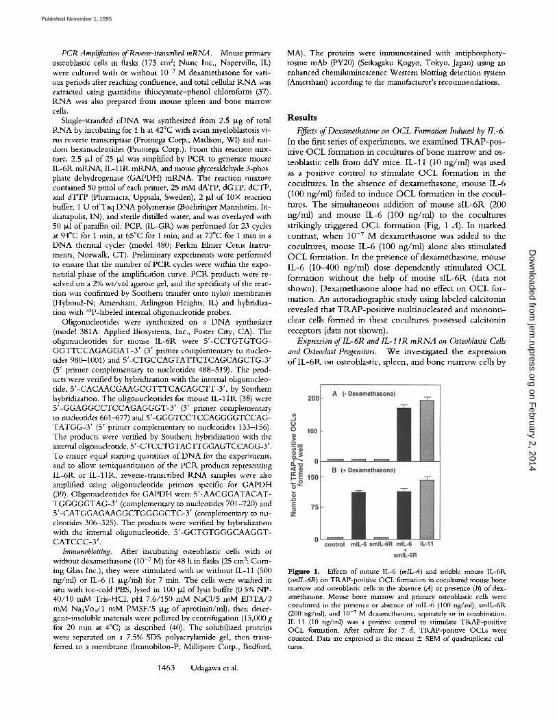
Effects of Dexamethasone on OCL Formation Induced by IL-6. In the first series o f experirnents, we examined T R A P - p o s - itive O C L formation in cocultures o f bone marrow and os- teoblastic cells from ddY mice. IL-11 (10 ng/ml) was used as a positive control to stimulate O C L formation in the cocultures. In the absence o f dexamethasone, mouse IL-6 (100 ng/ml) failed to induce O C L formation in the cocul- tures. The simultaneous addit ion o f mouse s lL-6R (200 ng/ml) and mouse IL-6 (100 ng/ml) to the cocultures strikingly triggered O C L formation (Fig. 1 A). In marked contrast, when 10 -v M dexamethasone was added to the cocultures, mouse IL-6 (100 ng/ml) alone also stimulated O C L formation. In the presence ofdexamethasone , mouse IL-6 (10-400 ng/ml) dose dependent ly stimulated O C L formation wi thout the help o f mouse s lL-6R (data not shown). Dexamethasone alone had no effect on O C L for- mation. An autoradiographic study using labeled calcitonin revealed that TRAP-pos i t ive mult inucleated and m o n o n u - clear cells formed in these cocultures possessed calcitonin receptors (data not shown).
Expression of lL-6R and IL-11R m R N A on Osteoblastic Cells and Osteoclast Progenitors. W e investigated the expression o f I L - 6 R on osteoblastic, spleen, and bone marrow cells by
Figure 1. Effects of mouse IL-6 (rolL-6) and soluble mouse IL-6R (smlL-6R) on TRAP-positive OCL formation in cocultured mouse bone marrow and osteoblastic cells in the absence (A) or presence (B) of dex- amethasone. Mouse bone marrow and primary osteoblastic cells were cocultured in the presence or absence of raiL-6 (100 ng/ml), smlL-6R (200 ng/ml), and 10 -7 M dexamethasone, separately or in combination. IL-11 (10 ng/ml) was a positive control to stimulate TRAP-positive OCL formation. After culture for 7 d, TRAP-positive OCLs were counted. Data are expressed as the means + SEM of quadruplicate cul- tures.
1463 Udagawa et al.
on February 2, 2014
jem.rupress.org
Dow
nloaded from
Published November 1, 1995

Figure 2. Expression oflL-6R mRNA on mouse osteoblastic cells and the osteoclast progenitors, mouse spleen (SP) and bone marrow (BM) cells. Osteoblastic cells were incubated with (+) or without ( - ) 10 -7 M dexamethasone (Dex) for 12, 24, and 48 h. The spleen and bone marrow cells were isolated immediately before use. Total 1LNA was reverse tran- scribed and amplified by 23 cycles of PCtL for mouse IL-6tL mB.NA and 20 cycles for mouse GAPDH mRNA using the specific primers described in Materials and Methods. PCtL products were transferred to a nylon mem- brane and hybridized with 32p-labeled internal sense oligonucleotides spe- cific to the mouse IL-6IL or the GAPDH sequences, respectively.
reverse transcript ion P C I k wi th mouse IL -6R-spec i f i c ol i - gonucleot ides . M o u s e pr imary osteoblastic stromal cells ex- pressed only a l o w level o f IL-6R. m R N A . T r e a t m e n t o f osteoblastic cells w i th 10 .7 M dexamethasone for 12-48 h markedly increased IL-61:( m t ( N A expression (Fig. 2). Fresh mouse spleen and b o n e m a r r o w cells, howeve r , c o n -
sti tutively expressed relatively h igh levels o f I L - 6 R m R N A (Fig. 2). In contrast, I L - 1 1 R m R N A was const i tut ively ex- pressed on osteoblastic cells i rrespective o f the presence or absense o f dexamethasone (Fig. 3). Fresh mouse spleen and b o n e m a r r o w cells also expressed I L - 1 1 R r n R N A (Fig. 3).
T h e results suggest that O C L format ion in the cocul tures in response to gp130-s t imula tory cytokines may be deter - m i n e d by the expression o f a l igand-b ind ing chain o f the receptor c o m p l e x in osteoblastic cells. A sandwich ELISA did no t detect mouse slL-6IL (30) in the cond i t i oned m e - d i u m o f cocul tures o f bone m a r r o w and osteoblastic cells, e i ther in the presence or absense o f dexamethasone (detec- t ion l imit , 1.0 n g / m l ; data no t shown).
Analysis of Protein Tyrosine Phosphorylation in Osteoblasts. T h e signals induced by cytokines that use gp130 as a signal t ransducer tr igger the act ivat ion o f some tyrosine kinases at an early step o f the signal t ransduct ion pa thway (29, 40). T o evaluate w h e t h e r the IL-6 and IL-11 signaling pathways
Figure 4. Protein tyrosine phosphorylation in osteoblastic cells. Osteo- blastic cells were incubated with (+) or without ( - ) 10 -7 M dexametha- sone (Dex) for 48 h, then stimulated with or without mouse IL-6 (1 Ixg/ ml) or IL-11 (500 ng/ml) for 7 rain. Antiphosphotyrosine imnmnoblots of NP-40 detergent-soluble protein (30 ~g/sample) were performed as described in Materials and Methods.
are init iated by the act ivat ion o f tyrosine kinases in os teo- blastic cells, w e examined pro te in tyrosine phosphoryla t ion in osteoblastic cells incubated w i t h dexamethasone fol- l owed by IL-6 or IL-11. Fig. 4 shows Wes t e rn blots against an ant iphosphotyrosine antibody. W i t h o u t dexamethasone pre t rea tment , IL-6 s t imulat ion did no t ty ros ine -phosphory- late the 130-kD prote in in osteoblastic cells, whereas IL-11 st imulat ion did. W h e n osteoblastic cells were first incu- bated wi th 10 -7 M dexamethasone for 48 h, bo th IL-6 and IL-11 tyrosine-phosphorylated the 130-kD protein (Fig. 4).
OCL Formation in Cocultures of Spleen and Osteoblastic Cells from Transgenic Mice Overexpressing Human IL-6R. T o ex- amine w h e t h e r O C L format ion in our cocul ture system is
Figure 3. Expression of IL-11tl. mtLNA on mouse osteoblastic cells and the osteoclast progenitors, mouse spleen (SP) and bone marrow (BM) cells. Osteoblastic cells were incubated with (+) or without (-) 10 -7 M dexamethasone (Dex) for 12, 24, and 48 h. The spleen and bone marrow cells were isolated immediately before use. Total tLNA was reverse tran- scribed and amplified by 30 cycles of PCIL for mouse IL-11R mR.NA and 20 cycles for mouse GAPDH mILNA using the specific primers de- scribed in Materials and Methods. PCR. products were transferred to ny- lon membranes and hybridized with 32p-labeled internal sense oligonucle- otides specific to the mouse IL-11R or GAPDH sequences, respectively.
Figure 5. TtLAP-posidve OCL formation in cocuhures of spleen or osteoblastic cells obtained from transgenic mice overexpressing human IL-6IL [hlL-6R(+)] and their counterparts from normal ddY mice (Normal), Cells were cocultured in the presence or absence of human IL- 6 (50 ng/ml), let,25(OH)2D 3 (1,251)3) (10 -8 M) was used as a positive control to stimulate TRAP-positive OCL formation. (A) Cocultured spleen cells from transgenic mice overexpressing human IL-6R and nor- mal ddY osteoblastic cells. (B) Cocultured normal ddY spleen and osteo- blastic cells from transgenic mice overexpressing human IL-6P,. After cul- ture for 7 d, TRAP-positive OCLs were counted. Data are expressed as the means "4- SEM of quadruplicate cultures.
1464 Mechanism of IL-6-induced Osteoclast Formation
on February 2, 2014
jem.rupress.org
Dow
nloaded from
Published November 1, 1995

G > ~E -" 300
o = 150
z
control hlL-6 mlL-6 mlL-6 +
smlL-6R
Figure 6. TRAP-positive OCL formation in cocultures of normal ddY mouse-derived spleen cells and osteoblastic cells obtained from transgenic mice overexpressing human IL-6R. Cocultures were incubated in the presence or absence of mouse IL-6 (mlL-6) (100 ng/ml), soluble mouse IL-6tk (smlL-6R) (200 ng/ml), and human IL-6 (hlL-6) (50 ng/ml), sep- arately or in combination. After 7 d, TRAP-positive OCLs were counted. Data are expressed as the means + SEM of quadruplicate cul- tures.
mediated by the gpl30 signal, which is initiated in osteo- blastic cells, cocultures were made using different combina- tions of osteoblastic cells and osteoclast progenitors either from transgenic mice overexpressing human IL-6R or from normal mice and were stimulated with human IL-6. In cocultured spleen cells containing osteoclast progenitors ob- tained from transgenic mice overexpressing human IL-6R and osteoblastic cells from normal ddY mice, 10t,25(OH)2D 3 (10 -8 M) stimulated OCL formation, whereas human IL-6 (50 ng/ml) did not (Fig. 5 A). In contrast, osteoblastic cells obtained from transgenic mice overexpressing human [L-6R supported OCL formation in response to human IL-6 (50 ng/ml) when cocultured with normal spleen cells (Fig. 5 B). Osteoblastic cells obtained from normal littermates did not exhibit such a capability (data not shown). Mouse IL-6 (100 ng/rnl) did not stimulate OCL formation, even in the cocultures of spleen cells from normal ddY mice and os- teoblastic cells from transgenic mice overexpressing human IL-6Fk (Fig. 6). This is because mouse IL-6 is specific to mouse IL-6R, whereas human IL-6 binds to both mouse IL-6R and human IL-6Fk (30). Incubating these cocultures with mouse IL-6 (100 ng/ml) together with mouse slL-6R (200 ng/ml) resulted in OCL formation (Fig. 6).
IL-6 § slL6R
Osteoblastic Stromal Cells
Dexamethasone Osteoclasts
Figure 7. The mechanism of IL-6-induced osteoclast formation and the role of dexamethasone.
1465 Udagawa et al.
Discussion
We reported that mouse IL-6 alone does not stimulate OCL formation in cocultures of mouse bone marrow and osteoblastic cells (11). Mouse slL-6R strikingly triggered OCL formation in response to mouse IL-6 in the cocul- tures. IL-11, leukemia inhibitory factor, and oncostatin M, all of which use gp130 as a signal transducer, similarly stim- ulated OCL formation in the cocultures without the help of their respective soluble receptors (11). Our findings indi- cate that the stimulation of gp130 signaling in the cells in cocultures (osteoclast progenitors and/or osteoblastic stro- mal cells) plays an important role in OCL formation and that expression of cytokine-specific receptor chains deter- mines responsiveness to gpl30-stimulatory cytokines, for example, IL-11 and IL-6.
Both 1L-11 and IL-6 stimulate mouse myeloid and lym- phoid cells (41). Not only IL-6, but also IL-11 induced acute phase proteins in hepatoma cells and isolated hepato- cytes (17, 42). A series of studies have shown that the bio- logical actions of lL-11 in hematopoietic cells overlap those of other gp130-mediated cytokines. IL-11 inhibits lipopro- tein lipase activity and preadipocyte differentiation in mouse stromal cells (43-45). In contrast, the activity of IL-6 to in- hibit adipogenesis is very low compared with that of IL-11, leukemia inhibitory factor, and oncostatin M in mouse preadipocyte cell lines (44, 46).
IL-6 as such has no direct effect on mouse osteoblastic stromal cells (47, 48). However, simultaneous addition of slL-6R and IL-6 to osteoblastic cell cultures caused a marked change in cell growth and alkaline phosphatase ac- tivity (48). These findings suggest that mouse osteoblastic stromal cells do not express functional IL-6R. Consistent with these results, our study clearly shows that the expres- sion of IL-6R m R N A on mouse primary osteoblastic cells was not sufficient to mediate an IL-6 effect. Dexametha- sone strikingly upregulated IL-6R m R N A expression and tyrosine phosphorylation in response to IL-6 in osteoblastic cells. In contrast, IL-11R m R N A was constitutively ex- pressed in mouse primary osteoblastic cells, which was con- sistent with the finding that a transcript homologous with IL-11R m R N A is expressed in skeletogenic cells (chon- droblastic and osteoblastic progenitor cells) (49). IL-11 in- duced OCL differentiation in cocultures of bone marrow and osteoblastic cells, whereas IL-6 failed to do so. How- ever, in the presence of dexamethasone, IL-6 stimulated OCL formation without the help of mouse slL-6R. These results indicate that dexamethasone induces functional IL-6R. expression in osteoblastic stromal cells, which confers upon them the ability to trigger the differentiation of postmitotic osteoclast precursors into osteoclasts (Fig. 7).
We established transgenic mice overexpressing human IL-6 (34). Although there was massive plasmacytosis in these mice overexpressing human IL-6, no abnormality was ap- parent in bone. Likewise, normal bone and bone marrow were formed in mice overexpressing human IL-6R (Hirota, H., unpublished observation). In cocultures of spleen cells from transgenic mice overexpressing human IL-6R and nor- mal osteoblastic cells, osteoclasts were not formed in response
on February 2, 2014
jem.rupress.org
Dow
nloaded from
Published November 1, 1995

to human IL-6. In contrast, osteoblastic cells obtained from transgenic mice overexpressing human IL-61L supported osteoclast differentiation in response to human IL-6 in co- cultures with normal spleen cells. These results strongly in- dicate that IL-6- induced osteoclast differentiation depends on signal transduction mediated by IL-6tk expressed on os- teoblastic cells but not on osteoclast progenitors (Fig. 7).
Cytokines that share gpl30 as the c o m m o n signal trans- ducer trigger activation of unknown tyrosine kinases at an early step in their signal transduction pathways (28, 29). The JAK family of protein kinases (JAK1, JAK2, and TYK2) and the latent cytoplasmic transcription factor A P R F / S T A T 3 participate in IL-6 signaling (40, 50, 51). Yin et al. (52) have identified a 130-kD tyrosine-phospho- rylated protein induced by IL-11 as JAK2 tyrosine kinase in a preadipocyte cell line. Levy et al. (53) have reported that the JAK family of tyrosine kinases (JAK1, JAK2, and TYK2) is tyrosine-phosphorylated by oncostatin M in mouse primary osteoblastic cells. Lowe et al. (54) have'also reported that IL-11 causes a modest elevation in JAK2 phosphorylation in mouse primary osteoblastic cells. Our results show that, like IL-11, IL-6 tyrosine-phosphorylates a protein with a molecular mass of 130 kD in osteoblastic cells exposed to dexamethasone, although further studies are necessary to determine whether this protein is a JAK- related kinase.
Glucocorticoids regulate some hormone and cytokine actions in a variety o f ceils and tissues. Insulin-like growth factor I receptors in rat bone cells (55), [32-adrenergic re- ceptors in smooth muscle cells (56), and calcitonin recep- tors in osteoclasts (57) are upregulated by glucocorticoids. Snyers et al. (58) have reported that dexamethasone upreg-
ulated the number o f IL -6R on human epithelial cells and hepatoma cells. Like the occurrence of osteoporotic disor- ders in Cushing's syndrome, prolonged treatment with phar- macological doses of glucocorticoids causes secondary osteo- porosis (59). This clinical phenomenon is well characterized by increased osteoclastic bone resorption and suppression of osteoblastic bone formation. Although there are several re- ports discussing the effect o f glucocorticoids on osteoclastic bone resorption (60-62), the mechanism of the glucocorti- cold action in stimulating osteoclastogenesis and bone re- sorption has not been elucidated. This study indicates that dexamethasone acts on osteoblastic cells to induce osteo- clast differentiation in response to IL-6 (Fig. 7). It is there- fore possible that the early increase in bone resorption dur- ing glucocorticoid therapy is mediated at least in part by the increase in IL-6 sensitivity ofosteoblastic cells.
Serum levels o f s l L - 6 R progressively increase after ovari- ectomy in women, and estrogen prevents this increase (63). Such a pathological role of IL-6 has also been recognized in patients with rheumatoid arthritis. Morever, we reported that synovial fluids from patients with rheumatoid arthritis contained both IL-6 and s lL-6R at sufficiently elevated levels to induce O C L formation in mouse cocultures (64). These findings suggest that the elevated levels o f both IL-6 and slL-6P, form a complex that causes bone destruction in these patients.
In conclusion, IL-6- induced osteoclast differentiation depends on signal transduction mediated by IL -6R that is expressed on osteoblastic cells but not on osteoclast pro- genitors. This study will further our understanding of sig- nal transduction during osteoclast development induced by gp130-mediated cytokines.
We thank Dr. H. Murakami (Pharmaceutical Research Center, Meiji Seika Kaisha, Kanagawa, Japan) and Dr. I. Nakamura (Department of Orthopedic Surgery, School of Medicine, University of Tokyo, Tokyo, Ja- pan) for valuable discussions.
This work was supported by grants-in-aid 06404067 and 06771640 from the Ministry of Science, Education, and Culture of Japan, and a Program Grant from the National Health and Medical Research Council of Australia. N. Udagawa is C. R. Roper Research Fellow, University of Melbourne.
Address correspondence to Dr. Tatsuo Suda, Department of Biochemistry, School of Dentistry, Showa Uni- versity, 1-5-8 Hatanodai, Shinagawa-ku, Tokyo 142, Japan.
Received for publication 14 April 1995 and in revised form 5July 1995.
References 1. Martin, T.J., N.C. Partridge, M. Greaves, D. Atkins, and K.J.
Ibbotson. 1979. Prostaglandin effects on bone and role in cancer hypercalcemia. In Molecular Endocrinology. I. Mac- Intyre and M. Szelke, editors. Elsevier Science Publishers, Amsterdam. pp. 251-264.
2. Chambers, T.J. 1980. The cellular basis of bone resorption. Clin. Orthop. 151:283-293.
3. Rodan, G.A., and T.J. Martin. 1981. Role of osteoblasts in hormonal control of bone resorption: a hypothesis. Calcif.
Tissue Int. 33:349-351. 4. Martin, T.J., and K.W. Ng. 1994. Mechanisms by which cells
of the osteoblast lineage control osteoclast formation and ac- tivity.J. Cell. Biochem. 56:357-366.
5. Kurihara, N., T. Suda, Y. Miura, H. Nakauchi, H. Kodama, K. Hiura, Y. Hakeda, and M. Kumegawa. 1989. Generation ofosteoclasts from isolated hemopoietic progenitor cells. Blood. 74:1295-1302.
6. Lee, M.Y., D.R. Eyre, and W.R.A. Osborne. 1991. Isolation
1466 Mechanism of IL-6--induced Osteoclast Formation
on February 2, 2014
jem.rupress.org
Dow
nloaded from
Published November 1, 1995

of a murine osteoclast colony-stimulating factor. Proc. Natl. Acad. Sci. USA. 88:8500-8504,
7. Sugimoto, T., K. Kanatani, H. Kaji, T. Yamaguchi, M. Fukase, and K. Chihara. 1993. Second messenger signaling of PTH- and PTHrP-stimulated osteoclast-like cell formation from hemopoietic blast cells. Am. J. Physiol. 265:E367-E373.
8. Suda, T., N. Takahashi, and T.J. Martin. 1992. Modulation ofosteoclast differentiation. Endoc. Rev. 13:66-88.
9. Takahashi, N., T. Akatsu, N. Udagawa, T. Sasaki, A. Yamagnchi, J.M. Moseley, T.J. Martin, and T. Suda. 1988. Osteoblastic cells are involved in osteoclast formation. Endo- crinology. 123:2600-2602.
10. Akatsu, T., N. Takahashi, N. Udagawa, K. Imamura, A. Yamaguchi, K. Sato, N. Nagata, and T. Suda. 1991. Role of prostaglandins in interleukin-l-induced bone resorption in mice in vitro.J. Bone Miner. Res. 6:183-190.
11. Tamura, T., N. Udagawa, N. Takahashi, C. Miyaura, S. Tanaka, Y. Yamada, Y. Koishihara, Y. Ohsugi, K. Kumaki, T. Taga, et al. 1993. Soluble interleukin-6 receptor triggers osteoclast formation by interleukin 6. Proc. Natl. Acad. Sci. USA. 90:11924-11928.
12. Hattersley, G., and T.J. Chambers. 1989. Calcitonin recep- tors as markers for osteoclastic differentiation: correlation be- tween generation of bone-resorptive cells and cells that ex- press calcitonin receptors in mouse bone marrow cultures. Endocrinology. 125:1606-1612.
13. Ikegame, M., M. Kakopoulos, H. Zhou, S. Houssami, T.J. Martin, J.M. Moseley, and D.M. Findlay. 1995. Calcitonin receptor isoforms in mouse and rat osteoclasts. J. Bone Miner. Res. 10:59-65.
14. Udagawa, N., N. Takahashi, T. Akatsu, T. Sasaki, A. Yamagnchi, H. Kodama, T.J. Martin, and T. Suda. 1989. The bone marrow-derived stromal cell lines MC3T3-G2/ PA6 and ST2 support osteoclast-like cell differentiation in co-cultures with mouse spleen cells. Endocrinology. 125:1805- 1813.
15. Udagawa, N., N. Takahashi, T. Akatsu, H. Tanaka, T. Sasaki, T. Nishihara, T. Koga, T.J. Martin, and T. Suda. 1990. Origin of osteoclasts: mature monocytes and macro- phages are capable of differentiating into osteoclasts under a suitable microenvironment prepared by bone marrow- derived stromal cells. Proc. Natl. Acad. Sci. USA. 87:7260- 7264.
16. Chambers, T.J., J.M. Owens, G. Hattersley, P.S. Jat, and M.D. Noble. 1993. Generation of osteoclast-inductive and osteoclastogenic cell lines from the H-2KbtsA58 transgenic mouse. Proc. Natl. Acad. Sci. USA. 90:5578-5582.
17. Kishimoto, T., S. Akira, and T. Taga. 1992. Interleukin 6 and its receptor: a paradigm for cytokines. Science (Wash. DC). 258:593-597.
18. Ishimi, Y., C. Miyaura, H. Jin, T. Akatsu, E. Abe, Y. Naka- mura, A. Yamaguchi, S. Yoshiki, T. Matsuda, T. Hirano, et al. 1990. IL-6 is produced by osteoblasts and induces bone re- sorption.J. Immunol. 145:3297-3303.
19. Littlewood, A.J., J. l:(ussell, G.R.. Harvey, D.E. Hughes, R.G.G. Russell, and M. Goweu. 1991. The modulation of the expression of IL-6 and its receptor in human osteoblasts in vitro. Endocrinology. 129:1513-1520.
20. L6wik, C.W.G.M., G. van der Pluijm, H. Bloys, K. Hoek- man, O.L.M. Bijvoet, L.A. Aarden, and S.E. Papapoulos. 1989. Parathyroid hormone (PTH) and PTH-like protein (PLP) stimulate interleukin-6 production by osteogenic cells: a possible role ofinterleukin-6 in osteoclastogenesis. Biochem.
Biophys. Res. Commun. 162:1546-1552. 21. Ohsaki, Y., S. Tanahashi, T. Scarcez, A. Demulder, T. Nish-
ihara, R. Williams, and G.D. Roodman. 1992. Evidence for an autocrine/paracrine role for interleukin-6 in bone resorp- tion by giant cells from giant cell tumors of bone. Endocrinol- ogy. 131:2229-2234.
22. Yoneda, T., M. Nakai, K. Moriyama, L. Scott, N. Ida, T. Kunitomo, and G.R.. Mundy. 1993. Neutralizing antibodies to human interleukin 6 reverse hypercalcemia associated with a human squamous carcinoma. Cancer Res. 53:737-740.
23. Black, K., K. Garrett, and G.R. Mundy. 1991. Chinese ham- ster ovarian cells transfected with the murine interleukin-6 gene cause hypercalcemia as well as cathexia, leukocytosis and thrombocytosis in tumor-bearing nude mice. Endocrinol- ogy. 128:2657-2659.
24. Kalu, D.N. 1990. Proliferation oftartrate-resistant acid phos- phatase-positive multinucleated cells in ovariectomized ani- mals. Proc. Soc. Exp. Biol. Med. 195:70-74.
25. Jilka, R.L., G. Hangoc, G. Girasole, G. Passeri, D.C. Will- iams, J.S. Abrams, B. Boyce, H. Broxmeyer, and S.C. Mano- lagas. 1992. Increased osteoctast development after estrogen loss: mediation by interleukin-6. Science (Wash. DC). 257:88- 91.
26. Poli, V., 1~. Balena, E. Fattori, A. Markatos, M. Yamamoto, H. Tanaka, and G.A. Kodan. 1994. Interleukin-6 deficient mice are protected from bone loss caused by estrogen deple- tion. EMBO (Eur. Mol. Biol. Organ.)J. 13:1189-1196.
27. Taga, T., M. Hibi, Y. Hirata, K. Yamasaki, K. Yasukawa, T. Matsuda, T. Hirano, and T. Kishimoto. 1989. Interleukin-6 triggers the association of its receptor with a possible signal transducer, gp130. Cell. 58:573-581.
28. Taga, T., and T. Kishimoto. 1992. Cytokine receptors and signal transduction. FASEB (Fed. Am. Soc. Exp. Biol.)J. 6: 3387-3396.
29. Kishimoto, T., T. Taga, and S. Akira. 1994. Cytokine signal transduction. Cell. 76:253-262.
30. Saito, T., K. Yasukawa, H. Suzuki, K. Futatsugi, T. Fuku- naga, C. Yokomizo, Y. Koishihara, H. Fukui, Y. Ohsugi, H. Yawata, et al. 1991. Preparation of soluble murine IL-6 re- ceptor and anti-murine IL-6 receptor antibodies. J. Immunol. 147:168-173.
31. Yamazaki, K., T. Taga, Y. Hirata, H. Yawata, Y. Kawanishi, B. Seed, T. Taniguchi, T. Hirano, and T. Kishimoto. 1988. Cloning and expression of the human interleukin-6 (BSF-2/ IFN~) receptors. Science (Wash. DC). 241:825-828.
32. Niwa, H., K.-I. Yamaura, andJ.-l. Miyazaki. 1991. Efficient selection for high-expression transfectants with a novel eu- karyotic vector. Gene. 108:193-200.
33. Yamaura, K., H. Kikutani, V. Folsom, L.K. Clayton, M. Kimoto, S. Akira, S. Kashiwamura, S. Tonegawa, and T. Kishimoto. 1985. Functional expression of a microinjected Ea~ gene in C57BL/6 transgenic mice. Nature (Lond.). 316: 67-69.
34. Suematsu, S., T. Matsuda, K. Aozasa, S. Akira, T. Nakano, and T. Kishimoto. 1989. IgG1 plasmacytosis in interleukin 6 transgenic mice. Proc. Natl. Acad. Sci. USA. 86:7547-7551.
35. Hirota, H., K. Yoshida, T. Kishimoto, and T. Taga. 1995. Continuous activation ofgpl30, a signal transducing receptor component for IL-6-related cytokines, causes myocardial hy- pertrophy in mice. Proc. Natl. Acad. Sci. USA. 92:4862-4866.
36. Takahashi, N., N. Udagawa, T. Akatsu, H. Tanaka, Y. Isogai, and T. Suda. 1991. Deficiency ofosteoclasts in osteo- petrotic mice is due to a defect in the local microenvironment
1467 Udagawa et al.
on February 2, 2014
jem.rupress.org
Dow
nloaded from
Published November 1, 1995

provided by osteoblastic cells. Endocrinology. 128:1792-1796. 37. Chomczynski, P., and N. Sacchi. 1987. Single-step method
of RNA isolation by guanidium thiocyanate-phenol-chloro- form extraction. Anal. Biochem. 162:156-159.
38. Hilton, D.J., A.A. Hilton, A. Raicevic, S. Rakar, M. Harri- son-Smith, N.M. Gough, C.G. Begley, D. Metcalf, N.A. Nichola, and T.A. Willson. 1994. Cloning ofa murine IL-11 receptor 0~-chain: requirement for gp130 for high affinity binding and signal transduction. EMBO (Eur. Mol. Biol. Or- gan.)J. 13:4765-4775.
39. Tso, J.Y., X.H. Sun, T.H. Kao, K.S. Reece, and R. Wu. 1985. Isolation and characterization of rat and human glycer- aldehyde-3-phosphate dehydrogenase cDNAs: genomic com- plexity and molecular evolution of the gene. Nucleic Acids Res. 13:2485-2502.
40. Narazaki, M., B.A. Witthuhn, K. Yoshida, O. Silvennoinen, K. Yasukawa, J.N. Ihle, T. Kishimoto, and T. Taga. Activa- tion ofJAK2 kinase mediated by interleukin 6 signal trans- ducer gp130. Proc. Natl. Acad. Sci. USA. 91:2285-2289.
41. Paul, S.R., F. Bennett, J.A. Calvetti, K. Kelleher, C.R. Wood, R.M. O'Hare, Jr., A.C. Leary, B. Sibley, S.C. Clark, D.A. Williams, and Y.-C. Yang. 1990. Molecular cloning of a cDNA encoding interleukin-11: a stromal cell-derived lym- phopoietic and hematopoietic cytokine. Proc. Natl. Acad. Sci. USA. 87:7512-7516.
42. Baumann, H., and P. Schendel. 1991. Interleukin 11 regu- lates the hepatic expression of the same plasma protein genes as interleukin 6.J. Biol. Chem. 266:2024-2027.
43. Kawashima, I., J. Ohsumi, K. Mita-Honjo, K. Shimoda- Takano, H. Ishikawa, S. Sakakibara, K. Miyadai, and Y. Takiguchi. 1991. Molecular cloning of a cDNA encoding adipogenesis inhibitory factor and identity with interleukin- 11. FEBS (Fed. Eur. Biochem. Soc.) Lett. 283:199-202.
44. Yin, T., K. Miyazawa, and Y.-C. Yang. 1992. Characteriza- tion of interleukin-ll receptor and protein tyrosine phos- phorylation induced by interleukin-11 in mouse 3T3-L1 cells.J. Biol. Chem. 267:8347-8351.
45. Keller, D.C., X.X. Du, E.F Srour, R. Hoffman, and D.A. Williams. 1993. Interleukin-ll inhibits adipogenesis and stim- ulates myelopoiesis in human long-term marrow cultures. Blood. 82:1428-1435.
46. Gimble, J.M., F. Wanker, C.S. Wang, H. Bass, X. Wu, K. Kelly, G.D. Yancopoulos, and M.R. Hill. 1994. Regulation of bone marrow stromal cell differentiation by cytokines whose receptors share the gp130 protein.J. Cell. Biochem. 54: 122-133.
47. Littlewood, A.J., J. Russell, G.R. Harvey, D.E. Hughes Evans, R.G.G. Russell, and M. Gowen. 1991. Human osteoblast- like cells do not respond to interleukin 6 (IL-6). J. Bone Miner. Res. 6:141-148.
48. Moriyama, K., M. Niewolna, G.R. Mundy, and T. Yoneda. 1994. Recombinant human soluble interleukin-6 receptor (rhslL-6R) alters growth and phenotypic expression in hu- man osteoblastic osteosarcoma MG-63 through gp130-medi- ated mechanism.J. Bone Miner. Res. 9:$150. (Abstr.)
49. Neuhaus, H., B. Bettenhausen, P. Bilinski, D. Simon- Chazottes, J.-L. Guenet, and A. Gossler. 1994. Etl2, a novel putative type-I cytokine receptor expressed during mouse embryogenesis at high levels in skin and cells with skeletoge- nic potential. Dev. Biol. 166:531-542.
50. Lutticken, C., U.M. Wegenka, J. Yuan, J. Buschmann, C. Schindler, A. Ziemiecki, A.G. Harpur, A.F. Wilks, K. Ya- sukawa, T. Taga, et al. 1994. Association of transcription fac-
tor APRF and protein kinase Jakl with the interleukin-6 sig- nal transducer gp130. Science (Wash. DC). 263:89-92.
51. Stahl, N., T.G. Boulton, T. Farrugella, N.Y. Ip, S. Davis, B.A. Witthuhn, F.W. Quelle, O. Silvennoinen, G. Barbieri, $. Pellegrini, et al. 1994. Association and activation of Jak- Tyk kinases by CNTF-LIF-OSM-IL-61B receptor compo- nents. Science (Wash. DC). 263:92-95.
52. Yin, T., K. Yasukawa, T. Taga, T. Kishimoto, and Y.-C. Yang. 1994. Identification ofa 130-kilodalton tyrosine-phos- phorylated protein induced by interleukin-ll as JAK2 ty- rosine kinase, which associates with gp130 signal transducer. Exp. Haematol. 22:467-472.
53. Levy, J.B., R. Baron, and M.C. Horowitz. 1994. Oncosta- tin-M induces signal transduction by non-receptor tyrosine kinases in primary osteoblasts. 3". Bone Miner. Res. 9:$149. (Abstr.)
54. Lowe, C., G.A.J. Gillespie, and J.W. Pike. 1994. Activation of the JAK/STAT signal transduction pathway in murine os- teoblasts.J. Bone Miner. Res. 9:$234. (Abstr.)
55. Bennett, A., T. Chen, D. Feldman, R. Hintz, and R.G. Rosenfeld. 1984. Characterization of insulin-like growth fac- tor receptors on cultured rat bone cells: regulation of receptor concentration by glucocorticoids. Endocrinology. 115:1577- 1583.
56. Collins, S., M.G. Caron, and R.J. Lefkowitz. 1988. ~2-adr- energic receptors in hamster smooth muscle cells are tran- scriptionaUy regulated by glucocorticoids.J. Biol. Chem. 263: 9067-9070.
57. Wada, S., T. Akatsu, T. Tamura, N. Takahashi, T. Suda, and N. Nagata. 1994. Glucocorticoid regulation of calcitonin re- ceptor in mouse osteoclast-like multinucleated cells. J. Bone Miner. Res. 9:1705-1712.
58. Snyers, L., L.D. Wit, and J. Content. 1990. Glucocorticoid up-regulation of high-affinity interleukin 6 receptors on hu- man epithelial cells. Proc. Natl. Acad. Sci. USA. 87:2838- 2842.
59. Theodore, J., and J. Hahn. 1993. Steroid and drug-induced osteopenia. In Primer on the Metabolic Bone Diseases and Disorders of Mineral Metabolism. 2nd ed. M.J. Favus, editor. Raven Press, New York. pp. 250-255.
60. Mundy, G.R., M.E. Rick, R. Turcotte, and M.A. Kowalski. 1978. Pathogenesis of hypercalcemia in lymphosarcoma cell leukemia: role of an osteoclast activating factor-like substance and a mechanism of action for glucocorticoid therapy. Am. J. Med. 65:600-606.
61. Tobias, J., and T.J. Chambers. 1989. Glucocorticoids impair bone resorptive activity and viability of osteoclasts disaggre- gated from neonatal rat long bones. Endocrinology. 125:1290- 1295.
62. Gronowicz, G., M.B. McCarthy, and L.G. Raisz. 1990. Glu- cocorticoids stimulate resorption in fetal rat parietal bones in vitro.J. Bone Miner. Res. 5:1223-1230.
63. Girasole, G., M. Pedrazzoni, N. Giuliani, G. Passeri, A. Bac- chi, P. Sansoni, and M. Passeri. 1995. Increased levels of hu- man serum soluble interleukin-6 receptors in ovariectomized women: evidence of a relationship with parathyroid hormone and FSH. Bone. 16:98S. (Abstr.)
64. Kotake, S., K. Sato, K.J. Kim, N. Takahashi, N. Udagawa, I. Nakamura, A. Yamaguchi, T. Kishimoto, T. Suda, and S. Kashiwazaki. 1996. Interleukin-6 and soluble interleukin 6 receptors in the synovial fluids from rheumatoid arthritis pa- tients are responsible for osteoclast-like cell formation. J. Bone Miner. Res. In press.
1468 Mechanism of IL-6-induced Osteoclast Formation
on February 2, 2014
jem.rupress.org
Dow
nloaded from
Published November 1, 1995