Observation of magnetization reversal and negative magnetization in Sr2YbRuO6
© 2014 Lin et al. This work is published by Dove Medical Press Limited, and licensed under Creative Commons Attribution – Non Commercial (unported, v3.0) License. The full terms of the License are available at http://creativecommons.org/licenses/by-nc/3.0/. Non-commercial uses of the work are permitted without any further
permission from Dove Medical Press Limited, provided the work is properly attributed. Permissions beyond the scope of the License are administered by Dove Medical Press Limited. Information on how to request permission may be found at: http://www.dovepress.com/permissions.php
Drug Design, Development and Therapy 2014:8 973–982
Drug Design, Development and Therapy Dovepress
submit your manuscript | www.dovepress.com
Dovepress 973
O r i g i n a l r e s e a r c h
open access to scientific and medical research
Open access Full Text article
http://dx.doi.org/10.2147/DDDT.S65410
sulforaphane reverses glucocorticoid-induced apoptosis in osteoblastic cells through regulation of the nrf2 pathway
hao lin1,*Bo Wei1,*guangsheng li1
Jinchang Zheng1
Jiecong sun1
Jiaqi chu2
rong Zeng1
Yanru niu2
1Department of spinal surgery, affiliated hospital of guangdong Medical college, Zhanjiang, People’s republic of china; 2laboratory institute of Minimally invasive Orthopedic surgery, affiliated hospital of guangdong Medical college, Zhanjiang, People’s republic of china
*These authors contributed equally to this work
correspondence: rong Zeng Department of Spinal Surgery, Affiliated hospital of guangdong Medical college, 57 south renmin avenue, Zhanjiang 524001, People’s republic of china Tel +86 1 380 282 5311 email [email protected]
Abstract: Apoptosis of osteoblasts triggered by high-dose glucocorticoids (GCs) has been
identified as a major cause of osteoporosis. However, the underlying molecular mechanisms
accounting for this action remain elusive, which has impeded the prevention and cure of this side
effect. Sulforaphane (SFP) is a naturally occurring isothiocyanate that has huge health benefits for
humans. In this study, by using osteoblastic MC3T3-E1 cells as a model, we demonstrate the pro-
tective effects of SFP against dexamethasone (Dex)-induced apoptosis and elucidate the underly-
ing molecular mechanisms. The results show that SFP could effectively inhibit the Dex-induced
growth inhibition and release of lactate dehydrogenase in MC3T3-E1 cells. Treatment with Dex
induced caspase-dependent apoptosis in MC3T3-E1 cells, as evidenced by an increase in the
Sub-G1 phase, chromatin condensation, and deoxyribonucleic acid fragmentation, which were
significantly suppressed by coincubation with SFP. Mitochondria-mediated apoptosis pathway
contributed importantly to Dex-induced apoptosis, as revealed by the activation of caspase-3/-9
and subsequent cleavage of poly adenosine diphosphate ribose polymerase, which was also
effectively blocked by SFP. Moreover, treatments of Dex strongly induced overproduction of
reactive oxygen species and inhibited the expression of nuclear factor erythroid 2-related factor 2
(Nrf2) and the downstream effectors HO1 and NQO1. However, cotreatment with SFP effectively
reversed this action of Dex. Furthermore, silencing of Nrf2 by small interfering ribonucleic acid
significantly blocked the cytoprotective effects of SFP against Dex-induced apoptosis, which
suggest the important role of Nrf2 signaling pathway and cell apoptosis induced by Dex. Taken
together, this study provides a novel strategy for molecular intervention against Dex-induced
osteoporosis using phytochemicals.
Keywords: osteoporosis, glucocorticoid, apoptosis, sulforaphane, Nrf2 pathway
IntroductionIn the lives of vertebrates, bone is destroyed and reformed over and over in order to
maintain their bone volume and calcium homeostasis. Osteoporosis, a prevalent health
concern, is a bone disease characterized by a reduction in bone mineral density, disorder
of bone architecture, and an increase in bone fragility.1 Osteoblasts and osteoclasts are
two kinds of cells in charge of bone formation and resorption, respectively. No matter
decrease in osteoblastic activity or increase in osteoclastic activity in the bone could
result in osteoporosis.2 The function and differentiation of these have been found to
play important roles during the occurrence of osteoporosis. Osteoblasts could build
up the matrix of bone and enhance its mineralization. Many studies have found that
cytokines, hormones, and some transcription factors contribute to the differentiation of
osteoblasts. However, the underlying signaling mechanisms accounting for osteoporosis
are still not well characterized.3,4
Drug Design, Development and Therapy 2014:8submit your manuscript | www.dovepress.com
Dovepress
Dovepress
974
lin et al
Glucocorticoids (GCs) have been widely used in clinics
due to their anti-inflammatory and immunomodulatory
effects. However, the therapeutic use of GCs is almost always
limited by substantial adverse outcomes such as osteoporosis,
diabetes, and obesity. These unwanted outcomes are a
major dilemma for clinicians because improvements in the
primary disorder seem to be achievable only by accepting
substantial adverse effects that are often difficult to prevent
or treat.4 Nowadays, GC-induced osteoporosis has been
regarded as an important cause for osteoporosis and bone
loss.5,6 For instance, long-term and high-dose intake of
GCs, especially dexamethasone (Dex), has been identified
as a major reason for human osteoporosis.7,8 Clinical studies
have shown that over 30% of patients who receive overdosed
GCs would develop bone fractures, and their bone displayed
increased osteoblasts and osteocyte apoptosis.7,8 For the
action mechanisms of GCs, Espina et al9 showed that Bim
was an important regulator of osteoblast Dex-induced apopto-
sis in murine MBA-15.4 osteoblasts. O’Brien et al10 reported
that Dex could directly induce apoptosis in osteoblasts and
osteocytes and reduce the bone formation and architec-
ture via regulation of 11β-hydroxysteroid dehydrogenase
type 2. Studies also showed that inhibition of cytokines like
interleukin 11 via interaction with AP-1 also contributed
to the inhibitory effects of Dex on osteoblasts.6 Moreover,
glycogen synthase kinase 3β and p38 mitogen-activated pro-
tein kinase were also activated in MC3T3-E1 cells by Dex.11
However, the underlying molecular mechanisms accounting
for this action remain elusive, which has impeded the preven-
tion and cure of this side effect.
Recently, the search for natural ly occur ring
phytochemicals from edible materials (especially fruits
and vegetables) that can antagonize osteoporosis has
gained more and more attention. Studies have found that
tetrahydroxystilbene glucoside, dehydrocostus lactone,
puerarin, and chemical constituents of the fruits of Prunus
mume could reverse GC-induced apoptosis in osteoblasts
by regulation of various signaling pathways.12–15 Nowadays,
cruciferous vegetables such as broccoli are widely con-
sumed all over the world. Epidemiological studies have
revealed that consumption of a crucifers-rich diet could
reduce the risk of chronic degenerative diseases.16 Evidence
has demonstrated that glucosinolates are the major active
component accounting for the protective effects of crucifers
against chronic disease.17 Sulforaphane (SFP) is a naturally
occurring isothiocyanate that has been identified as one of
the most important components in vegetables that provide
health benefits for humans.18 Xu et al19 demonstrated that
SFP exhibited its antioxidant activity through regulation
of nuclear factor erythroid 2-related factor 2 (Nrf-2) sig-
naling pathways. Moreover, SFP was also identified as a
significant inducer of Nrf2 that could rouse cells’ intrinsic
defense system via regulation of cytoprotective genes.18
Based on this property, SFP also exhibited a potential pro-
tective phytochemical against neurodegenerative diseases
by activating the Nrf2/ antioxidant response element
pathway.20 Studies also demonstrated the antigenotoxicity
of SFP.21 Recently, Gross-Steinmeyer et al22 found that
SFP and phenethyl isothiocyanate could inhibit aflatoxin
B1-mediated genotoxicity in human hepatocytes through
regulation of the GSTM1 genotype and CYP3A4 gene
expression. Katoch et al23 also showed that SFP mitigated
the genotoxicity induced by radiation and anticancer drugs
in human lymphocytes. However, little information about
the protective effects of SFP on GC-induced osteoporosis
is available.
Therefore, in the present study, studies were carried
out to clarify the protective effects of SFN to antagonize
GC-induced apoptosis in osteoblasts and to examine the
diversified signaling pathways accounting for this effect. Our
results showed that SFP effectively reversed Dex-induced
apoptosis in osteoblastic cells through regulation of the
Nrf2 pathway. This study may provide a novel strategy for
molecular intervention against Dex-induced osteoporosis by
using phytochemicals.
Materials and methodsMaterialsSFP, propidium iodide, 3-(4,5-dimethylthiazol-2-yl)-
2,5-diphenyltetrazolium bromide (MTT), JC-1, 4′,6-
diamidino-2-phenylindole (DAPI), 2′,7′-dichlorofluorescin
diacetate (DCFH-DA), and bicinchoninic acid kit for protein
concentration measurement were purchased from Sigma-
Aldrich, St Louis, MO, USA. The water used in this study
was ultrapure Milli-Q® water (EMD Millipore, Billerica,
MA, USA).
cell cultureMC3T3-E1 preosteoblast (CRL-2593™) was purchased
from American Type Culture Collection, Manassas, VA,
USA. The cells were cultured in Alpha Minimum Essential
Medium with ribonucleosides, deoxyribonucleosides, 2 mM
L-glutamine, and 1 mM sodium pyruvate but without ascor-
bic acid (Life Technologies, Carlsbad, CA, USA), supple-
mented with 10% fetal bovine serum in a humid incubator
at 37°C (95% O2 and 5% CO
2).
Drug Design, Development and Therapy 2014:8 submit your manuscript | www.dovepress.com
Dovepress
Dovepress
975
reversal of osteoblastic cell apoptosis by sulforaphane
Drug treatmentsMC3T3-E1 cells at a density of 3 × 10³ cells/well were
seeded in 96-well plates or Petri dishes for 24 hours. For
protection experiments, the cells were pretreated with vari-
ous concentrations of SFP for 2 hours, and then cultured in
combination with 1 µM Dex for 24 hours. The cells were
then subjected to biological analysis.
MTT assayMC3T3-E1 cells at a density of 3 × 10³ cells/well were seeded
in 96-well plates for 24 hours and then treated with different
concentrations of Dex and SFP for the indicated time. The
cell viability was determined by MTT assay as previously
described.24
TUnel-DaPi costaining assayDeoxyribonucleic acid (DNA) fragmentation and nucleus
condensation induced by Dex at single cell level were
investigated by terminal deoxynucleotidyl transferase dUTP
nick end labeling (TUNEL)-DAPI staining assay according
to the procedure of the assay kit (Hoffman-La Roche Ltd,
Basel, Switzerland).25
Flow cytometric analysisAfter exposure to different treatments of Dex and SFP,
MC3T3-E1 cells were washed with phosphate buffered saline
two times and then fixed with 75% ethanol at −20°C in dark
for 24 hours. The fixed cells were stained with propidium
iodide working solution (1.21 mg/mL Tris, 700 U/mL RNase,
50.1 µg/mL PI, pH 8.0) for 4 hours in dark, and then analyzed
on a Coulter Epics XL flow cytometer (Beckman Coulter
GmbH, Krefeld, Germany). Cell apoptosis was determined
by quantifying the sub-G1 peak in the cell cycle distribution
histogram.
assessment of the caspase activityThe enzymatic activities of caspase-3, -8, and -9 in
MC3T3-E1 cells treated with Dex and SFP were moni-
tored by fluorometric method using specific caspase sub-
strates (Ac-DEVD-AFC for caspase-3, Ac-IETD-AFC for
caspase-8, and Ac-LEHD-AFC for caspase-9).25
Measurement of rOs generationIntracellular reactive oxygen species (ROS) generation was
determined by DCFH-DA fluorometric assay.24 The change in
intracellular ROS levels of each group was determined by cal-
culating ∆F = (F − F0)/F0, where F represents the fluorescence
read at each time point and F0 the control fluorescence.
Western blot analysisTreated MC3T3-E1 cells were harvested and collected as
cell pellets, which were lysed in lysis buffer (Cell Signaling
Technology, Inc., Beverly, MA, USA) on ice for 1 hour.
The protein concentration was determined by bicinchoninic
acid assay (Sigma-Aldrich) according to the manufacturer’s
instructions. Equal proteins of each treatment were separated
on 12% sodium dodecyl sulfate denaturing polyacrylamide
gel and electrophoretically transferred to polyvinylidene
difluoride membranes. After blocking with 5% nonfat milk,
the membranes were incubated with primary antibodies (Cell
Signaling Technology, Inc.) at 1:1,000 dilution overnight at
4°C. After washing, the membranes were incubated with
corresponding secondary antibodies and visualized by Pierce
ECL Western Blotting Substrate (Thermo Fisher Scientific,
Waltham, MA, USA).
Transient transfection with nrf2 sirnaSilencer® Select Pre-Designed siRNA (Thermo Fisher
Scientific) (sense 5′-GCCCAUUGAUGUUUCUGAUTT-3′, antisense 5′-AUCAGAAACAUCAAUGGGCTT-3′) was
used for knockdown of Nrf2. Briefly, MC3T3-E1 cells
at 50%–60% confluence were transfected with siRNA–
lipofectamine complex according to the manufacturer’s
instructions. After incubation for 5 hours, the medium
was replaced with complete Dulbecco’s Modified Eagle’s
Medium (Thermo Fisher Scientific), and the cells were cul-
tured for another 24 hours. The transfected cells were then
subjected to treatments of Dex and SFP. The expression
levels of Nrf2 and related proteins were determined by
Western blotting.
statistical analysisIn this study all the experiments were conducted at least in
triplicate and the results were expressed as mean ± standard
deviation. Significance in the difference among different
treatments was analyzed by one-way analysis of variance at
P,0.05 (*) or P,0.01 (**) level.
ResultssFP reverses Dex-induced cytotoxicity in osteoblasts through inhibition of cell apoptosisIn this study MC3T3-E1 cell line was used as a model to
examine the cytotoxic effects of Dex on osteoblasts and the
protective effects of SFP. As shown in Figure 1, the results
of MTT assay revealed that Dex at the concentrations of

Drug Design, Development and Therapy 2014:8submit your manuscript | www.dovepress.com
Dovepress
Dovepress
976
lin et al
0.25–8.0 µM inhibited cell growth in a dose-dependent
manner after treatment for 24 hours. In contrast, SFP
alone was relatively nontoxic toward MC3T3-E1 cells at
concentrations lower than 160 µM. Interestingly, the cyto-
toxic effect of Dex was significantly blocked by pretreatment
of cells with SFP for 2 hours (Figure 2A). For instance,
under the treatment of 1 µM Dex, SFP at 20 µM and 10 µM
increased the viability of MC3T3-E1 cells from 73% to 92%
and 86%, respectively. The cytotoxic effects of Dex were
also effectively inhibited by SFP under cotreatment mode
(Figure 2B). These results demonstrate that SFP is a novel
agent to attenuate the toxicity of Dex to MC3T3-E1 cells.
To understand the action mechanisms of Dex and SFP,
we next examined the effects of Dex on the cell cycle
distribution of MC3T3-E1 cells and the protection of SFP by
DNA flow cytometric analysis. As shown in Figure 3A, treat-
ment of the cells with Dex resulted in a remarkable increase
in Sub-G1 cell population from 0.5% (control) to 31.8%,
which suggests the induction of apoptosis induced by Dex.
However, with cotreatment with SFP, the sub-G1 population
was significantly decreased to 5.6%. Meanwhile, cells treated
with SFP alone did not change the cell cycle distribution.
The inhibition of Dex-induced apoptosis by SFP was further
confirmed by TUNEL-DAPI costaining assay. As shown
in Figure 3B, treatment with Dex led to the appearance of
typical apoptotic features, including DNA fragmentation and
chromatin condensation. However, pretreatment of the cells
with SFP effectively reduced these changes induced by Dex.
Taken together, SFP could reverse Dex-induced cytotoxicity
in osteoblasts through inhibition of cell apoptosis.
sFP suppresses Dex-induced activation of caspase and poly adenosine diphosphate ribose polymeraseCaspase is a family of endoproteases that control inflamma-
tion and cell apoptosis, while poly adenosine diphosphate
0
25
50
75
100
Cel
l via
bili
ty (
%)
Control 0.25 0.5 1.0 2.0 4.0 8.0
Dex (µM)
0
25
50
75
100
Cel
l via
bili
ty (
%)
Control 5 10 20 40 80 160
SFP (µM)
A B
* ***** **
****
Figure 1 cytotoxic effects of dexamethasone (Dex) and sulforaphane (sFP) on Mc3T3-e1 cells as determined by 3-(4,5-dimethylthiazol-2-yl)-2,5-diphenyltetrazolium bromide (MTT) assay. The cells were treated with Dex and sFP for 24 hours, and the cell viability was examined by MTT assay.Notes: *P,0.05; **P,0.01 versus untreated controls.
0
25
50
75
100
Cel
l via
bili
ty (
%)
Cel
l via
bili
ty (
%)
Dex (µM) – 1 1 1 1
SFP (µM) – – 20 10 5
0
25
50
75
100
B
aa a a
b
aa
a ab
Dex (µM) – 1 1 1 1
SFP (µM) – – 20 10 5
A
Figure 2 sulforaphane (sFP) rescues Mc3T3-e1 cells from dexamethasone (Dex)-induced cytotoxicity. Notes: Mc3T3-e1 cells were pretreated with sFP for 2 hours (A) or 0 hours (B) and then cultured in the presence of Dex for 24 hours, and the cell viability was examined by 3-(4,5-dimethylthiazol-2-yl)-2,5-diphenyltetrazolium bromide assay. Bars with different characters (a and b) are statistically different at P,0.05 level.
Drug Design, Development and Therapy 2014:8 submit your manuscript | www.dovepress.com
Dovepress
Dovepress
977
reversal of osteoblastic cell apoptosis by sulforaphane
ribose polymerase (PARP) is a downstream effecter of caspase
family proteins in the apoptotic pathways.26 Nowadays, cleav-
age of PARP after activation of caspase has been identified
as a biochemical hallmark of cell apoptosis. In this study, to
examine the possible roles of caspases in apoptosis induced
by Dex, we have examined the changes in the activities of
various initiator and effector caspases. As shown in Figure 4,
treatment of the cells with Dex resulted in a significant
increase in the activities of caspase-3, -8, and -9. The higher
activation degree of caspase-9 than caspase-8 suggests the
Cel
l nu
mb
er
DNA content
Sub-G1: 0.5%
G1/G0: 76.7%
S: 16.9%
G2/M: 6.4%
Sub-G1: 31.8%
G1/G0: 80.4%
S: 12.0%
G2/M: 7.6%
Sub-G1: 1.2%
G1/G0: 76.2%
S: 17.0%
G2/M: 6.8%
Sub-G1: 5.6%
G1/G0: 76.1%
S: 16.9%
G2/M: 7.0%
– Dex
-
SFP
A
B
309
258
131
285
Control Dex Dex + SFP SFP
2% 36% 4% 2%
TUNEL
DAPI
Merge
Figure 3 Protective effects of sulforaphane (SFP) on MC3T3-E1 cells against dexamethasone (Dex)-induced apoptosis as examined by flow cytometric analysis (A) and terminal deoxynucleotidyl transferase dUTP nick end labeling (TUnel) 4′,6-diamidino-2-phenylindole (DaPi) (TUnel-DaPi) costaining assay (B). Notes: cells were pretreated with 10 µM sFP for 2 hours and then cultured in combination with 1 µM Dex for 24 hours. The apoptotic percentages, as calculated by dividing the TUnel positive cell number by the total cell number (DaPi-positive) within the same area, are listed below the images.
Drug Design, Development and Therapy 2014:8submit your manuscript | www.dovepress.com
Dovepress
Dovepress
978
lin et al
more important role of the mitochondria-mediated apoptotic
pathway in Dex-induced apoptosis. Moreover, the addition
of SFP at different concentrations effectively reduced the
caspase-3, -8, and -9 activities in a dose-dependent manner.
Moreover, as shown in Figure 4D, PARP activity in cells
exposed to Dex was increased to 190% compared with the
control group. However, cotreatment with SFP at the con-
centrations of 20 µM, 10 µM, and 5 µM effectively inhibited
it to 161%, 132%, and 117%, respectively. Taken together,
these results indicate that SFP suppresses Dex-induced
apoptosis through inhibition of caspase activation and PARP
cleavage, and the intrinsic mitochondrial apoptotic pathway
plays an important role in the action of Dex and the protec-
tion by SFP.
To further evaluate the roles of different caspases in
Dex-induced apoptosis, we next examined the effects
of various caspase inhibitors, including general caspase
inhibitor z-VAD-fmk, caspase-3/7 inhibitor z-DEVD-
fmk, caspase-8 inhibitor Z-IETD-fmk, and caspase-9
inhibitor Z-LEHD-fmk, on the cell apoptosis induced
by 1 µM Dex. As shown in Figure 5, z-VAD-fmk and
0
50
100
150
200
0
50
100
150
200
Cas
pas
e-3
acti
vity
(%
)a a
b
cc
c
Cas
pas
e-8
acti
vity
(%
)
0
50
100
150
200
ab b b
a aC
asp
ase-
9 ac
tivi
ty (
%)
0
50
100
150
200
a a
b
cd
a
PA
RP
act
ivit
y (%
)
a a
bc
da
Dex (μM) 1 1 1 1 - –
– –
-SFP (μM) –
–
– 5 10 20 20Dex (μM) 1 1 1 1
SFP (μM) 5 10 20 20
Dex (μM) 1 1 1 1
SFP (μM) 5 10 20 20
Dex (μM) –-
––
–
– –
-1 1 1 1
SFP (μM) 5 10 20 20
A B
C D
Figure 4 sulforaphane (sFP) suppresses dexamethasone (Dex)-induced caspase activation (A, B, and C) and poly adenosine diphosphate ribose polymerase (ParP) cleavage (D) in Mc3T3-e1 cells. Notes: cells were pretreated with sFP for 2 hours and then cultured in combination with 1 µM Dex for 24 hours. Data are presented as means ± standard deviation from three independent experiments. Bars with different characters are statistically different at P,0.05 level.
0
10
20
30
40
Su
b-G
1 (%
)
Dex (1µM) – + + + + +
z-VAD fmk – – + – – –
z-DEVD fmk – – – + – –
z-IETD fmk – – – – + –
z-LEHD fmk – – – – – +
a
a
b
b
a
c
Figure 5 effects of various caspase inhibitors on apoptosis induced by dexamethasone (Dex). Notes: cells were pretreated with caspase inhibitors (40 µM) for 2 hours followed by coincubation with 1 µM Dex for 24 hours. apoptotic cells were determined by flow cytometry. All results were obtained from three independent experiments. Bars with different characters are statistically different at P,0.05 level.
Drug Design, Development and Therapy 2014:8 submit your manuscript | www.dovepress.com
Dovepress
Dovepress
979
reversal of osteoblastic cell apoptosis by sulforaphane
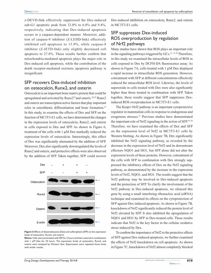
Dex-induced inhibition on osteocalcin, Runx2, and osterin
in MC3T3-E1 cells.
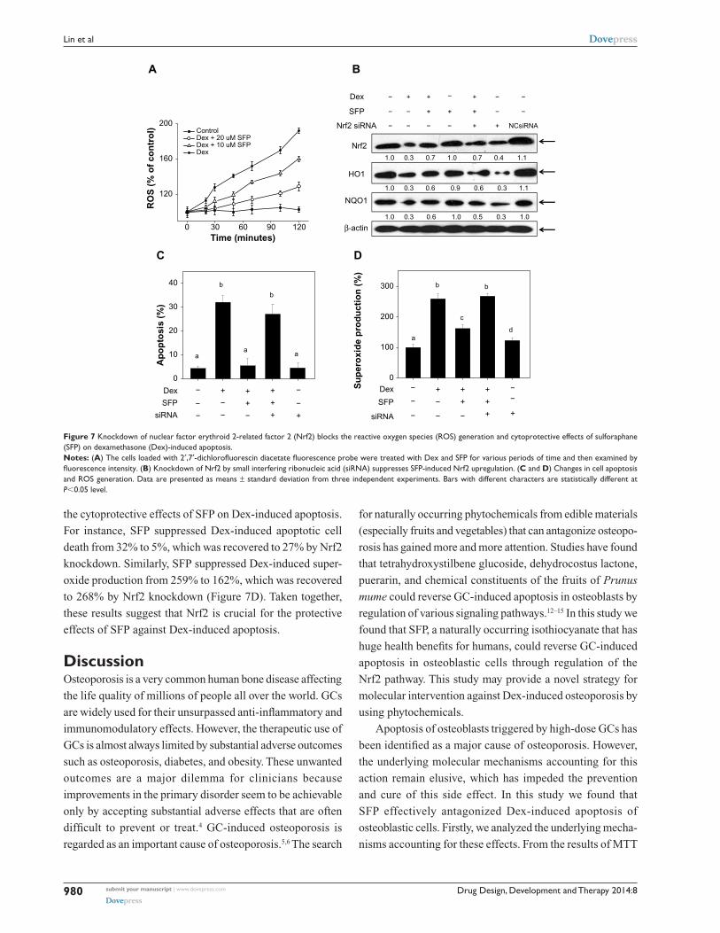
sFP suppresses Dex-induced rOs overproduction by regulation of nrf2 pathwaysMany studies have shown that ROS plays an important role
in the signaling pathways triggered by GCs.12–15,30 Therefore,
in this study we examined the intracellular levels of ROS in
cells exposed to Dex by DCFH-DA fluorescence assay. As
shown in Figure 7A, cells treated with 1 µM Dex displayed
a rapid increase in intracellular ROS generation. However,
cotreatment with SFP at different concentrations effectively
reduced the intracellular ROS level. Likewise, the levels of
superoxide in cells treated with Dex were also significantly
higher than those treated in combination with SFP. Taken
together, these results suggest that SFP suppresses Dex-
induced ROS overproduction in MC3T3-E1 cells.
The Keap1-Nrf2 pathway is an important cytoprotective
regulator in mammalian cells in response to endogenous and
exogenous stresses.31 Previous studies have demonstrated
the important role of Nrf2 signaling in the action of SFP.18–20
Therefore, we have examined the effects of Dex and SFP
on the expression level of Nrf2 in MC3T3-E1 cells by
Western blotting. As shown in Figure 7B, Dex significantly
inhibited the Nrf2 signaling pathway, as revealed by the
decrease in the expression level of Nrf2 and its downstream
effectors NQO1 and HO1, but SFP alone did not alter the
expression levels of these proteins. However, cotreatment of
the cells with SFP in combination with Dex strongly sup-
pressed the inhibitory effects of Dex on the Nrf2 signaling
pathway, as demonstrated by the increase in the expression
levels of Nrf2, NQO1, and HO1. The results suggest that the
Nrf2 pathway may be involved in Dex-induced apoptosis
and the protection of SFP. To clarify the involvement of the
Nrf2 pathway in Dex-induced apoptosis, we silenced this
gene by using a small interfering ribonucleic acid (siRNA)
technique and examined its effects on the cytoprotection of
SFP against Dex-induced apoptosis. As shown in Figure 7B,
knockdown of Nrf2 significantly reduced the protein level of
Nrf2 elevated by SFP. It also inhibited the upregulation of
NQO1 and HO1 by SFP in Dex-treated cells. These results
indicate that Nrf2 is the key factor in the cellular oxidative
stress induced by Dex.
To confirm the importance of Nrf2 to the protective effects
of SFP against Dex-induced apoptosis, we further examined
the effects of Nrf2 knockdown on cell apoptosis. As shown
in Figure 7C, knockdown of Nrf2 almost completely blocked
Osteocalcin
β-actin
Runx2
Osterin
SFP –
–
–
–
+ +
Dex + +
Figure 6 effects of dexamethasone (Dex) and sulforaphane (sFP) on the expression levels of osteocalcin, runx2, and osterin. Notes: cells were pretreated with sFP for 2 hours and then cultured in combination with 1 µM Dex for 24 hours. The expression levels of osteocalcin, runx2, and osterin were analyzed by Western blot. experiments were repeated three times with similar results.
z-DEVD-fmk effectively suppressed the Dex-induced
sub-G1 apoptotic peak from 32.0% to 6.8% and 9.4%,
respectively, indicating that Dex-induced apoptosis
occurs in a caspase-dependent manner. Moreover, addi-
tion of caspase-9 inhibitor (Z-LEHD-fmk) effectively
inhibited cell apoptosis to 13.9%, while caspase-8
inhibitor (Z-IETD-fmk) only slightly decreased cell
apoptosis to 27.8%. These results further confirm that
mitochondria-mediated apoptosis plays the major role in
Dex-induced cell apoptosis, while the contribution of the
death receptor-mediated extrinsic apoptosis pathway is
insignificant.
sFP recovers Dex-induced inhibition on osteocalcin, runx2, and osterinOsteocalcin is an important bone matrix protein that could be
upregulated and activated by Runx227and osterix.28,29 Runx2
and osterix are transcription active factors that play important
roles in osteoblastic differentiation and bone formation.27
In this study, to examine the effects of Dex and SFP on the
function of MC3T3-E1 cells, we have determined the changes
in the expression levels of osteocalcin, Runx2, and osterin
in cells exposed to Dex and SFP. As shown in Figure 6,
treatment of the cells with 1 µM Dex markedly reduced the
expression levels of osteocalcin. Interestingly, this effect
of Dex was significantly attenuated by the addition of SFP.
Moreover, Dex also significantly downregulated the levels of
Runx2 and osterix, and protective effects were also observed
by the addition of SFP. Taken together, SFP could recover
Drug Design, Development and Therapy 2014:8submit your manuscript | www.dovepress.com
Dovepress
Dovepress
980
lin et al
the cytoprotective effects of SFP on Dex-induced apoptosis.
For instance, SFP suppressed Dex-induced apoptotic cell
death from 32% to 5%, which was recovered to 27% by Nrf2
knockdown. Similarly, SFP suppressed Dex-induced super-
oxide production from 259% to 162%, which was recovered
to 268% by Nrf2 knockdown (Figure 7D). Taken together,
these results suggest that Nrf2 is crucial for the protective
effects of SFP against Dex-induced apoptosis.
DiscussionOsteoporosis is a very common human bone disease affecting
the life quality of millions of people all over the world. GCs
are widely used for their unsurpassed anti-inflammatory and
immunomodulatory effects. However, the therapeutic use of
GCs is almost always limited by substantial adverse outcomes
such as osteoporosis, diabetes, and obesity. These unwanted
outcomes are a major dilemma for clinicians because
improvements in the primary disorder seem to be achievable
only by accepting substantial adverse effects that are often
difficult to prevent or treat.4 GC-induced osteoporosis is
regarded as an important cause of osteoporosis.5,6 The search
for naturally occurring phytochemicals from edible materials
(especially fruits and vegetables) that can antagonize osteopo-
rosis has gained more and more attention. Studies have found
that tetrahydroxystilbene glucoside, dehydrocostus lactone,
puerarin, and chemical constituents of the fruits of Prunus
mume could reverse GC-induced apoptosis in osteoblasts by
regulation of various signaling pathways.12–15 In this study we
found that SFP, a naturally occurring isothiocyanate that has
huge health benefits for humans, could reverse GC-induced
apoptosis in osteoblastic cells through regulation of the
Nrf2 pathway. This study may provide a novel strategy for
molecular intervention against Dex-induced osteoporosis by
using phytochemicals.
Apoptosis of osteoblasts triggered by high-dose GCs has
been identified as a major cause of osteoporosis. However,
the underlying molecular mechanisms accounting for this
action remain elusive, which has impeded the prevention
and cure of this side effect. In this study we found that
SFP effectively antagonized Dex-induced apoptosis of
osteoblastic cells. Firstly, we analyzed the underlying mecha-
nisms accounting for these effects. From the results of MTT
Nrf2
β-actin
HO1
NQO1
SFP + + +
Dex + + − +
Nrf2 siRNA
− − − −
− − −
− − − − + + NCsiRNA
0
100
200
300
0
10
20
30
40
Su
per
oxi
de
pro
du
ctio
n (
%)
Dex + + +
SFP + +
siRNA−
−
− −
−
− − − + +
a
c
d
bb
Ap
op
tosi
s (%
)
a
b
b
aa
Dex + + +
SFP −
−
− − −
−−− + +
siRNA + +
B
C D
A
0 30 60 90 120
120
160
200ControlDex + 20 uM SFPDex + 10 uM SFPDex
RO
S (
% o
f co
ntr
ol)
Time (minutes)
1.0
1.0
1.0
0.7 1.0 0.7 0.4 1.1
0.6 0.9 0.6 0.3 1.1
0.6 1.0 0.5 0.30.3
0.3
0.3
1.0
Figure 7 Knockdown of nuclear factor erythroid 2-related factor 2 (nrf2) blocks the reactive oxygen species (rOs) generation and cytoprotective effects of sulforaphane (sFP) on dexamethasone (Dex)-induced apoptosis. Notes: (A) The cells loaded with 2′,7′-dichlorofluorescin diacetate fluorescence probe were treated with Dex and SFP for various periods of time and then examined by fluorescence intensity. (B) Knockdown of nrf2 by small interfering ribonucleic acid (sirna) suppresses sFP-induced nrf2 upregulation. (C and D) changes in cell apoptosis and rOs generation. Data are presented as means ± standard deviation from three independent experiments. Bars with different characters are statistically different at P,0.05 level.

Drug Design, Development and Therapy 2014:8 submit your manuscript | www.dovepress.com
Dovepress
Dovepress
981
reversal of osteoblastic cell apoptosis by sulforaphane
assay, cotreatment of the cells with SFP in combination with
Dex can reduce the cytotoxicity of Dex in MC3T3-E1 cells.
The results of flow cytometric analysis and DAPI-TUNEL
staining assay proved that SFP blocked Dex-induced apop-
tosis in MC3T3-E1 cells with the inhibition of caspase
activation and PARP cleavage. Caspase-3 was considered
to be a central mediator of cell apoptosis pathway, while
caspase-8 and -9 act as initiators of extrinsic (death receptor-
mediated) and intrinsic (mitochondria-mediated) apoptotic
pathways, respectively. The higher activation degree of
caspase-9 than caspase-8 suggests the more important
role of the mitochondria-mediated apoptotic pathway in
Dex-induced apoptosis. Moreover, cotreatment of the cells
with different concentrations of SFP effectively reduced
the caspase-3, -8, and -9 activities in a dose-dependent
manner. These results suggest that mitochondria-mediated
apoptosis plays an important role in the protective action of
SFP. To further evaluate the roles of the different caspases
in Dex-induced apoptosis, we next examined the effects of
various caspase inhibitors. The results showed that Dex-
induced apoptosis occurs in a caspase-dependent manner.
Moreover, mitochondria-mediated apoptosis plays the major
role in Dex-induced cell apoptosis, while the contribution
of the death receptor-mediated extrinsic apoptosis pathway
is insignificant.
To examine the effects of Dex and SFP on the function of
MC3T3-E1 cells, we determined the changes in the expression
levels of osteocalcin, Runx2, and osterin. The results showed
that treatment of the cells with Dex markedly reduced the
expression levels of osteocalcin, which was significantly
attenuated by SFP. Interestingly, Dex also significantly
downregulated the levels of Runx2 and osterix, and protective
effects were also observed by the addition of SFP. These data
suggest that SFP may block cell apoptosis induced by GCs
and recover the function of MC3T3-E1 cells.
Oxidative stress is a result of overproduction of ROS,
which could induce apoptosis and decrease activities of
osteoblasts. Bone mass is kept balanced by bone remodeling
via regulation of bone formation and resorption.32 A recent
study showed that ROS could enhance bone resorption
through promotion of osteoclast formation and an increase
in their activities.33 Moreover, ROS also induced apoptosis of
osteoblasts and led to a reduction of osteoblast differentiation
and bone formation.30 Therefore, administration of
antioxidants that could scavenge excess intracellular ROS
could be a potential approach for treatment of osteoporosis.
In this study we found that SFP could effectively suppress
Dex-induced cytotoxicity in osteoblastic cells through
inhibition of intracellular ROS generation and regulation of
the Nrf2 pathway.
Previous studies have reported that Nrf2 could protect
human cells from oxidative and electrophilic damage.27,28
In this study, due to the detection of ROS in MC3T3-E1
cells by Dex, the changes in the expression level of Nrf2
and two representative target genes NQO1 and HO1 in cells
exposed to Dex and SFP were examined by Western blot-
ting. Dex significantly inhibited the Nrf2 signaling pathway,
as revealed by the decrease in the expression level of Nrf2
and its downstream effectors NQO1 and HO1. However,
cotreatments of the cells with SFP strongly suppressed the
inhibitory effects of Dex on the Nrf2 signaling pathway.
To further clarify the importance of Nrf2, we examined
the effects of Nrf2 knockdown on the cytoprotection of
SFP against Dex-induced apoptosis. The results showed
that knockdown of Nrf2 almost completely blocked the
cytoprotective effects of SFP on Dex-induced apoptosis and
ROS overproduction. Overall, we conclude that Nrf2 plays
an important role in the protective effects of SFP against
Dex-induced apoptosis.
ConclusionIn summary, the present study demonstrates the protective
effects of SFP against Dex-induced cytotoxicity, and eluci-
dates the underlying molecular mechanisms. Specifically,
treatment of the cells with SFP attenuated Dex-induced cas-
pase activation, PARP cleavage, mitochondrial dysfunction,
and DNA damage. These effects were mainly mediated by
regulation of the Nrf2 pathway to inhibit the production of
ROS. Taken together, this study provides a novel strategy
for molecular intervention against Dex-induced osteoporosis
using phytochemicals.
AcknowledgmentsThis study was supported by grants from the Natural
Science Foundation of Guangdong Province, People’s
Republic of China (No S2013020012866), the Science
and Technology Planning Project of Guangdong Province,
People’s Republic of China (No 2011B031800212), the
Foundation for Youth Scholars of Guangdong Medical
College, People’s Republic of China (No XQ1322),
and the Science and Technology Development Planning
Project of Zhanjiang, People’s Republic of China
(No 2012C3101026).
DisclosureThe authors report no conflicts of interest in this work.

Drug Design, Development and Therapy
Publish your work in this journal
Submit your manuscript here: http://www.dovepress.com/drug-design-development-and-therapy-journal
Drug Design, Development and Therapy is an international, peer-reviewed open-access journal that spans the spectrum of drug design and development through to clinical applications. Clinical outcomes, patient safety, and programs for the development and effective, safe, and sustained use of medicines are a feature of the journal, which
has also been accepted for indexing on PubMed Central. The manu-script management system is completely online and includes a very quick and fair peer-review system, which is all easy to use. Visit http://www.dovepress.com/testimonials.php to read real quotes from published authors.
Drug Design, Development and Therapy 2014:8submit your manuscript | www.dovepress.com
Dovepress
Dovepress
Dovepress
982
lin et al
References 1. Hsu WL, Chen CY, Tsauo JY, Yang RS. Balance control in elderly
people with osteoporosis. J Formos Med Assoc. 2014;113(6): 334–339.
2. Reid IR. Anti-resorptive therapies for osteoporosis. Semin Cell Dev Biol. 2008;19(5):473–478.
3. Salam SN, Eastell R, Khwaja A. Fragility fractures and osteoporosis in CKD: pathophysiology and diagnostic methods. Am J Kidney Dis. 2014;S0272–6386(14):499–495.
4. Seibel MJ, Cooper MS, Zhou H. Glucocorticoid-induced osteoporosis: mechanisms, management, and future perspectives. Lancet Diabetes Endocrinol. 2013;1(1):59–70.
5. Brennan-Speranza TC, Henneicke H, Gasparini SJ, et al. Osteoblasts mediate the adverse effects of glucocorticoids on fuel metabolism. J Clin Invest. 2012;122(11):4172–4189.
6. Rauch A, Seitz S, Baschant U, et al. Glucocorticoids suppress bone formation by attenuating osteoblast differentiation via the monomeric glucocorticoid receptor. Cell Metab. 2010;11(6):517–531.
7. den Uyl D, Bultink IE, Lems WF. Advances in glucocorticoid-induced osteoporosis. Curr Rheumatol Rep. 2011;13(3):233–240.
8. Weinstein RS. Clinical practice. Glucocorticoid-induced bone disease. N Engl J Med. 2011;365(1):62–70.
9. Espina B, Liang M, Russell RG, Hulley PA. Regulation of Bim in glucocorticoid-mediated osteoblast apoptosis. J Cell Physiol. 2008;215(2):488–496.
10. O’Brien CA, Jia D, Plotkin LI, et al. Glucocorticoids act directly on osteoblasts and osteocytes to induce their apoptosis and reduce bone formation and strength. Endocrinology. 2004;145(4): 1835–1841.
11. Yun SI, Yoon HY, Jeong SY, Chung YS. Glucocorticoid induces apoptosis of osteoblast cells through the activation of glyco-gen synthase kinase 3beta. J Bone Miner Metab. 2009;27(2): 140–148.
12. Choi EM, Kim GH, Lee YS. Protective effects of dehydrocostus lactone against hydrogen peroxide-induced dysfunction and oxidative stress in osteoblastic MC3T3-E1 cells. Toxicol In Vitro. 2009;23(5): 862–867.
13. Wang Y, Wang WL, Xie WL, et al. Puerarin stimulates proliferation and differentiation and protects against cell death in human osteoblastic MG-63 cells via ER-dependent MEK/ERK and PI3K/Akt activation. Phytomedicine. 15, 2013;20(10):787–796.
14. Yan XT, Lee SH, Li W, et al. Evaluation of the antioxidant and anti-osteoporosis activities of chemical constituents of the fruits of Prunus mume. Food Chem. 2014;156:408–415.
15. Zhang JK, Yang L, Meng GL, et al. Protective effect of tetrahydrox-ystilbene glucoside against hydrogen peroxide-induced dysfunction and oxidative stress in osteoblastic MC3T3-E1 cells. Eur J Pharmacol. 2012;689(1–3):31–37.
16. Wiseman M. The second World Cancer Research Fund/American Institute for Cancer Research expert report. Food, nutrition, physical activity, and the prevention of cancer: a global perspective. Proc Nutr Soc. 2008;67(3):253–256.
17. Gupta P, Kim B, Kim SH, Srivastava SK. Molecular targets of iso-thiocyanates in cancer: recent advances. Mol Nutr Food Res. Epub February 10, 2014.
18. Houghton CA, Fassett RG, Coombes JS. Sulforaphane: translational research from laboratory bench to clinic. Nutr Rev. 2013;71(11): 709–726.
19. Xu T, Ren D, Sun X, Yang G. Dual roles of sulforaphane in cancer treatment. Anticancer Agents Med Chem. 2012;12(9):1132–1142.
20. Tarozzi A, Angeloni C, Malaguti M, Morroni F, Hrelia S, Hrelia P. Sulforaphane as a potential protective phytochemical against neurode-generative diseases. Oxid Med Cell Longev. 2013;2013:415078.
21. Barcelo S, Gardiner JM, Gescher A, Chipman JK. CYP2E1- mediated mechanism of anti-genotoxicity of the broccoli constituent sulforaphane. Carcinogenesis. 1996;17(2):277–282.
22. Gross-Steinmeyer K, Stapleton PL, Tracy JH, Bammler TK, Strom SC, Eaton DL. Sulforaphane- and phenethyl isothiocyanate-induced inhi-bition of aflatoxin B1-mediated genotoxicity in human hepatocytes: role of GSTM1 genotype and CYP3A4 gene expression. Toxicol Sci. 2010;116(2):422–432.
23. Katoch O, Kumar A, Adhikari JS, Dwarakanath BS, Agrawala PK. Sulforaphane mitigates genotoxicity induced by radiation and anticancer drugs in human lymphocytes. Mutat Res. 2013;758(1–2):29–34.
24. Fan C, Chen J, Wang Y, et al. Selenocystine potentiates cancer cell apoptosis induced by 5-fluorouracil by triggering reactive oxygen species-mediated DNA damage and inactivation of the ERK pathway. Free Radic Biol Med. 2013;65:305–316.
25. Su J, Lai H, Chen J, et al. Natural borneol, a monoterpenoid compound, potentiates selenocystine-induced apoptosis in human hepatocellular carcinoma cells by enhancement of cellular uptake and activation of ROS-mediated DNA damage. PLoS One. 2013;8(5):e63502.
26. McIlwain DR, Berger T, Mak TW. Caspase functions in cell death and disease. Cold Spring Harb Perspect Biol. 2013;5(4):a008656.
27. Xu ZS, Wang XY, Xiao DM, et al. Hydrogen sulfide protects MC3T3-E1 osteoblastic cells against H2O2-induced oxidative damage-implications for the treatment of osteoporosis. Free Radic Biol Med. 2011;50(10): 1314–1323.
28. Nakashima K, Zhou X, Kunkel G, et al. The novel zinc finger-containing transcription factor osterix is required for osteoblast differentiation and bone formation. Cell. 2002;108(1):17–29.
29. Komori T. Regulation of osteoblast differentiation by transcription factors. J Cell Biochem. 2006;99(5):1233–1239.
30. Arai M, Shibata Y, Pugdee K, Abiko Y, Ogata Y. Effects of reactive oxygen species (ROS) on antioxidant system and osteoblastic differ-entiation in MC3T3-E1 cells. IUBMB Life. 2007;59(1):27–33.
31. Kansanen E, Kuosmanen SM, Leinonen H, Levonen AL. The Keap1-Nrf2 pathway: mechanisms of activation and dysregulation in cancer. Redox Biol. 2013;1(1):45–49.
32. Seeman E, Delmas PD. Bone quality: the material and structural basis of bone strength and fragility. N Engl J Med. 2006;354(21):2250–2261.
33. Lee NK, Choi YG, Baik JY, et al. A crucial role for reactive oxygen species in RANKL-induced osteoclast differentiation. Blood. 2005; 106(3):852–859.
Copyright © 2022 FDOKUMEN