Magnetization reversal studies in structurally tailored cobalt nanowires

Reversal of Mortality for Congenital DiaphragmaticHernia with ECMO
KURT HEISS, M.D., PETER MANNING, M.D., KEITH T. OLDHAM, M.D., ARNOLD G. CORAN, M.D.,THEODORE Z. POLLEY, JR., M.D., JOHN R. WESLEY, M.D., and ROBERT H. BARTLETT, M.D.
Extracorporeal Membrane Oxygenation (ECMO) has beenavailable to neonates with respiratory failure at the Universityof Michigan School of Medicine since June 1981. In order toevaluate the impact of this type of pulmonary support, a retro-spective analysis of 50 neonates with posterolateral congenitaldiaphragmatic hernia (CDH) who were symptomatic during thefirst hour of life and were treated between June 1974 and De-cember 1987 was carried out. The patients were divided into twogroups, those treated before June 1981 (16 patients) and thosetreated after June 1981 (34 patients). Overall survival improvedfrom 50% (eight of 16 patients) during the pre-ECMO era to76% (26 of34 patients) during the post-ECM0 period (p = 0.06).During the period after June 1981, 21 neonates were unrespon-sive to conventional therapy and were therefore considered forECMO. Failure of conventional therapy was defined as acuteclinical deterioration with an expected mortality of> 80% basedon an objective formula previously reported. Six patients wereexcluded on the basis of specific contraindications to ECMO.Thirteen of 15 infants (87%) supported with ECMO survived.Three patients treated before 1981 met criteria for ECMO, allthree died while receiving treatment using conventional therapy.These survival differences are significant (p < 0.01). In addition,the survival of 87% for the infants treated with ECMO versusthe expected mortality of > 80% for these same patients whentreated with conventional therapy is highly significant (p< 0.005). Based on this data, ECMO appears to be a successful,reliable, and safe method of respiratory support for selected,critically ill infants with CDH.
D_ ESPITE IMPRESSIVE ADVANCEMENTS in surgicaland neonatal intensive care over the last twodecades, the mortality ofinfants born with con-
genital diaphragmatic hernia (CDH) presenting for treat-ment within the first 24 hours of life has remained ap-proximately 50% (Table 1). 1-9 Extracorporeal membraneoxygenation (ECMO) was introduced in 1975 as a treat-ment for acute respiratory failure in neonates unrespon-
From the Section of Pediatric Surgery and the Section ofGeneral Surgery, Department of Surgery, University of
Michigan School of Medicine, Ann Arbor, Michigan
sive to conventional mechanical ventilation.'° Since then,in the United States, over 1000 infants with diagnosessuch as sepsis, meconium aspiration syndrome, and CDHhave been cared for with ECMO, often with impressiveimprovements in survival."1 Children with CDH remainproblematic. Recent reports from established ECMOcenters have shown survival rates of 35-58% in infantswith CDH.12 '3 Our institutional survival rate for infantswith CDH who were treated with ECMO exceeds 85%,and the corresponding overall survival rate for infantswith CDH is 76%. This report presents our institutionalexperience, including selection criteria, methodology, andresults for ECMO in the treatment of severe CDH pre-senting within the first hour of life.
Materials and Methods
A retrospective review ofall patients with CDH treatedat the University of Michigan, Mott Children's Hospital,from June 1974 to December 1987, was performed.ECMO therapy became available for the management
ofCDH patients with postoperative pulmonary failure inJune 1981. This date was used to divide the series intotwo groups: Group 1 refers to patients treated before June1981 in the "pre-ECMO era," and Group 2 designatesthose seen during the "ECMO era". Patients in Group Iwere further divided into "ECMO candidate" or "non-candidate" groups by retrospectively applying ECMO se.:lection criteria to these patients. Those in Group 2 weredivided into "ECMO-treated" and "conventional man-agement" groups.The technique of extracorporeal support of neonates
with respiratory failure has been well-described else-
225
Reprint requests and correspondence: Keith T. Oldham, M.D., Sectionof Pediatric Surgery, F7516 Mott Children's Hospital, Ann Arbor, MI48109.0245.
Submitted for publication: May 20, 1988.
226Ann. Surs. Februay 1989HEISS AND OTHERS
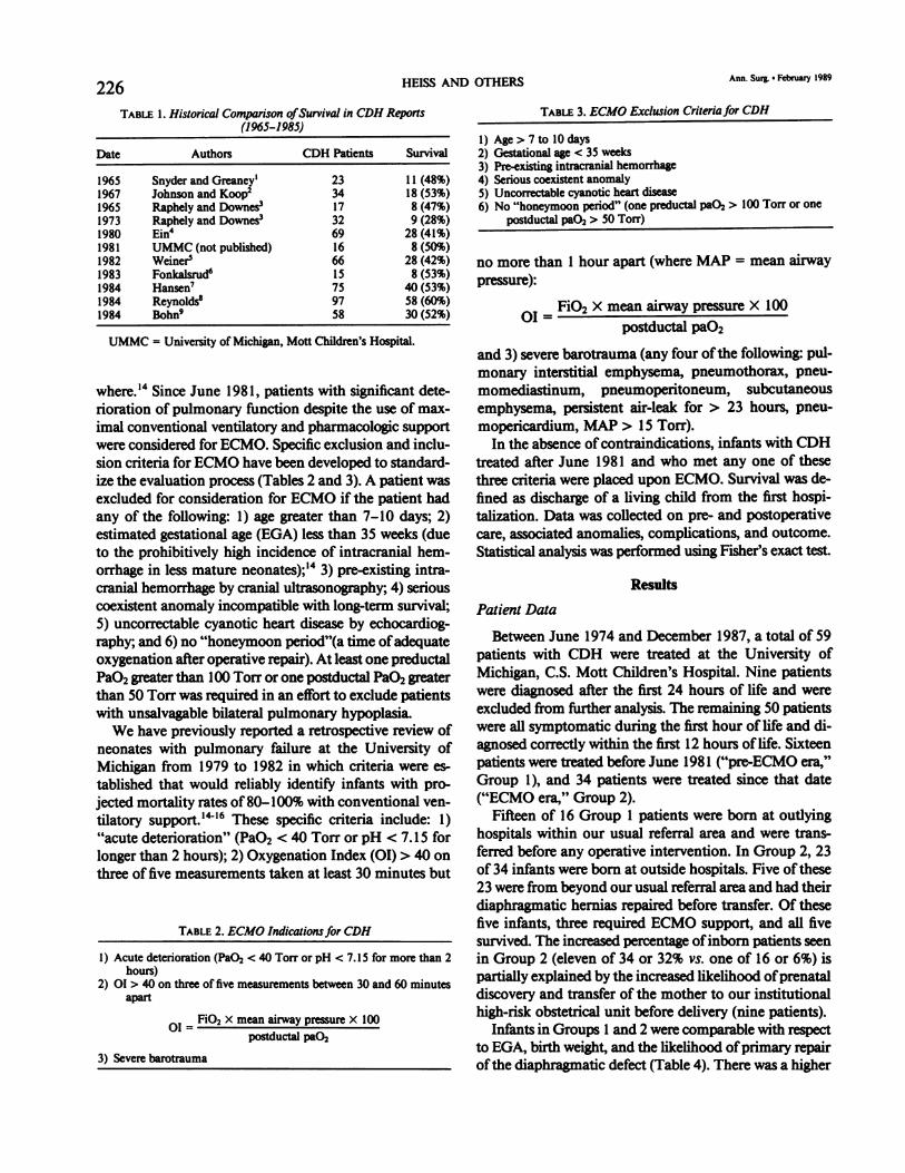
TABLE 1. Historical Comparison ofSurvival in CDH Reports(1965-1985)
Date Authors CDH Patients Survival
1965 Snyder and Greaney' 23 11 (48%)1967 Johnson and Koop2 34 18 (53%)1965 Raphely and Downes' 17 8 (47%)1973 Raphely and Downes3 32 9 (28%)1980 Ein4 69 28 (41%)1981 UMMC (not published) 16 8 (50%)1982 Weiner' 66 28 (42%)1983 Fonkalsrud' 15 8 (53%)1984 Hansen7 75 40 (53%)1984 Reynolds8 97 58 (60%)1984 Bohne 58 30 (52%)
UMMC = University of Michigan, Mott Children's Hospital.
where.'4 Since June 1981, patients with significant dete-rioration of pulmonary function despite the use of max-imal conventional ventilatory and pharmacologic supportwere considered for ECMO. Specific exclusion and inclu-sion criteria for ECMO have been developed to standard-ize the evaluation process (Tables 2 and 3). A patient wasexcluded for consideration for ECMO if the patient hadany of the following: 1) age greater than 7-10 days; 2)estimated gestational age (EGA) less than 35 weeks (dueto the prohibitively high incidence of intracranial hem-orrhage in less mature neonates);'4 3) pre-existing intra-cranial hemorrhage by cranial ultrasonography, 4) seriouscoexistent anomaly incompatible with long-term survival;5) uncorrectable cyanotic heart disease by echocardiog-raphy; and 6) no "honeymoon period"(a time ofadequateoxygenation after operative repair). At least one preductalPaO2 greater than 100 Torr or one postductal PaO2 greaterthan 50 Torr was required in an effort to exclude patientswith unsalvagable bilateral pulmonary hypoplasia.We have previously reported a retrospective review of
neonates with pulmonary failure at the University ofMichigan from 1979 to 1982 in which criteria were es-tablished that would reliably identify infants with pro-jected mortality rates of80-100% with conventional ven-tilatory support.""6 These specific criteria include: 1)"acute deterioration" (PaO2 < 40 Torr or pH < 7.15 forlonger than 2 hours); 2) Oxygenation Index (01) > 40 onthree of five measurements taken at least 30 minutes but
TABLE 2. ECMO Indications for CDH
1) Acute deterioration (PaO2 < 40 Torr or pH < 7.15 for more than 2hours)
2) OI > 40 on three of five measurements between 30 and 60 minutesapart
oI= FiO2 X mean airway pressure X 100postductal paO2
3) Severe barotrauma
TABLE 3. ECMO Exclusion Criteriafor CDH
1) Age>7tol0days2) Gestational age < 35 weeks3) Pre-existing intracrnial hemorrhage4) Serious coexistent anomaly5) Uncorrectable cyanotic heart disease6) No "honeymoon period" (one preductal paO2 > 100 Torr or one
postductal paO2 > 50 Torr)
no more than 1 hour apart (where MAP = mean airwaypressure):
OI =
FiO2 X mean airway pressure X 100postductal paO2
and 3) severe barotrauma (any four ofthe following: pul-monary interstitial emphysema, pneumothorax, pneu-momediastinum, pneumoperitoneum, subcutaneousemphysema, persistent air-leak for > 23 hours, pneu-mopericardium, MAP > 15 Torr).
In the absence of contraindications, infants with CDHtreated after June 1981 and who met any one of thesethree criteria were placed upon ECMO. Survival was de-fined as discharge of a living child from the first hospi-talization. Data was collected on pre- and postoperativecare, associated anomalies, complications, and outcome.Statistical analysis was performed using Fisher's exact test.
Results
Patient Data
Between June 1974 and December 1987, a total of 59patients with CDH were treated at the University ofMichigan, C.S. Mott Children's Hospital. Nine patientswere diagnosed after the first 24 hours of life and wereexcluded from further analysis. The remaining 50 patientswere all symptomatic during the first hour of life and di-agnosed correctly within the first 12 hours of life. Sixteenpatients were treated before June 1981 ("pre-ECMO era,"Group 1), and 34 patients were treated since that date("ECMO era," Group 2).
Fifteen of 16 Group 1 patients were born at outlyinghospitals within our usual referral area and were trans-ferred before any operative intervention. In Group 2, 23of 34 infants were born at outside hospitals. Five of these23 were from beyond our usual referral area and had theirdiaphragmatic hernias repaired before transfer. Of thesefive infants, three required ECMO support, and all fivesurvived. The increased percentage ofinborn patients seenin Group 2 (eleven of 34 or 32% vs. one of 16 or 6%) ispartially explained by the increased likelihood ofprenataldiscovery and transfer of the mother to our institutionalhigh-risk obstetrical unit before delivery (nine patients).
Infants in Groups 1 and 2 were comparable with respectto EGA, birth weight, and the likelihood ofprimary repairofthe diaphragmatic defect (Table 4). There was a higher
CONGENITAL DIAGPHRAGMATIC HERNIA
TABLE 4. CDH Patient Characteristics
Characteristics Group 1 Group 2
Average gestational age 38.4 ± 3.4 weeks 38.3 ± 2.6 weeksAverage birth weight 3.03 ± 0.69 kg 3.09 ± 0.63 kgAdditional anomalies 12.5% 18%Left-sided hernia 93.8% 76%Primary repair 84.6% 88%Age at operation 9.62 ± 5.0 hours 7.7 ± 3.7 hours
proportion of right-sided hernias seen in Group 2 (24%)and more patients in Group 2 had significant additionalcongenital anomalies. Patients in this group tended toundergo operation earlier, although this difference did notachieve statistical significance (p > 0.05). In Group 2, sixinfants were excluded from consideration forECMO sup-
port secondary to contraindications detailed in the Sur-vival Data section; [ofthese six patients, two were excludedon the basis ofchromosomal anomalies, two others wereexcluded because they who had no "honeymoon period,"and two because ofprematurity (EGA = 32 and 33 weeks,respectively)].As indicated by a review oftheir records, three patients
in Group 1 would have qualified for ECMO support hadit been available. In Group 2, 15 patients qualified andwere placed on ECMO support. Six of these infants suf-fered acute deterioration of their pulmonary status, andnine met 01 criteria as defined above. The average age atinitiation ofbypass was 41 hours (range of 13-130 hours).The mean time on bypass was 137 hours, slightly lessthan six days. This is roughly twice the average length oftime on bypass for infants with meconium aspiration atour institution. Two patients required support for greaterthan 200 hours, whereas four patients required less than100 hours. Table 5 shows the mean postductal arterialblood gases (ABG) of infants before the initiation ofECMO, as well as the average ventilator settings just beforeand just after bypass.The duration of hospitalization for Group 1 survivors
averaged 20.5 days. In Group 2 survivors, there was no
significant difference in length of stay between ECMOand non-ECMO patients at 46.1 and 43.4 days, respec-tively.
Survival Data
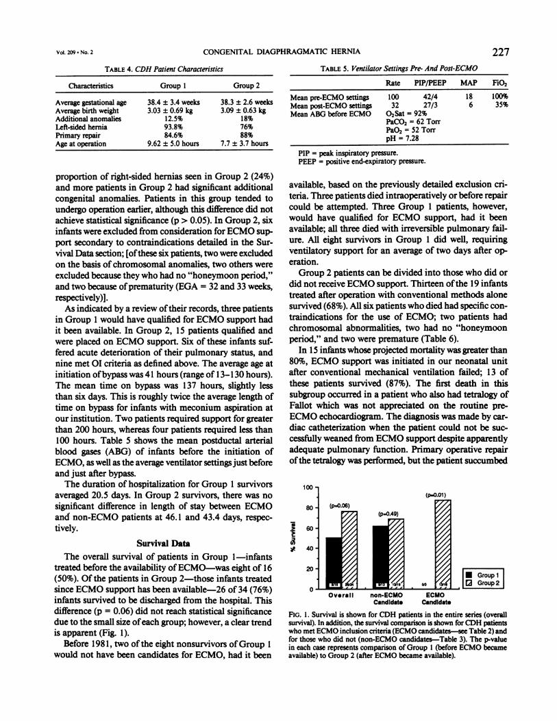
The overall survival of patients in Group 1-infantstreated before the availability ofECMO-was eight of 16(50%). Of the patients in Group 2-those infants treatedsince ECMO support has been available-26 of 34 (76%)infants survived to be discharged from the hospital. Thisdifference (p = 0.06) did not reach statistical significancedue to the small size ofeach group; however, a clear trendis apparent (Fig. 1).
Before 1981, two of the eight nonsurvivors ofGroup 1
would not have been candidates for ECMO, had it been
227TABLE 5. Ventilator Settings Pre- And Post-ECMO
Rate PIP/PEEP MAP FiO2
Mean pre-ECMO settings 100 42/4 18 100%Mean post-ECMO settings 32 27/3 6 35%Mean ABG before ECMO 02Sat = 92%
PaCO2 = 62 TorrPaO2 = 52 TorrpH = 7.28
PIP = peak inspiratory pressure.PEEP = positive end-expiratory pressure.
available, based on the previously detailed exclusion cri-teria. Three patients died intraoperatively or before repaircould be attempted. Three Group 1 patients, however,would have qualified for ECMO support, had it beenavailable; all three died with irreversible pulmonary fail-ure. All eight survivors in Group 1 did well, requiringventilatory support for an average of two days after op-
eration.Group 2 patients can be divided into those who did or
did not receive ECMO support. Thirteen ofthe 19 infantstreated after operation with conventional methods alonesurvived (68%). All six patients who died had specific con-traindications for the use of ECMO; two patients hadchromosomal abnormalities, two had no "honeymoonperiod," and two were premature (Table 6).
In 15 infants whose projected mortality was greater than80%, ECMO support was initiated in our neonatal unitafter conventional mechanical ventilation failed; 13 ofthese patients survived (87%). The first death in thissubgroup occurred in a patient who also had tetralogy ofFallot which was not appreciated on the routine pre-
ECMO echocardiogram. The diagnosis was made by car-
diac catheterization when the patient could not be suc-
cessfillly weaned from ECMO support despite apparentlyadequate pulmonary function. Primary operative repairofthe tetralogy was performed, but the patient succumbed
(p.0.01)
80 - (p.0.06)(p.0.49)
60
40-
20 IiGroup 2
0
Overall non-ECMO ECMOCandidate Candidate
FIG. 1. Survival is shown for CDH patients in the entire series (overallsurvival). In addition, the survival comparison is shown for CDH patientswho met ECMO inclusion criteria(ECMO candidates-see Table 2) andfor those who did not (non-ECMO candidates-Table 3). The p-valuein each case represents comparison of Group I (before ECMO becameavailable) to Group 2 (after ECMO became available).
Vol. 209 . No. 2

Ann. Surg. * February 1989
TABLE 6. CDH Patients Excluded From ECMO
Patient No. Reason for Exclusion
1 No "honeymoon period"2 No "honeymoon period"3 Premature (EGA of 32 weeks)4 Trisomy 185 Absent short arm of chromosome 216 Premature (EGA of 33 weeks)
because of postoperative intracranial hemorrhage afterbeing on ECMO bypass for a total of 13 days. The seconddeath occurred in an infant who was placed on ECMOat 82 hours ofage by reason ofOI criteria. A patent ductusarteriosus (PDA) ligation was required on the sixth dayof life while on ECMO. The ECMO course concludeduneventfully after 186 hours on bypass, with the infantreceiving minimal ventilatory support. During the ensuing48 hours, persistent fetal circulation recurred, complicatedby acute right heart failure. Aggressive ventilatory supportwas required, bronchopulmonary dysplasia developed,and the child eventually died of sepsis at 18 months ofage, having been weaned successfully from ventilatorysupport but never having left the hospital.
Complications
All deaths in both groups, with the exception of theinfant who had tetralogy of Fallot, were related to pul-monary failure. Two surviving patients in Group 1 de-veloped small bowel obstructions requiring enterolysis.
Five complications occurred in the Group 2 survivorswho did not require ECMO support. Three patients hadcontralateral pneumothoraces requiring thoracostomytube placement, and two infants had evidence of mildbronchopulmonary dysplasia on follow-up x-ray studies.
In the group of patients supported with ECMO, seven
had contralateral pneumothoraces (46.7%). Bleedingproblems secondary to the anticoagulation necessary foruse ofECMO were the most serious and frequently seen
complications ofthis form ofmanagement (Table 7). Fourinfants had such complications: three patients had intra-cranial hemorrhage (one fatal, one resulting in significantdevelopmental delay, and one minor intraparenchymal
TABLE 7. Complications Related to ECMO
Complication No. of Patients
Large volume gastrointestinal bleed due totolazoline
Dislodged umbilical artery catheter with 400 ccbleed
Large volume chest tube bleedHemopericardium causing tamponade, requiring
emergent thoracotomyIntracranial hemorrhage 3 (2 major, 1
minor)
bleed), and one patient had hemopericardium requiringoperative decompression, at which time PDA ligation wasalso performed. Chronic pulmonary disease was judgedmoderate to severe in three patients, leading in one infantindirectly to a late death at the age ofone year, after havingbeen discharged from the hospital. In three others, mildbronchopulmonary dysplasia was seen on follow-upx-rays with no clinically detectable compromise.
Discussion
Congenital diaphragmatic hernia may result in bilateralhypoplasia ofthe fetal airways and pulmonary vasculature.Ipsilateral hypoplasia is generally more severe. The phys-iologic consequences produce a spectrum of illness, rang-ing from an incidental finding on chest x-ray in anasymptomatic child to acute respiratory distress at birthmanifested by hypercapnia, hypoxemia, and acidosis. Pa-tients who present after 24 hours of life rarely have sig-nificant illness and have essentially no morbidity or mor-tality from operative repair. Those who present and re-quire treatment during the first hours of life are usuallyseverely ill and require aggressive care for salvage. Mostinfants ofthis latter group will experience a "honeymoonperiod." Unfortunately, many of these children developpulmonary artery hypertension and persistent fetal cir-culation with right to left shunting characterized by pro-gressive hypoxemia on maximal ventilator settings. His-torically, at major centers with state-of-the-art neonatalcare, the mortality for these infants has ranged from 40-60%. These figures have failed to improve over the last20 years, despite impressive improvements in neonatalintensive care. Table 1 gives a review of our experiencefrom 1974 to 1981 and a comparison with that of severalothers published during the last 20 years. Our survivalrate is consistent with other large series published at thattime. Despite significant changes in neonatal pulmonaryand critical care, including the use of tolazoline, alkalin-ization by hyperventilation, paralysis, and pressure-limitedventilation, the mortality for infants with early or im-mediate symptoms remained approximately 50% over this20-year period.
Keeping in mind the inherent weaknesses of retro-spective studies, a simple review ofthe survival data fromthis study presents four interesting comparisons. First, acomparison of the survival of infants treated before theavailability ofECMO (50%) with those of the ECMO era(76%) is not significant (p = 0.06). Although the 26%change suggests improvement, the sample size is currentlytoo small to reach significance. When the survival of pa-tients ofthe pre-ECMO period who would have qualifiedfor ECMO (0 of three patients, or 0%) is compared withthat of infants who were treated later and who qualifiedfor and were treated with ECMO (13 of 15 patients, or87%), the large difference in survival rates is significant at
HEISS AND OTHERS228
CONGENITAL DIAGPHRAGMATIC HERNIA.
p = 0.01 despite the small numbers. Third, and perhapsmost importantly, a comparison ofthe expected mortality(80-100%) with the observed mortality (13%) in theECMO group is highly significant at p = 0.0015. Thisstrongly suggests that ECMO impacts favorably on sur-vival of those infants who are the most ill and who haveno contraindications to the procedure. Finally, all serieswill have some patients who will never be viable, regardlessof the treatment. In Group 2, this number was six of 34,or approximately 18%. Ifthese patients are excluded, theresulting survival gives an idea ofthe sensitivity and spec-ificity ofthe selection criteria used during the ECMO era.With the exclusion of these six nonviable patients, theGroup 2 survival rate reaches 93% (26 of 28 patients).
There are a number of differences in the groups in-cluded in this study. The average length ofhospitalizationis shorter for Group 1 patients (20.5 vs. 45 days). Group2 patients managed conventionally required ventilatorsupport for 10 days, as compared to 2 days for patientsin Group 1. (ECMO patients required an average of 20days of ventilator support.) In addition, more patients ofGroup 2 had additional nonpulmonary anomalies. Thissuggests that more critically ill patients were being caredfor in the later group and that the difference in survivalbetween Groups 1 and 2 is perhaps more impressive thanwas first evident. The selection criteria used for ECMOare an obvious source ofbias in comparing these statisticswith those of other series. The well-publicized selectioncriteria may prevent some patients from being referred toour center, thus resulting in a form oflocal selection. Theincidence of fatal fetal anomalies associated with CDHranges from 5% to 60%.17-19 In our series, there were sixinfants in the post-ECMO group who had lethal anomaliesthat precluded ECMO (Table 6). Four of these patientshad anomalies that were nonpulmonary in nature, (twopatients were premature infants and two had chromo-somal abnormalities), and the remaining two infants werebelieved to have lethal bilateral pulmonary hypoplasiabecause no "honeymoon period" occurred. It is the treat-ment of this latter group of infants that is most contro-versial. No clinical event or observation has been proven
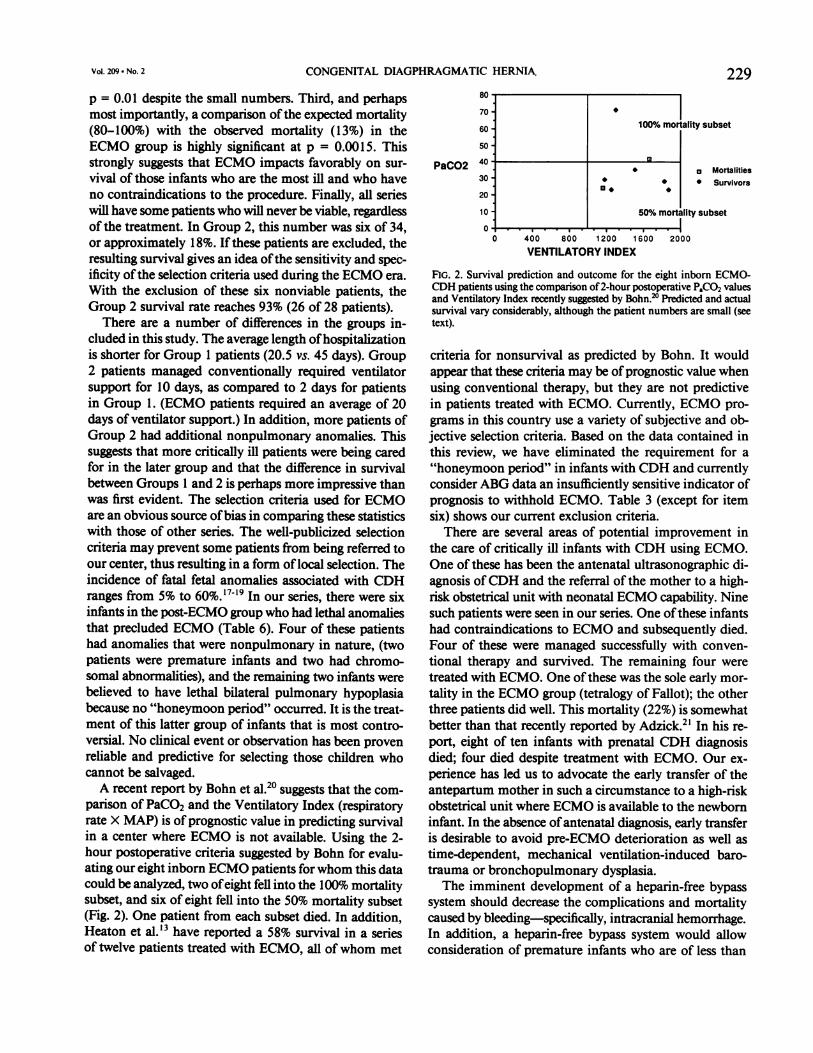
reliable and predictive for selecting those children whocannot be salvaged.A recent report by Bohn et al.20 suggests that the com-
parison of PaCO2 and the Ventilatory Index (respiratoryrate X MAP) is of prognostic value in predicting survivalin a center where ECMO is not available. Using the 2-hour postoperative criteria suggested by Bohn for evalu-ating our eight inborn ECMO patients forwhom this datacould be analyzed, two ofeight fell into the 100% mortalitysubset, and six of eight fell into the 50% mortality subset(Fig. 2). One patient from each subset died. In addition,Heaton et al.'3 have reported a 58% survival in a seriesof twelve patients treated with ECMO, all ofwhom met
70 -
60
50 -
PaCO2 40
30 -
20 -
10 -
100%/o mortality subset
400 800 1200VENTILATORY INDE
o Mortalities* Survivors
50% mortality subset
1600 2000
x
FIG. 2. Survival prediction and outcome for the eight inborn ECMO-CDH patients using the comparison of2-hour postoperative PaCO2 valuesand Ventilatory Index recently suggested by Bohn.?' Predicted and actualsurvival vary considerably, although the patient numbers are small (seetext).
criteria for nonsurvival as predicted by Bohn. It wouldappear that these criteria may be ofprognostic value whenusing conventional therapy, but they are not predictivein patients treated with ECMO. Currently, ECMO pro-grams in this country use a variety of subjective and ob-jective selection criteria. Based on the data contained inthis review, we have eliminated the requirement for a"honeymoon period" in infants with CDH and currentlyconsider ABG data an insufficiently sensitive indicator ofprognosis to withhold ECMO. Table 3 (except for itemsix) shows our current exclusion criteria.
There are several areas of potential improvement inthe care of critically ill infants with CDH using ECMO.One of these has been the antenatal ultrasonographic di-agnosis ofCDH and the referral of the mother to a high-risk obstetrical unit with neonatal ECMO capability. Ninesuch patients were seen in our series. One ofthese infantshad contraindications to ECMO and subsequently died.Four of these were managed successfully with conven-tional therapy and survived. The remaining four weretreated with ECMO. One ofthese was the sole early mor-tality in the ECMO group (tetralogy of Fallot); the otherthree patients did well. This mortality (22%) is somewhatbetter than that recently reported by Adzick.21 In his re-port, eight of ten infants with prenatal CDH diagnosisdied; four died despite treatment with ECMO. Our ex-perience has led us to advocate the early transfer of theantepartum mother in such a circumstance to a high-riskobstetrical unit where ECMO is available to the newborninfant. In the absence ofantenatal diagnosis, early transferis desirable to avoid pre-ECMO deterioration as well astime-dependent, mechanical ventilation-induced baro-trauma or bronchopulmonary dysplasia.The imminent development of a heparin-free bypass
system should decrease the complications and mortalitycaused by bleeding-specifically, intracranial hemorrhage.In addition, a heparin-free bypass system would allowconsideration of premature infants who are of less than
Vol. 209 * No. 2 229

230 HEISS AND OTHERS Ann. Surg * February 1989
35 weeks gestational age forECMO treatment. Currently,the cumulative risks of long-term extracorporeal supportrevolve around bleeding complications and oxygenatorfailure. The heparin-bound circuit would lessen these long-term risks and potentially allow exploration ofprolongedbypass in these infants.
Summary
Fifty infants with congenital diaphragmatic herniasymptomatic during the first hour of life were treated be-tween June 1974 and December 1987. The introductionofECMO in 1981, along with improved neonatal intensivecare, was followed by an improvement in survival (from50% to 76%) within this period. Ofthe ECMO era patientswho failed conventional therapy and had an expectedmortality of 80-100%, 13 of 15 (87%) survived, thus re-versing the expected mortality. This indicates thatECMOis a successful, reliable, and safe method of pulmonarysupport for selected critically ill infants with congenitaldiaphragmatic hernia.
References1. Snyder WH, Greaney EM. Congenital diaphragmatic hernia: 77
consecutive cases. Surgery 1965; 52:576-588.2. Johnson DG, Deaner RM, Koop CE. Diaphragmatic hernia in in-
fancy: factors affecting mortality rate. Surgery 1967; 62:1082-1091.
3. Raphely RC, Downes JJ Jr. Congential diaphragmatic hernia: pre-diction of survival. J Ped Surg 1973; 8:815-823.
4. Ein SH, Barber G, Olley P, et al. The pharmacologic treatment ofnewbom diaphragmatic hernia: a two year evaluation. J Ped Surg1980; 15:384-394.
5. Wiener ES. Congenital posterolateral diaphragmatic hernia: new di-mensions in management. Surgery 1982; 92:670-681.
6. Gibson C, Fonkalsrud EW. latrogenic pneumothorax and mortality
in congenital diaphragmatic hernia. J Peds Surg 1984; 19:385-388.
7. Hansen J, James S, Burrington J, Whitfield J. The decreasing inci-dence of pneumothorax and improving survival of infants withcongenital diaphragmatic hernia. J Ped Surg 1984; 19:385-388.
8. Reynolds, M, Luck SR, Lappan R. The "critical" neonate with dia-phragmatic hernia: a 21-year perspective. J Peds Surg 1984; 19:364-369.
9. Bohn DJ, James I, Filler RM, et al. The relationship between PaCO2and ventilatory parameters: predicting survival in congenital dia-phragmatic hernia. J Ped Surg 1984; 19:666-671.
10. Hirschl RB, Bartlett RH. ECMO support in cardiorespiratory failure.In R Tompkins, ed. Advances in Surgery. Chicago: YearbookMedical Publishers, 1987; 189-211.
11. Toomasian JM, Swedecor SM, Cornell RG, et al. National experiencewith extracorporeal membrane oxygenation for newborn respi-ratory failure. ASAIO 1988; 11:140-147.
12. Vacanti JP, O'Rourke P, Lillehei CW, Crone RK. The cardiopul-monary consequences ofhigh-risk congenital diaphragmatic her-nia. Ped Surg Intl 1988; 3:1-5.
13. Heaton JF, Redmond CR, Graves ED, et al. Congenital diaphra-matic hernia: improving survival with extracorporeal membraneoxygenation. Ped Surg Intl 1988; 3:6-10.
14. Bartlett RA, Gazaniga AB, Toomasian JM, et al. ECMO in neonatalrespiratory failure: 100 cases. Ann Surg 1986; 204:236-245.
15. Bartlett RH, Andrews AF, Toomasian JM, et al. ECMO for newbomrespiratory failure: 45 cases. Surgery 1982; 92:425-433.
16. University of Michigan ECMO Manual, 9th ed. Ann Arbor, Mich-igan, 1988.
17. Harrison MR, Bjordal RI, Langmark F, Knutrud 0. Congenitaldiaphragmatic hernia: the hidden mortality. J Ped Surg 1978;13: 227-230.
18. Puri P, Gorman F. Lethal nonpulmonary anomalies associated withcongenital diaphragmatic hernia: implications for early intra-uterine surgery. J Ped Surg 1984; 19:29-32.
19. Adzick NS, Harrison MR, Glick PH, Nakayama DK Diaphragmatichernia in the fetus: prenatal diagnosis and outcome in 94 cases.J Ped Surg 1985; 20:357-361.
20. Bohn DJ, Tamura M, Perrin D, et al. Ventilatory predictors of pul-monary hypoplasia in congenital diaphrgmatic hernia confirmedby morphologic assessment. J Ped 1987; 111:423-431.
21. Benacerraf BR, Adzick NS. Fetal diaphragmatic hernia: ultrasounddiagnosis and clinical outcome in 19 cases. Am J Obstet Gynecol1987; 156:573-576.
Copyright © 2022 FDOKUMEN