Isc1 regulates sphingolipid metabolism in yeast mitochondria

Introduction
During left ventricular (LV) remodelling and heart failure (HF) pro-gression, a marked and long-lasting reduction of circulating thy-roid hormone 3,5,3�-Levo-triiodothyronine (L-T3) has been foundin patients with severe cardiac disease, including acute myocar-
dial infarction (MI), by us [1, 2] and others [3–5]. Moreover, wehave previously demonstrated that this condition, known as ‘lowT3 syndrome’, represents a strong predictor of poor outcome inHF and that short-term synthetic L-T3 replacement therapyimproves LV function in patients with chronic HF [1, 2]. L-T3 is akey regulator of mitochondrial biogenesis, respiration and func-tion [6, 7]. In fact, changes in thyroid status are associated withbioenergetic remodelling of cardiac mitochondria and profoundalterations in the biochemistry of cardiac muscle, with repercus-sions on its structure and contractility [8]. As documented byprevious studies, mitochondrial dysfunctions are critical for theoccurrence and progression of HF, and the expression of several
Early long-term L-T3 replacement rescues mitochondria
and prevents ischemic cardiac remodelling in rats
Francesca Forini a, #, Vincenzo Lionetti a, b #, Hossein Ardehali c, Angela Pucci d, Federica Cecchetti e,Mohsen Ghanefar c, Giuseppina Nicolini a, Yoshihiko Ichikawa c, Monica Nannipieri e,
Fabio A. Recchia b, f, Giorgio Iervasi a, *
a Institute of Clinical Physiology, CNR, Pisa, Italyb Sector of Medicine, Scuola Superiore Sant’Anna, Pisa, Italy
c Division of Cardiology, Feinberg Cardiovascular Institute, Northwestern University, Chicago, IL, USAd Division of Surgical, Molecular and Ultrastructural Pathology, Pisa University Hospital, Pisa, Italy
e Department of Internal Medicine, University of Pisa and Pisa University Hospital, Pisa, Italyf Department of Physiology, New York Medical College, Valhalla, NY, USA
Received: November 3, 2009; Accepted: December 30, 2009
Abstract
3,5,3�-Levo-triiodothyronine (L-T3) is essential for DNA transcription, mitochondrial biogenesis and respiration, but its circulating lev-els rapidly decrease after myocardial infarction (MI). The main aim of our study was to test whether an early and sustained normaliza-tion of L-T3 serum levels after MI exerts myocardial protective effects through a mitochondrial preservation. Seventy-two hours afterMI induced by anterior interventricular artery ligation, rats were infused with synthetic L-T3 (1.2 �g/kg/day) or saline over 4 weeks.Compared to saline, L-T3 infusion restored FT3 serum levels at euthyroid state (3.0 � 0.2 versus 4.2 � 0.3 pg/ml), improved left ven-tricular (LV) ejection fraction (39.5 � 2.5 versus 65.5 � 6.9%), preserved LV end-systolic wall thickening in the peri-infarct zone (6.34 �3.1 versus 33.7 � 6.21%) and reduced LV infarct-scar size by approximately 50% (all P � 0.05). Moreover, L-T3 significantly increasedangiogenesis and cell survival and enhanced the expression of nuclear-encoded transcription factors involved in these processes. Finally,L-T3 significantly increased the expression of factors involved in mitochondrial DNA transcription and biogenesis, such as hypoxicinducible factor-1�, mitochondrial transcription factor A and peroxisome proliferator activated receptor � coactivator-1�, in the LV peri-infarct zone. To further explore mechanisms of L-T3 protective effects, we exposed isolated neonatal cardiomyocytes to H2O2 and foundthat L-T3 rescued mitochondrial biogenesis and function and protected against cell death via a mitoKATP dependent pathway. Early andsustained physiological restoration of circulating L-T3 levels after MI halves infarct scar size and prevents the progression towards heartfailure. This beneficial effect is likely due to enhanced capillary formation and mitochondrial protection.
Keywords: myocardial infarction • L-triiodothyronine • remodelling • mitoKATP • angiogenesis
J. Cell. Mol. Med. Vol 15, No 3, 2011 pp. 514-524
#These authors have contributed equally to the work.*Correspondence to: Giorgio IERVASI, M.D., Institute of Clinical Physiology, CNR via G. Moruzzi n. 1, 56122 Pisa, Italy.Tel.: �39-050-3152016Fax: �39-050-3152651E-mail: [email protected]
© 2011 The AuthorsJournal of Cellular and Molecular Medicine © 2011 Foundation for Cellular and Molecular Medicine/Blackwell Publishing Ltd
doi:10.1111/j.1582-4934.2010.01014.x
J. Cell. Mol. Med. Vol 15, No 3, 2011
515© 2011 The AuthorsJournal of Cellular and Molecular Medicine © 2011 Foundation for Cellular and Molecular Medicine/Blackwell Publishing Ltd
genes involved in mitochondrial biogenesis and oxidativecapacity is altered in the failing heart [9–11]. Moreover, thenuclear-mitochondrial cross-talk plays a pivotal role in signallingand cell death pathways, mitochondrial biogenesis, energy trans-duction in cardiomyocytes [12] and angiogenesis [13].Therefore, L-T3 administration to HF patients might hypotheti-cally promote angiogenesis and preserve cardiomyocytesintegrity by restoring mitochondria. If that is the case, the effectsof L-T3 might be particularly relevant during the ischemic cardiacremodelling, when an array of nuclear transcription factors,including hypoxic inducible factor-1� (HIF-1�), mitochondrialtranscription factor A (mt-TFA) and peroxisome proliferator acti-vated receptor � coactivator-1� (PGC-1�) are activated to controlcell survival, angiogenesis, mitochondrial activity and biogenesis[14]. In particular, mt-TFA is a nuclear-encoded protein thatpromotes the transcription of mt-DNA, regulates mt-DNA copynumber and mitochondrial function and preserves a mitochondr-ial gene expression program typical of the differentiated stage[15, 16]. On the other hand, PGC-1� acts upstream of mt-TFA,inducing mitochondrial biogenesis through its interaction withnuclear respiration factors [17] and can promote complete rever-sal of cardiac dysfunction [18]. The mt-TFA and PGC-1� genedown-regulation contributes to progression of several forms ofcardiac failure [19]. Thus, both mt-TFA and PGC-1� myocardialexpression are potential targets for L-T3 actions.
In the present study, we tested the hypothesis that early andsustained normalization of L-T3 serum levels in rats with MI exertsmyocardial protective effects through mitochondrial preservation.Because an important mechanism of cardioprotection duringischemia operates through mitochondrial ATP-sensitive K� chan-nels (mitoKATP) [20], we also tested the effects of L-T3 on thosechannels in isolated cardiomyocytes subjected to oxidative injury.
Methods
Animal procedures and experimental protocol
MI was produced in adult male Wistar rats (body weight 350–400 g) bypermanent ligation of the anterior interventricular artery, as described pre-viously [21]. Seventy-two hours after ligation, rats were randomly treatedfor 4 weeks with a constant subcutaneous infusion of L-T3 (1.2 �g/kg/day,T3�, n 8) or sterile vehicle (phosphate buffer solution, T3, n 8) viaa miniosmotic pump (Alzet, model 2ML4, Palo Alto, CA, USA), as previ-ously described [22, 23]. A group of sham-operated rats was used as con-trol (Sham, n 8). Pilot experiments were performed to select the L-T3dose to replace circulating levels free T3 (FT3) at euthyroid levels. (See Supporting Information.) After 4 weeks of infusion rats were killedand tissue samples of the left ventricle were obtained. Investigation wasapproved by the Animal Care Committee of the Italian Ministry of Health inaccordance with the Italian law (DL-116, January 27, 1992) and with theGuide for the Care and Use of Laboratory Animals published by the USNational Institutes of Health (NIH Publication No. 85–23, revised 1996).(See Supporting Information.)
Haemodynamic recordings
Haemodynamic values were recorded in sedated animals (Zoletil 100®, 40 mg/kg im) at the fourth week of the treatment, as previously described [24].
Global and regional LV function
Transthoracic echocardiography was performed before thoracotomy, at 72 hrs after MI and at 4 weeks of infusion in sedated rats (Zoletil 100®, 40 mg/kg im) using a commercially available echocardiography system(MyLab® 30, Esaote, Genoa, Italy) equipped with a 10 MHz linear transducer,as previously described [21]. All measurements were performed by anechocardiography specialist in blinded fashion. (See Supporting Information.)
Serum thyroid hormone levels
Arterial blood samples were drawn from the femoral artery in sedated ratsat the end of experimental protocol. Serum levels of total and free thyroidhormones were quantified as previously described [25].
Histological and immunohistochemical analysis
Hearts were arrested in diastole and five 2-mm-thick transverse slices werecut through the short axis of both ventricles, from the base to the apex. Afterparaffin embedding, each LV transverse slice was serially sliced into 4-�m-thick sections perpendicular to the long axis. For each animal we analysedfour sections per LV transverse slice (n 20 LV sections per animal). LVinfarct scar size, tissue viability and fibrosis were estimated through nitrob-lue tetrazolium and Masson’s trichrome staining, respectively, on fresh andformalin-fixed tissue [21]. As previously described [26], the area compris-ing 10% of the spared myocardium adjacent to the fibrotic tissue of trans-mural infarcts was identified as the border zone, and the viable myocardiumopposite to the previously defined region was considered the remote zone.The myocardial capillary density (number of capillaries/mm2) was assessedthrough immunohistochemical staining of CD31 (1:100; DAKO, Glostrup,Denmark), which is a specific endothelial marker [27]. Cell apoptosis in LVregions was detected by TUNEL staining (in situ cell death detection kit,Roche Diagnostic Corporation, Indianapolis, IN, USA), and confirmed byimmunostaining of caspase-3 activated (1:100; Santa Cruz Biotechnology,Santa Cruz, CA, USA). The apoptotic index (%) was calculated as the num-ber of TUNEL� cardiomyocyte nuclei on total cardiomyocyte nuclei permicroscopic field in each LV region, as previously described [28]. Regionalcapillary density and cell apoptosis were calculated using data obtained fromall analysed sections for each LV transverse slice. At least six randomlyselected high-power fields (40-fold of microscopic magnification) were ana-lyzed on each LV section (24 fields for each LV transverse slice) to providea coefficient of error � 0.1. (See Supporting Information.)
Mitochondria isolation and enzyme activities assays
Mitochondria were purified from LV fresh tissue and cultured neonatal ratcardiomyocytes (NRCM) (see below) according to the manufacturer’sprotocol provided with the mitochondria isolation kit (MITO-ISO1;

516 © 2011 The AuthorsJournal of Cellular and Molecular Medicine © 2011 Foundation for Cellular and Molecular Medicine/Blackwell Publishing Ltd
Sigma-Aldrich, St Louis, MO, USA), as previously described [29]. (SeeSupporting Information.) The activity of the cytochrome c oxidase-1 (CcO-1)of the mitochondrial respiratory chain, an essential subunit for cardiacoxidative phosphorylation [30], and of citrate synthase (CS), a representa-tive enzyme of the Krebs cycle, were measured in purified mitochondriawith commercially available kits (CYTOC-OX1, Sigma and CS0720, Sigma,respectively) according to the manufacturer’s instruction, and as previ-ously described [31, 32]. All assays were performed in duplicate. (SeeSupporting Information.)
Preparation of neonatal rat cardiomyocytes, caspase-3 activity and cell viability studies
NRCM were isolated and cultured as described previously [20]. Three to 5 days after isolation, cells were treated with T3 (1 �M) for 48 hrs or notsubjected to any treatment. Caspase-3 activation, an early marker of mito-chondrial-dependent apoptosis, was assessed through flow cytometryusing caspase-3 intracellular activity assay kit (PhiPhiLux® G2D2;Calbiochem, San Diego, CA, USA) and confirmed by anti-cleaved caspase-3 monoclonal antibody (dilution 1:100; Cell Signaling, Beverly, MA, USA)and secondary antibody (Alexa Fluor 488®, dilution 1:1000; InvitrogenCorporation, Carlsbad, CA, USA). The mitochondrial membrane potential(�m), a marker of cell injury, was assessed using the fluorescence dye,tetramethylrhodamine ethyl ester (TMRE). H2O2 (100 �M) was used toinduce oxidant stress on target cells, a well-known model system to studyregulation of cardiomyocyte cell death associated with ischemia [20, 33].This was applied for 30 min., after which �m were measured. 5-hydroxy-decanoate (5HD, 500 �M), a selective inhibitor of mitochondrial K(ATP)channels (mitoKATP) [34] was added 30 min. before the addition of H2O2.The trypan blue study was performed as previously described [20].
mtDNA content quantification
The relative mitochondrial DNA (mtDNA) copy number, an index of mito-chondrial biogenesis in cardiomyocytes, was calculated in each experi-mental condition by normalizing the mtDNA to 18S rRNA gene copy num-ber as previously described [35]. Details for mRNA and mtDNA quantifica-tion and primer sequences are provided in the Supporting Information.
Quantitative real-time RT PCR
Tissue samples were stored in RNAlater (Ambion, Milan, Italy) and totalRNA was isolated as previously described [36]. The gene expression of mt-TFA, PGC-1� and HIF-1�, was measured by real-time PCR (LightCycler,Roche Diagnostics, Mannheim, Germany), as previously described [21].Sense and antisense primer sequences are listed in Table 1. (SeeSupporting Information.)
Western blot analysis
Protein was extracted from frozen tissue as previously described [34].Twenty micrograms of total protein was resolved by SDS-PAGE on 15.0%gel. The membrane was probed with specific antibodies against mt-TFA
(dilution 1:200, Santa Cruz Biotechnology), HIF-1� (dilution 1:1000, SantaCruz Biotechnology) and thyroid hormone receptor type � (THR-�, dilution1:1000, Santa Cruz Biotechnology), a key modulator of L-T3-induced car-diac angiogenesis [37], and then reprobed for � actin (dilution 1:1000,Santa Cruz Biotechnology) to verify the uniformity of protein loading.Bands were visualized by autoradiography and quantified using commer-cially available software.
Statistical analysis
The statistical analysis of the data were performed by using a one- and two-way analysis of variance and the Bonferroni test assuming a P-value lessthan 0.05 as the limit of significance. Data are expressed as mean � S.E.M.
Results
Serum thyroid hormone levels
Seventy-two hours following coronary artery ligation, theuntreated animals showed a marked reduction of circulating TT3and FT3; this condition was reverted at euthyroid levels with long-term, low-dose infusion of synthetic L-T3 (Table 2). As also shownin Table 2, no changes in TT4 and FT4 levels were observed under
mRNA Oligonucleotide sequence GeneBanK locus
PGC-1�
Forward (682–701) 5�cgatgaccctcctcacacca 3� BC066868.1
Reverse (792–774) 5� ttggcttgagcatgttgcg 3�
HIF-1�
Forward (1968–1990) 5�atgaccactgctaaggcatcagc 3� NM024359.1
Reverse (2086–2064) 5� aggttaaggctccttggatgagc 3�
Mt-TFA
Forward (522–542) 5� gaaagcacaaatcaagaggag 3� AB014089.1
Reverse (696–676) 5� ctgcttttcatcatgagacag 3�
�-Actin NM031144.2
Forward (471–491) 5� agccatgtacgtagccatcca 3�
Reverse (551–531) 5� tctccggagtccatcacaatg 3�
PGC-1�: peroxisome proliferator activated receptor � coactivator-1�;HIF-1�: hypoxic inducible factor-1�; Mt-TFA: mitochondrial transcrip-tion factor A. The numbers in parentheses indicate the position in thereported sequences.
Table 1 Oligonucleotides sequences

J. Cell. Mol. Med. Vol 15, No 3, 2011
517© 2011 The AuthorsJournal of Cellular and Molecular Medicine © 2011 Foundation for Cellular and Molecular Medicine/Blackwell Publishing Ltd
any experimental condition. After 4 weeks of L-T3 administration,the animals did not present cardiovascular untoward effects, suchas arrhythmias or body weight loss.
Haemodynamics, LV function and remodelling
There were no significant differences in heart rate among groups(Table 3). However, L-T3 completely prevented the fall in mean arte-rial and LV systolic pressure and in LV dP/dtmax that occurred inT3. Moreover, T3� rats displayed less LV end diastolic pressureelevation compared to T3. Taken together, these data indicate thatlow-dose L-T3 preserved both systolic and diastolic functions. Wethen assessed global and regional LV function by echocardiography.The LV ejection fraction and internal diameter fractional shorteningwere significantly preserved in T3� animals compared to untreatedanimals (Fig. 1a and b). As shown in Fig. 1c, LV end-systolic diam-eter was significantly reduced in treated rats compared to T3, inthe absence of significant changes of LV end-diastolic diameter.
Changes in regional cardiac contractile function are shown in Fig.1d–f. After 4 weeks of L-T3 treatment, the LV end-systolic wallthickening in the infarct border zone was preserved compared tountreated rats (P � 0.05). No functional changes of the LV remoteregions were found in any experimental group. L-T3 infusion signif-icantly decreased infarct-scar size by approximately 50% comparedto untreated rats (Fig. 2a). As showed in Fig. 2b, the Massontrichrome staining confirmed a significant reduction in collagenfibres deposition in the myocardial perivascular and interstitialspaces of the T3�-LV border zone compared to untreated heart.
Angiogenesis and apoptosis in the infarct border zone
T3� LV slices from the infarct border zone presented significantlypreserved capillary density (Fig. 3a) in the presence of anincreased expression of THR-� (Fig. 3b), and reduced cardiomy-ocyte death compared to T3LV (Fig. 4), as confirmed byimmunostaining for caspase-3 activated (data not shown). No sig-nificant changes were observed in LV remote regions.
HIF-1� mitochondrial inducers and mitochondrialenzymes in the infarct border zone
mt-TFA, PGC-1� and HIF-1� are nuclear-encoded proteins thathave protective [38] and neoangiogenic roles [39] in response tomyocardial ischemia. As shown in Fig. 5, HIF1-� gene expressionwas significantly preserved in the LV border zone of T3� rats com-pared to untreated animals. Also myocardial expression of mt-TFAand PGC-1� was preserved in the border zone of infarcted T3�
compared to T3hearts (Fig. 6a–c). The CcO-1 activity normalizedto CS activity was significantly improved in the infarct border zoneof T3� compared to untreated animals, yet it remained signifi-cantly lower compared to the respective remote zone (Fig. 6d).
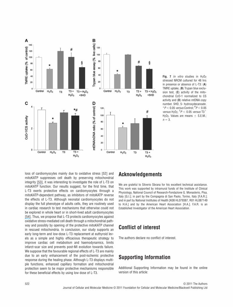
H2O2-induced cell death in isolated cardiomyocytes
The results confirmed thus far show that L-T3 exerts protectiveeffects against ischemic damage. To provide a mechanistic insightfor the observed phenomenon, we suggested that the protectiveeffects of L-T3 is through a mitoKATP-dependent pathway in res-cued mitochondria. We first tested 106 and 107 M concentra-tions of L-T3 in NRCM. There was no protection with 107 (data notshown), however, 106 M concentration for 48 hrs resulted in sig-nificant protection against H2O2-induced cell death, as assessed byTMRE uptake and flow cytometry (Fig. 7a). This concentration of L-T3 was used in all subsequent studies. Pre-treatment with 5HDsignificantly reduced TMRE uptake in H2O2 stressed NRCM in thepresence of L-T3. Moreover, the Trypan blue test (Fig. 7b) showed
Values are means � S.E.M. (n 8 for all groups); FT3: free fraction of T3; FT4: free fraction of T4; TT3: total T3; TT4: total T4. *P � 0.02versus Sham; #P � 0.05 versus T3�.
FT3 FT4 TT3 TT4
(pg/ml) (pg/ml) (ng/dl) (�g/dl)
SHAM 4.3 � 0.4 18.2 � 0.9 61.5 � 4.0 2.4 � 0.3
T3 3.0 � 0.2*# 17.4 � 1.1 42 � 2.8*# 3.1 � 0.2
T3� 4.2 � 0.3 16.3 � 1.5 65 � 4.0 2.8 � 0.2
Table 2 Serum thyroid hormone levels
Table 3 Haemodynamic values
Values are means � S.E.M. (n 8 for all groups); MAP: Mean arterialpressure; LVESP: Left ventricular end-systolic pressure; LVEDP: Leftventricular end-diastolic pressure. *P � 0.05 versus SHAM; #P � 0.05versus T3�.
SHAM T3� T3�
Heart rate,beats/min. 363.15 � 52.12 320.16 � 48.4 357.06 � 41.29
MAP, mmHg 92 � 10.2 80.5 � 9.14*# 90.4 � 12.03
LV dp/dtmax,mmHg/s 6162 � 882.71 3023 � 187.9*# 5756 � 693.52
LVESP, mmHg 122 � 1.45 105.2 � 6.5*# 121.5 � 5.5
LVEDP, mmHg 5.92 � 1.85 17.21 � 5.6*# 9.5 � 3.1*

518 © 2011 The AuthorsJournal of Cellular and Molecular Medicine © 2011 Foundation for Cellular and Molecular Medicine/Blackwell Publishing Ltd
the protective effects of L-T3 against cardiomyocyte necrosis deathat the same experimental condition. The caspase-3 activity did notsignificantly change in cells exposed to 100 �M H2O2 compared tounstressed cells (data not shown).
Mitochondrial function and biogenesis in isolated cardiomyocytes
As showed in panel C of Fig. 7, L-T3 rescued CS-normalized CcO-1activity, which is significantly reduced after H2O2 exposure.Moreover, L-T3 avoided a reduction of mitochondrial biogenesis inthe presence of oxidant stimuli (Fig. 7d).
Discussion
The present study shows that a constant, low-dose infusion of L-T3, initiated 72 hrs after MI and prolonged over 4 weeks to restoreand maintain euthyroid serum levels, markedly attenuatesmyocardial damage and remodelling and preserves LV systolicand diastolic function. Our data strongly suggest that physiologi-cal levels of circulating L-T3 might protect the post-MI heart, atleast in part, by sustaining the expression of key nuclear factorsthat drive mitochondrial integrity. We have previously reportedthat circulating FT3 levels in patients with ischemic HF wereprogressively reduced in relation to disease severity [1, 40]. This alteration could be reverted by short-term L-T3 treatment in
Fig. 1 Global (A–C) and regional(D–F) LV function. (G) RepresentativeLV M-Mode echocardiograms foreach experimental condition. MI:myocardial infarction; LVEF: LV ejec-tion fraction; LVFS: LV fractionalshortening; LVD: LV diameters;LVTBZ: LV thickness of the borderzone; LVTRZ: LV thickness of theremote zone; ED: end diastolic; ES:end systolic; LVESTHK: LV end-sys-tolic wall thickening; BZ: borderzone; RZ: remote zone; LVEDV: LVend-diastolic volume; LVESV: LVend-systolic volume; ED: end-dias-tolic; ES: end-systolic. Values aremeans � S.E.M.; n 8 animals pergroup. *P � 0.05 versus Sham; #P � 0.05 versus 72 hrs MI; †P �
0.05 versus T3–.

J. Cell. Mol. Med. Vol 15, No 3, 2011
519© 2011 The AuthorsJournal of Cellular and Molecular Medicine © 2011 Foundation for Cellular and Molecular Medicine/Blackwell Publishing Ltd
animal [41] and human [2] studies without affecting cardiacremodelling. Indeed, the effects of an early and sustained physio-logical L-T3 treatment on cardiac remodelling are still not welldefined. Consistent with the human disease, we observed reducedFT3 serum levels in rats 72 hrs after MI [4], a pathophysiolgical feature that supports the clinical relevance of our experimentalmodel. Because L-T3 has been shown to exert a wide spectrum ofcardiovascular effects [42], we first needed to assess whether thetherapeutic outcome of low-dose and long-lasting L-T3 infusioncould be related to changes in heart rate and arterial blood pres-sure. We found that the T3� rats displayed no changes in heartrate compared to other two groups, while mean arterial pressure
remained within a physiological range. Previous studies havedemonstrated that post-MI LV remodelling, a major determinant ofmorbidity and mortality in overt HF [43], is an early process, suchas the onset of post-ischemic hypothyroid state. New and moreefficacious interventions aimed at preventing the initial stages ofremodelling are highly needed to contrast the progressiontowards HF [44]. With long-term controlled L-T3 replacement, it iscritical to choose the right timing and dose in order to limit car-diac remodelling and avoid the potentially adverse systemiceffects (i.e. thyrotoxicosis). Henderson et al. recently showed thatL-T3 replacement, initiated 1 week after MI, improved ventricularperformance without reversing cardiac remodelling [22]. In a pre-vious study, an immediate long-term, but not controlled, supple-mentation of thyroid hormones at high dose in post-MI improvedLV function and prevented cardiac remodelling, but also induced athyrotoxic state; in this case it cannot be excluded that the haemo-dynamic actions of thyroid hormones may have contributed toattenuate LV remodelling [45]. In the present study we chose totreat rats with the minimum dose sufficient to restore normal cir-culating L-T3 levels (as established in pilot tests). To avoid the riskof treatment in presence of unstable cardiovascular and systemicconditions frequently observed during the first 2 days after MI, westarted the infusion immediately at the plateau (72 hrs) followingthe nadir (24–48 hrs) of L-T3 levels. Our approach proved veryefficacious in that it halved the infarct-scar size. Since the myocar-dial healing has already begun at 72 hrs after MI in rodent heart,we investigated whether L-T3 modulates some of the major factors involved in myocardial remodelling during the healingphase. Insufficient angiogenesis is one of the causes of myocar-dial dysfunction and there is solid evidence of reduced myocardialcapillary density in HF after MI [46]. Histological analysis of theperi-infarct myocardium revealed significantly less rarefaction ofcapillaries in the T3� hearts, consistent with a preserved expres-sion of the receptor THR-�, which plays a key role in determiningL-T3-induced coronary angiogenesis [37]. The improved capillarydensity might have in part favoured cardiomyocyte survival byenhancing oxygen supply to the border zone, the myocardial tis-sue survived to the ischemic insult. In fact, cardiomyocyte loss byapoptosis, a major cause of ventricular remodelling [47], was sig-nificantly reduced in the LV infarct border zone of L-T3-treatedrats. On the other hand, our in vitro findings did not show celldeath related to caspase-3 activation and confirmed previous find-ings recently published by others [48]. Accordingly, in our exper-imental model H2O2 rapidly damaged the plasma membraneintegrity in a manner similar to the necrosis death, as assessed bytrypan blue exclusion test. However, we cannot exclude that in thepresence of long-term oxidative stress at lower free radicals concentrations the L-T3 protective effect against cell death mightbe exerted, in vitro, even by an inhibition of apoptosis through adeactivation of caspases, as we observed in vivo. We exploredpotential effects of L-T3 on mitochondrial integrity and nuclear-mitochondrial cross-talk. Over the past decade, convincing evi-dence has been provided that the nuclear transcription factors mt-TFA and HIF1-� play an important role in mediating cell survival
Fig. 2 (A) Representative LV transverse slice cut at midseptal level fromT3� treated rats showed a significant reduced infarct-scar size comparedwith T3– animals (magnification, 10); (B) LV collagen accumulationrevealed by Masson trichrome staining (magnification, 40). Values aremeans � S.E.M.; n 8 animals per group. *P � 0.05 versus T3–.

520 © 2011 The AuthorsJournal of Cellular and Molecular Medicine © 2011 Foundation for Cellular and Molecular Medicine/Blackwell Publishing Ltd
mechanisms during myocardial ischemia [15, 38]. Their overex-pression limits LV remodelling and preserves cardiac performanceafter MI [15, 49]. We now found that the expression of mt-TFA andHIF1-� was enhanced in the peri-infarcted myocardium of T3�
hearts, but not in the remote zone. Mitochondrial function is reg-ulated by the coordinated expression of nuclear and mitochondr-ial genes encoding mitochondrial proteins, such as subunits ofrespiratory chain complexes. Recently, it has been found that L-T3modulates cardiac mitochondrial function increasing myocardialmitochondrial respiration, oxidative phosphorylation, mitochondr-ial protein synthesis and mtDNA content [50]. The down-regula-tion of CcO-I, a key enzyme of the mitochondrial respiratory chain,was significantly attenuated in the border zone of L-T3-treatedhearts, consistent with a preserved expression of nuclear tran-
scription factor PGC-1�, a powerful promoter of mitochondrialbiogenesis [17] that is involved in the control of cardiac cellmetabolism and signal transduction [35]. A cause-and-effect rela-tionship between mt-TFA expression, electron transport chainactivity and mtDNA copy number maintenance during the after MIremodelling process has been previously documented by others[51]. Here we demonstrated that L-T3 treatment preserved mito-chondrial biogenesis and function when cardiomyocytes areexposed to oxidative stress in vitro. Therefore, it is likely that thebeneficial effects of L-T3 in ischemic myocardium that we foundin vivo were in part due to rescue of mitochondria function and cellmetabolism during post-MI healing, which avoids a bioenergeticcatastrophe culminating in cell necrosis. In terms of mechanisticinvestigation, since LV remodelling after MI involves side-to-side
Fig. 3 (A) Immunohistochemical detection ofendothelial cells identified by CD31 (blackarrows) in border (BZ) and remote zone (RZ)of infarcted hearts to quantify LV capillarydensity (magnification, 400). (B) Westernblot analysis of THR-� normalized to �-actinin BZ and RZ of infarcted hearts. Values aremeans � S.E.M. (n 6 microscopic fieldsper LV section); n 8 animals per group. #P� 0.05 versus T3–; †P � 0.05 versus RZ.
Fig. 4 TUNEL staining to quantify apoptoticcardiomyocytes (black arrows) in T3� andT3 infarcted left ventricle and haematoxylincounterstaining (magnification, 400). Valuesare means � S.E.M. (n 6 microscopicfields per LV section); n 8 animals pergroup. #P � 0.05 versus T3–; †P � 0.05 versus RZ.

J. Cell. Mol. Med. Vol 15, No 3, 2011
521© 2011 The AuthorsJournal of Cellular and Molecular Medicine © 2011 Foundation for Cellular and Molecular Medicine/Blackwell Publishing Ltd
Fig. 5 Gene (A) and protein (B) expression of HIF-1� normalized to �-actin gene and protein expression in border (BZ) and remote zone (RZ)of infarcted hearts. Values are means � S.E.M.; n 8 animals per group.*P � 0.05 versus Sham; #P � 0.05 versus RZ; †P � 0.05 versus T3�.
Fig. 6 Gene (A) and protein (B)expression of Mt-TFA and (C) geneexpression of PGC-1� normalizedto �-actin gene and protein expres-sion in border (BZ) and remotezone (RZ) of infarcted hearts; (D)activity of the mitochondrial CcO-1normalized to CS activity in BZ andRZ zone of infarcted hearts. Valuesare means � S.E.M.; n 8 ani-mals per group. *P � 0.05 versusSham; #P � 0.05 versus RZ; †P �0.05 versus T3�.
522 © 2011 The AuthorsJournal of Cellular and Molecular Medicine © 2011 Foundation for Cellular and Molecular Medicine/Blackwell Publishing Ltd
loss of cardiomyocytes mainly due to oxidative stress [52] andmitoKATP suppresses cell death by preserving mitochondrialintegrity [53], it was interesting to investigate the role of L-T3 onmitoKATP function. Our results suggest, for the first time, that L-T3 exerts protective effects on cardiomyocytes through amitoKATP-dependent pathway, as inhibitors of mitoKATP reversethe effects of L-T3. Although neonatal cardiomyocytes do notdisplay the full phenotype of adults cells, they are routinely usedin cardiac research to test mechanisms that otherwise could notbe explored in whole heart or in short-lived adult cardiomyocytes[54]. Thus, we propose that L-T3 protects cardiomyocytes againstoxidative stress-mediated cell death through a mitochondrial path-way and possibly by opening of the protective mitoKATP channelin rescued mitochondria. In conclusion, our study supports anearly long-term and low-dose L-T3 replacement at euthyroid lev-els as a simple and highly efficacious therapeutic strategy toimprove cardiac cell metabolism and haemodynamics, limitsinfarct-scar size and prevents post-MI evolution towards failure.We suppose that the favourable regional effects of L-T3 are mainlydue to an early enhancement of the post-ischemic protectiveresponse during the healing phase. Although L-T3 displays multi-ple functions, enhanced capillary formation and mitochondrialprotection seem to be major protective mechanisms responsiblefor these beneficial effects by using low dose of L-T3.
Acknowledgements
We are grateful to Silverio Sbrana for his excellent technical assistance.This work was supported by intramural funds of the Institute of ClinicalPhysiology, National Council of Research-Fondazione G. Monasterio, Pisa,Italy [G.I.]; in part by the Compagnia di San Paolo, Torino, Italy [F.A.R.];and in part by National Institutes of Health [K08 HL079387, R01 HL087149to H.A.] and by the American Heart Association [H.A.]. F.A.R. is anEstablished Investigator of the American Heart Association.
Conflict of interest
The authors declare no conflict of interest.
Supporting Information
Additional Supporting Information may be found in the onlineversion of this article:
Fig. 7 In vitro studies in H2O2
stressed NRCM cultured for 48 hrsin presence or absence of L-T3: (A)TMRE uptake, (B) Trypan blue exclu-sion test, (C) activity of the mito-chondrial CcO-1 normalized to CSactivity and (D) relative mtDNA copynumber. 5HD, 5- hydroxydecanoate.*P � 0.05 versus Control; #P � 0.05versus H2O2; †P � 0.05 versus T3�
H2O2. Values are means � S.E.M.; n 3.
J. Cell. Mol. Med. Vol 15, No 3, 2011
523© 2011 The AuthorsJournal of Cellular and Molecular Medicine © 2011 Foundation for Cellular and Molecular Medicine/Blackwell Publishing Ltd
References
1. Iervasi G, Pingitore A, Landi P, et al.Low-T3 syndrome: a strong prognosticpredictor of death in patients with heartdisease. Circulation. 2003; 107: 708–13.
2. Pingitore A, Galli E, Barison A, et al.Acute effects of triiodothyronine (T3)replacement therapy in patients withchronic heart failure and low-T3 syn-drome: a randomized, placebo-controlledstudy. J Clin Endocrinol Metab. 2008; 93:1351–8.
3. Hamilton MA, Stevenson LW, Luu M, et al. Altered thyroid hormone metabolismin advanced heart failure. J Am CollCardiol. 1990; 16: 91–5.
4. Wiersinga WM, Lie KI, Touber JL. Thyroidhormones in acute myocardial infarction.Clin Endocrinol. 1981; 14: 367–74.
5. Friberg L, Drvota V, Bjelak AH, et al.Association between increased levels ofreverse triiodothyronine and mortality afteracute myocardial infarction. Am J Med.2001; 111: 699–703.
6. Wrutniak-Cabello C, Casas F, Cabello G.Thyroid hormone action in mitochondria.J Mol Endocrinol. 2001; 26: 67–77.
7. Goldenthal MJ, Ananthakrishnan R,Marín-García J. Nuclear-mitochondrialcross-talk in cardiomyocyte T3 signaling: atime-course analysis. J Mol Cell Cardiol.2005; 39: 319–26.
8. Goldenthal MJ, Weiss HR, Marín-GarcíaJ. Bioenergetic remodeling of heart mito-chondria by thyroid hormone. Mol CellBiochem. 2004; 265: 97–106.
9. Marín-García J, Goldenthal MJ.Mitochondrial centrality in heart failure.Heart Fail Rev. 2008; 13: 137–50.
10. Zhu L, Yu Y, Chua BH, et al. Regulation ofsodium-calcium exchange and mitochon-drial energetics by Bcl-2 in the heart oftransgenic mice. J Mol Cell Cardiol. 2001;33: 2135–44.
11. Huss JM, Kelly DP. Mitochondrial energymetabolism in heart failure: a question ofbalance. J Clin Invest. 2005; 115: 547–55.
12. Cannino G, Di Liegro CM, Rinaldi AM.Nuclear–mitochondrial interaction.Mitochondrion. 2007; 7: 359–66.
13. Chavez A, Miranda LF, Pichiule P, et al.Mitochondria and hypoxia-induced geneexpression mediated by hypoxia-induciblefactors. Ann NY Acad Sci. 2008; 1147:312–20.
14. Sun Y. Myocardial repair/remodelling fol-lowing infarction: roles of local factors.Cardiovasc Res. 2009; 81: 482–90.
15. Ikeuchi M, Matsusaka H, Kang D, et al.Overexpression of mitochondrial tran-scription factor A ameliorates mitochondr-ial deficiencies and cardiac failure aftermyocardial infarction. Circulation. 2005;112: 683–90.
16. Kang D, Kim SH, Hamasaki N.Mitochondrial transcription factor A(TFAM): roles in maintenance of mtDNAand cellular functions. Mitochondrion.2007; 7: 39–44.
17. Ventura-Clapier R, Garnier A, Veksler V.Transcriptional control of mitochondrialbiogenesis: the central role of PGC-1alpha.Cardiovasc Res. 2008; 79: 208–17.
18. Russell LK, Mansfield CM, Lehman JJ,et al. Cardiacspecific induction of the transcriptional coactivator peroxisomeproliferator-activated receptor gammacoactivator-1alpha promotes mitochondr-ial biogenesis and reversible cardiomyopa-thy in a developmental stage-dependentmanner. Circ Res. 2004; 94: 525–33.
19. Garnier A, Fortin D, Deloménie C, et al.Depressed mitochondrial transcription fac-tors and oxidative capacity in ratfailing car-diac and skeletal muscles. J Physiol. 2003;551: 491–501.
20. Ardehali H, O’Rourke B, Marbán E.Cardioprotective role of the mitochondrialATPbinding cassette protein 1. Circ Res.2005; 97: 740–2.
21. Ventura C, Cantoni S, Bianchi F, et al.Hyaluronan mixed esters of butyric andretinoic Acid drive cardiac and endothelial
fate in term placenta human mesenchymalstem cells and enhance cardiac repair ininfarcted rat hearts. J Biol Chem. 2007;282: 14243–52.
22. Henderson KK, Danzi S, Paul JT, et al.Physiological replacement of T3 improvesleft ventricular function in an animal modelof myocardial infarction-induced conges-tive heart failure. Circ Heart Fail. 2009; 2:243–52.
23. Ojamaa K, Kenessey A, Shenoy R, et al.Thyroid hormone metabolism and cardiacgene expression after acute myocardialinfarction in the rat. Am J PhysiolEndocrinol Metab. 2000; 279: E1319–24.
24. Shiomi T, Tsutsui H, Hayashidani S, et al. Pioglitazone, a peroxisome prolifera-tor–activated receptor-gamma agonist,attenuates lef ventricular remodeling andfailure after experimental myocardialinfarction. Circulation. 2002; 106:3126–32.
25. Kasdallah AG, Mornagui B, Gharbi N, et al. Metabolic and endocrine effects ofwater and/or food deprivation in rats. C RBiol. 2005; 328: 463–70.
26. Olivetti G, Ricci R, Beghi C, et al.Response of the border zone to myocardialinfarction in rats. Am J Pathol. 1986; 125:476–83.
27. Davani S, Marandin A, Mersin N, et al.Mesenchymal progenitor cells differentiateinto an endothelial phenotype, enhancevascular density, and improve heart func-tion in a rat cellular cardiomyoplastymodel. Circulation. 2003; 108: 253–8.
28. Pucci A, Zanini C, Granata R, et al.Myocardial insulinlike growth factor-1 andinsulin-like growth factor binding protein-3gene expression in failing hearts harvestedfrom patients undergoing cardiac trans-plantation. J Heart Lung Transplant. 2009;28: 402–5.
29. Morrish F, Buroker NE, Ge M, et al.Thyroid hormone receptor isoforms local-ize to cardiac mitochondrial matrix with
Fig S1 Circulating FT3 (A) and TT3 (B) levels before (Normal), 72 hours post-coronary ligation (72h MI) and after 1, 2, 3, 4 weeks ofexperimental T3 treatment. T3 supplementation at 0.5 or 1.2 �g/kg/dayin rodents with myocardial infarction began 72 hours after coronaryligation by osmotic pump. Values are means � SEM; n 4 animalsper group. *P � 0.05 versus Normal; #P � 0.05 versus 0.5 �g/kg/dayat the same experimental condition; †P � 0.05 versus 72 h MI.
Table S1 Oligonucleotides sequences.
Please note: Wiley-Blackwell are not responsible for the content orfunctionality of any supporting materials supplied by the authors.Any queries (other than missing material) should be directed tothe corresponding author for the article.

524 © 2011 The AuthorsJournal of Cellular and Molecular Medicine © 2011 Foundation for Cellular and Molecular Medicine/Blackwell Publishing Ltd
potential for binding to receptor elementson mtDNA. Mitochondrion. 2006; 6:143–8.
30. Guo D, Nguyen T, Ogbi M, et al. Proteinkinase C-epsilon coimmunoprecipitateswith cytochrome oxidase subunit IV and isassociated with improved cytochrome-coxidase activity and cardioprotection. AmJ Physiol Heart Circ Physiol. 2007; 293:H2219–30.
31. Hassouna A, Loubani M, Matata BM, et al. Mitochondrial dysfunction as thecause of the failure to precondition the dia-betic human myocardium. Cardiovasc Res.2006; 69: 450–8.
32. Morgunov I, Srere PA. Interaction betweencitrate synthase and malate dehydroge-nase. Substrate channeling of oxaloacetate.J Biol Chem. 1998; 273: 29540–4.
33. Valks DM, Kemp TJ, Clerk A. Regulationof Bcl-xL expression by H2O2 in cardiacmyocytes. J Biol Chem. 2003; 278:25542–7.
34. Korge P, Honda HM, Weiss JN. Protectionof cardiac mitochondria by diazoxide andprotein kinase C:implications for ischemicpreconditioning. Proc Natl Acad Sci USA.2002; 99: 3312–7.
35. Park JY, Wang P, Matsumoto T, et al. p53Improves aerobic exercise capacity andaugments skeletal muscle mitochondrialDNA content. Circ Res. 2009; 105: 705–12.
36. Fukui S, Kitagawa-Sakakida S, KawamataS, et al. Therapeutic effect of midkine oncardiac remodeling in infarcted rat hearts.Ann Thorac Surg. 2008; 85: 562–70.
37. Makino A, Suarez J, Wang H, et al.Thyroid hormone receptor-beta is associ-ated with coronary angiogenesis duringpathological cardiac hypertrophy.Endocrinology. 2009; 150: 2008–15.
38. Eckle T, Köhler D, Lehmann R, et al.Hypoxia-inducible factor-1 is central tocardioprotection: a new paradigm forischemic preconditioning. Circulation.2008; 118: 166–75.
39. Lee SH, Wolf PL, Escudero R, et al. Earlyexpression of angiogenesis factors inacute myocardial ischemia and infarction.N Engl J Med. 2000; 342: 626–33.
40. Pingitore A, Landi P, Taddei MC, et al.Triiodothyronine levels for risk stratifica-tion of patients with chronic heart failure.Am J Med. 2005; 118: 132–6.
41. Chen YF, Kobayashi S, Chen J, et al.Short term triiodo-Lthyronine treatmentinhibits cardiac myocyte apoptosis in bor-der area after myocardial infarction in rats.J Mol Cell Cardiol. 2008; 44: 180–7.
42. Kahaly GJ, Dillmann WH. Thyroid hor-mone action in the heart. Endocr Rev.2005; 26: 704–28.
43. Pfeffer MA, Braunwald E. Ventricularremodeling after myocardial infarction.Experimental observations and clinical impli-cations. Circulation. 1990; 81: 1161–72.
44. Sigurdsson A, Eriksson SV, Hall C, et al.Early neurohormonal effects of trandolaprilin patients with left ventricular dysfunctionand a recent acute myocardial infarction: adouble-blind, randomized, placebo-con-trolled multicentre study. Eur J Heart Fail.2001; 3: 69–78.
45. Pantos C, Mourouzis I, Markakis K, et al.Longterm thyroid hormone administrationreshapes left ventricular chamber andimproves cardiac function after myocardialinfarction in rats. Basic Res Cardiol. 2008;103: 308–18.
46. Karch R, Neumann F, Ullrich R, et al. Thespatial pattern of coronary capillaries inpatients with dilated, ischemic,or inflam-
matory cardiomyopathy. CardiovascPathol. 2005; 14: 135–44.
47. Dorn GW 2nd. Apoptotic and non-apop-totic programmed cardiomyocyte death inventricular remodelling. Cardiovasc Res.2009; 81: 465–73.
48. Goto K, Takemura G, Maruyama R, et al. Unique mode of cell death in freshlyisolated adult rat ventricular cardiomy-ocytes exposed to hydrogen peroxide.Med Mol Morphol. 2009; 42: 92–101.
49. Kido M, Du L, Sullivan CC, et al. Hypoxia-inducible factor 1-alpha reduces infarctionand attenuates progression of cardiac dys-function after myocardial infarction in themouse. J Am Coll Cardiol. 2005; 46:2116–24.
50. Marín-Garcia J. Thyroid hormone andmyocardial mitochondrial biogenesis.Vascul Pharmacol. 2009 [Epub ahead ofprint]. Doi:10.1016/j.vph.2009.10.008
51. Ide T, Tsutsui H, Hayashidani S, et al.Mitochondrial DNA damage and dysfunc-tion associated with oxidative stress in fail-ing hearts after myocardial infarction. CircRes. 2001; 88: 529 –35.
52. Yamaguchi O, Higuchi Y, Hirotani S, et al. Targeted deletion of apoptosis sig-nalregulating kinase 1 attenuates left ven-tricular remodeling. Proc Natl Acad SciUSA. 2003; 100: 15883–8.
53. Akao M, Ohler A, O’Rourke B, et al.Mitochondrial ATP-sensitive potassiumchannels inhibit apoptosis induced byoxidative stress in cardiac cells. Circ Res.2001; 88: 1267–75.
54. Pitts KR, Toombs CF. Studying ischemiaand reperfusion in isolated neonatal rat ven-tricular myocytes using coverslip hypoxia.Methods Mol Med. 2007; 139: 271–81.
Copyright © 2022 FDOKUMEN