Expression of transcripts for myelination-related genes in the anterior cingulate cortex in...
19
Expression of Transcripts for Myelination-Related Genes in the Anterior Cingulate Cortex in Schizophrenia Robert E McCullumsmith 1 , Daya Gupta 1 , Monica Beneyto 1 , Emily Kreger 2 , Vahram Haroutunian 3 , Kenneth L Davis 3 , and James H Meador-Woodruff 1 1 Department of Psychiatry and Behavioral Neurobiology, University of Alabama at Birmingham School of Medicine, Birmingham, AL 2 Neuroscience program, University of Michigan Medical School, 205 Zina Pitcher Place, Ann Arbor, MI 48109, USA 3 Department of Psychiatry, Mount Sinai School of Medicine, New York, NY, USA Abstract Several recent studies have found changes in the expression of genes functionally related to myelination and oligodendrocyte homeostasis in schizophrenia. These studies utilized microarrays and quantitative PCR (QPCR), methodologies which do not permit direct, unamplified examination of mRNA expression. In addition, these studies generally only examined transcript expression in homogenates of gray matter. In the present study, we examined the expression of myelination-related genes previously implicated in schizophrenia by microarray or QPCR. Using in situ hybridization, we measured transcript expression of 2′,3′-cyclic nucleotide 3′-phosphodiesterase (CNP), myelin- associated glycoprotein (MAG), transferrin (TF), quaking (QKI), gelsolin, myelin oligodendrocyte glycoprotein, v-erb-b2 erythroblastic leukemia viral oncogene homolog 3, erbb2 interacting protein, motility-related protein-1, SRY-box containing gene 10, oligodendrocyte transcription factor 2, peripheral myelin protein 22, and claudin-11 in both gray and white matter of the anterior cingulate cortex (ACC) in subjects with schizophrenia (n = 41) and a comparison group (n = 34). We found decreased expression of MAG, QKI, TF, and CNP transcripts in white matter. We did not find any differences in expression of these transcripts between medicated (n = 31) and unmedicated (n = 10) schizophrenics, suggesting that these changes are not secondary to treatment with antipsychotics. Finally, we found significant positive correlations between QKI and MAG or CNP mRNA expression, suggesting that the transcription factor QKI regulates MAG and CNP expression. Our results support the hypothesis that myelination and oligodendrocyte function are impaired in schizophrenia. Introduction While early hypotheses viewed schizophrenia as a dysfunction of discrete brain regions, recent models often conceptualize this illness as a disorder of the functional integrity of widely Address all correspondence to: Department of Psychiatry and Behavioral Neurobiology, University of Alabama at Birmingham School of Medicine, Civitan International Research Center, Room 576A, 1530 3rd Avenue South, Birmingham, AL, phone: (205) 996 6285, fax: (205) 975 4879, email: [email protected] Financial Support: This work was supported by MH45212 (JHMW, KLD & VH), a Pfizer Postdoctoral Fellowship (REM), VA Merit Review (VH), MH064673 (VH) and MH66392 (KLD). Publisher's Disclaimer: This is a PDF file of an unedited manuscript that has been accepted for publication. As a service to our customers we are providing this early version of the manuscript. The manuscript will undergo copyediting, typesetting, and review of the resulting proof before it is published in its final citable form. Please note that during the production process errors may be discovered which could affect the content, and all legal disclaimers that apply to the journal pertain. NIH Public Access Author Manuscript Schizophr Res. Author manuscript; available in PMC 2008 February 1. Published in final edited form as: Schizophr Res. 2007 February ; 90(1-3): 15–27. NIH-PA Author Manuscript NIH-PA Author Manuscript NIH-PA Author Manuscript
Transcript of Expression of transcripts for myelination-related genes in the anterior cingulate cortex in...
Expression of Transcripts for Myelination-Related Genes in theAnterior Cingulate Cortex in Schizophrenia
Robert E McCullumsmith1, Daya Gupta1, Monica Beneyto1, Emily Kreger2, VahramHaroutunian3, Kenneth L Davis3, and James H Meador-Woodruff11 Department of Psychiatry and Behavioral Neurobiology, University of Alabama at Birmingham School ofMedicine, Birmingham, AL
2 Neuroscience program, University of Michigan Medical School, 205 Zina Pitcher Place, Ann Arbor, MI48109, USA
3 Department of Psychiatry, Mount Sinai School of Medicine, New York, NY, USA
AbstractSeveral recent studies have found changes in the expression of genes functionally related tomyelination and oligodendrocyte homeostasis in schizophrenia. These studies utilized microarraysand quantitative PCR (QPCR), methodologies which do not permit direct, unamplified examinationof mRNA expression. In addition, these studies generally only examined transcript expression inhomogenates of gray matter. In the present study, we examined the expression of myelination-relatedgenes previously implicated in schizophrenia by microarray or QPCR. Using in situ hybridization,we measured transcript expression of 2′,3′-cyclic nucleotide 3′-phosphodiesterase (CNP), myelin-associated glycoprotein (MAG), transferrin (TF), quaking (QKI), gelsolin, myelin oligodendrocyteglycoprotein, v-erb-b2 erythroblastic leukemia viral oncogene homolog 3, erbb2 interacting protein,motility-related protein-1, SRY-box containing gene 10, oligodendrocyte transcription factor 2,peripheral myelin protein 22, and claudin-11 in both gray and white matter of the anterior cingulatecortex (ACC) in subjects with schizophrenia (n = 41) and a comparison group (n = 34). We founddecreased expression of MAG, QKI, TF, and CNP transcripts in white matter. We did not find anydifferences in expression of these transcripts between medicated (n = 31) and unmedicated (n = 10)schizophrenics, suggesting that these changes are not secondary to treatment with antipsychotics.Finally, we found significant positive correlations between QKI and MAG or CNP mRNAexpression, suggesting that the transcription factor QKI regulates MAG and CNP expression. Ourresults support the hypothesis that myelination and oligodendrocyte function are impaired inschizophrenia.
IntroductionWhile early hypotheses viewed schizophrenia as a dysfunction of discrete brain regions, recentmodels often conceptualize this illness as a disorder of the functional integrity of widely
Address all correspondence to: Department of Psychiatry and Behavioral Neurobiology, University of Alabama at Birmingham Schoolof Medicine, Civitan International Research Center, Room 576A, 1530 3rd Avenue South, Birmingham, AL, phone: (205) 996 6285,fax: (205) 975 4879, email: [email protected] Support: This work was supported by MH45212 (JHMW, KLD & VH), a Pfizer Postdoctoral Fellowship (REM), VA MeritReview (VH), MH064673 (VH) and MH66392 (KLD).Publisher's Disclaimer: This is a PDF file of an unedited manuscript that has been accepted for publication. As a service to our customerswe are providing this early version of the manuscript. The manuscript will undergo copyediting, typesetting, and review of the resultingproof before it is published in its final citable form. Please note that during the production process errors may be discovered which couldaffect the content, and all legal disclaimers that apply to the journal pertain.
NIH Public AccessAuthor ManuscriptSchizophr Res. Author manuscript; available in PMC 2008 February 1.
Published in final edited form as:Schizophr Res. 2007 February ; 90(1-3): 15–27.
NIH
-PA Author Manuscript
NIH
-PA Author Manuscript
NIH
-PA Author Manuscript
distributed circuitry. Connectivity within such circuits is dependent on myelination byoligodendroglia, a tightly regulated process that includes the initial myelination of axons duringbrain development and the coordination of myelin synthesis and turnover in the adult CNS.Converging lines of evidence suggest that the processes governing myelination and/or theregulation of oligodendroglia are abnormal in schizophrenia (Reviewed in: (Davis et al.,2003)). Studies using magnetic resonance, magnetization transfer, and diffusion tensor imagingsupport the hypothesis that there are white matter abnormalities in this illness, and pathologicalexamination by electron microscopy suggests a role for alterations of the myelin sheath(Buchsbaum et al., 1998;Lim et al., 1998;Lim et al., 1999;Pfefferbaum et al., 1999;Foong etal., 2000;Agartz et al., 2001;Foong et al., 2001;Sigmundsson et al., 2001;Steel et al.,2001;Uranova et al., 2001;Bagary et al., 2003;Sun et al., 2003;Hubl et al., 2004;Uranova et al.,2004;Wang et al., 2004;Szeszko et al., 2005). A subset of myelin-related genes has beenconsistently implicated in schizophrenia by genetic studies (Reviewed in: (Davis et al.,2003)), and subsequent microarray and QPCR analyses have found decreases in mRNAexpression of myelin-related genes in prefrontal and temporal cortices (Hakak et al.,2001;Tkachev et al., 2003;Aston et al., 2004;Dracheva et al., 2005a;Haroutunian et al.,2005).
One region in schizophrenia where myelination-dependent connectivity may be abnormal isthe anterior cingulate cortex (ACC). Comparison of region-specific gene expression bymicroarray analyses found the second highest number of abnormally expressed transcripts inthe ACC, after the superior temporal cortex, and well ahead of the DLPFC (Kastel et al.,2005). The ACC integrates cognitive function, motor control, and drive states; coordination ofthese modalities is often abnormal in schizophrenia (Paus, 2001;Heckers et al., 2004). Otherstudies have found abnormal activation of the ACC with tasks related to social functioning andbehavior, a reduction in ACC glial cell number, and changes in GABAergic and glutamatergicgene expression in the ACC in this illness (Quintana et al., 2004;Stark et al., 2004;Woo et al.,2004). In addition, reductions in white matter volumes were detected in the ACC inschizophrenia using magnetic resonance imaging (Riffkin et al., 2005). Taken together withthe aforementioned microarray data, these findings support a hypothesis of abnormalconnectivity secondary to alterations in myelination and oligodendrocyte function in the ACCin schizophrenia.
To investigate the hypothesis that myelination and oligodendrocyte function are abnormal inschizophrenia, we examined expression of myelination-related genes previously implicated inthis illness by microarray or QPCR analysis (Hakak et al., 2001;Tkachev et al., 2003;Aston etal., 2004;Dracheva et al., 2005a;Katsel et al., 2005). Since these techniques do not preservethe anatomy of the tissue and they rely on amplification of mRNA, we used in situ hybridizationto directly measure transcript expression of 2′,3′-cyclic nucleotide 3′-phosphodiesterase(CNP), myelin-associated glycoprotein (MAG), transferrin (TF), quaking (QKI), gelsolin(GEL), myelin oligodendrocyte glycoprotein (MOG), v-erb-b2 erythroblastic leukemia viraloncogene homolog 3 (ErbB3), erbb2 interacting protein (ErbB2IP), motility-related protein-1(CD9) , SRY-box containing gene 10 (SOX10), oligodendrocyte transcription factor 2(OLIG2), peripheral myelin protein 22 (PMP22), and Claudin-11 (CLDN11) in both gray andunderlying white matter of the ACC in schizophrenia.
Material and MethodsSubjects
A total of 75 subjects from the Mount Sinai Medical Center / Bronx VA Medical CenterDepartment of Psychiatry Brain Bank were studied, 41 individuals diagnosed withschizophrenia and 34 comparison subjects with no history of psychiatric illness (Table 1)
McCullumsmith et al. Page 2
Schizophr Res. Author manuscript; available in PMC 2008 February 1.
NIH
-PA Author Manuscript
NIH
-PA Author Manuscript
NIH
-PA Author Manuscript
(Davidson et al., 1995). Upon neuropathological examination, no evidence forneurodegenerative changes or Alzheimer disease was found in any of the subjects (Purohit etal., 1998). Brains were obtained after autopsy and one hemisphere was cut coronally into 10mm slabs and frozen. Brain pH was measured using an aliquot of homogenized brain tissueand a pH meter. pH measurements were validated using correlation analysis by determininglactate levels in the same tissue homogenates from randomly selected cases. Anterior cingulatecortex was dissected at the level of the genu of the corpus callosum. Sections included theadjacent cingulum bundle. Tissue blocks were dissected from the dorsal surface of the corpuscallosum extending 12–15mm dorsally and extending 12–15 mm laterally from the midline.15 μm sections were thawed onto slides previously treated with poly-L-lysine, dried, and storedat −80ºC. Two slides were studied per subject.
In situ hybridizationThe sensitivity and reproducibility of in situ hybridization has previously been wellcharacterized (Williams, 1982;Downs and Williams, 1984;Stolz et al., 1989). mRNAexpression was measured by in situ hybridization using subclones that were generated byamplifying unique segments of 2′,3′-cyclic nucleotide 3′-phosphodiesterase (Abbreviation:CNP, Genebank accession number: NM_03313, region used for probe: 523–1066), myelin-associated glycoprotein (MAG, XM_01287, 305–835), transferrin (TF, XM_00279, 1172–1940), quaking (QKI, AF142421, 508–715), gelsolin (GSN, XM_01654, 1249–1649), myelinoligodendrocyte glycoprotein (MOG, U18800, 109–263), v-erb-b2 erythroblastic leukemiaviral oncogene homolog 3 (ErbB3, NM_00198, 3081–3325), erbb2 interacting protein(ErbB2IP, NM_01869, 3531–3858), motility-related protein-1 (CD9, M38690, 330–666),SRY-box containing gene 10 (SOX10, BL007595, 1337–1560), oligodendrocyte transcriptionfactor 2 (OLIG2, NM_00580, 423–628), peripheral myelin protein 22 (PMP22, BC019040,293–637), and Claudin-11 (CLDN11, NM_00560, 610–821) from a human cDNA brain library(Human Adult Brain Unamplified cDNA Library, Edge Biosystems; Gaithersburg, MD) andPolymerase Chain Reaction (PCR). PCR-amplified cDNA segments were extracted (QIAquickGel Extraction Kit, Qiagen, Valencia, CA), subcloned (Zero Blunt TOPO PCR cloning kit;Invitrogen, Carlsbad, CA), and confirmed by nucleotide sequencing. Sense and antisenseprobes for in situ hybridization were synthesized using 100 μCi of dried [35S]UTP, 2.0 μl 5xtranscription buffer (40mM Tris-base, 6 mM Mg Cl2, 2mM spermidine,10mM NACl, pH7.9);1.0 μl each of 10 mM ATP, CTP, and GTP; 1.0 μg linearized plasmid DNA; 0.5μl RNAseinhibitor; and 1.5 μl SP6 or T7 RNA polymerase. After 2 hours incubation at 37°C, 1.0 μlDNAse (RNAse-free) was added and incubated for 15 min at room temperature (RT). [35S]Labeled cRNA was purified with a spin column (Micro Bio-Spin P-30 Tris Spin Columns,Bio-Rad Laboratories; Hercules, CA), diluted to 100 μl final volume, and 1.0 μl of 1Mdithiothreitol (DTT) was added to a final concentration of 0.01 M.
Sections were fixed with 4% (weight:volume) formaldehyde for 1 hour at room temperature(RT). Next, sections were rinsed three times in 2X SSC (300 mM NaCl/30 mM sodium citrate,pH 7.2) and incubated on a stir plate in 0.1 M triethanolamine (TAE), pH 8.0 / acetic anhydride,1:400 (volume/volume) for 10 min at RT. Sections were then washed in 2X SSC for 10 minat RT, the tissue was dehydrated through graded alcohols and air-dried. [35S] labeled riboprobe(1 x 10⁶ cpm), was applied in 100 μl of 50% formamide buffer (50% formamide, 10% dextransulfate, 3X SSC, 50 mM Na2HPO4, pH 7.4, 1X Denhardt’s solution, 100 μg/ml yeast tRNA)with 0.1% of 1M DTT per each slide. The slides were then covered with glass cover slips andstored in a humidified chamber saturated with 50% formamide overnight at 55°C for 18 hours.Next, the cover slips were removed and the sections were washed in 2X SSC for 2 min at RT,2X SSC for 10 min at RT, and then incubated with RNAse A (200 mg/ml in 10 mM Tris-HCl,pH 8.0/0.5 M NaCl) at 37°C for 30 min. Slides were then washed at RT two times for 15 min
McCullumsmith et al. Page 3
Schizophr Res. Author manuscript; available in PMC 2008 February 1.
NIH
-PA Author Manuscript
NIH
-PA Author Manuscript
NIH
-PA Author Manuscript
in 2X SSC, 15 min in 1X SSC, 5 min in 0.5X SSC, two times for 60 min in 0.1X SSC at 55°C and 15 min in 0.1X SSC at RT. The sections were then dehydrated in graded alcohol solutionsand air-dried. Finally the slides were placed in X-ray cassettes, apposed to film (KodakBIOMAX MR Film, New England, Nuclear, Boston, MA) and developed after 10–31 days.
Data analysisImages were digitized from films with a CCD camera with NIH Image software v16.1 andanalyzed with Scion Image Beta 3b. Gray and white matter margins were identified based uponcellular patterns as defined by cresyl violet staining of sections from each subject. The patternof expression for each of the probes was relatively uniform within the gray and underlyingwhite matter, and no lamination was observed for any of the transcripts. The region analysedfor each transcript included the entire profile of either the gray or the white matter of eachsection. Film background values were subtracted from gray scale values of either gray orunderlying white matter regions of each section and converted to optical density. Values fortwo sections per subject were averaged and used for statistical analysis.
Statistical analysesThe phrase “Main effect for diagnosis” refers to the statistical result from the factorial ANOVAor ANCOVA analysis for the independent variable, diagnosis. The phrase “diagnosis by regioninteraction” refers the statistical result from the factorial ANOVA or ANCOVA analysis forthe combined effects of the independent variables for diagnosis and isodense band. Correlationanalysis for each transcript was performed to investigate possible associations betweentranscript expression and age, postmortem interval (PMI), and tissue pH. When significantassociations with age, pH or PMI were found, analysis of covariance was utilized, otherwiseanalysis of variance was utilized, with diagnosis and region (gray matter or white matter) asthe independent variables and optical density as the dependent variable. Post-hoc analysis wasperformed by Tukey’s HSD. For all tests, α = 0.05. Correlation analyses were performed toprobe for associations in gene expression for transcript that were significantly altered inschizophrenia and were previously demonstrated to be functionally linked or coordinatelyregulated.
ResultsSense and antisense probes were tested in sections of human cortex; specific labeling was onlyobserved for sections incubated with antisense riboprobe (data not shown). We detectedtranscript expression in both white and gray matter of the ACC for all of the genes studied(Figures 1 and 2), although the gray matter labeling was considerably less than white matterfor each gene studied, as expected for oligodendrocyte-associated genes.
We did not detect any associations between QKI, MAG, SOX10, OLIG2, CD9, CLDN11,MOG, ErbB2IP, or ErbB3 mRNA expression and age, PMI or pH. We did detect associationsbetween CNP, GSN, and TF mRNA expression and pH (CNP: r = 0.27, p < 0.03, GSN: r =0.16, p < 0.05, TF r = 0.17, p < 0.03) but not age or PMI. We also detected associations betweenPMP22 and age (r = 0.20, p < 0.02) and pH (r = 0.17, p < 0.04), but not PMI.
We detected a main effect for diagnosis for QKI (F (1, 138) = 6.2, p < 0.02) and MAG (F (1,135) = 4.3, p < 0.04), but not SOX10, OLIG2, CD9, CLDN11, MOG, ErbB2, or ErbB3transcript expression (Figures 3 and 4). We detected a main effect for region for QKI, MAG,SOX10, OLIG2, CD9, CLDN11, MOG, ErbB2IP, and ErbB3 mRNA expression. We did notdetect any diagnosis by region interactions for these transcripts. Post-hoc analysis revealeddecreased QKI (p < 0.03) and MAG (p < 0.04) mRNA expression in the white matter in
McCullumsmith et al. Page 4
Schizophr Res. Author manuscript; available in PMC 2008 February 1.
NIH
-PA Author Manuscript
NIH
-PA Author Manuscript
NIH
-PA Author Manuscript
schizophrenia, and higher levels of mRNA expression for QKI, MAG, SOX10, OLIG2, CD9,CLDN11, MOG, ErbB2IP, and ErbB3 in the white matter compared to the gray matter (Figures3 and 4, p < 0.05 for all transcripts). Using ANCOVA with pH (and age for PMP22) as acovariate, we detected a main effect for diagnosis for CNP (F (1, 135) = 7.6, p < 0.01) and TF(F (1, 147) = 6.7, p < 0.01), but not GSN or PMP22 mRNA expression (Figures 3 and 4). Wedetected a main effect for region for CNP, TF, PMP22 and GSN mRNA expression. We didnot detect any diagnosis by region interactions for these transcripts. Post-hoc analysis revealeddecreased CNP (p < 0.04) and TF (p < 0.04) mRNA expression in the white matter inschizophrenia, and higher levels of mRNA expression for CNP, TF, PMP22 and GSN in thewhite matter compared to the gray matter (Figures 3 and 4, p < 0.05 for all transcripts).
To assess the role of antipsychotic treatment on the expression of CNP, TF, QKI, and MAGwe compared medicated (n = 31) and unmedicated (within six weeks of death) subjects withschizophrenia (Figure 5). We did not find any differences in transcript expression betweenunmedicated and medicated schizophrenics for these 4 genes in either the white or gray matter.To assess the role of gender on the expression of CNP, TF, QKI, and MAG we analysedtranscript expression in male versus female subjects (data not shown). We did not find anydifferences in transcript expression between males and females for these 4 genes in either thewhite or gray matter.
We also found significant positive correlations between QKI transcript expression and MAGor CNP mRNA expression (MAG: r = 0.529, p < 0.01; CNP: r = 0.647, p < 0.01)(Figure 6).
DiscussionUsing in situ hybridization, we found decreased mRNA expression in the white matter of theACC of four functionally related genes that have well-characterized roles in myelination andthe regulation of oligodendrocytes. These results confirm a subset of previous findings ofaltered expression of myelination-related genes in schizophrenia, supporting the hypothesisthat abnormalities of white matter axonal tracks contribute to schizophrenic pathophysiology(Hakak et al., 2001;Tkachev et al., 2003;Aston et al., 2004;Dracheva et al., 2005a;Haroutunianet al., 2005). Since we only found changes in expression of myelination-related genes in thewhite matter, and high levels of these genes (CNP, QKI, TF and MAG) are reportedly expressedby oligodendrocytes (Bloch et al., 1985;Barbarese et al., 1988;Sprinkle, 1989;Hardy, 1998),decreased gene expression is likely secondary to either a decrease in the number ofoligodendrocytes, a normal number of oligodendrocytes but with diminished gene expression,or both. Decreases in oligodendrocyte density and altered morphology have both been reportedin schizophrenia (Uranova et al., 2001;Hof et al., 2002;Davis et al., 2003;Hof et al.,2003;Uranova et al., 2004), suggesting that a combination of cell loss and oligodendrocytedysfunction could be contributing to the gene expression abnormalities we and others havedetected. However, we only found changes in some but not all oligodendrocyte-associatedtranscripts, suggesting that our findings are more likely due to perturbed gene expression ratherthan changes in oligodendrocyte density.
We found decreased expression of TF mRNA in the white matter in the ACC. This result isconsistent with microarray and QPCR findings of decreased TF mRNA expression in DLPFCsamples containing both gray and white matter in schizophrenia, and is further supported bysix positive linkage studies for the TF gene in schizophrenia (Hakak et al., 2001;Davis et al.,2003;Tkachev et al., 2003). Several preclinical studies have demonstrated a critical role for TFin myelination and oligodendrocyte maturation (Espinosa de los Monteros et al., 1999;Salehet al., 2003). TF is an iron transport glycoprotein synthesized in the liver by hepatocytes, bydistinct types of well-differentiated epithelial cells, and by oligodendrocytes and the choroid
McCullumsmith et al. Page 5
Schizophr Res. Author manuscript; available in PMC 2008 February 1.
NIH
-PA Author Manuscript
NIH
-PA Author Manuscript
NIH
-PA Author Manuscript
plexus in the CNS (Skinner and Griswold, 1980;Bloch et al., 1985;Bloch et al., 1987;Lee etal., 1987;Tu et al., 1991). TF made by oligodendrocytes is functionally distinct from TFsynthesized by other tissues, likely secondary to alternative splicing (de Arriba Zerpa et al.,2000;Duchange et al., 2002). TF expression correlates with the synthesis of myelin and thenormal development of oligodendrocytes in rodent models, and the replacement of TF withexogenous apo-transferrin restores myelination in P5 rat pups (Bartlett et al., 1991;Connor etal., 1993;Saleh et al., 2003). Thus, a decrease in TF expression is consistent with the hypothesisthat synthesis and maintenance of myelin is disrupted in schizophrenia.
We found a decrease in the expression of MAG in white matter in schizophrenia using a probethat will detect mRNAs for both the L-MAG and S-MAG splice variants. This result isconsistent with microarray findings of decreased MAG mRNA expression in the DLPFC andthe middle temporal gyrus, and QPCR findings of decreased MAG mRNA in the DLPFC, theACC and the hippocampus (HPC) in schizophrenia (Hakak et al., 2001;Tkachev et al.,2003;Aston et al., 2004;Dracheva et al., 2005a). The involvement of MAG in schizophrenia isfurther supported by three positive linkage studies for the MAG gene in this illness (reviewedin:(Davis et al., 2003)). However, a different study did not find statistically significant changesin MAG protein expression in gray matter from the anterior PFC using ELISA (Flynn et al.,2003), although MAG protein levels were reduced by 27%. MAG is a glycoprotein componentof the myelin sheath that is characterized as an inhibitor of axon outgrowth, an effect restrictedto adult neurons (Filbin, 2003;Grados-Munro and Fournier, 2003;Yiu and He, 2003;Cui,2006). Interestingly, iron deficient rats, used as an analogous model of iron deficiency inhumans that is linked to hypomyelination, have decreased MAG and TF protein expression inthe white matter, suggesting that these gene products are high yield markers of the fidelity ofmyelin synthesis and maintenance processes (Ortiz et al., 2004).
We also found a decreased expression of CNP in white matter in schizophrenia. This result isconsistent with findings of decreased CNP mRNA expression in DLPFC by microarrayanalysis and in the ACC and HPC by QPCR (Hakak et al., 2001;Dracheva et al., 2005a). Twoother studies have found decreases in CNP protein expression in the anterior PFC and the HPCby ELISA and Western blot analyses, respectively (Flynn et al., 2003;Dracheva et al.,2005a). The role of CNP in myelination is less well understood than for TF or MAG. CNP isexpressed by both mature oligodendrocytes as well as oligodendrocyte precursors (Lappe-Siefke et al., 2003). One function attributed to CNP is facilitation of the assembly and linkageof microtubules to membranes, an effect blocked by phosphorylation of CNP (Bifulco et al.,2002). While the specific role(s) of CNP in myelination remains unclear, CNP expression isabnormal in schizophrenia in a region specific manner at multiple levels of gene expressionusing distinct but complimentary techniques.
Similar to TF, MAG and CNP, QKI mRNA expression has recently been examined inschizophrenia (Haroutunian et al., 2005). We found decreased QKI transcript expression usinga probe that detects three isoforms of the QKI gene. The QKI gene codes for a family ofalternatively spliced gene products (QKI-5, QKI-6 and QKI-7) that regulate myelination bySchwann cells and oligodendrocytes in the CNS, and that have been extensively studied usingviable mutants (Hardy, 1998). For example, the 5′ promoter region of the QKI gene is disruptedin the quakingviable (qkv) mutant mouse, resulting in severe myelination defects in the CNS,including abnormal compaction of myelin and dysregulation of cytoplasmic loop formation(Hardy, 1998). Interestingly, these deficits are similar, albeit on a smaller scale, toultrastructural defects of the myelin sheath found in the prefrontal cortex and caudate nucleusin schizophrenia (Uranova et al., 2001;Uranova et al., 2004). Taken together with the abovefindings of alterations in TF, MAG, and CNP expression, these data suggest that myelination
McCullumsmith et al. Page 6
Schizophr Res. Author manuscript; available in PMC 2008 February 1.
NIH
-PA Author Manuscript
NIH
-PA Author Manuscript
NIH
-PA Author Manuscript
and oligodendrocyte function are impaired in schizophrenia, supporting the hypothesis ofaltered connectivity in this illness.
Preclinical studies suggest a functional link between QKI and the myelination-related genesMAG and CNP (Hardy, 1998). MAG mRNA expression is abnormal in qkv mice, andexpression of protein for both the L-MAG and S-MAG splice variants is decreased in theseanimals (Hardy, 1998). This effect might be mediated by QKI modulation of MAG mRNAsplicing, increased endocytosis of MAG from the plasma membrane, or differential activationof non-receptor tyrosine kinases such as Fyn (Hardy, 1998). The link between QKI and CNPis less well established. CNP protein expression is decreased in qkv mice, while CNP mRNAis only minimally diminished, suggesting that abnormal CNP levels are due to a post-transcriptional alteration in CNP expression or degradation (Zhang and Feng, 2001). SinceQKI apparently regulates expression of MAG and CNP, we performed correlation analysis andfound an association between expression of QKI and MAG or CNP in schizophrenia (Figure6), suggesting that in schizophrenia decreases in QKI mRNA expression may lead to decreasesin MAG and CNP gene expression.
We detected PMP22 mRNA expression in the gray and white matter in the ACC. This resultwas somewhat surprising given that PMP22 was until recently only reported to be expressedin the peripheral nervous system. Our finding is consistent with a recent report describingPMP22 expression in several regions of the human CNS (Ohsawa et al., 2006).
Reverse power calculations demonstrated that β < 0.05 for each of our transcript studies,indicating that transcript expression for SOX10, OLIG2, CD9, CLDN11, MOG, GSN, PMP22ErbB2IP, and ErbB3 was unchanged in the white and gray matter of the ACC in schizophrenia.The absence of changes in mRNA expression for these genes is divergent from previous studiesthat found decreases in transcript expression for SOX10, CLDN11, MAL, GEL, ErbB3, MOG,and OLIG2 (Hakak et al., 2001;Tkachev et al., 2003;Aston et al., 2004;Dracheva et al.,2005a). Differences in brain region and schizophrenic cohort could account for these disparatefindings. In one study using tissue from the Stanley Foundation Consortium (SFC), expressionof SOX10, MOG, CLDN11, OLIG2, and ErbB3 mRNA was performed via microarray analysisand QPCR in BA9 homogenates containing gray and white matter (Tkachev et al., 2003). Intwo other studies from the same brain bank as the tissue in the present study, changes in MAL,GEL, and ErbB3 were found by microarray analysis of mRNA from homogenates of graymatter from BA 46 (Hakak et al., 2001), and changes in SOX10, CLDN11, and PMP22 weredetected by QPCR in samples of gray matter from the ACC and the HPC (Dracheva et al.,2005a). A fourth study using tissue from the SFC that found decreased ErbB3 mRNA bymicroarray in BA 21 homogenate did not comment on whether the samples included whitematter (Aston et al., 2004).
In addition to differences in brain region and cohort, all of these studies relied on either QPCRor microarrays, techniques which require homogenization of the sample and amplification ofthe mRNA prior to quantification. We examined mRNA expression using in situ hybridization,a technique that does not involve amplification of the mRNA and that preserves the tissueanatomy permitting evaluation of mRNA expression in white and gray matter regions withinthe same section of brain tissue. Despite the anatomic advantages provided by in situhybridization, the significantly lower expression of the studied genes in the gray matter mayhave hampered our ability to observe significant reductions in their expression in gray matter.Similar to the region effects observed in the current study, preliminary QPCR studies of grayvs. white matter have suggested that oligodendrocyte-related gene expression may be 3–6 foldlower in gray matter than in the underlying white matter of the frontal cortex (unpublishedobservations, C. Copland & V. Haroutunian, 2005).
McCullumsmith et al. Page 7
Schizophr Res. Author manuscript; available in PMC 2008 February 1.
NIH
-PA Author Manuscript
NIH
-PA Author Manuscript
NIH
-PA Author Manuscript
There are several potential limitations of this study. We performed a secondary analysis oftranscript expression in unmedicated (no antipsychotic medications within six weeks of death,n = 10) versus medicated (n = 31) subjects with schizophrenia, and did not detect any significantchanges between these groups for CNP, MAG, TF, or QKI mRNA expression, suggesting thatour findings may not be due to treatment with antipsychotics. However, despite beingantipsychotic free within at least 6 weeks of death, all of the subjects in the unmedicated grouphad lifelong treatment with antipsychotics, leaving open the possibility that our findings aredue to a long-lasting effect of chronic drug treatment on transcript expression. Another concernis that we studied aged individuals, a potential limitation of this collection as compared to thosestudied from other brain banks. However, recent studies of the ontogeny of myelin-associatedgene expression across the human lifespan have indicated that the expression of many of thesegenes is relatively stable after age 30 in both gray and white matter (Copland et al., 2004).Finally, since we measured mRNA expression in brain sections, our findings do not indicatewhether or not there was a change in protein expression, and do not specify the type of cell orsynapse where gene expression is altered. However, studies conducted in this same cohort ofsubjects suggest that the protein expression of one of the studied genes, CNP, is significantlydownregulated in the hippocampal formation (Dracheva et al., 2005b).
In summary, this work is the first to systematically examine expression of myelination relatedgenes in the white matter in schizophrenia using a technique that does not rely on mRNAamplification. We found decreased expression of transcripts for QKI, MAG, CNP, and TF inthe white matter of the ACC in schizophrenia, supporting the hypothesis that there are deficitsin myelin synthesis and maintenance as well as oligodendrocyte function contributing to alteredconnectivity in this illness.
ReferencesAgartz I, Andersson JL, Skare S. Abnormal brain white matter in schizophrenia: a diffusion tensor
imaging study. Neuroreport 2001;12(10):2251–4. [PubMed: 11447344]Aston C, Jiang L, Sokolov BP. Microarray analysis of postmortem temporal cortex from patients with
schizophrenia. J Neurosci Res 2004;77(6):858–66. [PubMed: 15334603]Bagary MS, Symms MR, Barker GJ, Mutsatsa SH, Joyce EM, Ron MA. Gray and white matter brain
abnormalities in first-episode schizophrenia inferred from magnetization transfer imaging. Arch GenPsychiatry 2003;60(8):779–88. [PubMed: 12912761]
Barbarese E, Barry C, D’Occhio C, Edgar S, Akowitz A, Carson JH. Expression of myelin basic proteinmRNA and polypeptides in mouse oligodendrocytes in culture: differential regulation by genetic andepigenetic factors. Brain Res 1988;467(2):183–91. [PubMed: 2454146]
Bartlett WP, Li XS, Connor JR. Expression of transferrin mRNA in the CNS of normal and jimpy mice.J Neurochem 1991;57(1):318–22. [PubMed: 1711099]
Bifulco M, Laezza C, Stingo S, Wolff J. 2′,3′-Cyclic nucleotide 3′-phosphodiesterase: a membrane-bound, microtubule-associated protein and membrane anchor for tubulin. Proc Natl Acad Sci U S A2002;99(4):1807–12. [PubMed: 11842207]
Bloch B, Popovici T, Chouham S, Levin MJ, Tuil D, Kahn A. Transferrin gene expression in choroidplexus of the adult rat brain. Brain Res Bull 1987;18(4):573–6. [PubMed: 3300864]
Bloch B, Popovici T, Levin MJ, Tuil D, Kahn A. Transferrin gene expression visualized inoligodendrocytes of the rat brain by using in situ hybridization and immunohistochemistry. Proc NatlAcad Sci U S A 1985;82(19):6706–10. [PubMed: 3901006]
Buchsbaum MS, Tang CY, Peled S, Gudbjartsson H, Lu D, Hazlett EA, Downhill J, Haznedar M, FallonJH, Atlas SW. MRI white matter diffusion anisotropy and PET metabolic rate in schizophrenia.Neuroreport 1998;9(3):425–30. [PubMed: 9512384]
Connor JR, Roskams AJ, Menzies SL, Williams ME. Transferrin in the central nervous system of theshiverer mouse myelin mutant. J Neurosci Res 1993;36(5):501–7. [PubMed: 7511695]
McCullumsmith et al. Page 8
Schizophr Res. Author manuscript; available in PMC 2008 February 1.
NIH
-PA Author Manuscript
NIH
-PA Author Manuscript
NIH
-PA Author Manuscript
Copland C, Weickert C, Kleinman J, Davis KL, Haroutunian V. Myelin-associated glycoprotein (MAG)and other myelin-related gene expression profiles during postnatal development, maturation andaging of the human prefrontal cortex. Soc Neurosci 2004:109–5.
Cui Q. Actions of neurotrophic factors and their signaling pathways in neuronal survival and axonalregeneration. Mol Neurobiol 2006;33(2):155–79. [PubMed: 16603794]
Davidson M, Harvey PD, Powchik P, Parrella M, White L, Knobler HY, Losonczy MF, Keefe RS, KatzS, Frecska E. Severity of symptoms in chronically institutionalized geriatric schizophrenic patients.Am J Psychiatry 1995;152(2):197–207. [PubMed: 7840352]
Davis KL, Stewart DG, Friedman JI, Buchsbaum M, Harvey PD, Hof PR, Buxbaum J, Haroutunian V.White matter changes in schizophrenia: evidence for myelin-related dysfunction. Arch GenPsychiatry 2003;60(5):443–56. [PubMed: 12742865]
de Arriba Zerpa GA, Saleh MC, Fernandez PM, Guillou F, Espinosa de los Monteros A, de Vellis J,Zakin MM, Baron B. Alternative splicing prevents transferrin secretion during differentiation of ahuman oligodendrocyte cell line. J Neurosci Res 2000;61(4):388–95. [PubMed: 10931525]
Downs AM, Williams MA. An improved approach to the analysis of autoradiographs containing isolatedsources of simple shape: method, theoretical basis and reference data. J Microsc 1984;136(Pt 1):1–22. [PubMed: 6512851]
Dracheva S, Chin B, Woo DA, Davis KL, Gorman JM, Haroutunian V. Differential expression of myelinrelated genes in schizophrenic brain. Biological Psychiatry 2005a;57(8S):121S.
Dracheva S, Davis KL, Chin B, Woo DA, Schmeidler J, Haroutunian V. Myelin-associated RNA andprotein expression deficits in the anterior cingulate cortex and hippocampus in schizophrenia.Neurobiol Dis. 2005bIn Press
Duchange N, Saleh MC, de Arriba Zerpa G, Pidoux J, Guillou F, Zakin MM, Baron B. Alternative splicingin the brain of mice and rats generates transferrin transcripts lacking, as in humans, the signal peptidesequence. Neurochem Res 2002;27(11):1459–63. [PubMed: 12512950]
Espinosa de los Monteros A, Kumar S, Zhao P, Huang CJ, Nazarian R, Pan T, Scully S, Chang R, deVellis J. Transferrin is an essential factor for myelination. Neurochem Res 1999;24(2):235–48.[PubMed: 9972870]
Filbin MT. Myelin-associated inhibitors of axonal regeneration in the adult mammalian CNS. Nat RevNeurosci 2003;4(9):703–13. [PubMed: 12951563]
Flynn SW, Lang DJ, Mackay AL, Goghari V, Vavasour IM, Whittall KP, Smith GN, Arango V, MannJJ, Dwork AJ, Falkai P, Honer WG. Abnormalities of myelination in schizophrenia detected in vivowith MRI, and postmortem with analysis of oligodendrocyte proteins. Mol Psychiatry 2003;8(9):811–20. [PubMed: 12931208]
Foong J, Maier M, Barker GJ, Brocklehurst S, Miller DH, Ron MA. In vivo investigation of white matterpathology in schizophrenia with magnetisation transfer imaging. J Neurol Neurosurg Psychiatry2000;68(1):70–4. [PubMed: 10601405]
Foong J, Symms MR, Barker GJ, Maier M, Woermann FG, Miller DH, Ron MA. Neuropathologicalabnormalities in schizophrenia: evidence from magnetization transfer imaging. Brain 2001;124(Pt5):882–92. [PubMed: 11335691]
Grados-Munro EM, Fournier AE. Myelin-associated inhibitors of axon regeneration. J Neurosci Res2003;74(4):479–85. [PubMed: 14598291]
Hakak Y, Walker JR, Li C, Wong WH, Davis KL, Buxbaum JD, Haroutunian V, Fienberg AA. Genome-wide expression analysis reveals dysregulation of myelination-related genes in chronicschizophrenia. Proc Natl Acad Sci U S A 2001;98(8):4746–51. [PubMed: 11296301]
Hardy RJ. Molecular defects in the dysmyelinating mutant quaking. J Neurosci Res 1998;51(4):417–22.[PubMed: 9514195]
Haroutunian V, Katsel P, Dracheva S, Davis KL. The Human Homolog of QK1 Gene Affected in theSevere Dysmyelination2 “Quaking” Mouse Phenotype is Downregulated in Multiple Brain Regionsin Schizophrenia. Am J Psychiatry. 2005In Press
Heckers S, Weiss AP, Deckersbach T, Goff DC, Morecraft RJ, Bush G. Anterior cingulate cortexactivation during cognitive interference in schizophrenia. Am J Psychiatry 2004;161(4):707–15.[PubMed: 15056518]
McCullumsmith et al. Page 9
Schizophr Res. Author manuscript; available in PMC 2008 February 1.
NIH
-PA Author Manuscript
NIH
-PA Author Manuscript
NIH
-PA Author Manuscript
Hof PR, Haroutunian V, Copland C, Davis KL, Buxbaum JD. Molecular and cellular evidence for anoligodendrocyte abnormality in schizophrenia. Neurochem Res 2002;27(10):1193–200. [PubMed:12462417]
Hof PR, Haroutunian V, Friedrich VL Jr, Byne W, Buitron C, Perl DP, Davis KL. Loss and altered spatialdistribution of oligodendrocytes in the superior frontal gyrus in schizophrenia. Biol Psychiatry2003;53(12):1075–85. [PubMed: 12814859]
Hubl D, Koenig T, Strik W, Federspiel A, Kreis R, Boesch C, Maier SE, Schroth G, Lovblad K, DierksT. Pathways that make voices: white matter changes in auditory hallucinations. Arch Gen Psychiatry2004;61(7):658–68. [PubMed: 15237078]
Kastel P, Davis KL, Gorman JM, Haroutunian V. Variations in differential gene expression patternsacross multiple brain regions in schizophrenia. Schizophr Res. 2005In Press
Katsel P, Davis KL, Haroutunian V. Variations in myelin and oligodendrocyte-related gene expressionacross multiple brain regions: A gene ontology study. Schizophr Res. 2005In Press
Lappe-Siefke C, Goebbels S, Gravel M, Nicksch E, Lee J, Braun PE, Griffiths IR, Nave KA. Disruptionof Cnp1 uncouples oligodendroglial functions in axonal support and myelination. Nat Genet 2003;33(3):366–74. [PubMed: 12590258]
Lee EY, Barcellos-Hoff MH, Chen LH, Parry G, Bissell MJ. Transferrin is a major mouse milk proteinand is synthesized by mammary epithelial cells. In Vitro Cell Dev Biol 1987;23(3):221–6. [PubMed:3549678]
Lim KO, Adalsteinsson E, Spielman D, Sullivan EV, Rosenbloom MJ, Pfefferbaum A. Proton magneticresonance spectroscopic imaging of cortical gray and white matter in schizophrenia. Arch GenPsychiatry 1998;55(4):346–52. [PubMed: 9554430]
Lim KO, Hedehus M, Moseley M, de Crespigny A, Sullivan EV, Pfefferbaum A. Compromised whitematter tract integrity in schizophrenia inferred from diffusion tensor imaging. Arch Gen Psychiatry1999;56(4):367–74. [PubMed: 10197834]
Ohsawa Y, Murakami T, Miyazaki Y, Shirabe T, Sunada Y. Peripheral myelin protein 22 is expressedin human central nervous system. J Neurol Sci 2006;247(1):11–5. [PubMed: 16626749]
Ortiz E, Pasquini JM, Thompson K, Felt B, Butkus G, Beard J, Connor JR. Effect of manipulation of ironstorage, transport, or availability on myelin composition and brain iron content in three differentanimal models. J Neurosci Res 2004;77(5):681–9. [PubMed: 15352214]
Paus T. Primate anterior cingulate cortex: where motor control, drive and cognition interface. Nat RevNeurosci 2001;2(6):417–24. [PubMed: 11389475]
Pfefferbaum A, Sullivan EV, Hedehus M, Moseley M, Lim KO. Brain gray and white matter transverserelaxation time in schizophrenia. Psychiatry Res 1999;91(2):93–100. [PubMed: 10515464]
Purohit DP, Perl DP, Haroutunian V, Powchik P, Davidson M, Davis KL. Alzheimer disease and relatedneurodegenerative diseases in elderly patients with schizophrenia: a postmortem neuropathologicstudy of 100 cases. Arch Gen Psychiatry 1998;55(3):205–11. [PubMed: 9510214]
Quintana J, Wong T, Ortiz-Portillo E, Marder SR, Mazziotta JC. Anterior cingulate dysfunction duringchoice anticipation in schizophrenia. Psychiatry Res 2004;132(2):117–30. [PubMed: 15598546]
Riffkin J, Yucel M, Maruff P, Wood SJ, Soulsby B, Olver J, Kyrios M, Velakoulis D, Pantelis C. Amanual and automated MRI study of anterior cingulate and orbito-frontal cortices, and caudatenucleus in obsessive-compulsive disorder: comparison with healthy controls and patients withschizophrenia. Psychiatry Res 2005;138(2):99–113. [PubMed: 15766634]
Saleh MC, Espinosa de los Monteros A, de Arriba Zerpa GA, Fontaine I, Piaud O, Djordjijevic D, BaroukhN, Garcia Otin AL, Ortiz E, Lewis S, Fiette L, Santambrogio P, Belzung C, Connor JR, de Vellis J,Pasquini JM, Zakin MM, Baron B, Guillou F. Myelination and motor coordination are increased intransferrin transgenic mice. J Neurosci Res 2003;72(5):587–94. [PubMed: 12749023]
Sigmundsson T, Suckling J, Maier M, Williams S, Bullmore E, Greenwood K, Fukuda R, Ron M, TooneB. Structural abnormalities in frontal, temporal, and limbic regions and interconnecting white mattertracts in schizophrenic patients with prominent negative symptoms. Am J Psychiatry 2001;158(2):234–43. [PubMed: 11156806]
Skinner MK, Griswold MD. Sertoli cells synthesize and secrete transferrin-like protein. J Biol Chem1980;255(20):9523–5. [PubMed: 6776100]
McCullumsmith et al. Page 10
Schizophr Res. Author manuscript; available in PMC 2008 February 1.
NIH
-PA Author Manuscript
NIH
-PA Author Manuscript
NIH
-PA Author Manuscript
Sprinkle TJ. 2′,3′-cyclic nucleotide 3′-phosphodiesterase, an oligodendrocyte-Schwann cell and myelin-associated enzyme of the nervous system. Crit Rev Neurobiol 1989;4(3):235–301. [PubMed:2537684]
Stark AK, Uylings HB, Sanz-Arigita E, Pakkenberg B. Glial cell loss in the anterior cingulate cortex, asubregion of the prefrontal cortex, in subjects with schizophrenia. Am J Psychiatry 2004;161(5):882–8. [PubMed: 15121654]
Steel RM, Bastin ME, McConnell S, Marshall I, Cunningham-Owens DG, Lawrie SM, Johnstone EC,Best JJ. Diffusion tensor imaging (DTI) and proton magnetic resonance spectroscopy (1H MRS) inschizophrenic subjects and normal controls. Psychiatry Res 2001;106(3):161–70. [PubMed:11382538]
Stolz W, Scharffetter K, Abmayr W, Koditz W, Krieg T. An automatic analysis method for in situhybridization using high-resolution image analysis. Arch Dermatol Res 1989;281(5):336–41.[PubMed: 2802664]
Sun Z, Wang F, Cui L, Breeze J, Du X, Wang X, Cong Z, Zhang H, Li B, Hong N, Zhang D. Abnormalanterior cingulum in patients with schizophrenia: a diffusion tensor imaging study. Neuroreport2003;14(14):1833–6. [PubMed: 14534430]
Szeszko PR, Ardekani BA, Ashtari M, Kumra S, Robinson DG, Sevy S, Gunduz-Bruce H, Malhotra AK,Kane JM, Bilder RM, Lim KO. White matter abnormalities in first-episode schizophrenia orschizoaffective disorder: a diffusion tensor imaging study. Am J Psychiatry 2005;162(3):602–5.[PubMed: 15741480]
Tkachev D, Mimmack ML, Ryan MM, Wayland M, Freeman T, Jones PB, Starkey M, Webster MJ,Yolken RH, Bahn S. Oligodendrocyte dysfunction in schizophrenia and bipolar disorder. Lancet2003;362(9386):798–805. [PubMed: 13678875]
Tu GF, Achen MG, Aldred AR, Southwell BR, Schreiber G. The distribution of cerebral expression ofthe transferrin gene is species specific. J Biol Chem 1991;266(10):6201–8. [PubMed: 1848850]
Uranova N, Orlovskaya D, Vikhreva O, Zimina I, Kolomeets N, Vostrikov V, Rachmanova V. Electronmicroscopy of oligodendroglia in severe mental illness. Brain Res Bull 2001;55(5):597–610.[PubMed: 11576756]
Uranova NA, Vostrikov VM, Orlovskaya DD, Rachmanova VI. Oligodendroglial density in the prefrontalcortex in schizophrenia and mood disorders: a study from the Stanley Neuropathology Consortium.Schizophr Res 2004;67(2–3):269–75. [PubMed: 14984887]
Wang F, Sun Z, Cui L, Du X, Wang X, Zhang H, Cong Z, Hong N, Zhang D. Anterior cingulumabnormalities in male patients with schizophrenia determined through diffusion tensor imaging. AmJ Psychiatry 2004;161(3):573–5. [PubMed: 14992988]
Williams MA. Autoradiography: its methodology at the present time. J Microsc 1982;128(Pt 1):79–94.[PubMed: 7143439]
Woo TU, Walsh JP, Benes FM. Density of glutamic acid decarboxylase 67 messenger RNA-containingneurons that express the N-methyl-D-aspartate receptor subunit NR2A in the anterior cingulate cortexin schizophrenia and bipolar disorder. Arch Gen Psychiatry 2004;61(7):649–57. [PubMed:15237077]
Yiu G, He Z. Signaling mechanisms of the myelin inhibitors of axon regeneration. Curr Opin Neurobiol2003;13(5):545–51. [PubMed: 14630216]
Zhang Y, Feng Y. Distinct molecular mechanisms lead to diminished myelin basic protein and 2′,3′-cyclicnucleotide 3′-phosphodiesterase in qk(v) dysmyelination. J Neurochem 2001;77(1):165–72.[PubMed: 11279272]
McCullumsmith et al. Page 11
Schizophr Res. Author manuscript; available in PMC 2008 February 1.
NIH
-PA Author Manuscript
NIH
-PA Author Manuscript
NIH
-PA Author Manuscript
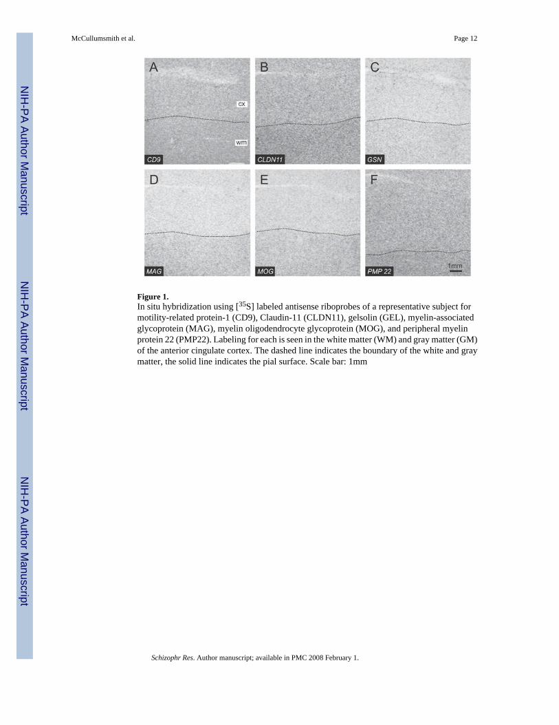
Figure 1.In situ hybridization using [35S] labeled antisense riboprobes of a representative subject formotility-related protein-1 (CD9), Claudin-11 (CLDN11), gelsolin (GEL), myelin-associatedglycoprotein (MAG), myelin oligodendrocyte glycoprotein (MOG), and peripheral myelinprotein 22 (PMP22). Labeling for each is seen in the white matter (WM) and gray matter (GM)of the anterior cingulate cortex. The dashed line indicates the boundary of the white and graymatter, the solid line indicates the pial surface. Scale bar: 1mm
McCullumsmith et al. Page 12
Schizophr Res. Author manuscript; available in PMC 2008 February 1.
NIH
-PA Author Manuscript
NIH
-PA Author Manuscript
NIH
-PA Author Manuscript
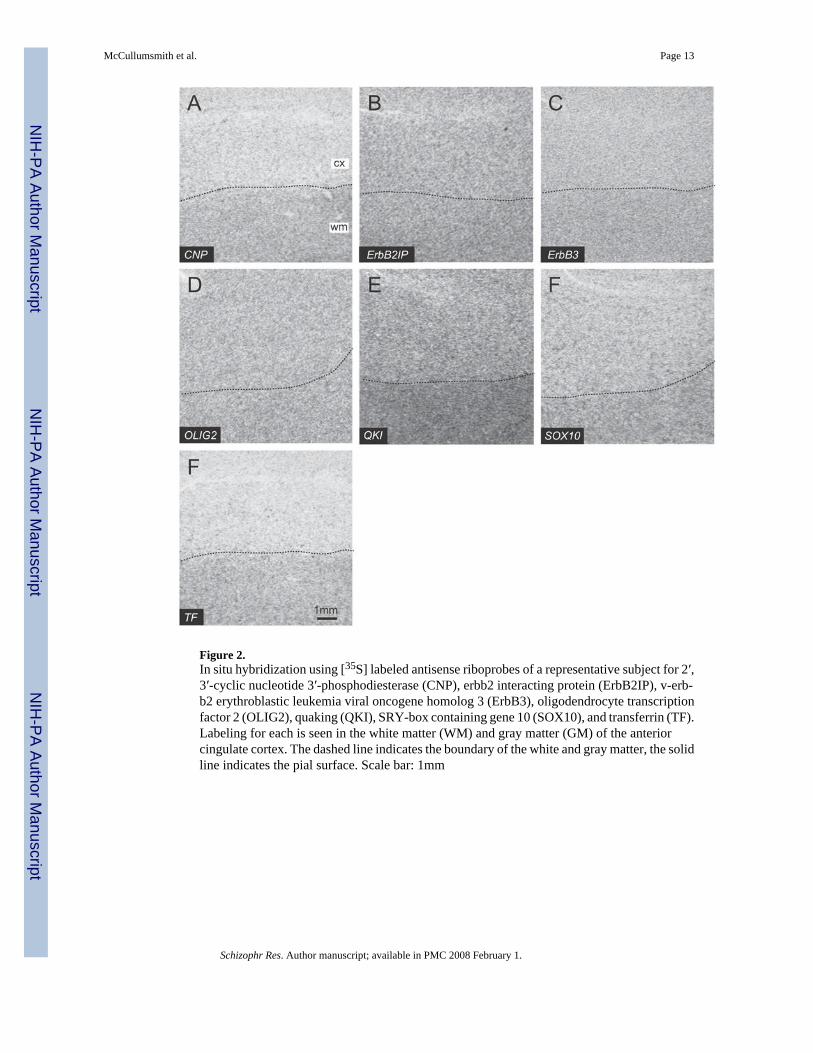
Figure 2.In situ hybridization using [35S] labeled antisense riboprobes of a representative subject for 2′,3′-cyclic nucleotide 3′-phosphodiesterase (CNP), erbb2 interacting protein (ErbB2IP), v-erb-b2 erythroblastic leukemia viral oncogene homolog 3 (ErbB3), oligodendrocyte transcriptionfactor 2 (OLIG2), quaking (QKI), SRY-box containing gene 10 (SOX10), and transferrin (TF).Labeling for each is seen in the white matter (WM) and gray matter (GM) of the anteriorcingulate cortex. The dashed line indicates the boundary of the white and gray matter, the solidline indicates the pial surface. Scale bar: 1mm
McCullumsmith et al. Page 13
Schizophr Res. Author manuscript; available in PMC 2008 February 1.
NIH
-PA Author Manuscript
NIH
-PA Author Manuscript
NIH
-PA Author Manuscript
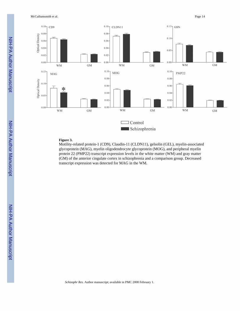
Figure 3.Motility-related protein-1 (CD9), Claudin-11 (CLDN11), gelsolin (GEL), myelin-associatedglycoprotein (MAG), myelin oligodendrocyte glycoprotein (MOG), and peripheral myelinprotein 22 (PMP22) transcript expression levels in the white matter (WM) and gray matter(GM) of the anterior cingulate cortex in schizophrenia and a comparison group. Decreasedtranscript expression was detected for MAG in the WM.
McCullumsmith et al. Page 14
Schizophr Res. Author manuscript; available in PMC 2008 February 1.
NIH
-PA Author Manuscript
NIH
-PA Author Manuscript
NIH
-PA Author Manuscript
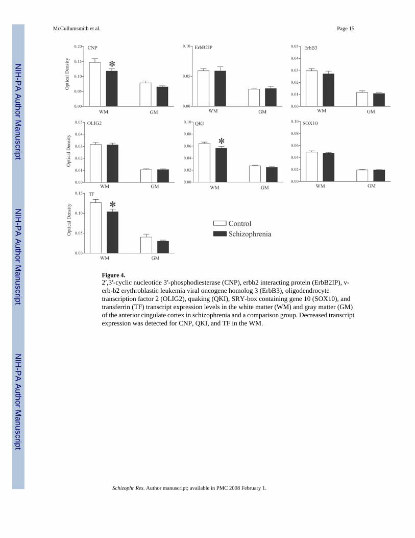
Figure 4.2′,3′-cyclic nucleotide 3′-phosphodiesterase (CNP), erbb2 interacting protein (ErbB2IP), v-erb-b2 erythroblastic leukemia viral oncogene homolog 3 (ErbB3), oligodendrocytetranscription factor 2 (OLIG2), quaking (QKI), SRY-box containing gene 10 (SOX10), andtransferrin (TF) transcript expression levels in the white matter (WM) and gray matter (GM)of the anterior cingulate cortex in schizophrenia and a comparison group. Decreased transcriptexpression was detected for CNP, QKI, and TF in the WM.
McCullumsmith et al. Page 15
Schizophr Res. Author manuscript; available in PMC 2008 February 1.
NIH
-PA Author Manuscript
NIH
-PA Author Manuscript
NIH
-PA Author Manuscript
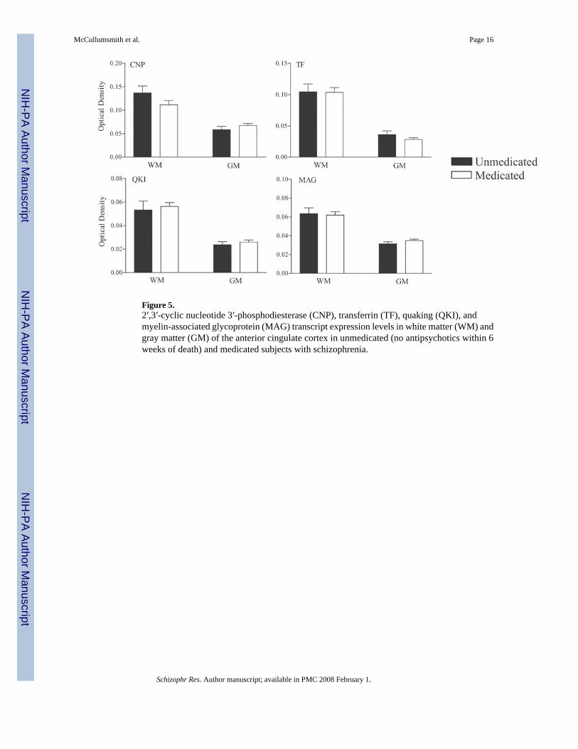
Figure 5.2′,3′-cyclic nucleotide 3′-phosphodiesterase (CNP), transferrin (TF), quaking (QKI), andmyelin-associated glycoprotein (MAG) transcript expression levels in white matter (WM) andgray matter (GM) of the anterior cingulate cortex in unmedicated (no antipsychotics within 6weeks of death) and medicated subjects with schizophrenia.
McCullumsmith et al. Page 16
Schizophr Res. Author manuscript; available in PMC 2008 February 1.
NIH
-PA Author Manuscript
NIH
-PA Author Manuscript
NIH
-PA Author Manuscript
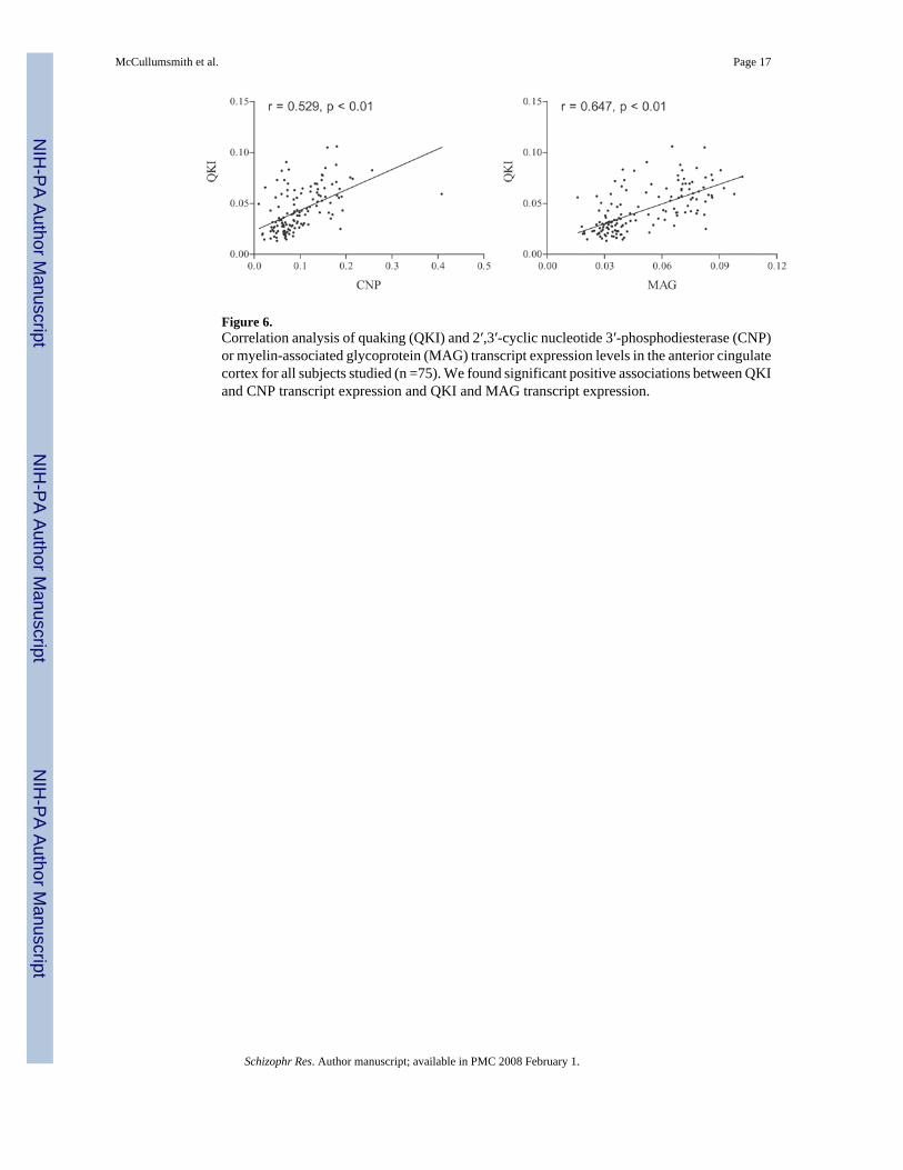
Figure 6.Correlation analysis of quaking (QKI) and 2′,3′-cyclic nucleotide 3′-phosphodiesterase (CNP)or myelin-associated glycoprotein (MAG) transcript expression levels in the anterior cingulatecortex for all subjects studied (n =75). We found significant positive associations between QKIand CNP transcript expression and QKI and MAG transcript expression.
McCullumsmith et al. Page 17
Schizophr Res. Author manuscript; available in PMC 2008 February 1.
NIH
-PA Author Manuscript
NIH
-PA Author Manuscript
NIH
-PA Author Manuscript
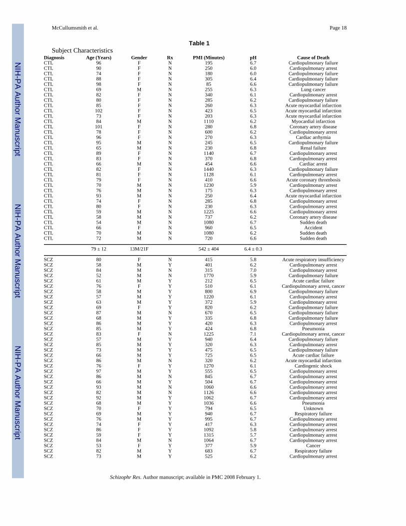
Table 1Subject Characteristics
Diagnosis Age (Years) Gender Rx PMI (Minutes) pH Cause of DeathCTL 96 F N 195 6.7 Cardiopulmonary failureCTL 90 F N 250 6.0 Cardiopulmonary arrestCTL 74 F N 180 6.0 Cardiopulmonary failureCTL 88 F N 305 6.4 Cardiopulmonary failureCTL 98 F N 85 6.6 Cardiopulmonary failureCTL 69 M N 255 6.3 Lung cancerCTL 82 F N 340 6.1 Cardiopulmonary arrestCTL 80 F N 285 6.2 Cardiopulmonary failureCTL 85 F N 260 6.3 Acute myocardial infarctionCTL 102 F N 423 6.5 Acute myocardial infarctionCTL 73 F N 203 6.3 Acute myocardial infarctionCTL 84 M N 1110 6.2 Myocardial infarctionCTL 101 F N 280 6.8 Coronary artery diseaseCTL 78 F N 600 6.2 Cardiopulmonary arrestCTL 96 F N 270 6.3 Cardiac arrhymiaCTL 95 M N 245 6.5 Cardiopulmonary failureCTL 65 M N 230 6.8 Renal failureCTL 89 F N 1140 6.7 Cardiopulmonary arrestCTL 83 F N 370 6.8 Cardiopulmonary arrestCTL 66 M N 454 6.6 Cardiac arrestCTL 82 F N 1440 6.3 Cardiopulmonary failureCTL 81 F N 1128 6.1 Cardiopulmonary arrestCTL 79 F N 410 6.6 Acute coronary thrombosisCTL 70 M N 1230 5.9 Cardiopulmonary arrestCTL 76 M N 175 6.3 Cardiopulmonary arrestCTL 93 M N 250 6.4 Acute myocardial infarctionCTL 74 F N 285 6.8 Cardiopulmonary arrestCTL 80 F N 230 6.3 Cardiopulmonary arrestCTL 59 M N 1225 6.6 Cardiopulmonary arrestCTL 58 M N 737 6.2 Coronary artery diseaseCTL 54 M N 1080 6.7 Sudden deathCTL 66 F N 960 6.5 AccidentCTL 70 M N 1080 6.2 Sudden deathCTL 72 M N 720 6.6 Sudden death
79 ± 12 13M/21F 542 ± 404 6.4 ± 0.3
SCZ 80 F N 415 5.8 Acute respiratory insufficiencySCZ 58 M Y 401 6.2 Cardiopulmonary arrestSCZ 84 M N 315 7.0 Cardiopulmonary arrestSCZ 52 M N 1770 5.9 Cardiopulmonary failureSCZ 61 M Y 212 6.5 Acute cardiac failureSCZ 76 F Y 510 6.1 Cardiopulmonary arrest, cancerSCZ 58 M Y 800 6.9 Cardiopulmonary failureSCZ 57 M Y 1220 6.1 Cardiopulmonary arrestSCZ 63 M Y 372 5.9 Cardiopulmonary arrestSCZ 69 F Y 820 6.2 Cardiopulmonary failureSCZ 87 M N 670 6.5 Cardiopulmonary failureSCZ 68 M Y 335 6.8 Cardiopulmonary failureSCZ 86 M Y 420 6.3 Cardiopulmonary arrestSCZ 85 M Y 424 6.8 PneumoniaSCZ 83 F N 1225 7.1 Cardiopulmonary arrest, cancerSCZ 57 M Y 940 6.4 Cardiopulmonary failureSCZ 85 M Y 320 6.3 Cardiopulmonary arrestSCZ 73 M Y 475 6.5 Cardiopulmonary failureSCZ 66 M Y 725 6.5 Acute cardiac failureSCZ 86 M N 320 6.2 Acute myocardial infarctionSCZ 76 F Y 1270 6.1 Cardiogenic shockSCZ 97 M Y 555 6.5 Cardiopulmonary arrestSCZ 86 M N 845 6.7 Cardiopulmonary arrestSCZ 66 M Y 504 6.7 Cardiopulmonary arrestSCZ 93 M N 1060 6.6 Cardiopulmonary arrestSCZ 82 M N 1126 6.6 Cardiopulmonary arrestSCZ 92 M Y 1062 6.7 Cardiopulmonary arrestSCZ 68 M Y 1036 6.6 PneumoniaSCZ 70 F Y 794 6.5 UnknownSCZ 69 M Y 940 6.7 Respiratory failureSCZ 76 M Y 995 6.7 Cardiopulmonary arrestSCZ 74 F Y 417 6.3 Cardiopulmonary arrestSCZ 86 F Y 1092 5.8 Cardiopulmonary arrestSCZ 59 F Y 1315 5.7 Cardiopulmonary arrestSCZ 84 M N 1064 6.7 Cardiopulmonary arrestSCZ 53 F Y 377 5.9 CancerSCZ 82 M Y 683 6.7 Respiratory failureSCZ 73 M Y 525 6.2 Cardiopulmonary arrest
McCullumsmith et al. Page 18
Schizophr Res. Author manuscript; available in PMC 2008 February 1.
NIH
-PA Author Manuscript
NIH
-PA Author Manuscript
NIH
-PA Author Manuscript
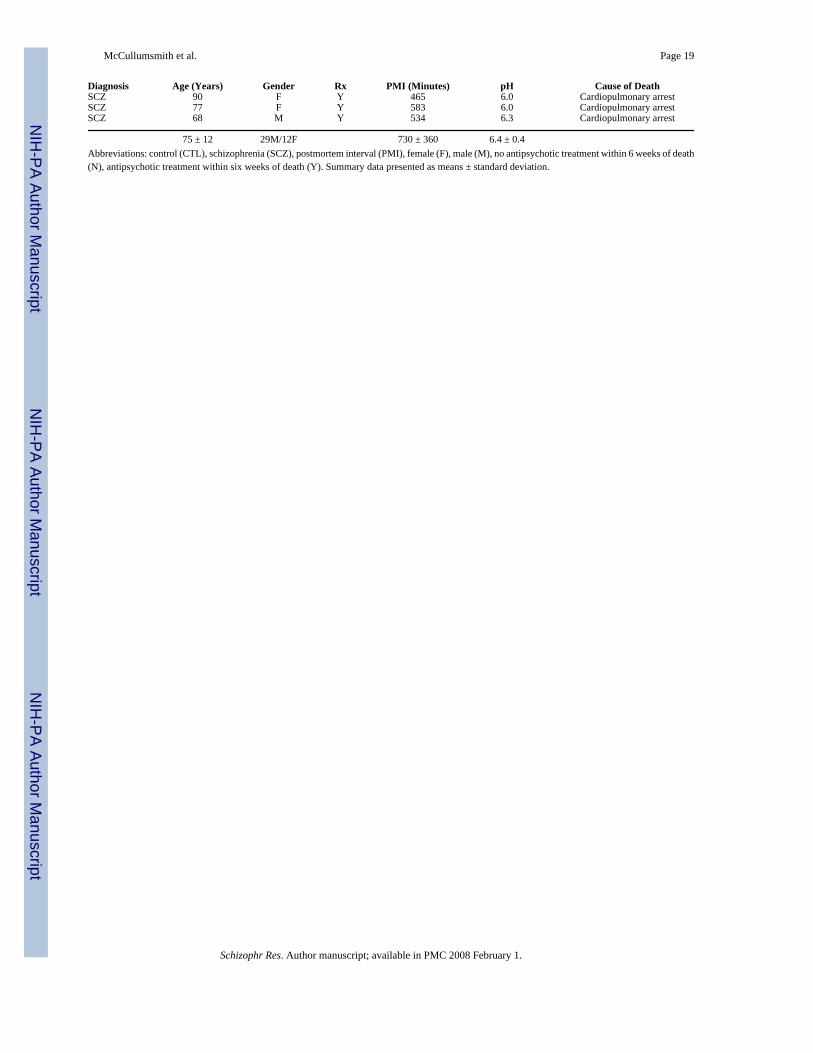
Diagnosis Age (Years) Gender Rx PMI (Minutes) pH Cause of DeathSCZ 90 F Y 465 6.0 Cardiopulmonary arrestSCZ 77 F Y 583 6.0 Cardiopulmonary arrestSCZ 68 M Y 534 6.3 Cardiopulmonary arrest
75 ± 12 29M/12F 730 ± 360 6.4 ± 0.4Abbreviations: control (CTL), schizophrenia (SCZ), postmortem interval (PMI), female (F), male (M), no antipsychotic treatment within 6 weeks of death(N), antipsychotic treatment within six weeks of death (Y). Summary data presented as means ± standard deviation.
McCullumsmith et al. Page 19
Schizophr Res. Author manuscript; available in PMC 2008 February 1.
NIH
-PA Author Manuscript
NIH
-PA Author Manuscript
NIH
-PA Author Manuscript