Plasmin-sensitive Dibasic Sequences in the Third Fibronectin-like Domain of L1Cell Adhesion Molecule...
17
The Rockefeller University Press, 0021-9525/2000/06/1485/17 $5.00 The Journal of Cell Biology, Volume 149, Number 7, June 26, 2000 1485–1501 http://www.jcb.org 1485 Plasmin-sensitive Dibasic Sequences in the Third Fibronectin-like Domain of L1–Cell Adhesion Molecule (CAM) Facilitate Homomultimerization and Concomitant Integrin Recruitment Steve Silletti,* ‡ Fang Mei, ‡ Dean Sheppard, §i and Anthony M.P. Montgomery* ‡ *Department of Pediatrics, University of California at San Diego, La Jolla, California 92037; ‡ Department of Immunology, The Scripps Research Institute, La Jolla, California 92037; and § Lung Biology Center, Center for Occupational and Environmental Health, Cardiovascular Research Institute, i Department of Medicine, University of California, San Francisco, California 94080 Abstract. L1 is a multidomain transmembrane neural recognition molecule essential for neurohistogenesis. While moieties in the immunoglobulin-like domains of L1 have been implicated in both heterophilic and ho- mophilic binding, the function of the fibronectin (FN)-like repeats remains largely unresolved. Here, we demonstrate that the third FN-like repeat of L1 (FN3) spontaneously homomultimerizes to form trimeric and higher order complexes. Remarkably, these complexes support direct RGD-independent interactions with several integrins, including a v b 3 and a 5 b 1 . A pep- tide derived from the putative C-C9 loop of FN3 (GSQRKHSKRHIHKDHV 852 ) also forms trimeric complexes and supports a v b 3 and a 5 b 1 binding. Substi- tution of the dibasic RK 841 and KR 845 sequences within this peptide or the FN3 domain limited multimerization and abrogated integrin binding. Evidence is presented that the multimerization of, and integrin binding to, the FN3 domain is regulated both by conformational con- straints imposed by other domains and by plasmin- mediated cleavage within the sequence RK ↓ HSK ↓ RH 846 . The integrin a 9 b 1 , which also recognizes the FN3 do- main, colocalizes with L1 in a manner restricted to sites of cell–cell contact. We propose that distal receptor li- gation events at the cell–cell interface may induce a conformational change within the L1 ectodomain that culminates in receptor multimerization and integrin re- cruitment via interaction with the FN3 domain. Key words: neural CAM • heterophilic ligation • mel- anoma • a v b 3 • a 5 b 1 • a 9 b 1 Introduction Human L1 is a member of a subfamily of phylogenetically conserved neural recognition molecules that share a com- plex ectodomain structure consisting of multiple immuno- globulin and fibronectin (FN) 1 type III repeats (Hortsch, 1996). Orthologues of human and mouse L1 have been de- scribed, including NILE (rat), NgCAM (chick), E587 (goldfish), L1.1/L1.2 (zebrafish), and neuroglian (Dro- sophila) (Bock et al., 1985; Lemmon and McLoon, 1986; Bieber et al., 1989; Bastmeyer et al., 1995; Tongiorgi et al., 1995). Pioneering studies implicated the L1 subfamily in a variety of dynamic neurological processes, including neu- rite fasciculation and outgrowth, as well as cerebellar cell migration (Lindner et al., 1983; Martini and Schachner, 1986; Lagenaur and Lemmon, 1987). With the recent gen- eration of L1-deficient mice, it has been confirmed that L1 is required for normal corticospinal axon guidance (Cohen et al., 1997) and for axonal ensheathment by nonmyelinat- ing Schwann cells (Dahme et al., 1997; Haney et al., 1999). Many of the neuropathologies now described in L1 knock- out mice, including dilated brain ventricles, abnormal den- dritic architecture, and developmental defects in the hip- pocampus and corpus callosum (Dahme et al., 1997; Demyanenko et al., 1999), are consistent with the manifes- tations of CRASH, a neurological syndrome associated with mutations in the human L1 gene (Fransen et al., 1997; Brummendorf et al., 1998). Although designated a neural cell adhesion molecule (CAM), both murine and human L1 homologues have been described on cells of diverse histological origin in- cluding epithelial cells associated with kidney collecting ducts (Debiec et al., 1998) and with the intestinal and uro- genital tract (Thor et al., 1987; Kujat et al., 1995). Interest- Address correspondence to Anthony Montgomery, Department of Pedi- atrics-0983, The Whittier Institute, University of California at San Diego, 9894 Genesee Avenue, La Jolla, CA 92037. Tel.: (858) 550-2909. Fax: (858) 558-3495. E-mail: [email protected] 1 Abbreviations used in this paper: CAM, cell adhesion molecule; ECD, ectodomain; FN, fibronectin; pAb, polyclonal antibody; RGD, arginine- glycine-aspartate. on October 6, 2014 jcb.rupress.org Downloaded from Published June 26, 2000
-
Upload
independent -
Category
Documents
-
view
0 -
download
0
Transcript of Plasmin-sensitive Dibasic Sequences in the Third Fibronectin-like Domain of L1Cell Adhesion Molecule...

The Rockefeller University Press, 0021-9525/2000/06/1485/17 $5.00The Journal of Cell Biology, Volume 149, Number 7, June 26, 2000 1485–1501http://www.jcb.org 1485
Plasmin-sensitive Dibasic Sequences in the Third Fibronectin-like Domain of L1–Cell Adhesion Molecule (CAM) Facilitate Homomultimerization and Concomitant Integrin Recruitment
Steve Silletti,*
‡
Fang Mei,
‡
Dean Sheppard,
§
i
and Anthony M.P. Montgomery*
‡
*Department of Pediatrics, University of California at San Diego, La Jolla, California 92037;
‡
Department of Immunology, The Scripps Research Institute, La Jolla, California 92037; and
§
Lung Biology Center, Center for Occupational and Environmental
Health, Cardiovascular Research Institute,
i
Department of Medicine, University of California, San Francisco, California 94080
Abstract.
L1 is a multidomain transmembrane neural recognition molecule essential for neurohistogenesis. While moieties in the immunoglobulin-like domains of L1 have been implicated in both heterophilic and ho-mophilic binding, the function of the fibronectin(FN)-like repeats remains largely unresolved. Here, we demonstrate that the third FN-like repeat of L1 (FN3) spontaneously homomultimerizes to form trimeric and higher order complexes. Remarkably, these complexes support direct RGD-independent interactions withseveral integrins, including
a
v
b
3
and
a
5
b
1
. A pep-tide derived from the putative C-C
9
loop of FN3 (GSQRKHSKRHIHKDHV
852
) also forms trimeric complexes and supports
a
v
b
3
and
a
5
b
1
binding. Substi-tution of the dibasic RK
841
and KR
845
sequences within this peptide or the FN3 domain limited multimerization
and abrogated integrin binding. Evidence is presented that the multimerization of, and integrin binding to, the FN3 domain is regulated both by conformational con-straints imposed by other domains and by plasmin-
mediated cleavage within the sequence RK
↓
HSK
↓
RH
846
. The integrin
a
9
b
1
, which also recognizes the FN3 do-main, colocalizes with L1 in a manner restricted to sites of cell–cell contact. We propose that distal receptor li-gation events at the cell–cell interface may induce a conformational change within the L1 ectodomain that culminates in receptor multimerization and integrin re-cruitment via interaction with the FN3 domain.
Key words: neural CAM • heterophilic ligation • mel-anoma •
a
v
b
3
•
a
5
b
1
•
a
9
b
1
Introduction
Human L1 is a member of a subfamily of phylogeneticallyconserved neural recognition molecules that share a com-plex ectodomain structure consisting of multiple immuno-globulin and fibronectin (FN)
1
type III repeats (Hortsch,1996). Orthologues of human and mouse L1 have been de-scribed, including NILE (rat), NgCAM (chick), E587(goldfish), L1.1/L1.2 (zebrafish), and neuroglian (
Dro-sophila
) (Bock et al., 1985; Lemmon and McLoon, 1986;Bieber et al., 1989; Bastmeyer et al., 1995; Tongiorgi et al.,1995). Pioneering studies implicated the L1 subfamily in avariety of dynamic neurological processes, including neu-rite fasciculation and outgrowth, as well as cerebellar cell
migration (Lindner et al., 1983; Martini and Schachner,1986; Lagenaur and Lemmon, 1987). With the recent gen-eration of L1-deficient mice, it has been confirmed that L1is required for normal corticospinal axon guidance (Cohenet al., 1997) and for axonal ensheathment by nonmyelinat-ing Schwann cells (Dahme et al., 1997; Haney et al., 1999).Many of the neuropathologies now described in L1 knock-out mice, including dilated brain ventricles, abnormal den-dritic architecture, and developmental defects in the hip-pocampus and corpus callosum (Dahme et al., 1997;Demyanenko et al., 1999), are consistent with the manifes-tations of CRASH, a neurological syndrome associatedwith mutations in the human L1 gene (Fransen et al., 1997;Brummendorf et al., 1998).
Although designated a neural cell adhesion molecule(CAM), both murine and human L1 homologues havebeen described on cells of diverse histological origin in-cluding epithelial cells associated with kidney collectingducts (Debiec et al., 1998) and with the intestinal and uro-genital tract (Thor et al., 1987; Kujat et al., 1995). Interest-
Address correspondence to Anthony Montgomery, Department of Pedi-atrics-0983, The Whittier Institute, University of California at San Diego,9894 Genesee Avenue, La Jolla, CA 92037. Tel.: (858) 550-2909. Fax:(858) 558-3495. E-mail: [email protected]
1
Abbreviations used in this paper:
CAM, cell adhesion molecule; ECD,ectodomain; FN, fibronectin; pAb, polyclonal antibody; RGD, arginine-glycine-aspartate.
on October 6, 2014
jcb.rupress.orgD
ownloaded from
Published June 26, 2000
The Journal of Cell Biology, Volume 149, 2000 1486
ingly, the L1 expressed by renal epithelium has beenshown to be important for normal branching morphogene-sis (Debiec et al., 1998). Cells of lymphoid and my-elomonocytic origin also express L1 (Ebeling et al., 1996;Pancook et al., 1997), however, the functional significanceof L1 within the immune system remains to be deter-mined. In this regard, Di Sciullo et al. (1998) have shownthat L1 is important for maintaining normal lymph nodearchitecture during an immune response and suggest amechanism based on the expression of L1 by reticular fi-broblasts. A potential function for L1 in tumor progres-sion is also suggested by widespread expression on manytumor cell lines including neuroectodermal tumors (mela-noma and neuroblastoma), carcinomas (lung, renal, andskin), and monocytic leukemias (Mujoo et al., 1986; Linne-mann et al., 1989; Reid and Hemperly, 1992; Katayama et al.,1997; Pancook et al., 1997). Supporting a role for L1 in tu-mor progression, Linnemann et al. (1989) reported findingelevated levels of L1 on a metastatic variant of a mela-noma cell line. Indeed, a recent study by Ohnishi et al.(1998) suggests that L1 may promote metastasis by facili-tating tumor cell invasion or migration.
Structure–function studies have defined multiple inter-active moieties within L1 that facilitate either homophilicor heterophilic interactions (Hortsch, 1996). Thus far,many of the interactions defined involve one or more of thesix Ig-like domains that constitute the NH
2
-terminal por-tion of the L1 ectodomain. An antiparallel alignment ofthe first four Ig-like domains of L1 is proposed to facilitatehomophilic L1–L1 binding (Su et al., 1998). The chon-droitin sulfate proteoglycan, neurocan, binds with highaffinity to the NH
2
-terminal Ig-like domain of L1 (Oles-zewski et al., 1999). An arginine-glycine-aspartate (RGD)motif in the sixth Ig-like domain of human L1 supportsheterophilic interactions with multiple members of the in-tegrin superfamily, including
a
v
b
3
and
a
5
b
1
(Montgomeryet al., 1996; Felding-Habermann et al., 1997; Oleszewski etal., 1999). Axonin-1/TAG-1, a neuron-specific CAM, un-dergoes a cis-interaction with the chick L1-orthologueNgCAM via multiple moieties including the first and sec-ond Ig-like domains as well as the third FN-like repeat(Kunz et al., 1998). Other cis-interacting elements includethe tetraspan signaling molecule CD9 (Schmidt et al.,1996), NCAM (Feizi, 1994), and the heat-stable antigenCD24 (Kadmon et al., 1995), although the regions of L1 re-sponsible for these interactions have yet to be determined.
The functional significance of the FN-like repeats thatconstitute the membrane-proximal portion of the L1ectodomain remains largely unresolved. An antibody di-rected to an epitope between FN-like repeats 2 and 3 hasbeen shown to promote signal transduction and concomi-tant neurite extension (Appel et al., 1995). Based on thisobservation the authors proposed that distal recognitionevents may induce conformational changes that are fun-neled to a region within the FN-like repeats, which thenrepresents the ultimate site for the induction of signalingevents. Recent rotary shadow analysis of the purified L1ectodomain showed that the FN-like repeats assume atight globular configuration (Drescher et al., 1996). Anyconformational lability within this structure may, there-fore, provide a mechanism for translating distal ligationevents into signaling events. In a further study, Holm et al.
(1995) demonstrated that certain FN-like domain frag-ments have a capacity for homoaggregation, suggestingthat one or more of the FN-like domains may have the po-tential for self-association, perhaps leading to the cluster-ing of L1 at the cell surface; such clustering may in turn besubject to conformational constraints imposed by the glob-ular configuration of the FN-like repeats.
In this study, we have identified moieties within thethird FN-like repeat of L1 (FN3) that can simultaneouslypotentiate receptor clustering and integrin recruitment.Both multimerization of, and integrin binding to, the FN3domain are shown to be critically regulated by conforma-tional constraints imposed by other domains and by pro-teolysis. Several integrins are implicated in binding to theFN3 domain including
a
v
b
3
,
a
5
b
1
, and
a
9
b
1
. Based on ourfindings, we propose that ligation events at the cell–cell in-terface may induce a conformation change within the L1ectodomain that culminates in receptor multimerizationand integrin recruitment via the FN3 domain. This para-digm has important implications for L1 signaling and forthe modulation of integrin activity during cell–cell interac-tions.
Materials and Methods
Reagents
Antiintegrin antibodies used in this study include the following: anti-
b
1
mAbs P4C10, LM534, B44, and Cl. 18; anti-
a
5
b
1
mAbs P1D6 and NKI-SAM-1, anti-
a
5
mAb Cl.1, anti-
a
9
b
1
mAb Y9A2; anti-
a
v
b
3
mAb LM609;anti-
b
3
antibody AP3; anti-
b
5
antibody 11D1; and an anti-
a
v
/anti-
b
3
(anti-
VNR) polyclonal antibody (pAb). mAbs B44, P1D6, and Y9A2 werepurchased from Chemicon International. mAbs Cl. 18 and Cl.1 were pur-chased from Transduction Laboratories. mAb NKI-SAM-1 was pur-chased from Southern Biotechnology. mAb P4C10 was provided by Dr.E.A. Wayner (University of Minnesota, Minneapolis, MN). LM609, theanti–human L1 mAb 5G3 (Mujoo et al., 1986), the anti–L1-Ig6 mAbLP1B9, the anti-VNR pAb, and the anti-5G3 Ag pAb were generated
within the Scripps Research Institute. Antibodies AP3 and 11D1 wereprovided by Dr. David Cheresh (The Scripps Research Institute). Ananti–L1 ectodomain (anti–L1-ECD) pAb was generated against, and af-finity-purified using the L1 ectodomain fusion protein. A pAb specific forglutathione S-transferase (GST) was purchased from Upstate Biotechnol-ogy Inc. L1 peptides were synthesized on an ABI 430A peptide synthe-sizer within The Scripps Research Institute Core Facility as describedpreviously (Felding-Habermann et al., 1997). For the purpose of immobi-lization, most peptides were made with NH
2
-terminal cysteine residues.RGD and RGE control peptides were as follows: GRGDSPC andGRGESPC. Human plasmin was purchased from Calbiochem.
Cell Lines and Culture
M21 human melanoma cells were derived from the UCLA-SO-M21 cellline, which was provided by Dr. D.L. Morton (University of California,Los Angeles, CA). Variant
a
v-integrin–deficient cells (M21-L) were nega-tively selected from M21 cells by FACS at The Scripps Research Institute.All cells were maintained in RPMI-1640 supplemented with 10% FCS.Transfected CHO cells bearing the human
a
9
integrin subunit were gener-ated in the laboratory of Dr. Sheppard and cultured as described (Taookaet al., 1999).
Construction and Expression of L1 Fusion Proteins
The recombinant L1 fusion proteins shown in Table I (schematic) weregenerated by PCR amplification of appropriate coding sequences and re-striction cloned into the appropriate fusion vector based upon the re-quired tag and reading frame. Primer sequences and their correspondingL1 amino acid start (sense oligos) or stop (antisense oligos) translation
on October 6, 2014
jcb.rupress.orgD
ownloaded from
Published June 26, 2000

Silletti et al.
L1 Multimerization and Integrin Ligation
1487
sites, as well as the restriction enzymes used for insertion of the respectivePCR products into the respective fusion protein vectors, are shown in Ta-ble I. Plasmids were purchased from Amersham Pharmacia Biotech orGIBCO BRL for pGEX GST fusion vectors or pProEX 6
3
His fusion vec-tors, respectively. Mutagenesis of the FN3 construct was performed essen-tially as described previously (Nayeem et al., 1999). In brief, mutagenicsense and antisense oligonucleotides (Table I) were annealed and ex-tended with
Pfu
polymerase for a total of 18 cycles. Nonmutant startingmaterial was digested with DpnI and the final product was transformedinto supercompetent
Escherichia coli
. Final constructs were confirmed bydideoxy sequencing.
Purification of the recombinant fusion proteins was performed from logphase BL21 strain
E. coli
induced with either 100
m
M (GST) or 600
m
Misopropyl-
b
-
D
-thiogalactopyranoside (6
3
His). GST fusion protein purifi-cation was performed as previously described (Nayeem et al., 1999). ForHis fusion protein purification, cultures were resuspended in lysis buffer(50 mM Tris-HCl, pH 8.5, 300 mM KCl, 20 mM imidazole, and 0.1% Tri-ton X-100–containing protease inhibitors) and incubated with 100
m
g/mllysozyme at 4
8
C. Lysates were clarified by centrifugation and fusion pro-teins were immobilized on Ni-NTA agarose (Qiagen) before extensivewashing of the matrix with lysis buffer, followed by washing with 50 mMTris-HCl, pH 8.5, 500 mM KCl, 40 mM imidazole, and elution with 20 mMTris-HCl, pH 8.5, 300 mM KCl, 250 mM imidazole. Purified GST and Hisfusion proteins were dialyzed extensively against PBS.
Adhesion Assays
Adhesion assays were performed essentially as described previously(Felding-Habermann et al., 1997). In brief, purified L1 fusion proteins
(100–250 nM) were spotted (2-
m
l spots) or coated (100
m
l) onto the bot-tom of 96-well Titertek plates (ICN Biomedicals) and allowed to coat for1–2 h at 37
8
C before blocking with 5% BSA. For adhesion studies involv-ing immobilized peptides, wells were precoated overnight with murineIgG2a antibody before incubation with the heterobifunctional cross-linkerSPDP (Pierce Chemical Co.), washing and incubation with peptides at100–200
m
g/ml for 2–3 h before blocking with 5% BSA. Control wells re-ceived antibody and SPDP alone without peptide. Cells were harvestedand resuspended in adhesion buffer (HBSS, 10 mM Hepes, 0.5% BSA,pH 7.4) containing divalent cations (0.4 mM MnCl
2
, 1 mM MgCl
2
, 1 mMCaCl
2
) with or without antiintegrin function-blocking antibodies. For as-says with
a
v
(
2
)M21-L cells, adhesion was determined in the presence of0.4 mM MnCl
2
alone. Cells were added at 10
5
cells/well in the presence orabsence of antibodies, and the plates were spun at 700 rpm to give a con-tinuous monolayer. After 15–40 min at 37
8
C wells were washed with PBS,and the remaining adherent cells were fixed with 1% paraformaldehydebefore counting the number of cells per high power field using a 40
3
ob-jective and an ocular grid at a minimum of four areas per well. Experi-mental treatments were performed in triplicate.
Fractionation and Detection of L1-His Fusion Proteins
L1-FN3 (His) fusion proteins (5
m
g) were fractionated at a flow rate of0.180 ml/min using a 40-ml bed volume Sephacryl S-200 column (Amer-sham Pharmacia Biotech). Fractions of 250
m
l were collected, and 100
m
lof each fraction was applied per well of a Ni-NTA HisSorb plate (Qiagen)for overnight immobilization at 4
8
C. Wells were subsequently washed with0.5% BSA in PBS (BSA/PBS) before detection of bound His fusion pro-tein as follows. Wells were incubated with anti–L1-ECD pAb for 1 h with
Table I. Oligonucleotide Primers Used in the Construction of Recombinant L1 Domains
Underlined bases represent sequences responsible for mutations in mutagenesis primers and restriction sites in PCR primers.
on October 6, 2014
jcb.rupress.orgD
ownloaded from
Published June 26, 2000
The Journal of Cell Biology, Volume 149, 2000 1488
constant shaking before being washed at least five times with BSA/PBSand subsequently incubated with HRP-conjugated goat anti–rabbit sec-ondary antibody (Jackson ImmunoResearch Laboratories). Wells werewashed further and bound antibody was detected colorimetrically withTMB (Bio-Rad Laboratories). Color development was arrested withH
2
SO
4
, and the plates were read at 450 nm on a microplate reader (Ki-netic Microplate Reader; Molecular Devices).
Integrin-binding Assays
Purified
a
v
b
3
and
a
5
b
1
integrin heterodimers were purchased from Chemi-con International. Integrin
a
v
b
3
was biotinylated using NHS-LC-biotin(Pierce Chemical Co.). L1 fusion proteins (10–40
m
g/ml) were adsorbedovernight at 4
8
C onto 96-well Titertek plates. Alternatively, 20
m
g/ml ofrabbit Ig was adsorbed before incubation with SPDP and immobilizationof various peptides as described above for adhesion assays. After coating,the wells were washed and blocked with 0.5% BSA in TBS buffer. Puri-fied integrin heterodimers were added at 1
m
g/ml in TBS supplementedwith 0.4 mM MnCl
2
and 0.5% BSA. After washing, bound
a
v
b
3
was de-tected with an HRP-conjugated antibiotin mAb (Sigma Chemical Co.).Bound
a
5
b
1
was detected with anti-
a
5
b
1
mAb NKI-SAM-1, followed byHRP-conjugated goat anti–mouse Ig (Jackson ImmunoResearch Labora-tories). Color was developed with TMB or OPD (Sigma Chemical Co.)and plates were read at 450 nm. Control wells received no integrin or werenot coated with the L1 fusion protein. To assess the equivalent coating ofimmobilized GST fusion proteins, parallel wells were blocked with BSA/PBS, incubated with an anti-GST pAb, washed further, and incubatedwith an HRP-conjugated goat anti–rabbit antibody before colorimetricdetection with either TMB or OPD at 450 nm.
Double Immunofluorescence
Aggregated nonadherent
a
v
(
2
)M21-L melanoma cells from routine tissueculture were harvested, washed, and resuspended in ice-cold FACS buffer(BSA/PBS with 0.05% NaN
3
) for incubation with both anti-
a
9
b
1
mAbY9A2 and the anti–L1-ECD pAb. Cell aggregates were washed and dou-ble stained with fluorochrome-conjugated affinity-purified donkey F(ab
9
)-specific for mouse IgG (Texas red) or rabbit IgG (FITC), which had beenpreadsorbed to minimize cross-reactivity (Jackson ImmunoResearch Lab-oratories). Stained cell aggregates were mounted and analyzed using anMRC 1024 confocal microscope (Bio-Rad Laboratories) or a NikonEclipse E800 fluorescent microscope. Some stained cell aggregates weregently disrupted by pipetting in the presence of 1% paraformaldehyde,and single cells were assessed for L1 and
a
9
b
1
colocalization. In furtherstudies, all of the experimental steps described above were performed at37
8
C in the absence of sodium azide.
Immunoprecipitation
For analysis of the coprecipitation of L1 with integrin
a9b1, av(2)M21-Lcells were cultured until the nonadherent fraction contained numerous ag-gregates, at which time the adherent population was composed largely ofindependent cells. The nonadherent population was harvested, washed,and lysed in 50 mM Tris 7.6, 300 mM NaCl, 0.5% Triton X-100–containingprotease inhibitors. Adherent cells were washed and lysed on the plate,and both adherent and nonadherent samples were clarified of Triton-insoluble material. Equal quantities of adherent and nonadherent cell ly-sate (3.5 mg) were precleared with protein G–Sepharose (Pierce ChemicalCo.) before overnight incubation with 5 mg anti-a9b1 mAb Y9A2. Anti-body–antigen complexes were precipitated with protein G–Sepharose,washed with lysis buffer, and boiled in reducing SDS-PAGE samplebuffer before being subjected to SDS-PAGE and immunoblotting with ananti–L1-ECD pAb and a combination of anti–b1 integrin mAbs B44 andCl. 18 as described below.
For studies on the coimmunoprecipitation of integrin subunits with L1,M21 or M21-L cells were aggregated by rotation, harvested, allowed tosettle, and lysed as described above for the nonadherent M21-L popula-tion. After clarification of the Triton-insoluble fraction, equal quantitiesof lysate (7.5 mg) were precleared with protein A–Sepharose (SigmaChemical Co.) before overnight incubation with 15 mg of either anti-5G3Ag pAb or a control anti-GST pAb. Antibody–antigen complexeswere precipitated with protein A–Sepharose and washed extensively withlysis buffer (M21-L cells) or RIPA buffer (M21 cells) before boiling innonreducing SDS-PAGE buffer and separation by SDS-PAGE and im-munoblotting as described below. Precipitated L1 and associated integrinswere detected with the appropriate antibody as follows: L1, mAb 5G3;
b1 integrin, mAb LM534; integrin a5, mAb Cl. 1; integrin b3, mAb AP3;integrin b5, mAb 11D1.
SDS-PAGE and ImmunoblottingPeptides, purified proteins, or immunoprecipitates were prepared as de-scribed and separated in the presence or absence of 2-mercaptoethanol(as required) by SDS-PAGE on precast Tris-glycine gels (Novex). Sepa-rated proteins were electroblotted as required to a PVDF membrane (Im-mobilon-P; Millipore), which was subsequently blocked with 5% milk inPBS. Appropriate primary antibodies were incubated for 1–2 h in TBScontaining 0.1% Tween 20 (TBS-T) and 0.5% milk, and bound primaryantibody was detected with either an HRP-conjugated goat anti–rabbit Ig(Southern Biotechnology) or donkey anti–mouse Ig (Jackson ImmunoRe-search Laboratories) preadsorbed to minimize cross-reactivity with theprecipitating immunoglobulins. Antibody complexes were visualized withthe chemiluminescent substrate PS-3 (Lumigen Inc.). Alternatively, gelswere fixed and processed for staining.
Results
The Third FN-like Repeat of L1 PromotesRGD-independent b1 Integrin Interaction with theL1 Ectodomain
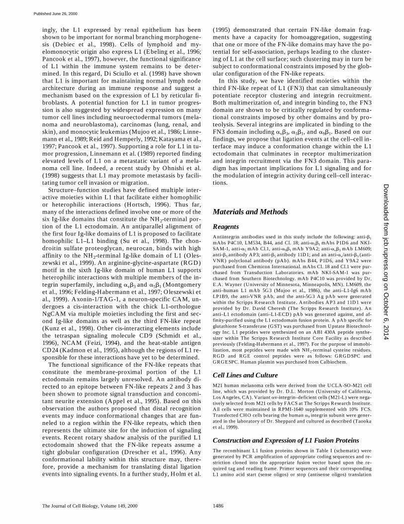
Previously, we have shown that the single RGD motif inthe sixth Ig-like domain (Ig6) of human L1 is recognizedby multiple integrin heterodimers, with the contribution ofeach of these integrins being dictated by cell type and thecation environment (Felding-Habermann et al., 1997). In aphysiological cation environment, the adhesion of M21melanoma cells to the Ig6 domain was found to be solelydependent on integrin avb3 (Montgomery et al., 1996).While these studies established the importance of theRGD motif in the context of individual domain fragments,integrin recognition of the intact L1 ectodomain was notaddressed.
Significant dose-dependent adhesion of M21 cells wasobserved on immobilized fusion proteins consisting of ei-ther the Ig6 domain alone or the entire L1 ectodomain(Fig. 1 a). However, inhibition studies using function-blocking antibodies to either avb3 or b1 integrin demon-strated a significant disparity in the contribution of theseintegrins to adhesion on the two substrates (Fig. 1 b).Thus, blocking ligation by avb3 resulted in a complete ab-rogation of adhesion to Ig6, but was only partially effectivewhen the entire L1 ectodomain was used as a substrate(Fig. 1 b). This disparity can be attributed to supplementalb1 integrin recognition of the L1 ectodomain, as adhesionto the L1 ectodomain could only be fully blocked with acombination of antibodies to both avb3 and b1 integrins(Fig. 1 b; right).
These findings indicate that recognition of the RGDmotif in L1-Ig6 can only partially account for the full mea-sure of integrin binding to the entire L1 ectodomain. Onepossible explanation for this disparity is the presence of asecond non-RGD motif recognized by one or more b1integrins. To identify the location of this motif, we gener-ated multidomain L1 fragments containing the Ig6 domainand adjacent FN-like repeats. While the addition of thefirst and second proximal FN-like repeats of L1 (Ig6-FN1-2)failed to result in supplemental b1 integrin binding (Fig. 1c, left) inclusion of the third FN-like repeat (Ig6-FN1-3)did result in adhesion by both avb3 and b1 integrin(s) (Fig.1 c, right). These data indicate that recognition of sites
on October 6, 2014
jcb.rupress.orgD
ownloaded from
Published June 26, 2000

Silletti et al. L1 Multimerization and Integrin Ligation 1489
within Ig6 (RGD) and the third FN-like repeat (FN3; non-RGD), can fully account for integrin binding to the L1ectodomain. It should be noted that the Ig-like domainsproximal to Ig6 (i.e., Ig5 and Ig4) had no influence on inte-grin recognition (data not shown).
The Heterodimers a5b1 and a9b1 Are Responsible for b1 Recognition of the Third FN-like Repeat of L1
A panel of antiintegrin antibodies was tested to identifythe b1 integrin(s) responsible for recognition of the L1ectodomain. These antibodies were first tested in adhesionassays using M21 cells selected for a lack of av integrin ex-pression (i.e., M21-L cells). Because of the absence of avb3expression, M21-L cell adhesion to L1-Ig6 or L1-Ig6-FN1-2was minimal (Fig. 2 a). Consistent with b1 integrin recogni-tion of the third FN-like repeat, a marked increase in ad-hesion was observed on either Ig6-FN1-3 or on the L1ectodomain (Fig. 2 a). Adhesion of av(2)M21-L cells toboth of these substrates was examined in the presence offunction-blocking antibodies to a variety of b1 integrins in-cluding a1b1, a2b1, a3b1, a4b1, a5b1, a6b1, and a9b1. Fromthese studies, it was confirmed that adhesion could only
be abrogated using a combination of mAbs specific forboth a5b1 and a9b1 (Fig. 2 b). Results obtained with theav(2)M21-L cells could be reproduced with wild-typeM21 cells provided avb3 binding by these cells was alsoblocked (Fig. 2 c). It should be noted that whereas wild-type M21 cells were able to utilize a5b1 and a9b1 for adhe-sion in a physiological cation environment (i.e., 1 mMCa21, 1 mM Mg21, 0.4 mM Mn21), av(2)M21-L cells onlyshowed significant adhesion via these integrins in an acti-vating cation environment consisting of 0.4 mM Mn21
alone. This unexpected finding suggests that the selectionof M21 cells lacking av integrin expression has had an un-foreseen effect on the activation state of these b1 integrins.
The Third FN-like Repeat (FN3) Alone Supports Integrin Ligation But Recognition of This Domain Is Regulated by Upstream FN1
To confirm that a second integrin recognition motif ispresent in the FN3 repeat, we generated a further series ofL1 domain fragments consisting of single or multiple FN-like repeats, including FN3 alone as well as FN2-3 andFN1-3. L1 domain fragments consisting of FN1 alone andFN1-2 were generated as controls.
M21 cells failed to adhere to either FN1 or FN1-2, but
Figure 1. RGD-independent b1 integrin binding to the L1ectodomain involves the third FN-like repeat. (a) Adhesion of M21cells to immobilized L1 ectodomain or domain Ig6 alone. (b) M21adhesion to L1 ectodomain or Ig6 in the presence or absence offunction-blocking antibody to avb3 (LM609), b1 integrins (P4C10),av integrins (anti-VNR), or both av and b1 integrins. (c) M21 ad-hesion to Ig6 with adjacent FN-like repeats (Ig6-FN1-2 and Ig6-FN1-3) in the presence or absence of function-blocking antibodies.Data shown is the mean of triplicate measurements 6 SD.
Figure 2. The third FN-like repeat of L1 is recognized by inte-grins a5b1 and a9b1. (a) Adhesion of av(2)M21-L cells to immo-bilized L1 ectodomain, the Ig6 domain alone, or Ig6 in conjunc-tion with adjacent FN-like repeats (Ig6-FN1-2 and Ig6-FN1-3). (band c) Adhesion of av(2)M21-L (b) or M21 cells (c) to L1ectodomain or Ig6-FN1-3 in the presence or absence of function-blocking antibody to avb3 (LM609), a5b1 (P1D6), or a9b1(Y9A2), alone or in combination. Data shown are the mean oftriplicate measurements 6 SD. on O
ctober 6, 2014jcb.rupress.org
Dow
nloaded from
Published June 26, 2000
The Journal of Cell Biology, Volume 149, 2000 1490
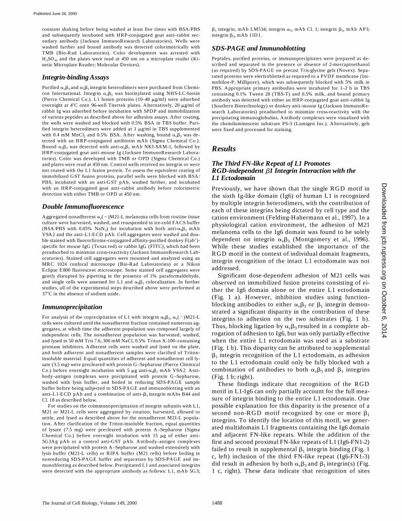
did display significant dose-dependent adhesion to theFN3 domain alone (Fig. 3 a). These findings confirm theimportance of FN3 in adhesion and demonstrate that thisdomain can support adhesion independent of the RGDmotif in Ig6. However, it is important to note that adhe-sion to FN3 was markedly reduced when this domain wasoffered together with both FN1 and FN2 domains (Fig. 3a, FN1-3). This effect can be attributed to the presence ofthe first FN repeat (FN1) since adhesion to FN2-3 did notdiffer markedly from adhesion to FN3 alone (Fig. 3 a).Several explanations could account for the disparity in ad-hesion observed between the FN3 and FN1-3 domain con-structs, including unequal adsorption of the GST fusionproteins. To exclude this possibility, we assessed the rela-tive coating efficiency of the FN domain constructs bymeasuring the amount of immobilized GST present incoated wells. When offered at slightly different concentra-tions, which resulted in equalization of GST immunoreac-tivity (Fig. 3 b, inset), the significant disparity in adhesionbetween FN3 and FN1-3 was still observed, even thoughrelatively high amounts of fusion protein were offered(Fig. 3 b). It is important to note that cell spreading wasalso markedly reduced on FN1-3, and that at lower coatingconcentrations, the disparity in adhesion between FN3 andFN1-3 was even more marked (Fig. 3 a). A further expla-nation for this reduced adhesion associated with the pres-ence of FN1 would be an interaction between this domainand a cellular ligand that negatively regulates integrin liga-tion. However, it should be noted that FN1 did not impact
integrin-mediated adhesion to Ig6. Thus, adhesion to aconstruct consisting of Ig6 and FN1 was not significantlydifferent from that to Ig6 alone (Fig. 3 c).
To confirm that the adhesion observed with the FN3domain alone is due to ligation of a5b1 and a9b1, furtherinhibition studies were performed with both M21 andav(2)M21-L cells. As expected, av(2)M21-L cell adhesionto both FN3 and FN1-3 was reduced by function-blockingantibodies to both a5b1 and a9b1 (Fig. 4 a). Wild-type M21
Figure 3. Adhesion to thethird FN-like repeat of L1(FN3) is regulated by the firstFN-like repeat (FN1). (a) M21adhesion to immobilized sin-gle domain fragments (FN1and FN3) and multiple do-main fragments (FN1-2,FN1-3, and FN2-3). (b) M21adhesion to the immobilizedFN domain fragments afterequalizing for protein adsorp-tion as determined by ELISAdetection of GST (b, inset).(c) Adhesion of M21 cells toimmobilized Ig6 or Ig6-FN1.Data shown are the mean oftriplicate measurements 6 SD.
Figure 4. The FN3 domain alone is recognized by avb3 as well asa5b1 and a9b1, but this interaction is markedly inhibited by theFN1 domain. (a) Adhesion of av(2)M21-L cells to immobilizedFN3 alone or FN3 with the adjacent first and second FN-like do-mains (FN1-3) in the presence or absence of function-blockingantibodies to a5b1 (P1D6) or a9b1 (Y9A2). (b) Adhesion of M21cells to immobilized FN 3 or FN1-3 in the presence or absence offunction blocking antibody to avb3 (LM609), a5b1 (P1D6), ora9b1 (Y9A2), alone or in combination. (c) Mock-transfectedCHO cells were compared with CHO cells transfected with thehuman integrin a9 subunit in the presence or absence of function-blocking antibody to a9b1 (Y9A2). (d) Direct binding of purifiedavb3 or a5b1 integrins to immobilized FN 3 or FN1-3 as deter-mined by ELISA. The relative coating efficiency of the con-structs was determined by anti-GST ELISA (inset). Data shownare the mean of triplicate measurements 6 SD.
on October 6, 2014
jcb.rupress.orgD
ownloaded from
Published June 26, 2000

Silletti et al. L1 Multimerization and Integrin Ligation 1491
cell adhesion to FN1-3 was similarly abrogated using acombination of antibodies to a5b1 and a9b1 (Fig. 4 b, right).Remarkably, and in contrast to adhesion on FN1-3, M21cell adhesion to FN3 could only be completely abrogatedwith the further addition of an antibody to avb3 (Fig. 4 b,left). These findings suggest that part of the antiadhesiveactivity of FN1 can be attributed to its capacity to signifi-cantly limit recognition of FN3 by avb3. However, sincethe adhesion of av(2)M21-L cells was also reduced in thepresence of FN1 (Fig. 4 a), it is likely that this domain alsolimits recognition by a5b1 and a9b1. To further establishthat a9b1 can directly support adhesion on FN3, it was de-termined that CHO cells transfected with the a9 subunit(Taooka et al., 1999) are significantly more adherent onFN3 than mock transfectants, and that the increased adhe-sion observed can be inhibited with a function-blockingantibody to a9b1 (Fig. 4 c).
To confirm direct integrin binding to FN3 and to dem-onstrate regulation of this interaction by FN1, we per-formed binding assays with purified a5b1 and avb3 het-erodimers. The binding of these integrins to FN3 or FN1-3substrates was compared in an ELISA-based assay (Fig. 4d). Significant direct binding between both avb3 and a5b1and FN3 was observed, and both of these interactionswere significantly reduced when FN1-3 was used as a sub-strate (Fig. 4 d). This difference was observed despite aslight disparity in coating efficiency, resulting in the avail-ability of more FN1-3 substrate (Fig. 4 d, inset).
The findings presented clearly demonstrate that thethird FN-like repeat of L1 can be recognized by multipleintegrins including avb3, a5b1, and a9b1. However, such in-teractions are markedly reduced in the presence of FN1.This inhibition is most evident in the case of avb3 which,when present, appears to have a dominant role in adhesionto FN3, but recognizes FN1-3 poorly.
Sequences within the Putative B-C and C-C9 Loop Regions of FN3 Support Integrin Ligation
Integrin binding motifs in the FN- or Ig-like domainsof matrix components or cell adhesion molecules arecommonly situated on exposed loops or turns betweenb-strands. To identify integrin recognition sequences inFN3, we generated a series of peptides corresponding toputative loop regions (Bateman et al., 1996). Two activepeptide sequences were identified within the putative B-Cand C-C9 loop regions of FN3.
A peptide based on the putative B-C loop sequenceRPVDLAQVKGHLR827 was found to support significantM21 cell attachment (Fig. 5 a). Overlapping truncationpeptides from this sequence established the minimal activesequence to be QVKGHLR827. Confirming an integrin-dependent interaction, M21 adhesion to this peptide wasblocked using a combination of antibodies to both avb3and a5b1 (Fig. 5 b). The adhesion of av(2)M21-L cells tothe QVKGHLR827 sequence was blocked by an antibodyto a5b1 alone but was unaffected by an mAb to a9b1 (Fig. 5c). Based on amino acid substitution, both lysine823 and theCOOH-terminal arginine827 residues are essential for ad-hesion to QVKGHLR827 (Fig. 5 d). Substitution of theNH2-terminal glutamine821 residue partially reduced adhe-sion, whereas mutation of histidine825 had no effect. Im-
portantly, the corresponding sequence in the mouse L1homologue (i.e., QVKGHLK) was also able to support ad-hesion (Fig. 5 d). To confirm direct integrin binding to im-mobilized QVKGHLR827 peptide, binding assays wereperformed with purified a5b1 and avb3 heterodimers (Fig.5, e and f). Significant binding by both avb3 and a5b1 wasobserved and, consistent with results obtained in the adhe-sion assays, mutation of the lysine823 residue resulted inthe complete loss of binding by both integrins (Fig. 5, e
Figure 5. A sequence in the putative B-C loop of the FN3 do-main supports adhesion via both avb3 and a5b1. (a) M21 adhesionto an immobilized peptide corresponding to the putative B-Cloop of FN3 (RPVDLAQVKGHLR827) or overlapping trunca-tion peptides of this sequence. (b) Adhesion of M21 cells to im-mobilized peptide QVKGHLR827 in the presence or absence ofantibody to avb3 (LM609) or a5b1 (P1D6), alone or in combina-tion. (c) Adhesion of av(2)M21-L cells to immobilized peptideQVKGHLR827 in the presence or absence of antibodies to a9b1(Y9A2) or a5b1 (P1D6). (d) M21 adhesion to peptides resultingfrom alanine substitutions within the sequence QVKGHLR827. (eand f) Direct binding of purified avb3 (e) or a5b1 (f) integrins toimmobilized QVKGHLR827 or QVAGHLR827 (mutant) peptideas determined by ELISA. Soluble RGD or RGE peptide (50 mg/ml)was added concurrently with the purified integrins. Mutated resi-dues are underlined, and the data shown are the mean of tripli-cate measurements 6 SD.
on October 6, 2014
jcb.rupress.orgD
ownloaded from
Published June 26, 2000

The Journal of Cell Biology, Volume 149, 2000 1492
and f, mutant). Significantly, specific inhibition of integrinbinding by the soluble RGD peptide (GRGDSPC) wasnot detected (Fig. 5, e and f).
A further peptide that was derived from a sequence inthe putative C-C9 loop region of FN3 (GSQRKHSKR-HIHKDHV852) also supported significant cell adhesion(Fig. 6 a). Independent alanine substitution of either of thetwo dibasic RK841 and KR845 sequences within the peptideresulted in a minor loss of cell adhesion, whereas simulta-neous mutation of both dibasic sequences abrogated celladhesion almost completely (Fig. 6 a). Alanine replace-ment of a downstream KD850 sequence within the peptidehad a negligible effect on cell adhesion, demonstrating thespecificity of cell adhesion for the two dibasic sequences.Consistent with these findings, significant cell adhesionwas also observed on a 9-mer peptide corresponding to thefirst half of the entire C-C9 loop peptide (GSQRKH-SKR845), but not a peptide corresponding to the secondhalf of the C-C9 loop (HIHKDHV852; Fig. 6 a). M21 adhe-sion to the wild-type C-C9 loop peptide GSQRKHSKR-HIHKDHV852 was partially blocked using a combinationof antibodies to a5b1 and avb3 (Fig. 6 b), whereas the adhe-sion of av(2)M21-L cells was also partially blocked by in-hibition of a5b1, but was not significantly affected by anantibody to a9b1 (Fig. 6 c). It is important to note thata component of M21 cell adhesion to the wild-typeGSQRKHSKRHIHKDHV852 peptide was found to be in-tegrin-independent, with some degree of adhesion evidenteven in the presence of EDTA (data not shown).
To confirm direct integrin binding to the immobilizedGSQRKHSKRHIHKDHV852 peptide, binding assays wereperformed with purified a5b1 and avb3 heterodimers (Fig.6, d and e). Significant binding to the wild-type peptide byboth avb3 and a5b1 was observed, and independent alaninesubstitution of either of the two dibasic RK841 and KR845
sequences resulted in some loss of binding by both inte-grins. Concurrent mutation of both dibasic sequencescompletely abolished binding by both avb3 and a5b1 (Fig.6, d and e), which is consistent with results obtained in theadhesion assays. As expected, both integrins bound to thefirst half of the wild-type peptide exclusively, demonstrat-ing little or no interaction with the second half of the pep-tide. As with integrin binding to the B-C loop peptide,binding of avb3 and a5b1 integrins to the C-C9 loop peptidewas not specifically inhibited by the soluble RGD peptide(data not shown).
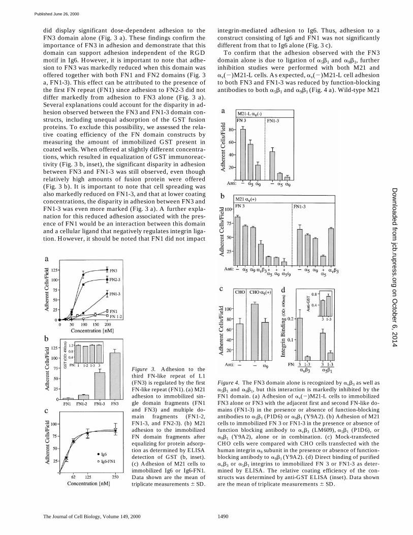
Site-directed mutagenesis of key residues within the pu-tative B-C and C-C9 loop regions of FN3 was performedto confirm that they are required for integrin recognitionin the context of the whole domain. Specifically, the se-quence QVKGHLR827 in the putative B-C loop was mu-tated to QVAGHLR827, whereas the GSQRKHSKRHI-HKDHV852 sequence constituting the C-C9 loop wasmutated to GSQNNHSNNHIHKDHV852. Conservativeasparagine substitutions were generated within the C-C9loop to minimize unforeseen effects on domain structure,and both of the dibasic RK841 and KR845 sequences weresubstituted since both were found to contribute to integrinbinding (Fig. 6). While the wild-type FN3 domain sup-ported both M21 and av(2)M21-L adhesion at concentra-tions as low as 50–75 nM, this adhesion was markedly re-duced after substitution of the dibasic sets of lysine and
arginine residues in the putative C-C9 loop (Fig. 7, a andb). Mutation of the single lysine823 residue in the putativeB-C loop had a relatively small but significant effect on ad-hesion, which was principally evident at lower coating con-centrations (Fig. 7, a and b). To confirm the primary im-portance of the C-C9 loop sequence, direct binding assayswere performed with purified a5b1 and avb3 integrins. Inagreement with the integrin binding results obtained usingpeptides, ligation of both integrins to the FN3 domain wasdose-dependent and saturable, and exhibited significant
Figure 6. A sequence in the putative C-C9 loop of the FN3 do-main supports adhesion and is recognized by both avb3 and a5b1.(a) M21 adhesion to an immobilized peptide derived from a se-quence in the putative C-C9 loop of FN3 (GSQRKHSKRHIH-KDHV852) or peptides resulting from alanine substitution ortruncation. (b) Adhesion of M21 cells to immobilized wild-typepeptide GSQRKHSKRHIHKDHV852 in the presence or absenceof antibodies to avb3 (LM609), a5b1 (P1D6), or b1 integrins(P4C10), alone or in combination. (c) Adhesion of av(2)M21-Lcells to immobilized wild-type peptide GSQRKHSKRHIH-KDHV852 in the presence or absence of antibodies to a9b1(Y9A2) or a5b1 (P1D6). (d and e) ELISA determination of thedirect binding of purified avb3 (d) or a5b1 integrins (e) to immobi-lized wild-type GSQRKHSKRHIHKDHV852 or mutant peptidesderived by alanine substitution or truncation. Mutated residuesare underlined, and the data shown are the mean of triplicatemeasurements 6 SD.
on October 6, 2014
jcb.rupress.orgD
ownloaded from
Published June 26, 2000
Silletti et al. L1 Multimerization and Integrin Ligation 1493
susceptibility to mutations within the C-C9 loop (Fig. 7, cand d).
Based on our findings, we propose that the FN3 domainof L1 contains two novel integrin binding motifs that ac-count for RGD-independent recognition by a5b1 and avb3.Substitution studies demonstrate that the dibasic RK841
and KR845 sequences present in the putative C-C9 loop ofFN3 are of primary importance for integrin recognition. Asecond motif in the putative B-C loop of the FN3 domain(QVKGHLR/K827) also contributes to a5b1 and avb3 bind-ing, albeit to a lesser degree. It is important to note thatboth QVKGHLR827 and GSQRKHSKRHIHKDHV852 pep-tides were ineffective as soluble antagonists in as much asthey failed to inhibit adhesion to themselves. Indeed, aparadoxical increase in adhesion was often observed whenthese peptides were offered during the adhesion assay(data not shown). One explanation for this effect would bethat the soluble peptides can self-associate with the immo-bilized peptide, thereby resulting in multimerized peptidewhich is then recognized by integrin avb3 and/or a5b1.While we have obtained clear evidence that a9b1 can sup-port adhesion to FN3, we failed to identify the binding mo-tif. It may prove that recognition by this integrin is subjectto conformational constraints that are violated by the useof linear peptides.
Integrins Recognize Multimerized FN3 and Homoaggregation of This Domain Is Regulated by the Dibasic Sequences in the Putative C-C9 Loop and by the Presence of FN1
Purified FN3 and FN2-3 fusion proteins (GST or His)were both observed to precipitate at high protein concen-trations, suggesting a propensity for homoaggregation. Incontrast, such precipitation was not observed with eitherIg6-FN1-3 or FN1-3 fusion proteins. These observations,coupled with the prior observation that the presence ofFN1 is inhibitory to cell adhesion on FN3, raised the possi-bility that integrins preferentially recognize the FN3 do-main as a homomultimer and that inhibition of integrinrecognition by FN1 is related to the ability of this domainto inhibit such multimerization. It was also observed thatprecipitation of the FN3 domain was markedly reduced af-ter substitution of the dibasic RK841 and KR845 sequencespresent in the putative C-C9 loop sequence of FN3. Thisraised the further possibility that the peptide sequenceGSQRKHSKRHIHKDHV852, which is of primary impor-tance for integrin recognition, is also important for FN3homomultimerization. Indeed, a propensity for self-associ-ation by the GSQRKHSKRHIHKDHV852 peptide couldexplain the paradoxical finding that this peptide can func-tion as a soluble agonist in adhesion assays. Confirmationof this hypothesis would require that several stipulationsbe fulfilled: (1) FN3 is multimerized at the time of integrinrecognition; (2) the presence of FN1 limits FN3-medi-ated multimerization; and (3) the GSQRKHSKRHIHK-DHV852 peptide self-associates and mutation of the C-C9loop sequence, which results in a loss of integrin recogni-tion, also prevents multimerization.
As a first step, we looked for evidence of FN3 multimer-ization by both column chromatography and SDS-PAGE.To avoid potential complexities associated with the pres-ence of GST as a fusion partner, these studies were per-formed with an FN3-His construct. After gel filtration on aSephracryl S-200 column, the FN3 domain was observedto elute as a series of high molecular mass complexes (Fig.8 a, top). Based on size relative to molecular mass stan-dards, the smallest complexes were trimers (33) and hexa-mers (63), with relatively little monomer (13) evident.The relative proportion of monomer present in differentpreparations varied with some preparations having littleor no evidence of any monomer whatsoever. It should benoted that any precipitate present in the FN3 preparationswas removed by centrifugation before fractionation bycolumn chromatography. In support of these findings withthe FN3-His protein, separation of the FN3-GST prepara-tion also revealed the presence of trimeric and higher or-der complexes (data not shown).
Upon SDS-PAGE resolution under nondenaturing con-ditions (i.e., without boiling), most of the FN3–His com-plexes were resolved into the monomer (16 kD, 13; Fig. 8b, lane 1). However, even in the presence of SDS, a signifi-cant amount of trimeric FN3 (48 kD, 33) was detected.Depending upon the amount of material loaded, smallamounts of hexameric FN3 (96 kD, 63) and sometimeseven higher order species could also be detected (Fig. 8 b,lane 1). Taken together, these data demonstrate that, un-der native conditions, the FN3 domain self-associates to
Figure 7. Substitution of the dibasic sequences in the putativeC-C9 loop of the FN3 domain suppresses adhesion and direct in-tegrin binding. (a and b) Adhesion of M21 (a) or av(2)M21-Lcells (b) to wild-type FN3 domain or mutant FN3 domains con-taining conservative asparagine substitution of the two dibasic se-quences within the C-C9 loop (RK841 and KR845), or a singlelysine823 to alanine823 substitution within the B-C loop. (c and d)ELISA determination of the direct binding of purified avb3 (c) ora5b1 integrins (d) to immobilized wild-type FN3 domain or mu-tant FN3 containing asparagine substitutions within the C-C9loop. Data shown are the mean of triplicate measurements 6 SD.
on October 6, 2014
jcb.rupress.orgD
ownloaded from
Published June 26, 2000

The Journal of Cell Biology, Volume 149, 2000 1494
form large multimeric complexes and that the most stablemultimeric configuration appears to be a trimer (SDS-PAGE), which can further self-associate to form higherorder complexes (gel filtration).
Importantly, substitution of the dibasic RK841 and KR845
sequences present in the putative C-C9 loop sequence ofFN3, which effectively abrogated integrin binding, wasalso found to limit FN3 multimerization. This is demon-strated by the large amount of monomeric FN3 evident on
fractionation (Fig. 8 a, bottom) and by an almost completeabsence of trimeric FN3 (33) evident on SDS-PAGE (Fig.8 b, lane 2). The small amount of complexed FN3 remain-ing despite mutation of the dibasic sequences likely re-flects the conservative substitution of the arginine andlysine residues with asparagines. In contrast, alanine muta-tion of the lysine823 residue in the putative B-C loop, whichonly marginally effected adhesion, did not obviously effectthe multimerization of FN3 (Fig. 8 b, lane 3). This lack ofeffect on domain multimerization was also observed uponfractionation of the lysine823 mutant by gel filtration (datanot shown). Together, these data suggest that integrinbinding to FN3 primarily involves recognition of mul-timers.
Confirming the hypothesis proposed above, a compari-son of complex formation by FN1-3 versus FN3 dem-onstrates that the presence of FN1 does indeed limit mul-timerization mediated by the FN3 domain. Thus, theproportion of trimeric FN1-3 (140 kD, 33) evident onSDS-PAGE was found to be markedly lower than that ob-served with both FN3 (Fig. 8 b, lane 1) and FN2-3 (Fig. 8c). That the FN2-3 construct forms complexes as effi-ciently as FN3 is consistent with the ability of this two-domain construct to support adhesion at levels equivalentto FN3 alone (Fig. 3 a). The observation that FN1-3 is stillcapable of limited multimerization may explain why FN1-3can still be recognized by integrins at high coating concen-trations.
A final requirement of the hypothesis proposed above isthat the GSQRKHSKRHIHKDHV852 peptide derivedfrom the C-C9 loop of FN3 can self-associate and, as a re-sult, support integrin recognition as a peptide complex.This would support the concept that the C-C9 loop of FN3promotes both multimerization and integrin binding. OnSDS-PAGE the 16-mer GSQRKHSKRHIHKDHV852
peptide, which has a predicted molecular mass of 2 kD,was found to migrate as a primary species of z6 kD, indic-ative of an SDS-stable tripeptide complex (Fig. 8 d, lane2). A 15-mer peptide of 1.95 kD derived from the Ig6 do-main of L1 (PSITWRGDGRDLQEL544) is shown forcomparison (Fig. 8 d, lane 1). Interestingly, separate ala-nine substitution of either of the two dibasic RK841 orKR845 sequences in the GSQRKHSKRHIHKDHV852 pep-tide resulted in a reduction in molecular mass, which isconsistent with the formation of di- rather than tripeptidecomplexes (Fig. 8 d, lanes 3 and 4). Finally, simultaneousalanine replacement of both sets of dibasic residues re-sulted in a peptide that resolved as a monomeric species(Fig. 8 d, lane 5). Importantly, these same alanine substitu-tions also abrogated cell adhesion and integrin binding(Fig. 6). Taken together, these data indicate that optimalintegrin binding to the GSQRKHSKRHIHKDHV852 pep-tide under native conditions involves recognition of tri-peptide or higher order peptide complexes and confirms arole for the dibasic RK841 and KR845 sequences in domainand peptide multimerization.
Plasmin Regulates FN3 Multimerization andIntegrin Binding
Human L1, as well as related molecules in the mouse, rat(NILE), and chick (Ng-CAM and Nr-CAM) have been
Figure 8. Homomultimerization of the FN3 domain is mediatedby dibasic sequences in the putative C-C9 loop and regulated bythe presence of FN1. (a) Sephacryl S-200 gel filtration elutionprofiles of wild-type FN3 domain (top) or mutant FN3 containingconservative asparagine substitution of the two dibasic sequences(RK841 and KR845) within the C-C9 loop (bottom). Elution pointsof monomer (13), trimer (33), and hexamer (63) in relation tomolecular mass standards are denoted. (b) SDS-PAGE analysisof wild-type FN3 (lane 1), FN3 in which the two dibasic pairswithin the C-C9 loop are replaced with asparagines (lane 2), orFN3 in which lysine823 within the B-C loop is replaced with ala-nine (lane 3). Separated proteins were detected by immunoblot-ting with the anti–L1 ECD pAb. (c) SDS-PAGE and immuno-blot comparison of FN3 in conjunction with adjacent FN-likedomains (FN2-3 or FN1-3). The relative migration of monomeric(13), trimeric (33), and hexameric species (63) are indicated.(d) SDS-PAGE comparison of wild-type GSQRKHSKRH-IHKDHV852 peptide (lane 2), with single dibasic alanine mutantpeptides (GSQAAHSKRHIHKDHV852, lane 3; GSQRKHS-AAHIHKDHV852, lane 4) and the double dibasic alanine mutantpeptide (GSQAAHSAAHIHKDHV852, lane 5). A peptide de-rived from the Ig6 domain of L1 (PSITWRGDGRDLQEL544) isshown for comparison (lane 1). The relative migration of the pep-tides in relation to molecular mass standards is shown at the left.
on October 6, 2014
jcb.rupress.orgD
ownloaded from
Published June 26, 2000

Silletti et al. L1 Multimerization and Integrin Ligation 1495
shown to be sensitive to posttranslational cleavage withinthe FN3 domain (Faissner et al., 1985; Sadoul et al., 1988;Nybroe et al., 1990; Burgoon et al., 1995). Importantly, thiscleavage has been shown to involve the same dibasicsequences that we have identified as important for inte-grin binding and multimerization. Thus, trypsin has beenshown to cleave after the second dibasic sequence(GSQRKHSKR↓HIHKDHV852; Faissner et al., 1985; Sa-doul et al., 1988), whereas we have recently demonstratedthat plasmin cleaves both dibasic sequences (GSQ-RK↓HSK↓RHIHKDHV852; Nayeem et al., 1999). Basedon this information, we questioned whether posttransla-tional cleavage of the FN3 domain represents a mecha-nism for regulating both multimerization and integrinbinding.
Treatment of FN3–His complexes with plasmin resultedin a dose-dependent dissolution of the trimeric FN3 com-plexes evident on SDS-PAGE (Fig. 9 a). A loss of trimericFN3 complexes was evident at plasmin concentrations aslow as 0.01 U/ml. Consistent with this finding, treatment ofimmobilized FN3 substrate with plasmin at a concentra-tion of 0.01 U/ml also resulted in a .60% inhibition of ad-hesion by both M21 and av(2)M21-L cells (Fig. 9 b, right).At the same concentration, plasmin had no effect on theRGD-dependent adhesion of M21 cells to the Ig6 domainof L1 (Fig. 9 b, left). Interestingly, plasmin treatment ofthe L1 ectodomain also resulted in a marked inhibition ofadhesion by av(2)M21-L cells, but only marginally de-creased adhesion by M21 cells (Fig. 9 b, middle). Thisresult is consistent with the finding that av(2)M21-L ad-hesion to the L1 ectodomain is highly dependent on inter-actions with the FN3 domain, while M21 cells can stilladhere via interaction with the RGD motif in the Ig6 do-main. Together, these data support the concept that serineprotease–mediated cleavage within the FN3 domain is apotentially important mechanism for regulating its func-tional activity.
A Paradigm for L1–Integrin Interactions
Based on our findings, it is evident that FN1 limits homo-multimerization via the FN3 domain. One possible expla-nation for this would be steric hindrance in which the FN1and FN3 domains are folded back upon one another in aclosed globular conformation. Homomultimerization viathe FN3 domain and concomitant integrin recruitmentmay only occur after a change in conformation that pro-motes a more extended open configuration. HomophilicL1–L1 ligation via the Ig-like domains and/or integrin in-teraction with the RGD motif in Ig6 may be mechanismsfor inducing such a change in conformation. A schematicrepresentation of such a model is shown in Fig. 10 a.
Certain predictions can be made based on the model pro-posed. First, it should be possible to show that, as a result offolding between FN1 and FN3, accessibility to certain do-main regions will be limited. In this regard, an mAb specificfor an epitope in the Ig6 domain of L1 (mAb LP1B9) wasable to recognize Ig6-FN1 and Ig6-FN1-2, but was very lim-ited in its ability to recognize Ig6-FN1-3 (Fig. 10 b). This re-sult is hard to reconcile if Ig6-FN1-3 simply forms a linearstructure, but can be explained readily if there is a foldingevent that juxtaposes FN3 with FN1 and Ig6.
A further prediction of the model presented is that inhi-bition of upstream L1 ligation (e.g., homophilic L1–L1 li-gation) will also inhibit integrin-dependent adhesion viaFN3 because the FN-like repeats will remain in a closedconformation. In this regard, we observed that integrin-dependent adhesion of av(2)M21-L cells to the L1 ec-todomain could be significantly reduced by an antibody(5G3) that blocks homophilic L1–L1 ligation by binding toan NH2-terminal Ig-like domain (Montgomery et al., 1996;Nayeem et al., 1999; Fig. 10 c, left). It is unlikely that thisantibody is inhibiting av(2)M21-L adhesion by directlyblocking integrin recognition of the FN3 domain since itrecognizes a distal epitope and fails to prevent integrinbinding to the Ig6 domain, which is located closer to theantibody binding site. However, since av(2)M21-L cellsexpress high levels of L1, a homophilic interaction with theimmobilized L1 could induce the conformation change re-quired for binding of these cells to FN3. The 5G3 antibodywas markedly less effective at preventing M21 cell adhe-sion to the L1 ectodomain (Fig. 10 c, right), presumablybecause these cells are still able to recognize the RGD mo-tif in the Ig6 domain via avb3. It is interesting to note thatthe 5G3 antibody also had some minimal inhibitory activ-ity when the FN3 domain alone was offered as a substrate(Fig. 10 c). This inhibition may indicate a limited but directinteraction between cellular L1 and the immobilized FN3
Figure 9. Plasmin regulates FN3 multimerization and integrinbinding. (a) SDS-PAGE and immunoblotting analysis of com-plexed wild-type FN3 treated with plasmin. FN3 in solution wastreated for 90 min with plasmin before SDS-PAGE and immuno-blotting with the anti–L1-ECD pAb. The FN3 trimer band (48 kD)was analyzed by scanning densitometry and graphed in arbitrarydensity units. (b) Adhesion of M21 or av(2)M21-L cells to Ig6,FN3, or L1 ectodomain treated with plasmin. Immobilized pro-teins were treated with or without 0.01 U/ml plasmin for 90 min,washed, and blocked before the addition of cells. Data shown arethe mean of triplicate measurements 6 SD.
on October 6, 2014
jcb.rupress.orgD
ownloaded from
Published June 26, 2000

The Journal of Cell Biology, Volume 149, 2000 1496
domain. In this regard, it has been shown that FN con-structs containing the FN3 domain can interact with otherIg-like domains present in the L1 ectodomain (Holm et al.,1995).
Based on the premise that distal L1 ligation events arerequired to promote FN3 multimerization and integrin re-cruitment, it is also to be expected that L1 and integrinswill only colocalize at the cell–cell interface, where suchdistal ligation events are expected to occur. Double im-munofluorescence was performed to test this prediction.The a9b1 integrin was selected for analysis since previousstudies with the av(2)M21-L cells demonstrated that thisintegrin is primarily involved in adhesion to the FN3 do-main rather than the RGD motif in Ig6. Aggregates of
av(2)M21-L cells were analyzed after simultaneous stain-ing for a9b1 and L1. Both L1 and a9b1 were observed to berecruited to sites of cell–cell contact (Fig. 11, a and b) andsignificant colocalization is evident at these sites (Fig. 11, cand d). However, it is also important to note that theseligands do not appear to colocalize unless recruited to thecell–cell interface as demonstrated by confocal micro-scopic analysis (Fig. 11 d). Since the juxtaposition of twocell membranes could give the illusion of colocalization, itwas further determined whether such colocalization is stillevident on single cells obtained after the gentle disruptionof stained cell aggregates. The disruption of cell aggre-gates was performed with simultaneous fixation. Usingthis approach, we observed significant modulation of bothL1 and a9b1 on some of the single cells obtained (Fig. 11, eand f). Based on the absence of such obvious modulationin the absence of prior aggregation, it is likely that areas ofmodulation are a result of prior cell–cell contact. Impor-tantly, even under these conditions, we still observed sig-nificant colocalization of L1 and a9b1 (Fig. 11 g). Signifi-cant colocalization was not observed on those single cellsthat failed to display evidence of modulation (Fig. 11 g, as-terisk). Adopting the same experimental approach, butwith all steps performed at 378C in the absence of sodiumazide, we observed further marked modulation of L1 anda9b1 expression (Fig. 11, h and i), and again significantcolocalization was observed (Fig. 11 j). Colocalization be-tween a9b1 and L1 on individual cells that were separatedfrom cell aggregates supports the concept of a cis-interac-tion between these two ligands.
As a further test of a direct association between a9b1and L1, coimmunoprecipitation studies were performedusing the anti-a9b1 mAb Y9A2. Cell lysates were madefrom av(2)M21-L cells maintained as aggregates or assubconfluent monolayers. These lysates were treated withmAb Y9A2 and Western blot analysis was performed todetect the presence of coprecipitated L1. Consistent withan interaction at sites of cell–cell contact, L1 was readilydetected in Y9A2 immunoprecipitates derived from ly-sates of aggregated av(2)M21-L cells (Fig. 11 k, Agg.). Incontrast, only minimal reactivity was evident in the lysatederived from monolayer cultures (Fig. 11 k, Mono.). Asfurther confirmation of L1–integrin association, reverseimmunoprecipitation was performed using an anti-L1polyclonal antibody (anti-5G3 Ag-pAb). In these studies,Western blot analysis was performed to detect the pres-ence of coprecipitated b1, a5, b3, and b5 integrin subunits.Providing further evidence for a possible association be-tween L1 and a9b1, Western blot analysis confirmed thepresence of the b1 integrin subunit in immunoprecipitatesderived from aggregated av(2)M21-L cells (Fig. 11 l). Be-cause of a lack of antibodies suitable for the detection ofthe a9 subunit by Western blotting, we cannot definitivelyclaim that the presence of the b1 integrin subunit is due tothe coprecipitation of a9b1. However, given that a9b1 bothcolocalizes with and coprecipitates L1, it is likely that atleast a component of the b1
present is due to its associationwith a9. Consistent with reports that have indicated an as-sociation between L1 and a5b1 and avb3, both b3 and a5 in-tegrin subunits were also identified in immunoprecipitatesof aggregated M21 or M21-L cells (Fig. 11 l). In contrast,despite the presence of significant amounts of avb5 in M21
Figure 10. L1 conformation and L1–L1 homophilic interactionmay regulate integrin recruitment via the FN3 domain. (a) A po-tential model for the interaction between integrins and the FN3domain of L1. The Ig- and FN-like domains of L1 are assumed toform a closed globular conformation according to the findings ofSu et al. (1998) and Drescher et al. (1996). Distal ligation eventsinvolving the Ig-like domains of L1 (L1–L1 or L1–integrin) arepostulated to cause a conformational change, resulting in a per-missive open conformation that can support L1 clustering viaFN3 and subsequent integrin recruitment. Potential implicationsof this multimerization and integrin recruitment include L1-inte-grin–dependent signal transduction. (b) Anti-Ig6–specific mAb(LP1B9) recognition of its epitope in the Ig6 domain alone, orIg6 with adjacent FN-like domains (Ig6-FN1-2 and Ig6-FN1-3) asdetermined by ELISA. Immobilized proteins were incubatedwith the anti–L1-Ig6 mAb LP1B9, washed, and were detectedcolorimetrically with HRP-conjugated secondary antibody andOPD. (c) Adhesion of M21 or av(2)M21-L cells to the L1ectodomain (L1-ECD) or FN3 after pretreatment of cells withthe function-blocking anti-L1 mAb 5G3. Data shown are themean of triplicate measurements 6 SD.
on October 6, 2014
jcb.rupress.orgD
ownloaded from
Published June 26, 2000

Silletti et al. L1 Multimerization and Integrin Ligation 1497
cell lysates, no significant coprecipitation of the b5 subunitwas detected (Fig. 11 l). Importantly, none of the integrinsubunits were detected after immunoprecipitation using acontrol antibody to GST (Fig. 11 l).
Based on the observation that L1–integrin colocaliza-tion is primarily restricted to sites of cell–cell contact, it isto be expected that only a small fraction of the available
integrin pool will be directly associated with L1. In our sys-tem, despite attempts to maximize aggregation, we still ob-served that many cells remained single and, even in aggre-gates, we often observed variable recruitment into cell–cellcontact sites. It also needs to be recognized that some inte-grins are sequestered in large intracellular pools. In thecase of the b1 integrin subunit, the amount specifically co-
Figure 11. Colocalizationand coimmunoprecipitationof L1 and integrins after cell–cell interaction. (a–c) Stain-ing of M21-L cell cluster withanti–L1-ECD pAb and theanti-a9b1 mAb Y9A2. L1staining is shown in green (a,FITC), a9b1 staining in red(b, Texas red), and overlap-ping fluorescence or colocal-ization is evidenced as yellow(c, Merge). Arrows denoteareas of colocalization atpoints of cell–cell contact.Images were obtained usinga 403 objective and a NikonEclipse E800 fluorescent mi-croscope. (d) Merged imageof L1 (green) and a9b1 (red)staining of M21-L cell clus-ters obtained using an MRC1024 confocal microscope.Note that colocalization isprimarily limited to cell–cellinterfaces (arrows). (e–g)Disruption of stained M21-Lcell clusters with simulta-neous fixation results insome single cells with areasof modulated L1 and a9b1 ex-pression (e and f). Note thatthere is still evidence of sig-nificant colocalization be-tween L1 and a9b1(g). Nosignificant localization wasobserved in another cell thatfailed to show evidence of L1or a9b1 modulation (g, aster-isk). (h–j) M21-L cell clusterswere stained at 378C and inthe absence of azide. Afterdisruption of cell aggregates,some single cells showed evi-dence of significant L1 anda9b1 modulation (h and i).Significant areas of colocal-ization were observed (j). (k)Coimmunoprecipitation ofL1 with an anti-a9b1 mAb.Lysates were obtained fromaggregated av(2)M21-L cells(Agg.) or their adherent sub-
confluent counterparts (Mono.). Precipitated proteins were immunoblotted to detect coprecipitated L1 or the immunoprecipitated b1subunit of a9b1. (l) Reverse immunoprecipitation was performed using an anti-L1 pAb and lysates of aggregated M21-L or M21 cells. Apolyclonal antibody to GST was used as a control. Immunoprecipitated material was immunoblotted using antiintegrin antibodies as de-scribed in Materials and Methods. Membranes were reprobed with anti-L1 mAb 5G3 to confirm precipitation of L1. Integrin subunitsidentified have molecular masses consistent with that reported in the literature.
on October 6, 2014
jcb.rupress.orgD
ownloaded from
Published June 26, 2000

The Journal of Cell Biology, Volume 149, 2000 1498
precipitated with L1 appears to be ,1% of the total avail-able cellular b1. However, since the L1-reactive b1 inte-grins (i.e., a5b1 and a9b1) constitute only a small fractionof the total b1 integrin expression by M21-L cells (,10%)this is not unexpected. Consistent with this, we estimatethat significantly more of the available a5b1 integrin poolcoprecipitated with L1 (z5%). However, such estimatesmay be underestimates since it is not clear how stable L1–integrin complexes are under the detergent conditionsused during the immunoprecipitation.
DiscussionIn this study, we have shown that integrin binding to intactL1 cannot be solely attributed to the RGD motif presentin the sixth Ig-like domain, but rather involves additionalrecognition of sequences in the third FN-like domain(FN3). As such, we describe two novel integrin-bindingmotifs located within the putative B-C and C-C9 loops ofthe FN3 domain (QVKGHLR827 and GSQRKHSKR845).Site-directed mutagenesis of key residues within these mo-tifs confirmed the primary importance of the sequenceGSQRKHSKR845 for both cell adhesion and direct inte-grin binding to the FN3 domain. Remarkably, the samedibasic sequences responsible for supporting integrinbinding to this motif (RK841 and KR845) are also shown topromote the multimerization of the FN3 domain. In thisregard, multimerization of the GSQRKHSKR845 sequenceprecedes, and may be required for, interaction with bothavb3 and a5b1 integrins. Importantly, both homomultimer-ization of, and integrin binding to FN3 is shown to be regu-lated both by conformational constraints imposed by otherFN-like domains (FN1) and by plasmin-mediated cleavagewithin the sequence RK↓HSK↓RH846. Based on our find-ings, we propose that L1 may have an expanded role in theregulation of integrin function, especially at the cell–cellinterface, where bridging events appear to allow L1–inte-grin (a9b1) colocalization.
The large multidomain organization of L1 has permittedthe evolution of domains and domain regions with distinctfunctional attributes. A recent model based on the crystalstructure of hemolin suggests that an antiparallel align-ment involving the first four Ig-like domains of L1 is re-sponsible for homophilic L1–L1 binding (Su et al., 1998).Importantly, the same Ig-like domains involved in ho-mophilic binding may also adopt a tertiary horseshoe con-figuration because of acute folding and strong interdomainpairing (Su et al., 1998), suggesting that homophilic L1–L1binding at the cell surface may be regulated by an equilib-rium between open/accessible (extended) and closed/inac-cessible (globular or horseshoe shaped) conformationswithin the distal Ig domains.
In this study, we propose that the functional activity ofthe FN-like domains of L1 may also be regulated by open(extended) and closed (globular) conformations. In thisregard, a recent structural analysis of L1 by rotary shadow-ing and transmission electron microscopy confirmed thatthe FN-like repeats do indeed form a tight globular struc-ture (Drescher et al., 1996). In addition, an antibody di-rected towards an epitope located between domains FN2and FN3 mimics the effects of homophilic L1–L1 binding,promoting neurite outgrowth and signal transduction (Ap-
pel et al., 1995). Based on their findings, the authors sug-gest that conformational changes resulting from L1 liga-tion could funnel to this region of L1, a region that may beconformationally labile and the ultimate site for the induc-tion of signaling events. Together, these findings are con-sistent with the paradigm that the FN-like repeats assumea closed globular conformation and that extrinsic ligation,by an antibody or by L1 itself, has important functionalconsequences by virtue of exposing a region that is essen-tial for receptor multimerization, integrin recruitment, andconsequent cell signaling.
While we suggest that homophilic ligation can alter theconformation of the FN-like repeats, thereby exposing theFN3 domain and promoting integrin recruitment, otherheterophilic interactions may also have a role. In this re-gard, it is of particular interest that TAG-1/axonin-1, aneuron-specific CAM, has been shown to promote neuriteextension via a mechanism that requires both an L1-likemolecule and b1 integrins (Felsenfeld et al., 1994). The au-thors suggest a mechanism involving a direct interactionbetween TAG-1 and L1, leading to a concomitant cis-interaction between L1 and b1 integrin. The result of theseinteractions is signaling via both types of receptors,thereby culminating in neurite extension. A further possi-bility is that a direct trans-interaction between integrinsand the RGD motif in the Ig6 domain of L1 is sufficient tofunnel a conformational change to the adjacent FN-likerepeats. This type of interaction would facilitate L1 multi-merization and further integrin recruitment, even whenthe interacting cell type fails to express neuron-associatedCAMs.
The concept that the FN3 domain is involved in self-association is supported by earlier work, which showed thatbeads coated with FN-like domains 3–5 of L1 had a strongtendency to self-associate (Holm et al., 1995). Consistentwith a central role for the FN3 domain in this process, littleor no aggregation was observed with beads coated withFN-like domains 1 and 2 or 4 and 5. Furthermore, and inaccordance with our finding that the FN1 domain limitsmultimerization through FN3, the authors also observedthat beads coated with FN-like domains 1–5 showed littletendency towards self-association. Our finding that theFN3 domain can support cell adhesion is also supported inthe literature. Thus, Appel et al. (1993) demonstratedstrong neural cell adhesion to an L1 fragment consisting ofFN-like domains 3–5. In agreement with our findings, littleadhesion was observed on FN-like domains 1 and 2. Againsupporting a negative regulatory function for the FN1 do-main in this process, the adhesion observed on FN-like do-mains 1–5 was markedly reduced in comparison to thatevident on FN-like domains 3–5. While the authors sug-gested that the neuronal adhesion observed was primarilydue to homophilic ligation between cellular L1 and the L1fragments, they also proposed a role for an unidentifiedheterophilic ligand. Based on the findings presented in thismanuscript, neuronal integrins would be likely candidatesfor this role.
Based on our findings, we propose that the FN3 domainis of essential importance for the biological activity of L1.As a consequence, it is expected that the activity of thisdomain will be tightly regulated. While we propose thatconformational constraints are a primary mechanism for
on October 6, 2014
jcb.rupress.orgD
ownloaded from
Published June 26, 2000

Silletti et al. L1 Multimerization and Integrin Ligation 1499
regulating the activity of FN3, we also detail a secondmechanism based on proteolytic cleavage. An importantand unifying property of L1-like proteins is a susceptibilityto posttranslational cleavage, thus, human and mouse L1,as well as related molecules in the rat and chick, are sensi-tive to cleavage within the FN3 domain, yielding frag-ments that have been detected in neural tissue in vivo(Rathjen and Schachner, 1984; Faissner et al., 1985; Sadoulet al., 1988; Wolff et al., 1988; Nybroe et al., 1990; Burgoonet al., 1995). Cleavage within the FN3 domain is due to thepresence of a highly conserved serine protease–sensitiveregion that incorporates dibasic arginine/lysine sequences(Faissner et al., 1985; Sadoul et al., 1988). Significantly, wehave now shown that this same sequence of basic aminoacids is important for FN3 multimerization and integrinbinding. Furthermore, we show that low concentrations ofplasmin will disrupt such interactions, even in the case ofpre-formed complexes, based upon the ability of plasminto cleave within the sequence RK↓HSK↓RH (Nayeem etal., 1999). This suggests that plasmin-mediated proteolysisof homomultimerized FN3 domains may provide an im-portant mechanism for regulating initiation and/or inacti-vation of L1–integrin interactions. Potential regulation ofL1 function by plasmin is of particular interest, given theextensive overlap of neurological functions that involveboth plasmin and L1 including neurite extension (Lage-naur and Lemmon, 1987; Seeds et al., 1997), granule cellmigration (Krystosek and Seeds, 1981; Moos et al., 1988),nerve regeneration (Salles et al., 1990; Jung et al., 1997),and long-term potentiation in the hippocampus (Luthl et al.,1994; Mizutani et al., 1996).
The extent to which the FN3 domain promotes eithercis- or trans-integrin interactions remains to be resolved.Multimerization of L1 in the plane of the cell membranevia a domain (i.e., FN3) that is in close proximity to themembrane may favor the recruitment of integrins in a cis-type of interaction. The demonstration of colocalizationbetween a9b1 and L1 on individual cells separated fromcell aggregates supports the concept of a cis-interaction. Inthis regard, cis-interactions between integrins and otherreceptors, including members of the IgSF, are well docu-mented and are believed to be an important mechanismfor both regulating the activational status of the integrin,and for the formation of signaling complexes at the cell–cell or cell–substrate interface (Porter and Hogg, 1998).While conformational constraints are proposed to regulateintegrin access to the FN3 domain, such constraints areless likely to limit integrin recognition of the RGD motifin the Ig6 domain of L1. Thus, Drescher et al. (1996) con-clude that the RGD motif in human L1 is available for in-termolecular interactions based both on the high surfacehydrophobicity of the Ig6 domain and the position of themotif within a loop structure. Based on this, it is conceiv-able that RGD-dependent integrins can recognize L1(Ig6) in its resting conformation via a trans-interaction atthe cell–cell interface. Whether such an interaction is suffi-cient to initiate a conformational change in L1 and furtherintegrin recruitment via a cis-non-RGD–dependent mech-anism remains to be determined. It is also unclear whetherrecognition of FN3 by integrins involves the typical ligand-binding pocket or is the result of an interaction with alter-native sites on either or both of the integrin subunits.
Specificity for a5b1 and a9b1 to the apparent exclusion ofother b1 integrins does, however, suggest the involvementof the a-subunit.
Changes in L1 conformation and concomitant L1 clus-tering may also be important for the recruitment of otherheterophilic ligands reported to undergo cis-interactionswith L1. Candidate ligands include the following: CD9(Schmidt et al., 1996), NCAM (Feizi, 1994), CD24 (Kad-mon et al., 1995), axonin-1/TAG-1 (Kunz et al., 1998), andthe FGF receptor (FGFr; Viollet and Doherty, 1997). It isconceivable that L1 clustering results in the recruitment ofa variety of signaling elements (e.g., CD9, CD24, andFGFr) culminating in the formation of large signalingcomplexes at the cell–cell interface. In this regard, it hasbeen suggested that trans-homophilic L1–L1 ligation leadsto L1 clustering and the recruitment of FGFr (Viollet andDoherty, 1997). Downstream signaling events resultingfrom the activation of the FGF tyrosine kinase receptorhave now been shown to be responsible for L1-mediatedneurite extension (Viollet and Doherty, 1997). The poten-tial recruitment of CD9 is also of particular interest sincethis tetraspan signaling molecule is also known to associ-ate with certain integrins (Berditchevski and Odintsova,1999) and could, therefore, serve to stabilize cis-complexesformed between integrins and L1. Given the pivotal im-portance of the third FN-like repeat of L1, it may provesignificant that this domain also has been implicated in thecis-interaction between the chick L1 orthologue NgCAMand axonin-1/TAG-1 (Kunz et al., 1998). While we specu-late that changes in L1 conformation and concomitant L1clustering could result in the recruitment of a variety ofsignaling molecules, we do not exclude the possibility thatcertain cis-interacting ligands may compete with or inhibitintegrin recruitment or that some heterophilic ligands willpreferentially interact with L1 in its resting conformation.
The integrin a9b1 identified as an L1 binding partner inthis study has a limited tissue distribution that includes ep-ithelia, muscle, and cells of the myelomonocytic series(Palmer et al., 1993; Taooka et al., 1999); no expressionhas been reported in the nervous system. The a9b1 integrinis also present on melanoma cells (Smith and Giachelli,1998) and a variety of carcinoma cell lines (Palmer et al.,1993), suggesting a potential role in tumor development.An overlap in expression between L1 and a9b1 is expectedsince L1 also has been described on a variety of epithelialstructures (Thor et al., 1987; Debiec et al., 1998; Kujat et al.,1995), on carcinoma and melanoma cell lines, and on nor-mal and transformed myelomonocytic cells (Mujoo et al.,1986; Linnemann et al., 1989; Reid and Hemperly, 1992;Pancook et al., 1997). Recently, it has been demonstratedthat a9b1 can promote neutrophil transendothelial cell mi-gration via an interaction with VCAM-1 (Taooka et al.,1999). L1–a9b1 interactions may also modulate transen-dothelial cell migration either directly by promoting adhe-sion to the endothelium or indirectly by modulating theactivity of a9b1 and its ability to bind to VCAM-1. Simi-larly, the b1-mediated adhesion of eosinophils to endothe-lial cells has also been shown to be modulated by ligationof PECAM-1 on endothelial cells (Chiba et al., 1999). Inthis regard, L1 has not only been described on my-elomonocytic cells and metastatic tumor lines (Mujoo etal., 1986; Linnemann et al., 1989; Reid and Hemperly,
on October 6, 2014
jcb.rupress.orgD
ownloaded from
Published June 26, 2000

The Journal of Cell Biology, Volume 149, 2000 1500
1992; Pancook et al., 1997), but also on activated small ves-sel endothelium in certain diseases (Felding-Habermannet al., 1997).
The observation that the integrins identified in thisstudy are interacting with multimeric FN3 is consistentwith the concept that L1 clustering is important for the re-cruitment of integrins, and would also explain why a9b1can only be colocalized with L1 at cell–cell interfaces,where classical L1–L1 homophilic interactions and conse-quent clustering would be expected to occur. The conceptthat ligand polymerization can potentiate integrin bindingis supported by recent work that demonstrates preferen-tial integrin-mediated adhesion to multivalent, rather thanmonovalent extracellular matrix components in lymphoidcells (Stupak et al., 1999). Indeed, a primary regulator ofinflammatory cell entry into sites of wounding is the accu-mulation of polymerized provisional matrix componentsincluding fibrin, fibronectin, and vitronectin (Dvorak et al.,1995). Although the enhanced affinity of the aforemen-tioned matrices primarily rely upon appropriate spacing orexposure of canonical cell adhesion motifs (e.g., RGD), itis not clear whether the integrin–L1 interactions describedin this report are due to the creation of neorecognitionsites within the complexed ligand, or rather reflect the op-timal presentation of multiple, appropriately spaced, pre-viously available, low affinity binding motifs in such amanner that the clustered motifs stabilize integrin bindingthrough avidity modulation.
An important implication of the work presented here isthat cell–cell contact and concomitant L1 clustering canpotentiate integrin activation and/or signaling. As a conse-quence, L1 may directly modulate those processes nor-mally associated with integrin-binding to the extracellularmatrix, including cell motility, survival, and proliferation.The fact that avb3 can associate with L1 via interactionwith its RGD motif (Montgomery et al., 1996; Felding-Habermann et al., 1997; Oleszewski et al., 1999) as well asvia an interaction with the multimerized FN3 domain sug-gests an interdependent relationship that may have impor-tant ramifications for cell survival and motility. For exam-ple, a recent study has shown that avb3 is essential for thesurvival of melanoma cells in the dermal microenviron-ment, however, detailed examination of these cells indi-cates that they are surviving as cell clusters (Hsu et al.,1998). In this context cell–cell interaction and concomitantL1–avb3 ligation may represent an important survivalmechanism. The ability of L1 to induce signaling via its in-teraction with integrins requires confirmation, however,the early steps of neurite extension on L1 are dependenton protein phosphorylation events involving the nonre-ceptor tyrosine kinase pp60c-src, a known mediator of in-tegrin signaling (Ignelzi et al., 1994), suggesting that L1 in-teractions may indeed facilitate signaling in this manner.
The findings reported here significantly extend our cur-rent knowledge of L1–integrin interactions. We have doc-umented two novel integrin-binding sequences within pu-tative loops in the third FN-like domain of L1. One ofthese sequences is shown to self-associate, resulting inboth domain multimerization and integrin ligation. To ourknowledge, this is the first report of a spontaneously self-associating peptide sequence that can also support directintegrin binding. Dual regulation of FN3-integrin binding
is proposed based on conformational constraints and sen-sitivity to proteolysis. Based on our observations, andthose of others, we propose a model in which distal trans-ligation of L1 at the cell–cell interface induces a conforma-tion change within the L1 ectodomain that culminates inreceptor multimerization and integrin recruitment via theFN3 domain. The findings presented in this study furthersuggest an intimate association between integrins and L1,an association that may have important implications fordownstream signaling events.
This study was supported by the National Cancer Institute (NCI) RO1grant CA69112-01 and by National Heart, Lung and Blood Institute(NHLB) RO1 grant HL62477-01 (to A.M.P. Montgomery) and by theNCI research fellowship (S. Silletti) 1F32 CA72192-01. This is Scrippsmanuscript #12868-IMM.
Submitted: 13 December 1999Revised: 1 May 2000Accepted: 23 May 2000
Note Added in Proof. While this manucript was being reviewed, it was re-ported that the third fibronectin repeat of L1 is required for L1 to serve asan optimal substratum for neurite extension. Stallcup, W.B. 2000. J. Neu-rosci. Res. 61:33–43.
References
Appel, F., J. Holm, J.-F. Conscience, and M. Schachner. 1993. Several extracel-lular domains of the neural cell adhesion molecule L1 are involved in neuriteoutgrowth and cell body adhesion. J. Neurosci. 13:4764–4775.
Appel, F., J. Holm, J.-F. Conscience, F. von Bohlen und Halbach, A. Faissner,P. James, and M. Schachner. 1995. Identification of the border between fi-bronectin type III homologous repeats 2 and 3 of the neural cell adhesionmolecule L1 as a neurite outgrowth promoting and signal transducing do-main. J. Neurobiol. 28:297–312.
Bastmeyer, M., H. Ott, C.A. Leppert, and C.A. Stuermer. 1995. Fish E587 gly-coprotein, a member of the L1 family of cell adhesion molecules participatesin axonal fasciculation and the age-related order of ganglion cell axons ingoldfish retina. J. Cell Biol. 130:969–976.
Bateman, A., M. Jouet, J. MacFarlane, J.-S. Du, S. Kenwrick, and C. Chothia.1996. Outline structure of the human L1 cell adhesion molecule and the siteswhere mutations cause neurological disorders. EMBO (Eur. Mol. Biol. Or-gan.) J. 15:6050–6059.
Berditchevski, F., and E. Odintsova. 1999. Characterization of integrin-tet-raspanin adhesion complexes: role of tetraspanins in integrin signaling. J.Cell Biol. 146:477–492.
Bieber, A.J., P.M. Snow, M. Hortsch, N.H. Patel, J.R. Jacobs, Z.R. Traquina, J.Schilling, and C.S. Goodman. 1989. Drosophila neuroglian: a member of theimmunoglobulin superfamily with extensive homology to the vertebrateneural cell adhesion molecule L1. Cell. 59:447–460.
Bock, E.C., C. Richter-Landsberg, A. Faissner, and M. Schachner. 1985. Dem-onstration of immunochemical identity between the nerve growth factor-inducible large external (NILE) glycoprotein and the cell adhesion moleculeL1. EMBO (Eur. Mol. Biol. Organ.) J. 4:2765–2768.
Brummendorf, T., S. Kenwick, and F.G. Rathjen. 1998. Neural recognition mol-ecule L1: from cell biology to human hereditary brain malformations. Curr.Opin. Neurobiol. 8:87–97.
Burgoon, M.P., R.B. Hazan, G.R. Phillips, K.L. Crossin, G.M. Edelman, andB.A. Cunningham. 1995. Functional analysis of posttranslational cleavageproducts of the neuron–glia cell adhesion molecule Ng-CAM. J. Cell Biol.130:733–744.
Chiba, R., N. Nakagawa, K. Kurasawa, Y. Tanaka, Y. Saito, and I. Iwamoto.1999. Ligation of CD31 (PECAM-1) on endothelial cells increases adhesivefunction of avb3 integrin and enhances b1 integrin-mediated adhesion ofeosinophils to endothelial cells. Blood. 94:1319–1329.
Cohen, N.R., J.S.H. Taylor, L.B. Scott, R.W. Guillery, P. Soriano, and A.J. Fur-ley. 1997. Errors in corticospinal axon guidance in mice lacking the neuralcell adhesion molecule L1. Curr. Biol. 8:26–33.
Dahme, M., U. Bartsch, R. Martini, B. Anliker, M. Schachner, and N. Mantei.1997. Disruption of the gene coding for the cell adhesion molecule L1 leadsto malformations of the nervous system in mice. Nat. Genet. 17:346–349.
Debiec, H., E.I. Christensen, and P.M. Ronco. 1998. The cell adhesion moleculeL1 is developmentally regulated in the renal epithelium and is involved inkidney branching morphogenesis. J. Cell Biol. 143:2067–2079.
Demyanenko, G.P., A.Y. Tsai, and P.F. Maness. 1999. Abnormalities in neu-ronal process extension, hippocampal development, and the ventricular sys-tem of L1 knockout mice. J. Neurosci. 19:4907–4920.
Di Sciullo, G., T. Donahue, M. Schachner, and S.A. Bogen. 1998. L1 antibodies
on October 6, 2014
jcb.rupress.orgD
ownloaded from
Published June 26, 2000

Silletti et al. L1 Multimerization and Integrin Ligation 1501
block lymph node fibroblastic reticular matrix remodeling in vivo. J. Exp.Med. 187:1953–1963.
Drescher, B., E. Spiess, M. Schachner, and R. Probstmeier. 1996. Structuralanalysis of the murine cell adhesion molecule L1 by electron microscopy andcomputer-assisted modelling. Eur. J. Neurosci. 8:2467–2478.
Dvorak, H.F., L.F. Brown, M. Detmar, and A.M. Dvorak. 1995. Vascular per-meability factor/vascular endothelial growth factor, microvascular hyperper-meability, and angiogenesis. Am. J. Pathol. 146:1029–1039.
Ebeling, O., A. Duczmal, S. Aigner, C. Geiger, S. Schollhammer, J.T. Kems-head, P. Moller, R. Schwartz-Albiez, and P. Altevogt. 1996. L1 adhesionmolecule on human lymphocytes and monocytes: expression and involve-ment in binding to avb3 integrin. Eur. J. Immunol. 26:2508–2516.
Faissner, A., D.B. Teplow, D. Kübler, G. Keilhauer, V. Kinzel, and M.Schachner. 1985. Biosynthesis and membrane topography of the neural celladhesion molecule L1. EMBO (Eur. Mol. Biol. Organ.) J. 4:3105–3113.
Feizi, A. 1994. Evidence for carbohydrate-mediated interactions between theneural-cell-adhesion molecules NCAM and L1. Trends Biochem. Sci. 19:233–234.
Felding-Habermann, B., S. Silletti, F. Mei, C.-H. Siu, P. Yip, P.C. Brooks, D.A.Cheresh, T.E. O’Toole, M.H. Ginsberg, and A.M.P. Montgomery. 1997. Asingle immunoglobulin-like domain of the human neural adhesion moleculeL1 supports adhesion by multiple vascular and platelet integrins. J. Cell Biol.139:1567–1581.
Felsenfeld, D.P., M.A. Hynes, K.M. Skoler, A.J. Furley, and T.M. Jessell. 1994.TAG-1 can mediate homophilic binding, but neurite outgrowth on TAG-1requires an L1-like molecule and b1 integrins. Neuron. 12:675–690.
Fransen, E., G.V. Camp, L. Vits, and P.J. Willems. 1997. L1-associated dis-eases: clinical geneticists divide, molecular geneticists unite. Hum. Mol.Genet. 6:1625–1632.
Haney, C.A., Z. Sahenk, C. Li, V.P. Lemmon, J. Roder, and B.D. Trapp. 1999.Heterophilic binding of L1 on unmyelinated sensory axons mediatesSchwann cell adhesion and is required for axonal survival. J. Cell Biol. 1463:1173–1183.
Holm, J., F. Appel, and M. Schachner. 1995. Several extracellular domains ofthe neural cell adhesion molecule L1 are involved in homophilic interac-tions. J. Neurosci. Res. 42:9–20.
Hortsch, M. 1996. The L1 family of neural cell adhesion molecules: old proteinsperforming new tricks. Neuron. 17:587–593.
Hsu, M.Y., D.T. Shih, F.E. Meier, P. Van Belle, J.Y. Hsu, D.E. Elder, C.A.Buck, and M. Herlyn. 1998. Adenoviral gene transfer of b3 integrin subunitinduces conversion from radial to vertical growth phase in primary mela-noma. Am. J. Pathol. 153:1435–1442.
Ignelzi, M.A., D.R. Miller, P. Soriano, and P.F. Maness. 1994. Impaired neuriteoutgrowth of src-minus cerebellar neurons on the cell adhesion molecule L1.Neuron. 12:873–884.
Jung, M., B. Petrausch, and C.A.O. Stuermer. 1997. Axon-regenerating retinalganglion cells in adult rats synthesize the cell adhesion molecule L1 but notTAG-1 or SC-1. Mol. Cell. Neurosci. 9:116–131.
Kadmon, G., A.B. Imhof, P. Altevogt, and M. Schachner. 1995. Adhesive hier-archy involving the cell adhesion molecules L1, CD24, and a6 integrin in mu-rine neuroblastoma N2A cells. Biochem. Biophys. Res. Commun. 214:94–101.
Katayama, M., A. Iwamatsu, H. Mastani, K. Furuke, K. Takeda, H. Wada, T.Masuda, and K. Ishii. 1997. Expression of neural cell adhesion molecule L1in human lung. Cell Struct. Funct. 22:511–516.
Krystosek, A., and N.W. Seeds. 1981. Plasminogen activator release at the neu-ronal growth cone. Science. 213:1532–1534.
Kujat, R., F. Miragall, D. Krause, R. Dermietzel, and K.H. Wrobel. 1995. Im-munolocalization of the neural cell adhesion molecule L1 in non-proliferat-ing epithelial cells of the male urogenital tract. Histochem. Cell Biol. 103:311–321.
Kunz, S., M. Spirig, C. Ginsberg, A. Buchstaller, P. Berger, R. Lanz, C. Rader,L. Vogt, B. Kunz, and P. Sonderegger. 1998. Neurite fasciculation mediatedby complexes of axonin-1 and Ng cell adhesion molecule. J. Cell Biol. 143:1673–1690.
Lagenaur, C., and V. Lemmon. 1987. An L1-like molecule, the 8D9 antigen, is apotent substrate for neurite extension. Proc. Natl. Acad. Sci. USA. 84:7753–7757.
Lemmon, V., and S.C. McLoon. 1986. The appearence of an L1-like moleculein the chick visual pathway. J. Neurosci. 6:2987–2994.
Lindner, J., F.G. Rathjen, and M. Schachner. 1983. L1 mono- and polyclonalantibodies modify cell migration in early postnatal mouse cerebellum. Na-ture. 305:427–430.
Linnemann, D., A. Raz, and E. Bock. 1989. Differential expression of cell adhe-sion molecules in variants of the K1735 melanoma cells differing in meta-static capacity. Int. J. Cancer. 43:709–712.
Luthl, A., J.-P. Laurent, A. Figurov, D. Muller, and M. Schachner. 1994. Hip-pocampal long term potentiation and neural cell adhesion molecules L1 andNCAM. Nature. 372:777–779.
Martini, R., and M. Schachner. 1986. Immunoelectron microscopic localization
of neural cell adhesion molecules (L-1, N-CAM, and MAG) and theirshared carbohydrate epitope and myelin basic protein in developing sciaticnerve. J. Cell Biol. 103:2439–2448.
Mizutani, A., H. Saito, and N. Matsuki. 1996. Possible involvement of plasminin long term potentiation of rat hippocampal slices. Brain Res. 739:276–281.
Montgomery, A.M.P., J.C. Becker, C.-H. Siu, V. Lemmon, D.A. Cheresh, J.D.Pancook, X. Zhoa, and R.A. Reisfeld. 1996. Human neural cell adhesionmolecule L1 and rat homologue NILE are ligands for integrin avb3. J. CellBiol. 132:475–485.
Moos, M., R. Tacke, H. Scherer, D. Teplow, K. Fruh, and M. Schachner. 1988.Neural adhesion molecule L1 as a member of the immunoglobulin super-family with binding domains similar to fibronectin. Nature. 334:701–703.
Mujoo, K., R.C. Spiro, and R.A. Reisfeld. 1986. Characterization of a uniqueglycoprotein antigen expressed on the surface of human neuroblastomacells. J. Biol. Chem. 261:10299–10309.
Nayeem, N., S. Silletti, X.-M. Yang, V.P. Lemmon, R.A. Reisfeld, W.B. Stall-cup, and A.M.P. Montgomery. 1999. A potential role for the plasmin(ogen)system in the posttranslational cleavage of the neural cell adhesion moleculeL1. J. Cell Sci. 112:4739–4749.
Nybroe, O., A.M. Dalseg, and E. Bock. 1990. A developmental study of solubleL1. Int. J. Dev. Neurosci. 8:273–281.
Ohnishi, T., H. Matsumura, S. Izumoto, S. Hiraga, and T. Hayakawa. 1998. Anovel model of glioma cell invasion using organotypic brain slice culture.Cancer Res. 58:2935–2940.
Oleszewski, M., S. Beer, S. Katich, C. Geiger, Y. Zeller, U. Rauch, and P. Al-tevogt. 1999. Integrin and neurocan binding to L1 involves distinct Ig do-mains. J. Biol. Chem. 274:24602–24610.
Palmer, E.L., C. Ruegg, R. Ferrando, R. Pytela, and D. Sheppard. 1993. Se-quence and tissue distribution of the integrin a9b1 subunit, a novel partner ofb1 that is widely distributed in epithelia and muscle. J. Cell Biol. 123:1289–1297.
Pancook, J.D., R.A. Reisfeld, N. Varki, A. Vitiello, R.I. Fox, and A.M.P. Mont-gomery. 1997. Expression and regulation of the neural cell adhesion mole-cule L1 on human cells of myelomonocytic and lymphoid origin. J. Immunol.158:4413–4421.
Porter, J.C., and N. Hogg. 1998. Integrins take partners: cross-talk between in-tegrins and other membrane receptors. Trends Cell Biol. 8:390–396.
Rathjen, F.G., and M. Schachner. 1984. Immunocytological and biochemicalcharacterization of a new neuronal cell surface component (L1 antigen)which is involved in cell adhesion. EMBO (Eur. Mol. Biol. Organ.) J. 3:1–10.
Reid, R.A., and J.J. Hemperly. 1992. Variants of human L1 cell adhesion mole-cule arise through alternate splicing of RNA. J. Mol. Neurosci. 3:127–135.
Sadoul, K., R. Sadoul, A. Faissner, and M. Schachner. 1988. Biochemical char-acterization of different molecular forms of the neural cell adhesion mole-cule L1. J. Neurochem. 50:510–521.
Salles, F.J., N. Schechter, and S. Strickland. 1990. A plasminogen activator is in-duced during goldfish optic nerve regeneration. EMBO (Eur. Mol. Biol. Or-gan.) J. 9:2471–2477.
Schmidt, C., V. Kunemund, E.S. Wintergerst, B. Schmitz, and M. Schachner.1996. CD9 of mouse brain is implicated in neurite outgrowth and cell migra-tion in vitro and is associated with the a6b1 integrin and the neural adhesionmolecule L1. J. Neurosci. Res. 43:12–31.
Seeds, N.W., L.B. Siconolfi, and S.P. Haffke. 1997. Neuronal extracellular pro-teases facilitate cell migration, axonal growth, and pathfinding. Cell TissueRes. 290:367–370.
Smith, L.L., and C.M. Giachelli. 1998. Structural requirements for a9b1-medi-ated adhesion and migration to thrombin-cleaved osteopontin. Exp. CellRes. 242:351–360.
Stupak, D.G., E. Li, S.A. Silletti, J.A. Kehler, R.L. Geahlen, K. Hahn, G.R.Nemerow, and D.A. Cheresh. 1999. Matrix valency regulates integrin-medi-ated lymphoid adhesion via syk kinase. J. Cell Biol. 144:777–787.
Su, X.-D., L.N. Gastinel, D.E. Vaughn, I. Faye, P. Poon, and P.J. Bjorkman.1998. Crystal structure of hemolin: a horseshoe shape with implications forhomophilic adhesion. Science. 281:991–995.
Taooka, Y., J. Chen, T. Yednock, and D. Sheppard. 1999. The integrin a9b1 me-diates adhesion to activated endothelial cells and transendothelial neutro-phil migration through interaction with vascular cell adhesion molecule 1. J.Cell Biol. 145:413–420.
Thor, G., R. Probstmeier, and M. Schachner. 1987. Characterization of the celladhesion molecules L1, N-CAM and J1 in the mouse intestine. EMBO (Eur.Mol. Biol. Organ.) J. 6:2581–2586.
Tongiorgi, E., R.R. Bernhardt, and M. Schachner. 1995. Zebrafish neurons ex-press two L1-related molecules during early axonogenesis. J. Neurosci. Res.42:547–561.
Viollet, C., and P. Doherty. 1997. CAMs and the FGF receptor: an interactingrole in axonal growth. Cell Tissue Res. 290:451–455.
Wolff, J.M., R. Frank, K. Mujoo, R. Spiro, R.A. Reisfeld, and F.G. Rathjen.1988. A human brain glycoprotein related to the mouse cell adhesion mole-cule L1. J. Biol. Chem. 263:11943–11947.
on October 6, 2014
jcb.rupress.orgD
ownloaded from
Published June 26, 2000