Urinary Plasmin Inhibits TRPV5 in Nephrotic-Range Proteinuria
11
BASIC RESEARCH www.jasn.org Urinary Plasmin Inhibits TRPV5 in Nephrotic-Range Proteinuria Kukiat Tudpor,* Sergio Laínez,* Arjan J. Kwakernaak, † Nadezda V. Kovalevskaya,* ‡ Sjoerd Verkaart,* Siebe van Genesen, § Annemiete van der Kemp,* Gerjan Navis, † René J.M. Bindels,* and Joost G.J. Hoenderop* *Department of Physiology, Nijmegen Centre for Molecular Life Sciences, ‡ Department of Protein Biophysics, Institute of Molecules and Materials, and § Department of Biomolecular Chemistry, Nijmegen Centre for Molecular Life Sciences, Radboud University Nijmegen Medical Centre, Nijmegen, the Netherlands, and † Department of Internal Medicine, Division of Nephrology, University Medical Center Groningen, University of Groningen, Groningen, the Netherlands ABSTRACT Urinary proteins that leak through the abnormal glomerulus in nephrotic syndrome may affect tubular transport by interacting with membrane transporters on the luminal side of tubular epithelial cells. Patients with nephrotic syndrome can develop nephrocalcinosis, which animal models suggest may de- velop from impaired transcellular Ca 2+ reabsorption via TRPV5 in the distal convoluted tubule (DCT). In nephrotic-range proteinuria, filtered plasminogen reaches the luminal side of DCT, where it is cleaved into active plasmin by urokinase. In this study, we found that plasmin purified from the urine of patients with nephrotic-range proteinuria inhibits Ca 2+ uptake in TRPV5-expressing human embryonic kidney 293 cells through the activation of protease-activated receptor-1 (PAR-1). Preincubation with a plasmin inhibitor, a PAR-1 antagonist, or a protein kinase C (PKC) inhibitor abolished the effect of plasmin on TRPV5. In addition, ablation of the PKC phosphorylation site S144 rendered TRPV5 resistant to the action of plasmin. Patch- clamp experiments showed that a decreased TRPV5 pore size and a reduced open probability accompany the plasmin-mediated reduction in Ca 2+ uptake. Furthermore, high-resolution nuclear magnetic resonance spectroscopy demonstrated specific interactions between calmodulin and residues 133–154 of the N-terminus of TRPV5 for both wild-type and phosphorylated (S144pS) peptides. In summary, PAR-1 activa- tion by plasmin induces PKC-mediated phosphorylation of TRPV5, thereby altering calmodulin-TRPV5 binding, resulting in decreased channel activity. These results indicate that urinary plasmin could contribute to the downstream effects of proteinuria on the tubulointerstitium by negatively modulating TRPV5. J Am Soc Nephrol 23: ccc–ccc, 2012. doi: 10.1681/ASN.2011111126 In the kidney, the fine regulation of Ca 2+ balance occurs through the activity of the epithelial Ca 2+ channel TRPV5. 1 TRPV5 is mostly expressed in the distal convoluted tubule (DCT) and connecting tu- bule of the nephron, where it constitutes the apical entry mechanism for transcellular Ca 2+ reabsorption. TRPV5 is a constitutively active ion channel that bears unique electrophysiologic characteristics, in- cluding calmodulin (CaM) and Ca 2+ -dependent inactivation and high selectivity for Ca 2+ . 2,3 The activity of TRPV5 is tightly controlled at multiple levels by an array of different factors, including parathyroid hormone and the serine protease tissue kallikrein. Both parathyroid hormone and tissue kallikrein initiate the phosphorylation of TRPV5 through the cAMP/protein kinase A (PKA) and phos- pholipase C (PLC)/ diacylglycerol (DAG)/protein kinase C (PKC) signaling cascades, respectively. 4,5 Received November 29, 2011. Accepted August 8, 2012. Published online ahead of print. Publication date available at www.jasn.org. Correspondence: Dr. Joost G.J. Hoenderop, Department of Physiology (286), Radboud University, Nijmegen Medical Centre, P.O. Box 9101, 6500 HB Nijmegen, the Netherlands. Email: J. [email protected] Copyright © 2012 by the American Society of Nephrology J Am Soc Nephrol 23: ccc–ccc, 2012 ISSN : 1046-6673/2311-ccc 1
-
Upload
independent -
Category
Documents
-
view
0 -
download
0
Transcript of Urinary Plasmin Inhibits TRPV5 in Nephrotic-Range Proteinuria

BASIC RESEARCH www.jasn.org
Urinary Plasmin Inhibits TRPV5 in Nephrotic-RangeProteinuria
Kukiat Tudpor,* Sergio Laínez,* Arjan J. Kwakernaak,† Nadezda V. Kovalevskaya,*‡
Sjoerd Verkaart,* Siebe van Genesen,§ Annemiete van der Kemp,* Gerjan Navis,†
René J.M. Bindels,* and Joost G.J. Hoenderop*
*Department of Physiology, Nijmegen Centre for Molecular Life Sciences, ‡Department of Protein Biophysics,Institute of Molecules and Materials, and §Department of Biomolecular Chemistry, Nijmegen Centre for MolecularLife Sciences, Radboud University Nijmegen Medical Centre, Nijmegen, the Netherlands, and †Department ofInternal Medicine, Division of Nephrology, University Medical Center Groningen, University of Groningen, Groningen,the Netherlands
ABSTRACTUrinary proteins that leak through the abnormal glomerulus in nephrotic syndrome may affect tubulartransport by interacting with membrane transporters on the luminal side of tubular epithelial cells.Patients with nephrotic syndrome can develop nephrocalcinosis, which animal models suggest may de-velop from impaired transcellular Ca2+ reabsorption via TRPV5 in the distal convoluted tubule (DCT). Innephrotic-range proteinuria, filtered plasminogen reaches the luminal side of DCT, where it is cleaved intoactive plasmin by urokinase. In this study, we found that plasmin purified from the urine of patients withnephrotic-range proteinuria inhibits Ca2+ uptake in TRPV5-expressing human embryonic kidney 293 cellsthrough the activation of protease-activated receptor-1 (PAR-1). Preincubation with a plasmin inhibitor, aPAR-1 antagonist, or a protein kinaseC (PKC) inhibitor abolished the effect of plasmin on TRPV5. In addition,ablation of the PKC phosphorylation site S144 rendered TRPV5 resistant to the action of plasmin. Patch-clamp experiments showed that a decreased TRPV5 pore size and a reduced open probability accompanythe plasmin-mediated reduction in Ca2+ uptake. Furthermore, high-resolution nuclear magnetic resonancespectroscopy demonstrated specific interactions between calmodulin and residues 133–154 of theN-terminus of TRPV5 for both wild-type and phosphorylated (S144pS) peptides. In summary, PAR-1 activa-tion by plasmin induces PKC-mediated phosphorylation of TRPV5, thereby altering calmodulin-TRPV5binding, resulting in decreased channel activity. These results indicate that urinary plasmin could contributeto the downstream effects of proteinuria on the tubulointerstitium by negatively modulating TRPV5.
J Am Soc Nephrol 23: ccc–ccc, 2012. doi: 10.1681/ASN.2011111126
In the kidney, the fine regulation of Ca2+ balanceoccurs through the activity of the epithelial Ca2+
channel TRPV5.1 TRPV5 is mostly expressed in thedistal convoluted tubule (DCT) and connecting tu-bule of the nephron, where it constitutes the apicalentrymechanism for transcellularCa2+ reabsorption.TRPV5 is a constitutively active ion channel thatbears unique electrophysiologic characteristics, in-cluding calmodulin (CaM) and Ca2+-dependentinactivation and high selectivity for Ca2+.2,3 Theactivity of TRPV5 is tightly controlled at multiplelevels by an array of different factors, includingparathyroid hormone and the serine protease tissuekallikrein. Both parathyroid hormone and tissue
kallikrein initiate the phosphorylation of TRPV5through the cAMP/protein kinaseA (PKA) andphos-pholipase C (PLC)/ diacylglycerol (DAG)/proteinkinase C (PKC) signaling cascades, respectively.4,5
Received November 29, 2011. Accepted August 8, 2012.
Published online ahead of print. Publication date available atwww.jasn.org.
Correspondence: Dr. Joost G.J. Hoenderop, Department ofPhysiology (286), Radboud University, Nijmegen Medical Centre,P.O. Box 9101, 6500 HB Nijmegen, the Netherlands. Email: [email protected]
Copyright © 2012 by the American Society of Nephrology
J Am Soc Nephrol 23: ccc–ccc, 2012 ISSN : 1046-6673/2311-ccc 1

Proteinuria is a hallmark of nephrotic syndrome, in whichlarge plasma proteins pass through the disrupted glomerularbasementmembrane (GBM).Pathologic leakageof glomerularproteins causes multiple tubulointerstitial abnormalities, suchas interstitial inflammation and eventually fibrosis, but doesnot affect tubular structure.6However, recent data showed thattubular transport processes could be affected by direct effect ofurinary protein on membrane transporters at the luminal sideof the DCT, such as epithelial sodium channel.7 The obser-vation of nephrocalcinosis in patients with nephrotic-rangeproteinuria,8,9 associated with impaired transcellular Ca2+
reabsorption via TRPV5 in a mouse model,10 suggests that pro-teinuria might affect tubular Ca2+ handling.
Patients with nephrotic syndrome (NS)have elevated serum plasminogen levels.11
After leakage into the urine, plasminogen isconverted into active plasmin by tubularurokinase-type plasminogen activator andhas recently been reported to regulate renalion transport in nephrotic syndrome by ef-fects on the epithelial sodium channel.7,12
Whether urinary plasmin also can affecttubular Ca2+ handling and, if so, by whichmechanisms, has not been established sofar. This study aimed to investigate the ef-fects andmechanism of urinary plasmin onTRPV5-mediated Ca2+ reabsorption.
RESULTS
Plasmin in Nephrotic Urine InhibitsTRPV5-Mediated Ca2+ InfluxTo investigate the effect of plasmin onTRPV5-mediated Ca2+ transport, humanembryonic kidney (HEK) 293 cells weretransfected with TRPV5 and treated with10 nM plasmin for 1 hour before radio-tracer 45Ca2+ influx measurements. Plas-min inhibited TRPV5-mediated Ca2+
influx to an extent similar to that seen withruthenium red, thus indicating completelyinhibited TRPV5 activity (Figure 1A).Plasmin inhibits Ca2+ influx with a 50%inhibitory concentration of approxi-mately 3 nM (Figure 1B) after at least30 minutes of incubation (SupplementalFigure 1). The specific plasmin inhibitora2-antiplasmin reversed this block (Fig-ure 1A). Purified plasmin from the urineof five nephrotic patients mimicked theinhibitory effect compared with commer-cial plasmin. This urinary activity couldbe reversed by heat inactivation and a2-antiplasmin (Figure 1C). The presence of
plasmin in urine samples and their activities are depicted inFigure 1D.
Plasmin Does Not Affect TRPV5 Membrane AbundanceBecause plasmin has been reported to cleave the membrane-bound epithelial sodium channel,12 we investigated whetherplasmin could exert its effects via cleavage of TRPV5 using cellsurface biotinylation experiments. Figure 2A shows no changein plasma membrane or total expression of TRPV5 (proteininput) after plasmin treatment (immunoblot intensities areshown in Figure 2B). The positive control tissue kallikreincould enhance TRPV5 membrane abundance without affect-ing the channel total expression5 (Figure 2A).
Figure 1. Plasmin in nephrotic urine inhibits TRPV5-mediated Ca2+ influx in HEK293 cells.(A) One-hour incubation of 10 nM commercial plasmin was effective to inhibit Ca2+ influxand was reversed by 50 nM a2-antiplasmin (a2), specific plasmin inhibitor. Ruthenium red(RR), 10 mM, was administered to determine TRPV5-mediated Ca2+ influx. Cells werepretreated with 25 mM BAPTA-AM for 30 minutes. (B) Dose-response curve of Ca2+ up-take. (C) Plasmin purified from nephrotic urine inhibited Ca2+ influx, and this effect wasabolished by a heat inactivation (5 minutes at 80°C) and a2. (D) Plasmin expression in urinesamples of five nephrotic patients was shown on immunoblot (C, control urine sample;P, plasmin-added sample). Plasmin activities of the commercial plasmin, nephrotic urine–purified, and urine from healthy person are shown in the unit of relative fluorescenceunit (RFU) under control (black), heat-inactivated (striped), and a2-antiplasmin (white),respectively. §P,0.05 versus mock. †P,0.05 versus respective control.
2 Journal of the American Society of Nephrology J Am Soc Nephrol 23: ccc–ccc, 2012
BASIC RESEARCH www.jasn.org

TRPV5 Inhibition by Plasmin Is Mediated by Protease-Activated Receptor-1Because plasmin failed to cleave TRPV5 at the plasma mem-brane, we hypothesized that the reduced TRPV5 activity ismediated by a cell surface membrane receptor. Plasmin hasbeen previously shown to bind the protease-activated receptor-1(PAR-1), prostasin, and megalin.13–16 Therefore, we examinedmRNA expressions of F2R, PRSS8, and LRP2, which encodePAR-1, prostasin, and megalin, respectively. Our results showed
that only PAR-1 is endogenously expressed in HEK293 cells(Figure 3A). PAR-1 protein expression is shown by immunoblot(Figure 3B). Co-localization of PAR-1 and TRPV5 in late DCTand connecting tubules of mouse kidney cortex is depicted byimmunohistochemistry (Figure 3C). Fura-2–based Ca2+ imag-ing experiments showed that PAR-1 was functional because itstimulated intracellular Ca2+ release (Figure 3D). The PAR-1inhibitor SCH79797 and PAR-1 antibody prevented TRPV5 in-hibition by plasmin, indicating a specific action of plasminthrough PAR-1 (Figure 3E).
Plasmin-Induced Inhibition of TRPV5 Depends on PKCPhosphorylation of Serine-144 ResiduePAR-1 is a member of the G protein–coupled receptor family,which normally exerts its effect through the PKA17 or PLC/PKCpathways.18 We investigated whether these protein kinases areinvolved in plasmin-activated PAR-1 signaling. TRPV5possessesthree potential phosphorylation sites for PKA: One is on theN-terminus (Nter) and the other two are on the C-terminus(Cter).4 In addition, there are six sites for PKC: three sites onboth Nter and Cter.
5 HEK293 cells were transfected withNter-PKA, Cter-PKA, or six-PKC phosphorylation site-mutatedTRPV5. All PKA- and PKC-TRPV5 mutants were still ableto transport Ca2+ to the same extent as wild-type TRPV5.However, plasmin failed to inhibit Ca2+ uptake in the six-PKC-phosphorylation site mutant, suggesting that at least oneof these sites is responsible for plasmin action (Figure 4A).This finding prompted us to find the PKC phosphoryla-tion site responsible for the inactivation of TRPV5. In com-bination with the Ca2+ uptake assay, serine (S) residue locatedin the ankyrin repeat domain 3 of the Nter mutated toalanine (A), S144A,5 was found to be unresponsive to plas-min action (Figure 4C). The involvement of the PLC/PKCsignaling pathway was further confirmed by establishing thereversal effect of PLC and PKC antagonists (U73122 and che-lerythrine, respectively) on the inhibitory action of plasmin(Figure 4B).
Plasmin Specifically Decreases TRPV5-Mediated Ca2+
Current by Reducing Its Pore Size and OpenProbabilityThe functional effect of plasmin on TRPV5-mediated Ca2+
current measured by patch-clamp technique was studied inHEK293 cells expressing TRPV5, by incubating cells for 1hour with 10 nM plasmin. Before Ca2+ currents were mea-sured, noninactivating inward-rectifying Na+ currents wererecorded in the presence of 50 mM EDTA. Neither the ampli-tude of the Na+ current nor current-to-voltage relationshipwas affected by plasmin (Figure 5, A and B). However, in thepresence of 10 mM 1,2-bis(o-aminophenoxy)ethane- N,N,N’,N’-tetraacetic acid (BAPTA) intracellularly, a significant de-crease in the Ca2+ peak current from 1.0260.07–0.7360.06nA/pF was observed in plasmin-treated group without affect-ing TRPV5 inactivation kinetics (Figure 5, C–E). This effectwas not observed in the S144A mutant. Similar results were
Figure 2. Plasmin does not affect surface plasma membraneabundance of TRPV5. (A) TRPV5-transfected HEK293 cells weretreated with 10 nM plasmin. Tissue kallikrein (TK), 100 nM, wasused as positive control. N-hydroxysuccinimide-long chain-longchain-biotin (NHS-LC-LC-biotin) was added to media for 30 minutesbefore cells were lysed, and the biotinylated fraction was pulleddown with neutravidin-agarose beads. TRPV5 was eluted andanalyzed by immunoblot for plasma membrane expression. b-actinwas used as an internal control. (B) Immunoblot densities calculatedfrom A. C, control; P, plasmin-treated.
J Am Soc Nephrol 23: ccc–ccc, 2012 Plasmin Inhibits TRPV5 Activity 3
www.jasn.org BASIC RESEARCH

seen in the presence of 50 mM ethylene glycol tetraacetic acid(Supplemental Figure 2).
To further substantiate the inhibitory effect of plasmin onthe Ca2+-mediated current, we performed sieving experi-ments after plasmin treatment to estimate changes inTRPV5 effective pore diameter by measuring relative perme-ability to organic monovalent cations of increasing size.Divalent-free intra- and extracellular solutions were used toavoid Mg2+ and Ca2+ block of monovalent currents. Starting
with Na+ as the sole intra- and extracellular cation, TRPV5currents reversed close to 0 mV. Subsequently, all Na+ in theextracellular solution was replaced by mono-, di-, tri-, ortetra-methyl substituents. Then permeability ratios of the sub-stituents relative to Na+ (PX/PNa) from the bi-ionic reversalpotential were determined. Figure 5F shows the permeabilityratios of the different cations versus their estimated diametersobtained for TRPV5 treated with plasmin. Data points werefitted using a modified excluded volume equation:
ðPX=PNaÞ1=2 ¼ dP � dX=dP � dNa;
where dP, dX, and dNa are diameters ofthe pore and specific cation X+ and Na+,respectively. Plasmin-treated cells showeda significant decrease in the estimatedpore diameter (mean 6 SEM, 6.7260.07and 6.1860.04 Å for control and plasmin-treated cells, respectively; P,0.05) (Figure5G).
In different cell types, PKC activationvia PAR-2 affected open probability ofTRPV1.19,20 The cell-attached single-channel recordings keep intracellular sec-ond messengers and kinases intact, therebymaking them suitable for investigatingTRPV5 function in the present study. Therecordings were made from HEK293 cellsexpressing TRPV5-wild type (WT) (Figure6A) or TRPV5-S144A (Figure 6B). The av-eraged calculated slope conductance was6864 picosiemens (pS) for TRPV5-WTand 7264 pS for TRPV5-WT preincubatedwith 10 nM plasmin for 1 hour (Figure 6B).Similarly, the averaged calculated slope con-ductance was 6563 pS for TRPV5-S144Aand6662pS forTRPV5-S144Apreincubated
Figure 3. PAR-1 is responsible for plasmin-induced reduction in TRPV5-mediated Ca2+
influx. (A) F2R, PRSS8, and LRP2 mRNA expression were examined by RT-PCR.GAPDH was used as a control. Plus and minus signify PCR products of reverse-transcribed and non–reverse-transcribed mRNA, respectively. (B) Immunoblot showsendogenous PAR-1 expression in HEK293 cells in mock (M), TRPV5-transfected (C),and TRPV5-transfected and treated with 10 nM plasmin (P). (C) Immunohistochemistrydepicts PAR-1 and TRPV5 co-localization. (D) Fura-2 experiment tracer shows a PAR-1induction of Ca2+i release in the presence of 10 mM EDTA. (E) Inhibitory action ofplasmin (P) on TRPV5-mediated 45Ca2+ influx is reversed by 30-minute pretreatment of20 mM SCH79797 (S), a PAR-1 antagonist; and 2 mg/mL PAR-1 antibody (Ab). †P,0.05versus control (C).
Figure 4. Inhibition of TRPV5 activity by plasmin requires the presence of S144 residue on amino-terminus. (A) Effects of plasmin on Ca2+
uptake were measured in wild-type (WT) TRPV5, amino- and carboxy-terminal PKA-site mutants (N-PKA and C-PKA, respectively),and the six-site PKC TRPV5 mutant (PKC). (B) Incubation of 10 mM U73122 (U), a PLC inhibitor, and 10 mM chelerythrine (Ch), a cell-permeable PKC inhibitor, for 30 minutes reversed the inhibitory effect of plasmin (P) to control (C) level. (C) Effect of plasmin on Ca2+
uptake when TRPV5 contains single-point mutations of six PKC phosphorylation sites. *P,0.05 versus a nonplasmin condition (-) and†P,0.05 versus control (C).
4 Journal of the American Society of Nephrology J Am Soc Nephrol 23: ccc–ccc, 2012
BASIC RESEARCH www.jasn.org

with 10 nM plasmin for 1 hour (Figure 6D). In Figure 6, Cand F, the averaged open probability upon plasmin incuba-tion was assessed by averaging 10-second intervals for 1minute using a holding potential of280 mV (for representa-tive traces, see Supplemental Figure 3). Plasmin significantlydecreases the open probability in TRPV5-WT–expressingcells, but does not influence open probability in cells expressingTRPV5-S144A.
Calmodulin Selectively Binds TRPV5,and This Binding Is Affected byPhosphorylation of S144 in the N-Terminus of TRPV5The effect of plasmin on TRPV5 has beenshown to be highly specific for Ca2+. Wedecided, therefore, to test whether theubiquitous Ca2+-sensor CaM was requiredfor plasmin-mediated TRPV5 inhibition.The Calmodulin Target Database (2002Ikura Lab, Ontario Cancer Institute; http://calcium.uhnres.utoronto.ca) indicated thatthe specific region surrounding the highlyconserved S144 amino acid holds thehighest probability for potential CaM bind-ing (RGASVSARA) (Figure 7A). 15N-1
H-heteronuclear single quantum coherence(HSQC) spectra of 15N-labeled CaM weremeasured by high-resolution nuclear mag-netic resonance (NMR) spectroscopy undervarious concentrations of the synthetic pep-tides corresponding to residues 133–154 ofNter. In the presence of Ca2+, both WT andS144pS mutant peptides specifically boundCaM. As seen from the overlaid 15N-1
H-HSQC spectra of CaM in Figure 7, B andC, CaM peaks shift in a similar fashion uponaddition of the peptides. This indicates thatboth peptides interact with CaM in a gener-ally similar way, with some slight differences.Namely, in case of the WT peptide, the in-tensity of the peaks remains the same in thecourse of the titration, which implies fast ex-change (on the NMR time scale). However,in case of the S144pSmutant, the intensity ofthe shifted signals significantly decreases (e.g.,I27, A57, D64), indicating that the CaM-peptide complex is now in intermediateexchange regime. Thus, phosphorylation ofS144 clearly affects CaM-TRPV5 bindingand alters its dynamic properties.
DISCUSSION
This study shows that plasmin present in theurine of patients with nephrotic syndromeinhibits TRPV5-mediated Ca2+ transport.
This inhibition is mediated by PAR-1 through PKC phosphory-lation of the Ca2+ channel at the S144 residue. Our conclusion isbased on the following findings: (1) Plasmin, both the commer-cially available form and that concentrated from the urine ofpatients with nephrotic syndrome, inhibits TRPV5-mediated Ca2+
transport without affecting TRPV5 surface membrane expres-sion; inhibition of Ca2+ transport could be prevented by thespecific plasmin inhibitor a2-antiplasmin and heat inactivation.
Figure 5. Plasmin-mediated inhibition of TRPV5 Ca2+ currents in HEK293 cells. (A)Representative current traces (current-to-voltage relationship) obtained from TRPV5incubated with plasmin. Voltage ramps were applied in presence of EDTA (50 mM)until currents reached steady state (left, wild type; right, S144A mutant; solid line,control; dotted line, plasmin-treated). (B) Averaged Na+ current density at 280 mV inthe presence of EDTA for TRPV5 wild type and TRPV5-S144A mutant. No differencesin the Na+ currents were reported in those conditions. (C) Representative TRPV5 Ca2+
currents evoked by application of a hyperpolarizing voltage step to 2100 mV inpresence of 10 mM Ca2+ (left, wild type; right, S144A mutant). Plasmin promotesa significant reduction in the peak of Ca2+currents without affecting the inactivationkinetics. (D) Averaged density of the Ca2+-mediated peak currents shows that muta-tion of the S144 to A abolishes effect of plasmin on Ca2+ currents. (E) Inactivationkinetics analysis from all the Ca2+ recordings in both wild type TRPV5 and S144Amutant in the presence and absence of plasmin. No change was reported at any of theconditions. (F) Relative permeabilities (Px/PNa) of the different organic cations plottedas a function of the cation diameter for TRPV5 in the absence and presence of plasmin(solid line and dotted line, respectively). MA, monomethylammonium; DMA, di-methylammonium; TriMA, trimethylammonium; TetMA, tetramethylammonium. Datapoints were fitted to a first order exponential decay equation and used to calculatepore diameter (G). *P,0.05 versus a nonplasmin condition (-).
J Am Soc Nephrol 23: ccc–ccc, 2012 Plasmin Inhibits TRPV5 Activity 5
www.jasn.org BASIC RESEARCH

(2) Plasmin activity requires an intact PAR-1/PLC/PKC signal-ing pathway, which phosphorylates TRPV5 at S144. (3) Plasmindecreases TRPV5-mediated Ca2+ currents mainly by reduc-ing the open probability of TRPV5 without affecting mono-valent currents. (4) Residues 133–154 of TRPV5 selectivelybind CaM, and this binding is affected by phosphorylation ofS144.
Plasmin is well known for its action in fibrinolytic pro-cesses. Our data suggest that plasmin may be involved inmodulating Ca2+ reabsorption in the kidney under pathophys-iologic conditions, namely glomerular protein leakage. Plasminhas recently been identified as a crucial factor for Ca2+ homeo-stasis and normal bone maintenance.21 Plasminogen-deficientmice manifest decreased trabecular and cortical bone densitybecause of enhanced osteoclast activity and retarded osteoblastactivity. In the kidney, plasmin is known to play a major role inthe degradation and turnover of the extracellular matrix andespecially the glomerular mesangial matrix.22 In addition,
plasmin has long been hypothesized to be involved in renalstone formation because plasmin and urokinase-type plasmin-ogen activator activities are decreased in stone-forming pa-tients.23 In that study, van Aswegen and colleagues reportedthat urate inhibited urokinase/plasmin activities at a pH of3.9 (in the presence of acetate) but is minimal under alkalinecondition (in the absence of acetate). Therefore, it can beinferred that plasmin is still stable in our patients’ urine,with its pH of 7.5–7.7. Our laboratory previously showedthat extracellular alkalinization resulted in a pool ofTRPV5-containing vesicles recruited to the cell surface.24
Our findings suggest that, once present at the cell surface,TRPV5 activity is inhibited by plasmin in nephritic urinevia its receptor PAR-1. In vivo studies on Ca2+ balance, how-ever, would be required to fully substantiate this assumption.Of note, in line with our data, pharmacologic reduction ofproteinuria leads to a corresponding reduction in calciuria inclinical study.25
Figure 6. Plasmin inhibits single-channel activity of TRPV5-WT but not TRPV5-S144A. (A and D) Cell-attached single-channel recordingswere made from HEK293 cells expressing TRPV5-WT or TRPV5-S144A. Channel activity was elicited by step potentials varying from2100 to 80 mV for TRPV5-WT (A) and TRPV5-S144A (D). Downward currents indicate channel opening state. (B and E) Amplitudehistograms were constructed from regions of the single-channel recordings and were fitted by three Gaussian functions correspondingto closed, one open, or two open levels. (C and F) The averaged open probability upon plasmin incubation was assessed by averaging10-second intervals for 1 minute using a holding potential of 280 mV. *P,0.05 versus nonplasmin condition (n=5–7).
6 Journal of the American Society of Nephrology J Am Soc Nephrol 23: ccc–ccc, 2012
BASIC RESEARCH www.jasn.org

PAR-1 has been reported to couple to various hetero-trimeric G proteins and conveys its signal through severalkinases, including Src family tyrosine kinases, JNK, Rhokinases, mitogen-activated protein K, PKA, and PKC.17,19 Pro-tein kinases have been found to differentially regulate ionchannel gating and trafficking. Upon stimulation, PAR-1 en-hances PLC-b–dependent hydrolysis of PIP2, which results inDAG and inositol trisphosphate production. DAG furtherstimulates PKC-dependent phosphorylation of TRPV5. Inthis study we show that S299 and S654 double mutant is stillresponsive to plasmin effect (Supplemental Figure 4A). On theother hand, the S144 mutant is also stimulated by tissuekallikrein. Therefore, plasmin and tissue kallikrein inducePKC phosphorylation of TRPV5 at independent sites. 12-O-tetradecanoylphorbol-13-acetate has been previously shownto downregulate a, b, and «.5 Accordingly, the present datashowed that 24-hour 12-O-tetradecanoylphorbol-13-acetatetreatment prevented the effects of both tissue kallikrein and
plasmin (Supplemental Figure 4B). The specific subtype ofPKC involved in this process has not yet been characterized.However, PKC-a and PKC-« have been reported to mediatePAR-1 signaling,26 and we would like to suggest that the Ca2+
-independent PKC-« isoform is more likely to be involvedin this pathway because plasmin-mediated PAR-1 activationis sufficient to inhibit TRPV5 in the presence of intracellularCa2+ chelator BAPTA. The atypical PKC isoforms were prob-ably not involved because the effect of plasmin is DAG depen-dent (Supplemental Figure 4B).
TRPV5 is the most Ca2+-selective member of the TRP su-perfamily involved in Ca2+ reabsorption in renal epithelia, asshown by knockout mice studies.27 TRPV5 activity dependson calciotropic hormones to ensure proper Ca2+ reabsorption,28
but other molecules are also involved in the TRPV5 regulation.Activation of PKC by tissue kallikrein via the bradykininreceptor phosphorylates TRPV5 at S299 and S654, resultingin an inhibition of channel internalization.5 This process retains
Figure 7. S144 residue located on the third ankyrin repeat domain-3 (ANK3) of the Nter-TRPV5 is conserved among mammalian species.The potential CaM-binding sequence (RGASVSARA) is depicted (A). 15N-1H-HSQC spectra of 15N-calmodulin at various concentrationsof the synthetic peptides: (B) WT, (C) S144pS mutant. Calmodulin-to-peptide molar ratios are represented as follows: black, 1:0; red,1:1; blue, 1:2. Each peak corresponds to a unique proton attached to a 15N nucleus. Spectra were acquired at 308 K (600 MHz BrukerAvance III spectrometer).
J Am Soc Nephrol 23: ccc–ccc, 2012 Plasmin Inhibits TRPV5 Activity 7
www.jasn.org BASIC RESEARCH

the channel at the plasma membrane, thus increasing Ca2+ re-absorption. A similar effect of plasmin on TRPV5 surface ex-pression was, however, not observed.
Electrophysiologic analysis of HEK293 cells expressingTRPV5 showed that plasmin specifically decreased Ca2+ perme-ability but had no effect on Na+ permeability. This can be ex-plained by examining the selectivity filter of Ca2+ channels,which have the terminal carboxyl side chains of their aspartatesand glutamates in the permeation pathway, directly interactingwith passing Ca2+ ions.29,30 This structural organization isthought to be flexible, and, therefore, its configuration canchange as a consequence of rearrangements of the tertiary andquaternary structure of the channel. Here, we have shown thatphosphorylation of the S144 amino acid in the Nter of TRPV5 isrequired to observe the inhibitory effect of plasmin. We can,therefore, infer an alteration of pore structure, probablythrough changes in ion-ion interactions in the pore. This no-tion is supported by the observation that the effective porediameter of TRPV5 (6.18 Å) after plasmin treatment is smallerthan the hydrated size of Ca2+ ion (6.2 Å).31 At present it isunclear whether this reduction contributed to the diminishedTRPV5 activity. Further analysis suggests that our reduction inCa2+ transport is probably a consequence of a decrease in openprobability induced by plasmin. This observation furthersuggests a role of plasmin in regulating Ca2+ reabsorption inthe context of nephrotic syndrome because the glomerularprotein leakage allows this enzyme to enter into the pro-urineto reach the DCT.
Because of the specific effect of plasmin on TRPV5 Ca2+
current and open probability, we hypothesized that anotherplayer could be involved in the plasmin-activated pathway.CaM has been recently shown to enhance Ca2+-dependentTRPV5 inactivation by binding to the distal region of theCter domain.32 The phosphorylation of T709 by PTH (viathe PKA pathway) impedes CaM binding to the Cter and sub-sequently increases the open probability of TRPV5.32 CaMcan be activated by Ca2+ influx via TRPV532
or the release of Ca2+ from intracellularstores.33 During the resting state, intracel-lular free Ca2+ ([Ca2+]i) is relatively low andCaM remains inactive. Once [Ca2+]i rises,Ca2+ binds to CaM via Ca2+-binding EF-hand structures,2 which in turn stretchesCaM as each pair of EF hands exposes a hy-drophobic patch available to bind targetamino acid sequences.34 One of the conse-quences is the inhibition of Ca2+ channelactivities.35 In addition to binding of CaMto the Cter domain, several studies haveshown that CaM is able to bind the Nter
domain of TRPV5 in a Ca2+-dependentmanner,32,36,37 although no functional ex-periments have been performed to assessthe effects of Nter binding on channel activ-ity. In fact, CaM has recently been reported
to bind in vitro to the Nter (residues 133–154 and 310–330).38
The present study revealed that plasmin-induced S144 phos-phorylation modifies CaM binding affinity of the surroundingregion of S144, in addition to a decrease of TRPV5 open prob-ability and changing the channel pore diameter (Figure 8).
In conclusion, this studydemonstrates that plasmin inhibitsTRPV5 activity through the activation of the PAR-1 receptorvia a PLC/PKC-dependent pathway. PKC activation results inphosphorylation of the S144 residue and consequentlyinhibits TRPV5 activity by decreasing its open probabilityand pore diameter. Urinary plasmin, by inhibiting TRPV5activity, could potentially be involved in disturbances in renalCa2+ handling found in nephrotic patients andmay play a rolein the tubulo-toxic effects of proteinuric urine.
CONCISE METHODS
Collection of Urine SamplesTwenty-four-hour urine samples were collected from five nephrotic
patients (allmale;mean age, 5362.6 years;mean creatinine clearance,
2469 ml/min) before their visits to the outpatient nephrology clinic.
Their underlying diseases proven by biopsies were myeloperoxidase-
ANCA–associated GN, IgA nephropathy, membranous nephropathy,
FSGS, and diabetic nephropathy. All patients were overtly proteinuric
(i.e., the median urinary protein excretion was 6.5 [interquartile
range, 5.3–8.2] g/d). Collections were performed at University Med-
ical Centre Groningen, and all patients provided informed consent.
Plasmin Purification and Activity AssayUrine samples were dialyzed in Spectra/Por dialysis membrane (Spec-
trum Laboratories) for 12 hours with dialysis buffer (10 mM Tris HCl
[pH, 8.1] and 5mMEDTA). Urine precipitates were purifiedwith high-
affinity ion exchange chromatography with mono-Q column based on
the theoretical isoelectricpoint ofplasmin (7.08;http://expasy.org/tools/
pi_tool.html). Urine samples were injected in the columnwith buffer A
Figure 8. Molecular model of plasmin action on TRPV5 in nephrotic syndrome. Thebasal level of TRPV5-dependent Ca2+ influx is constitutively established by the en-dogenous CaM binding (A). Plasmin (PL) from nephrotic urine catalyzes PAR-1, pro-moting S144 phosphorylation by PKC. This results in decreased open probability(NPo) and pore size of TRPV5 and modification of CaM binding to the CaM-bindingsequence (B).
8 Journal of the American Society of Nephrology J Am Soc Nephrol 23: ccc–ccc, 2012
BASIC RESEARCH www.jasn.org

(10 mM Tris [pH, 8.1] and 5 mM EDTA) and eluted with buffer B (10
mM Tris [pH, 8.1], 5 mM EDTA, and 1 M NaCl). Chromatography
fractions were concentrated with 50K Amicon Ultra Centrifugal Filter
Units (Millipore), and the presence of plasmin was determined by im-
munoblotting. Plasmin activity wasmeasuredwith SensoLyteAFCplas-
min activity assay kit (AnaSpec) according to a provided protocol.
PCR AnalysisTo evaluate mRNA expression of F2R, PRSS8, and LRP2 (encoding
PAR-1, prostasin, and megalin, respectively), total RNAwas extracted
from HEK293 cells using TriZol Total RNA Isolation Reagent (Life
Technologies BRL, the Netherlands) according to the manufacturer’s
protocol. The obtained RNA was subjected to deoxyribonuclease
treatment (Promega) to prevent genomic DNA contamination.
Thereafter, 2 mg of RNAwas reverse transcribed by Moloney-murine
leukemia virus-reverse transcription (Invitrogen, the Netherlands).
The cDNAwas used to determine mRNA expression levels by PCR of
the target genes of interest and of the housekeeping gene GAPDH as
an endogenous control. Primers targeting the genes of interest were
designed using the computer program Primer3 (version 0.4.0) and
are listed in Table 1.
DNA Constructs and Cell CultureTRPV5 pCINeo/internal ribosome entry site-green fluorescent protein
(IRES-GFP) constructswere generated, as described previously.39 Single
and combined PKC mutants were generated by alanine substitution of
the six putative phosphorylation sites of TRPV5 (S144A, S299A, S316A,
S654A, S664A, S698A) using in vitro mutagenesis (QuikChange Site-
Directed Mutagenesis kit, Stratagene).5 HEK293 cells were transfected
at 70% of confluency using polyethylenimine (Polysciences, Inc.) or
Lipofectamine 2000 (Invitrogen). After 48 hours, cells were used for 45
Ca2+ uptake assays, patch-clamp, or biotinylation experiments. Before
the assays, cells were incubated for 1 hour in serum-free cell culture
medium containing the particular compounds.
45Ca2+ Uptake AssayHEK293 cells were transfected with TRPV5-HA pCINeo/IRES-GFP
constructs. Ca2+ uptake was determined in uptake medium (110 mM
NaCl, 5 mM KCl, 1.2 mMMgCl2, 0.1 mM CaCl2, 10 mMNa-acetate,
2 mM NaH2PO4, and 20 mM HEPES–Tris [pH, 7.4], supplemented
with 10 mM felodipine, 10 mMmethoxy-verapamil, and 1 mCi/ml 45
CaCl2) for 10minutes at 37°C. Eachwell was washed extensively with
stop buffer (110 mM NaCl, 5 mM KCl, 1.2 mM MgCl2, 0.5 mM
CaCl2, 1.5 mM LaCl3, 10 mM Na-acetate, 20 mM HEPES–Tris
[pH, 7.4]) at 4°C, incubated with 0.05% wt/vol SDS, and the lysates
were counted for radioactivity using liquid scintillation.
Cell Surface Biotinylation and ImmunoblottingHEK293 cells (9☓104 cells/cm2) were plated and transfected with
15 mg TRPV5 pCINeo/IRES-GFP or pCINeo/IRES-GFP in poly-L-
lysine (Sigma) coated 10-cm dishes. At 48 hours after transfection, cells
were incubated for 1 hour with 10 nM plasmin and 50 nM
a2-antiplasmin inhibitor. Cells were homogenized in 1 ml lysis buffer,
as described previously4 using the NHS-LC-LC-biotin (Pierce, the
Netherlands). Finally, biotinylated proteins were precipitated using
NeutrAvidin beads (Pierce). TRPV5 expression was analyzed by immu-
noblotting for the precipitates (plasma membrane fraction) and for
the total cell lysates using the guinea-pig antirabbit TRPV5 antibody.1
Ca2+ Imaging Using Fura-2/AcetoxymethylHEK293 cells were seeded on fibronectin-coated coverslips. After 24
hours, cells were loaded with 3 mM fura-2-acetoxymethyl and 0.01%
(vol/vol) Pluronic F-129 in DMEM medium at 37°C for 20 minutes.
After loading, cells were washed twice with PBS and allowed to equil-
ibrate at 37°C for another 10 minutes in 132 mM NaCl, 4.2 mM KCl,
1.0mMMgCl2, 5.5mMD-glucose, 10mMEDTA, and 10mMHEPES,
and titrated to a pHof 7.4 with Tris. Plasminwith final a concentration
of 10 nM was added directly to buffer solution. Changes in intracel-
lular Ca2+ concentration ([Ca2+]i) weremonitored with fura-2 excited
at 340 and 380 nm using a monochromator (Polychrome IV, Ger-
many). All measurements were performed at room temperature. De-
tails of this experiment were described previously.32
ImmunohistochemistryMouse kidney sections were incubated for 16 hours at 4°C with rabbit
polyclonal antibody against PAR-1 (1:100). To visualize PAR-1, goat anti-
rabbit Alexa Fluor 488-conjugated antibody (1:300) (Molecular Probes)
was used. TRPV5 staining has been performed as previously described.40
All negative controls, including sections incubated with preimmune
serum or conjugated antibodies solely, were devoid of any staining.
Electrophysiology and SolutionsPatch-clamp experiments were performed under the whole-cell
configuration using an EPC-9 patch-clamp amplifier controlled
by the Pulse software (HEKA Elektronik, Germany). Borosilicate
patch pipettes had a resistance between 3 and 4MV after being filled
with the intracellular solutions. Series resistance (3–10 MV) was
continuously monitored with the automatic capacitance compen-
sation of Pulse software. The extracellular solution consisted of (in
mM) 150 NaCl, 6 CsCl, 10 glucose, 10 HEPES, 44 mannitol, and
0.05 EDTA (divalent-free solution with EDTA [pH, 7.4] and
NaOH). The intracellular solution included (in mM) 20 CsCl,
100 aspartate, 1 MgCl2, 10 BAPTA, 1 Na2ATP, and 10 HEPES
Table 1. Homo sapiens oligonucleotide sequences used in RT-PCR
Gene Forward Primer Reverse Primer Product Length (bp)
F2R CTCTGCCTGCCGCGAAGACC CCTGCGATGGCCAAAGCCA 813PRSS8 GGCCAGACGGTGCTGGTGAC GGGTGCTGACCTTGGCGTCC 582LRP2 GGCGGGTCGCTCGTGTCAAA GTGACCGGGGCTGGACTTGC 981GAPDH GTGAAGGTCGGAGTCAACGG TCTTCTGGGTGGCAGTGATG 650F2R, coagulation factor II receptor gene; PRSS8, serine protease 8 gene; LRP2, low-density lipoprotein receptor-related protein 2 gene;GAPDH, glyceraldehyde-3-phosphate dehydrogenase gene.
J Am Soc Nephrol 23: ccc–ccc, 2012 Plasmin Inhibits TRPV5 Activity 9
www.jasn.org BASIC RESEARCH

(pH, 7.2 CsOH). For the recording of Ca2+ current, 10 mM CaCl2was added to the extracellular solution. Cells were exposed for a
maximum of 1 minute to a Krebs solution containing 1 mM
Ca2+ before sealing the patch pipette to the cell. The analysis and
display of patch-clamp data were performed using Igor Pro soft-
ware (WaveMetrics).
NMR SpectroscopyTitration of 15N-labeled CaM by nonlabeled peptides was done as
previously described.32,38 Briefly, 15N-labeled CaM was titrated by
synthetic peptides with CaM-to-peptide molar ratios ranging from
1:0–1:3 in steps of 0.5. Before titrations, both CaM and peptides were
dialyzed against the same buffer (1/100 of 50 mMKCl, 10 mMCaCl2,
20 mM Tris [pH, 7.0], HCl), freeze-dried and reconstituted in the
same buffer. D2O and NaN3 (both from Sigma) were added to the
NMR samples to the final concentrations of 5%–7% (vol/vol) and
0.01% (vol/vol), respectively. The concentration of CaM in the sam-
ples was 0.4 mM. Spectra were acquired at 308 K, 600 MHz Bruker
Avance III spectrometer.
CompoundsPlasmin, plasminogen, and a2-antiplasmin inhibitor were purchased
from Sigma. PAR-1 antagonist (SCH79797) was from Axon
Medchem (the Netherlands). A polyclonal PAR-1 antibody (rabbit
IgG) was from Santa Cruz Biotechnology. A polyclonal plasminogen
antibody (goat IgG) was obtained from Abcam (United Kingdom).
Chelerythrine was from Research Biochemicals International (Ger-
many). 15NH4Cl was from Buchem BV (the Netherlands). Synthetic
peptides corresponding to the residues 133–154 of hTRPV5 (WTand
with phosphorylated S144, S144pS) were purchased from GenicBio
Limited (China).
Statistical AnalysesIf not specified otherwise, the data are expressed as mean 6 SEM.
Multiple sets of data were compared by ANOVA. The significant dif-
ferences between the means of two groups were analyzed by unpaired
t test, and multiple comparisons between groups were performed by
Tukey post hoc analysis. The level of statistical significance is P,0.05.
All data were analyzed by GraphPad Prism (version 4.0c for Mac OS
X; GraphPad Software).
ACKNOWLEDGMENTS
We thank Dr. G.W. Vuister for valuable discussion.
This work was supported by the Netherlands Organization for
Scientific Research (grant number ZonMw 9120.6110, ZonMw
9120.8026). J.G.J.H. is supported by a EURYI award from the Euro-
pean Science Foundation, and the Dutch Kidney Foundation (grant
number C05.2134).
DISCLOSURESNone.
REFERENCES
1. Hoenderop JG, van der Kemp AW, Hartog A, van de Graaf SF, van OsCH,Willems PH, Bindels RJ: Molecular identification of the apical Ca2+channel in 1, 25-dihydroxyvitaminD3-responsive epithelia. J Biol Chem274: 8375–8378, 1999
2. Lambers TT, Weidema AF, Nilius B, Hoenderop JG, Bindels RJ: Reg-ulation of the mouse epithelial Ca2(+) channel TRPV6 by the Ca(2+)-sensor calmodulin. J Biol Chem 279: 28855–28861, 2004
3. NiliusB,VennekensR, PrenenJ,HoenderopJG,DroogmansG,BindelsRJ:The single pore residue Asp542 determines Ca2+ permeation and Mg2+block of the epithelial Ca2+ channel. J Biol Chem 276: 1020–1025, 2001
4. de Groot T, Lee K, Langeslag M, Xi Q, Jalink K, Bindels RJ, HoenderopJG: Parathyroid hormone activates TRPV5 via PKA-dependent phos-phorylation. J Am Soc Nephrol 20: 1693–1704, 2009
5. Gkika D, Topala CN, Chang Q, Picard N, Thébault S, Houillier P,Hoenderop JG, Bindels RJ: Tissue kallikrein stimulates Ca(2+) re-absorption via PKC-dependent plasma membrane accumulation ofTRPV5. EMBO J 25: 4707–4716, 2006
6. Kuusniemi AM, Lapatto R, Holmberg C, Karikoski R, Rapola J, JalankoH: Kidneys with heavy proteinuria show fibrosis, inflammation, andoxidative stress, but no tubular phenotypic change. Kidney Int 68: 121–132, 2005
7. Svenningsen P, Bistrup C, Friis UG, Bertog M, Haerteis S, Krueger B,Stubbe J, JensenON, Thiesson HC, Uhrenholt TR, Jespersen B, JensenBL, Korbmacher C, Skøtt O: Plasmin in nephrotic urine activates theepithelial sodium channel. J Am Soc Nephrol 20: 299–310, 2009
8. Burke EC, Holley KE, Stickler GB: Familial nephrotic syndrome withnephrocalcinosis and tubular dysfunction. J Pediatr 82: 202–206, 1973
9. Mocan H, Yildiran A, Camlibel T, Kuzey GM: Microscopic nephro-calcinosis and hypercalciuria in nephrotic syndrome. Hum Pathol 31:1363–1367, 2000
10. Alexander RT, Woudenberg-Vrenken TE, Buurman J, Dijkman H, vander Eerden BC, van Leeuwen JP, Bindels RJ, Hoenderop JG: Klothoprevents renal calcium loss. J Am Soc Nephrol 20: 2371–2379, 2009
11. Vaziri ND, Gonzales EC, Shayestehfar B, Barton CH: Plasma levels andurinary excretion of fibrinolytic and protease inhibitory proteins in ne-phrotic syndrome. J Lab Clin Med 124: 118–124, 1994
12. Passero CJ, Mueller GM, Rondon-Berrios H, Tofovic SP, Hughey RP,Kleyman TR: Plasmin activates epithelial Na+ channels by cleaving thegamma subunit. J Biol Chem 283: 36586–36591, 2008
13. Kanalas JJ, Makker SP: Identification of the rat Heymann nephritis au-toantigen (GP330) as a receptor site for plasminogen. J Biol Chem 266:10825–10829, 1991
14. Kuliopulos A, Covic L, Seeley SK, Sheridan PJ, Helin J, Costello CE:Plasmin desensitization of the PAR1 thrombin receptor: Kinetics, sitesof truncation, and implications for thrombolytic therapy. Biochemistry
38: 4572–4585, 199915. MajumdarM,Tarui T, Shi B,AkakuraN,RufW,TakadaY: Plasmin-induced
migration requires signaling through protease-activated receptor 1 andintegrin alpha(9)beta(1). J Biol Chem 279: 37528–37534, 2004
16. Svenningsen P, Uhrenholt TR, Palarasah Y, Skjødt K, JensenBL, SkøttO:Prostasin-dependent activation of epithelial Na+ channels by lowplasmin concentrations. Am J Physiol Regul Integr Comp Physiol 297:R1733–R1741, 2009
17. Zieger M, Tausch S, Henklein P, Nowak G, Kaufmann R: A novel PAR-1-type thrombin receptor signaling pathway: Cyclic AMP-independentactivation of PKA in SNB-19 glioblastoma cells. Biochem Biophys Res
Commun 282: 952–957, 200118. Harper MT, Sage SO: PAR-1-dependent pp60src activation is de-
pendent on protein kinase C and increased [Ca2+]: Evidence thatpp60src does not regulate PAR-1-dependent Ca2+ entry in humanplatelets. J Thromb Haemost 4: 2695–2703, 2006
19. Macfarlane SR, Seatter MJ, Kanke T, Hunter GD, Plevin R: Proteinase-activated receptors. Pharmacol Rev 53: 245–282, 2001
10 Journal of the American Society of Nephrology J Am Soc Nephrol 23: ccc–ccc, 2012
BASIC RESEARCH www.jasn.org
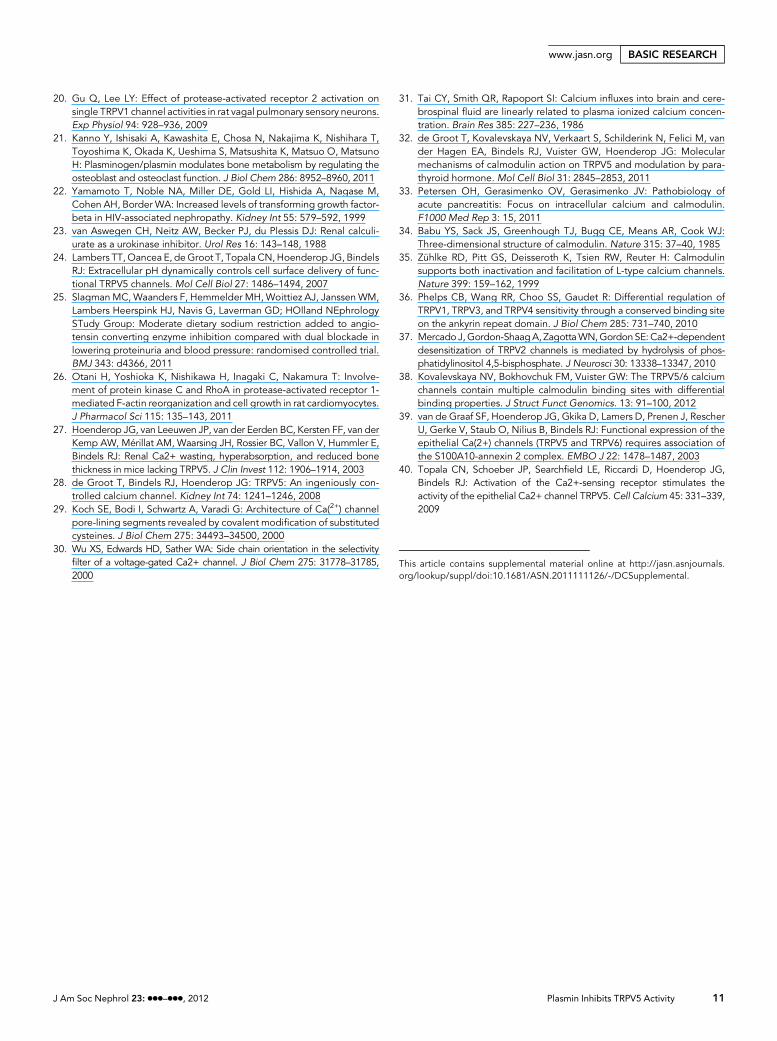
20. Gu Q, Lee LY: Effect of protease-activated receptor 2 activation onsingle TRPV1 channel activities in rat vagal pulmonary sensory neurons.Exp Physiol 94: 928–936, 2009
21. Kanno Y, Ishisaki A, Kawashita E, Chosa N, Nakajima K, Nishihara T,Toyoshima K, Okada K, Ueshima S, Matsushita K, Matsuo O, MatsunoH: Plasminogen/plasmin modulates bone metabolism by regulating theosteoblast and osteoclast function. J Biol Chem 286: 8952–8960, 2011
22. Yamamoto T, Noble NA, Miller DE, Gold LI, Hishida A, Nagase M,Cohen AH, Border WA: Increased levels of transforming growth factor-beta in HIV-associated nephropathy. Kidney Int 55: 579–592, 1999
23. van Aswegen CH, Neitz AW, Becker PJ, du Plessis DJ: Renal calculi-urate as a urokinase inhibitor. Urol Res 16: 143–148, 1988
24. Lambers TT,Oancea E, deGroot T, Topala CN,Hoenderop JG, BindelsRJ: Extracellular pH dynamically controls cell surface delivery of func-tional TRPV5 channels. Mol Cell Biol 27: 1486–1494, 2007
25. Slagman MC, Waanders F, Hemmelder MH, Woittiez AJ, Janssen WM,Lambers Heerspink HJ, Navis G, Laverman GD; HOlland NEphrologySTudy Group: Moderate dietary sodium restriction added to angio-tensin converting enzyme inhibition compared with dual blockade inlowering proteinuria and blood pressure: randomised controlled trial.BMJ 343: d4366, 2011
26. Otani H, Yoshioka K, Nishikawa H, Inagaki C, Nakamura T: Involve-ment of protein kinase C and RhoA in protease-activated receptor 1-mediated F-actin reorganization and cell growth in rat cardiomyocytes.J Pharmacol Sci 115: 135–143, 2011
27. Hoenderop JG, van Leeuwen JP, van der Eerden BC, Kersten FF, van derKemp AW, Mérillat AM, Waarsing JH, Rossier BC, Vallon V, Hummler E,Bindels RJ: Renal Ca2+ wasting, hyperabsorption, and reduced bonethickness in mice lacking TRPV5. J Clin Invest 112: 1906–1914, 2003
28. de Groot T, Bindels RJ, Hoenderop JG: TRPV5: An ingeniously con-trolled calcium channel. Kidney Int 74: 1241–1246, 2008
29. Koch SE, Bodi I, Schwartz A, Varadi G: Architecture of Ca(2+) channelpore-lining segments revealed by covalent modification of substitutedcysteines. J Biol Chem 275: 34493–34500, 2000
30. Wu XS, Edwards HD, Sather WA: Side chain orientation in the selectivityfilter of a voltage-gated Ca2+ channel. J Biol Chem 275: 31778–31785,2000
31. Tai CY, Smith QR, Rapoport SI: Calcium influxes into brain and cere-brospinal fluid are linearly related to plasma ionized calcium concen-tration. Brain Res 385: 227–236, 1986
32. de Groot T, Kovalevskaya NV, Verkaart S, Schilderink N, Felici M, vander Hagen EA, Bindels RJ, Vuister GW, Hoenderop JG: Molecularmechanisms of calmodulin action on TRPV5 and modulation by para-thyroid hormone. Mol Cell Biol 31: 2845–2853, 2011
33. Petersen OH, Gerasimenko OV, Gerasimenko JV: Pathobiology ofacute pancreatitis: Focus on intracellular calcium and calmodulin.F1000 Med Rep 3: 15, 2011
34. Babu YS, Sack JS, Greenhough TJ, Bugg CE, Means AR, Cook WJ:Three-dimensional structure of calmodulin. Nature 315: 37–40, 1985
35. Zühlke RD, Pitt GS, Deisseroth K, Tsien RW, Reuter H: Calmodulinsupports both inactivation and facilitation of L-type calcium channels.Nature 399: 159–162, 1999
36. Phelps CB, Wang RR, Choo SS, Gaudet R: Differential regulation ofTRPV1, TRPV3, and TRPV4 sensitivity through a conserved binding siteon the ankyrin repeat domain. J Biol Chem 285: 731–740, 2010
37. Mercado J, Gordon-ShaagA, ZagottaWN,GordonSE: Ca2+-dependentdesensitization of TRPV2 channels is mediated by hydrolysis of phos-phatidylinositol 4,5-bisphosphate. J Neurosci 30: 13338–13347, 2010
38. Kovalevskaya NV, Bokhovchuk FM, Vuister GW: The TRPV5/6 calciumchannels contain multiple calmodulin binding sites with differentialbinding properties. J Struct Funct Genomics. 13: 91–100, 2012
39. van de Graaf SF, Hoenderop JG, Gkika D, Lamers D, Prenen J, RescherU, Gerke V, Staub O, Nilius B, Bindels RJ: Functional expression of theepithelial Ca(2+) channels (TRPV5 and TRPV6) requires association ofthe S100A10-annexin 2 complex. EMBO J 22: 1478–1487, 2003
40. Topala CN, Schoeber JP, Searchfield LE, Riccardi D, Hoenderop JG,Bindels RJ: Activation of the Ca2+-sensing receptor stimulates theactivity of the epithelial Ca2+ channel TRPV5.Cell Calcium 45: 331–339,2009
This article contains supplemental material online at http://jasn.asnjournals.org/lookup/suppl/doi:10.1681/ASN.2011111126/-/DCSupplemental.
J Am Soc Nephrol 23: ccc–ccc, 2012 Plasmin Inhibits TRPV5 Activity 11
www.jasn.org BASIC RESEARCH