Experimental and Numerical Investigation of the Acoustic Response of Multi-slit Bunsen Burners
CLINICAL AND TRANSLATIONAL RESEARCH
Sirolimus and Proteinuria in Renal Transplant Patients:Evidence for a Dose-Dependent Effect on Slit
Diaphragm-Associated ProteinsGiovanni Stallone,1 Barbara Infante,1 Paola Pontrelli,1 Maddalena Gigante,1 Eustacchio Montemurno,1
Antonia Loverre,2 Michele Rossini,2 Francesco Paolo Schena,2 Giuseppe Grandaliano,2
and Loreto Gesualdo1
Background. The mechanisms underlying the development of proteinuria in renal-transplant recipients convertedfrom calcineurin inhibitors to sirolimus are still unknown.Methods. This is a single-center cohort study. One hundred ten kidney transplant recipients converted from calcineu-rin inhibitors to sirolimus in the period from September 2000 to December 2005 were included in the study. All patientsunderwent a graft biopsy before conversion (T0) and a second protocol biopsy 2 years thereafter (T2), according to ourstandard clinical protocol. On the basis of the changes observed in proteinuria between T0 and T2 (median 70%), thepatients were divided into two groups: group I (�70%) and group II (�70%). The authors blinded the sirolimus bloodtrough levels. We investigated in vivo the effects of sirolimus on nephrin, podocin, CD2ap, and actin protein expres-sion. Slit diaphragm (SD)-associated protein expressions were evaluated in T0 and T2 biopsies. The same analysiswas performed in cultured human podocytes treated with different doses of sirolimus (5, 10, 20, and50 ng/mL).Results. The SD protein expression in group II T2 biopsies was significantly reduced compared with the T0 biopsies andwith T2 group I biopsies. In addition, sirolimus blood trough levels directly and significantly correlated with the SDprotein expression at T2 graft biopsies. Group II patients presented significantly higher sirolimus blood levels thangroup I. In vitro study confirmed that sirolimus effect on podocytes was dose dependent.Conclusions. Our data suggest that sirolimus-induced proteinuria may be a dose-dependent effect of the drug on keypodocyte structures.
Keywords: Sirolimus, Slit diaphragm proteins, Renal allograft.
(Transplantation 2011;91: 997–1004)
Nephrotic range proteinuria was reported in 64% of renaltransplant recipients converted from a calcineurin in-
hibitor (CNI)-based to a sirolimus-based immunosuppres-sive regimen (1). The mechanism of this event is stillunknown. Several studies emphasized the role of podocytes inthe pathogenesis of proteinuria and glomerular damage (2, 3).
Podocytes are differentiated cells with a complex cyto-architecture. They have a voluminous cell body and interdig-itated foot processes, covering the glomerular basementmembrane, connected by an electron-dense structure, the slitdiaphragm (SD) (3). Proteinuria, the most common clinicalmanifestation of glomerular diseases, is invariably associatedwith podocyte foot-process effacement and flattening (3).The SD is a specialized cell-adhesion structure essential forglomerular ultrafiltration that differentiates from typicaljunctional complexes during development and share similar-
This work was supported by Ministero dell’Universita e della Ricerca Scien-tifica (PRIN 2005 granted to L.G.), the University of Foggia (ex 60%,2005, granted to L.G.), and by Ministero della Salute (Ricerca Finalizzata2006, granted to G.G.).
G.S. and B.I. equally contributed to this study.G.S., G.G., F.P.S., and L.G. received honorary fees for lectures and advisory
boards from Wyeth, Novartis, Astellas, and Roche; and L.G., G.G., andF.P.S. have received an unrestricted research grant from Wyeth, Astellas,Roche, and Novartis.
1 Section of Nephrology, Department of Biomedical Sciences, University ofFoggia, Italy.
2 Department of Emergency and Organ Transplantation, Nephrology,Dialysis and Transplantation Unit, University of Bari, Italy.
Address correspondence to: Giovanni Stallone, M.D., Division of Nephrol-ogy, Department of Biomedical Sciences, University of Foggia, VialeLuigi Pinto, 1, 71100 Foggia, Italy.
E-mail: [email protected] or [email protected]
G.S. and B.I. designed research and wrote the article; G.G. analyzed data andrevised the article; M.R. and A.L. performed histological analysis; E.M.and M.G. performed in vitro study; P.P. analyzed data and supervisedresults; and F.P. S. and L.G. critically revised the article.
Supplemental digital content is available for this article. Direct URLcitations appear in the printed text, and links to the digital files areprovided in the HTML text of this article on the journal’s Web site(www.transplantjournal.com).
Received November 23, 2010. Revision requested December 12, 2010.Accepted January 21, 2011.Copyright © 2011 by Lippincott Williams & WilkinsISSN 0041-1337/11/9109-997DOI: 10.1097/TP.0b013e318211d342
Transplantation • Volume 91, Number 9, May 15, 2011 www.transplantjournal.com | 997
ities with tight junctions (4). SD, like other adhering junc-tions, includes immunoglobulin (Ig) and cadherin superfam-ily cell-adhesion receptors. Nephrin and its homologueNeph1 are members of the Ig superfamily and have been sug-gested to form the framework of the SD through homophilicinteractions between podocytes (5– 8). Nephrin is crucial forpodocyte maturation and glomerular function, because itsmutation is associated with congenital Finnish type nephrotic(5, 9). Mutations in several other SD-associated proteins leadto the loss of podocyte foot-process architecture and the de-velopment of nephrotic syndrome (10 –14).
Recent studies suggest that nephrin, podocin, andCD2-associated protein (CD2ap) might participate in a com-mon cell-signaling pathway. Huber et al. (15) demonstratedthat both nephrin and CD2ap interact in vivo with thep85 regulatory subunit of phosphoinositide-3-OH-kinase(PI3K). PI3K is the first signaling protein demonstrated tointeract with the cytoplasmic surface of SD–protein complexin vivo. Nephrin and CD2ap recruit PI3K to the plasma mem-brane and, together with podocin, stimulate PI3K-dependentactivation of the serine-threonine kinase, AKT. These find-ings reveal a novel role for the SD proteins and demonstrate thatnephrin, CD2ap, and podocin, in addition to their structuralfunctions, initiate signal transduction in podocytes (15, 16).
Sirolimus is an immunosuppressive drug currently usedin the antirejection regimen in kidney transplantation (17).Sirolimus inhibits mammalian target of rapamycin (mTOR), aserine/threonine-kinase, lying immediately downstream ofAKT, which modulates proliferation and clonal expansion ofactivated T cells (18). In addition to its immunosuppressive ac-tions, sirolimus inhibits growth factor-mediated proliferation ofseveral nonimmune cells (19).
To date, there are no reports in vivo on the effect ofsirolimus on SD-associated protein function and structure.Moreover, little is known about the molecular mechanismsleading to proteinuria in renal transplant patients receivingsirolimus. Thus, our work focused on the effects of sirolimuson nephrin, podocin, CD2ap expression, and their interac-tion with the podocyte cytoskeleton, in the attempt to definethe influence of this drug on SD functions and whether theseeffects might be dose dependent.
RESULTS
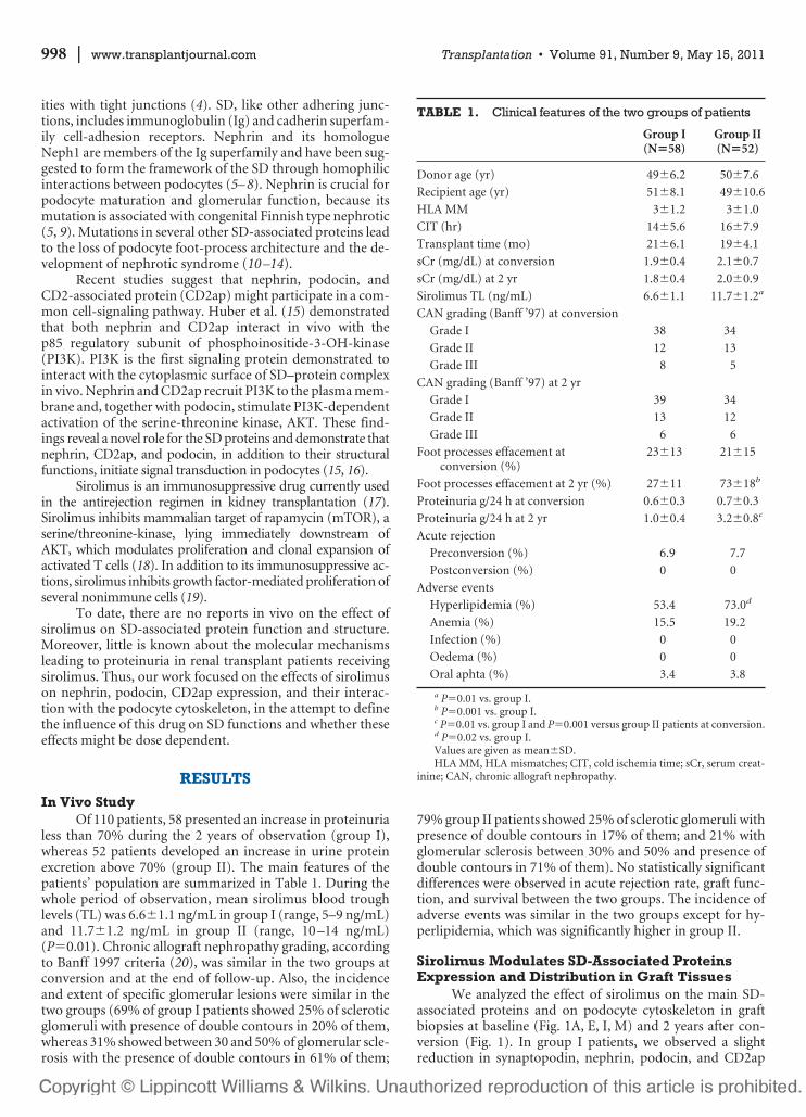
In Vivo StudyOf 110 patients, 58 presented an increase in proteinuria
less than 70% during the 2 years of observation (group I),whereas 52 patients developed an increase in urine proteinexcretion above 70% (group II). The main features of thepatients’ population are summarized in Table 1. During thewhole period of observation, mean sirolimus blood troughlevels (TL) was 6.6�1.1 ng/mL in group I (range, 5–9 ng/mL)and 11.7�1.2 ng/mL in group II (range, 10 –14 ng/mL)(P�0.01). Chronic allograft nephropathy grading, accordingto Banff 1997 criteria (20), was similar in the two groups atconversion and at the end of follow-up. Also, the incidenceand extent of specific glomerular lesions were similar in thetwo groups (69% of group I patients showed 25% of scleroticglomeruli with presence of double contours in 20% of them,whereas 31% showed between 30 and 50% of glomerular scle-rosis with the presence of double contours in 61% of them;
79% group II patients showed 25% of sclerotic glomeruli withpresence of double contours in 17% of them; and 21% withglomerular sclerosis between 30% and 50% and presence ofdouble contours in 71% of them). No statistically significantdifferences were observed in acute rejection rate, graft func-tion, and survival between the two groups. The incidence ofadverse events was similar in the two groups except for hy-perlipidemia, which was significantly higher in group II.
Sirolimus Modulates SD-Associated ProteinsExpression and Distribution in Graft Tissues
We analyzed the effect of sirolimus on the main SD-associated proteins and on podocyte cytoskeleton in graftbiopsies at baseline (Fig. 1A, E, I, M) and 2 years after con-version (Fig. 1). In group I patients, we observed a slightreduction in synaptopodin, nephrin, podocin, and CD2ap
TABLE 1. Clinical features of the two groups of patients
Group I(N�58)
Group II(N�52)
Donor age (yr) 49�6.2 50�7.6
Recipient age (yr) 51�8.1 49�10.6
HLA MM 3�1.2 3�1.0
CIT (hr) 14�5.6 16�7.9
Transplant time (mo) 21�6.1 19�4.1
sCr (mg/dL) at conversion 1.9�0.4 2.1�0.7
sCr (mg/dL) at 2 yr 1.8�0.4 2.0�0.9
Sirolimus TL (ng/mL) 6.6�1.1 11.7�1.2a
CAN grading (Banff ’97) at conversion
Grade I 38 34
Grade II 12 13
Grade III 8 5
CAN grading (Banff ’97) at 2 yr
Grade I 39 34
Grade II 13 12
Grade III 6 6
Foot processes effacement atconversion (%)
23�13 21�15
Foot processes effacement at 2 yr (%) 27�11 73�18b
Proteinuria g/24 h at conversion 0.6�0.3 0.7�0.3
Proteinuria g/24 h at 2 yr 1.0�0.4 3.2�0.8c
Acute rejection
Preconversion (%) 6.9 7.7
Postconversion (%) 0 0
Adverse events
Hyperlipidemia (%) 53.4 73.0d
Anemia (%) 15.5 19.2
Infection (%) 0 0
Oedema (%) 0 0
Oral aphta (%) 3.4 3.8
a P�0.01 vs. group I.b P�0.001 vs. group I.c P�0.01 vs. group I and P�0.001 versus group II patients at conversion.d P�0.02 vs. group I.Values are given as mean�SD.HLA MM, HLA mismatches; CIT, cold ischemia time; sCr, serum creat-
inine; CAN, chronic allograft nephropathy.
998 | www.transplantjournal.com Transplantation • Volume 91, Number 9, May 15, 2011

protein expressions when compared with baseline (Fig.1B, F, J, N). Interestingly, in group II, synaptopodin, neph-rin, podocin, and CD2ap protein expressions were reduced(Fig. 1C, G, K, O) compared with baseline. Specific signalquantification demonstrated that all the differences observedwere statistically significant (Fig. 1D, H, L, P).
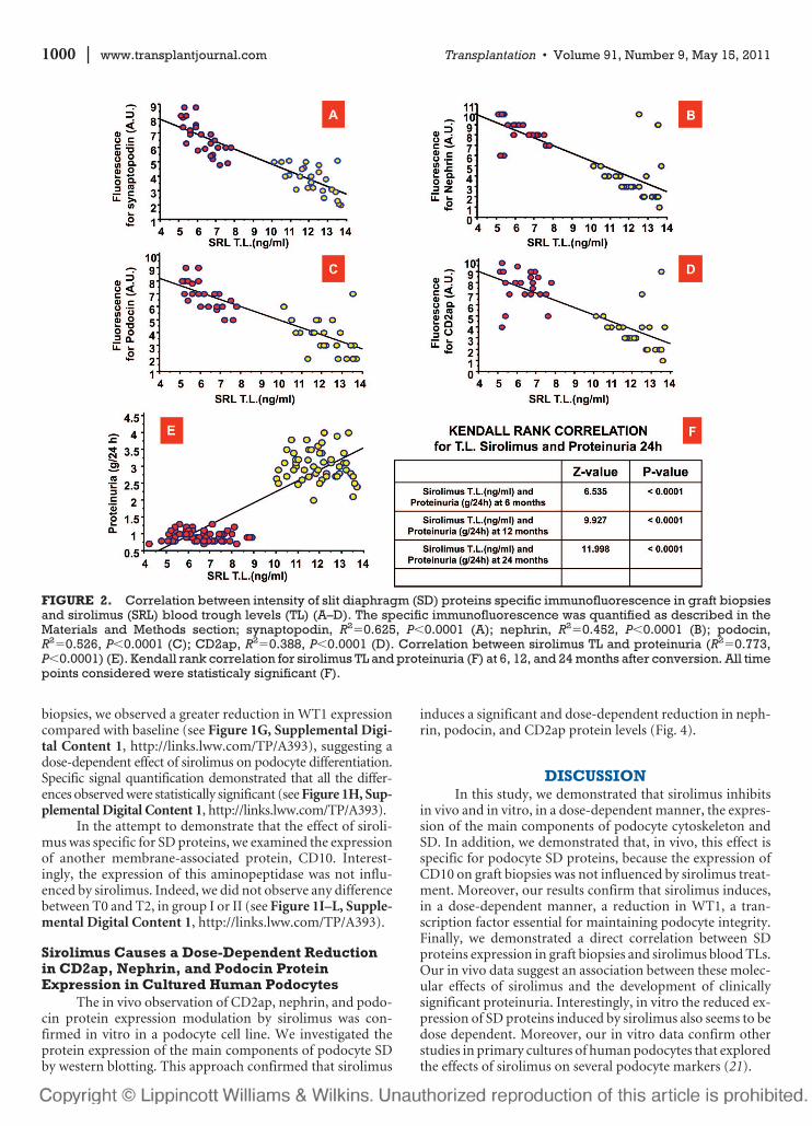
Interestingly, synaptopodin, nephrin, podocin, andCD2ap specific-immunofluorescence signals were inverselyand significantly correlated with sirolimus TL at the time ofsecond biopsy (R2�0.625, P�0.0001; R2�0.452, P�0.0001;R2�0.526, P�0.0001; R2�0.388, P�0.0001, respectively)(Fig. 2A–D). In addition, we observed a significant correlationbetween sirolimus TL and proteinuria (R2�0.597, P�0.0001;Fig. 2E). Finally, we performed a Kendall rank correlation forsirolimus TL and proteinuria (Fig. 2F) at 6, 12, and 24 monthafter conversion. At all time points, sirolimus TL was signifi-cantly associated with urine protein excretion rate (Fig. 2F).
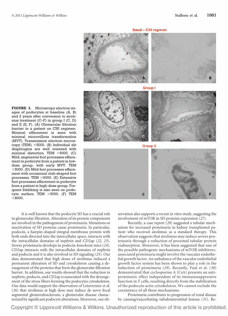
Electron microscopy study (Fig. 3) showed a differentgrade of podocyte damage in the two groups of patients. Ingroup I, we observed a mild, segmental foot processes efface-ment with occasional early microvillous transformation andclub-shaped foot processes (Fig. 3B–D). In group II, we noted
an extensive foot processes effacement, resembling those ob-served in minimal change nephropathy and idiopathic focalsegmental glomerulosclerosis (Fig. 3E–F). These podocyte al-terations were absent at baseline (Fig. 3A, B).
Sirolimus Inhibits p70S6 Kinase-Phosphorylationand WT1 Expression But Does Not ModulateCD10 Expression in Graft Biopsies
We analyzed the effect of sirolimus on phospho-p70S6-kinase expression on graft biopsies at baseline (see Figure 1A,Supplemental Digital Content 1, http://links.lww.com/TP/A393)and 2 years after conversion (see Figure 1B and C, Supple-mental Digital Content 1, http://links.lww.com/TP/A393).In both groups, we observed a reduction in p70S6 kinasephosphorylation. This reduction was significantly greater ingroup II patients (see Figure 1D, Supplemental Digital Con-tent 1, http://links.lww.com/TP/A393).
We, then, analyzed a marker of podocyte differentia-tion on graft tissue. WT1 expression in group I biopsies wasslightly reduced compared with baseline (see Figure 1Fand 1E, respectively, Supplemental Digital Content 1,http://links.lww.com/TP/A393). On the contrary, in group II
FIGURE 1. In vivo effect of sirolimus (SRL) on synaptopodin (A–D), nephrin (E–H), podocin (I–L), CD2ap (M–P) expres-sions and distribution on graft tissues at conversion (A, E, I, M) and 2 years thereafter (B, C, F, G, J, K, N, O) in patients fromgroup I (B, F, J, N) and group II (C, G, K, O). Specific immunofluorescence signal was quantified as described in Materialsand Methods section and expressed as arbitrary units/pixel (D, H, L, P). *P�0.004 group II vs. group I and P�0.0004 groupII vs. basal; #P�0.003 group II vs. group I and P�0.0005 group II vs. basal; §P�0.001 group II vs. group I and P�0.0002 groupII vs. basal; °P�0.005 group II vs. group I and P�0.0001 group II vs. basal. For this analysis, we evaluated 25 biopsies ofgroup I patients and 25 for group II patients.
© 2011 Lippincott Williams & Wilkins 999Stallone et al.
biopsies, we observed a greater reduction in WT1 expressioncompared with baseline (see Figure 1G, Supplemental Digi-tal Content 1, http://links.lww.com/TP/A393), suggesting adose-dependent effect of sirolimus on podocyte differentiation.Specific signal quantification demonstrated that all the differ-ences observed were statistically significant (see Figure 1H, Sup-plemental Digital Content 1, http://links.lww.com/TP/A393).
In the attempt to demonstrate that the effect of siroli-mus was specific for SD proteins, we examined the expressionof another membrane-associated protein, CD10. Interest-ingly, the expression of this aminopeptidase was not influ-enced by sirolimus. Indeed, we did not observe any differencebetween T0 and T2, in group I or II (see Figure 1I–L, Supple-mental Digital Content 1, http://links.lww.com/TP/A393).
Sirolimus Causes a Dose-Dependent Reductionin CD2ap, Nephrin, and Podocin ProteinExpression in Cultured Human Podocytes
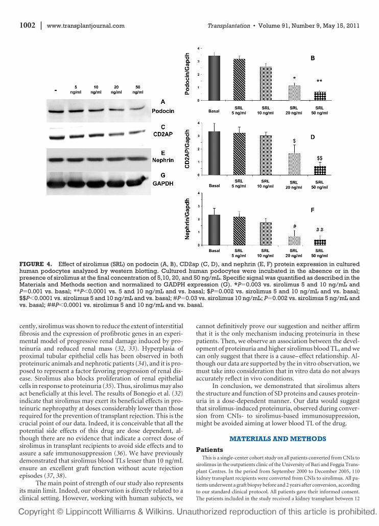
The in vivo observation of CD2ap, nephrin, and podo-cin protein expression modulation by sirolimus was con-firmed in vitro in a podocyte cell line. We investigated theprotein expression of the main components of podocyte SDby western blotting. This approach confirmed that sirolimus
induces a significant and dose-dependent reduction in neph-rin, podocin, and CD2ap protein levels (Fig. 4).
DISCUSSIONIn this study, we demonstrated that sirolimus inhibits
in vivo and in vitro, in a dose-dependent manner, the expres-sion of the main components of podocyte cytoskeleton andSD. In addition, we demonstrated that, in vivo, this effect isspecific for podocyte SD proteins, because the expression ofCD10 on graft biopsies was not influenced by sirolimus treat-ment. Moreover, our results confirm that sirolimus induces,in a dose-dependent manner, a reduction in WT1, a tran-scription factor essential for maintaining podocyte integrity.Finally, we demonstrated a direct correlation between SDproteins expression in graft biopsies and sirolimus blood TLs.Our in vivo data suggest an association between these molec-ular effects of sirolimus and the development of clinicallysignificant proteinuria. Interestingly, in vitro the reduced ex-pression of SD proteins induced by sirolimus also seems to bedose dependent. Moreover, our in vitro data confirm otherstudies in primary cultures of human podocytes that exploredthe effects of sirolimus on several podocyte markers (21).
FIGURE 2. Correlation between intensity of slit diaphragm (SD) proteins specific immunofluorescence in graft biopsiesand sirolimus (SRL) blood trough levels (TL) (A–D). The specific immunofluorescence was quantified as described in theMaterials and Methods section; synaptopodin, R2�0.625, P�0.0001 (A); nephrin, R2�0.452, P�0.0001 (B); podocin,R2�0.526, P�0.0001 (C); CD2ap, R2�0.388, P�0.0001 (D). Correlation between sirolimus TL and proteinuria (R2�0.773,P�0.0001) (E). Kendall rank correlation for sirolimus TL and proteinuria (F) at 6, 12, and 24 months after conversion. All timepoints considered were statisticaly significant (F).
1000 | www.transplantjournal.com Transplantation • Volume 91, Number 9, May 15, 2011
It is well known that the podocyte SD has a crucial rolein glomerular filtration. Alteration of its protein componentsare involved in the pathogenesis of proteinuria. Mutations orinactivation of SD proteins cause proteinuria. In particular,podocin, a hairpin-shaped integral membrane protein withboth ends directed into the intercellular space, interacts withthe intracellular domains of nephrin and CD2ap (22, 23).Severe proteinuria develops in podocin-knockout mice (24).CD2ap interacts with the intracellular domains of nephrinand podocin and it is also involved in SD signaling (25). Ourdata demonstrated that high doses of sirolimus induced apermanent alteration of SD and cytoskeleton causing a de-rangement of the proteins that form the glomerular filtrationbarrier. In addition, our results showed that the reduction innephrin, podocin, and CD2ap is associated with the derange-ment of the stress fibers forming the podocytes cytoskeleton.Our data would support the observation of Letavernier et al.(26) that sirolimus at high dose may induce de novo focalsegmental glomerulosclerosis, a glomerular disease charac-terized by significant podocyte alterations. Moreover, our ob-
servation also supports a recent in vitro study, suggesting theinvolvement of mTOR in SD proteins expression (27).
Recently, a case report (28) suggested a tubular mech-anism for increased proteinuria in kidney transplanted pa-tient who received sirolimus as a standard therapy. Thisobservation suggests that sirolimus may induce severe pro-teinuria through a reduction of proximal tubular proteinreabsorption. Moreover, it has been suggested that one ofthe possible pathogenic mechanisms of mTOR inhibitors-associated proteinuria might involve the vascular endothe-lial growth factor. An unbalance of the vascular endothelialgrowth factor system has been shown to play a role in theinduction of proteinuria (29). Recently, Faul et al. (30)demonstrated that cyclosporine A (CsA) presents an anti-proteinuric effect independent of its immunosuppressivefunction in T cells, resulting directly from the stabilizationof the podocyte actin cytoskeleton. We cannot exclude thecoexistence of all these mechanisms.
Proteinuria contributes to progression of renal diseaseby causing/exacerbating tubulointerstitial lesions (31). Re-
FIGURE 3. Microscopy electron im-ages of podocytes at baseline (A, B)and 2 years after conversion to siroli-mus treatment (C–F) in group I (C, D)and II (E, F). (A) Glomerular filtrationbarrier in a patient on CNI regimen.Minimal effacement is seen withminimal microvillous transformation(MVT). Transmission electron micros-copy (TEM) �5000. (B) Individual slitdiaphragms are well oriented withminimal distortion. TEM �9000. (C)Mild, segmental foot processes efface-ment in podocyte from a patient in low-dose group, with early MVT. TEM�5000. (D) Mild foot processes efface-ment with occasional club-shaped footprocesses. TEM �6000. (E) Extensivefoot processes effacement in podocytefrom a patient in high-dose group. Fre-quent blebbing is also seen on podo-cyte surface. TEM �5000. (F) TEM�8000.
© 2011 Lippincott Williams & Wilkins 1001Stallone et al.
cently, sirolimus was shown to reduce the extent of interstitialfibrosis and the expression of profibrotic genes in an experi-mental model of progressive renal damage induced by pro-teinuria and reduced renal mass (32, 33). Hyperplasia ofproximal tubular epithelial cells has been observed in bothproteinuric animals and nephrotic patients (34), and it is pro-posed to represent a factor favoring progression of renal dis-ease. Sirolimus also blocks proliferation of renal epithelialcells in response to proteinuria (35). Thus, sirolimus may alsoact beneficially at this level. The results of Bonegio et al. (32)indicate that sirolimus may exert its beneficial effects in pro-teinuric nephropathy at doses considerably lower than thoserequired for the prevention of transplant rejection. This is thecrucial point of our data. Indeed, it is conceivable that all thepotential side effects of this drug are dose dependent, al-though there are no evidence that indicate a correct dose ofsirolimus in transplant recipients to avoid side effects and toassure a safe immunosuppression (36). We have previouslydemonstrated that sirolimus blood TLs lesser than 10 ng/mLensure an excellent graft function without acute rejectionepisodes (37, 38).
The main point of strength of our study also representsits main limit. Indeed, our observation is directly related to aclinical setting. However, working with human subjects, we
cannot definitively prove our suggestion and neither affirmthat it is the only mechanism inducing proteinuria in thesepatients. Then, we observe an association between the devel-opment of proteinuria and higher sirolimus blood TL, and wecan only suggest that there is a cause– effect relationship. Al-though our data are supported by the in vitro observation, wemust take into consideration that in vitro data do not alwaysaccurately reflect in vivo conditions.
In conclusion, we demonstrated that sirolimus altersthe structure and function of SD proteins and causes protein-uria in a dose-dependent manner. Our data would suggestthat sirolimus-induced proteinuria, observed during conver-sion from CNIs- to sirolimus-based immunosuppression,might be avoided aiming at lower blood TL of the drug.
MATERIALS AND METHODS
PatientsThis is a single-center cohort study on all patients converted from CNIs to
sirolimus in the outpatients clinic of the University of Bari and Foggia Trans-plant Centres. In the period from September 2000 to December 2005, 110kidney transplant recipients were converted from CNIs to sirolimus. All pa-tients underwent a graft biopsy before and 2 years after conversion, accordingto our standard clinical protocol. All patients gave their informed consent.The patients included in the study received a kidney transplant between 12
FIGURE 4. Effect of sirolimus (SRL) on podocin (A, B), CD2ap (C, D), and nephrin (E, F) protein expression in culturedhuman podocytes analyzed by western blotting. Cultured human podocytes were incubated in the absence or in thepresence of sirolimus at the final concentration of 5,10, 20, and 50 ng/mL. Specific signal was quantified as described in theMaterials and Methods section and normalized to GADPH expression (G). *P�0.003 vs. sirolimus 5 and 10 ng/mL andP�0.001 vs. basal; **P�0.0001 vs. 5 and 10 ng/mL and vs. basal; $P�0.002 vs. sirolimus 5 and 10 ng/mL and vs. basal;$$P�0.0001 vs. sirolimus 5 and 10 ng/mL and vs. basal; #P�0.03 vs. sirolimus 10 ng/mL; P�0.002 vs. sirolimus 5 ng/mL andvs. basal; ##P�0.0001 vs. sirolimus 5 and 10 ng/mL and vs. basal.
1002 | www.transplantjournal.com Transplantation • Volume 91, Number 9, May 15, 2011
and 36 months before conversion, presented stable renal function, a PRA lessthan 20%, histologic diagnosis of chronic allograft nephropathy, accordingto the Banff 1997 criteria (20), a preconversion proteinuria less than 1 g/24 hrand received our standard immunosuppressive regimen, including cortico-steroids (prednisone), CsA (Neoral, Novartis), or tacrolimus (Prograf,Astellas), and mycophenolate mofetil (MMF; Cell-Cept, Roche). All patientsat the time of conversion received a standard dose of angiotensin-convertingenzyme inhibithors (ramipril 5 mg/day). The patients were then dividedaccording to the increase of proteinuria throughout the period of observa-tion. Specifically, the patients who showed an increase in proteinuria lessthan 70% (the median of the changes in proteinuria between pre- and post-treatment period) were included in group I. Group II included all patientswho presented an increase in proteinuria higher than 70%. Two authorsblinded to sirolimus TL performed the group assignment. To this purpose,proteinuria levels were measured three times over 1 month immediatelybefore and 2 years after conversion and the changes were calculated on themean of the three pre- and posttreatment determinations. The study wasapproved by our internal institutional review board.
Immunosuppressive Regimens
Before ConversionSeventy-four patients received 2.9 to 4.1 mg/kg per day of CsA in two
equally divided doses; 36 patients received 0.1 to 0.2 mg/kg per day of tacroli-mus in two equally divided doses; all patients received 500 mg of MMF twotimes per day; 5 mg/day of prednisone was administered to all patients.
After ConversionCNIs were abruptly withdrawn and sirolimus was introduced with an
initial loading dose of 0.1 mg/kg for the first day and a maintaining dose of0.02 to 0.04 mg/kg per day thereafter, aiming at blood TL of 6 to 14 ng/mL. Inall patients, the dose of MMF and prednisone was maintained.
Renal Allograft BiopsyRenal specimens were obtained by needle-core biopsies performed
under ultrasonographic guidance, fixed in 4% formaldehyde, and thenprocessed for routine histologic staining. Portions of cortex were fixed in2.5% glutaraldehyde in 0.1 M phosphate-buffered saline (PBS), pro-cessed, and embedded in Epon resin. Thin sections were examined usinga ZEISS910 transmission electron microscope (Zeiss, Arese, Italy). Semi-quantitative scoring for acute and chronic tubular, interstitial, vascular, andglomerular alterations was performed according to Banff 1997 criteria (20).To limit imprecision in score estimation, only biopsy specimens with at least8 to 10 glomeruli and at least one artery per section were considered asrepresentative. All biopsies satisfied listed criteria. Renal tissue for immuno-fluorescence, confocal laser scanning microscopy, immunohistochemistry,and electron microscopy studies was available for 25 patients/group. Exten-sion of foot processes effacement was semiquantitatively assessed on at least2 glomeruli/biopsy (avoiding partially/completely scarred glomeruli) andthe results were given averaging at least 5 capillary loops/glomerulus.
Tissue Immunofluorescence and Confocal LaserScanning Microscopy
Synaptopodin, Nephrin, CD2AP, Podocin, phospho-P70S6-kinase andCD10 protein expression and distribution in graft biopsies at conversion and2 years thereafter were evaluated by indirect immunofluorescence and con-focal microscopy analysis using specific antibodies. The sections were incu-bated for 1 hr in blocking buffer (2% bovine serum albumin, 0.5% fetalbovine serum) and then with the primary antibodies.
Synaptopodin was detected using a monoclonal antibody from Progen Bio-technik (Heidelberg, Germany) (ready to use, 1 hr at room temperature[RT]).Nephrin expression and distribution were evaluated using a rabbit poly-clonal antibody (39) (1:1500 dilution, overnight at 4°C). Podocin and CD2apexpressions were investigated using a respective monoclonal and goat poly-clonal antibody (Santa Cruz Biotechnology; 1:100 and 1:200 dilution, respec-tively, overnight at 4°C). Phospho-P70S6 kinase protein expression was
investigated on paraffin-embedded renal biopsies using a goat polyclonalantibody (Santa Cruz Biotechnology; 1:100 dilution, overnight at 4°C).CD10 was detected on paraffin-embedded renal sections using a monoclonalantibody from Abcam (Cambridge, MA; 1:50 dilution, 1 hr at RT).
The immune complexes were then identified incubating the slides withthe secondary antibodies Alexa-Fluor555 goat anti-mouse IgG-TRITC con-jugate (1:400 dilution), Alexa-Fluor488 rabbit anti-goat IgG-FITC conjugate(1:400 dilution), Alexa-Fluor488 goat anti-mouse IgG-FITC conjugate (1:200 dilution), and Alexa-Fluor goat anti-rabbit IgG-FITC conjugate (1:300dilution; Molecular Probes) for 1 hr at RT. Nuclei were stained withTO-PRO. The slides were then mounted in Gel/Mount (Biomeda, Milan,Italy) and sealed. Negative control was obtained incubating tissue sectionswith the blocking solution and then substituting the primary antibody with anonimmune poly-IgG serum. The specific fluorescence was analyzed by con-focal laser scanning microscopy using the Leica TCS SP2 (Leica, Wetzlar,Germany) equipped with argon– krypton (488 nm) and green–neon (543nm) lasers. Mean fluorescence was measured by confocal laser scanning mi-croscopy software and expressed as unit of fluorescence intensity/pixel.
ImmunohistochemistryWT1 protein expression was evaluated on paraffin-embedded kidney sec-
tions, using a specific mouse monoclonal anti-human WT1 antibody at 1:40dilution. Immobilized mouse antibodies were detected by the Avidin BiotinComplex method with the Vectastain Avidin Biotin Complex system, ac-cording to the manufacturer’s instructions (Vector, Burlingame, CA). Neg-ative controls were obtained by omitting the primary or secondary antibodiesand using nonimmune antiserum as first layer. The protein expression levelswere assessed semiquantitatively by our pathologist blinded to the origin ofthe slides.
Cell CultureAn immortalized human podocyte cell line, obtained by infection of pri-
mary glomerular epithelial cells with a hybrid Adeno5/SV40 virus (40), wasused for the experiments. The cell line was cultured in Dulbecco’s modifiedEagle’s medium containing 10% heat-inactivated fetal calf serum, 100 U/mLpenicillin, and 100 mg/mL streptomycin at 37°C with 5% CO2.
Western BlotPodocytes were plated in six-well dishes and grown to confluence. The
cells were starved for 24 hr and, then, exposed to sirolimus (Alexis Biochem-icals, San Diego, CA) for 48 hr at the final concentration of 5, 10, 20, or 50ng/mL. At the end of the treatment, the cell monolayer was rapidly rinsedtwice with ice-cold PBS and lysed in 100 �L of RIPA buffer (1 mM phenyl-methylsulfonyl fluoride, 5 mM EDTA, 1 mM sodium orthovanadate, 150mM NaCl, 8 �g/mL leupeptin, 1.5% Nonidet P-40, 20 mM Tris-HCl,pH7.4). The lysates were centrifuged at 10,000g at 4°C for 5 min. The super-natants were collected and aliquots containing 40 �g of proteins from eachlysate were subjected to SDS/PAGE and then electrotransferred onto nitro-cellulose membrane (Hybond C, Amersham, UK). The filter was blockedovernight at RT with 2% bovine serum albumin in PBS containing 0.1%Tween-20 (TBS) and then incubated with the goat polyclonal antinephrinantibody (Santa Cruz, 1:200 dilution, overnight at 4°C) or the monoclonalanti-CD2ap antibody (Santa Cruz, 1:50 dilution, overnight at 4°C) or thegoat polyclonal antipodocin antibody (Santa Cruz, 1:100 dilution, at RT for2 hr). The membranes were washed twice in TBS and incubated for 1 hr at RTwith HRP-conjugated goat anti-mouse-IgG or the rabbit anti-goat-IgG at1:1500 dilution in TBS, respectively. The membranes were washed three times atRT in TBS and then once with 0.1%SDS in PBS. The enhanced chemilumines-cence system (ECL, Amersham) was used for detection. The same membraneswere then stripped and immunoblotted again with rabbit polyclonalanti-GAPDH-antibody (Santa Cruz, 1:1000 dilution, overnight at 4°C).
Statistical AnalysisContinuous variables were expressed as mean�SD and compared by anal-
ysis of variance and paired Student’s t test, as appropriate. Association be-tween two continuous variables were investigated by Spearman and Kendal
© 2011 Lippincott Williams & Wilkins 1003Stallone et al.
rank correlations, as appropriate. A two-sided P less than 0.05 was consideredstatistically significant.
ACKNOWLEDGMENTSThe authors acknowledge Dr. Michelangelo Corcelli and
Mr. Vincenzo Gesualdo (both from the Department of Emer-gency and Organ Transplantation, University of Bari) for theirskilful technical help.
REFERENCES1. Izzedine H, Brocheriou I, Frances C. Post-transplantation proteinuria
and sirolimus. N Engl J Med 2005; 353: 2088.2. Somlo S, Mundel P. Getting a foothold in nephrotic syndrome. Nat
Genet 2000; 24: 333.3. Tryggvason K, Patrakka J, Wartiovaara J. Hereditary proteinuria syn-
dromes and mechanisms of proteinuria. N Engl J Med 2006; 354: 1387.4. Reiser J, Kriz W, Kretzler M, et al. The glomerular slit diaphragm is a
modified adherens junction. J Am Soc Nephrol 2000; 11: 1.5. Kestila M, Lenkkeri U, Mannikko M, et al. Positionally cloned gene for
a novel glomerular protein nephrin is mutated in congenital nephroticsyndrome. Mol Cell 1998; 1: 575.
6. Donoviel DB, Freed DD, Vogel H, et al. Proteinuria and perinatal le-thality in mice lacking NEPH1, a novel protein with homology toNEPHRIN. Mol Cell Biol 2001; 21: 4829.
7. Khoshnoodi J, Sigmundsson K, Ofverstedt LG, et al. Nephrin promotescell-cell adhesion through homophilic interactions. Am J Pathol 2003;163: 2337.
8. Wartiovaara J, Ofverstedt LG, Khoshnoodi J, et al. Nephrin strandscontribute to a porous slit diaphragm scaffold as revealed by electrontomography. J Clin Invest 2004; 114: 1475.
9. Done SC, Takemoto M, He L, et al. Nephrin is involved in podocytematuration but not survival during glomerular development. KidneyInt 2008; 73: 697.
10. Boute N, Gribouval O, Roselli S, et al. NPHS2, encoding the glomerularprotein podocin, is mutated in autosomal recessive steroid-resistantnephrotic syndrome. Nat Genet 2000; 24: 349.
11. Shih NY, Li J, Karpitskii V, et al. Congenital nephrotic syndrome inmice lacking CD2-associated protein. Science 1999; 286: 312.
12. Ciani L, Patel A, Allen ND, et al. Mice lacking the giant protocadherinmFAT1 exhibit renal slit junction abnormalities and a partially pene-trant cyclopia and anophthalmia phenotype. Mol Cell Biol 2003; 23:3575.
13. Winn MP, Conlon PJ, Lynn KL, et al. A mutation in the TRPC6 cautionchannel causes familial focal segmental glomerulosclerosis. Science2005; 308: 1801.
14. Reiser J, Polu KR, Moller CC, et al. TRPC6 is a glomerular slit dia-phragm associated channel required for normal renal function. NatGenet 2005; 37: 739.
15. Huber TB, Kottgen M, Shilling B, et al. Interaction with podocin facil-itates nephrin signaling. J Biol Chem 2001; 276: 41543.
16. Huber TB, Hartleben B, Kim J, et al. Nephrin and CD2AP associatewith phosphoinositide 3-OH kinase and stimulate AKT-dependent sig-naling. Mol Cell Biol 2003; 23: 4917.
17. Vasquez EM. Sirolimus: A new agent for prevention of renal allograftrejection. Am J Health Syst Pharm 2000; 57: 437.
18. Bierer BE, Mattila PS, Standaert RF, et al. Two distinct signal transmis-sion pathways in T lymphocytes are inhibited by complexes formedbetween an immunophilin and either FK506 or sirolimus. Proc NatlAcad Sci U S A 1990; 87: 9231.
19. Kim G, Jun JB, Elkon KB. Necessary role of phosphatidylinositol3-kinase in transforming growth factor beta-mediated activation of Aktin normal and rheumatoid arthritis synovial fibroblasts. ArthritisRheum 2002; 46: 1504.
20. Racusen LC, Solez K, Colvin RB, et al. Banff 97 working classification ofrenal allograft pathology. Kidney Int 1999; 55: 713.
21. Letavernier E, Bruneval P, Vandermeersch S, et al. Sirolimus interactswith pathways essential for podocyte integrity. Nephrol Dial Transplant2009; 24: 630.
22. Sellin L, Huber TB, Gerke P, et al. NEPH1 defines a novel family ofpodocin interacting proteins. FASEB J 2003; 17: 115.
23. Schwarz K, Simons M, Reiser J, et al. Podocin, a raft-associated com-ponent of the glomerular slit diaphragm, interacts with CD2AP andnephrin. J Clin Invest 2001; 108: 1621.
24. Roselli S, Heidet L, Sich M, et al. Early glomerular filtration defect andsevere renal disease in podocin-deficient mice. Mol Cell Biol 2004; 24: 550.
25. Schiffer M, Mundel P, Shaw AS, et al. A novel role for the adaptormolecule CD2-associated protein in transforming growth factor-beta-induced apoptosis. J Biol Chem 2004; 279: 37004.
26. Letavernier E, Bruneval P, Mandet C, et al. High sirolimus levels mayinduce focal segmental glomerulosclerosis de novo. Clin J Am SocNephrol 2007; 2: 326.
27. Vollenbroeker B, George B, Wolfgart M, et al. mTOR regulates expres-sion of slit diaphragm proteins and cytoskeleton structure in podo-cytes. Am J Physiol Renal Physiol 2009; 296: F418.
28. Straathof-Galema L, Wetzels JF, Dijkman HB, et al. Sirolimus-associated heavy proteinuria in a renal transplant recipient: Evidencefor a tubular mechanism. Am J Transplant 2006; 6: 429.
29. Diekmann F, Rovira J, Carreras J, et al. Mammalian target of sirolimusinhibition halts the progression of proteinuria in a rat model of reducedrenal mass. J Am Soc Nephrol 2007; 18: 2653.
30. Faul C, Donnelly M, Merscher-Gomez S, et al. The actin cytoscheletonof kidney podocytes is a direct target of the antiproteinuric effect ofcyclosporine A. Nat Med 2008; 14: 931.
31. Eddy A. Role of cellular infiltrates in response to proteinuria. Am JKidney Dis 2001; 37: 25.
32. Bonegio RGB, Fuhro R, Wang Z, et al. Sirolimus ameliorates proteinuria-associated tubulointerstitial inflammation and fibrosis in experimentalmembranous nephropathy. J Am Soc Nephrol 2005; 16: 2063.
33. Torras J, Herrero-Fresneda I, Gulias O, et al. Sirolimus has dual oppos-ing effects on proteinuric experimental nephropathies: Is it a matter ofpodocyte damage? Nephrol Dial Transplant 2009; 24: 3632.
34. Hebert LA, Agarwal G, Sedmak D, et al. Proximal tubular epithelialhyperplasia in patients with chronic glomerular proteinuria. Kidney Int2000; 57: 1962.
35. Pallet N, Thervet E, Le Corre D, et al. Sirolimus inhibits human renalepithelial cell proliferation: Effect on cyclin D3 mRNA expression andstability. Kidney Int 2005; 67: 2422.
36. Stallone G, Infante B, Grandaliano G, et al. Management of side effectsof sirolimus therapy. Transplantation 2009; 87: S23.
37. Stallone G, Infante B, Schena A, et al. Sirolimus for treatment ofchronic allograft nephropathy in renal transplant patients. J Am SocNephrol 2005; 16: 3755.
38. Stallone G, Schena A, Infante B, et al. Sirolimus for Kaposi’s sarcoma inrenal transplant patients. N Engl J Med 2005; 352: 1317.
39. Ruotsalainen V, Ljungberg P, Wartiovaara J, et al. Nephrin is specifi-cally located at the slit diaphragm of glomerular podocytes. Proc NatlAcad Sci USA 1999; 96: 7962.
40. Conaldi PG, Bottelli A, Baj A, et al. Human immunodeficiency virus-1tat induces hyperproliferation and dysregulation of renal glomerularepithelial cells. Am J Pathol 2002; 161: 53.
1004 | www.transplantjournal.com Transplantation • Volume 91, Number 9, May 15, 2011
Copyright © 2022 FDOKUMEN