Histopathology of Diaphragm Disease of the Small Intestine A ...
Upload
khangminh22Category
view
0download
0
REVIEW Open Access
Ventilator-induced diaphragm dysfunction:translational mechanisms lead totherapeutical alternatives in the critically illOscar Peñuelas1,2* , Elena Keough1, Lucía López-Rodríguez1, Demetrio Carriedo1, Gesly Gonçalves1,Esther Barreiro2,3,4 and José Ángel Lorente1,2,5
From The 3rd International Symposium on Acute Pulmonary Injury Translational Research, under the auspices of the: ‘IN-SPIRES®'Amsterdam, the Netherlands. 4-5 December 2018
* Correspondence: [email protected] Care Unit, HospitalUniversitario de Getafe, Carretera deToledo, km 12.5, 28905 Getafe,Madrid, Spain2Centro de Investigación en Red deEnfermedades Respiratorias[CIBERES], Instituto de Salud CarlosIII [ISCIII], Madrid, SpainFull list of author information isavailable at the end of the article
Abstract
Mechanical ventilation [MV] is a life-saving technique delivered to critically ill patientsincapable of adequately ventilating and/or oxygenating due to respiratory or otherdisease processes. This necessarily invasive support however could potentially resultin important iatrogenic complications. Even brief periods of MV may result indiaphragm weakness [i.e., ventilator-induced diaphragm dysfunction [VIDD]], whichmay be associated with difficulty weaning from the ventilator as well as mortality.This suggests that VIDD could potentially have a major impact on clinical practicethrough worse clinical outcomes and healthcare resource use. Recent translationalinvestigations have identified that VIDD is mainly characterized by alterationsresulting in a major decline of diaphragmatic contractile force together with atrophyof diaphragm muscle fibers. However, the signaling mechanisms responsible forVIDD have not been fully established. In this paper, we summarize the currentunderstanding of the pathophysiological pathways underlying VIDD and highlightthe diagnostic approach, as well as novel and experimental therapeutic options.
Keywords: Diaphragm dysfunction, Mechanical ventilation, Critically ill patient,Diaphragmatic fatigue, Respiratory muscles, Weaning failure
BackgroundInvasive mechanical ventilation (MV) is a life-saving procedure applied to critically ill
patients to achieve adequate pulmonary gas exchange and unload excessive respiratory
muscle work. Observational studies report that the use of mechanical ventilation has
led to a decrease in the mortality rate of critically ill patients [1–3].
Nevertheless, and based on the advances in the understanding that mechanical venti-
lation itself can cause and potentiate lung injury [4, 5], translational research has focused
on ventilatory strategies and adjunctive measures aimed at mitigating this so-called
ventilator-induced lung injury (VILI) [6].
Recently, a similar concern has emerged with regard to the potential adverse effects
of invasive mechanical ventilation on the respiratory muscles. This entity was originally
© The Author(s). 2019 Open Access This article is distributed under the terms of the Creative Commons Attribution 4.0 InternationalLicense (http://creativecommons.org/licenses/by/4.0/), which permits unrestricted use, distribution, and reproduction in any medium,provided you give appropriate credit to the original author(s) and the source, provide a link to the Creative Commons license, andindicate if changes were made.
Intensive Care MedicineExperimental
Peñuelas et al. Intensive Care Medicine Experimental 2019, 7(Suppl 1):48https://doi.org/10.1186/s40635-019-0259-9

termed ventilator-induced diaphragmatic dysfunction (VIDD) [7], although it may also
involve other respiratory muscles. Both animal [8, 9] and recent human [10, 11] studies
have demonstrated that complete diaphragm muscle unloading or inactivity during
invasive MV induces a rapid and profound loss of diaphragm muscle force-generating
capacity. In 2008, it was [12] demonstrated that VIDD occurs in critically ill patients
and is characterized by marked diaphragm atrophy of both slow-twitch and fast-twitch
fibers, with evidence of oxidative stress in addition to proteolysis.
It is well known that muscle dysfunction in critically ill patients defined as ICU-
acquired weakness is associated with weaning failure and unfavorable outcomes in
critically ill patients [13, 14]. However, it remains unclear whether the specific changes
in the diaphragm caused by mechanical ventilation significantly impact clinical out-
comes. A recent study [15] confirmed the results of prospective studies that associated
VIDD with difficulty weaning and poor clinical outcomes. The authors also found that
the development of a decreased diaphragm thickness was associated with a lower daily
probability of liberation from ventilation adjusted hazard ratio (0.69; 95% confidence
interval (CI), 0.54–0.87; per 10% decrease), prolonged ICU admission (adjusted
duration ratio, 1.71; 95% CI, 1.29–2.27), and a higher risk of complications (adjusted
odds ratio, 3.00; 95% CI, 1.34–6.72).
Currently, VIDD represents one of the most challenging fields within translational
research applied to critically ill patients. This review provides the knowledge necessary
for intensivists to better understand the signaling mechanisms responsible for VIDD, as
well as summarizes the development of diagnostic bedside tools. Based on these
findings, future therapeutical approaches may have the ability to prevent VIDD and to
improve weaning success as well as other relevant clinical outcomes.
Epidemiology of diaphragm dysfunction in mechanically ventilated patientsMultiple recent studies have shown that VIDD is reported in up to 53% of mechanically
ventilated patients within 24 h of intubation. An additional 26% may develop VIDD
while on mechanical ventilation during their stay in the intensive care unit (ICU).
In the literature, the variations observed in the incidence of this complication are
based fundamentally on the diagnostic tool used. The assessment of the transdiaphrag-
matic twitch pressure (PdiTw) generated in response to bilateral anterior magnetic
phrenic nerve stimulation (BAMPS) is an objective, nonvolitional measurement which
represents the gold standard technique used to specifically assess diaphragm strength.
According to several observational studies, mechanically ventilated patients in the ICU,
on average, generate a PdiTw that is only 20% of normal [16]. The use of ultrasono-
graphy as a diagnostic tool is easier in everyday practice when compared to performing
a PdiTw, and it is becoming an increasingly popular alternative method for the diagno-
sis of VIDD [17]. Additional studies have confirmed that around 60 to 80% of mechan-
ically ventilated patients manifest clinically significant diaphragm dysfunction as
evaluated by bedside diaphragmatic ultrasound [18–20]. Overall, a recent study
indicates that diaphragm weakness is present twice as often as limb weakness in
critically ill patients [21, 22].
The potential improvement in the outcome that might be obtained by implementing
prevention or therapeutic strategies for diaphragm atrophy caused by ventilation is
therefore also uncertain.
Peñuelas et al. Intensive Care Medicine Experimental 2019, 7(Suppl 1):48 Page 2 of 25

Pathogenic mechanisms underlying ventilator-induced diaphragmdysfunctionMost studies show that VIDD occurs in a progressive, time-dependent manner
[11], although its degree is influenced by different MV modes and other clinical
variables [23, 24].
The molecular mechanisms underlying VIDD currently represents one of the more
attractive fields of translational research in critically ill patients. We summarize some
of the key discoveries that may provide a unified mechanistic framework for under-
standing the pathogenesis of VIDD (Fig. 1).
The mechanical properties of diaphragm muscle fibers are characterized by the
relationship between cytosolic-free calcium, cross-bridge attachment/cross-bridge
cycling rate, and sarcomere length [25]. Any factor that influences one or more of these
variables can impact diaphragm muscle force generation.
Nonetheless, it seems likely that the cause of MV-induced diaphragm contractile
dysfunction is multiplicative and includes oxidative modifications to contractile
proteins resulting in depressed fiber sensitivity to calcium, protease activation leading
to sarcomere disruption, and a loss of myosin heavy chain protein.
Mitochondrial oxidative stress (MOS)
Overall, we can consider that VIDD is characterized by a disbalance in protein homeosta-
sis defined as a decrease in diaphragm protein synthesis and an increase in diaphragm
protein degradation. The activation of catabolic pathways, induced in the diaphragm
under mechanical ventilation, occurs in the short-term under controlled modes of
Fig. 1 Summary of the current understanding of the molecular pathways contributing to ventilator-induceddiaphragm dysfunction (VIDD) in critically ill patients. As shown, different conditions can lead to diaphragmatrophy via an imbalance between proteolysis and protein synthesis [11, 14], whereas remaining muscleproteins may be impaired by enhanced oxidation and dephosphorylation [15–17]. Inflammation andoxidative stress are proposed to be the major drivers of these impairments [17]. In addition, certain drugscan impair neural drive and excitation-contraction coupling
Peñuelas et al. Intensive Care Medicine Experimental 2019, 7(Suppl 1):48 Page 3 of 25

mechanical ventilation [26]. This appears to be related to an upregulation of the oxidative
stress-mediated ubiquitin–proteasome pathway that would lead to an increase in protein
degradation and thus atrophy.
Therefore, mitochondrial oxidative stress (MOS) may play an integral role in
MV-induced atrophy of the diaphragm by inducing catabolism [27, 28].
The force component of VIDD can also be affected by MOS in the following two
ways: firstly, by producing free radicals that can induce a post-translational modifi-
cation of muscle proteins [e.g., oxidation] and, hence, alter their structure and function,
specifically reducing the calcium sensitivity of myofilaments [29], and secondly, MOS
can cause a metabolic switch, by reducing mitochondrial oxidative phosphorylation and
increasing glycolysis [30]. The lipid accumulation seen in diaphragms under mechanical
ventilation [31] could trigger important metabolic pathways as an accelerated glycolysis.
This allows for more intermediate metabolites to be converted into fatty acids and so
the decreased breakdown of fatty acids due to altered mitochondrial function. In fact,
lipotoxicity could serve as a stimulus to VIDD, but further studies are required to
determine if these are the triggers or the consequence of MOS.
Autophagy
Autophagy is a catabolic process that degrades cytosolic proteins and organelles using
lysosomal proteases. More specifically, lysosomal proteases (i.e., cathepsins) have the
biological function of removing both organelles and nonmyofibril cytosolic protein
aggregates [32]. During autophagy, targeted cytosolic components are encapsulated into
a double-membrane vacuole called the autophagosome. When complete, the auto-
phagosome moves through the cytosol to the lysosome where the contents of the
autophagosome are degraded.
A recent study found that prolonged mechanical ventilation increases the expression
of key autophagy proteins [e.g., ATG5, ATG7, and beclin 1] and the number of auto-
phagosomes in the human diaphragm [33]. Furthermore, emerging evidence reveals an
increase in autophagy biomarkers in rodent diaphragms following 8–18 h of controlled
mechanical ventilation [34, 35].
However, it is not clear whether autophagy is an effector of VIDD, or a protective
mechanism that may actually offset VIDD [36, 37]. Critically ill patients have been
shown to have both impaired autophagic vacuole formation and mitophagy in their
skeletal muscles, resulting in a reduced clearance of proteins that result from cellular
damage [38]. On the other hand, several experimental studies have shown a reduction
of VIDD by enhanced autophagic pathways. These include fragmentation of intermyo-
fibrillar mitochondria as an early event in a mouse model of VIDD [39], and stimulation
of autophagy with n-acetylcysteine or rapamycin [40]. Mechanical ventilation-associated
increases in autophagy may actually help to clear cells of dysfunctional mitochondria and
thereby improve muscle function, as has been shown to be the case in other murine
models of muscle disease [41].
Calpains
Calpains are cysteine proteases that promote muscle atrophy by cleaving over 100 dif-
ferent cellular proteins, and the two ubiquitous calpains located in skeletal muscle are
Peñuelas et al. Intensive Care Medicine Experimental 2019, 7(Suppl 1):48 Page 4 of 25

calpain I and II [42]. Numerous studies reveal that calpain cleavage of Z line-associated
proteins (i.e., titin and nebulin) is responsible for the release of myofilament proteins
that are then degraded by the ubiquitin-proteasome system and perhaps other
proteases as well [37, 43].
Importantly, it has been established that prolonged mechanical ventilation activates
calpain in the diaphragms of both humans and animals [44]. In fact, recent experimen-
tal studies revealed that pharmacological inhibition of calpain activation could partially
protect against VIDD [45].
Specific diaphragm muscle proteins have been identified that are degraded during
mechanical ventilation-induced sarcomere damage, concretely both titin and myosin. A
recent work found a significant reduction in rat diaphragm single muscle fiber force
production after 18 h of mechanical ventilation, which was accompanied by a pro-
portional loss of myosin heavy chain concentration together with a titin dysfunc-
tion in diaphragm fibers [46]. Interestingly, these findings were not found in other
peripheral muscles.
Another possible link between mechanical ventilation-induced oxidative stress and
diaphragm contractile dysfunction is the connection between reactive oxygen species
(ROS) and calpain activation. MV-induced ROS production in diaphragm muscle
promotes calpain activation [47, 48]; in turn, active calpain can degrade key cytoskeletal
proteins involved in maintaining sarcomere structure. This results in sarcomere dis-
ruption, which impairs the muscle’s ability to generate force.
Finally, recent studies have shown that regulatory cross-talk exists between calpain
and caspase-3 in the diaphragm during controlled MV in rats. Caspase-3 belongs to a
large family of cysteine proteases and plays an important role in apoptosis. This pro-
tease may also contribute to muscle protein degradation during a variety of muscle
wasting conditions.
There is a body of research demonstrating that full support MV activates caspase-3
in both the rodent and human diaphragm and induces myonuclear apoptosis in the
diaphragm [25, 44, 49, 50].
The ubiquitin-proteasome system of proteolysis plays an important role in muscle
protein catabolic pathways during a variety of wasting conditions [51]. Recent ex-
perimental studies have demonstrated that prolonged and controlled mechanical
ventilation may increase the activation of the ubiquitin-proteasome system of proteolysis
in both human and rat diaphragms [52, 53]. Studies on the effects of partial support or
assisted mechanical ventilation on protease activation in humans remain unpublished.
Ventilator-induced impairment of diaphragmatic contractile function
Both experimental and human studies have shown that prolonged mechanical ventilation
induces diaphragm contractile dysfunction [11]. In an animal study, the investigators
found a depression of in vivo diaphragm contractility at 5 days of mechanical ventilation
in piglets [54]. Several experimental studies consistently report that invasive mechanical
ventilation results in a rapid and time-dependent decrease in diaphragmatic force pro-
duction measured in vitro through the electrical stimulation of the diaphragm [55–58]. A
recent murine model showed diaphragm contractile dysfunction after 6 h of ventilatory
support [59]. The ventilator-induced diaphragmatic contractile dysfunction in animals
Peñuelas et al. Intensive Care Medicine Experimental 2019, 7(Suppl 1):48 Page 5 of 25

may be directly associated to diaphragm contractile inactivity since partial ventilator
modes or short periods of intermittent spontaneous breathing can reduce the magnitude
of full ventilator support-induced contractile dysfunction [9, 36]. In this respect, there are
few experimental studies focused on the impact of prolonged mechanical ventilation on
diaphragm weakness in animals [60–64].
Risk factors for acquiring VIDDIn critically ill patients, some factors have been identified that along with prolonged mechan-
ical ventilation adversely impact diaphragm contractile function including senescence [65, 66],
intravenous medications such as neuromuscular blockers [67], and/or glucocorticoids [68].
These studies however present divergent findings, and further research is needed to
address the effect of treatments on diaphragm function in animals using both partial
and controlled mechanical ventilation.
Although human information is limited, age-related respiratory muscle dysfunction
resulting in a decrease in strength has been demonstrated, with the fall of 0.8 to
2.7 cm H2O per year from the maximal inspiratory pressure (MIP) between the
ages of 65 and 85 years [67]. A 25% drop in transdiaphragmatic pressure in adults
between 65 and 75 years of age has also been described [69, 70].
One mechanism that explains these changes is the cumulative effect of active oxygen
radicals, which can trigger proteolytic processes. It has also been suggested that with
increasing age, there is a remodeling of the muscle fibers in which the fast myosin
fibers are replaced by slow type isoforms [71].
The mechanisms involved in the deleterious effect of mechanical ventilation on dia-
phragmatic dysfunction are not fully elucidated. Recently, a translational study found
that invasive mechanical ventilation with positive end-expiratory pressure (PEEP) re-
sults in longitudinal atrophy of the diaphragm fibers that is modulated by the elasticity
of the titin of giant sarcomeric protein [72].
Hypercapnia, on the other hand, is more clearly protective against VIDD. In this re-
gard, piglets ventilated with increased dead space to achieve moderate hypercapnic
acidosis [PaCO2 55–70 mmHg] showed preserved diaphragmatic force production after
72 h of mechanical ventilation [73]. A similar level of hypercapnia induced by adding
inspired CO2 also attenuated several aspects of VIDD in rats [74].
The cellular mechanisms underlying the improvements associated with hypercapnia are yet
to be fully understood but likely involve anti-inflammatory and/or antioxidant effects. VIDD
however does not appear to be correlated with ventilator-induced lung injury [60, 75, 76].
The majority of VIDD seen in critically ill patients, however, does not appear to be
the consequence of any easily treatable conditions. In many cases, the potential mecha-
nisms underlying diaphragmatic injury are induced by mechanical ventilation. There is
also strong evidence that processes other than VIDD, including sepsis and other
systemic infections, are responsible for various forms of diaphragmatic myotrauma.
Diagnostic approach for VIDDTests of respiratory muscle strength
Accurate evaluation of diaphragmatic contractile function in the setting of critically ill pa-
tients undergoing mechanical ventilation continues to be difficult due to multiple factors.
Peñuelas et al. Intensive Care Medicine Experimental 2019, 7(Suppl 1):48 Page 6 of 25
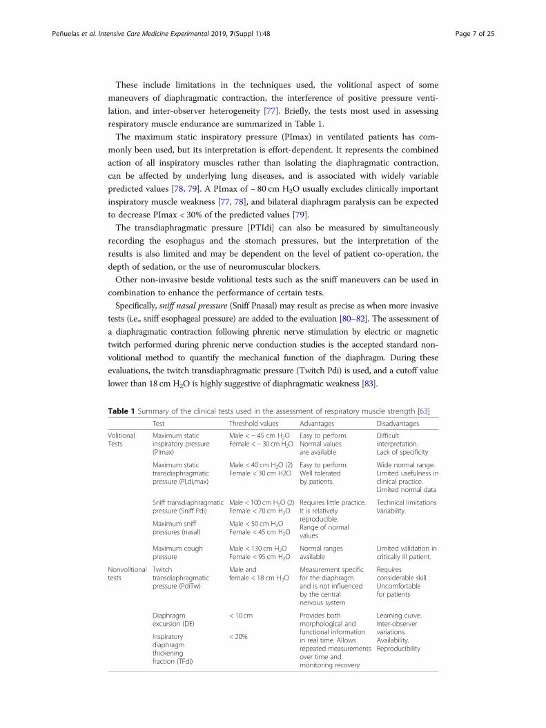
These include limitations in the techniques used, the volitional aspect of some
maneuvers of diaphragmatic contraction, the interference of positive pressure venti-
lation, and inter-observer heterogeneity [77]. Briefly, the tests most used in assessing
respiratory muscle endurance are summarized in Table 1.
The maximum static inspiratory pressure (PImax) in ventilated patients has com-
monly been used, but its interpretation is effort-dependent. It represents the combined
action of all inspiratory muscles rather than isolating the diaphragmatic contraction,
can be affected by underlying lung diseases, and is associated with widely variable
predicted values [78, 79]. A PImax of − 80 cm H2O usually excludes clinically important
inspiratory muscle weakness [77, 78], and bilateral diaphragm paralysis can be expected
to decrease PImax < 30% of the predicted values [79].
The transdiaphragmatic pressure [PTIdi] can also be measured by simultaneously
recording the esophagus and the stomach pressures, but the interpretation of the
results is also limited and may be dependent on the level of patient co-operation, the
depth of sedation, or the use of neuromuscular blockers.
Other non-invasive beside volitional tests such as the sniff maneuvers can be used in
combination to enhance the performance of certain tests.
Specifically, sniff nasal pressure (Sniff Pnasal) may result as precise as when more invasive
tests (i.e., sniff esophageal pressure) are added to the evaluation [80–82]. The assessment of
a diaphragmatic contraction following phrenic nerve stimulation by electric or magnetic
twitch performed during phrenic nerve conduction studies is the accepted standard non-
volitional method to quantify the mechanical function of the diaphragm. During these
evaluations, the twitch transdiaphragmatic pressure (Twitch Pdi) is used, and a cutoff value
lower than 18 cm H2O is highly suggestive of diaphragmatic weakness [83].
Table 1 Summary of the clinical tests used in the assessment of respiratory muscle strength [63]
Test Threshold values Advantages Disadvantages
VolitionalTests
Maximum staticinspiratory pressure(PImax)
Male < − 45 cm H2OFemale <− 30 cm H2O
Easy to perform.Normal valuesare available
Difficultinterpretation.Lack of specificity
Maximum statictransdiaphragmaticpressure (PI,di,max)
Male < 40 cm H2O (2)Female < 30 cm H2O
Easy to perform.Well toleratedby patients.
Wide normal range.Limited usefulness inclinical practice.Limited normal data
Sniff transdiaphragmaticpressure (Sniff Pdi)
Male < 100 cm H2O (2)Female < 70 cm H2O
Requires little practice.It is relativelyreproducible.Range of normalvalues
Technical limitationsVariability.
Maximum sniffpressures (nasal)
Male < 50 cm H2OFemale < 45 cm H2O
Maximum coughpressure
Male < 130 cm H2OFemale < 95 cm H2O
Normal rangesavailable
Limited validation incritically ill patient.
Nonvolitionaltests
Twitchtransdiaphragmaticpressure (PdiTw)
Male andfemale < 18 cm H2O
Measurement specificfor the diaphragmand is not influencedby the centralnervous system
Requiresconsiderable skill.Uncomfortablefor patients
Diaphragmexcursion (DE)
< 10 cm Provides bothmorphological andfunctional informationin real time. Allowsrepeated measurementsover time andmonitoring recovery
Learning curve.Inter-observervariations.Availability.Reproducibility
Inspiratorydiaphragmthickeningfraction (TFdi)
< 20%
Peñuelas et al. Intensive Care Medicine Experimental 2019, 7(Suppl 1):48 Page 7 of 25

However, these techniques depend to a great extent on the effort of the patient in
addition to other factors. For instance, these maneuvers require experience and special-
ized equipment as well as access to this equipment in routine ICU clinical practice. The
capacity to predict clinical outcomes, such as weaning failure, with the use of any of the
abovementioned tests of inspiratory, specific diaphragm, or expiratory muscle strength, is
broadly similar to any other test [84].
Nevertheless, a combination of tests can substantially increase the diagnostic
accuracy [85–88].
Diaphragmatic contraction can be assessed using magnetic resonance imaging and
fluoroscopy, but the problems associated with ionizing radiation, patient transport, and
high cost are factors limiting daily bedside use [89, 90].
Ultrasonography
In recent years, diaphragmatic ultrasound has become the most useful bedside for the
clinician to identify patients with VIDD subjected to invasive mechanical ventilation.
This is most likely because it has been shown to be safe and easy to perform, while
allowing both morphologic assessment and functional evaluation of the muscle with
high inter-observer agreement [91, 92]. It is also possible to perform a follow-up of the
initial clinical evaluation, providing information on time course and recovery.
There are two main ultrasonography parameters used to assess diaphragmatic function.
These include the dynamic evaluation of diaphragmatic excursion (DE) and the inspi-
ratory diaphragm thickening fraction (TFdi). Diaphragmatic excursion (DE) can be easily
measured with a 3–5-MHz probe in M-mode and represents the mobility of the
diaphragm in inspiration and expiration during a spontaneous mode of mechanical venti-
lation. However, DE depends on the amount of ventilator support and PEEP; accordingly,
a recent study indicated that DE should not be used to assess diaphragmatic contractility
in patients receiving mechanical ventilation [93, 94].
The second parameter, TFdi, measures muscle thickening in the zone of apposition of
the diaphragm to the rib cage with a 10–15MHz probe in B or M mode.
The transducer is placed in the same way as for the measurement of the dia-
phragmatic excursion. Following placement, measurements of the diaphragmatic thick-
ness in both inspiration and expiration are taken (Fig. 2).
TFdi is defined as [[thickness at end-inspiration − thickness at end-expiration]/thick-
ness at end-expiration]. The most accepted cutoff values are 10–14mm for DE and
30–36% for TFdi [95].
Both DE and TFdi have been shown to correlate with functional measurements of
diaphragmatic function in spontaneously breathing patients, but the use of ultrasound
in the process of weaning remains controversial [96, 97].
Several clinical studies suggest that these ultrasound parameters can be reliable
predictors of weaning and extubation outcomes. Indeed, a recent study [98] found
a prevalence of VIDD of 29% (by DE < 10 mm or paradoxical movements of the
diaphragm). Additionally, patients diagnosed with VIDD had a longer duration of
mechanical ventilation.
On the other hand, a study [96] reported in 63 adult critically ill patients with me-
chanical ventilation that a TFdi ≥ 30% presented a sensitivity of 88% and a specificity of
Peñuelas et al. Intensive Care Medicine Experimental 2019, 7(Suppl 1):48 Page 8 of 25

71% for extubation success, with a positive predictive value of 91% and a negative value of
63%, with an area under the curve of 0.79. In combination, these two methods of assess-
ment of diaphragmatic function during spontaneous breathing are useful tools for the
determination of patients at risk of invasive mechanical ventilation withdrawal failure.
Recently, a systematic review that included 13 studies on 742 adult critically ill
patients found that ultrasound had excellent intra- and inter-observer reproducibility,
as well as higher accuracy in investigating diaphragm dysfunction compared with other
weaning indexes or fluoroscopy [99]. Finally, another metaanalysis that included
nineteen studies (1071 participants) found that ultrasound may help predict weaning
outcome, but its accuracy and interpretation involve several pitfalls [100]. Therefore,
ultrasound is emerging as a new noninvasive tool used to monitor respiratory workload
during assisted mechanical ventilation and to assess the progression of VIDD. Never-
theless, further randomized clinical trials are needed to determine whether ultrasound
could guide clinical decisions for improving outcomes in critically ill patients.
A novel field of research is the implementation of tissue Doppler as a diagnostic tool
in the analysis of diaphragm function. Tissue Doppler imaging (PW-TDI, pulse wave
tissue Doppler imaging) is an echocardiographic technique and promises a number of
applications to further extend the unique diagnostic role in echocardiography. In
particular relation to the diaphragm, it might allow the assessment of regional dia-
phragmatic contractile function at rest and with stress. It should also provide a reliable
identification of viable respiratory contractile muscle. There is currently a French
clinical trial which aims to look for correlations between the time movement mode and
tissue Doppler in the assessment of a threshold value of diaphragm mobility in patients
in the postoperative period following scheduled cardiac surgery (ClinicalTrials.gov
Identifier: NCT03295344).
Diaphragm electromyography (EMGdi) reflects the muscle electrical activity, and it is
considered the gold standard in the assessment of neural respiratory drive.
Electromyography (EMG) comprises the temporal and spatial summation of neural
impulses from the brain that are translated into muscle fiber action potentials. There
are different approaches to obtaining a diaphragm EMG, and at the bedside, it can be
recorded using an esophageal catheter with multiple electrodes [101].
Fig. 2 Representative ultrasound image at the zone of apposition in B-mode view of the diaphragm duringinspiration (a) and expiration (b). The diaphragm is identified as a 3-layer comprising two hyperechoic linesrepresenting the pleural and peritoneal membranes and a middle hypoechoic layer representing thediaphragmatic muscle itself (with permission of the Intensive Care Unit from the Hospital Universitariode Getafe)
Peñuelas et al. Intensive Care Medicine Experimental 2019, 7(Suppl 1):48 Page 9 of 25

The processed signal can be obtained easily and continuously in ICU patients and is re-
ferred to as the amplitude of the electrical activity of the diaphragm (EAdi). EAdi records
action potentials from the diaphragm and can therefore assess whether the phrenic nerve
is intact. This may be helpful in monitoring respiratory muscle loading, patient–ventilator
synchrony, and efficiency of breathing in critically ill patients [102]. EAdi is tightly
correlated to a patient’s inspiratory effort and is a good estimate of diaphragm function.
Combining EAdi with the breathing pattern provides indices that evaluate the diaphragm’s
contribution to the generation of tidal volume (VT). For example, the ratio of VT to EAdi
(VT/EAdi) represents the neuroventilatory efficiency of the diaphragm.
This index reflects the ability of the diaphragm to convert the respiratory drive into
ventilation. A low VT/EAdi suggests severe impairment of neuromechanical coupling.
EAdi-derived indices have been used to identify patients with weaning failure [103].
Interestingly, monitoring neuroventilatory efficiency during a SBT enables very early
detection of patients likely to fail the test. However, the performance of EAdi-derived
indices to predict weaning failure is not better than the performance of other clinical,
non-invasive, and bedside weaning tests.
Similarly, the electrical activity of the diaphragm (EAdi) records action potentials
from the diaphragm and therefore assesses whether the phrenic nerve is intact [104].
A clinical study [105] reported that the pressure developed by the respiratory muscles
(Pmusc) is related to the electrical activity of the diaphragm (EAdi) and that the ratio of
Pmusc/EAdi could estimate inspiratory effort. However, a study published by the same
author found no correlation between the Pmusc/EAdi ratio and ventilator variables [106].
The second tool is the diaphragm electromyography (EMGdi), which measures the
activation of action potentials along the diaphragm muscle and can be used to assess
muscle contractility. Intramuscular electrodes can be used to record, relatively select-
ively, from the diaphragm and intercostal muscles [107].
However, the techniques are invasive and technically difficult.
There is a body of research showing that EMGdi is feasible in routine clinical care
for monitoring respiratory muscle function. However, there are some limitations in the
interpretation of EAdi: it is still necessary to identify the threshold level of EAdi that
should be achieved in the daily management of mechanically ventilated patients [108];
respiratory muscle activity may be suppressed by sedatives; the insertion of an esopha-
geal catheter carries a low complication risk, but it is an uncomfortable and invasive
procedure in non-sedated patients.
Nevertheless, there are nasogastric tubes for enteral feeding, which are already com-
mercially available, with EMG electrodes.
Taken together, EAdi is a well-evaluated parameter to monitor respiratory muscle
unloading and patient–ventilator synchrony from an early phase of critical illness. The
implementation of EAdi would be clinically relevant as an important tool for respi-
ratory muscle monitoring during mechanical ventilation and weaning.
Potential therapeutical approaches for VIDDOne of the challenges in translational research is the development of a management
strategy in order to avoid VIDD in the critically ill patient, with the aim of improving
clinical outcomes.
No preventive or therapeutic interventions have been tested in clinical trials.
Peñuelas et al. Intensive Care Medicine Experimental 2019, 7(Suppl 1):48 Page 10 of 25

Potential treatments arising from translational or from small clinical studies should be
considered as initial steps towards large clinical trials. These larger trials are necessary in
order to establish a definitive approach to VIDD in the critically ill patient. The current
strategies which target preventive, experimental, and/or therapeutic approaches might
support the development of well-designed clinical trials. Any VIDD management strategies
should firstly consider the optimal medical treatment of concurrent conditions, such as
malnutrition [109], in addition to any other associated factors such as sepsis [110].
Experimental drugs
A better understanding of the pathophysiological mechanisms involved in VIDD at the
cellular, transcriptional, and molecular levels is leading the search for new therapeutic
approaches. This includes experimental pharmacological agents such as the use of
mitochondria-targeted antioxidants [111], JAK-STAT inhibitors [112, 113], different
modulators of proteolysis pathways including autophagy [40], and myofilament calcium
sensitizers [114]. Future studies should be focused at translating these findings into the
clinical setting in critically ill patients.
Transcription factors of the FOXO and signal transducer and activator of transcrip-
tion (STAT) families are upregulated and act as important mediators of VIDD [115].
Data from both animal [28, 116, 117] and human studies [118] have highlighted that
the main source of oxidative stress in the diaphragm during mechanical ventilation is
excess mitochondrial reactive oxygen species (ROS) production. However, the mecha-
nisms triggered by invasive mechanical ventilation leading to the development of
mitochondria-derived oxidative stress are poorly understood.
An experimental study found in a rat model of mechanical ventilation that the intra-
venous infusion of an antioxidant (Trolox, an analog of vitamin E with antioxidant
activity) prevented VIDD by attenuating the oxidative stress and subsequent proteolysis
and contractile dysfunction during mechanical ventilation [119].
In summary, antioxidant agents acting on more than one molecular target may
mediate VIDD produced by oxidative stress, and therefore, this represents an important
clinical issue that warrants further investigation.
Preventives
Since VIDD has been identified as a cause of weaning failure, improving respiratory
muscle function is an important strategic option. In the ICU, the most promising
therapy seems to be inspiratory muscle training (IMT); however, only a few studies
have been conducted. When looking at studies on inspiratory muscle training, two
factors must be considered. First, the modalities of the “control” arm to which inspi-
ratory muscle training is compared should be carefully examined because there is
considerable heterogeneity in practice. Second, the impact of the intervention should
have clinical relevance. There is no real benefit of improving inspiratory muscle force
itself; attention has to be paid to clinical outcomes, such as shortened duration of
mechanical ventilation, successful extubation, or greater survival. The largest study
enrolled 69 long-term ventilated patients and randomized them to receive either IMT
(defined as a 5-day-a-week program with four sets breathing through a threshold
inspiratory muscle trainer of 6 to 10 breaths per day) or control training [118]. Patients
Peñuelas et al. Intensive Care Medicine Experimental 2019, 7(Suppl 1):48 Page 11 of 25

in the control group used a resistive inspiratory muscle training device set at the largest
opening. In this study, IMT significantly improved maximal inspiratory pressure and
successful weaning was more likely. A randomized study [119] performed on ventilated
patients allocated them to either inspiratory strength training in addition to usual care
or usual care only. Each training session consisted of five sets of loaded breaths [40%
maximum inspiratory pressure], twice a day, 7 days a week.
In this study, IMT significantly increased maximum inspiratory pressure but did not
affect clinical outcomes such as weaning time. In another study, it was recently
reported that IMT performed after successful extubation improved respiratory muscle
function, but without any clinical benefit [120]. In conclusion, IMT appears to be
effective by improving markers of respiratory muscle function but fails to produce any
significant clinical outcomes, in particular on the duration of mechanical ventilation.
Therefore, IMT is feasible and appears safe in critically ill patients with respiratory
muscle weakness and weaning failure [121]. Studies in other patient categories, inclu-
ding COPD, indicate that IMT improves outcome. In our opinion, IMT can be included
as an endurance training strategy in stable, difficult-to-wean patients with confirmed
VIDD. Further studies are needed to determine the optimal training protocol and
appropriate timing for initiation of IMT.
When choosing ventilatory modes, maintaining spontaneous breathing efforts during
invasive mechanical ventilation seems protective and seems to alleviate VIDD in healthy
animals [122, 123]. Early in acute critical illness, modes of controlled mechanical ventila-
tion may often be required, but an early switch to an assisted mode seems clearly desirable
[124]. A recent study [125] found that the daily reductions in thickness were 7.5% during
controlled mechanical ventilation, 5.3% during high pressure support ventilation, and
1.5% during low pressure support ventilation, whereas the diaphragm thickness increased
2.3% under conditions of spontaneous breathing and CPAP. In another large obser-
vational and prospective study [126], diaphragm thickness was assessed in 107 critically ill
patients during the first week of invasive mechanical ventilation. The authors found a
rapid decrease in more than 10% of diaphragm thickness during the first several days of
mechanical ventilation in 44% of subjects. The factors significantly associated with higher
contractile activity were the use of lower ventilator driving pressures and partially assisted
modes of ventilation (p value 0.01 and 0.02, respectively).
The preferred ventilation mode, the optimal level of support (“unloading”), or the rate
of support reduction for patients is currently unknown. Spontaneous breathing is well
maintained with so-called effort-adapted modes [127], for example neurally adjusted
ventilatory assist (NAVA) or adaptive support ventilation (ASV). ASV seems to mitigate
deleterious effects of mechanical ventilation on the diaphragm of piglets [128–130].
NAVA provides support in unison with the patient’s inspiratory neural effort [131]. As the
support level is titrated against the patient’s respiratory demand, patients are protected
against over-assist [132, 133]. This approach was successfully used in various clinical ICU
settings, especially in patients with critical illness myoneuropathy [134, 135].
Pharmacological agents
Methylxanthines, especially aminophylline and theophylline, have been widely prescribed
in poorly controlled asthma or COPD based on some of their pathophysiological
Peñuelas et al. Intensive Care Medicine Experimental 2019, 7(Suppl 1):48 Page 12 of 25

mechanisms of action. These drugs mainly relax airway smooth muscle by inhibiting
phosphodiesterase-3 activity, leading to bronchodilation [136]. They antagonize adenosine
A1 and A2 receptors, which also results in bronchodilation; they act as anti-inflammatory
agents increasing the effect of interleukin-10 and preventing the translocation of the
proinflammatory transcription factor nuclear factor-B [137]. Theophylline also enhances
histone deacetylase-2 activity, which is reduced by oxidative stress.
The increased histone deacetylase-2 activity reduces formation of peroxynitrite
radicals, stimulates the respiratory neuronal network, and increases the activity of
respiratory muscles, including the intercostal and transversus abdominis muscles, as
well as the diaphragm [138–142].
These findings suggest that methylxanthines may have a role as therapeutic agents in
ventilated patients with weaning difficulties associated with VIDD [143–146].
A study using phrenic nerve conduction showed that theophylline infusion rapidly
reversed the reduction of transdiaphragmatic pressure resulting from resistive loaded
breathing in normal human subjects [147].
In subjects with severe COPD, theophylline significantly increased the maximal trans-
diaphragmatic pressure and suppressed diaphragmatic fatigue when compared with
placebo [148]. Several studies have shown the favorable effects of theophylline on
human respiratory muscle function, and the drug is commonly used in patients
weaning from mechanical ventilation. However, few studies have reported clinical
experience with theophylline with regard to clinical outcomes such as weaning from
mechanical ventilation. Recently, a clinical trial found that low-dose (median, 200 mg/d
intravenously) theophylline treatment significantly improved the diaphragmatic move-
ments in ventilated patients with evidence of VIDD [149]. In this study, no significant
adverse effects were reported. This effect may be related to the relatively low serum
concentration [mean 4.6 mg/L on day 3] of theophylline compared with studies that
used higher doses of theophylline (mean serum concentration ≥ 10 mg/L) due to a
proven pathophysiological effect even at these low concentrations. Another study
analysis from a Taiwanese group included 160 patients that required invasive mecha-
nical ventilation longer than 21 days and compared retrospectively 84 patients that
received aminophylline (200 mg/twice a day) with 76 patients included in the nontheo-
phylline group. The mean treatment duration was 23 [9–34] days, and the mean serum
concentration of theophylline after the 3rd day of treatment, which was checked in 49
(58%) of the 84 patients, was 9.3 ± 3.3 mg/ml [150].
The investigators found that the primary outcome (PImax) was significantly better in
the theophylline group than in the nontheophylline group (30.1 ± 9.7 cm H2O vs.
26.9 ± 9.1 cm H2O; p value = 0.034), but no statistically significant differences in clinical
outcomes were found (successful weaning in the aminophylline group 66/84, 79%,
versus 50/76, 66%, in the control group, p = 0.071; duration of mechanical ventilation
in the aminophylline group mean 20 ± 14 days, 79%, versus mean 23 ± 14.5 days, in
the control group, p = 0.192), and with no significant adverse reactions among the
study patients.
The finding that low-dose theophylline significantly improved VIDD is encouraging,
considering that the drug is often withheld due to concerns over its adverse effects,
and therefore, further large randomized clinical trials are needed to implement this
pharmacological approach in the management of VIDD.
Peñuelas et al. Intensive Care Medicine Experimental 2019, 7(Suppl 1):48 Page 13 of 25

Although the ultimate pathophysiological mechanisms are not as yet fully under-
stood, changes in intracellular calcium level also appear to be an important basis for
VIDD. In a murine model of VIDD, a rapid remodeling of the sarcoplasmic reticulum
(SR) Ca [2+] release channel/ryanodine receptor (RyR1) in the diaphragm has been
found [151]. Calcium sensitizers have been developed to treat similar pathology in
cardiac muscle [152]. Indeed, levosimendan is the only calcium sensitizer approved for
the treatment of heart failure in patients.
Experimental studies have shown that levosimendan improves calcium sensitivity of
diaphragm muscle fibers in patients with COPD [153]. In healthy subjects, levosimendan
reverses diaphragm fatigue and improves neuromechanical efficiency of the diaphragm
[154]. In an experimental model of endotoxemia and mechanical ventilation, levosimen-
dan decreased markers of oxidative and nitrosative stress in the diaphragm of endotoxe-
mic mechanically ventilated mice, but did not attenuate diaphragmatic and systemic
inflammatory responses [155].
However, in a recent trial [156] in which levosimendan was tested to prevent acute
organ dysfunction in sepsis, patients who received the drug were more likely to fail
weaning from mechanical ventilation [95%CI 0.60 to 0.97, p = 0.03].
Further studies are therefore needed to elucidate the potential indication of levosimen-
dan in patients with diaphragm dysfunction.
Finally, a randomized clinical trial [ClinicalTrials.gov identifier NCT01721434] is
currently investigating whether levosimendan facilitates liberation from the ventilator
in patients difficult to wean from the ventilator.
In contrast to levosimendan, the effectiveness of other calcium sensitizers has so far only
been studied in vitro. For example, exposure to EMD 57033 [a troponin activator] partially
restored calcium sensitivity in diaphragm fibers isolated from piglets after 5 days of me-
chanical ventilation [157]. Taken together, calcium sensitizers might trigger a beneficial
effect on diaphragm work [158]. However, further clinical trials are needed to prove the
benefits of these calcium sensitizers in ICU patients with respiratory muscle weakness, and
they are currently not recommended for the management of difficult-to-wean patients.
Pharmacological interventions aimed at restoring protein balance during muscle
dysfunction in the critically ill patient seem a consistent pathophysiological approach. The
most important endogenous anabolic hormones are growth hormone, insulin-like growth
factor-1, insulin, and the anabolic steroid testosterone and its analogues. Although several
trials have been conducted in patients with chronic disease, no clear clinical benefits have
been identified and they are not indicated in ICU patients [159, 160].
Non-pharmacological alternatives
It is well known that invasive mechanical ventilation may suppress the electrical activity
of disused diaphragm muscle [9, 161]. Indeed, mechanical ventilation in a fully controlled
support mode can abolish electrical activity in the diaphragm [56]. Even with an intact
phrenic nerve function, there is no diaphragmatic stimulation during mechanical venti-
lation. A clinical study [56] found that 3 days of controlled mechanical ventilation (CMV)
in the rabbit diaphragm completely inhibited the electromyogram signal compared with
those animals randomized to continuous positive airway pressure (CPAP) for 3 days. In
the rabbits that received CMV, there was accompanying histological changes in the
Peñuelas et al. Intensive Care Medicine Experimental 2019, 7(Suppl 1):48 Page 14 of 25

diaphragm consisting of myofibril injury, findings which were not observed in the soleus
muscle of the same animals.
Similar findings have been consistently demonstrated in different studies in rat [59],
piglet [162], and both healthy humans and patients [163, 164]. As a result of these find-
ings, an attractive hypothesis emerged some years ago that electrically pacing the dia-
phragm could prevent VIDD [165, 166].
Diaphragm pacing can be applied through a transvenous phrenic nerve pacing system
designed for percutaneous placement into the left subclavian vein. In an experimental
study, pigs subjected to mechanical ventilation that received transvenous phrenic nerve
pacing in synchrony with ventilation exhibited less diaphragm atrophy [167]. Identifying
the population who would benefit from this strategy will be the next clinical challenge.
Surgically implanted systems for pacing the phrenic nerves and diaphragm have been used
for the last 40 years in more than 2500 adult and pediatric patients with high-level
spinal cord injury [167] and in patients with late-stage amyotrophic lateral sclerosis.
Implanted systems are not suitable for critically ill patients because of the invasive and
complex surgical procedure.
Brief transcutaneous stimulation of phrenic nerves is often used for diagnostic
purposes, but is not suitable for therapeutic purposes. Critically ill patients requiring
prolonged mechanical ventilation are not ideal candidates for surgically implanted
systems, but may benefit from short-term phrenic nerve pacing to diminish VIDD.
Transvenous unilateral phrenic nerve stimulation has been shown to be safe in the
treatment of central sleep apnea [168]. Another option is intermittent magnetic
stimulation of the phrenic nerves, which is painless and noninvasive [169]. In rats,
bilateral phrenic nerve stimulation preserved diaphragmatic force production after 18 h
of mechanical ventilation [170]. A pilot study of unilateral phrenic nerve stimulation in
sheep showed reduced atrophy and muscle fiber injury in the stimulated hemi-
diaphragm after 72 h of mechanical ventilation [171]. Another observational study
[172] performed unilateral phrenic nerve stimulation in patients undergoing mecha-
nical ventilation during cardiothoracic surgery and reported increased mitochondrial
respiration rates in the stimulated hemidiaphragm.
Recent investigations based on a semiblinded, nonrandomized preclinical interven-
tional study test the hypothesis that early phrenic nerve pacing stimulation is a means
of increasing diaphragmatic activity during mechanical ventilation [173]. Further
research is needed to determine whether phrenic nerve stimulation is effective and
feasible as a strategy for preventing VIDD in ICU patients. Phrenic pacing has the
potential to provide full ventilatory support for ventilator-dependent patients who have
bilateral diaphragmatic paralysis and intact phrenic nerves. Candidates for this treat-
ment method are primarily patients with high cervical cord quadriplegia or patients
with central hypoventilation.
Despite technical improvements in phrenic-pacing systems, activation of the diaphragm
does not provide sustained, full ventilatory support [174], and evaluation for phrenic pacing
should be performed in specialized centers with experience in the technique. Newer methods
that use laparoscopic mapping of motor points and intramuscular stimulation of the dia-
phragm have shown promising results in patients with non-pulmonary diseases [175–179].
As described above, diaphragm pacing is a novel method that can stimulate the dia-
phragm. This method could be viewed as an effort to support, maintain, and strengthen
Peñuelas et al. Intensive Care Medicine Experimental 2019, 7(Suppl 1):48 Page 15 of 25

the diaphragm in patients with weaning failure. It is in these situations that diaphragm
pacing (DP) could be proposed in order to remove positive pressure ventilation and
restore a more “physiological” breathing obtained through diaphragm contraction [180].
The pathophysiological basis underlying electrical intramuscular DP is seen in experi-
mental studies that have demonstrated that DP increases type 1 slow-twitch fatigue-
resistant fibers, thereby reinforcing the resistance of the muscle to fatigue [181]. The
process through which an unused muscle progressively reacquires contractile efficiency
and resistance to fatigue is called conditioning. This is a well-known process in
diaphragm muscles subjected to pacing [182, 183]. It has been known for some time
now that such electrical stimulation has an effect on the muscle fiber of patients with
spinal cord injuries [184, 185]. These studies support evidence that diaphragm pacing
can be used to remodel the composition of the muscle fiber itself.
The use of DP has been proven in canine animal models to provoke the conversion
of diaphragm muscle fibers into slow-twitch muscle fibers [186].
These studies portray a clear model of muscle atrophy from disuse and how electrical
stimulation (i.e., pacing) can have effects in its own right, independently of any superior
voluntary control. The inactivation of a skeletal muscle, due to disuse or direct trauma,
seems to lead to the transformation of type I muscles (slow-twitch, fatigue-resistant,
oxidative metabolism) into type II muscles (fast twitch glycolytic metabolism) [187, 188].
The two techniques for the implantation of the intramuscular DP system are the
thoracic approach, which has been regularly performed with success in a few centers
around the world [189], and a laparoscopic approach, which was developed following
anatomical studies [190, 191].
Recently, this technique has also been tested as a temporary tool to avoid diaphrag-
matic amyotrophy in patients requiring MV for whom spontaneous breathing recovery
seemed possible. Thus, these two approaches have led to broadened indications not all
validated to date. In fact, some controversy exists as the DP system has been accepted
in the USA under compassionate use for some non-pulmonary diseases [191], and
recently approved in Europe as first medical device for the treatment of VIDD (CE
Mark Approval). Currently, there are two ongoing randomized clinical trials
(ClinicalTrials.gov Identifier: NCT03107949 and NCT03096639) to investigate the
safety, effectiveness, and performance of a temporary percutaneous phrenic nerve pacing
device. The Lungpacer Diaphragm Pacing Therapy System™ (DPTS, Lungpacer Medical
Inc.) is a central venous catheter that incorporates pacing electrodes.
The current clinical trials include patients undergoing invasive mechanical ventilation
≥ 7 days and/or have failed two or more spontaneous breathing trials [SBT]. The pacing
was performed by stimulating the diaphragm through daily therapy sessions, with
the intention of exercising and rehabilitating the diaphragm muscle and with the
objective of accelerating the process of liberation from mechanical ventilation. A
multicenter, randomized, controlled, and open-label interventional study [192] will
carry out (ClinicalTrials.gov Identifier NCT03096639) to investigate the safety and
effectiveness of a temporary transvenous diaphragm pacing (TTVDP). The aim is
to improve weaning from MV in up to 88 mechanically ventilated adult patients
who have failed at least two spontaneous breathing trials over at least 7 days.
Patients will be randomized to TTVDP as the intervention group or standard of care
as the control group. The primary endpoint is time to successful extubation with no
Peñuelas et al. Intensive Care Medicine Experimental 2019, 7(Suppl 1):48 Page 16 of 25

reintubation within 48 h. Secondary endpoints include physiological parameters such as
the maximal inspiratory pressure and ultrasound-measured changes in diaphragm
thickness and diaphragm thickening fraction over time. It is expected that enrollment
will be completed by September 2019, and this will be the first clinical trial of TTVDP
in critically ill patients with difficultly weaning.
Considering the current evidence, there are limitations regarding the effectiveness
and safety on the use of DP in critically ill patients with VIDD.
Until more results are available from forthcoming clinical and translational studies in
this patient population, this therapy should for the moment be considered as a rescue
intervention and always performed in specialized centers with experience.
Future directionsThe majority of critically ill patients admitted to the ICU require mechanical ventilation,
and a substantial amount of time spent in the ICU is dedicated to withdrawal from the
ventilator [1–3]. It is during the weaning process that diaphragmatic function becomes so
important, as it is a major determinant of weaning success and clinical outcomes [192].
The novel concept of myotrauma may have several implications for research and
practice [193]. First, future observational studies and clinical trials regarding acute
respiratory failure should consider investigating myotrauma as an explanatory mecha-
nism underlying treatment effects.
The concept of “bundling” adjunctive therapies is well established in the ICU, and
one could even envision the concurrent use of several interventions such as diaphragm
pacing with pharmacological interventions to prevent or treat VIDD in the future
(Fig. 3). Until further research addresses these clinical challenges, clinicians are left with
limited options: to reduce where possible controlled mechanical ventilation through
the use of assisted modes and frequent reassessments of the patient’s ability to resume
effective spontaneous breathing [194, 195].
Fig. 3 A practical approach for the management of diaphragm dysfunction in critically ill patients.Abbreviations: DU, diaphragmatic ultrasound; DE, diaphragmatic excursion; PIM maximum inspiratorypressure; TFdi, thickening fraction. Asterisk refers to reference [196]
Peñuelas et al. Intensive Care Medicine Experimental 2019, 7(Suppl 1):48 Page 17 of 25

While using interventions that might affect patient inspiratory effort or patient–ven-
tilator synchrony—i.e., invasive or non-invasive ventilation strategies, sedation stra-
tegies, high-flow oxygen therapy, and extracorporeal life support techniques— the
possibility of myotrauma should be considered as a mechanism which may impact on
outcomes. The benefits of early mobilization and sedation avoidance strategies might,
in part, result from improved recruitment of respiratory muscle effort and avoidance of
over-assistance myotrauma.
Ultrasound is a powerful tool used to diagnose VIDD in the clinical setting—it
enables assessment of both respiratory effort and detection of structural changes in the
muscle and the development of muscle weakness. Future studies should consider
incorporating simple diaphragm ultrasound measurements to explore the role of the
various forms of myotrauma and diaphragm weakness in the determination of
outcomes and hence integrate ultrasound in algorithms for decision-making process.
Concerns about myotrauma are further increased by the possibility that it might
contribute to long-term functional disability in ICU survivors.
The major implication of the myotrauma paradigm might be to provide a conceptual
framework for how to titrate ventilator support to prevent diaphragm injury.
Evidence suggests that the optimal level of respiratory muscle effort might be that of
healthy individuals breathing at rest, equivalent to a respiratory muscle pressure swing
of 5–8 cm H2O [197]. The aforementioned mediation analyses provide further support
for a potential causal relationship between optimal effort and clinical outcome. Impor-
tantly, the potential impact of modulating inspiratory effort on major outcomes re-
mains uncertain because of the wide confidence intervals associated with the data
regarding effect mediation by diaphragm injury.
Monitoring of respiratory effort should become a routine aspect of clinical prac-
tice in the ICU. In view of the current evidence, clinicians should attend to patient
inspiratory effort and minimize the duration of diaphragm inactivity during invasive
mechanical ventilation. If there is no clear indication for neuromuscular blockade,
clinicians should aim to maintain a normal level of spontaneous inspiratory effort
as well as minimize the volume and transpulmonary pressure applied to the lung
in accordance with the paradigm of protective ventilatory strategy. Indeed, the
implementation of a diaphragm-protective ventilation strategy aimed in avoiding
changes in diaphragm structure, function and myotrauma should be examined in
further clinical translational studies.
ConclusionsVentilator-induced diaphragmatic dysfunction is a common deleterious effect of
invasive mechanical ventilation and a current relevant clinical challenge. The patho-
physiological pathways include several alterations mainly produced by a protein im-
balance and oxidative stress. VIDD is often associated with poor clinical outcomes. A
thorough evaluation is required to identify its origin and properly manage its effects on
symptoms, sleep homeostasis, and exercise capacity.
The increasing availability of diaphragmatic ultrasound has provided a simple and
effective means of routinely evaluating diaphragm function at the bedside that should
help clinicians identify the optimal treatment for the correct patient.
Peñuelas et al. Intensive Care Medicine Experimental 2019, 7(Suppl 1):48 Page 18 of 25

Referral to a center with experience in this disease and access to diaphragmatic ultra-
sound, phrenic stimulation or pacing and surgical expertise in diaphragm stimulation
should be considered, where applicable.
The integration of evidence from a large body of research based on preclinical and
experimental studies of animal models and critically ill patients should allow us to
develop a conceptual framework for understanding VIDD. Implementing this knowledge
to patient management will eventually result in improved clinical outcomes.
AbbreviationsATG5: Autophagy-related 5; BAMPS: Bilateral anterior magnetic phrenic nerve stimulation; CI: Confidence interval;CMV: Controlled mechanical ventilation; CMVC: CMV treated with calpeptin; CON: Control animals; CSA: Cross-sectionalarea; EAdi: Diaphragm electrical activity; EMG: Electromyography; EMGdi: Diaphragm electromyography;GADPH: Glyceraldehyde-3-phosphate dehydrogenase; ICU: Intensive care unit; MHC: Myosin heavy chain; MIP: Maximalinspiratory pressure; MOS: Mitochondrial oxidative stress; MV: Mechanical ventilation; PdiTw: Transdiaphragmatic twitchpressure; PEEP: Positive end-expiratory pressure; PW-TDI: Pulse wave tissue Doppler imaging; ROS: Reactive oxygenspecies; TTVDP: Temporary transvenous diaphragm pacing; UPP: Ubiquitin–proteasome pathway; VIDD: Ventilator-induced diaphragm dysfunction; VILI: Ventilator-induced lung injury; VT: Tidal volume
AcknowledgementsThe authors would like to especially thank Nicole Juffermans, Marcus Schultz, and Liewe Boss for their valuableassistance and unconditional support in organizing the INSPIRES III Conference.
About this supplementThis article has been published as part of the supplement Intensive Care Medicine Experimental Volume 7 Supplement1 2019: Proceedings from the Third International Symposium on Acute Pulmonary Injury and Translational Research(INSPIRES III). The full contents of the supplement are available at https://icm-experimental.springeropen.com/articles/supplements/volume-7-supplement-1.
Authors’ contributionsOP designed the strategy, performed the literature review, and drafted the manuscript. EK and LLR contributed withillustrative material and revised the manuscript. DC and GG contributed in drafting the manuscript and revised thetop-cited articles of diaphragmatic dysfunction in critically ill patients. JAL and EB revised the manuscript for importantintellectual content. OP revised the manuscript for important intellectual content. All authors read and approved thesubmitted version of the manuscript.
FundingThis article did not receive sponsorship for publication.
Availability of data and materialsData sharing is not applicable to this article as no datasets were generated or analyzed during the current study.Not applicable.
Ethics approval and consent to participateNot applicable.
Consent for publicationNot applicable.
Competing interestsThe authors declare that they have no competing interests.
Author details1Intensive Care Unit, Hospital Universitario de Getafe, Carretera de Toledo, km 12.5, 28905 Getafe, Madrid, Spain.2Centro de Investigación en Red de Enfermedades Respiratorias [CIBERES], Instituto de Salud Carlos III [ISCIII], Madrid,Spain. 3Pulmonology Department-Muscle Wasting and Cachexia in Chronic Respiratory Diseases and Lung CancerResearch Group, IMIM-Hospital del Mar, Parc de Salut Mar, Health and Experimental Sciences Department [CEXS],Barcelona, Spain. 4Universitat Pompeu Fabra [UPF], Barcelona Biomedical Research Park [PRBB], Barcelona, Spain.5Universidad Europea, Madrid, Spain.
Received: 22 May 2019 Accepted: 23 May 2019Published: 25 July 2019
References1. Esteban A, Frutos-Vivar F, Muriel A, Ferguson ND, Peñuelas O, Abraira V et al (2013) Evolution of mortality over time in
patients receiving mechanical ventilation. Am J Respir Crit Care Med 188(2):220–2302. Esteban A, Ferguson ND, Meade MO, Frutos-Vivar F, Apezteguia C, Brochard L et al (2008) Evolution of mechanical
ventilation in response to clinical research. Am J Respir Crit Care Med 177(2):170–177
Peñuelas et al. Intensive Care Medicine Experimental 2019, 7(Suppl 1):48 Page 19 of 25

3. Peñuelas O, Frutos-Vivar F, Fernández C, Anzueto A, Epstein SK, Apezteguía C et al (2011) Characteristics and outcomesof ventilated patients according to time to liberation from mechanical ventilation. Am J Respir Crit Care Med 184:430–437
4. Fan E, Needham DM, Stewart TE (2005) Ventilatory management of acute lung injury and acute respiratory distresssyndrome. JAMA 294:2889–2896
5. Slutsky AS, Ranieri VM (2013) Ventilator-induced lung injury. N Engl J Med 369:2126–21366. Fan E, Del Sorbo L, Goligher EC, Hodgson CL, Munshi L, Walkey AJ (2017) An Official American Thoracic Society/
European Society of Intensive Care Medicine/Society of Critical Care Medicine Clinical Practice Guideline: mechanicalventilation in adult patients with acute respiratory distress syndrome; American Thoracic Society, European Society ofIntensive Care Medicine, and Society of Critical Care Medicine. Am J Respir Crit Care Med 195(9):1253–1263
7. Vassilakopoulos T, Petrof BJ (2004) Ventilator-induced diaphragmatic dysfunction. Am J Respir Crit Care Med 169:336–3418. Sassoon CSH, Caiozzo VJ, Manka A, Sieck GC (2002) Altered diaphragm contractile properties with controlled mechanical
ventilation. J Appl Physiol 92:2585–25959. Anzueto A, Peters JI, Tobin MJ et al (1997) Effects of prolonged controlled mechanical ventilation on diaphragmatic
function in healthy adult baboons. Crit Care Med 25:1187–119010. Laghi F, Cattapan SE, Jubran A, Parthasarathy S, Warshawsky P, Choi YS et al (2003) Is weaning failure caused by low
frequency fatigue of the diaphragm? Am J Respir Crit Care Med 167:120–12711. Jaber S, Petrof BJ, Jung B, Chanques G, Berthet JP, Rabuel C et al (2011) Rapidly progressive diaphragmatic weakness
and injury during mechanical ventilation in humans. Am J Respir Crit Care Med 183:364–37112. Levine S, Nguyen T, Taylor N, Friscia ME, Budak MT, Rothenberg P et al (2008) Rapid disuse atrophy of diaphragm fibers
in mechanically ventilated humans. N Engl J Med 358:1327–133513. Peñuelas O, Muriel A, Frutos-Vivar F, Fan E, Raymondos K, Rios F et al (2018) Prediction and outcome of intensive care
unit-acquired paresis. J Intensive Care Med 33(1):16–2814. Grosu HB, Ost DE, Lee YI, Song J, Li L, Eden E et al (2017) Diaphragm muscle thinning in subjects receiving mechanical
ventilation and its effect on extubation. Respir Care 62:904–91115. Goligher EC, Dres M, Fan E, Rubenfeld GD, Scales DC, Herridge MS et al (2018) Mechanical ventilation-induced
diaphragm atrophy strongly impacts clinical outcomes. Am J Respir Crit Care Med 197(2):204–21316. Watson AC, Hughes PD, Louise Harris M, Hart N, Ware RJ, Wendon J et al (2001) Measurement of twitch
transdiaphragmatic, esophageal, and endotracheal tube pressure with bilateral anterolateral magnetic phrenic nervestimulation in patients in the intensive care unit. Crit Care Med 29(7):1325–1331
17. Hermans G, Agten A, Testelmans D, Decramer M, Gayan-Ramirez G (2010) Increased duration of mechanical ventilationis associated with decreased diaphragmatic force: a prospective observational study. Crit Care 14(9):R127
18. Supinski GS, Callahan LA (2013) Diaphragm weakness in mechanically ventilated critically ill patients. Crit Care 17(3):R12019. Demoule A, Jung B, Prodanovic H, Molinari N, Chanques G, Coirault C et al (2013) Diaphragm dysfunction on admission
to the intensive care unit. Prevalence, risk factors, and prognostic impact-a prospective study. Am J Respir Crit Care Med188(2):213–219
20. Kim WY, Suh HJ, Hong S-B, Koh Y, Lim C-M (2011) Diaphragm dysfunction assessed by ultrasonography: influence onweaning from mechanical ventilation. Crit Care Med 39:2627–2630
21. Jung B, Moury PH, Mahul M, de Jong A, Galia F, Prades A et al (2016) Diaphragmatic dysfunction in patients with ICU-acquired weakness and its impact on extubation failure. Intensive Care Med 42:853–861
22. Dres M, Dube BP, Mayaux J, Delemazure J, Reuter D, Brochard L et al (2017) Coexistence and impact of limb muscleand diaphragm weakness at time of liberation from mechanical ventilation in medical intensive care unit patients. Am JRespir Crit Care Med 195(3):57–66
23. Hooijman PE, Paul MA, Stienen GJ, Beishuizen A, Van Hees HW, Singhal S et al (2014) Unaffected contractility ofdiaphragm muscle fibers in humans on mechanical ventilation. Am J Physiol Lung Cell Mol Physiol 307:L460–L470
24. Shanely RA, Zergeroglu MA, Lennon SL, Sugiura T, Yimlamai T, Enns D et al (2002) Mechanical ventilation–induced diaphragmaticatrophy is associated with oxidative injury and increased proteolytic activity. Am J Respir Crit Care Med 166:1369–1374
25. Tang H, Lee M, Budak MT, Pietras N, Hittinger S, Vu M et al (2011) Intrinsic apoptosis in mechanically ventilated humandiaphragm: linkage to a novel Fos/FoxO1/Stat3-Bim axis. FASEB J 25:2921–2936
26. Whidden MA, Smuder AJ, Wu M, Hudson MB, Nelson WB, Powers SK (2010) Oxidative stress is required for mechanicalventilation-induced protease activation in the diaphragm. J Appl Physiol [1985] 108:1376–1382
27. Falk DJ, Kavazis AN, Whidden MA, Smuder AJ, McClung JM, Hudson MB et al (2011) Mechanical ventilation–inducedoxidative stress in the diaphragm: role of heme oxygenase-1. Chest 139:816–824
28. Picard M, Jung B, Liang F, Azuelos I, Hussain S, Goldberg P et al (2012) Mitochondrial dysfunction and lipidaccumulation in the human diaphragm during mechanical ventilation. Am J Respir Crit Care Med 186:1140–1149
29. Tang H, Shrager JB (2018) The signaling network resulting in ventilator-induced diaphragm dysfunction. Am J RespirCell Mol Biol 59(4):417–427
30. Tang H, Lee M, Sharpe O, Salamone L, Noonan EJ, Hoang CD et al (2012) Oxidative stress-responsive microRNA-320regulates glycolysis in diverse biological systems. FASEB J 26:4710–4721
31. Lecuona E, Sassoon CS, Barreiro E (2012) Lipid overload: trigger or consequence of mitochondrial oxidative stress inventilator-induced diaphragmatic dysfunction? Am J Respir Crit Care Med 186:1074–1076
32. Mizushima N, Klionsky DJ (2007) Protein turnover via autophagy: implications for metabolism. Annu Rev Nutr 27:19–4033. Hussain SN, Mofarrahi M, Sigala I, Kim HC, Vassilakopoulos T, Maltais F et al (2010) Mechanical ventilation-induced
diaphragm disuse in humans triggers autophagy. Am J Respir Crit Care Med 182:1377–138634. Schellekens WJ, van Hees HW, Vaneker M, Linkels M, Dekhuijzen PN, Scheffer GJ et al (2012) Toll-like receptor 4
signaling in ventilator-induced diaphragm atrophy. Anesthesiology 117:329–33835. Tang H, Lee M, Khuong A, Wright E, Shrager JB (2012) Diaphragm muscle atrophy in the mouse after long-term
mechanical ventilation. Muscle Nerve 48:272–27836. Mrozek S, Jung B, Petrof BJ, Pauly M, Roberge S, Lacampagne A et al (2012) Rapid onset of specific diaphragm
weakness in a healthy murine model of ventilator-induced diaphragmatic dysfunction. Anesthesiology 117:560–56737. Smuder AJ, Sollanek KJ, Nelson WB, Min K, Talbert EE, Kavazis AN et al (2018) Crosstalk between autophagy and
oxidative stress regulates proteolysis in the diaphragm during mechanical ventilation. Free Radic Biol Med 115:179–190
Peñuelas et al. Intensive Care Medicine Experimental 2019, 7(Suppl 1):48 Page 20 of 25

38. Vanhorebeek I, Gunst J, Derde S, Derese I, Boussemaere M, Guiza F et al (2011) Insufficient activation of autophagyallows cellular damage to accumulate in critically ill patients. J Clin Endocrinol Metab 96:E633–E645
39. Picard M, Azuelos I, Jung B, Giordano C, Matecki S, Hussain S et al (2015) Mechanical ventilation triggers abnormalmitochondrial dynamics and morphology in the diaphragm. J Appl Physiol [1985] 118:1161–1171
40. Azuelos I, Jung B, Picard M, Liang F, Li T, Lemaire C et al (2015) Relationship between autophagy and ventilator-induceddiaphragmatic dysfunction. Anesthesiology 122:1349–1361
41. Grumati P, Coletto L, Sabatelli P, Cescon M, Angelin A, Bertaggia E et al (2010) Autophagy is defective in collagen VImuscular dystrophies, and its reactivation rescues myofiber degeneration. Nat Med 16:1313–1320
42. Goll DE, Thompson VF, Li H, Wei W, Cong J (2003) The calpain system. Physiol Rev 83:731–80143. Shanely RA, Zergeroglu MA, Lennon SL, Sugiura T, Yimlamai T, Enns D et al (2002) Mechanical ventilation-induced
diaphragmatic atrophy is associated with oxidative injury and increased proteolytic activity. Am J Respir Crit Care Med166:1369–1374
44. Nelson WB, Smuder AJ, Hudson MB, Talbert EE, Powers SK (2012) Cross-talk between the calpain and caspase-3proteolytic systems in the diaphragm during prolonged mechanical ventilation. Crit Care Med 40:1857–1863
45. Maes K, Testelmans D, Powers S, Decramer M, Gayan-Ramirez G (2007) Leupeptin inhibits ventilator-induced diaphragmdysfunction in rats. Am J Respir Crit Care Med 175:1134–1138
46. van Hees HW, Schellekens WJ, Andrade Acuna GL, Linkels M, Hafmans T, Ottenheijm CA et al (2012) Titin anddiaphragm dysfunction in mechanically ventilated rats. Intensive Care Med 38:702–709
47. Whidden MA, Smuder AJ, Wu M, Hudson MB, Nelson WB, Powers SK (2010) Oxidative stress is required for mechanicalventilation-induced protease activation in the diaphragm. J Appl Physiol 108:1376–1382
48. Maes K, Testelmans D, Powers SK, Decramer M, Gayan-Ramirez G (2007) Leupeptin inhibits ventilator-induceddiaphragm dysfunction in rats. Am J Respir Crit Care Med 175:1134–1138
49. McClung JM, Kavazis AN, Deruisseau KC, Falk DJ, Deering MA, Lee Y et al (2007) Caspase-3 regulation of diaphragmmyonuclear domain during mechanical ventilation-induced atrophy. Am J Respir Crit Care Med 175:150–159
50. McClung JM, Kavazis AN, Whidden MA, DeRuisseau KC, Falk DJ, Criswell DS et al (2007) Antioxidant administrationattenuates mechanical ventilation induced rat diaphragm muscle atrophy independent of protein kinase B [PKB Akt]signalling. J Physiol 585:203–215
51. Bodine SC, Latres E, Baumhueter S, Lai VK, Nunez L, Clarke BA et al (2001) Identification of ubiquitin ligases required forskeletal muscle atrophy. Science 294:1704–1708
52. Deruisseau KC, Kavazis AN, Deering MA, Falk DJ, Van Gammeren D, Yimlamai T et al (2005) Mechanical ventilationinduces alterations of the ubiquitin proteasome pathway in the diaphragm. J Appl Physiol 98:1314–1132
53. Levine S, Biswas C, Dierov J, Barsotti R, Shrager JB, Nguyen T et al (2011) Increased proteolysis, myosin depletion, andatrophic AKT-FOXO signaling in human diaphragm disuse. Am J Respir Crit Care Med 183:483–490
54. Radell PJ, Remahl S, Nichols DG, Eriksson LI (2002) Effects of prolonged mechanical ventilation and inactivity on pigletdiaphragm function. Intensive Care Med 8:358–364
55. Gayan-Ramirez G, Testelmans D, Maes K, Racz GZ, Cadot P, Zador E et al (2005) Intermittent spontaneous breathingprotects the rat diaphragm from mechanical ventilation effects. Crit Care Med 3:2804–2809
56. Sassoon CS, Caiozzo VJ, Manka A, Sieck GC (2002) Altered diaphragm contractile properties with controlled mechanicalventilation. J Appl Physiol 92:2585–2595
57. Powers SK, Shanely RA, Coombes JS, Koesterer TJ, McKenzie M, Van Gammeren D et al (2002) Mechanical ventilationresults in progressive contractile dysfunction in the diaphragm. J Appl Physiol 92:1851–1858
58. Shanely RA, Coombes JS, Zergeroglu AM, Webb AI, Powers SK (2003) Short duration mechanical ventilation enhancesdiaphragmatic fatigue resistance but impairs force production. Chest 123:195–201
59. Le Bourdelles G, Viires N, Boczkowski J, Seta N, Pavlovic D, Aubier M (1994) Effects of mechanical ventilation ondiaphragmatic contractile properties in rats. Am J Respir Crit Care Med 149:1539–1544
60. Hudson MB, Smuder AJ, Nelson WB, Bruells CS, Levine S, Powers SK (2012) Both high level pressure support ventilationand controlled mechanical ventilation induce diaphragm dysfunction and atrophy. Crit Care Med 40:1254–1260
61. Sassoon CS, Zhu E, Caiozzo VJ (2004) Assist-control mechanical ventilation attenuates ventilator-induced diaphragmaticdysfunction. Am J Respir Crit Care Med 170:626–632
62. Le Dinh M, Carreira S, Obert J, Gayan-Ramirez G, Riou B, Beuvin M et al (2018) Prolonged mechanical ventilationworsens sepsis-induced diaphragmatic dysfunction in the rat. PLoS One 13(8):e0200429
63. Kavazis AN, Talbert EE, Smuder AJ, Hudson MB, Nelson WB, Powers SK (2009) Mechanical ventilation inducesdiaphragmatic mitochondrial dysfunction and increased oxidant production. Free Radic Biol Med 46(6):842–850
64. Smuder AJ, Sollanek KJ, Min K, Nelson WB, Powers SK (2015) Inhibition of forkhead boxO specific transcription preventsmechanical ventilation-induced diaphragm dysfunction. Crit Care Med 43(5):e133–e142
65. Criswell DS, Powers SK, Herb RA, Dodd SL (1997) Mechanism of specific force deficit in the senescent rat diaphragm.Respir Physiol 107:149–155
66. Criswell DS, Shanely RA, Betters JJ, McKenzie MJ, Sellman JE, Van Gammeren DL et al (2003) Cumulative effects of agingand mechanical ventilation on in vitro diaphragm function. Chest 124:2302–2308
67. Testelmans D, Maes K, Wouters P, Powers SK, Decramer M, Gayan-Ramirez G (2007) Infusions of rocuronium andcisatracurium exert different effects on rat diaphragm function. Intensive Care Med 33:872–879
68. Maes K, Testelmans D, Cadot P, Deruisseau K, Powers SK, Decramer M et al (2008) Effects of acute administration ofcorticosteroids during mechanical ventilation on rat diaphragm. Am J Respir Crit Care Med 178:1219–1226
69. Tolep K, Higgins N, Muza S, Criner G, Kelsen SG (1995) Comparison of diaphragm strength between healthy adultelderly and young men. Am J Respir Crit Care Med 152:677–682
70. Polkey MI, Harris ML, Hughes PD, Hamnegärd CH, Lyons D, Green M (1997) The contractile properties of the elderlyhuman diaphragm. Am J Respir Crit Care Med 155:1560–1564
71. Polla B, D’Antona G, Bottinelli R, Reggiani C (2004) Respiratory muscle fibres: specialisation and plasticity. Thorax 59:808–81772. Lindqvist J, Van Den Berg M, Van Der Pijl R, Hooijman PE, Beishuizen A, Elshof J et al (2018) Positive end-
expiratory pressure ventilation induces longitudinal atrophy in diaphragm fibers. Am J Respir Crit Care Med198(4):472–485
Peñuelas et al. Intensive Care Medicine Experimental 2019, 7(Suppl 1):48 Page 21 of 25

73. Jung B, Sebbane M, Le Goff C, Rossel N, Chanques G, Futier E et al (2013) Moderate and prolonged hypercapnic acidosismay protect against ventilator-induced diaphragmatic dysfunction in healthy piglet: an in vivo study. Crit Care 17:R15
74. Schellekens W-JM, van Hees HW, Kox M, Linkels M, Acuña M, Dekhuijzen PN et al (2014) Hypercapnia attenuatesventilator-induced diaphragm atrophy and modulates dysfunction. Crit Care 18:R28
75. Bruells CS, Smuder AJ, Reiss LK, Hudson MB, Nelson WB, Wiggs MP et al (2013) Negative pressure ventilation andpositive pressure ventilation promote comparable levels of ventilator-induced diaphragmatic dysfunction in rats.Anesthesiology 119:652–662
76. Chacon-Cabrera A, Rojas Y, Martínez-Caro L, Vila-Ubach M, Nin N, Ferruelo A et al (2014) Influence of mechanicalventilation and sepsis on redox balance in diaphragm, myocardium, limb muscles, and lungs. Transl Res 164:477–495
77. Steier J, Kaul S, Seymour J, Jolley C, Rafferty G, Man W et al (2007) The value of multiple tests of respiratory musclestrength. Thorax 62(11):975–980
78. Supinski GS, Westgate P, Callahan LA (2016) Correlation of maximal pressure to transdiaphragmatic twitch pressure inintensive care unit patients. Crit Care 20:77
79. American Thoracic Society/European Respiratory Society (2002) ATS/ERS Statement on Respiratory Muscle Testing. Am JRespir Crit Care Med 166:518–624
80. Stefanutti D, Benoist MR, Scheinmann P, Chaussain M, Fitting JW (2000) Usefulness of sniff nasal pressure in patientswith neuromuscular or skeletal disorders. Am J Respir Crit Care Med 162:1507–1511
81. Uldry C, Fitting JW (1995) Maximal values of sniff nasal inspiratory pressure in healthy subjects. Thorax 50:371–37582. Supinski GS, Callahan LA (2013) Diaphragm weakness in mechanically ventilated critically ill patients. Crit Care 17:R12083. Mills GH, Ponte J, Hamnegard CH, Kyroussis D, Polkey MI, Moxham J et al (2001) Tracheal tube pressure change during
magnetic stimulation of the phrenic nerves as an indicator of diaphragm strength on the intensive care unit. Br JAnaesth 87:876–884107
84. Mills G, Kyroussis D, Hamnegard C, Polkey M, Green M, Moxham J (1996) Bilateral magnetic stimulation of the phrenicnerves from an anterolateral approach. Am J Respir Crit Care Med 154:1099–1105
85. Hamnegaard CH, Wragg S, Kyroussis D, Mills G, Bake B, Green M et al (1995) Mouth pressure in response to magneticstimulation of the phrenic nerves. Thorax 50:620–624
86. Cattapan SE, Laghi F, Tobin MJ (2003) Can diaphragmatic contractility be assessed by airway twitch pressure inmechanically ventilated patients? Thorax 58:58–62
87. Watson AC, Hughes PD, Louise Harris M, Hart N, Ware RJ, Wendon J et al (2001) Measurement of twitchtransdiaphragmatic, esophageal, and endotracheal tube pressure with bilateral anterolateral magnetic phrenic nervestimulation in patients in the intensive care unit. Crit Care Med 29:1325–1331
88. Heritier F, Rahm F, Pasche P, Fitting JW (1994) Sniff nasal inspiratory pressure. A noninvasive assessment of inspiratorymuscle strength. Am J Respir Crit Care Med 150:1678–1683
89. Chavhan GB, Babyn PS, Cohen RA, Langer JC (2010) Multimodality imaging of the pediatric diaphragm: anatomy andpathologic conditions. Radiographics 30(7):1797–1817
90. Kiryu S, Loring SH, Mori Y, Rofsky NM, Hatabu H, Takahashi M (2006) Quantitative analysis of the velocity andsynchronicity of diaphragmatic motion: dynamic MRI in different postures. Magn Reson Imaging 24(10):1325–1332
91. Boussuges A, Gole Y, Blanc P (2009) Diaphragmatic motion studied by m-mode ultrasonography: methods,reproducibility, and normal values. Chest 135(2):391–400
92. Matamis D, Soilemezi E, Tsagourias M, Akoumianaki E, Dimassi E, Boroli F et al (2013) Sonographic evaluation of thediaphragm in critically ill patients. Technique and clinical applications. Intensive Care Med 39:801–810
93. Goligher EC, Laghi F, Detsky ME, Farias P, Murray A, Brace D et al (2015) Measuring diaphragm thickness with ultrasoundin mechanically ventilated patients: feasibility, reproducibility and validity. Intensive Care Med 41(4):642–649
94. Summerhill EM, El-Sameed YA, Glidden TJ, McCool FD (2008) Monitoring recovery from diaphragm paralysis withultrasound. Chest 133(3):737–743
95. Zambon M, Greco M, Bocchino S, Cabrini L, Beccaria PF, Zangrillo A (2017) Assessment of diaphragmatic dysfunction inthe critically ill patient with ultrasound: a systematic review. Intensive Care Med 43(1):29–38
96. Dubé BP, Dres M, Mayaux J, Demiri S, Similowski T, Demoule A et al (2017) Ultrasound evaluation of diaphragm function inmechanically ventilated patients: comparison to phrenic stimulation and prognostic implications. Thorax 72(9):811–818
97. Kim WY, Suh HJ, Hong SB, Koh Y, Lim CM (2011) Diaphragm dysfunction assessed by ultrasonography: influence onweaning from mechanical ventilation. Crit Care Med 39:2627–2630
98. DiNino E, Gartman EJ, Sethi JM, McCool FD (2014) Diaphragm ultrasound as a predictor of successful extubation frommechanical ventilation. Thorax 69:423–427
99. Li C, Li X, Han H, Cui H, Wang G, Wang Z (2018) Diaphragmatic ultrasonography for predicting ventilator weaning: ameta-analysis. Medicine (Baltimore) 97(22):e10968
100. Llamas-Álvarez AM, Tenza-Lozano EM, Latour-Pérez J (2017) Diaphragm and lung ultrasound to predict weaningoutcome: systematic review and meta-analysis. Chest 152(6):1140–1150
101. Dres M, Schmidt M, Ferre A, Mayaux J, Similowski T, Demoule A (2012) Diaphragm electromyographic activity as apredictor of weaning failure. Intensive Care Med 38:2017–2025
102. Luo YM, Moxham J, Polkey MI (2008) Diaphragm electromyography using an oesophageal catheter: current concepts.Clin Sci (Lond) 115:233–244
103. Roze H, Richard JC, Mercat A, Brochard L (2011) Recording of possible diaphragm fatigue under neurally adjustedventilatory assist. Am J Respir Crit Care Med 184:1213–1214
104. Beck J, Gottfried SB, Navalesi P, Skrobik Y, Comtois N, Rossini M et al (2001) Electrical activity of the diaphragm duringpressure support ventilation in acute respiratory failure. Am J Respir Crit Care Med 164(3):419–424
105. Bellani G, Mauri T, Coppadoro A, Grasselli G, Patroniti N, Spadaro S et al (2013) Estimation of patient’s inspiratory effortfrom the electrical activity of the diaphragm. Crit Care Med 41(6):1483–1491
106. Bellani G, Coppadoro A, Pozzi M, Bronco A, Albiero D, Eronia N et al (2016) The ratio of inspiratory pressure overelectrical activity of the diaphragm remains stable during ICU stay and is not related to clinical outcome. RespirCare 61(4):495–501
Peñuelas et al. Intensive Care Medicine Experimental 2019, 7(Suppl 1):48 Page 22 of 25

107. Saadeh PB, Crisafulli CM, Sosner J, Wolf E (1993) Needle electromyography of the diaphragm: a new technique. MuscleNerve 16:15–20
108. Jansen D, Jonkman AH, Roesthuis L, Gadgil S, van der Hoeven JG, Scheffer GJ et al (2018) Estimation of the diaphragmneuromuscular efficiency index in mechanically ventilated critically ill patients. Crit Care 22:238
109. Hermans G, Casaer MP, Clerckx B, Güiza F, Vanhullebusch T, Derde S et al (2013) Effect of tolerating macronutrientdeficit on the development of intensive-care unit acquired weakness: a subanalysis of the EPaNIC trial. Lancet RespirMed 1:621–629
110. Jung B, Nougaret S, Conseil M, Conseil Y, Futier E, Chanques G et al (2014) Sepsis is associated with apreferential diaphragmatic atrophy: a critically ill patient study using tridimensional computed tomography.Anesthesiology 120:1182–1191
111. Powers SK, Hudson MB, Nelson WB, Talbert EE, Min K, Szeto HH et al (2011) Mitochondria-targeted antioxidants protectagainst mechanical ventilation induced diaphragm weakness. Crit Care Med 39:1749–1759
112. Smith IJ, Godinez GL, Singh BK, McCaughey KM, Alcantara RR, Gururaja T et al (2014) Inhibition of Janus kinase signalingduring controlled mechanical ventilation prevents ventilation-induced diaphragm dysfunction. FASEB J 28:2790–2803
113. Tang H, Smith IJ, Hussain SN, Goldberg P, Lee M, Sugiarto S et al (2015) The JAK-STAT pathway is critical in ventilator-induced diaphragm dysfunction. Mol Med 20:579–589
114. Hooijman PE, Beishuizen A, de Waard MC, de Mans FS, Vermeijden JW, Steenvoorde P et al (2014) Diaphragm fiberstrength is reduced in critically ill patients and restored by a troponin activator. Am J Respir Crit Care Med 189:863–865
115. Tang H, Lee M, Budak MT, Pietras N, Hittenger S, Vu M et al (2011) Intrinsic apoptosis in mechanically ventilated humandiaphragm: linkage to a novel Fos/FoxO1/Stat3-Bim axis. FASEB J 25:2921–2936
116. Whidden MA, McClung JM, Falk DJ, Hudson MB, Smuder AJ, Nelson WB et al (2009) Xanthine oxidase contributesto mechanical ventilation-induced diaphragmatic oxidative stress and contractile dysfunction. J Appl Physiol[1985] 106(2):385–394
117. Picard M, Azuelos I, Jung B, Giordano C, Matecki S, Hussain S et al (2015) Mechanical ventilation triggers abnormalmitochondrial dynamics and morphology in the diaphragm. J Appl Physiol 118:1161–1171
118. Marin-Corral J, Minguella J, Ramirez-Sarmiento AL, Hussain SN, Gea J, Barreiro E (2009) Oxidised proteins and superoxideanion production in the diaphragm of severe COPD patients. Eur Respir J 33(6):1309–1319
119. Betters JL, Criswell DS, Shanely RA, Van Gammeren D, Falk D, Deruosseau KC et al (2004) Trolox attenuates mechanicalventilation–induced diaphragmatic dysfunction and proteolysis. Am J Respir Crit Care Med 170:1179–1184
120. Martin AD, Smith BK, Davenport PD, Harman E, Gonzalez-Rothi RJ, Baz M et al (2011) Inspiratory muscle strengthtraining improves weaning outcome in failure to wean patients: a randomized trial. Crit Care 15(2):R84
121. Condessa RL, Brauner JS, Saul AL, Baptista M, Silva AC, Vieira SR (2013) Inspiratory muscle training did not accelerateweaning from mechanical ventilation but did improve tidal volume and maximal respiratory pressures: a randomisedtrial. J Physiother 59:101–107
122. Bissett BM, Leditschke IA, Neeman T, Boots R, Paratz J (2016) Inspiratory muscle training to enhance recovery frommechanical ventilation: a randomised trial. Thorax 71(9):812–819
123. Elkins M, Dentice R (2015) Inspiratory muscle training facilitates weaning from mechanical ventilation among patients inthe intensive care unit: a systematic review. J Physiother 61:125–134
124. Sassoon CSH, Zhu E, Caiozzo VJ (2004) Assist-control mechanical ventilation attenuates ventilator-induceddiaphragmatic dysfunction. Am J Respir Crit Care Med 170:626–632
125. Gayan-Ramirez G, Testelmans D, Maes K, Rácz GZ, Cadot P, Zador E et al (2005) Intermittent spontaneous breathingprotects the rat diaphragm from mechanical ventilation effects. Crit Care Med 33:2804–2809
126. Powers SK, Smuder AJ, Fuller D, Levine S (2013) Cross-talk proposal: mechanical ventilation-induced diaphragm atrophyis primarily due to inactivity. J Physiol 591:5255–5257
127. Zambon M, Beccaria P, Matsuno J, Gemma M, Frati E, Colombo S et al (2016) Mechanical ventilation and diaphragmaticatrophy in critically ill patients: an ultrasound study. Crit Care Med 44(7):1347–1352
128. Goligher EC, Fan E, Herridge MS, Murray A, Vorona S, Brace D et al (2015) Evolution of diaphragm thickness duringmechanical ventilation: impact of inspiratory effort. Am J Respir Crit Care Med 192(9):1080–1088
129. Moerer O (2012) Effort-adapted modes of assisted breathing. Curr Opin Crit Care 18:61–69130. Jung B, Constantin J, Rossel N, Le Goff C, Sebbane M, Coisel Y et al (2010) Adaptive support ventilation prevents
ventilator induced diaphragmatic dysfunction in piglet: an in vivo and in vitro study. Anesthesiology 112:1435–1443131. Sinderby C, Navalesi P, Beck J, Skrobik Y, Comtois N, Friberg S (1999) Neural control of mechanical ventilation in
respiratory failure. Nat Med 5:1433–1436132. Brander L, Leong-Poi H, Beck J, Brunet F, Hutchison SJ, Slutsky AS et al (2009) Chest 135:695–703133. Terzi N, Pelieu I, Guittet L, Ramakers M, Seguin A, Daubin C (2010) Neurally adjusted ventilator assist in patients recovering
spontaneous breathing after acute respiratory distress syndrome: physiological evaluation. Crit Care Med 38:1830–1837134. Tuchscherer D, Z’Graggen WJ, Passath C, Takala J, Sinderby C, Brander L (2011) Neurally adjusted ventilatory assist in
patients with critical illness-associated polyneuromyopathy. Intensive Care Med 37:1951–1961135. Passath C, Takala J, Tuchscherer D, Jakob SM, Sinderby C, Brander L (2010) Physiologic response to changing positive
end-expiratory pressure during neurally adjusted ventilator assist in sedated, critically ill adults. Chest 138:578–587136. Rabe KF, Magnussen H, Dent G (1995) Theophylline and selective PDE inhibitors as bronchodilators and smooth muscle
relaxants. Eur Respir J 8(4):637–642137. Polosa R, Blackburn MR (2009) Adenosine receptors as targets for therapeutic intervention in asthma and chronic
obstructive pulmonary disease. Trends Pharmacol Sci 30(10):528–535138. Mascali JJ, Cvietusa P, Negri J, Borish L (1996) Anti-inflammatory effects of theophylline: modulation of cytokine
production. Ann Allergy Asthma Immunol 77(1):34–38139. Ichiyama T, Hasegawa S, Matsubara T, Hayashi T, Furukawa S (2001) NF-kappa B activation and I kappa B alpha
degradation in human pulmonary epithelial cells. Naunyn Schmiedeberg’s Arch Pharmacol 364(6):558–561140. Cosio BG, Tsaprouni L, Ito K, Jazrawi E, Adcock IM, Barnes PJ (2004) Theophylline restores histone deacetylase activity
and steroid responses in COPD macrophages. J Exp Med 200(5):689–695
Peñuelas et al. Intensive Care Medicine Experimental 2019, 7(Suppl 1):48 Page 23 of 25

141. Hirano T, Yamagata T, Gohda M, Yamagata Y, Ichikawa T, Yanagisawa S et al (2006) Inhibition of reactive nitrogenspecies production in COPD airways: comparison of inhaled corticosteroid and oral theophylline. Thorax 61(9):761–766
142. Nantwi KD, El-Bohy A, Goshgarian HG (1996) Actions of systemic theophylline on hemidiaphragmatic recovery in ratsfollowing cervical spinal cord hemisection. Exp Neurol 140(1):53–59
143. Tzelepis GE, Bascom AT, Safwan Badr M, Goshgarian HG (2006) Effects of theophylline on pulmonary function inpatients with traumatic tetraplegia. J Spinal Cord Med 29(3):227–233
144. Nishii Y, Okada Y, Yokoba M, Katagiri M, Yanaihara T, Masuda N et al (2008) Aminophylline increases parasternalintercostal muscle activity during hypoxia in humans. Respir Physiol Neurobiol 161(1):69–75
145. Yokoba M, Ichikawa T, Takakura A, Ishii N, Kurosaki Y, Yamada Y et al (2015) Aminophylline increases respiratory muscleactivity during hypercapnia in humans. Pulm Pharmacol Ther 30:96–101
146. Jagers JV, Hawes HG, Easton PA (2009) Aminophylline increases ventilation and diaphragm contractility in awakecanines. Respir Physiol Neurobiol 167(3):273–280
147. Aubier M, De Troyer A, Sampson M, Macklem PT, Roussos C (1981) Aminophylline improves diaphragmatic contractility.N Engl J Med 305(5):249–252
148. Murciano D, Aubier M, Lecocguic Y, Pariente R (1984) Effects of theophylline on diaphragmatic strength and fatigue inpatients with chronic obstructive pulmonary disease. N Engl J Med 311(6):349–353
149. Kim WY, Park SH, Kim WY, Huh JW, Hong SB, Koh Y et al (2016) Effect of theophylline on ventilator-induceddiaphragmatic dysfunction. J Crit Care 33:145–150
150. Yu TJ, Liu YC, Chu CM, Hu HC, Kao KC (2019) Effects of theophylline therapy on respiratory muscle strength in patientswith prolonged mechanical ventilation: a retrospective cohort study. Medicine (Baltimore) 98(2):e13982
151. Matecki S, Dridi H, Jung B, Saint N, Reiken SR, Scheuermann V et al (2016) Leaky ryanodine receptors contribute todiaphragmatic weakness during mechanical ventilation. Proc Natl Acad Sci U S A 113(32):9069–9074
152. Kass DA, Solaro RJ (2006) Mechanisms and use of calcium-sensitizing agents in the failing heart. Circulation 113:305–315153. van Hees HWH, Dekhuijzen PNR, Heunks LMA (2009) Levosimendan enhances force generation of diaphragm muscle
from patients with chronic obstructive pulmonary disease. Am J Respir Crit Care Med 179:41–47154. Doorduin J, Sinderby CA, Beck J, Stegeman DF, Van Hees HW, van der Hoeven JG et al (2012) The calcium sensitizer
levosimendan improves human diaphragm function. Am J Respir Crit Care Med 185:90–95155. Schellekens WJ, van Hees HW, Linkels M, Dekhuijzen PN, Scheffer GJ, Hoeven JG et al (2015) Levosimendan affects
oxidative and inflammatory pathways in the diaphragm of ventilated endotoxemic mice. Crit Care 19:69156. Gordon AC, Perkins GD, Singer M, McAuley DF, Orme RM, Santhakumaran S et al (2016) Levosimendan for the
prevention of acute organ dysfunction in sepsis. N Engl J Med 375(17):1638–1648157. Ochala J, Radell PJ, Eriksson LI, Larsson L (2010) EMD 57033 partially reverses ventilator induced diaphragm muscle fibre
calcium desensitisation. Pflugers Arch 459:475–483158. Schellekens WJ, van Hees HW, Doorduin J, Roesthuis LH, Scheffer GJ, van der Hoeven JG et al (2016) Strategies to
optimize respiratory muscle function in ICU patients. Crit Care 20:103159. Takala J, Ruokonen E, Webster NR, Nielsen MS, Zandstra DF, Vundelinckx G et al (1999) Increased mortality associated
with growth hormone treatment in critically ill adults. N Engl J Med 341:785–792160. Creutzberg EC, Wouters EF, Mostert R, Pluymers RJ, Schols AM (2003) A role for anabolic steroids in the rehabilitation of
patients with COPD? A double blind, placebo-controlled, randomized trial. Chest 124:1733–1742161. Yang L, Luo J, Bourdon J, Lin MC, Gottfried SB, Petrof BJ (2002) Controlled mechanical ventilation leads to remodeling
of the rat diaphragm. Am J Respir Crit Care Med 166:1135–1140162. Jung B, Constantin JM, Rossel N, Le Goff C, Sebbane M, Coisel Y et al (2010) Adaptive support ventilation prevents
ventilator-induced diaphragmatic dysfunction in piglet: an in vivo and in vitro study. Anesthesiology 112:1435–1443163. Fauroux B, Isabey D, Desmarais G, Brochard L, Harf A, Lofaso F (1998) Nonchemical influence of inspiratory pressure
support on inspiratory activity in humans. J Appl Physiol [1985] 85:2169–2175164. Beck J, Gottfried SB, Navalesi P, Skrobik Y, Comtois N, Rossini M et al (2001) Electrical activity of the diaphragm during
pressure support ventilation in acute respiratory failure. Am J Respir Crit Care Med 164:419–424165. Sarnoff SJ, Hardenbergh E, Whittenberger JL (1948) Electrophrenic respiration. Science 108:482166. Pavlovic D, Wendt M (2003) Diaphragm pacing during prolonged mechanical ventilation of the lungs could prevent
from respiratory muscle fatigue. Med Hypotheses 60:398–403167. Reynolds SC, Meyyappan R, Thakkar V, Tran BD, Nolette MA, Sadarangani G et al (2017) Mitigation of ventilator-induced
diaphragm atrophy by transvenous phrenic nerve stimulation. Am J Respir Crit Care Med 195(3):339–348168. Abraham WT, Jagielski D, Oldenburg O, Augostini R, Krueger R, Kolodziej A et al (2015) Phrenic nerve stimulation for the
treatment of central sleep apnea. JACC Heart Fail 3:360–369169. Adler D, Gottfried SB, Bautin N, Mirkovic T, Schmidt M, Raux M et al (2011) Repetitive magnetic stimulation of the
phrenic nerves for diaphragm conditioning: a normative study of feasibility and optimal settings. Appl Physiol NutrMetab 36:1001–1008
170. Yang M, Wang H, Han G, Chen L, Huang L, Jiang J et al (2013) Phrenic nerve stimulation protects against mechanicalventilation-induced diaphragm dysfunction in rats. Muscle Nerve 48(6):958–962
171. Masmoudi H, Coirault C, Demoule A, Mayaux J, Beuvin M, Romero N et al (2013) Can phrenic stimulation protect thediaphragm from mechanical ventilation induced damage? Eur Respir J 42(1):280–283
172. Martin AD, Joseph AM, Beaver TM, Smith BK, Martin TD, Berg K et al (2014) Effect of intermittent phrenic nervestimulation during cardiothoracic surgery on mitochondrial respiration in the human diaphragm. Crit Care Med42(2):e152–e156
173. Reynolds S, Ebner A, Meffen T, Thakkar V, Gani M, Taylor K et al (2017) Diaphragm activation in ventilated patients usinga novel transvenous phrenic pacing catheter. Crit Care Med 45(7):e691–e694
174. DiMarco AF, Kowalski KE (2009) High-frequency spinal cord stimulation of inspiratory muscles in dogs: a new method ofinspiratory muscle pacing. J Appl Physiol 107:662–669
175. Onders RP, Elmo M, Khansarinia S, Bowman B, Yee J, Ruta J et al (2009) Complete worldwide operative experience inlaparoscopic diaphragm pacing: results and differences in spinal cord injured patients and amyotrophic lateral sclerosispatients. Surg Endosc 23:1433–1440
Peñuelas et al. Intensive Care Medicine Experimental 2019, 7(Suppl 1):48 Page 24 of 25

176. Romero-Ganuza FJ, Gambarrutta-Malfatti C, Diez de la Lastra-Buigues E, Marín-Ruiz MA, Merlo-González VE, Sánchez-Aranzueque Pantoja AM (2011) Diaphragmatic pacemaker as an alternative to mechanical ventilation in patients withcervical spinal injury. Med Int 35:13–21
177. Gonzalez-Bermejo J, Morélot-Panzini C, Tanguy ML, Meininger V, Pradat PF, Lenglet T et al (2016) Early diaphragmaticpacing in patients with amyotrophic lateral sclerosis [RespiStimALS]: a randomised controlled triple-blind trial. LancetNeurol 15(12):1217–1227
178. DiMarco AF, Onders RP, Kowalski KE, Miller ME, Ferek S, Mortimer JT (2002) Phrenic nerve pacing in a tetraplegic patientvia intramuscular diaphragm electrodes. Am J Respir Crit Care Med 166:1604–1606
179. Marzocchi M, Brouillette RT, Klemka-Walden LM, Heller SL, Weese Mayer DE, Brozanski BS et al (1990) Effects ofcontinuous low-frequency pacing on immature canine diaphragm. J Appl Physiol 69:892–898
180. Glenn WW, Hogan JF, Loke JS, Ciesielski TE, Phelps ML, Rowedder RN (1984) Ventilatory support by pacing theconditioned diaphragm in quadriplegia. N Engl J Med 310(18):1150–1155
181. Peterseon DK, Nochomovitz ML, Stellato TA, Mortimer JT (1994) Long-term intramuscular electrical activation of thephrenic nerve: safety and reliability. IEEE Trans Biomed Eng 41(2):1115–1126
182. Peckham PH, Mortimer JT, Marsolais EB (1976) Upper and lower motor neuron lesions in the upper extremity muscles oftetraplegics. Paraplegia 14(2):115–121
183. Ragnarsson KT (2008) Functional electrical stimulation after spinal cord injury: current use, therapeutic effects and futuredirections. Spinal Cord 46(4):255–274
184. Acker MA, Mannion JD, Brown WE, Salmon S, Henriksson J, Bitto T et al (1987) Canine diaphragm muscle after 1 yr ofcontinuous electrical stimulation: its potential as a myocardial substitute. J Appl Physiol 62(3):1264–1270
185. Burnham R, Martin T, Stein R, de Bell G, MacLean I, Steadward R et al (1997) Skeletal muscle fibre type transformationfollowing spinal cord injury. Spinal Cord 35(2):86–91
186. Scott W, Stevens J, Binder-Macleod SA (2001) Human skeletal muscle fiber type classifications. Phys Ther 81(11):1810–1816187. Romero FJ, Gambarrutta C, Garcia-Forcada A et al (2012) Long-term evaluation of phrenic nerve pacing for respiratory
failure due to high cervical spinal cord injury. Spinal Cord 50:895–898188. Onders RP, Elmo M, Kaplan C et al (2014) Extended use of diaphragm pacing in patients with unilateral or bilateral
diaphragm dysfunction: a new therapeutic option. Surgery 156:776–784189. Onders RP, Dimarco AF, Ignagni AR, Aiyar H, Mortimer JT et al (2004) Mapping the phrenic nerve motor point: the key
to a successful laparoscopic diaphragm pacing system in the first human series. Surgery 136:819–826190. Le Pimpec-Barthes F, Legras A, Arame A, Pricopi C, Boucherie JC, Badia A et al (2016) Diaphragm pacing: the state of
the art. J Thorac Dis 8(Suppl 4):S376–S386191. Onders RP, Elmo M, Kaplan C, Nearman H, Schilz R (2016) Diaphragm pacing as successful salvage therapy in failure to
wean patients: should this novel technology be used earlier? Am J Respir Crit Care Med 193:A7653192. Evans D, Shure D, Claks L, Crinr GJ, Dres M, de Abreu MG et al (2019) Temporary transvenous diaphragm pacing vs.
standard of care for weaning from mechanical ventilation: study protocol for a randomized trial. Trials 20(1):60193. Vassilakopoulos T, Zakynthinos S, Roussos C (1998) The tension-time index and the frequency/ tidal volume ratio are the
major pathophysiologic determinants of weaning failure and success. Am J Respir Crit Care Med 158:378–385194. Goligher EC, Brochard L, Reid WD, Fan E, Saarela O, Slutsky AS et al (2019) Diaphragmatic myotrauma: a mediator of
prolonged ventilation and poor patient outcomes in acute respiratory failure. Lancet Respir Med 7:90–98195. Vassilakopoulos T, Petrof BJ (2017) A stimulating approach to ventilator-induced diaphragmatic dysfunction. Am J Respir
Crit Care Med 195(3):281–282196. Atkins D, Eccles M, Flottorp S, Guyatt GH, Henry D, Hill S et al (2004) Systems for grading the quality of evidence and
the strength of recommendations I: critical appraisal of existing approaches The GRADE Working Group. BMC HealthServ Res 4(1):38
197. Jubran A, Grant BJB, Laghi F, Parthasarathy S, Tobin MJ (2005) Weaning prediction. Am J Respir Crit Care Med 171:1252–1259
Publisher’s NoteSpringer Nature remains neutral with regard to jurisdictional claims in published maps and institutional affiliations.
Peñuelas et al. Intensive Care Medicine Experimental 2019, 7(Suppl 1):48 Page 25 of 25
Copyright © 2022 FDOKUMEN