Pathogenicity Assay for Cochliobolus heterostrophus G-Protein and MAPK Signaling Deficiency Strains
Upload
independentCategory
view
4download
0
JOURNAL OF CELLULAR PHYSIOLOGY 197:110–121 (2003)
Kaempferol-Induced Growth Inhibition and Apoptosisin A549 Lung Cancer Cells Is Mediated by
Activation of MEK-MAPK
T.T.T. NGUYEN, E. TRAN, C.K. ONG, S.K. LEE, P.T. DO, T.T. HUYNH, T.H. NGUYEN, J.J. LEE,Y. TAN, C.S. ONG, AND H. HUYNH*
Laboratory of Molecular Endocrinology, Division of Cellular and Molecular Research,National Cancer Centre of Singapore, Singapore
A vast variety of naturally occurring substances have been shown to protect againstexperimental carcinogenesis and an increasing amount of evidence suggests thatkaempferol may have cancer chemopreventative properties. However, the preciseunderlying protective mechanisms are poorly understood. To elucidate thesemechanisms, we challenged human lung cancer cell line A549 with kaempferoland investigated its effects upon cellular growth and signal transduction pathways.Treatment of A549 cells with kaempferol resulted in a dose- and time-dependentreduction in cell viability and DNA synthesis with the rate of apoptosis equivalentto 0.9� 0.5, 5.2� 1.5, 16.8� 2.0, 25.4� 2.6, and 37.8� 4.5% on treatmentwith 0, 17.5, 35.0, 52.5, and 70.0 mM kaempferol, respectively. Concomitantly,kaempferol treatments led to a 1.2-, 2.7-, 3.3-, and 3.4-fold increase in Bax. Similarelevationswere also observed in Badwhich increased 1.2-, 3.3-, 3.7-, and 4.7-fold,respectively, ascompared tocontrol.Bcl-2andBcl-xLexpressionwere inhibited inadose-dependent fashion.While theAkt-1 andphosphorylatedAkt-1were inhibited,the mitogen-activated protein kinase (MAPK) was activated upon kaempferoltreatment. Kaempferol induced apoptosis was associated with the cleavage ofcaspase-7 and poly ADP-ribose polymerase (PARP). Inhibition of MEK1/2 but notPI-3 kinase blocked kaempferol-induced cleavage of caspase-7, PARP cleavage,and apoptosis. The results suggest that inactivation of Akt-1 and alteration of Bcl-2family of proteins are not sufficient for kaempferol to induce apoptosis and activ-ation ofMEK-MAPK is a requirement for kaempferol-induced cell deathmachineryin A549 cells. J. Cell. Physiol. 197: 110–121, 2003. � 2003 Wiley-Liss, Inc.
Lung cancer is one of the most common cancers inthe world and accounts for approximately 28% of allcancer death. It has been estimated that more than 75%of lung cancer is non-small cell lung cancer and the restis small cell lung cancer (Midthun and Jett, 1997).Treatment outcomes for lung cancer have remainedgenerally poor. The average 5-year survival rate forlocalized and metastatic lung cancer was 48 and 2.5%,respectively (Feng et al., 2001; Gargiullo et al., 2002).Five-year survival in stage I lung cancer patients withsurgical resection may reach to 60% (Feng et al., 2001).The majority of patients with lung cancer have in-operable disease with very poor prognosis. Only 15% ofpeople are diagnosed at an early, localized stage becausemost lung cancer begins to grow silently without anysymptoms until the cancer is in an advanced stage(Gargiullo et al., 2002). There is also currently noaccepted adjuvant or palliative treatment modalitiesthat have been conclusively shown to prolong survival inlung cancer (Feng et al., 2001). Thus, there is an urgentneed for novel diagnosis, prevention, and/or treatmentof lung cancer.
One of the most frequent targets downstream ofreceptor and non-receptor tyrosine kinases and the ras
family of GTP-binding proteins is the MEK-MAPKsignal transduction pathway (Lewis et al., 1998; Ballifand Blenis, 2001). Elevated levels of constitutivelyactivated MEK1 are seen frequently in carcinoma celllines (Amundadottir and Leder, 1998; Hoshino et al.,1999). Constitutive MEK1 activation contributes to cellsurvival (Gupta et al., 1999; Ballif and Blenis, 2001),migration (Krueger et al., 2001), transformation offibroblasts and epithelial cells (Mansour et al., 1994;Greulich and Erikson, 1998; Montesano et al., 1999).
� 2003 WILEY-LISS, INC.
Contract grant sponsor: National Medical Research Council ofSingapore; Contract grant numbers: NMRC/0541/2001, A*STAR-BMRC (LS/00/019), A*STAR-BMRC (LS/00/017).
*Correspondence to: Hung Huynh, Laboratory of MolecularEndocrinology, Division of Cellular and Molecular Research,National Cancer Centre of Singapore, Singapore 169610.E-mail: [email protected]
Received 16 January 2003; Accepted 14 April 2003
DOI: 10.1002/jcp.10340

Studies with small molecule inhibitors of MEK activity(Dudley et al., 1995; Favata et al., 1998) demonstrate arole for MEK in mediating expression of proteinasesimplicated in invasion and metastasis (Reddy et al.,1999; Liu et al., 2000), and disruption of normalepithelial morphology (Lu et al., 1998; Chen et al.,2000). No substrates of MEK have been identified otherthan p44/42 MAPK (reviewed in Anderson et al., 1990).Treatment of cells with various growth factors or chemo-therapeutic agents produces activation of MEK1/2 andits downstream target, MAPK, resulting in prolifera-tion, differentiation, and survival (reviewed in Ballifand Blenis, 2001). Activation of MAPK regulates theactivity of a number of substrates including trans-cription factor p62TCF (Elk-1), c-myc, ATF2, and AP-1components, c-Jun and c-fos (Favata et al., 1998). MAPKis also involved in nuclear transport, nucleosome as-sembly, and cytoskeletal regulation (Lewis et al., 2000).MAPK activation may exert either anti-apoptotic(reviewed in Walter et al., 2002) or pro-apoptotic (Moosand Fitzpatrick, 1998; Bhat and Zhang, 1999) influencedepending upon the cellular context.
Regulation of apoptosis is a complex process andinvolves a number of cellular genes, including Bcl-2(Fisher et al., 1993), and Bcl-2 related family memberssuch as Bcl-xL, Bcl-xs, Bad, and Bax (Boise et al., 1993).Suppressing of Bcl-2 has been shown to promoteapoptosis in response to a number of stimuli, includinganticancer drugs (Hickman, 1992; Fisher et al., 1993).Bcl-2 and Bcl-xL exert their anti-apoptotic effect, at leastin part by binding to Bax and related pro-apoptoticproteins. They also prevent Bax and pro-apoptotic pro-teins from inducing the release of cytochrome c andactivation of the caspase-9. Recent work into apoptosishas demonstrated the importance of PI-3 kinase and itsdownstream substrate protein kinase B (Akt) (Frankeet al., 1995; Kulik et al., 1997). Akt exerts an anti-apoptosis effect against various stimuli (Franke et al.,1995) and confers resistance to taxol (Page et al., 2000).A direct link between the PI-3 kinase and apoptosis-regulating proteins was established through Akt phos-phorylation of Bad (Zha et al., 1996; Datta et al., 1997).
A number of epidemiological studies have document-ed the relationship between diet and cancer and hasprovided evidence that consumption of fruits and vege-tables is associated with a low risk of various types ofcancers (Steinmetz and Potter, 1991; Block et al., 1992).Flavonoids are polyphenolic compounds that are widelydistributed in fruits and vegetables (Leighton et al.,1992; Messina et al., 1994; Stavric, 1994). The mostcommon flavonoid glycones found in the diet are quercetin,kaempferol, rutin, and robinin (Anton, 1988). Amongthe dietary flavonoids, quercetin has been extensivelystudied (Constantinou et al., 1995; Lee et al., 1998b;Aligiannis et al., 2001). In the gastrointestinal tract,robinin is hydrolyzed to kaempferol by the b-glucosidaseactivity of microorganisms (Bokkenheuser and Winter,1988). It has been proposed that the action of flavonoidssuch as kaempferol and quercetin is mediated by inter-action with the type II estrogen binding sites (Ranellettiet al., 1992). In vitro, kaempferol inhibits growth ofhuman leukaemic cells (Dimas et al., 2000) and v-H-rasNIH3T3 transformed cells (Kuo et al., 1994), butprotects PC12 and T47D cells from b-amyloid-induced
toxicity (Roth et al., 1999). It has been reported thatkaempferol can function as an estrogen agonist orgrowth inhibitor depending on concentrations used. Atthe low concentrations (1–10 mM), kaempferol acts as anestrogen agonist to enhance MCF-7 cell growth andDNA synthesis and induces the activity of estrogen-responsive genes and several reporter gene constructsin the presence of ER-a. At higher concentrations (20–90 mM), kaempferol inhibits DNA synthesis and growthof MCF-7 cells (Sathyamoorthy et al., 1994). Kaempferolalso induces nuclear DNA degradation concurrent withlipid peroxidation (Sahu and Gray, 1994). It inhibits theactivity of several enzymes involved in cell growth andsignal transduction pathway including cAMP-phospho-diesterase and tyrosine kinase (Ferrell et al., 1979;Landolfi et al., 1984), cdc25 phosphatase (Aligianniset al., 2001), DNA topoisomerase II (Constantinou et al.,1995), topoisomerase I catalyzed DNA religation (Boegeet al., 1996), proline-directed protein kinase fatty acid inhuman prostate carcinoma cells (Lee et al., 1998b), andmyosin light chain kinase (Rogers and Williams, 1989).In vivo studies have shown that kaempferol has estro-genic and uterotrophic activities on rat uterus (Whittenand Naftolin, 1991) and causes relaxation of smoothmuscle contraction (Kostrzewska et al., 1993).
To further understand the molecular mechanisticbasis for the chemopreventative properties of kaemp-ferol, we herein demonstrate that kaempferol inhibitedcell proliferation and induced apoptosis in A549 lungcancer cells. Although early and sustained activationof MAPK, inhibition of Akt activation, up-regulation ofpro-apoptotic: Bax and Bad, and down-regulation ofanti-apoptotic Bcl-2 and Bcl-xL expressions took placeduring kaempferol-induced growth inhibition and apop-tosis, only activation MEK-MAPK was proven to play acritical role in kaempferol-induced apoptosis in A549cells. Our studies, employing pharmacological inhibitorfor MAPK revealed that prolonged MAPK activationby kaempferol mediated apoptosis machinery and thatMEK-MAPK blockage modified the cytotoxicity ofkaempferol, indicating that the prolonged MEK-MAPKactivation may link to cell death.
MATERIALS AND METHODSReagents
U0126, LY294002, Rabbit anti-phospho MEK1/2(Ser217/221), rabbit anti-cleaved caspase-7 (20 kDa),rabbit anti-caspase-3, rabbit anti-caspase-9, rabbit anti-phospho Akt (Ser473), mouse anti-phospho p44/42 MAPkinase (Thr202/Tyr204), rabbit anti-Akt and rabbitanti-MAPK, and rabbit anti-cleaved PARP antibodieswere purchased from New England Biolabs (Beverly,MA). Mouse anti-Bax, mouse anti-a-tubulin, rabbit anti-Bcl-2, rabbit anti-Bcl-xL, rabbit anti-Bad antibodieswere obtained from Santa Cruz, Inc. (Santa Cruz, CA).Horseradish peroxidase-conjugated donkey anti-mouseoranti-rabbitsecondaryantibodieswerepurchasedfromPierce (Rockford, IL). Chemiluminescent detectionsystem was supplied from Amersham, PharmaciaBiotech (Arlington Heights, IL). Tissue culture petri-dishes, 6-well plates, 96-well plates, and 8-chamberslides were purchased from Lab-Tek Chamber SlideSystem, Nunc, Inc. (Naperville, IL). Cell ProliferationELISA Kit (BrdU, colorimetric assay) and in situ Cell
KAEMPFEROL INHIBITS GROWTH AND INDUCES APOPTOSIS IN A549 CELLS 111

Death Detection Kit (Fluorescein) were supplied fromRocheDiagnosticsCorporation(Indianapolis, IN).RPMI1640 medium, fetal bovine serum (FBS), and penicillin–streptomycin were from Gibco-BRL (Grand Island, NY).
U0126 and LY294002 compounds were dissolved indimethylsulfoxide (DMSO) (with final concentrationnever exceeding 0.1%). Kaempferol (Sigma) was dissolv-ed in DMSO at a concentration of 100 mg/ml. They werestored frozen under light-protected conditions at�208C.
Cell culture and treatment
Human A549 lung epithelial cells were obtained fromAmerican Type Culture Collection and cultured in RPMI1640 medium supplemented with 10% FBS, 1% peni-cillin and streptomycin (growth medium) at 378C in a 5%CO2 incubator. To study the effects of kaempferol on cellmorphology, A549 cells were seeded onto 6-well platesat a density of 5�104 per well in the growth medium for24 h. Cells were washed with serum-free RPMI 1640(SRF) medium and then allowed to grow in serum SRFmedium for another 6 h. Cells were then treated withincreased concentrations of kaempferol (from 17.5 to70.0 mM) in SRF medium. Photographs taken were 48 hafter treatment using the inverse microscope (NikonTMS, Tokyo, Japan).
Detection of apoptosis
A549 cells were plated onto 8-chamber slides at adensity of 5� 103 cells per well and allowed to grow inthe growth medium for 24 h. Cells were then washedonce with SRF medium and then allowed to grow inserum SRF medium for another 6 h. Cells were thentreated with indicated concentrations of kaempferolin fresh SRF medium for 48 h. Cells were fixed withphosphate buffer saline (PBS) containing 4% formalde-hyde for 1 h at room temperature, washed with PBS,and stored at �808C until analysis. Apoptosis was de-tected by the terminal deoxynucleotidyl transferase-mediated dUTP nick-end labeling (TUNEL) assay usingthe in situ Cell Death Detection Kit (Roche) as describedby the manufacturer. Slides were visualized underfluorescent microscope (Olympus BX60) equipped withan FITC filter. Labelling indices were obtained by count-ing cell number of labeled cells among at least 500 cellsper region and expressed as a percentage values.
Cell viability and proliferation
To study the effects of kaempferol on cell proliferationand viability, A549 cells were plated at 1�104 cells perwell in 96-well plates and allowed to grow in the growthmedium for 24 h. Cells were then washed once with SRFmedium and allowed to grow in serum SRF medium foranother 6 h. Cells then treated with indicated concen-trations of kaempferol in fresh SRF medium for 24 or48 h. Cell proliferation was determined daily for 2 daysusing the Cell Proliferation ELISA Kit as described bythe manufacturer. Cell viability was determined usingthe MTT assay as described (Lim et al., 2001). Experi-ments were repeated at least three times, and the datawere expressed as the mean�SE.
Western blot analysis
To examine the effects of kaempferol on Bax, Bad,Bcl-2, Bcl-xL, cleaved caspase-3 and -7, cleaved PARP,
PI-3 kinase, Akt, and phosphorylation of MAPK, c-Jun,JNK, p38, and Akts. A549 cells were plated at a density of5� 106 cells per 100 mm petri-dish in the growth media.After 24 h, the cell monolayer was washed and allowed togrow in serum SRF medium for another 6 h. Cells werethen treated with indicated concentrations of kaemp-ferol in SRF medium as described above. Following thetreatment, cells were harvested at the indicated timesand lysed in a lysis buffer. Equal amount of proteins(100 mg/sample) was used for Western blot analysis asdescribed (Huynh et al., 2002). Blots were incubatedwith the indicated antibodies and 1:7,500 horseradishperoxidase-conjugated donkey anti-mouse or anti-rab-bit secondary antibody. All the primary antibodies wereused at the final concentration of 1 mg/ml. The blots were
Fig. 1. Effects of kaempferol on the viability and proliferation ofA549 cells. A549 lung cancer cells were grown and treated with serumfree RPMI 1640 (SRF) medium containing either 0.1% DMSO orindicated doses of kaempferol for 24 and 48 h as described underMaterials and Methods. Cell proliferation (A) and cell viability (B)were determined by bromo-uridine incorporation and MTT assay,respectively, as described under Materials and Methods. Experimentswere performed in quadruplicate, with the results reflecting the meanand standard deviation of the quadruplicate of each group. For a giventime, the differences among the treatments were compared and barswith different letters are indicated significantly different at P<0.01as determined by Kruskal–Wallis test. The experiments wererepeated three times with similar results.
112 NGUYEN ET AL.

then visualized with a chemiluminescent detectionsystem as described by the manufacturer.
Statistical analysis
For quantitation analysis, the sum of the density ofbands corresponding to protein blotting with the anti-body under study was calculated, and the amount ofa-tubulin normalized. Differences in cell number andthe levels of proteins under study were analyzed by theKruskal–Wallis test.
RESULTS
We determined the effect of kaempferol on prolifera-tion of A549 cells in vitro. For the time course and dose-response experiments, human A549 lung cancer cellswere treated with 17.5, 35.0, 52.5, and 70.0 mM ofkaempferol for 24 and 48 h, and then the cell viabilityand cell growth were assessed by the MTT assay andBrdU incorporation, respectively. Control cells weretreated with 0.1% DMSO. Figure 1 shows that kaemp-ferol caused a time- and dose-dependent reduction inDNA synthesis and cell viability. Significant inhibitionin BrdU incorporation was observed as early as 24 hpost-treatment (P<0.01). Fifty percent reduction incell viability was seen at a dose of 35.0 mM after 48 hincubation (Fig. 1B).
Phase-contrast photomicrographs taken 48 h afterkaempferol treatment revealed a dose-dependent decre-ase in cell density (Fig. 2). Changes in cell morphologyand cell membrane blebbing, which are characteristics
of apoptosis, were also detected. Apoptosis was con-firmed by TUNEL assay showing nuclear condensationand DNA fragmentation in kaempferol-treated cells(Fig. 3B,C,D). In cells treated with 0.1% DMSO, 17.5,35.0, 52.5, and 70.0 mM kaempferol for 48 h, approxi-mately 0.9�0.5, 5.2�1.5, 16.8� 2.0, 25.4�2.6, and37.8�4.5% of apoptotic cells, respectively (Fig. 3E). Thepercentage of apoptotic cells in the treatment groupscompared with the control group were statisticallysignificant at P<0.01 as determined by the Kruskal–Wallis test.
Becauseapoptosis inmammaliancellshasbeenshownto be regulated by Bax, Bcl-xL, Bad, Bcl-2 (Boise et al.,1993), we determined whether kaempferol-inducedapoptosis in A549 cells was also associated with themodulation of these proteins. To test this possibility, celllysate from A549 cells treated with different concen-trations of kaempferol was examined by Western blotanalysis. As shown in Figure 4, kaempferol induced asignificant increase in the expression of pro-apoptoticBax and Bad. The maximal induction of Bax and Badexpression was seen at 52.5 and 70.0 mM of kaempferol,respectively. Treatment of A549 cells with 17.5, 35.0,52.5, and 70.0 mM of kaempferol led to 1.15-, 2.70-, 3.29-,and 3.41-fold increase in Bax. Similar elevations werealso observed in Bad levels, which increased 1.25-, 3.36,3.78-, and 4.73-fold, respectively, in the kaempferoltreatment as compared to 0.1% DMSO-treated cells.Bcl-2 and Bcl-xL levels were decreased in a dose-dependent fashion following kaempferol (Fig. 4D,E).
Fig. 2. Effects of kaempferol on A549 cell morphology. A549 lung cancer cells were grown andtreated with SRF medium containing 0.1% DMSO (A), 17.5 mM (B), 35.0 mM (C), and 70.0 mM (D)of kaempferol for 48 h. Bright field views of A549 cells under various treatments are demonstrated.Original magnification, �200. [Color figure can be viewed in the online issue, which is available atwww.interscience.wiley.com.]
KAEMPFEROL INHIBITS GROWTH AND INDUCES APOPTOSIS IN A549 CELLS 113

Because phosphorylation of Bad at Serine 112 and 136created consensus sites for interaction with 14-3-3protein; phosphorylated Bad then bound to 14-3-3 in-stead of Bcl-2 or Bcl-xL, resulting in the liberation of theanti-apoptotic proteins and the consequent promotionof cell survival (Downward, 1999), we determined thephosphorylation status of Bad following kaempferoltreatment. Using anti-phospho-specific Bad (Ser112)and Bad (Ser136) antibodies, we observed that Bad wasnot phosphorylated at these positions (data not shown).The results indicate that kaempferol-induced apoptosisin A549 cells is associated with the elevation of pro-apoptotic, Bax and Bad, and the decrease in anti-apoptotic, Bcl-2 and Bcl-xL, proteins.
It has been demonstrated that PI-3 kinase pathwayis activated by a variety of growth factors (Datta et al.,1997; Page et al., 2000). Recent work on apoptosis
signaling has demonstrated the importance of PI-3kinase and its downstream substrate, Akt (Frankeet al., 1995; Kulik et al., 1997). Because a link betweenPI-3 kinase and apoptosis-regulating protein Bcl-2family of proteins through Akt phosphorylation of Bad(Zha et al., 1996; Datta et al., 1997), we determined theeffects of kaempferol on the levels of p85 subunit of PI-3kinase, Akt-1, and phosphorylated Akt-1 in A549 cells.Figure 5D shows that the expression of p85 subunitof PI-3 kinase was significantly decreased followingkaempferol treatment. Treatment of A549 cells with17.5, 35.0, 52.5, and 70.0 mM of kaempferol resulted in a8, 35, 48, and 67% decrease in total Akt protein, respec-tively (Fig. 5D). The basal phosphorylation of Akt-1 wassharply reduced and barely detectable in cells treatedwith 52.5 mM of kaempferol (Fig. 5C). The resultsindicate that kaempferol was effective in inhibiting
Fig. 3. Induction of apoptosis by kaempferol in A549 cells. A549 lungcancer cells were grown and treated with escalating doses ofkaempferol (17.5, 35.0, 52.5, and 70.0 mM) in SRF medium for 48 h.Apoptotic cells were determined by TUNEL assay as described underMaterials and Methods. Apoptotic cells were visualized under afluorescent microscope (A). The rate of apoptosis was expressed as
percentage of total cells counted and is shown in (B). Bars withdifferent letters are significantly different from one another atP< 0.01 as determined by Kruskal–Wallis test. Experiments wererepeated three times with similar results. [Color figure can be viewedin the online issue, which is available at www.interscience.wiley.com.]
114 NGUYEN ET AL.

both Akt-1 expression and Akt basal phosphorylation.Subsequent blotting with anti-a tubulin antibodyshowed relatively equal amounts of total protein loadedper lane (Fig. 5A).
It has been demonstrated that the proteolytic clea-vage of PARP, which synthesizes (poly ADP-ribo)from b-nicotinamide adenine dinucleotide (NDA) inresponse to DNA strand breaks, is an early biochemicalevent during apoptosis (Germain et al., 1999). SincePARP cleavage is a hallmark of caspase activation,
Fig. 4. Effects of kaempferol on the levels of Bcl-2, Bax, Bad, and Bcl-xL in A549 cells. A549 cells were cultured as described underMaterials and Methods. Cells were treated with 0.1% DMSO orindicated concentrations of kaempferol in SRF medium for 48 h. Cellswere harvested and lysed for Western blot analysis as described underMaterials and Methods. Blots were incubated with mouse anti-a-tubulin (A), mouse anti-Bax (B), rabbit anti-Bad (C), rabbit anti-Bcl-2(D), and rabbit anti-Bcl-xL (E) antibodies. Changes in the levels ofBax, Bad, Bcl-2, and Bcl-xL proteins after being normalized to thelevels of a-tubulin are shown below each blot. Representative blots areshown. Experiments were repeated three times with similar results.
Fig. 5. Effects of kaempferol on the basal levels of p85 subunit of PI-3kinase, Akt-1, and phosphorylated Akt (Ser473) in A549 cells. A549cells were cultured as described in Materials and Methods. Cells weretreated with SRF medium containing 0.1% DMSO or indicatedconcentrations of kaempferol for 48 h. Cells were harvested and lysedfor Western blot analysis as described under Materials and Methods.Blots were incubated with mouse anti-a-tubulin (A), rabbit anti-p85subunit of PI-3 kinase (B), rabbit anti-phospho Akt-1 (Ser473) (C),and rabbit anti-Akt-1 (D) antibodies. Changes in the levels of theAkt-1 and phospho-Akt-1 after being normalized to the levels ofa-tubulin are shown below each blot. Experiments wererepeated three times with similar results. Representative blots areshown.
Fig. 6. Effects of kaempferol on the levels of MEK1, MAPK, andphosphorylated MEK1/2 (Ser217/221), phosphorylated MAPK(Thr202/Tyr204), phosphorylated JNK (Thr183/Tyr185), phospho-p38, phosphorylated c-Jun (Ser39), cleaved caspase-3, cleaved cas-pase-7, and cleaved PARP in A549 cells. A549 cells were culturedas described under Materials and Methods. Cells were treated withSRF medium containing 0.1% DMSO or indicated concentrations ofkaempferol for 48 h. Cells were harvested and lysed for Western blotanalysis as described under Materials and Methods. Blots wereincubated with mouse anti-a-tubulin (A), rabbit anti-phospho MEK1/2(Ser217/221) (B), rabbit anti-MEK1 (C), mouse anti-phospho p44/42MAP kinase (Thr202/Tyr204) (D), rabbit anti-MAPK (E), and mouseanti-phospho c-Jun (Ser39) (F), mouse anti-phospho-p38 (G) mouseanti-phospho JNK (Thr183/Tyr185) (H), cleaved caspase-3 (I), cleavedcaspase-7 (J), and cleaved PARP (K). Changes in the levels of phos-phorylated MAPK, phosphorylated MEK1/2, phosphorylated c-Jun,phospho-p38, phosphorylated JNK, cleaved caspase-3, cleavedcaspase-7, and cleaved PARP after being normalized to the levels ofa-tubulin are shown below each blot. Experiments were repeatedthree times with similar results.
KAEMPFEROL INHIBITS GROWTH AND INDUCES APOPTOSIS IN A549 CELLS 115

we determined whether the apoptosis machinery wasactivated by kaempferol treatment, using an anti-spe-cific-cleaved- PARP antibody that detects only cleavedproducts of PARP. As shown in Figure 6K the 89 kDacleaved PARP fragment was detected in kaempferol-treated samples. Since caspase-3 and -7 have a centralrole in PARP cleavage (Germain et al., 1999), we deter-mined the activation of these two caspases by Westernblot analysis using antibodies capable of detectingactivated (cleaved) caspase-3 and -7. Figure 6I showsthat the cleaved forms of caspase-3 (19 and 17 kDafragments) were not detected while cleaved caspase-7fragments (19 and 20 kDa) were readily seen at a dose aslow as 17.5 mM of kaempferol and reached high levels atthe dose of 70.0 mM (Fig. 6J). The data indicate thatkaempferol-induced apoptosis is associated with activa-tion of caspase-7 but not caspase-3.
It has been demonstrated that the MAPK can triggercellular apoptosis and predict chemosensitivity of thetumors (Akata et al., 1995; Sansbury et al., 1997; Lieuet al., 1998; Chen et al., 1999; Koo et al., 1999; Petracheet al., 1999; Alechman et al., 2000). To investigatewhether kaempferol-induced growth inhibition and ap-optosis in A549 cells were associated with the activationof MAPK, cell lysates from kaempferol-treated cells atdifferent times were subjected to Western blot analysisusing an anti-phospho-MAPK antibody to detect phos-phorylated (and, therefore, activated) MAPK. The sameblots were subsequently stripped and reblotted with anantibody that recognized total MAPK to verify equal
amounts of the protein in various samples. As shown inFigure 6D, treatment of A549 cell with 17.5, 35.0, 52.5,and 70.0 mM of kaempferol for 48 h, all of which inducedapoptosis, led to a dose-dependent phosphorylation ofMAPK. The MEK1/2 was phosphorylated in a dose-dependent manner following kaempferol treatmentover the same time frame as seen for MAPK (Fig. 6B).Because c-Jun is a target for MAPK activity, the levelsof c-Jun phosphorylation were determined. Figure 6Fshows that c-Jun was phosphorylated following kaemp-ferol treatment and the pattern of c-Jun was similar toMAPK activation suggesting that phosphorylation ofMAPK by MEK1/2 increased MAPK activity.
Since JNK and p38 activation is thought to be adetermining factor in cell cycle arrest and apoptosis(Sanchez et al., 1994; MacKeigan et al., 2000), we set outto determine whether kaempferol-induced apoptosis inA549 cells was also associated with the activation ofJNK and p38. The activation of JNK and p38 was deter-mined by examining its degree of phosphorylation byWestern blot analysis with anti-phospho-JNK1/2 andanti-phospho p38 antibodies. Figure 5G,H show thatthe basal phosphorylation of JNK1/2 and p38 was highin A549 cells and their levels were slightly reducedfollowing kaempferol treatment. The results suggestthat it is very unlikely that p38 and JNK play a signi-ficant role in kaempferol-induced apoptosis in A549cells.
To study the time-course of MEK1/2, MAPK, c-Jun,and caspase-7 and activation, cells were treated with0.1% DMSO or 70.0 mM of kaempferol and then har-vested at different times for Western blot analysis.As shown in Figure 7D, activation of MAPK was de-tected as early as 3 h, reached maximum levels 9 h afterkaempferol treatment, and sustained over the 48 hperiod (Fig. 6D). MAPK activity, as determined by thelevels of c-Jun phosphorylation, was also increased byfollowing kaempferol treatment (Fig. 7F). Cleavedcaspase-7 fragments and 89 kDa cleaved PARP weredetected at 24 h after kaempferol treatment (Fig. 7G,H).
It has been demonstrated that MEK-MAPK activa-tion may exert pro-apoptotic influence depending uponthe cellular context (Moos and Fitzpatrick, 1998; Bhatand Zhang, 1999; MacKeigan et al., 2000). To determinewhether kaempferol-induced apoptosis is mediated byactivation of MEK-MAPK, blockers of MEK1/2 was usedto inhibit kaempferol-induced MAPK activation andtheir downstream effects. Kaempferol, in combinationwith a MEK1/2 inhibitor, U0126, or a PI-3 kinase in-hibitor, LY294002 was used to treat human A549 lungcarcinoma cells. The cellular apoptosis was determinedby TUNEL assay, the levels of cleaved caspase-7, andcleaved PARP. Figure 8 shows the morphology of cellstreated with DMSO, kaempferol, U0126, LY294002, andthe combination. Cells treated with kaempferol and thecombination of kaempferol and LY294002 displayedtypical features of apoptosis: shrinkage of cytoplasm,membrane blebbing, and condensation of nuclei(Fig. 8D,F). Blocking PI-3 kinase with LY294002 didnot prevent kaempferol-induced shrinkage of cytoplasmand membrane blebbing (Fig. 8F). TUNEL assayconfirmed that kaempferol and combined kaempferol-LY294002 treatments caused apoptosis in A549 cells(Fig. 9D,F). Neither U0126 nor LY294002 treatment had
Fig. 7. Time dependent phosphorylation of MEK1/2, MAPK, andc-Jun, and cleavage of caspase-7 and PARP in A549 cells. A549 cellswere cultured as described under Materials and Methods. Cells weretreated with SRF medium containing 0.1% DMSO or 70.0 mM ofkaempferol for 3, 6, 9, and 24 h. Cells were harvested at indicatedtime and lysed for Western blot analysis as described underMaterials and Methods. Blots were incubated with mouse anti-a-tubulin (A), rabbit anti-phospho MEK 1/2 (Ser217/221) (B), rabbitanti-MEK1 (C), mouse anti-phospho p44/42 MAP kinase (Thr202/Tyr204) (D), rabbit anti-MAPK (E), phospho c-Jun (Ser39) (F), rabbitanti-cleaved caspase-7 (20 kDa) (G), and rabbit anti-cleaved PARP(H) antibodies. Experiments were repeated three times with similarresults.
116 NGUYEN ET AL.

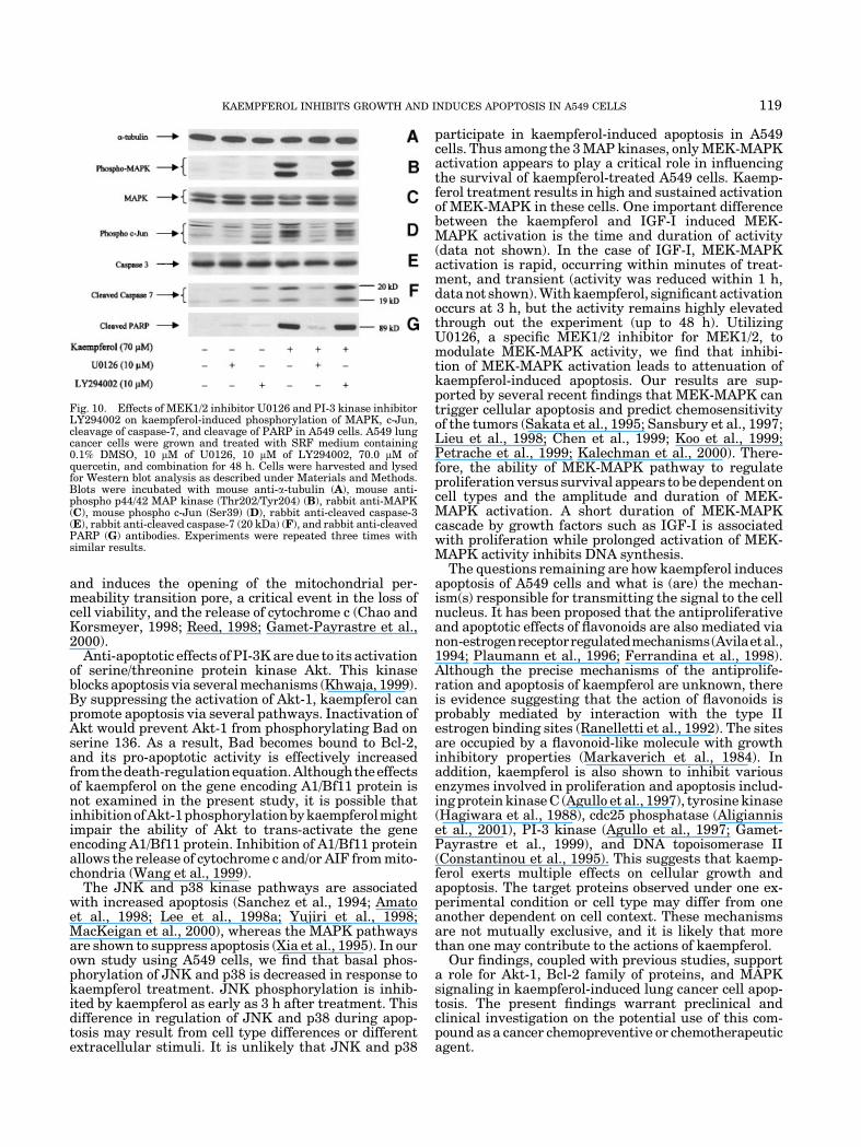
any effect on cell morphology (Fig. 8B,E). Co-treatmentof A549 cells with U0126 and kaempferol preventedkaempferol-induced shrinkage of cytoplasm and mem-brane blebbing (Fig. 8C). This combination effectivelyblocked kaempferol-induced apoptosis as determined byTUNEL assay (Fig. 9C). Western blot analysis revealedthat kaempferol alone significantly increased phos-phorylation of MAPK and c-Jun, cleaved PARP, andcleaved caspase-7 (Fig. 10). Figure 10F,G show that bothLY294002 and U0126 also caused a mild increase incleaved PARP and cleaved caspase-7. Cotreatment ofcells with U0126 and kaempferol prevented kaempferol-induced phosphorylation of MAPK, phosphorylationof c-Jun, cleavage of caspase-7, and cleavage of PARP(Fig. 10). Blocking PI-3 kinase by LY294002 inhibitordid not enhance kaempferol-induced apoptosis, cleavageof caspase-7, and cleavage of PARP (Fig. 10). The resultsindicate that activation of MEK-MAPK play a criticalrole in kaempferol-induced apoptosis and MEK-MAPKacts upstream of caspase-7 to exert its apoptotic in-fluence in the kaempferol-treated A549 cells.
DISCUSSION
The relationship between diet and cancers has beenimplicated in several epidemiological studies (Blocket al., 1992). The cancer incidence is significantly lowerin people whom diet consists of largely fruits andvegetables than people whom diet consists mainly ofanimal products (Steinmetz and Potter, 1991; Blocket al., 1992). The results from several studies indicatethat vegetables and fruits contain components thathave antiproliferative and antineoplastic properties(Leighton et al., 1992; Messina et al., 1994; Stavric,1994). Kaempferol, a natural occurring compound pre-sent in fruits and other vegetables, has been shown toprovide antiproliferative effects in different systemsbased on its striking inhibition of diverse cellular eventsassociated with tumor initiation, promotion, and pro-gression (Ferrell et al., 1979; Landolfi et al., 1984; Kuoet al., 1994; Sahu and Gray, 1994; Sathyamoorthy et al.,1994; Constantinou et al., 1995; Boege et al., 1996; Rothet al., 1999; Dimas et al., 2000; Aligiannis et al., 2001).
Fig. 8. Effects of MEK1/2 inhibitor U0126 and PI-3 kinase inhibitor LY294002 on kaempferol-inducedchanges in A549 cell morphology. A549 lung cancer cells were grown and treated with SRF mediumcontaining 0.1% DMSO (A), 10 mM of U0126 (B), 70.0 mM of kaempferol plus 10 mM of U0126 (C), 70.0 mMof kaempferol (D), 10 mM of LY294002 (E), and 70.0 mM of kaempferol plus 10 mM of LY294002 (F) for 48 h.Bright field views of A549 cells under various treatments are demonstrated. Original magnification,�200. [Color figure can be viewed in the online issue, which is available at www.interscience.wiley.com.]
KAEMPFEROL INHIBITS GROWTH AND INDUCES APOPTOSIS IN A549 CELLS 117

However, the precise mechanisms of its antitumori-genic or chemopreventative activities remain largelyunknown. In the present study, we have shown thatkaempferol inhibits proliferation and induces apop-tosis in A549 lung cancer cells. Morphologically, A549cells exhibit ruffling, blebbing, and condensation of theplasma and nuclear membranes, subsequently, aggre-gation of nuclear chromatin. These observations areconfirmed by TUNEL assay which clearly shows DNAfragmentation. In addition to changes in Bcl-2 familyof proteins and inhibition of Akt-1 phosphorylation,MEK-MAPK activation was required for kaempferol-induced apoptosis. Kaempferol treatment results indose- and time-dependent activation of MEK-MAPK.The elevated MAPK activity contributed to cell death bykaempferol is supported by the observations: U0126chemical inhibitor of the MEK-MAPK signaling path-way attenuates apoptosis. Kaempferol-induced apopto-sis is associated with PARP cleavage and cleavage ofcaspase-7, all of which can be blocked by treatment withthe MEK1/2 inhibitor. Our findings suggest that besideinhibition of Akt activation and alteration of Bcl-2family of proteins, MEK-MAPK activation also plays a
critical role in mediating kaempferol-induced apoptosisof A549 cells and MEK-MAPK functions upstream ofcaspase activation to initiate the apoptosis signal.
Two major distinct apoptosis pathways have beendescribed for mammalian cells. One involves caspase-8,which is recruited by the adapter molecule Fas/APO-1associated death domain protein to death receptors uponextracellular ligand binding (Muzio et al., 1998). We donot observe any change in either Fas or FasL expressionin kaempferol-treated A549 cells (data not shown).We do, however, observe that kaempferol treatmentresults in a dose-dependent increase in expression ofpro-apoptotic proteins Bax and Bad while expression ofanti-apoptotic Bcl-xL and Bcl-2 proteins is inhibited.Thus, there is a shift in the dynamic balance between theoutputs of pro-apoptotic and anti-apoptotic pathwaysfollowing kaempferol treatment. It is possible that thereduction in Bcl-2 and Bcl-xL by kaempferol would allowless Bcl-2-Bax complex. Increase in Bad by kaempferolallows more interaction of Bcl-2 and Bcl-xL with Bad.By this way, Bad sequesters Bcl-2 and Bcl-xL away fromthe Bax. The net effect is the release of more free Bax.Bax then translocates into the mitochondrial membrane
Fig. 9. Effects of MEK1/2 inhibitor U0126 and PI-3 kinase inhibitorLY294002 on kaempferol-induced apoptosis in A549 cells. A549 cellswere grown and treated with SRF medium containing 0.1% DMSO(A), 10 mM of U0126 (B), 70.0 mM of kaempferol plus 10 mM of U0126(C), 70.0 mM of kaempferol (D), 10 mM of LY294002 (E), and 70.0 mM of
kaempferol plus 10 mM of LY294002 (F) for 48 h. Cells were subjectedto TUNEL assay as described under Materials and Methods. Cellnuclei were visualized under a fluorescent microscope. Originalmagnification, �200. [Color figure can be viewed in the online issue,which is available at www.interscience.wiley.com.]
118 NGUYEN ET AL.
and induces the opening of the mitochondrial per-meability transition pore, a critical event in the loss ofcell viability, and the release of cytochrome c (Chao andKorsmeyer, 1998; Reed, 1998; Gamet-Payrastre et al.,2000).
Anti-apoptotic effects of PI-3K are due to its activationof serine/threonine protein kinase Akt. This kinaseblocks apoptosis via several mechanisms (Khwaja, 1999).By suppressing the activation of Akt-1, kaempferol canpromote apoptosis via several pathways. Inactivation ofAkt would prevent Akt-1 from phosphorylating Bad onserine 136. As a result, Bad becomes bound to Bcl-2,and its pro-apoptotic activity is effectively increasedfromthedeath-regulationequation.Althoughtheeffectsof kaempferol on the gene encoding A1/Bf11 protein isnot examined in the present study, it is possible thatinhibitionofAkt-1phosphorylationbykaempferolmightimpair the ability of Akt to trans-activate the geneencoding A1/Bf11 protein. Inhibition of A1/Bf11 proteinallows the release of cytochrome c and/or AIF from mito-chondria (Wang et al., 1999).
The JNK and p38 kinase pathways are associatedwith increased apoptosis (Sanchez et al., 1994; Amatoet al., 1998; Lee et al., 1998a; Yujiri et al., 1998;MacKeigan et al., 2000), whereas the MAPK pathwaysare shown to suppress apoptosis (Xia et al., 1995). In ourown study using A549 cells, we find that basal phos-phorylation of JNK and p38 is decreased in response tokaempferol treatment. JNK phosphorylation is inhib-ited by kaempferol as early as 3 h after treatment. Thisdifference in regulation of JNK and p38 during apop-tosis may result from cell type differences or differentextracellular stimuli. It is unlikely that JNK and p38
participate in kaempferol-induced apoptosis in A549cells. Thus among the 3 MAP kinases, only MEK-MAPKactivation appears to play a critical role in influencingthe survival of kaempferol-treated A549 cells. Kaemp-ferol treatment results in high and sustained activationof MEK-MAPK in these cells. One important differencebetween the kaempferol and IGF-I induced MEK-MAPK activation is the time and duration of activity(data not shown). In the case of IGF-I, MEK-MAPKactivation is rapid, occurring within minutes of treat-ment, and transient (activity was reduced within 1 h,data not shown). With kaempferol, significant activationoccurs at 3 h, but the activity remains highly elevatedthrough out the experiment (up to 48 h). UtilizingU0126, a specific MEK1/2 inhibitor for MEK1/2, tomodulate MEK-MAPK activity, we find that inhibi-tion of MEK-MAPK activation leads to attenuation ofkaempferol-induced apoptosis. Our results are sup-ported by several recent findings that MEK-MAPK cantrigger cellular apoptosis and predict chemosensitivityof the tumors (Sakata et al., 1995; Sansbury et al., 1997;Lieu et al., 1998; Chen et al., 1999; Koo et al., 1999;Petrache et al., 1999; Kalechman et al., 2000). There-fore, the ability of MEK-MAPK pathway to regulateproliferation versus survival appears to be dependent oncell types and the amplitude and duration of MEK-MAPK activation. A short duration of MEK-MAPKcascade by growth factors such as IGF-I is associatedwith proliferation while prolonged activation of MEK-MAPK activity inhibits DNA synthesis.
The questions remaining are how kaempferol inducesapoptosis of A549 cells and what is (are) the mechan-ism(s) responsible for transmitting the signal to the cellnucleus. It has been proposed that the antiproliferativeand apoptotic effects of flavonoids are also mediated vianon-estrogenreceptorregulatedmechanisms(Avilaetal.,1994; Plaumann et al., 1996; Ferrandina et al., 1998).Although the precise mechanisms of the antiprolife-ration and apoptosis of kaempferol are unknown, thereis evidence suggesting that the action of flavonoids isprobably mediated by interaction with the type IIestrogen binding sites (Ranelletti et al., 1992). The sitesare occupied by a flavonoid-like molecule with growthinhibitory properties (Markaverich et al., 1984). Inaddition, kaempferol is also shown to inhibit variousenzymes involved in proliferation and apoptosis includ-ing protein kinase C (Agullo et al., 1997), tyrosine kinase(Hagiwara et al., 1988), cdc25 phosphatase (Aligianniset al., 2001), PI-3 kinase (Agullo et al., 1997; Gamet-Payrastre et al., 1999), and DNA topoisomerase II(Constantinou et al., 1995). This suggests that kaemp-ferol exerts multiple effects on cellular growth andapoptosis. The target proteins observed under one ex-perimental condition or cell type may differ from oneanother dependent on cell context. These mechanismsare not mutually exclusive, and it is likely that morethan one may contribute to the actions of kaempferol.
Our findings, coupled with previous studies, supporta role for Akt-1, Bcl-2 family of proteins, and MAPKsignaling in kaempferol-induced lung cancer cell apop-tosis. The present findings warrant preclinical andclinical investigation on the potential use of this com-pound as a cancer chemopreventive or chemotherapeuticagent.
Fig. 10. Effects of MEK1/2 inhibitor U0126 and PI-3 kinase inhibitorLY294002 on kaempferol-induced phosphorylation of MAPK, c-Jun,cleavage of caspase-7, and cleavage of PARP in A549 cells. A549 lungcancer cells were grown and treated with SRF medium containing0.1% DMSO, 10 mM of U0126, 10 mM of LY294002, 70.0 mM ofquercetin, and combination for 48 h. Cells were harvested and lysedfor Western blot analysis as described under Materials and Methods.Blots were incubated with mouse anti-a-tubulin (A), mouse anti-phospho p44/42 MAP kinase (Thr202/Tyr204) (B), rabbit anti-MAPK(C), mouse phospho c-Jun (Ser39) (D), rabbit anti-cleaved caspase-3(E), rabbit anti-cleaved caspase-7 (20 kDa) (F), and rabbit anti-cleavedPARP (G) antibodies. Experiments were repeated three times withsimilar results.
KAEMPFEROL INHIBITS GROWTH AND INDUCES APOPTOSIS IN A549 CELLS 119

LITERATURE CITED
Agullo G, Gamet-Payrastre L, Manenti S, Viala C, Remesy C, Chap H,Payrastre B. 1997. Relationship between flavonoid structure andinhibition of phosphatidylinositol 3-kinase: A comparison withtyrosine kinase and protein kinase C inhibition. Biochem Pharma-col 53:1649–1657.
Aligiannis N, Mitaku S, Mitrocotsa D, Leclerc S. 2001. Flavonoids ascycline-dependent kinase inhibitors: Inhibition of cdc 25 phospha-tase activity by flavonoids belonging to the quercetin and kaemp-ferol series. Planta Med 67:468–470.
Amato SF, Swart JM, Berg M, Wanebo HJ, Mehta SR, Chiles TC.1998. Transient stimulation of the c-Jun-NH2-terminal kinase/activator protein 1 pathway and inhibition of extracellular signal-regulated kinase are early effects in paclitaxel-mediated apoptosisin human B lymphoblasts. Cancer Res 58:241–247.
Amundadottir LT, Leder P. 1998. Signal transduction pathwaysactivated and required for mammary carcinogenesis in response tospecific oncogenes. Oncogene 16:737–746.
Anderson NG, Maller JL, Tonks NK, Sturgill TW. 1990. Requirementfor integration of signals from two distinct phosphorylation path-ways for activation of MAP kinase. Nature 343:651–653.
Anton R. 1988. Flavonoids and traditional medicine. New York: AlanR. Liss, Inc.
Avila MA, Velasco JA, Cansado J, Notario V. 1994. Quercetin mediatesthe down-regulation of mutant p53 in the human breast cancer cellline MDA-MB468. Cancer Res 54:2424–2428.
Ballif BA, Blenis J. 2001. Molecular mechanisms mediating mam-malian mitogen-activated protein kinase (MAPK) kinase (MEK)-MAPK cell survival signals. Cell Growth Differ 12:397–408.
Bhat NR, Zhang P. 1999. Hydrogen peroxide activation of multiplemitogen-activated protein kinases in an oligodendrocyte cell line:Role of extracellular signal-regulated kinase in hydrogen peroxide-induced cell death. J Neurochem 72:112–119.
Block G, Patterson B, Subar A. 1992. Fruit, vegetables, and cancerprevention: A review of the epidemiological evidence. Nutr Cancer18:1–29.
Boege F, Straub T, Kehr A, Boesenberg C, Christiansen K, AndersenA, Jakob F, Kohrle J. 1996. Selected novel flavones inhibit the DNAbinding or the DNA religation step of eukaryotic topoisomerase I.J Biol Chem 271:2262–2270.
Boise LH, Gonzalez-Garcia M, Postema CE, Ding L, Lindsten T, TurkaLA, Mao X, Nunez G, Thompson CB. 1993. bcl-x, a bcl-2-related genethat functions as a dominant regulator of apoptotic cell death. Cell74:597–608.
Bokkenheuser VD, Winter J. 1988. Hydrolysis of flavonoids by humanintestinal bacteria. In: Cody V, Middleton E, Harbourne JB, BeretzA, editors. Plant flavonoids in biology and medicine II. New York:Alan R. Liss, Inc.
Chao DT, Korsmeyer SJ. 1998. BCL-2 family: Regulators of cell death.Annu Rev Immunol 16:395–419.
Chen N, Ma W, Huang C, Dong Z. 1999. Translocation of proteinkinase Cepsilon and protein kinase Cdelta to membrane is requiredfor ultraviolet B-induced activation of mitogen-activated proteinkinases and apoptosis. J Biol Chem 274:15389–15394.
Chen Y, Lu Q, Schneeberger EE, Goodenough DA. 2000. Restorationof tight junction structure and barrier function by down-regulationof the mitogen-activated protein kinase pathway in ras-transformedMadin–Darby canine kidney cells. Mol Biol Cell 11:849–862.
Constantinou A, Mehta R, Runyan C, Rao K, Vaughan A, Moon R.1995. Flavonoids as DNA topoisomerase antagonists and poisons:Structure–activity relationships. J Nat Prod 58:217–225.
Datta SR, Dudek H, Tao X, Masters S, Fu H, Gotoh Y, Greenberg ME.1997. Akt phosphorylation of BAD couples survival signals to thecell-intrinsic death machinery. Cell 91:231–41.
Dimas K, Demetzos C, Mitaku S, Marselos M, Tzavaras T,Kokkinopoulos D. 2000. Cytotoxic activity of kaempferol glycosidesagainst human leukaemic cell lines in vitro. Pharmacol Res 41:85–88.
Downward J. 1999. How BAD phosphorylation is good for survival[news]. Nat Cell Biol 1:E33–E35.
Dudley DT, Pang L, Decker SJ, Bridges AJ, Saltiel AR. 1995. Asynthetic inhibitor of the mitogen-activated protein kinase cascade.Proc Natl Acad Sci USA 92:7686–7689.
Favata MF, Horiuchi KY, Manos EJ, Daulerio AJ, Stradley DA,Feeser WS, Van Dyk DE, Pitts WJ, Earl RA, Hobbs F, Copeland RA,Magolda RL, Scherle PA, Trzaskos JM. 1998. Identification of anovel inhibitor of mitogen-activated protein kinase kinase. J BiolChem 273:18623–18632.
Feng G, Xu X, Youssef EM, Lotan R. 2001. Diminished expression ofS100A2, a putative tumor suppressor, at early stage of human lungcarcinogenesis. Cancer Res 61:7999–8004.
Ferrandina G, Almadori G, Maggiano N, Lanza P, Ferlini C, CattaniP, Piantelli M, Scambia G, Ranelletti FO. 1998. Growth-inhibitoryeffect of tamoxifen and quercetin and presence of type II estrogenbinding sites in human laryngeal cancer cell lines and primarylaryngeal tumors. Int J Cancer 77:747–754.
Ferrell JE, Jr., Chang Sing PD, Loew G, King R, Mansour JM,Mansour TE. 1979. Structure/activity studies of flavonoids asinhibitors of cyclic AMP phosphodiesterase and relationship toquantum chemical indices. Mol Pharmacol 16:556–568.
Fisher TC, Milner AE, Gregory CD, Jackman AL, Aherne GW,Hartley JA, Dive C, Hickman JA. 1993. bcl-2 modulation ofapoptosis induced by anticancer drugs: Resistance to thymidylatestress is independent of classical resistance pathways. Cancer Res53:3321–3326.
Franke TF, Yang SI, Chan TO, Datta K, Kazlauskas A, Morrison DK,Kaplan DR, Tsichlis PN. 1995. The protein kinase encoded by theAkt proto-oncogene is a target of the PDGF-activated phosphatidy-linositol 3-kinase. Cell 81:727–736.
Gamet-Payrastre L, Manenti S, Gratacap MP, Tulliez J, Chap H,Payrastre B. 1999. Flavonoids and the inhibition of PKC and PI3-kinase. Gen Pharmacol 32:279–286.
Gamet-Payrastre L, Li P, Lumeau S, Cassar G, Dupont MA,Chevolleau S, Gasc N, Tulliez J, Terce F. 2000. Sulforaphane, anaturally occurring isothiocyanate, induces cell cycle arrest andapoptosis in HT29 human colon cancer cells. Cancer Res 60:1426–1433.
Gargiullo P, Wingo PA, Coates RJ, Thompson TD. 2002. Recent trendsin mortality rates for four major cancers, by sex and race/ethnicity—United States. MMWR 51:49–53.
Germain M, Affar EB, D’Amours D, Dixit VM, Salvesen GS, PoirierGG. 1999. Cleavage of automodified poly(ADP-ribose) polymeraseduring apoptosis. Evidence for involvement of caspase-7. J BiolChem 274:28379–28384.
Greulich H, Erikson RL. 1998. An analysis of Mek1 signaling incell proliferation and transformation. J Biol Chem 273:13280–13288.
Gupta K, Kshirsagar S, Li W, Gui L, Ramakrishnan S, Gupta P,Law PY, Hebbel RP. 1999. VEGF prevents apoptosis of humanmicrovascular endothelial cells via opposing effects on MAPK/ERKand SAPK/JNK signaling. Exp Cell Res 247:495–504.
Hagiwara M, Inoue S, Tanaka T, Nunoki K, Ito M, Hidaka H. 1988.Differential effects of flavonoids as inhibitors of tyrosine proteinkinases and serine/threonine protein kinases. Biochem Pharmacol37:2987–2992.
Hickman JA. 1992. Apoptosis induced by anticancer drugs. CancerMetastasis Rev 11:121–139.
Hoshino R, Chatani Y, Yamori T, Tsuruo T, Oka H, Yoshida O,Shimada Y, Ari-i S, Wada H, Fujimoto J, Kohno M. 1999.Constitutive activation of the 41-/43-kDa mitogen-activated proteinkinase signaling pathway in human tumors. Oncogene 18:813–822.
Huynh H, Chow PK, Ooi LL, Soo KC. 2002. A possible role for insulin-like growth factor-binding protein-3 autocrine/paracrine loops incontrolling hepatocellular carcinoma cell proliferation. Cell GrowthDiffer 13:115–122.
Kalechman Y, Longo DL, Catane R, Shani A, Albeck M, Sredni B.2000. Synergistic anti-tumoral effect of paclitaxel (Taxol)þAS101 ina murine model of B16 melanoma: Association with ras-dependentsignal-transduction pathways. Int J Cancer 86:281–288.
Khwaja A. 1999. Akt is more than just a Bad kinase. Nature 401:33–34.
Koo HM, Gray-Goodrich M, Kohlhagen G, McWilliams MJ, Jeffers M,Vaigro-Wolff A, Alvord WG, Monks A, Paull KD, Pommier Y,Vande Woude GF. 1999. The ras oncogene-mediated sensitization ofhuman cells to topoisomerase II inhibitor-induced apoptosis. J NatlCancer Inst 91:236–244.
Kostrzewska A, Laudanski T, Batra S. 1993. Effect of ovarian steroidsand diethylstilbestrol on the contractile responses of the humanmyometrium and intramyometrial arteries. Eur J Pharmacol 233:127–134.
Krueger JS, Keshamouni VG, Atanaskova N, Reddy KB. 2001.Temporal and quantitative regulation of mitogen-activated proteinkinase (MAPK) modulates cell motility and invasion. Oncogene20:4209–4218.
Kulik G, Klippel A, Weber MJ. 1997. Antiapoptotic signalling by theinsulin-like growth factor I receptor, phosphatidylinositol 3-kinase,and Akt. Mol Cell Biol 17:1595–1606.
120 NGUYEN ET AL.

Kuo ML, Lin JK, Huang TS, Yang NC. 1994. Reversion of thetransformed phenotypes of v-H-ras NIH3T3 cells by flavonoidsthrough attenuating the content of phosphotyrosine. Cancer Lett87:91–97.
Landolfi R, Mower RL, Steiner M. 1984. Modification of plateletfunction and arachidonic acid metabolism by bioflavonoids. Struc-ture–activity relations. Biochem Pharmacol 33:1525–1530.
Lee LF, Li G, Templeton DJ, Ting JP. 1998a. Paclitaxel (taxol)-induced gene expression and cell death are both mediated by theactivation of c-Jun NH2-terminal kinase (JNK/SAPK). J Biol Chem273:28253–28260.
Lee SC, Kuan CY, Yang CC, Yang SD. 1998b. Bioflavonoids commonlyand potently induce tyrosine dephosphorylation/inactivation ofoncogenic proline-directed protein kinase FA in human prostatecarcinoma cells. Anticancer Res 18:1117–1121.
Leighton T, GInther C, Fluss L, Harter WK, Cansado J, Notario V.1992. Phenolic compounds in food and their effects on health II.Washington: American Chemical Society.
Lewis TS, Shapiro PS, Ahn NG. 1998. Signal transduction throughMAP kinase cascades. Adv Cancer Res 74:49–139.
Lewis TS, Hunt JB, Aveline LD, Jonscher KR, Louie DF, Yeh JM,Nahreini TS, Resing KA, Ahn NG. 2000. Identification of novel MAPkinase pathway signaling targets by functional proteomics andmass spectrometry. Mol Cell 6:1343–1354.
Lieu CH, Liu CC, Yu TH, Chen KD, Chang YN, Lai YK. 1998. Role ofmitogen-activated protein kinase in taxol-induced apoptosis inhuman leukemic U937 cells. Cell Growth Differ 9:767–776.
Lim IJ, Phan TT, Song C, Tan WT, Longaker MT. 2001. Investigationof the influence of keloid-derived keratinocytes on fibroblast growthand proliferation in vitro. Plast Reconstr Surg 107:797–808.
Liu E, Thant AA, Kikkawa F, Kurata H, Tanaka S, Nawa A, MizutaniS, Matsuda S, Hanafusa H, Hamaguchi M. 2000. The Ras-mitogen-activated protein kinase pathway is critical for the activation ofmatrix metalloproteinase secretion and the invasiveness in v-crk-transformed 3Y1. Cancer Res 60:2361–2364.
Lu Q, Paredes M, Zhang J, Kosik KS. 1998. Basal extracellular signal-regulated kinase activity modulates cell–cell and cell–matrixinteractions. Mol Cell Biol 18:3257–3265.
MacKeigan JP, Collins TS, Ting JP. 2000. MEK inhibition enhancespaclitaxel-induced tumor apoptosis. J Biol Chem 275:38953–38956.
Mansour SJ, Matten WT, Hermann AS, Candia JM, Rong S, FukasawaK, Vande Woude GF, Ahn NG. 1994. Transformation of mammaliancells by constitutively active MAP kinase kinase. Science 265:966–970.
Markaverich BM, Roberts RR, Alejandro MA, Clark JH. 1984. Anendogenous inhibitor of [3H]estradiol binding to nuclear type IIestrogen binding sites in normal and malignant tissues. Cancer Res44:1515–1519.
Messina MJ, Persky V, Setchell KD, Barnes S. 1994. Soy intake andcancer risk: A review of the in vitro and in vivo data. Nutr Cancer21:113–131.
Midthun DE, Jett JR. 1997. Chemotherapy for advanced lung cancer.When to expect a response. Postgrad Med 101:187-2–194.
Montesano R, Soriano JV, Hosseini G, Pepper MS, Schramek H. 1999.Constitutively active mitogen-activated protein kinase kinaseMEK1 disrupts morphogenesis and induces an invasive phenotypein Madin–Darby canine kidney epithelial cells. Cell Growth Differ10:317–332.
Moos PJ, Fitzpatrick FA. 1998. Taxanes propagate apoptosis via twocell populations with distinctive cytological and molecular traits.Cell Growth Differ 9:687–697.
Muzio M, Stockwell BR, Stennicke HR, Salvesen GS, Dixit VM. 1998.An induced proximity model for caspase-8 activation. J Biol Chem273:2926–2930.
Page C, Lin HJ, Jin Y, Castle VP, Nunez G, Huang M, Lin J. 2000.Overexpression of Akt/AKT can modulate chemotherapy-inducedapoptosis. Anticancer Res 20:407–416.
Petrache I, Choi ME, Otterbein LE, Chin BY, Mantell LL, Horowitz S,Choi AM. 1999. Mitogen-activated protein kinase pathway mediateshyperoxia-induced apoptosis in cultured macrophage cells. Am JPhysiol 277:L589–L595.
Plaumann B, Fritsche M, Rimpler H, Brandner G, Hess RD. 1996.Flavonoids activate wild-type p53. Oncogene 13:1605–1614.
Ranelletti FO, Ricci R, Larocca LM, Maggiano N, Capelli A,Scambia G, Benedetti-Panici P, Mancuso S, Rumi C, Piantelli M.1992. Growth-inhibitory effect of quercetin and presence of type-IIestrogen- binding sites in human colon-cancer cell lines and primarycolorectal tumors. Int J Cancer 50:486–492.
Reddy KB, Krueger JS, Kondapaka SB, Diglio CA. 1999. Mitogen-activated protein kinase (MAPK) regulates the expression of proge-latinase B (MMP-9) in breast epithelial cells. Int J Cancer 82:268–273.
Reed JC. 1998. Bcl-2 family proteins. Oncogene 17:3225–3236.Rogers JC, Williams DL, Jr. 1989. Kaempferol inhibits myosin light
chain kinase. Biochem Biophys Res Commun 164:419–425.Roth A, Schaffner W, Hertel C. 1999. Phytoestrogen kaem-
pferol (3,40,5,7-tetrahydroxyflavone) protects PC12 and T47Dcells from beta-amyloid-induced toxicity. J Neurosci Res 57:399–404.
Sahu SC, Gray GC. 1994. Kaempferol-induced nuclear DNA damageand lipid peroxidation. Cancer Lett 85:159–164.
Sakata N, Patel HR, Terada N, Aruffo A, Johnson GL, Gelfand EW.1995. Selective activation of c-Jun kinase mitogen-activatedprotein kinase by CD40 on human B cells. J Biol Chem 270:30823–30828.
Sanchez I, Hughes RT, Mayer BJ, Yee K, Woodgett JR, Avruch J,Kyriakis JM, Zon LI. 1994. Role of SAPK/ERK kinase-1 in thestress-activated pathway regulating transcription factor c-Jun.Nature 372:794–798.
Sansbury HM, Wisehart-Johnson AE, Qi C, Fulwood S, Meier KE.1997. Effects of protein kinase C activators on phorbol ester-sensitive and -resistant EL4 thymoma cells. Carcinogenesis 18:1817–1824.
Sathyamoorthy N, Wang TT, Phang JM. 1994. Stimulation of pS2expression by diet-derived compounds. Cancer Res 54:957–961.
Stavric B. 1994. Quercetin in our diet: From potent mutagen toprobable anticarcinogen. Clin Biochem 27:245–248.
Steinmetz KA, Potter JD. 1991. Vegetables, fruit, and cancer. I.Epidemiology. Cancer Causes Control 2:325–357.
Walter Kolch, Ashwin Kotwaliwale, Keith Vass, Petra Janosch. 2002.The role of Raf kinases in malignant transformation. Expertreviews in molecular medicine. UK: Cambridge University Press.pp 1–18.
Wang CY, Guttridge DC, Mayo M, Baldwin AS, Jr. 1999. NF-kappaBinduces expression of the Bcl-2 homologue A1/Bfl-1 to preferentiallysuppress chemotherapy-induced apoptosis. Mol Cell Biol 19:5923–5929.
Whitten P, Naftolin F. 1991. The new biology of steroid hormones.New York: Raven Press.
Xia Z, Dickens M, Raingeaud J, Davis RJ, Greenberg ME. 1995.Opposing effects of ERK and JNK-p38 MAP kinases on apoptosis.Science 270:1326–1331.
Yujiri T, Sather S, Fanger GR, Johnson GL. 1998. Role of MEKK1 incell survival and activation of JNK and ERK pathways defined bytargeted gene disruption. Science 282:1911–1914.
Zha J, Harada H, Yang E, Jockel J, Korsmeyer SJ. 1996. Serinephosphorylation of death agonist BAD in response to survivalfactor results in binding to 14-3-3 not BCL-X(L). Cell 87:619–628.
KAEMPFEROL INHIBITS GROWTH AND INDUCES APOPTOSIS IN A549 CELLS 121
Copyright © 2022 FDOKUMEN