Distribution of β-enolase in normal and tumor rat cells
6
©Polish Histochemical et Cytochemical Society Folia Histochem Cytobiol. 2008:46(4): 519 (519-524) doi: 10.2478/v10042-008-0075-7 Introduction Enolase (2-phospho-D-glycerate hydrolyase, EC 4.2.1.11) is a glycolytic enzyme in the cell's cytosol compartment of all organisms metabolizing glucose along the Embden-Meyerhoff-Parnas pathway. In higher vertebrates, enolase is active as a dimer formed from three different subunits α, β and γ, which are coded by separate genes. The expression of these genes are developmentally regulated in a tissue-specif- ic manner. The ubiquitous αα isoform remains widely distributed in embryonic most adult cell types. In adult muscle tissues, the β-enolase subunit accumulates preferentially in fast-twitch fibres, where the ββ homodimer accounts for more than 90% of the total enolase activity [1]. Many reports are available demonstrating the direct correlation between increased expression of enolase and progression of neuroendocrine tumors, neuroblas- toma and lung cancer [2]. The expression level of eno- lase is too high for its contribution to glycolysis alone [3]. On the other hand, enolase is recognized as a mul- tifunctional protein. It is ubiquitously located in the cytoplasm as a glycolytic enzyme, but also identified on the surfaces of many eukaryotic and prokaryotic cells, where it plays a role as a receptor of some lig- ands. Numerous data present enolase as an effective plasminogen receptor [4]. In this way enolase may arm various cells with proteolytic properties and facilitate tissue growth by the degradation of extracellular matrix (ECM). The degradation of extracellular matrix proteins in this system is very important in the spread of tumor cells [5,6], in myogenesis and in the process of muscle tissue remodeling after injury [7,8]. In the present report we demonstrate the localiza- tion of enolase in normal (H9c2) and transformed rat muscle cells (the rat rhabdomyosarcoma R1 line) by fluorescence microscopy and immunocytochemical analysis using rabbit antibodies specific against β-eno- lase from human striated muscle. Determining enolase localization in these cells may give the rise to further study of its receptor function in tumor cells. Materials and methods Cell cultures. The rat sarcoma (R1) cell line was obtained from the Institute of Immunology and Experimental Therapy of the Polish FOLIA HISTOCHEMICA ET CYTOBIOLOGICA Vol. 46, No. 4, 2008 pp. 519-524 Distribution of β-enolase in normal and tumor rat cells Ewa Seweryn 1 , Iwona S. Bednarz-Misa 1 , Regina Danielewicz 1 , Jolanta Saczko 1 , Julita Kulbacka 1 , Tomasz Dawiskiba 2 , Jadwiga Pietkiewicz 1 1 Department of Medical Biochemistry, Wroclaw Medical University, Wroclaw, Poland; 2 Department of Vascular, General and Transplantation Surgery, Wroclaw Medical University, Wroclaw, Poland Abstract: Enolase – a glycolytic enzyme is also expressed on the surface of eukaryotic cells such as macrophages, neu- trophils, endothelial, neuronal, tumor cells. Surface enolase as plasminogen receptor plays an important role in myogenesis, tumorgenesis and angiogenesis. Determination of enolase localization in the cell lines may give rise to the elucidation of its receptor function in tumor cells. The cellular localization of the muscle-specific isoform of the enolase in normal rat car- diomyocytes (H9c2, an embryonic rat heart-derived cell line) and a rat sarcoma (R1) cell line is reported here. Immunocy- tochemical assays showed that this enolase isoform is freely diffused in the sarcoplasm of rat cells. The evident location of enolase molecules on the perinuclear surface is observed in immunofluorescence assays. Enolase localization on the surface of some intact normal rat cardiomyocytes was also observed. This surface protein maintains enolase catalytic activity. Key words: beta-enolase, cellular localization, rat cardiomyocytes, rat sarcoma Correspondence: J. Pietkiewicz, Dept. of Medical Biochemistry, Wroclaw Medical University, Chalubinskiego 10, 50-368 Wroclaw, Poland; fax.: (+4871) 7840085, e-mail: [email protected] Abbreviations: ERK, extracellular-signal-regulated kinase; MEK/ERK kinase; 2-PGA, 2-phosphoglycerate; PEP, phospho- enolpyruvate; TRITC, rhodamine isothiyocyanate;
Transcript of Distribution of β-enolase in normal and tumor rat cells

©Polish Histochemical et Cytochemical SocietyFolia Histochem Cytobiol. 2008:46(4): 519 (519-524) doi: 10.2478/v10042-008-0075-7
IntroductionEnolase (2-phospho-D-glycerate hydrolyase, EC4.2.1.11) is a glycolytic enzyme in the cell's cytosolcompartment of all organisms metabolizing glucosealong the Embden-Meyerhoff-Parnas pathway. Inhigher vertebrates, enolase is active as a dimer formedfrom three different subunits α, β and γ, which arecoded by separate genes. The expression of thesegenes are developmentally regulated in a tissue-specif-ic manner. The ubiquitous αα isoform remains widelydistributed in embryonic most adult cell types. In adultmuscle tissues, the β-enolase subunit accumulatespreferentially in fast-twitch fibres, where the ββhomodimer accounts for more than 90% of the totalenolase activity [1].
Many reports are available demonstrating the directcorrelation between increased expression of enolaseand progression of neuroendocrine tumors, neuroblas-toma and lung cancer [2]. The expression level of eno-lase is too high for its contribution to glycolysis alone[3]. On the other hand, enolase is recognized as a mul-tifunctional protein. It is ubiquitously located in the
cytoplasm as a glycolytic enzyme, but also identifiedon the surfaces of many eukaryotic and prokaryoticcells, where it plays a role as a receptor of some lig-ands. Numerous data present enolase as an effectiveplasminogen receptor [4]. In this way enolase may armvarious cells with proteolytic properties and facilitatetissue growth by the degradation of extracellularmatrix (ECM). The degradation of extracellular matrixproteins in this system is very important in the spreadof tumor cells [5,6], in myogenesis and in the processof muscle tissue remodeling after injury [7,8].
In the present report we demonstrate the localiza-tion of enolase in normal (H9c2) and transformed ratmuscle cells (the rat rhabdomyosarcoma R1 line) byfluorescence microscopy and immunocytochemicalanalysis using rabbit antibodies specific against β-eno-lase from human striated muscle. Determining enolaselocalization in these cells may give the rise to furtherstudy of its receptor function in tumor cells.
Materials and methodsCell cultures. The rat sarcoma (R1) cell line was obtained from theInstitute of Immunology and Experimental Therapy of the Polish
FOLIA HISTOCHEMICAET CYTOBIOLOGICAVol. 46, No. 4, 2008pp. 519-524
Distribution of β-enolase in normal and tumor rat cells
Ewa Seweryn1, Iwona S. Bednarz-Misa1, Regina Danielewicz1, Jolanta Saczko1, Julita Kulbacka1, Tomasz Dawiskiba2, Jadwiga Pietkiewicz1
1Department of Medical Biochemistry, Wroclaw Medical University, Wroclaw, Poland;2Department of Vascular, General and Transplantation Surgery, Wroclaw Medical University,
Wroclaw, Poland
Abstract: Enolase – a glycolytic enzyme is also expressed on the surface of eukaryotic cells such as macrophages, neu-trophils, endothelial, neuronal, tumor cells. Surface enolase as plasminogen receptor plays an important role in myogenesis,tumorgenesis and angiogenesis. Determination of enolase localization in the cell lines may give rise to the elucidation of itsreceptor function in tumor cells. The cellular localization of the muscle-specific isoform of the enolase in normal rat car-diomyocytes (H9c2, an embryonic rat heart-derived cell line) and a rat sarcoma (R1) cell line is reported here. Immunocy-tochemical assays showed that this enolase isoform is freely diffused in the sarcoplasm of rat cells. The evident location ofenolase molecules on the perinuclear surface is observed in immunofluorescence assays. Enolase localization on the surfaceof some intact normal rat cardiomyocytes was also observed. This surface protein maintains enolase catalytic activity.
Key words: beta-enolase, cellular localization, rat cardiomyocytes, rat sarcoma
Correspondence: J. Pietkiewicz, Dept. of Medical Biochemistry,Wroclaw Medical University, Chalubinskiego 10, 50-368 Wroclaw, Poland; fax.: (+4871) 7840085, e-mail: [email protected]
Abbreviations: ERK, extracellular-signal-regulated kinase;MEK/ERK kinase; 2-PGA, 2-phosphoglycerate; PEP, phospho-enolpyruvate; TRITC, rhodamine isothiyocyanate;

Academy Sciences (Wroclaw, Poland) and normal rat cardiomy-ocytes (the embryonic rat heart-derived cell line H9c2) were a giftfrom prof. F. Ursini from University of Padova (Italy). The cellswere grown in DMEM medium (Sigma) with 3% glutamine, 10%foetal calf serum and antibiotics: gentamycin (100 μg/ml) andpenicillin (100 U/ml). The cell lines were incubated at 37°C with5% CO2.
Antibodies against human β-enolase. Human tibialis anteriormuscle was obtained from postoperative material from the Depart-ment of Vascular, General and Transplantation Surgery of the Wro-claw Medical University in accordance with the Polish legalrequirements, under a license issued by the Commission ofBioethics of the Wroclaw Medical University.
Purification of enolase from the human striated muscle, prepa-ration of rabbit polyclonal serum against human β-enolase and iso-lation of specific antibodies from the rabbit serum by affinitychromatography were preformed according to our previous report[9]. The specificity of the obtained antibodies was confirmed onhomogenates of human and rat striated muscles and β-enolasepurified from human and rat tissue. Gel electrophoresis andimmunoblotting were performed under the conditions previouslydescribed [9].
Immunocytochemical staining. Immunocytochemical reactionswere performed using the avidin biotinylated peroxidase (ABC)technique (DAKO LSAB2 System HRP, Denmark). Specific poly-clonal rabbit antibodies against human β-enolase were applied.
Cell surface enolase activity. Enolase activity of intact normaland tumor cells was determined by a direct assay [10].The cellstock solutions contained 6.73×10⁵ H9c2 cells and 12.8×10⁵ R1cells were resuspended in 50 mM imidazole pH 6.8 buffer con-taining 10 mM MgSO4 and 10 mM KCl and subsequent dilutions(1:1; 1:2; 1:4; 1:8 and 1:16 v/v) were used to determined of surfaceenolase activity.
The enolase substrate 2-PGA was added to the reaction mixturein a 6 mM concentration and conversion of the substrate to phos-phoenolpyruvate (PEP) was followed at 37°C for 3 min. The cellswere removed by centrifugation (3000 rpm for 1 min). The level ofPEP production of the supernatant was measured at 240 nm. In the
same manner enolase activity was detected in the normal andtumor rat cells under thermal and osmotic shock conditions.
Immunofluorescence studies. All cell cultures were suspended inPBS with Triton X-100 (Sigma). To stain β-enolase, the cells wereincubated first with rabbit polyclonal antibodies against β-enolase(1:1000 v/v) and then with rhodamine isothiocyanate-labeledmouse anti-rabbit IgG (Sigma). The control for specificityinvolved omission of the primary antibodies and the cells wereincubated only with 2% BSA. The specimens were evaluated usingfluorescence microscope (Zeiss, Germany).
Statistical analysis. Experiments were performed in triplicates.Data for activity of surface enolase were analyzed by Student's t-test. The mean value was presented on figures.
Results
Enolase immunolocalization was performed on thenormal and sarcoma rat cells. For this study the anti-bodies were affinity-purified from rabbit polyclonalantiserum on a Sepharose CL-4B column with immo-bilized human muscle enolase as described previously[9]. Their specificity was confirmed by immunoblot-ting with homogenates of human and rat normal striat-ed muscle and pure enolase from both tissues (Fig. 1).Rabbit antibody produced against human β-enolasereacts also with rat muscle enolase. These antibodieswe applied to detect β-enolase reactivity in the sar-coplasmatic compartment of normal rat cells (H9c2)by immunocytochemical staining. Most of the enolasemolecules were diffused throughout the sarcoplasm(Fig. 2).
The distribution of enolase in the normal rat and ratsarcoma cells were observed by immunofluorescencemicroscopy (Figs 3 and 4). We observed different size
520 E. Seweryn et al.
©Polish Histochemical et Cytochemical SocietyFolia Histochem Cytobiol. 2008:46(4): 520 (519-524) doi: 10.2478/v10042-008-0075-7
Fig. 1. Immunereactivity of rabbit specificantibodies against human β-enolase withhuman and rat enzyme – 10% SDS/PAGE(A) and immunoblotting (B): samples of 25μg of human /2/ and rat muscles /4/homogenates and 2 μg of purified β-enolasefrom human /3/ and rat /5/ striated muscle.Line /1/: protein standards of molecularmass (Bio-Rad).

areas of fluorescence-labeled proteins in the cytoplasmof the normal, and sarcoma cells. Clearly visible fluores-cence of the cytosol compartment in both cell lines andless expressed fluorescence on the surfaces of some cellswere observed. Furthermore, a more intensive fluores-cence of β-enolase near the perinuclear surface of ratsarcoma sarcoplasm was noted in comparison with nor-mal rat cardiomyocytes (Figs. 4 and 3, respectively).
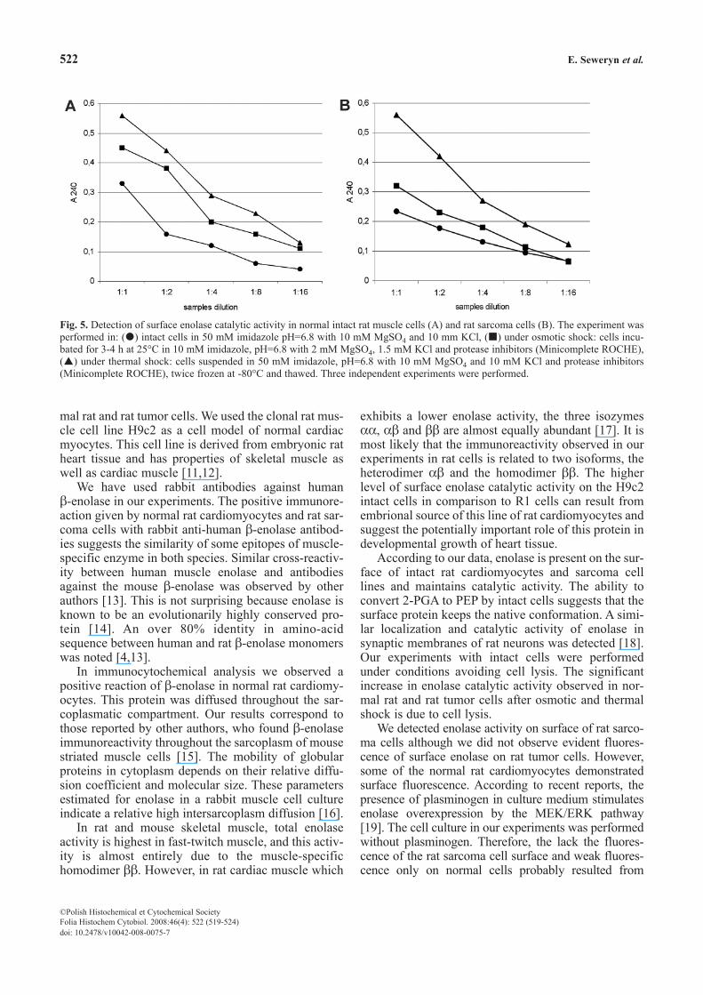
We found enolase catalytic activity in intact musclecells of normal rat cardiomyocytes as well as in the cul-ture of rat tumor cells (Fig. 5 A,B). A dose-dependentenolase activity i.a. 2-PGA conversion to PEP in seriallydiluted intact cells was noted. In the absence of 2-PGAthe intact cells did not reveal any enzymatic activity andwere used as a negative control. We used log-phasegrown cell cultures to rule out the possibility that enolaseactivity was not due to enzyme released as a result of celllysis. In additional experiments we compared the pro-duction of PEP in normal and sarcoma rat cells under
osmotic and thermal shock conditions (Fig. 5 A,B). Thelysis of rat cells caused under these conditions revealedhigher PEP levels in the assay medium compared withexperiments with intact cells.
Some differences between the surface enolaseactivities of normal rat cardiomyocytes and tumorcells were observed. The 12.8×10⁵ cells from the ratsarcoma culture used in the experiment demonstrated asignificantly lower (0.230±0.02 vs. 0.330±0.04,p=0.016) enolase activity than 6.73 × 10⁵ normal cells.This suggests weak availability to the substrate or lowlevel of surface enolase expression on the rat tumorcells used in the experiments.
Discussionβ-enolase is expressed in the striated muscle of mam-mals and humans. In the present study we demonstrat-ed the distribution of muscle-specific enolase in nor-
521β-enolase in normal and tumor rat cells
©Polish Histochemical et Cytochemical SocietyFolia Histochem Cytobiol. 2008:46(4): 521 (519-524) doi: 10.2478/v10042-008-0075-7
Fig. 2. Immunocytochemical detection of β-enolase in rat normalcardiomyocytes (the embryonic rat heart-derived cell line H9c2)using rabbit anti-human β-enolase antibodies (magnification,×400).
Fig. 4. Immunofluorescence detection of enolase in a rat sarcoma cell line (R1) incubated with rabbit anti-human β-enolase antibodies(magnification, x 400). Arrows indicate of surface (A) and intracellular localization (B) of enolase.
Fig. 3. Immunofluorescence detection of enolase in normal rat car-diomyocytes (H9c2) incubated with rabbit anti-human β-enolaseantibodies (magnification, ×400). Arrows indicate cytoplasmicand surface localization of enolase.
mal rat and rat tumor cells. We used the clonal rat mus-cle cell line H9c2 as a cell model of normal cardiacmyocytes. This cell line is derived from embryonic ratheart tissue and has properties of skeletal muscle aswell as cardiac muscle [11,12].
We have used rabbit antibodies against human β-enolase in our experiments. The positive immunore-action given by normal rat cardiomyocytes and rat sar-coma cells with rabbit anti-human β-enolase antibod-ies suggests the similarity of some epitopes of muscle-specific enzyme in both species. Similar cross-reactiv-ity between human muscle enolase and antibodiesagainst the mouse β-enolase was observed by otherauthors [13]. This is not surprising because enolase isknown to be an evolutionarily highly conserved pro-tein [14]. An over 80% identity in amino-acidsequence between human and rat β-enolase monomerswas noted [4,13].
In immunocytochemical analysis we observed apositive reaction of β-enolase in normal rat cardiomy-ocytes. This protein was diffused throughout the sar-coplasmatic compartment. Our results correspond tothose reported by other authors, who found β-enolaseimmunoreactivity throughout the sarcoplasm of mousestriated muscle cells [15]. The mobility of globularproteins in cytoplasm depends on their relative diffu-sion coefficient and molecular size. These parametersestimated for enolase in a rabbit muscle cell cultureindicate a relative high intersarcoplasm diffusion [16].
In rat and mouse skeletal muscle, total enolaseactivity is highest in fast-twitch muscle, and this activ-ity is almost entirely due to the muscle-specifichomodimer ββ. However, in rat cardiac muscle which
exhibits a lower enolase activity, the three isozymesαα, αβ and ββ are almost equally abundant [17]. It ismost likely that the immunoreactivity observed in ourexperiments in rat cells is related to two isoforms, theheterodimer αβ and the homodimer ββ. The higherlevel of surface enolase catalytic activity on the H9c2intact cells in comparison to R1 cells can result fromembrional source of this line of rat cardiomyocytes andsuggest the potentially important role of this protein indevelopmental growth of heart tissue.
According to our data, enolase is present on the sur-face of intact rat cardiomyocytes and sarcoma celllines and maintains catalytic activity. The ability toconvert 2-PGA to PEP by intact cells suggests that thesurface protein keeps the native conformation. A simi-lar localization and catalytic activity of enolase insynaptic membranes of rat neurons was detected [18].Our experiments with intact cells were performedunder conditions avoiding cell lysis. The significantincrease in enolase catalytic activity observed in nor-mal rat and rat tumor cells after osmotic and thermalshock is due to cell lysis.
We detected enolase activity on surface of rat sarco-ma cells although we did not observe evident fluores-cence of surface enolase on rat tumor cells. However,some of the normal rat cardiomyocytes demonstratedsurface fluorescence. According to recent reports, thepresence of plasminogen in culture medium stimulatesenolase overexpression by the MEK/ERK pathway[19]. The cell culture in our experiments was performedwithout plasminogen. Therefore, the lack the fluores-cence of the rat sarcoma cell surface and weak fluores-cence only on normal cells probably resulted from
522 E. Seweryn et al.
©Polish Histochemical et Cytochemical SocietyFolia Histochem Cytobiol. 2008:46(4): 522 (519-524) doi: 10.2478/v10042-008-0075-7
Fig. 5. Detection of surface enolase catalytic activity in normal intact rat muscle cells (A) and rat sarcoma cells (B). The experiment wasperformed in: ( ) intact cells in 50 mM imidazole pH=6.8 with 10 mM MgSO4 and 10 mm KCl, ( ) under osmotic shock: cells incu-bated for 3-4 h at 25°C in 10 mM imidazole, pH=6.8 with 2 mM MgSO4, 1.5 mM KCl and protease inhibitors (Minicomplete ROCHE),( ) under thermal shock: cells suspended in 50 mM imidazole, pH=6.8 with 10 mM MgSO4 and 10 mM KCl and protease inhibitors(Minicomplete ROCHE), twice frozen at -80°C and thawed. Three independent experiments were performed.

another, unknown mechanism regulating the enolasesurface expression. No known muscle enolases pos-sess a signaling sequence, which may act as asequence targeting the protein to the plasma mem-brane [20].
We detected an evident accumulation of rat mus-cle enolase molecules near the nuclear surface in bothcell lines. We observed that the perinuclearimmunoreactivity of β-enolase in the rat sarcomacells was more intensive than in normal rat cells. Thiscorresponds to other reports on the localization ofmouse muscle enolase in regenerating tissue [1].However other authors report a nuclear location ofenolase molecules in rat neurons [21] and HeLa cells[22]. Enolase was shown to be a nuclear factor nega-tively regulating c-myc promoter activity. c-Myc pro-tooncogene plays an important role in the regulationof cell growth and differentiation. Overexpression ofc-myc gene is a common characteristic feature ofmany malignant cell types [23]. Some other enzymeswhose primary described function is participation inthe glycolytic pathway in the cytoplasm have so farbeen found in nuclei. Their physiological roles in thenucleus seem to be different from those in the cytosolcompartment. Phosphoglycerate kinase is supposedto participate in DNA synthesis and cell cycle pro-gression [24]. Glyceraldehyde-3-phosphate dehydro-genase recognizes the sequence and structural fea-tures of RNA and is involved in DNA replication andrepair [25]. Lactate dehydrogenase has been found tobe a stabilizing nuclear factor participating in DNArepair [24].
According to recent reports, the expression ofenolase is too high to be justified by its contributionto glycolysis alone [3,4] and may explain the pres-ence of enolase on the surface of some intact normalrat cardiomyocytes and rat sarcoma cell lines. Sur-face enolase exposition on myogenic cells duringmuscle development has also been observed [26].This may suggest that muscle-specific enolase canexhibit functions unrelated to its pivotal role in thesarcoplasm as a glycolytic enzyme. Surface enolasewas recently recognized to be a receptor for plas-minogen [27]. The binding of plasminogen and itssubsequent pericellular activation is an importantprocess in the development of normal muscle tissue[26] in remodeling after injury [7] and in tumorgrowth [6,28]. The influence of some effectors on theinteraction between plasminogen and surface enolasein selected cell cultures should be examined in thefuture in more detail.
Acknowledgements: Authors are grateful to Prof. Andrzej Gami-an for helpful discussions and criticism of the manuscript. Thiswork was financially supported by Wroclaw Medical Universitygrant no. 1602.
References[ 1] Merkulova T, Dehaupas M, Nevers MC, Creminon C,
Alameddine H, Keller A. Differential modulation of α, β andγ enolase isoforms in regenerating mouse skeletal muscle.Eur J Biochem. 2000b;267:3735-3743.
[ 2] Eriksson B, Oberg K, Stridsberg M. Tumor markers in neu-roendocrine tumors. Digestion. 2000;62:33-38.
[ 3] Chai G, Brewer JM, Lovelace L, Aoki T, Minor V, Lebioda L.Expression, purification and the 1.8Å resolution crystal struc-ture of human neuron specific enolase. J Mol Biol. 2004;341:1015-1021.
[ 4] Pancholi V. Multifunctional α-enolase: its role in diseases.Cell Mol Life Sci. 2001;58:902-920.
[ 5] Liu KJ, Shih NY. The role of enolase in tissue invasion andmetastasis of pathogens and tumor cells. J Cancer Mol. 2007;3:45-48.
[ 6] Ranson M, Andronicos NM. Plasminogen binding and cancerpromises and pitfalls. Front Biosci. 2003;8:294-304.
[ 7] Suelves M, Lopez-Alemany R, Lluis F. Plasmin activity isrequired for myogenesis in vitro and skeletal muscle regener-ation in vivo. Blood. 2002;99:2835-2844.
[ 8] Vignaud A, Hourde C, Torres S. Functional, cellular andmolecular aspects of skeletal muscle recovery after injuryinduced by snake venom from Notechis scutatus scutatus.Toxicon. 2005;45:789-801.
[ 9] Witkowska D, Pietkiewicz J, Szostko B, Danielewicz D,Mas³owski L, Gamian A. Antibodies against human muscleenolase recognize a 45-kDa bacterial cell wall outer mem-brane enolase-like protein. FEMS Immunol Med Microbiol.2005;45:53-62.
[10] Pietkiewicz J, Kustrzeba-Wójcicka I, Wolna E. Purificationand properties of enolase from carp (Cyprinus Carpio). Com-parison with enolases from mammals' muscles and yeast.Comp Biochem Physiol(B). 1983;75:693-698.
[11] Gerrelli D, Huntis JD, Latchman DS. Antagonistic effects ofretinoid acid and thyroid hormone on the expression of thetissue-specific splicing protein SmN in a clonal cell linederived from rat heart. J Mol Cell Cardiol. 1994;26:713-719.
[12] Brostrom MA, Reilly BA, Wilson FJ, Brostrom CO. Vaso-pressin-induced hypertrophy in H9c2 heart-derivedmyocytes. Int J Biochem Cell Biol. 2000;32:993-1006.
[13] Merkulova T, Thornell LE, Butler-Browne G. The β enolasesubunit displays three different patterns of microheterogene-ity in human striated muscle. J Muscle Res Cell Motil. 1999;20:55-63.
[14] Tracy MR, Hedges SB. Evolutionary history of the enolasegene family. Gene. 2000;259:129-138.
[15] Keller A, Demeurie J, Merkulova T, et al. Fibre-type distribu-tion and subcellular localisation of α and β enolase in mousestriated muscle. Biol Cell. 2000;92:527-535.
[16] Arrio-Dupont M, Foucault G, Vacher M, Devaux PF, CribierS. Translational diffusion of globular proteins in the cyto-plasm of cultured muscle cells. Biophys J. 2000;78:901-907.
[17] Merkulova T, Keller A, Oliviero P. Thyroid hormones differ-entially modulate enolase isozymes during rat skeletal andcardiac muscle development. Am J Physiol EndocrinolMetab. 2000a;278:E330-E339.
[18] Ueta H, Nagasawa H, Oyabu-Manabe Y, Toida K, IshimuraK, Hori H. Localization of enolase in synaptic plasma mem-brane as an αγ heterodimer in rat brain. Neurosci Res.2004;48:379-386.
[19] Sousa LP, Silva BM, Brasil BSAF. Plasminogen/plasmin reg-ulates α-enolase expression through the MEK/ERK pathway.Biochem Biophys Res Commun. 2005;337:1065-1071.
[20] Redlitz A, Fowler BJ, Plow EF, and Miles LA. The role of anenolase-related molecule in plasminogen binding to cells. EurJ Biochem. 1995;227:407-415.
523β-enolase in normal and tumor rat cells
©Polish Histochemical et Cytochemical SocietyFolia Histochem Cytobiol. 2008:46(4): 523 (519-524) doi: 10.2478/v10042-008-0075-7

[21] Angelov DN, Neiss WF, Gunkel A, Guntinas-Lichius O, Sten-nert E. Axotomy induces intanuclear immunolocalization ofneuron-specific enolase in facial and hypoglosal neurons ofthe rat. J Neurocytol. 1994;23:218-233.
[22] Subramanian A, Miller DM. Structural analysis of α-enolase.Mapping the functional domains involved in down-regulationof the c-myc protooncogene. J Biol Chem. 2000;275:5958-5965.
[23] Cole M. The myc oncogene: its role in transformation and dif-ferentiation. Annu Rev Genet. 1986;20:361-384.
[24] Popanda O, Fox G, Thielmann HW. Modulation of DNApolymerases by lactate dehydrogenase and 3-phosphoglycer-ate kinase. Biochim Biophys Acta. 1998;1397:102-117.
[25] Sirover MA. Role of the glycolytic protein, glyceraldehyde-3-phosphate dehydrogenase in normal cell function and in cellpathology. J Cell Biochem. 1997;66:133-140.
[26] Fougerousse F, Edom-Vovard F, Merkulova T, et al. The mus-cle-specific enolase is an early marker of human myogenesis.J Muscle Res Cell Motyl. 2001;22:535-544.
[27] Seweryn E, Pietkiewicz J, Szamborska A, Gamian A. Enolaseon the surfach of prockaryotic and eukaryotic cells is a recep-tor for human plasminogen. Postepy Hig Med Dosw [serialonline]. 2007;61:672-682. Available at: http://www.phmd.pl/Accessed November 15,2007.
[28] Bergaman AC, Linder C, Sakaguchi K, et al. Increasedexpression of alpha-enolase in c-jun transformed rat fibrob-lasts without increased activation of plasminogen. FEBS Lett.1997;417:17-20.
Submitted: 3 April, 2008Accepted after reviews: 10 June, 2008
524 E. Seweryn et al.
©Polish Histochemical et Cytochemical SocietyFolia Histochem Cytobiol. 2008:46(4): 524 (519-524) doi: 10.2478/v10042-008-0075-7