C-Npys (S-3-nitro-2-pyridinesulfenyl) and peptide derivatives can inhibit a serine-thiol proteinase...
13
C-Npys (S-3-nitro-2-pyridinesulfenyl) and peptide derivatives can inhibit a serine-thiol proteinase activity from Paracoccidioides brasiliensis Alisson L. Matsuo a , Adriana K. Carmona b , Luiz S. Silva a , Carlos E. L. Cunha b , Ernesto S. Nakayasu c , Igor C. Almeida c , Maria A. Juliano b , and Rosana Puccia a,* a Department of Microbiology, Immunology and Parasitology, Cell Biology Division, Federal University of São Paulo, São Paulo, SP 04023-062, Brazil b Department of Biophysics, Federal University of São Paulo, São Paulo, SP 04023-062, Brazil c Department of Biological Sciences, University of Texas at El Paso, El Paso, TX 79968, USA Abstract The inhibitory capacity of C-Npys (S-[3-nitro-2-pyridinesulfenyl]) derivatives over thiol- containing serine proteases has never been tested. In the present work we used an extracellular serine-thiol proteinase activity from the fungal pathogen Paracoccidioides brasiliensis (PbST) to describe a potent inhibitory capacity of Bzl-C(Npys)KRLTL-NH 2 and Bzl-MKRLTLC(Npys)- NH 2 . The assays were performed with PbST enriched upon affinity chromatography in a p- aminobenzamidine (pABA)-Sepharose column. Although PbST can cleave the fluorescence resonance energy transfer peptide Abz-MKRLTL-EDDnp between L-T, the C(Npys) derivatives were not substrates nor were they toxic in a cell detachment assay, allowing therapeutic use. The best inhibitor was Bzl-C(Npys)KRLTL-NH 2 (K i = 16 nM), suggesting that the peptide sequence promoted a favorable interaction, especially when C(Npys) was placed at a further position from the L-T bond, at the N-terminus. Inhibition was completely reverted with dithioerythritol, indicating that it was due to the reactivity of the C(Npys) moiety with a free SH- group. Keywords Paracoccidioides brasiliensis; serine-thiol proteinase; Npys Introduction Extracellular proteinases can facilitate the process of invasion and dissemination of microorganisms through the human tissues by degrading extracellular-associated proteins [1]. Our group has previously characterized an extracellular subtilisin-like serine proteinase activity in the yeast phase of the human pathogen Paracoccidioides brasiliensis [2]. This activity is able to selectively degrade, in vitro, murine laminin, human fibronectin, type IV- collagen, and proteoglycans, while common proteins like bovine serum albumin (BSA) or © 2007 Elsevier Inc. All rights reserved. * Corresponding author. Mailing address: Disciplina de Biologia Celular, UNIFESP, Rua Botucatu, 862, oitavo andar, São Paulo, SP, 04023-062 Brazil, Tel: 55-11-5084-2991. Fax #: 55-11-5571-5877. [email protected]. Publisher's Disclaimer: This is a PDF file of an unedited manuscript that has been accepted for publication. As a service to our customers we are providing this early version of the manuscript. The manuscript will undergo copyediting, typesetting, and review of the resulting proof before it is published in its final citable form. Please note that during the production process errors may be discovered which could affect the content, and all legal disclaimers that apply to the journal pertain. NIH Public Access Author Manuscript Biochem Biophys Res Commun. Author manuscript; available in PMC 2013 March 24. Published in final edited form as: Biochem Biophys Res Commun. 2007 April 20; 355(4): 1000–1005. doi:10.1016/j.bbrc.2007.02.070. NIH-PA Author Manuscript NIH-PA Author Manuscript NIH-PA Author Manuscript
-
Upload
independent -
Category
Documents
-
view
4 -
download
0
Transcript of C-Npys (S-3-nitro-2-pyridinesulfenyl) and peptide derivatives can inhibit a serine-thiol proteinase...
C-Npys (S-3-nitro-2-pyridinesulfenyl) and peptide derivatives caninhibit a serine-thiol proteinase activity from Paracoccidioidesbrasiliensis
Alisson L. Matsuoa, Adriana K. Carmonab, Luiz S. Silvaa, Carlos E. L. Cunhab, Ernesto S.Nakayasuc, Igor C. Almeidac, Maria A. Julianob, and Rosana Pucciaa,*
aDepartment of Microbiology, Immunology and Parasitology, Cell Biology Division, FederalUniversity of São Paulo, São Paulo, SP 04023-062, BrazilbDepartment of Biophysics, Federal University of São Paulo, São Paulo, SP 04023-062, BrazilcDepartment of Biological Sciences, University of Texas at El Paso, El Paso, TX 79968, USA
AbstractThe inhibitory capacity of C-Npys (S-[3-nitro-2-pyridinesulfenyl]) derivatives over thiol-containing serine proteases has never been tested. In the present work we used an extracellularserine-thiol proteinase activity from the fungal pathogen Paracoccidioides brasiliensis (PbST) todescribe a potent inhibitory capacity of Bzl-C(Npys)KRLTL-NH2 and Bzl-MKRLTLC(Npys)-NH2. The assays were performed with PbST enriched upon affinity chromatography in a p-aminobenzamidine (pABA)-Sepharose column. Although PbST can cleave the fluorescenceresonance energy transfer peptide Abz-MKRLTL-EDDnp between L-T, the C(Npys) derivativeswere not substrates nor were they toxic in a cell detachment assay, allowing therapeutic use. Thebest inhibitor was Bzl-C(Npys)KRLTL-NH2 (Ki = 16 nM), suggesting that the peptide sequencepromoted a favorable interaction, especially when C(Npys) was placed at a further position fromthe L-T bond, at the N-terminus. Inhibition was completely reverted with dithioerythritol,indicating that it was due to the reactivity of the C(Npys) moiety with a free SH- group.
KeywordsParacoccidioides brasiliensis; serine-thiol proteinase; Npys
IntroductionExtracellular proteinases can facilitate the process of invasion and dissemination ofmicroorganisms through the human tissues by degrading extracellular-associated proteins[1]. Our group has previously characterized an extracellular subtilisin-like serine proteinaseactivity in the yeast phase of the human pathogen Paracoccidioides brasiliensis [2]. Thisactivity is able to selectively degrade, in vitro, murine laminin, human fibronectin, type IV-collagen, and proteoglycans, while common proteins like bovine serum albumin (BSA) or
© 2007 Elsevier Inc. All rights reserved.*Corresponding author. Mailing address: Disciplina de Biologia Celular, UNIFESP, Rua Botucatu, 862, oitavo andar, São Paulo, SP,04023-062 Brazil, Tel: 55-11-5084-2991. Fax #: 55-11-5571-5877. [email protected].
Publisher's Disclaimer: This is a PDF file of an unedited manuscript that has been accepted for publication. As a service to ourcustomers we are providing this early version of the manuscript. The manuscript will undergo copyediting, typesetting, and review ofthe resulting proof before it is published in its final citable form. Please note that during the production process errors may bediscovered which could affect the content, and all legal disclaimers that apply to the journal pertain.
NIH Public AccessAuthor ManuscriptBiochem Biophys Res Commun. Author manuscript; available in PMC 2013 March 24.
Published in final edited form as:Biochem Biophys Res Commun. 2007 April 20; 355(4): 1000–1005. doi:10.1016/j.bbrc.2007.02.070.
NIH
-PA Author Manuscript
NIH
-PA Author Manuscript
NIH
-PA Author Manuscript

casein are not cleavable substrates [3]. The proteinase is able to hydrolyze fluorescenceresonance energy (FRET) peptides homologous to MKRLTL, which are flanked by Abz(ortho-aminobenzoic acid) and EDDnp (N-[2,4-dinitrophenyl]ethylenediamine). Cleavageoccurs between the Leu-Thr bond at an optimum alkaline pH and is inhibited by PMSF,mercuric acetate, and p-HMB (sodium 7-hydroxymercuribenzoate), but not by E-64 [2].Therefore, the enzymatic activity has been classified as a thiol-containing subtilisin-likeserine proteinase (PbST, for P. brasiliensis serine-thiol proteinase) belonging to the group ofproteinase K. In addition, we have recently described a novel type of regulation of the PbSTactivity against Abz-MKALTLQ-EDDnp by neutral polysaccharides, including a fungalextracellular galactomannan, which might help stabilize the enzyme [4].
P. brasiliensis is a dimorphic fungus that causes paracoccidioidomycosis (PCM), apotentially lethal systemic mycosis with multiple clinical forms that is prevalent in SouthAmerica [5]. The fungus grows in the infective mycelial phase at environmentaltemperatures below 26°C and in the yeast pathogenic phase at body temperatures of about37°C. PCM is a granulomatous mycosis that starts in the lungs and tends to disseminaterapidly through the lymphatic system in juvenile (acute and subacute) forms. In adult(chronic) forms, active disease affects especially the lungs, however dissemination to thecutaneous, mucocutaneous tissues or other organs is likely to occur through the blood andlymphatic route. In this context, PbST is a potential virulence factor that could contribute tofungal spread to surrounding and/or distant tissues.
The S-(3-nitro-2-pyridinesulfenyl) group – C(Npys) – has originally been used forprotection in peptide synthesis [6] and later demonstrated to be effective in the design andsynthesis of thiol proteinase irreversible inhibitors [7]. These compounds have been found tobe highly specific to cysteine proteinases due to their capacity of selectively reacting withfree thiol groups to form unsymmetrical disulfide bonds [8] and their inability to inhibitserine or acid proteinases [7]. A variety of cysteine proteinase inhibitors have beendeveloped such as diazomethyl ketones, halomethyl ketones, and epoxides [9]. However,they are strong electrophilic reagents and may alkylate non-targeted enzymes and otherbiomolecules in vivo [10], preventing any therapeutic use. The C(Npys) compoundsrepresent an alternative to overcome this problem, considering that they promote theformation of disulfide bonds without non-specific alkylation, but still keep the ability ofbeing specific inhibitors.
The inhibitory capacity of C(Npys) derivatives over thiol-containing subtilisins has neverbeen tested. In the present work, we used PbST to describe a potent inhibitory capacity oftwo peptides derived from MKRLTL coupled with the C(Npys) group.
Materials and methodsIsolation of the serino-thiol proteinase activity
Culture supernatants containing PbST activity against Abz-MKALTLQ-EDDnp wereobtained from P. brasiliensis B-339 as previously described [3,4] and buffered to a finalconcentration of 50 mM Tris-HCl, pH 8.0, containing 1.0 M NaCl, which did not alter theinitial proteolytic activity. Aliquots (40 ml) were chromatographed, using a peristaltic pump,in an affinity column of p-aminomethylbenzamidine (pABA) Sepharose (Pharmacia/LKB)previously equilibrated in the same buffer. After a washing step with 10 volumes of thesame buffer, bound compounds were eluted with 50 mM glycine, pH 3.0, into 1.0-mLfractions immediately neutralized with Tris-HCl. The fractions were analyzed for proteolyticactivity against Abz-MKALTLQ-EDDnp, as described below, and against fibronectin (FN,2 µg) as described by Puccia et al. [3]. Activity was assayed in 50 mM Tris-HCl, pH 8.0.Fibronectin was purified from sera donated from healthy individuals by chromatography in
Matsuo et al. Page 2
Biochem Biophys Res Commun. Author manuscript; available in PMC 2013 March 24.
NIH
-PA Author Manuscript
NIH
-PA Author Manuscript
NIH
-PA Author Manuscript
gelatin-Sepharose (Amersham Biosciences, GE Healthcare), as described [11]. Proteincontents were visualized in Coomassie blue and/or silver-stained SDS-PAGE gels [12].
Peptide synthesisThe FRET substrate Abz-MKALTLQ-EDDnp was synthesized using the solid phasesynthesis method [13] according to the Fmoc procedure. An automated bench-topsimultaneous multiple solid-phase peptide synthesizer (PSSM 8 system; Shimadzu) wasused in the synthesis. The inhibitor peptides Bzl-MKRLTLC(Npys)-NH2 and Bzl-C(Npys)KRLTL-NH2 were prepared by the solid phase methodology using Boc-amino acidsaccording to the general procedure described previously [14]. All the peptides were purifiedby semi-preparative h.p.l.c. using an Econosil C-18 column. The molecular mass and puritywere checked by amino acid analysis and by mass spectrometry using a matrix assistedlaser-desorption ionization-time-of-flight-mass spectrometer (MALDI-TOF-MS) TOFSpecE instrument (Micromass, Manchester, Waters, U.K.).
Enzymatic assaysThe proteolytic activity of PbST against Abz-MKALTLQ-EDDnp was determined at 37°Cin 50 mM Tris-HCl, pH 8.0, in a Hitachi F-2000 spectrofluorometer (excitation=320 nm;emission=420 nm), as described previously [2]. The effect of Boc-C(Npys), Bzl-MKRLTLC(Npys)-NH2 and Bzl-C(Npys)KRLTL-NH2 in the enzymatic activity wasdetermined using the same procedure, after 5 min of pre-incubation with the enzymepreparation. Kinetic parameters were determined by measuring the initial rate of hydrolysisat various inhibitor concentrations and the residual activity was evaluated. Fluorescenceemission was continuously measured and inhibition constant values (Ki) were obtainedusing the GraFit 3.0 computer program (Erithacus Software Ltd.).
Cell detachment assaysLLC-MK2 cells were expanded overnight in 24-well culture plates (2 × 10⁴ cells/mL/well)at 37°C, in a 5% CO2 chamber. The cells were maintained in DMEM (Dulbecco’s ModifiedEagle Medium, Gibco) containing 0.37% NaHCO3 and 10% FBS. The plastic-attached cellswere washed five times with 0.5 mL of phosphate-buffered saline (PBS), then incubated for5, 10, 15, 20, 30, 45 or 60 min with 200 µL of PBS/0.05 M EDTA containing 10 µL of PbSTpreparation (fraction 4, Fig. 1) previously inactivated or not by incubation with C(Npy)inhibitors for 10 minutes at 37°C. Controls were assayed in the absence of enzyme. Classicalinhibitors were used to characterize the activity at the following excess concentrations: 2mM PMSF, 1 mM p-HMB, 0.13 mM E-64, and 13 µM pepstatin. The tests were performedin the presence of EDTA, which inhibits metallo proteinases. Cells were harvested andcounted in a Neubauer chamber using Trypan blue staining for viability. The experimentswere carried out in triplicates and statistical analyses were performed using the one-taileddistribution Student’s t-test.
In-gel protein digestion and LC-MS/MS analysisProteins fractionated by SDS-PAGE were silver-stained using the Vorum method [15]. In-gel protein digestion was performed as described [16]. Resulting peptides were recoveredfrom the gel and desalted in a Poros R2-50 (Applied Biosystems) zip-tip, dried in a vacuumcentrifuge, dissolved in 30 µL of 0.1% formic acid (FA), and subjected (1 µL) to liquidchromatography-mass spectrometry (LC-MS) analysis. LC was performed in a PepMapreversed-phase column (15 cm × 75 µm, 3-µm C18, LC Packings, Dionex) coupled to anUltimate (LC Packings, Dionex) nanoHPLC. Bound peptides were eluted with 5–42.5%acetonitrile/0.1% FA over 30 min and directly analyzed in a Q-tof 1 (Micromass, Waters)mass spectrometer. Spectra in positive-ion mode were collected in the 400–1800 m/z range,
Matsuo et al. Page 3
Biochem Biophys Res Commun. Author manuscript; available in PMC 2013 March 24.
NIH
-PA Author Manuscript
NIH
-PA Author Manuscript
NIH
-PA Author Manuscript

and each peptide was fragmented (MS/MS) for 3 seconds in the 50–2050 m/z range. MSdata were converted into peak lists (PKL format) and analyzed by the Phenyx (GeneBio)and Mascot (Matrix Science) softwares through the NCBInr and P. brasiliensis protein andnucleotide sequences from GenBank. Peptides with no match in these databases weresubjected to de novo sequencing using the MassLynx v4.0 software (Waters).
Results and discussionIn previous publications we characterized the PbST activity using preparations of P.brasiliensis supernatants enriched by either ion exchange/gel-filtration chromatography [2,3]or hydrophobic phenyl-Superose columns [4,17]. To date, we have tried to purify theproteinase using many different chromatographic methods [not published], however totalpurification of the active proteinase has never been achieved due to aggregation to otherfungal components and/or loss of enzymatic activity after several chromatographic steps.
Presently, a cell-free supernatant from P. brasiliensis B-339 yeast-cell culture bearing PbSTproteolytic activity was fractionated in a pABA Sepharose column. The p-aminobenzamidine (pABA) compound is a commercially available competitive inhibitor oftrypsin that binds to the specificity cleavage pocket of the enzyme [18]. We observed thatthe PbST activity capable of hydrolyzing the FRET peptide substrate Abz-MKALTLQ-EDDnp was bound to the column and predominantly eluted in fractions 4 and 5, as seen inFig. 1. These fractions also concentrated the proteolytic activity towards fibronectin found intotal supernatants, as suggested by extensive degradation of the protein resulting fromincubation with fractions 4 or 5 (Fig. 1). A time-course assay showed that the proteolyticactivity contained in fraction 4 (PbST-4) was so strong that after only 5 min of incubationprotein degradation was already evident (Fig. 1, bottom). Enzymatic cleavage of Abz-MKALTLQ-EDDnp or human fibronectin was due to PbST, since hydrolysis wasspecifically inhibited by p-HMB and PMSF (not shown), as previously reported [2]. In ourexperimental conditions, the activity against Abz-MKALTLQ-EDDnp was fully bound tothe column and was recovered in the eluted fractions at a yield of about 65%. In addition,PbST-4 was not active against casein or BSA (not shown), as previously described [3]. Thechromatographic profile in SDS-PAGE silver-stained gels showed that fractions 4 and 5 hada predominant 55-kDa component (Fig. 1), among others that appeared after longerdevelopment times of silver-stained gels. Preparation PbST-4 could be kept at 4°C for about7 days before activity started to gradually decrease.
Previously, we have been able to detect in situ activity of PbST in SDS-PAGE gels [17].The PbST activity was revealed as a broad smeared band migrating between 43 and 68 kDain gelatin zymograms or using an Abz-MKALTLQ-EDDnp-agarose overlay. It is uncertainwhether the proteinase molecular mass is around 50 kDa or if migrates as such due toaggregation in our electrophoretic conditions, where the samples were denatured at low SDSconcentration and without boiling. Presently, pABA preparation PbST-4 was tested ingelatin zymograms, but a proteolysis band has not been detected, possibly because pABA-isolated PbST was unstable after electrophoresis. We then chose to carry out an LC-MS/MSanalysis of the whole PbST-4 preparation and also of individual main gel bands present inoverloaded gels. We consistently identified NADP-dependent glutamate dehydrogenase(correspondent to the 55-kDa band), LPD1 (lipoamide dehydrogenase), and chitinase basedon the high number of tryptic peptides bearing over 10 amino acids identical to peptidesfound in the NCBInr and GenBank database containing fungal proteins. The identifiedproteins should contain motifs that were recognized by pABA or they were unspecificallybound to the resin as aggregates. Although a couple of peptides would match those ofsubtilisins, the number of identical amino acids was not enough to guarantee reliable proteinidentification. One difficulty to analyze generated peptides is the lack of complete genome
Matsuo et al. Page 4
Biochem Biophys Res Commun. Author manuscript; available in PMC 2013 March 24.
NIH
-PA Author Manuscript
NIH
-PA Author Manuscript
NIH
-PA Author Manuscript

information for P. brasiliensis, although there are two GenBank databases of partialtranslated sequences [19,20]. It is important to point out that there is a 481-amino acid P.brasiliensis subtilase of the S8 family deposited in the GeneBank (Accession numberAAP83193). However, our present analysis suggests that PbST does not correspond to thisproteinase, since identical peptides have not been identified.
We used pABA-eluted PbST-4 to evaluate the inhibitory effect of peptides derived fromMKRLTL coupled with C(Npys). A general mechanism of action of C(Npys)-peptides isschematically represented in Fig. 2, which shows that a peptide sequence containingC(Npys) can block a free thiol group, liberating Npys. We presently synthesized 2 analogouspeptides, namely Bzl-MKRLTLC(Npys)-NH2 and Bzl-C(Npys)KRLTL-NH2, which bearthe C(Npys) group respectively at the C- and N-terminus. A third compound, Boc-Cys(Npys), was used to test the inhibitory capacity of C(Npys) devoid of a peptidesequence.
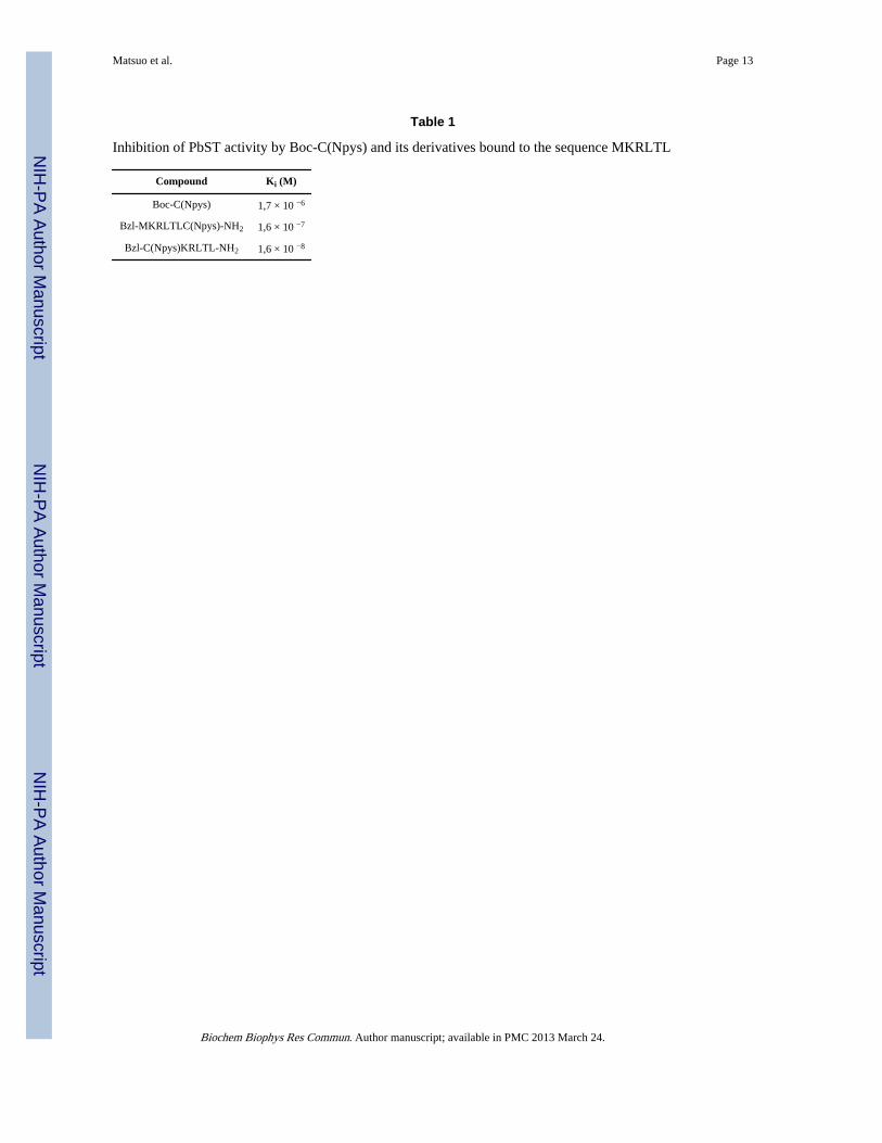
We observed that the three compounds inhibited the enzymatic activity against Abz-MKALTLQ-EDDnp [17] at different levels (Table 1). The best inhibitory activity was seenwith Bzl-C(Npys)KRLTL-NH2 (Ki = 16 nM), where the C(Npys) group was placed at theN-terminus substituting the original methionine residue. The peptide bearing C(Npys) at theC-terminus was one order of magnitude less effective (Ki = 160 nM), whereas the Boc-C(Npys) compound was a weaker inhibitor (Ki = 1,7 µM). These results indicated that thepeptide sequence promotes a favorable interaction increasing the magnitude of inhibition,especially when C(Npys) was placed at a further position from L-T, at the N-terminus. Theinhibitory effect of all tested peptides was completely reverted with 5 mM dithioerythritol,indicating that it was due to the reactivity of the C(Npys) moiety with a free SH- group ofthe proteinase (Fig. 2). It is noteworthy that the inhibitor peptides have not been cleaved bythe proteinase activity, as revealed by h.l.p.c. analysis (not shown).
We have previously reported on the PbST capacity to cleave components associated with thebasement membrane [3]. Presently, we have not been able to test the inhibitory capacity ofC(Npys) peptides on the cleavage of fibronectin visualized in SDS-PAGE gels (as in Fig. 1)because they altered fibronectin resolution. Therefore, we developed a cell detachment assayfor this purpose. We tested PbST ability to detach cells of the LLC-MK2 lineage fromplastic dishes, which is generally achieved using trypsin. Cell detachment depends oncleavage of extracellular matrix components or of surface integrins that directly interact withthem. Fig. 3A shows that PbST-4 was able to detach LLC-MK2 at increased rates over time,which reached a plateau corresponding to approximately 50% of total cells detached after 20min. Consequently, this incubation time was chosen for the inhibition experiments shownbelow. All experiments were carried out in triplicates and harvested cells were 70–80%viable. It is worth mentioning for comparison that 2.5 mg/mL of trypsin can detach all thecells (90–95% viable) in 5 min (not shown). Fig. 3B shows that the detachment activity wasdue to PbST, considering that pre-incubations with E-64 and pepstatin (or EDTA containedin the PBS) did not affect the activity, whereas p-HMB and PMSF totally abolished it (Fig.3B).
In order to evaluate the inhibitory capacity of C(Npys) peptides in the cell detachmentactivity of PbST-4, the experiments were performed with the enzyme preparation previouslyincubated or not (control) with 6 µM or 12 µM of each peptide for 20 min at 37°C. Fig 3Cshows that both Bzl-C(Npys)KRLTL-NH2 and Bzl-MKRLTLC(Npys)-NH2 exhibitedstatistically significant inhibitory effects, in a dose-dependent fashion. Bzl-C(Npys)KRLTL-NH2 was a better inhibitor than Bzl-MKRLTLC(Npys)-NH2, in agreement with the Kiresults seen in Table 1. Boc-Cys(Npys) showed no statistically significant effect at the
Matsuo et al. Page 5
Biochem Biophys Res Commun. Author manuscript; available in PMC 2013 March 24.
NIH
-PA Author Manuscript
NIH
-PA Author Manuscript
NIH
-PA Author Manuscript

concentrations tested, probably due to its lower binding affinity, as suggested from theresults in Table 1.
This is the first time C(Npys) compounds are used to inhibit a thiol-containing subtilisin. Itremains to be tested whether these compounds would effectively decrease P. brasiliensisvirulence in vivo. For that purpose, C(Npys) might be bound to peptides of the D-anomericity, for e.g., to avoid proteolysis by local endopeptidases.
The results presented here elegantly show that PbST activity can be blocked withcompounds interfering with a free cysteine, confirming that it belongs to the subtilisin S8family of serine proteinases, from which proteinase K from Tritirachium album is the beststudied member [21]. By analogy with the members of this group [22], we speculate thatthere is a thiol group lying near the imidazole of the histidine from the Ser-His-Asp catalytictriad. The presence of such a residue does not change the catalytic activity of subtilisinSavinase, although it makes it susceptible to mercurials [23]. In proteinase K, the thiol group(Cys78) is buried in the molecule and covalently binds to the first Hg+ molecule, whichdisrupts the catalytic triad by changing the imidazole conformation [24]. A second Hg+
complexes non-covalently with His72, Cys78 and Thr76, decreasing substrate-bindingaffinity. However, 100% inhibition by mercurials is never achieved, possibly due toflexibility of the molecule [24]. In contrast to proteinase K, PbST is selective regardingsubstrate specificity and is inhibited 100% by pHMB [2]. On the other hand, proximity ofthe thiol group to the catalytic triad would explain why the C(Npys)-peptide derivatives arebetter inhibitors than Boc-C(Npys), considering that the peptide used here fits in thesubstrate pocket to target the inhibitor.
AcknowledgmentsWe thank Dr. Ivarne Tersariol for critical review of the manuscript. This work and fellowships involved weresupported fully or partially by grants from FAPESP, PRONEX/CNPq, CNPq, and NIH/NCRR (grant no.5G12RR008124).
Abbreviations
Abz ortho-aminobenzoic acid
AUF arbitrary units of fluorescence
Bzl benzoyl
Boc tert-butyloxycarbonyl
C(Npys) S-(3-nitro-2-pyridinesulfenyl)
E-64 (L-trans-expoxysuccinyl-leucylamido[4-guanidino butane])
EDDnp N-(2,4-dinitrophenyl)ethylenediamine
FRET fluorescence resonance energy transfer
pABA p-aminobenzamidine
PbST exocellular serine-thiol activity from Paracoccidioides brasiliensis
PCM paracoccidioidomycosis
p-HMB sodium 7-hydroxymercuribenzoate
PMSF phenylmethylsulfonyl fluoride
Matsuo et al. Page 6
Biochem Biophys Res Commun. Author manuscript; available in PMC 2013 March 24.
NIH
-PA Author Manuscript
NIH
-PA Author Manuscript
NIH
-PA Author Manuscript

References1. Monod M, Capoccia S, Lechenne B, Zaugg C, Holdom M, Jousson O. Secreted proteases from
pathogenic fungi. Int. J. Med. Microbiol. 2002; 292:405–419. [PubMed: 12452286]
2. Carmona AK, Puccia R, Oliveira MCF, Rodrigues EG, Juliano L, Travassos LR. Characterization ofan exocellular serine-thiol proteinase activity in Paracoccidioides brasiliensis. Biochem. J. 1995;309:209–214. [PubMed: 7619058]
3. Puccia R, Carmona AK, Gesztesi JL, Juliano L, Travassos LR. Exocellular proteolytic activity ofParacoccidioides brasiliensis: cleavage of components associated with the basement membrane.Med. Mycol. 1998; 36:345–348. [PubMed: 10075506]
4. Matsuo AL, Tersariol IIL, Kobata SI, Travassos LR, Carmona AK, Puccia R. Modulation of theexocellular serine-thiol proteinase activity of Paracoccidioides brasiliensis by neutralpolysaccharides. Microbes Infect. 2006; 8:84–91. [PubMed: 16153872]
5. San Blas G, Nino-Vega G, Iturriaga T. Paracoccidioides brasiliensis and paracoccidioidomycosis:Molecular approaches to morphogenesis, diagnosis, epidemiology, taxonomy and genetics. Med.Mycol. 2002; 40:225–242. [PubMed: 12146752]
6. Matsueda R, Walter R. 3-nitro-2-pyridinesulfenyl (Npys) group - a novel selective protecting groupwhich can be activated for peptide-bond formation. Int. J. Pept. Protein Res. 1980; 16:392–401.[PubMed: 7216615]
7. Matsueda R, Umeyama H, Kominami E, Katunuma N. Design and synthesis of cathepsin-Binhibitors by an affinity labeling approach. Chem. Lett. 1988:1857–1860.
8. Bernatowicz MS, Matsueda R, Matsueda GR. Preparation of Boc-[S-(3-nitro-2-pyridinesulfenyl)]-cysteine and its use for unsymmetrical disulfide bond formation. Int. J. Pept. Protein Res. 1986;28:107–112. [PubMed: 3771096]
9. Rich, DH. Proteinase inhibitors. Barret, AJ.; Salvesen, G., editors. Elsevier Science Publishers;1986. p. 153
10. Matsueda R, Umeyama H, Puri RN, Bradford HN, Colman RW. Potent affinity labeling peptideinhibitors of calpain. Chem. Lett. 1990:191–194.
11. Dejana, E.; Corada, M. Adhesion protocols. In: Walker, JM., editor. Methods in MolecularBiology. Editora Humana; 1990. p. 119-124.
12. Laemmli UK. Cleavage of structural proteins during assembly of head of bacteriophage-T4.Nature. 1970; 227:680–685. [PubMed: 5432063]
13. Hirata M, Nemoto K, Hirata A. Solubility of N-(benzyloxycarbonyl)-L-aspartyl-L-phenylalaninemethyl-ester forming a complex with L-phenylalanine methyl-ester in aqueous system. J. Chem.Eng. 1994; 27:72–76.
14. Barany, G.; Merrifield, RB. The peptides: analysis, synthesis and biology. New York: AcademicPress; 1980.
15. Mortz E, Krogh TN, Vorum H, Gorg A. Improved silver staining protocols for high sensitivityprotein identification using matrix-assisted laser desorption/ionization-time of flight analysis.Proteomics. 2001; 1:1359–1363. [PubMed: 11922595]
16. Shevchenko A, Wilm M, Vorm O, Mann M. Mass spectrometric sequencing of proteins from silverstained polyacrylamide gels. Anal. Chem. 1996; 68:850–858. [PubMed: 8779443]
17. Puccia R, Juliano MA, Juliano L, Travassos LR, Carmona AK. Detection of the basementmembrane-degrading proteolytic activity of Paracoccidioides brasiliensis after SDS-PAGE usingagarose overlays containing Abz-MKALTLQ-EDDnp. Braz. J. Med. Biol. Res. 1999; 32:645–649.[PubMed: 10412577]
18. Kanamori A, Seno N, Matsumoto I. Chromatogr J. Preparation of high-capacity affinity adsorbentsusing formyl carriers and their use for low-performance and high-performance liquid affinity-chromatography of trypsin-family proteases. J. Chromatogr. 1986; 363:231–242.
19. Goldman GH, Marques ED, Ribeiro DCD, Bernardes LAD, Quiapin AC, Vitorelli PM, Savoldi M,Semighini CP, Oliveira RC, Nunes LR, Travassos LR, Puccia R, Batista WL, Ferreira LE, MoreiraJC, Bogossian AP, Tekaia F, Nobrega MP, Nobrega FG, Goldman MHS. Expressed sequence taganalysis of the human pathogen Paracoccidioides brasiliensis yeast phase: Identification of
Matsuo et al. Page 7
Biochem Biophys Res Commun. Author manuscript; available in PMC 2013 March 24.
NIH
-PA Author Manuscript
NIH
-PA Author Manuscript
NIH
-PA Author Manuscript

putative homologues of Candida albicans virulence and pathogenicity genes. Eukaryot. Cell. 2003;2:34–48. [PubMed: 12582121]
20. Felipe MSS, Andrade RV, Arraes FBM, Nicola AM, Maranhao AQ, Torres FAG, Silva-Pereira I,Pocas-Fonseca MJ, Campos EG, Moraes LMP, Andrade PA, Tavares AHFP, Silva SS, Kyaw CM,Souza DP, Network P, Pereira M, Jesuino RSA, Andrade EV, Parente JA, Oliveira GS, BarbosaMS, Martins NF, Fachin AL, Cardoso RS, Passos GAS, Almeida NF, Walter MEMT, SoaresCMA, Carvalho MJA, Brigido MM. Transcriptional profiles of the human pathogenic fungusParacoccidioides brasiliensis in mycelium and yeast cells. J. Biol.Chem. 2005; 280:24706–24714.[PubMed: 15849188]
21. Pahler A, Banerjee A, Dattagupta JK, Fujiwara T, Lindner K, Pal GP, Suck D, Weber G, SaengerW. Three-dimensional structure of fungal proteinase K reveals similarity to bacterial subtilisin.EMBO J. 1984; 3:1311–1314. [PubMed: 6378621]
22. Siezen RJ, Leunissen JA. Subtilases: the superfamily of subtilisin-like serine proteases. Protein Sci.1997; 6:501–523. [PubMed: 9070434]
23. Muller A, Saenger W. Studies on the inhibitory action of mercury upon proteinase K. J Biol.Chem.1993; 268:26150–26154. [PubMed: 8253733]
24. Bech LM, Branner S, Hastrup S, Breddam K. Introduction of a free cysteinyl residue at position 68in the subtilisin Savinase, based on homology with proteinase K. FEBS Lett. 1992; 297:164–166.[PubMed: 1551423]
Matsuo et al. Page 8
Biochem Biophys Res Commun. Author manuscript; available in PMC 2013 March 24.
NIH
-PA Author Manuscript
NIH
-PA Author Manuscript
NIH
-PA Author Manuscript

Fig 1.Isolation of PbST by chromatography in a pABA-Sepharose column. Upper panel: silver-stained 10% SDS-PAGE gel showing the profile of culture supernatant (10 µl) from Pb339yeast cells before (In) and after (FT) chromatography. Eluted fractions are numbered. Thehydrolytic activity against Abz-MKALTLQ-EDDnp produced by a 10 µl-aliquot of eachfraction is shown as arbitrary units of fluorescence per minute (AUF/min). The results arenot presented in terms of specific activity due to the low protein contents. The hydrolyticactivity against fibronectin (FN) correspondent to incubation with each fraction at 37°C for30 min is shown in a Coomassie blue-stained 8% SDS-PAGE gel. A time-course of FNcleavage after incubation for 5, 10, 15, and 30 min with peak fraction 4 (10 µl) is also
Matsuo et al. Page 9
Biochem Biophys Res Commun. Author manuscript; available in PMC 2013 March 24.
NIH
-PA Author Manuscript
NIH
-PA Author Manuscript
NIH
-PA Author Manuscript

depicted. The positions of molecular mass markers (kDa) and of one of the FN chain areshown on the left.
Matsuo et al. Page 10
Biochem Biophys Res Commun. Author manuscript; available in PMC 2013 March 24.
NIH
-PA Author Manuscript
NIH
-PA Author Manuscript
NIH
-PA Author Manuscript

Fig. 2.Shematic representation of the mechanism of inhibition of thiol proteinases by Cys(Npy).
Matsuo et al. Page 11
Biochem Biophys Res Commun. Author manuscript; available in PMC 2013 March 24.
NIH
-PA Author Manuscript
NIH
-PA Author Manuscript
NIH
-PA Author Manuscript

Fig. 3.Evaluation of PbST inhibition in a cell detachment assay. A, Time-course of cell detachmentwhen incubated with PbST-4 (10 µL, fraction 4, Fig. 1). B, Percentage of cell detachmentwhen compared to the control (100%), as counted after 20 min of incubation with PbST-4 inthe presence of either standard proteinase inhibitors or Bzl-MKRLTLC(Npys)-NH2 (C-),Bzl-C(Npys)KRLTL-NH2 (N-) and Boc-Cys(Npys) (Boc-) at the indicated concentrations.Incubation of cell cultures with equivalent concentrations of the inhibitors alone has notresulted in cell detachment or loss of cell viability (not shown). Statistically significantvalues (p < 0.05) are indicated with an asterisk.
Matsuo et al. Page 12
Biochem Biophys Res Commun. Author manuscript; available in PMC 2013 March 24.
NIH
-PA Author Manuscript
NIH
-PA Author Manuscript
NIH
-PA Author Manuscript
NIH
-PA Author Manuscript
NIH
-PA Author Manuscript
NIH
-PA Author Manuscript
Matsuo et al. Page 13
Table 1
Inhibition of PbST activity by Boc-C(Npys) and its derivatives bound to the sequence MKRLTL
Compound Ki (M)
Boc-C(Npys) 1,7 × 10 −6
Bzl-MKRLTLC(Npys)-NH2 1,6 × 10 −7
Bzl-C(Npys)KRLTL-NH2 1,6 × 10 −8
Biochem Biophys Res Commun. Author manuscript; available in PMC 2013 March 24.