Diversity and functional significance of cellulolytic microbes living in termite, pill-bug and...
11
Diversity and functional significance of cellulolytic microbes living in termite, pill-bug and stem-borer guts Zeenat Bashir 1 *, Vamsi Krishna Kondapalli 1 *, Nidhi Adlakha 1 , Anil Sharma 2 , Raj Bhatnagar 2 , Girish Chandel 3 & Syed Shams Yazdani 1 1 Synthetic Biology and Biofuels Group, 2 Insect Resistant Group, International Centre for Genetic Engineering and Biotechnology, Aruna Asaf Ali Marg, New Delhi, 3 Department of Plant Molecular Biology and Biotechnology, College of Agriculture, Indira Gandhi Agricultural University, Raipur (C.G.). Arthropods living on plants are able to digest plant biomass with the help of microbial flora in their guts. This study considered three arthropods from different niches - termites, pill-bugs and yellow stem-borers - and screened their guts for cellulase producing microbes. Among 42 unique cellulase-producing strains, 50% belonged to Bacillaceae, 26% belonged to Enterobacteriaceae, 17% belonged to Microbacteriaceae, 5% belonged to Paenibacillaceae and 2% belonged to Promicromonosporaceae. The distribution of microbial families in the three arthropod guts reflected differences in their food consumption habits. Most of the carboxymethylcellulase positive strains also hydrolysed other amorphous substrates such as xylan, locust bean gum and b-D-glucan. Two strains, A11 and A21, demonstrated significant activity towards Avicel and p-nitrophenyl-b-D-cellobiose, indicating that they express cellobiohydrolase. These results provide insight into the co-existence of symbionts in the guts of arthropods and their possible exploitation for the production of fuels and chemicals derived from plant biomass. R ising concern over the cost and sustained availability of fossil fuels has increased demand for the develop- ment of alternative and preferably renewable fuel resources, with lignocellulosic biomass as one promising resource 1,2 . Lignocellulose represents ,90% of the dry weight of all plant materials 3 , is primarily composed of the sugar polymers cellulose (35–50%) and hemicellulose (20–35%) together with lignin (5–30%) that provides structural support for the plant 4 . Cellulose and hemicellulose represent abundant and inexpensive sources of fermentable sugar for the production of various chemicals and biofuels, but finding efficient catalysts for their saccharification to monomeric sugar and producing those catalysts in a cost-effective manner are major hurdles 5 . Biological catalysts such as cellulase/hemicellulase enzymes play significant roles in the production of ethanol using plant biomass 6 . The enzymatic hydrolysis of cellulose polymers is achieved by the synergistic activity of three different groups of cellulase enzymes 1 : endoglucanase 2 , exoglucanase or cellobiohydrolase, and 3 b-gluco- sidase 7,8 . Arabinoxylanases and xylanases are groups of enzymes that hydrolyse hemicellulose components. A large number of these hydrolytic enzymes have been discovered in fungi 9 , but they are typically associated with certain limitations, such as stability to low pH and thermal stresses and low specific activities 10 . As bacteria are able to tolerate environmental extremes, they represent an ideal source for the screening and isolation of novel cellulolytic enzymes to help overcome these challenges 10 . Therefore, researchers are investigating natural systems that have evolved to decompose plant cell wall polymers as sources for enzyme discovery. Arthropods are the most abundant and successful species on earth that can survive in diverse ecological niches 11,12 . The digestive tracts of arthropods contain microbiota members from the bacterial, fungal, protozoan and archaeal groups, which assist with various physiological functions including food digestion, nitrogen fixation, nutrition and pheromone production 13 . Comprehensive analyses of the transcriptomes and metagenomes of termites and symbiotic microbes have generated huge amounts of genomic information for the screening of cellulase encoding genes 14,15 . However, enzymes screened through metagenomic and metatranscriptomic studies could have issues with heterologous overexpression in terms of folding and yield 16,17 because little is known about the host organism. Therefore, natural cellulolytic bacteria that can be cultured under laboratory conditions are excellent platforms for the study of the properties of hydrolytic enzymes. In the present work, we have selected three arthropods with different dietary habits, i.e., two insects (termites and yellow stem-borers) and a crustacean (pill-bugs), and explored their guts as sources for the isolation and SCIENTIFIC REPORTS SREP-13-01671-T.3d 20/8/13 14:51:51 OPEN SUBJECT AREAS: BIOTECHNOLOGY MICROBIOLOGY BIOCATALYSIS MICROBIAL ECOLOGY Received 22 April 2013 Accepted 16 August 2013 Published 30 August 2013 Correspondence and requests for materials should be addressed to S.S.Y. (shams@icgeb. res.in) * These authors contributed equally to this work. SCIENTIFIC REPORTS | 3 : 2558 | DOI: 10.1038/srep02558 1
-
Upload
independent -
Category
Documents
-
view
0 -
download
0
Transcript of Diversity and functional significance of cellulolytic microbes living in termite, pill-bug and...

Diversity and functional significance ofcellulolytic microbes living in termite,pill-bug and stem-borer gutsZeenat Bashir1*, Vamsi Krishna Kondapalli1*, Nidhi Adlakha1, Anil Sharma2, Raj Bhatnagar2,Girish Chandel3 & Syed Shams Yazdani1
1Synthetic Biology and Biofuels Group, 2Insect Resistant Group, International Centre for Genetic Engineering and Biotechnology,Aruna Asaf Ali Marg, New Delhi, 3Department of Plant Molecular Biology and Biotechnology, College of Agriculture, Indira GandhiAgricultural University, Raipur (C.G.).
Arthropods living on plants are able to digest plant biomass with the help of microbial flora in their guts.This study considered three arthropods from different niches - termites, pill-bugs and yellow stem-borers -and screened their guts for cellulase producing microbes. Among 42 unique cellulase-producing strains,50% belonged to Bacillaceae, 26% belonged to Enterobacteriaceae, 17% belonged to Microbacteriaceae, 5%belonged to Paenibacillaceae and 2% belonged to Promicromonosporaceae. The distribution of microbialfamilies in the three arthropod guts reflected differences in their food consumption habits. Most of thecarboxymethylcellulase positive strains also hydrolysed other amorphous substrates such as xylan, locustbean gum and b-D-glucan. Two strains, A11 and A21, demonstrated significant activity towards Avicel andp-nitrophenyl-b-D-cellobiose, indicating that they express cellobiohydrolase. These results provide insightinto the co-existence of symbionts in the guts of arthropods and their possible exploitation for theproduction of fuels and chemicals derived from plant biomass.
Rising concern over the cost and sustained availability of fossil fuels has increased demand for the develop-ment of alternative and preferably renewable fuel resources, with lignocellulosic biomass as one promisingresource1,2. Lignocellulose represents ,90% of the dry weight of all plant materials3, is primarily composed
of the sugar polymers cellulose (35–50%) and hemicellulose (20–35%) together with lignin (5–30%) that providesstructural support for the plant4. Cellulose and hemicellulose represent abundant and inexpensive sources offermentable sugar for the production of various chemicals and biofuels, but finding efficient catalysts for theirsaccharification to monomeric sugar and producing those catalysts in a cost-effective manner are major hurdles5.
Biological catalysts such as cellulase/hemicellulase enzymes play significant roles in the production of ethanolusing plant biomass6. The enzymatic hydrolysis of cellulose polymers is achieved by the synergistic activity ofthree different groups of cellulase enzymes1: endoglucanase2, exoglucanase or cellobiohydrolase, and3 b-gluco-sidase7,8. Arabinoxylanases and xylanases are groups of enzymes that hydrolyse hemicellulose components. Alarge number of these hydrolytic enzymes have been discovered in fungi9, but they are typically associated withcertain limitations, such as stability to low pH and thermal stresses and low specific activities10. As bacteria areable to tolerate environmental extremes, they represent an ideal source for the screening and isolation of novelcellulolytic enzymes to help overcome these challenges10. Therefore, researchers are investigating natural systemsthat have evolved to decompose plant cell wall polymers as sources for enzyme discovery.
Arthropods are the most abundant and successful species on earth that can survive in diverse ecologicalniches11,12. The digestive tracts of arthropods contain microbiota members from the bacterial, fungal, protozoanand archaeal groups, which assist with various physiological functions including food digestion, nitrogen fixation,nutrition and pheromone production13. Comprehensive analyses of the transcriptomes and metagenomes oftermites and symbiotic microbes have generated huge amounts of genomic information for the screening ofcellulase encoding genes14,15. However, enzymes screened through metagenomic and metatranscriptomic studiescould have issues with heterologous overexpression in terms of folding and yield16,17 because little is known aboutthe host organism. Therefore, natural cellulolytic bacteria that can be cultured under laboratory conditions areexcellent platforms for the study of the properties of hydrolytic enzymes.
In the present work, we have selected three arthropods with different dietary habits, i.e., two insects (termitesand yellow stem-borers) and a crustacean (pill-bugs), and explored their guts as sources for the isolation and
SCIENTIFIC REPORTS SREP-13-01671-T.3d 20/8/13 14:51:51
OPEN
SUBJECT AREAS:BIOTECHNOLOGY
MICROBIOLOGY
BIOCATALYSIS
MICROBIAL ECOLOGY
Received22 April 2013
Accepted16 August 2013
Published30 August 2013
Correspondence andrequests for materials
should be addressed toS.S.Y. (shams@icgeb.
res.in)
* These authorscontributed equally to
this work.
SCIENTIFIC REPORTS | 3 : 2558 | DOI: 10.1038/srep02558 1

characterisation of cellulose-degrading bacteria. The ability of thesearthropods to feed on wood, foliage and detritus is likely to involvecatalysis by different types of cellulases/hemicellulases that aresecreted by gut microbiota to digest the structural and recalcitrantlignocellulosic residues in their foods. Thus, the main goals of thisstudy were to explore the guts of these arthropods that feed onpotential biofuel feedstocks and to compare their hydrolytic activ-ities. We successfully isolated 42 cellulose-degrading bacterial strainsfrom these arthropods and characterised their abilities to producehydrolytic enzymes.
ResultsScreening and characterisation of cellulolytic bacteria from theguts of arthropods. The guts of arthropods feeding on woody andagricultural biomass are an ideal place for symbiotic cellulolyticmicrobes to thrive. We selected two insects, termites and yellowstem borers, and a crustacean, pill-bugs, because of their ability tolive in diverse niches. Termites were collected from the trunks androots of dead trees; pill-bugs were collected from dampenedsoil containing decomposed leaf litters; and yellow stem borersliving in rice stems were collected from an agricultural rice field(Figure 1). Arthropod families were identified by performingBLAST with the nucleotide sequences of the mitochondrial cyto-chrome oxidase I (CO-I) genes from each organism and furtherconfirmed by morphological characterisation. The nearest possiblehits for termites, pill-bugs and yellow stem borers were Odontotermeshiananensis, Armadillidium sp. and Scirpophaga incertulas, respec-tively. The guts of these arthropods were dissected as described in theMethods section, and the microbes living in the guts were grown onagar plates containing CMC and trypan blue at various dilutionsunder both aerobic and anaerobic conditions. Unique isolated colo-nies showing clearance zones on a blue background were purified byre-streaking, and their cellulolytic capabilities were further con-firmed by Congo red staining of agar plates18. Approximately 42bacterial colonies were selected by the above screening method forphylogenetic analysis and enzymatic activity analysis. Among these,26 bacteria were isolated by screening under aerobic conditions, and16 were isolated under anaerobic conditions (Table 1).
Phylogenetic analysis of gut bacteria. Among the 42 bacterialcolonies selected for further characterisation, 17 were isolated fromtermite guts, 8 were isolated from pill-bug guts and 17 were isolatedfrom yellow stem borer guts (Table 1). Phylogenetic analysis of the16S rDNA sequences revealed that all identified bacterial speciesbelonged to five families - 50% belonged to Bacillaceae, 26%belonged to Enterobacteriaceae, 17% belonged to Microbacteria-ceae, 5% belonged to Paenibacillaceae and 2% belonged toPromicromonosporaceae (Figure 2A). Between 60 and 70% of thebacteria from the guts of termites and pill-bugs were from theBacillaceae family, while bacteria from the Microbacteriaceae andEnterobacteriaceae families were more dominant in the guts ofyellow stem borers (Figure 2B).
Characterisation of bacterial enzymes hydrolysing plant biomass.Microbes residing in the guts of biomass feeding arthropods arelikely to produce a variety of hydrolytic enzymes. In previousstudies we showed that Paenibacillus ICGEB2008 isolated from thegut of the cotton bollworm produced several biomass-degradingenzymes, including cellulases and hemicellulases18,19. Therefore, wetested the expression levels in the gut microbes of major glycosylhydrolase enzymes belonging to the categories of cellulase,hemicellulase, mannanase and glucanase. The expression levels ofendoglucanase, b-glucosidase, xylanase, b-xylosidase, mannanaseand b-D-glucanase were determined using the substrates CMC,pNPG, Xylan, pNPX, locust bean gum and barley b-D-glucan,respectively. To test exocellulase or cellobiohydrolase expression,both Avicel and pNPC (in the presence of the b-glucosidaseinhibitor glucono-d-lactone) were used as substrates. A potentialbiofuel feedstock was also used, namely alkali treated rice straw,and it mainly contained crystalline cellulose as well as someamorphous forms of cellulose. These assays were used to test bothextracellular and cell associated enzymes. Except for A13, A16 andT3, all natural isolates produced at least one type of glycosylhydrolase (Figure 3A and 3B). The reason that these three strainsare negative in the liquid assay despite being positive for CMCactivity on the agar plates could be related to the stringency criteriaset as described in the Methods section. The majority of microbesproduced more than one kind of hydrolytic enzyme (Figure 3C).There were 23 microbes that produced at least one hydrolyticenzyme from each category of biomass degrading enzymes. Severalstrains produced only one category of enzyme, such as cellulases (5strains) and hemicellulases (1 strain), which hydrolysed pNPG andpNPX, respectively, but did not produce significant amounts of anyother type of hydrolase (Figure 3). Extracellular fractions were foundto be rich in enzymes specific for amorphous polymers (Figure 3A).Only a few microbes secreted enzymes that were able to hydrolysedimeric or oligomeric substrates, such as pNPG, pNPC and pNPX, orcomplex structures, such as rice straw. On the other hand, themajority of cell associated fractions hydrolysed dimeric substratessuch as pNPG and pNPX (Figure 3B). However, cell associatedfractions of none of the microbes were able to hydrolyse pNPC.Some strains also produced enzymes in the extracellular or cell-associated fractions for the hydrolysis of complex structures suchas rice straw and Avicel.
Quantitative assessments of the hydrolytic activities in both theextracellular and cell-associated fractions enabled us to analyse theirrelative contributions to biomass degradation (Figure 4, 5 and 6). Themajority of strains secreted high quantities of enzymes specific foramorphous polymers such as CMC, xylan, locust bean gum andbarley b-D-glucan (Figure 4). Relatively fewer strains producedenzymes that were able to hydrolyse significant amounts of crystal-line polymers such as Avicel and rice straw (Figure 5D, 6A and 6D).The strains hydrolysing the greatest amounts of rice straw were A11,A18, A21, A22, S2 and U8 (Figure 5D and 6D), while strains A11,A17, A21, A24, U5 and U10 hydrolysed Avicel (Figure 6A). Enzymes
Figure 1 | Pictures of termites, pill-bugs and rice stem-borers collected from various ecological niches.
www.nature.com/scientificreports
SCIENTIFIC REPORTS | 3 : 2558 | DOI: 10.1038/srep02558 2

Tabl
e1
|Cod
ing
ofce
llulo
lytic
mic
robe
sis
olat
edfr
omin
sect
guts
Inse
ct
Aer
obic
scre
enin
gA
naer
obic
scre
enin
g
Stra
inID
Gen
Bank
Acc
essi
onN
eare
stre
lativ
eSt
rain
IDG
enBa
nkA
cces
sion
Nea
rest
rela
tive
Term
iteA
1KC
4349
60Ba
cillu
ssp
.606
3T1
KC43
4986
Baci
llus
subt
ilis
M50
A4
KC43
4961
Kleb
siel
lasp
.clo
neF7
T2KC
4349
87Ba
cillu
slic
heni
form
isA
CO
1A
5KC
4349
62Tr
abul
siel
lagu
amen
sis
GTC
1379
T3KC
4349
88Tr
abul
siel
lagu
amen
sis
GTC
1379
A6
KC43
4963
Baci
llus
pum
ilus
BSH
4T4
KC43
4989
Baci
llus
sp.D
V9-3
5A
7KC
4349
64Ba
cillu
ssp
.SC
SSS1
0A
9KC
4349
65Pa
ntoe
aag
glom
eran
sW
AB1
927
A10
KC43
4966
Baci
llus
liche
nifo
rmis
EdyK
olBl
23A
11KC
4349
67Ba
cillu
slic
heni
form
isBC
RC15
413
A12
KC43
4968
Baci
llus
liche
nifo
rmis
Suba
Muc
Bl16
A13
KC43
4969
Baci
llus
cere
usTA
UC
5A
16KC
4349
70Ba
cillu
sce
reus
F837
/76
A17
KC43
4971
Baci
llus
subt
ilis
K21
A18
KC43
4972
Paen
ibac
illus
poly
myx
aD
SM36
T
Pill-B
ugA
19KC
4349
73Pa
enib
acill
uspo
lym
yxa
YRL1
3W
1KC
4349
90Ba
cillu
ssu
btili
sKL
-073
A21
KC43
4974
Baci
llus
subt
ilis
M50
W6
KC43
4991
Baci
llus
thur
ingi
ensi
s2P
R56-
10A
22KC
4349
75Ba
cillu
ssu
btili
sM
16K
W7
KC43
4992
Baci
llus
tequ
ilens
isVI
TJA
AM
2A
23KC
4349
76En
tero
bact
erae
roge
nes
KCTC
2190
A24
KC43
4977
Cel
lulo
sim
icro
bium
sp.T
UT1
242
Yello
wSt
emBo
rer
S1KC
4349
78Ba
cillu
ssp
ecie
sBB
2_1A
U2
KC43
4993
Baci
llus
sp.P
R1.
7S2
KC43
4979
Baci
llus
subt
ilis
AQ
1U
3KC
4349
94Pa
ntoe
asp
ecie
sN
CC
P116
S3KC
4349
80M
icro
bact
eria
ceae
bact
eriu
mH
LB-6
U4
KC43
4995
Pant
oea
aggl
omer
ans
WA
B192
7S4
KC43
4981
Mic
roba
cter
iace
aeba
cter
ium
BMC
-3U
5KC
4349
96Kl
ebsi
ella
pneu
mon
iae
L-13
S5KC
4349
82M
icro
bact
eriu
mol
eivo
rans
CC
GE2
277
U6
KC43
4997
Kleb
siel
lapn
eum
onia
eL-1
3S6
KC43
4983
Mic
roba
cter
ium
arbo
resc
ens
DSM
2075
4U
7KC
4349
98M
icro
bact
eria
ceae
bact
eriu
mBM
C-3
S7KC
4349
84M
icro
bact
eriu
mar
bore
scen
sD
SM20
754
U8
KC43
4999
Baci
llus
subt
ilis
C1Y
001
S8KC
4349
85M
icro
bact
eriu
mar
bore
scen
sJB
8_2B
U9
KC43
500
Ente
roba
cter
spec
ies
DH
M1T
U10
KC43
501
Serr
atia
mar
cesc
ens
PS1
www.nature.com/scientificreports
SCIENTIFIC REPORTS | 3 : 2558 | DOI: 10.1038/srep02558 3

Figure 2 | Classification and phylogenetic analysis of natural isolates from the guts of termites, pill-bugs and yellow stem-borers. (A) 16s rDNA
sequences of 42 isolates screened for their ability to produce cellulolytic enzymes obtained from the guts of termites (.), pill bugs (#) and yellow stem
borers (&) were used to perform blast searches to identify their nearest neighbors, and ClustalW was used to construct the phylogenetic tree. The natural
isolates were found to belong to five families. (B) Relative abundance of various bacterial families in the guts of these three arthropods.
www.nature.com/scientificreports
SCIENTIFIC REPORTS | 3 : 2558 | DOI: 10.1038/srep02558 4

secreted from strains A11 and A21 also hydrolysed pNPC, indicatingthat these strains might produce either cellobiohydrolase or proces-sive endoglucanase (Figure 5B). None of the cell bound fractionsfrom any of the strains hydrolysed pNPC. Among b-glucosidaseproducers, a distinct set of strains exhibited extracellular and cell-associated b-glucosidase activity (Figure 5A and Figure 6B). Onlythree strains, A11, A19 and U8, produced extracellular b-xylosidase(Figure 5C). The remaining xylosidase producers expressed the b-xylosidase enzyme intracellularly (Figure 6C).
Cellulase and hemicellulase production were further evaluatedusing a zymogram. We selected 18 strains from the CMC positivebatch and tested the extracellular enzymatic activity by a zymogram.The strains that secreted high quantities of cellulase, A11, A17, A18,A21, A22, U8 and U9 (Figure 4A), also showed significant clearancezones on the zymogram (Figure 7A). Most of the strains secretedcellulases in the range of 72–130 kDa, and a few secreted cellulases inthe range of 43–53 kDa. The zymogram results were somewhatambiguous for xylanase activity. Although 16 of the 18 strains testedby zymogram exhibited significant xylanase activity in the liquidculture assay (Figure 4B), only 5 strains showed xylanolytic activityon the zymogram (Figure 7B). The samples of these 5 strains con-tained high molecular weight bands, possibly protein oligomers, thatcould not be resolved by the gel. Similar problems might haveoccurred with other xylanase positive strains, thus preventing the
detection of activity by the zymogram. Four strains, A10, A12, T4and W7, exhibited xylanase activity at ,55 kDa, while strain S7exhibited xylanase activity at ,100 kDa (Figure 7B).
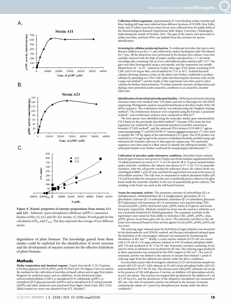
Kinetic properties of pNPC hydrolysing cellulases. Two strains,A11 and A21, displayed enzymatic activities against pNPC, indi-cating that they were secreting either cellobiohydrolase or proces-sive endoglucanase. Therefore, we further characterised the enzymespresent in these strains by partially purifying extracellular fractionsvia ultrafiltration using a 10-kDa cut-off membrane and then usingthat preparation to analyse their kinetic properties. The enzymeprepared in this way from the A11 strain exhibited a 4-fold higheractivity at the saturating substrate concentration compared to theA21 strain (Figure 8A and 8B). The Km and Vmax values calculatedfrom the Hanes Woolf plot (Figure 8C) were 8.47 mM and263 nmol/min for A11, respectively, and 7.06 mM and 158 nmol/min for A21, respectively. These parameters suggest that althoughthe enzyme preparations from both microbes exhibit similarsubstrate affinities, the enzyme preparation from A11 has a 1.7times higher catalytic efficiency than A21.
DiscussionFinding optimal enzymes for biomass hydrolysis remains a chal-lenge for scientists developing methods of lignocellulosic ethanol
Figure 3 | Qualitative assessment of hydrolytic enzymes produced by various natural isolates. Enzyme production was tested in the extracellular (A)
and cell-bound fractions (B) against amorphous, crystalline and chromogenic substrates. Black and white boxes represent significant and non-significant
activities, respectively, in the liquid culture assay. (C) Venn diagram for natural isolates exhibiting cellulase, hemicellulase and other glycosidase activities.
Cellulase positive strains were considered to be those that hydrolysed CMC, Avicel, rice straw, pNPC or pNPG. Hemicellulase positive strains were those
that hydrolysed xylan or pNPX. Strains that hydrolysed locust bean gum or b-D-glucan were considered to be positive for other glycosyl hydrolases.
www.nature.com/scientificreports
SCIENTIFIC REPORTS | 3 : 2558 | DOI: 10.1038/srep02558 5

Figure 4 | Quantitative assessment of the enzymatic activities of extracellular fractions against amorphous polysaccharides. All bacterial strains that
were found to be cellulase positive by the agar plate assay were grown in liquid medium, and their extracellular fractions were tested for the hydrolysis of
various substrates. Each graph indicates the substrate. Black bars represent strains that exhibited significantly higher activities compared to the negative
controls, while white bars represent negative strains. The data represent the average and standard deviation of two different assays.
www.nature.com/scientificreports
SCIENTIFIC REPORTS | 3 : 2558 | DOI: 10.1038/srep02558 6
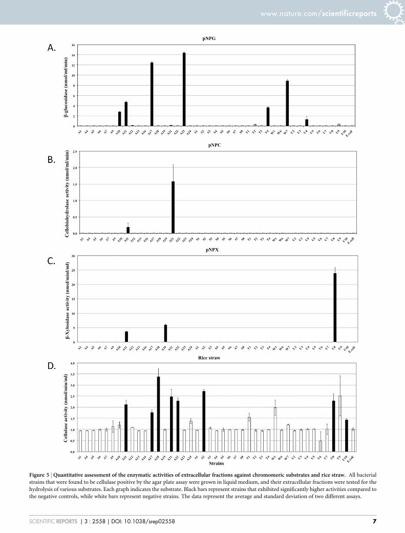
Figure 5 | Quantitative assessment of the enzymatic activities of extracellular fractions against chromomeric substrates and rice straw. All bacterial
strains that were found to be cellulase positive by the agar plate assay were grown in liquid medium, and their extracellular fractions were tested for the
hydrolysis of various substrates. Each graph indicates the substrate. Black bars represent strains that exhibited significantly higher activities compared to
the negative controls, while white bars represent negative strains. The data represent the average and standard deviation of two different assays.
www.nature.com/scientificreports
SCIENTIFIC REPORTS | 3 : 2558 | DOI: 10.1038/srep02558 7
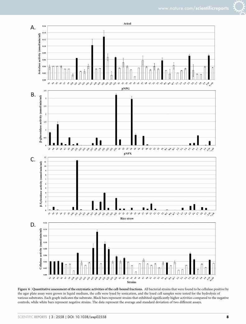
Figure 6 | Quantitative assessment of the enzymatic activities of the cell-bound fractions. All bacterial strains that were found to be cellulase positive by
the agar plate assay were grown in liquid medium, the cells were lysed by sonication, and the lysed cell samples were tested for the hydrolysis of
various substrates. Each graph indicates the substrate. Black bars represent strains that exhibited significantly higher activities compared to the negative
controls, while white bars represent negative strains. The data represent the average and standard deviation of two different assays.
www.nature.com/scientificreports
SCIENTIFIC REPORTS | 3 : 2558 | DOI: 10.1038/srep02558 8

production. Nature has created reservoirs of catalysts that performthis function in a variety of niches. We have attempted to mine thesebiological reservoirs by selecting arthropods from three diverseniches and screening for microbes capable of biomass hydrolysis intheir guts. While other methodologies such as metagenomics andmetatranscriptomics are becoming popular approaches to screeningfor biomass hydrolysing enzymes, they are often associated withissues of low heterologous expression yield and protein misfolding16.Our approach of identifying culturable microbes with biomasshydrolysing capabilities has the advantage of studying the enzymecharacteristics in both homologous and heterologous hosts.
This study has revealed significant knowledge regarding thediversity of microbes existing in the guts of arthropods and theirroles in biomass degradation. Among the arthropods we selectedfor this study, termites are the best-studied arthropods in terms ofcellulolytic microbes13–15, although the majority of the cellulolyticmicrobes that have been identified are difficult to culture in thelab. No previous studies have reported on the nature of cellulolyticmicrobes existing in the guts of pill-bugs and yellow stem borers. Wefound that greater than 60% of the cellulolytic microbes isolated fromthe guts of termites and pill-bugs belonged to the Bacillaceae family,while greater than 75% of the cellulolytic microbes isolated from theguts of yellow stem borers belonged to the Microbacteriaceae andEnterobacteriaceae families. These differences in the microbial florareflected differences in the food habits of the arthropods. Whiletermites and pill-bugs were collected from decomposed trees andlitter, yellow stem borer larvae were collected from rice plants atthe time of seeding. The pill-bug gut also contained Cellulosimicro-bium sp., a known natural producer of cellulases and xylanases.
Hydrolytic enzymes have been reported in both the extracellularand cell-bound fractions18–21. More often, the enzymes hydrolysingamorphous polysaccharides are secreted, while those hydrolysingcrystalline substrates are found in the membrane-bound fraction ofthe bacteria18,20. Enzymes hydrolysing dimeric sugars into mono-meric forms can be found in both fractions19,22. Therefore, weselected substrates to analyse the hydrolysing enzymes present inboth the extracellular and cell-bound fractions. The majority of thenatural isolates were expected to produce CMCases as they wereselected on CMC containing agar plates. Among a total of 42 selectedstrains, 17 secreted significant amounts of endocellulase. Theremaining strains did not pass the stringency criteria of the liquidassay. Most of the CMCase producing strains also secreted hydrolyticenzymes for other amorphous substrates. We found a large numberof strains producing cell-bound enzymes to hydrolyse dimeric sub-strates, although few strains also secreted these enzymes into theextracellular medium. Few strains demonstrated significant Avi-celase activity in the cell-bound fraction or pNPC activity in theextracellular fraction, indicating the presence of hydrolytic enzymesagainst crystalline substrates.
Enzyme production by the major CMCase and xylanase producerswas further confirmed by performing zymograms. A number ofenzymes have been reported in the literature with CMCase activityin the ranges of 40 kDa and 90 kDa and xylanase activity in the rangeof 60 kDa18,23,24. The xylanase activity with a much higher molecularmass is intriguing and could represent oligomers.
In conclusion, the results of this study provide insight into thepresence of diverse microbial flora in the guts of three arthropodsdiffering in their food habits and into the roles of these floras in the
Figure 7 | Zymograms to determine the enzymatic activities of the extracellular fractions of natural isolates against (A) CMC and (B) xylan.
www.nature.com/scientificreports
SCIENTIFIC REPORTS | 3 : 2558 | DOI: 10.1038/srep02558 9
degradation of plant biomass. The knowledge gained from thesestudies could be exploited for the identification of novel enzymesand the development of enzyme systems for the effective hydrolysisof plant biomass.
MethodsMedia composition and chemical reagents. Tryptic Soya broth (1.7% Tryptone,0.3% Soya peptone, 0.25% K2HPO4 and 0.5% NaCl, pH 7.0) (Sigma, USA) was used asthe medium for the cultivation of microbes in liquid culture and on agar Petri-plates.Reagents for analytical assays such as cellobiose, Avicel, birchwood xylan,carboxymethylcellulose (CMC), p-nitrophenyl b-D-cellobioside (pNPC), p-nitrophenyl b-D-glucopyranoside (pNPG) and p-nitrophenyl b-D-xylopyranoside(pNPX) and other chemicals were purchased from Sigma (Saint Louis, MO, USA).Alkali-treated rice straw was obtained from ICT, Mumbai25.
Collection of host organisms. Approximately 25 wood-feeding worker termites andlitter-feeding pill bugs were collected from different locations of ICGEB, New Delhi,India, and 25 yellow stem-borer insect larvae were collected from the paddy fields ofthe Biotechnological Research Experiments field, Raipur University, Chhattisgarh,India during the month of October, 2011. The guts of the insects were processed toisolate microbes, and host DNA was isolated from the carcasses for speciesidentification.
Screening for cellulase producing bacteria. To isolate gut microbes, the insects werefirst pre-chilled on ice for 2–3 min, followed by surface sterilisation with 70% ethanolfor 1 min. All the dissections were performed in the laminar flow cabinet. Guts werecarefully removed with the help of aseptic needles and placed in a 1.5 ml sterilemicrofuge tube containing 100 ml of ice-cold buffered saline solution (pH 7.2)18. Theguts were then homogenised using a micropestle, and the suspension was seriallydiluted from 1021 to 1027, plated on Tryptic Soya agar (TSA) plates containing 0.5%CMC and 0.1% trypan blue, and incubated for 72 h at 30uC. Isolated bacterialcolonies showing clearance zones on the plates were further confirmed to producecellulase by spreading on a TSA-CMC plate and observing the clearance zone via theCongo-red method18, and the results of this experiment were then used to selectcolonies for further characterisation. To isolate anaerobic bacteria, all dissections andplatings were performed under anaerobic conditions in an anaerobic chamber(Shel Lab).
Identification of microbial and arthropod families. All the bacterial strains showingclearance zones were streaked onto TSA plates and sent to Macrogen for 16S rDNAsequencing. Phylogenetic analysis was performed based on the blast results of the 16SrDNA sequence. The evolutionary history was inferred using the Neighbor-Joiningmethod26. The evolutionary distances were computed using the Kimura 2-parametermethod27, and evolutionary analyses were conducted in MEGA528.
The host species were identified using the molecular marker gene mitochondrialCO-I based on the previously described method29. Genomic DNA from the hostorganisms was isolated by the conventional hexadecyl-trimethyl-ammoniumbromide (CTAB) method30, and the universal DNA primers LCO1490 (59-ggtcaa-caaatcataaagatattgg-39) and HCO2198 (59-taaacttcagggtgaccaaaaaatca-39) were usedto amplify the 710-bp region of the mitochondrial CO-I gene. The PCR product wasanalysed on a 1% agarose gel in the presence of ethidium bromide, purified using a gelextraction kit (Genetix) and sent to Macrogen for sequencing. The nucleotidesequences were then used in a Blast search to identify the arthropod families. Thearthropod families were further confirmed by morphological identification31,32.
Cultivation of microbes under laboratory conditions. Microbial strains isolatedfrom the guts of insects were grown in Tryptic soya broth medium supplemented with1% alkali pretreated rice straw at 37 uC at 150 rpm for 48 h. To grow natural isolatesunder anaerobic conditions, the cultures were grown at 37 uC for 72 h in anaerobicchambers. After the cell growth reached the stationary phase, the culture broth wascentrifuged at 8000 3 g for 20 min, and then the supernatant was used as the source ofextracellular enzymes. The cells were re-suspended in sodium phosphate buffer (pH7.0) and lysed either by sonication in the case of aerobically grown cultures or by glassbeads inside the anaerobic chamber in the case of anaerobically grown cultures. Theresulting crude lysate was used as the cell-bound fraction.
Assays for enzymatic activity. The enzymatic activities of endocellulase (b-1,4-endoglucanase), cellobiohydrolase (b-1,4-exoglucanase), glucosidase (b-1,4-glucosidase), xylanase (b-1,4-endoxylanase), xylosidase (b-1,4-xylosidase), glucanase(b-D-glucanase) and mannanase (b-1,4-mannanase) were assayed using CMC,Avicel and pNPC, pNPG, birchwood xylan, pNPX, barley b-D-glucan and locustbean gum, respectively. Alkali pre-treated rice straw was also used as a substrate, as itwas a potential feedstock for biofuel generation. The enzymatic activities in thesupernatant were tested for their ability to hydrolyse CMC, pNPC, pNPG, xylan,pNPX, glucan, locust bean gum and rice straw. The enzymatic activities in the celllysate were measured based on their activity against Avicel, pNPC, pNPG, pNPX andrice straw.
The reducing sugar released upon the hydrolysis of sugar polymers was measuredby the dinitrosalicylic acid (DNSA) method, and the para-nitrophenol released uponthe hydrolysis of the chromogenic substrate was measured by monitoring theabsorbance at 405 nm19,27. Briefly, a crude enzyme solution (0.125 ml) was mixedwith 0.125 ml of a 1% sugar polymer solution in 0.05 M sodium phosphate buffer(pH 7.0) and incubated at 50 uC for 30 min. Enzymatic reactions containing Aviceland rice straw as substrates were incubated for 60 min. The reducing sugar producedin these experiments was measured by the DNS reagent at 540 nm18. One unit ofenzymatic activity was defined as the amount of enzyme that released 1 mmol ofreducing sugar from the substrate per minute under the above conditions.
For enzymatic assays with chromogenic substrates, 0.125 ml of enzyme sample wasmixed with 0.55 ml of 5 mM substrate in 50 mM citrate phosphate buffer (pH 6.0)and incubated at 50uC for 30 min. The enzyme assay with pNPC substrate was set upin the presence of 100 mM glucono-d-lactone, an inhibitor of b-glucosidase activity,in a 0.2 ml volume. The reaction was stopped after incubation by the addition of 1 mlof 2 M Na2CO3, and the absorbance of the released p-nitrophenol was measured at405 nm. One unit of enzymatic activity was defined as the amount of enzymecatalysing the release of 1 mmol of p-nitrophenol per minute under the aboveconditions19.
Figure 8 | Kinetic properties of enzyme preparations from strains A11and A21. Substrate (para-nitrophenyl cellobiose (pNPC)) saturation
kinetics of the (A) A11 and (B) A21 strains. (C) Hanes-Woolf graph for the
calculation of Km and Vmax. The data represent the average of two different
assays.
www.nature.com/scientificreports
SCIENTIFIC REPORTS | 3 : 2558 | DOI: 10.1038/srep02558 10

All assays were performed in duplicate, and the data presented here represent theaverage of the two readings. E. coli DH5a grown under similar conditions was takenas a negative control for all of the enzymatic assays. The data obtained for theexperimental conditions and negative controls were compared in multiple compar-isons using Student’s t-test. The statistical significance of differences between varioustest samples and the control sample were determined using two-tailed unpairedStudent’s t-tests. P values #0.05 were considered statistically significant.
The kinetic parameters of enzymes produced by strains A11 and A21 against pNPCwere determined as follows. The strains were grown in 1 L of TSB medium andharvested to obtain the extracellular fractions. The extracellular fractions were con-centrated 50-fold using a 10 kDa cut-off poly-ether-sulphone membrane in aLabscale XL system (Merck Millipore) and buffer exchanged. This partially purifiedenzyme preparation was used for enzyme assays with different concentrations ofpNPC ranging from 100 mM to 5120 mM. The substrate affinity (Km) and catalyticefficiency (Vmax) of these enzyme preparations were calculated using the Hanes-Woolf linear transformation33.
Analytical methods. To assess the CMCase or xylanase activity of the protein byzymograms, the samples were resolved on a 12% SDS-PAGE gel containing either0.5% (wt/vol) CMC or 0.5% (wt/vol) xylan. After electrophoresis, the gel was washedonce with 20% (vol/vol) isopropanol in PBS for 1 min followed by three washes of20 min each in PBS. The gel was incubated in PBS at 37uC for 1 h, stained with 0.1%(wt/vol) Congo red for 30 min, and destained with 1 M NaCl. Clear bands against thered background indicated CMCase or xylanase activity. Protein concentrations wereestimated with the bicinchoninic acid (BCA) protein assay kit (Pierce) using bovineserum albumin as a standard.
Nucleotide sequence accession numbers. The nucleotide sequences of 16S rDNAobtained in this study have been deposited in the NCBI gene bank, and the GenBankaccession numbers of the bacterial isolates are listed in Table 1.
1. Himmel, M. E. & Bayer, E. A. Lignocellulose conversion to biofuels: currentchallenges, global perspectives. Curr. Opin. Biotechnol. 20, 316–317 (2009).
2. Jager, G. & Buchs, J. Biocatalytic conversion of lignocellulose to platformchemicals. Biotechnol. J. 7, 1122–1136 (2012).
3. Howard, R. L., Abotsi, E., Jansen van Rensburg, E. L. & Howard, S. Lignocellulosebiotechnology: issues of bioconversion and enzyme production. Afr. J. Biotechnol.2, 602–619 (2003).
4. Lange, J. P. Lignocellulose conversion: an introduction to chemistry, process andeconomics. Biofuels Bioprod. Bioref. 1, 39–48 (2007).
5. Stephanopoulos, G. Challenges in engineering microbes for biofuels production.Science 315, 801–804 (2007).
6. Beguin, P. & Aubert, J. P. The biological degradation of cellulose. FEMS Microbiol.Rev. 13, 25–58 (1994).
7. Bhat, M. K. & Bhat, S. Cellulose degrading enzymes and their potential industrialapplications. Biotechnol. Adv. 15, 583–620 (1997).
8. Dashtban, M., Maki, M., Leung, K. T., Mao, C. & Qin, W. Cellulase activities inbiomass conversion: measurement methods and comparison. Crit. Rev.Biotechnol. 30, 302–309 (2010).
9. Dashtban, M., Schraft, H. & Qin, W. Fungal bioconversion of lignocellulosicresidues; opportunities & perspectives. Int. J. Biol. Sci. 5, 578–595 (2009).
10. Maki, M., Leung, K. T. & Qin, W. The prospects of cellulase-producing bacteria forthe bioconversion of lignocellulosic biomass. Int. J. Biol. Sci. 5, 500–516 (2009).
11. May, R. M. The dimensions of life on earth. Nature and Human. Society: the Questfor a Sustainable World (ed. by Raven, P. H. & Williams, T.), pp. 30–45 (2000).
12. Levine, M. How insects lose their limbs. Nature 415, 848–849 (2002).13. Hongoh, Y. Diversity and genomes of uncultured microbial symbionts in the
termite gut. Biosci. Biotechnol. Biochem. 74, 1145–1151 (2010).14. Warnecke, F. et al. Metagenomic and functional analysis of hindgut microbiota of
a wood-feeding higher termite. Nature 450, 560–565 (2007).15. Tartar, A. et al. Parallel metatranscriptome analyses of host and symbiont gene
expression in the gut of the termite Reticulitermes flavipes. Biotechnol. Biofuels 2,25 (2009).
16. de Lorenzo, V. Problems with metagenomic screening. Nat. Biotechnol. 23, 1045(2005).
17. Uchiyama, T. & Miyazaki, K. Functional metagenomics for enzyme discovery:challenges to efficient screening. Curr. Opin. Biotechnol. 20, 616–622 (2009).
18. Adlakha, N., Rajagopal, R., Kumar, S., Reddy, V. S. & Yazdani, S. S. Synthesis andcharacterization of chimeric proteins based on cellulase and xylanase from aninsect gut bacterium. Appl. Environ. Microbiol. 77, 4859–4866 (2011).
19. Adlakha, N., Sawant, S., Anil, A., Lali, A. & Yazdani, S. S. Specific fusion of b-1,4-endoglucanase and b-1,4-glucosidase enhances cellulolytic activity and helps inchanneling of intermediates. Appl. Environ. Microbiol. 78, 7447–7454 (2012).
20. Fontes, C. M. & Gilbert, H. J. Cellulosomes: highly efficient nanomachinesdesigned to deconstruct plant cell wall complex carbohydrates. Annu. Rev.Biochem. 79, 655–681 (2010).
21. Wood, T. M., Wilson, C. A. & Stewart, C. S. Preparation of the cellulase from thecellulolytic anaerobic rumen bacterium Ruminococcus albus and its release fromthe bacterial cell wall. Biochem. J. 205, 129–137 (1982).
22. Ma, L., Zhang, J., Zou, G., Wang, C. & Zhou, Z. Improvement of cellulase activityin Trichoderma reesei by heterologous expression of a beta-glucosidase gene fromPenicillium decumbens. Enzyme Microb. Technol. 49, 366–371 (2011).
23. Kleman-Leyer, K. M., Siika-Aho, M., Teeri, T. T. & Kirk, T. K. The CellulasesEndoglucanase I and Cellobiohydrolase II of Trichoderma reesei ActSynergistically To Solubilize Native Cotton Cellulose but Not To Decrease ItsMolecular Size. Appl. Environ. Microbiol. 62, 2883–2887 (1996).
24. Ng, T. K. & Zeikus, J. G. Purification and characterization of an endoglucanase(1,4-beta-D-glucan glucanohydrolase) from Clostridium thermocellum.Biochem. J. 199, 341–350 (1981).
25. Lali, A. M. et al. Method for preparation of fermentable sugars from biomass.patent application PCT/IN2010/000355 (2010).
26. Saitou, N. & Nei, M. The neighbor-joining method: a new method forreconstructing phylogenetic trees. Mol. Biol. Evol. 4, 406–425 (1987).
27. Kimura, M. A simple method for estimating evolutionary rates of basesubstitutions through comparative studies of nucleotide sequences. J. Mol. Evol.16, 111–20 (1980).
28. Tamura, K. et al. MEGA5: molecular evolutionary genetics analysis usingmaximum likelihood, evolutionary distance, and maximum parsimony methods.Mol. Biol. Evol. 28, 2731–2739 (2011).
29. Folmer, O., Black, M., Hoeh, W., Lutz, R. & Vrijenhoek, R. DNA primers foramplification of mitochondrial cytochrome c oxidase subunit I from diversemetazoan invertebrates. Mol. Mar. Biol. Biotechnol. 3, 294–299 (1994).
30. Ausubel, F. M. et al. Current Protocols in Molecular Biology. New York City, NY:John Wiley & Sons Inc (1994).
31. Brusca, R. C., Coelho, V. & Taiti, S. A guide to the coastal Isopods of California.Internet address: http://tolweb.org/notes/?note_id53004.x (2001).
32. Roonwal, M. L. & Chhotani, O. B. Termite fauna of Assam region, Eastern India.Proc. Natn. Inst. Sci. India 28, 282–406 (1962).
33. Segel, I. H. Biochemical Calculations: How to Solve Mathematical Problems inGeneral Biochemistry, 2nd edn. New York: John Wiley (1976).
AcknowledgementsWe acknowledge financial support from the Department of Biotechnology of theGovernment of India.
Author contributionsS.S.Y., R.B. and N.A. designed the experiments; Z.B., V.K.K., N.A. and G.C. collected thearthropods; Z.B., V.K.K., N.A. and A.S. conducted the experiments; Z.B. and S.S.Y.performed the computational analysis and prepared the figures; Z.B., V.K.K. and S.S.Y.wrote the manuscript. All authors reviewed the manuscript.
Additional informationSupplementary information accompanies this paper at http://www.nature.com/scientificreports
Competing financial interests: The authors declare no competing financial interests.
How to cite this article: Bashir, Z. et al. Diversity and functional significance of cellulolyticmicrobes living in termite, pill-bug and stem-borer guts. Sci. Rep. 3, 2558; DOI:10.1038/srep02558 (2013).
This work is licensed under a Creative Commons Attribution-NonCommercial-NoDerivs 3.0 Unported license. To view a copy of this license,
visit http://creativecommons.org/licenses/by-nc-nd/3.0
www.nature.com/scientificreports
SCIENTIFIC REPORTS | 3 : 2558 | DOI: 10.1038/srep02558 11