Liability of Birth Control Pill Manufacturers - UC Hastings ...
Upload
khangminh22Category
view
1download
0
IJARCCE
ISSN (Online) 2278-1021 ISSN (Print) 2319 5940
International Journal of Advanced Research in Computer and Communication Engineering Vol. 5, Issue 4, April 2016
Copyright to IJARCCE DOI 10.17148/IJARCCE.2016.5485 329
Pill Camera
Miss. Anchal Khadse1, Prof. S.O. Dahad
2
P.G Student, Electronics and Telecommunication, GCOEJ, Jalgaon, India1
Associate Professor, Electronics and Telecommunication, GCOEJ, Jalgaon, India2
Abstract: The aim of technology is to make products in a large scale for cheaper price and increased quality. As the
current manufacturing technology is at macro level but the future lies in manufacturing product at molecular level. On
the basis & advent of nanotechnology one such product manufactured is pill camera. “Camera Pill” or Capsule
endoscopy is a new diagnostic tool that permits a direct visual examination of the small intestine, an area of the body
not previously accessible using upper endoscopy from above or colonoscopy from below. The pill, known as the
Capsule Endoscopy, is about the size of a multivitamin and is swallowed with a sip of water. The pill is made of
specially sealed biocompatible material that is resistant to stomach acid and powerful digestive enzymes and thus every
care is taken such that the caps will not rupture or burst. Its non-invasive diagnostic alternative that is relatively quick,
easy, office based test that will encourage people to see their doctors to get checked for diseases, Capsule endoscopy
helps your doctor evaluate the small intestine. This part of the bowel cannot be reached by traditional upper endoscopy
or by colonoscopy. The most common reason for doing capsule endoscopy is to search for a cause of bleeding from the
small intestine. It may also be useful for detecting polyps, inflammatory bowel disease (Crohn‟s disease), ulcers,
cancers, andanemia and tumors of the small intestine. It takes picture of our intestine and transmits the same to the
receiver of the computer for analysis of our digestive system. This process can help in tracking any kind of disease
related to digestive system.
Keywords: colonoscopy, endoscopy, polyps, crohn‟s disease.
I. INTRODUCTION
1.1 PREAMBLE
Technology is like an expanding universe. As there is a
great progress in manufacturing products, humans are still
thinking more complex about innovative ideas. With our
present technology we manufacture products by casting,
milling, grinding, chipping and integrated fabrication.
With these technologies we have made more things at a
lower cost and greater precision than ever before. In the
manufacture of these products we have been arranging
atoms in great thundering statistical herds. All
manufactured products are made from atoms.
The next step in manufacturing technology is to
manufacture products at molecular level. The technology
used to achieve manufacturing at molecular level is
“NANOTECHNOLOGY”. Nanotechnology is the
creation of useful materials, devices and system through
manipulation of such miniscule matter (nanometre).
Nanotechnology deals with objects measured in
Nanometers. Nanometre can be visualized as billionth of a
meter or millionth of a millimetre or it is 1/80000 width of
human hair. These technologies we have made more
things at a lower cost and greater precision than before [3]
[5].
Millions of assembler needed to build products. In order to
create enough assemblers to build consumer goods, some
Nano machines called explicators will be developed using
self-replication process. Self-replication is a process in
which devices whose diameters are of atomic scales, on
the order of nanometres, create copies of themselves.
1.2. OBJECTIVE
1) Pill camera endoscopy is used to detect intestinal
cancer, oesophageal diseases like crohn‟s disease.
2) Its major use is to capture live colour footage of small
intestinal track and detect any digestive system
disease at very early stage.
1.3ORGANIZATION
This topic is mainly focus on cancer treatment at early
stage. It includes information as follows.
Chapter 2: This chapter includes about capsule endoscopy
literature survey and history.
Chapter 3: This chapter contains detail information about
Pill camera.
Chapter 4: This chapter contains information about total
pil camera endoscopy from mouth to anus [6] [7] (M2A).
II. LITERATURE SURVEY
2.1 INTRODUCTION
All manufactured products are made from atoms and
properties of those products depend on how those atoms
are arranged in great thundering statistical herds. If we
rearrange atoms in dirt, water and air we get grass. The
next step in manufacturing technology is to manufacture
products at molecular level. The technology used to
achieve manufacturing at molecular level is
Nanotechnology. And pill camera is one of its example
which takes pictures of our intestine and transmits the
same to the receiver of the Computer for analysis of our
digestive system. This process can help in tracking any

IJARCCE
ISSN (Online) 2278-1021 ISSN (Print) 2319 5940
International Journal of Advanced Research in Computer and Communication Engineering Vol. 5, Issue 4, April 2016
Copyright to IJARCCE DOI 10.17148/IJARCCE.2016.5485 330
kind of disease related to digestive system. Also some
drawbacks of PILL CAMERA are mentioned and how
these drawbacks can be overcome using Grain sized motor
and bi -directional wireless telemetry capsule [3] [1] [5].
2.2 HISTORICAL OVERVIEW
Manipulation of atoms is first talked about by noble
laureate Dr.Richard Feyngmanlong ago in 1959 at the
annual meeting of the American Physical Society at
Fig2.1 Nickel Crystal Board
The California institute of technology -Caltech and at that
time it was laughed about. Nothing was pursued init till
80‟s. The technology used to achieve it takes pictures of
our intestine and transmits the same to the receiver of the
Computer analysis of our digestive system.In 1990, IBM
researchers showed that it is possible to manipulate single
atoms. They positioned 35 Xenon atoms on the surface of
nickel crystal fig 2.1, using an atomic force microscopy
instrument. These positioned atoms spelled out the letters”
IBM”.
2.3 MANUFACTURING PRODUCTS USING
NANOTECHNOLOGY
There are two steps to achieving nanotechnology-
produced goods:
Atoms are the building blocks for all matter in our
Universe. All the products that are manufactured are
made from atoms and properties of those products
depend of how those atoms are arranged for e.g. If we
rearrange the atoms in coal we get diamonds, if we
rearrange the atoms in sand and add a pinch of
impurities we get computer chips.Scientists must be
able to manipulate individual atoms. This means that
they will have to develop a technique to grab single
atoms and move them to desired positions.
The next step will be to develop nanoscopic machines,
called assemblers, that can beprogrammed to
manipulate atoms and molecules at will. It would take
thousands of years for a single assembler to produce
any kind of material one atom at a time.In order to
create enough assemblers to build consumer goods,
some Nano machines called explicators will be
developed using self-re-plication process, will be
programmed to build more assemblers. Self-replication
is a process in which devices whose diameters are of
atomic scales, on the order of nanometres, create
copies of themselves. For of self-replication to take
place in a constructive manner, three conditions must
be met [3-2].
2.3.1 Nano Robot
The 1st requirement is that each unit be a specialised
machine called Nano robot, one of whose functions is
to construct at least one copy of itself during its
operational life apart from performing its intended task.
An e.g. of self-replicating Nano robot is artificial
antibody. In addition to reproducing itself, it seeks and
destroys disease causing organism.
2.3.2 Ingredients
The 2nd requirement is existence of all energy and
ingredients necessary tobuild complete copies of Nano
robot in question. Ideally the quantities of
eachIngredient should be such that they are consumed
in the correct proportion theprocess is intended to be
finite, then when desired number of Nano robot has
been constructed, there should be non-use quantities of
any ingredient.
2.3.3 Replication Process
The 3rd requirement is that the environment be
controlled so that the replication process can proceed
efficiently and without malfunctions. Excessive
turbulence, temperature extremes, intense radiation, or
other adverse circumstances might prevent the proper
functioning of the Nano robot and cause the process to
fail or falter. Once Nano robots are made in sufficient
numbers, the process of most of the Nano robots is
changed from self-replication to mass manufacturing of
products. The Nano robots are connected and
controlled by super computer which has the design
details of the product to be manufactured. These Nano
robots now work in tandem and start placing each
molecules of product to be manufactured in the
required position. The process of most of the Nano
robots is changed from self-replication to mass
manufacturing of products.
2.4 POTENTIAL EFFECTS OF
NANOTECHNOLOGY
Nanotechnology [4] [5] is likely to change the way almost
everything, including medicine, computers and cars, are
designed and constructed. The resolution is better than 100
microns, or more than 500 lines per inch.
Although conventional endoscopes produce images at
higher resolution, the tethered-capsule endoscope is
designed specifically for low-cost screening.Using the
scanning device is cheap because it's so small it doesn't
require anaesthesia and sedation, which increase the cost
of the traditional procedure.
The capsule must be expelled before you can have an MRI
(Magnetic Resonance Imaging) study. This can easily be
checked by an x-ray if you‟re not sure. A year after Given
Imaging received U.S. Food and Drug Administration
approval to begin clinical trials in the United States, the
FDA granted Given Imaging permission to begin
marketing the capsule. In FDA testing, the Given Imaging
Diagnostic System detected physical abnormalities more
successfully than push enteroscopy and surgical
techniques.

IJARCCE
ISSN (Online) 2278-1021 ISSN (Print) 2319 5940
International Journal of Advanced Research in Computer and Communication Engineering Vol. 5, Issue 4, April 2016
Copyright to IJARCCE DOI 10.17148/IJARCCE.2016.5485 331
2.4.1 Why not use large endoscope?
Since scope tests were first invented, doctors have wanted
to be able to visualize the entire gut - all 30 feet. But, a
direct view of the small intestine has remained elusive.
Attempt shave madeto develop longer endoscopic
instruments. This Technique called push enteroscopy has
had only limited success. [1] [2] The longer instruments
are difficult to control and manipulate and are hard to
maintain. The accuracy of push enteroscopy is still limited
since even in the best of hands the entire small intestine is
not visualized. About the size of a large vitamin, the
capsule is made of specially sealed biocompatible material
that is resistant to stomach acid and powerful digestive
enzymes. Another name for this new technique is Wireless
Capsule Endoscopy.
Fig 2.2 View of Capsule
2.4.2 Peristalic Activity
Patients report that the video capsule is easier to swallow
than an aspirin as shown in fig 2.2. It seems that the most
important factor in ease of swallowing is the lack of
friction. The capsule is very smooth, enabling it to slip
down the throat with just a sip of water. After the Given
M2A [7] [6] capsule is swallowed, it moves through the
digestive track naturally with the aid of the peristaltic
activity of the intestinal muscles. The patient comfortably
continues with regular activities throughout the
examination without feeling sensations resulting from the
capsule's passage. After the exam, the patient returns to the
doctor's office and the recording device is removed. The
stored images are transferred to a computer PC
workstation where they are transformed into a digital
movie which the doctor can later examine on the computer
monitor. Patients are not required to retrieve and return the
video capsule to the physician. It is disposable and
expelled normally and effortlessly with the next bowel
movement[4].If you've ever been plagued by temporary
amnesia and forgottenwhetherornotyoutook your
medication, Take heart U.S. researchers have engineered a
pill that will jog your memory.
The pill is intended to improve patient compliance with
prescriptions. Many people forget to take their
medications regularly, which can exacerbate their medical
problems, result in unexpected hospitalizations and
undermine clinical trial results. The pill has yet to be
tested on humans. To date, it has been tried out on
cadavers and models of humans. Scientists have also
conducted experiments on the pill to see how effectively it
dissolves in stomach acid.
2.4.3 Gastrointestinal Tract
Research shows that the pill leaves behind a trace of silver
when it passes through the body. [7] [6] Silver coats the
pill and also makes up the antenna, however the amount
left behind in the body is less than is absorbed by the
average person drinking tap water, according to
researchers.
Scientific advances in areas such as nanotechnology and
gene therapy promise to revolutionize the way we discover
and develop drugs, as well as how we diagnose and treat
disease. The 'camera in a pill' is one recent development
that is generating considerable interest. Until recently,
only the proximal (oesophagus, stomach and duodenum)
and the distal (colon) portions of the gastrointestinal tract
were easily visible using available technology. The twenty
feet or so of small intestine in between these two portions
was essentially unreachable. This hurdle might soon be
overcome.
2.5 SUMMARY
This survey comes to the point that capsule endoscopy is
superior to push enteroscopy in the diagnosis of recurrent
bleeding in patients who had a negative gastroscopy and
colonoscopy. It is safe and well tolerated. Wireless capsule
endoscopy represents a significant technical breakthrough
for the investigation of the small bowel, especially in light
of the shortcomings of other available techniques.
The capsule endoscopy seems can suit to patients with
gastrointestinal bleeding of unclear etiology who have had
non diagnostic traditional testing and in whom the distal
small bowel (beyond reach of a push enteroscopy) needs
to be visualised.
III. PILL CAMERA
3.1 INTRODUCTION
The pill camera is a new diagnostic tool that permits a
direct visual examination of the small intestine. It is that
area of the body which is not previously accessible using
upper endoscopy or colonoscopy. The pill is known as
M2A capsule endoscopy.
3.1.1 PILL –SIZED CAMERA
Imagine a vitamin pill-sized camera as shown in fig 3.1
that could travel through your body taking pictures,
helping diagnose a problem which doctor previously
would have found only through surgery. No longer is such
technology the stuff of science fiction films.

IJARCCE
ISSN (Online) 2278-1021 ISSN (Print) 2319 5940
International Journal of Advanced Research in Computer and Communication Engineering Vol. 5, Issue 4, April 2016
Copyright to IJARCCE DOI 10.17148/IJARCCE.2016.5485 332
Fig 3.1 Pill Sized Camera
3.2 CONVENTIONAL METHOD
Currently, standard method of detecting abnormalities in
the intestines is through endoscopic examination in which
doctors advance a scope down into the small intestine via
the mouth. However, these scopes are unable to reach
through all of the 20-foot-long small intestine, and thus
provide only a partial view of that part of the bowel. With
the help of capsule which contains conventional camera as
shown in fig 3.2 not only can diagnoses, be made for
certain conditions routinely missed by other tests, but
disorders can be detected at an earlier stage, enabling
treatment before complications develop. However, the
amount left behind in the body is less than is absorbed by
the average person drinking tap water, according to
researchers. Scientific advances in areas such as
nanotechnology and gene therapy promise to revolutionize
the way we discover and develop drugs, as well as how we
diagnose and treat disease. The 'camera in a pill' is one
recent development that is generating considerable
interest.
Fig 3.2 Conventional Camera
3.2.1 Diagnostic Imaging System
The device, called the given Diagnostic Imaging System,
comes in capsule form and contains a camera, lights,
transmitter and batteries. The capsule has a clear end that
allows the camera to view the lining of the small intestine.
As shown in fig 3.3 Capsule endoscopy consists of a
disposable video camera encapsulated into a pill like form
that is swallowed with water. The wireless camera takes
thousands of high-quality digital images within the body
as it passes through the entire length of the small intestine.
The latest pill camera is sized at 26*11 mm and is capable
of transmitting 50,000 colour images during its traversal
through the digestive system of patient [2] [3] [6].
The tiny cameras are swallowed by patients who want less
invasive examinations of their digestive track. Until now
U.S. DRAM maker Micron Technology Inc. had been the
biggest promoter of the camera-in-a-pill concept, with
companies such as Israel's Given Imaging charging as
much as $450 for its PillCam. MagnaChip is highlighting
the low-light sensitivity of the camera, but provided no
specification detail. Usually, an LED flash is used to
illuminate the area around the capsule.
Fig3.3 Disposable Video Camera
3.2.2 Video Chip:
Video chip consists of the IC CMOS image sensor which
is used to take pictures of intestine .The lamp is used for
proper illumination in the intestine for taking photos.
Micro actuator acts as memory to store the software code
that is the instructions. The antenna is used to transmit the
images to the receiver. For the detection of reliable and
correct information, capsule should be able to design, to
transmit several biomedical signals, such as pH, temp and
pressure.
3.3 COMPONENTS OF CAPSULE CAMERA
As shown in fig 3.4 capsules consist of eight components
with their respective function as below:-
Fig 3.4 Components of Capsule Camera
3.3.1 Optical Dome
This shape results in easy orientation of the capsule
axis along the central axis ofsmall intestine and so
helps propel the capsule forward easily.
The Optical Dome contains the Light Receiving
Window.
3.3.2 Lens Holder
The Lens Holder is that part of the capsule which
accommodates the lens.
The lens is tightly fixed to the holder so that it doesn‟t
get damage.

IJARCCE
ISSN (Online) 2278-1021 ISSN (Print) 2319 5940
International Journal of Advanced Research in Computer and Communication Engineering Vol. 5, Issue 4, April 2016
Copyright to IJARCCE DOI 10.17148/IJARCCE.2016.5485 333
3.3.3 Lens
The Lens is an integral component of the capsule. It is
arranged behind the Light Receiving Window.
3.3.4 Illuminating LED’s
Around the Lens & CMOS Image Sensor, four LED‟s
(Light Emitting Diodes) are present.These plural
lighting devices are arranged in doughnut shape.
3.3.5 CMOS Image Sensor
CMOS Image Sensor is the most important part of the
capsule. It is highly sensitive and produces very high
quality images. It has 140º field of view and can
detect objects as small as possible
3.3.6 Battery
Battery used in the capsule is button shaped and two in
number as shown in fig 3.4. Batteries are arranged
together just behind the CMOS Image Sensor. Silver
Oxide primary batteries are used (Zinc/Alkaline
Electrolyte/Silver Oxide).Such a battery has an even
discharge voltage, disposable and doesn‟t cause harm to
the body.
3.3.7 ASIC Transmitter
The ASIC (Application Specific Integrated Circuit)
Transmitter is arranged behind the Batteries as shown.
Two Transmitting Electrodes are connected to the outlines
of the ASIC Transmitter. These electrodes are electrically
isolated from each other.
3.3.8 Antenna
The Antenna is arranged at the end of the capsule. It is
enclosed in a dome shaped chamber. Once swallowed, the
missile pill travels through the small intestine propelled by
the contractions of the gastrointestinal tract. The squeezing
motion acts as a squeegee, wiping the lens clean for clear
pictures. Along the way it films digital images and
transmits them to a receiver worn by the patient. The
recorder also tracks the capsule's location within the body.
The capsule itself is larger than an aspirin, about 11 mm x
26 mm in size and about 4 grams in weight. Called the
M2A [6], it is not a medication, but rather a single-use
video colour-imaging capsule.
Besides the miniature colour video camera, the capsule
contains a light source, batteries, a transmitter, and an
antenna. Once swallowed this capsule/camera travels
easily through the digestive tract and is naturally excreted.
It is never absorbed in the body. The patient wears a
wireless Given Data Recorder on a belt around his or her.
Standard CMOS APS pixel today consists of a photo
detector (a pinned photodiode), a floating diffusion, a
transfer gate, reset gate, selection gate and source-follower
readout transistor the so-called 4T cell. The pinned
photodiode was originally used in interline.
3.4 DATA RECORDER
Once the patient swallows the capsule they can continue
with their daily activities. After eight hours they return to
the physician‟s office with the Data Recorder so the
images can be downloaded, and a diagnosis can be made.
A patient will fast for at least two hours before swallowing
the PillCam ESO video capsule. The capsule is easily
swallowed with water while the patient lies on his or her
back. The patient is then raised by 30 degree angles every
two minutes until the patient is sitting upright. Similar to
the PillCam SB procedure, the patient is wearing the Data
Recorder on a belt around the waist.A PillCam capsule
endoscopy requires no preparation or sedation, and
recovery is immediate. Both the PillCam SB and PillCam
ESO disposable capsules make their way through the rest
of the gastrointestinal tract and then are passed naturally
and painlessly from the body, usually within 24 hours.
Both PillCam SB and ESO video capsules are 11 mm x 26
mm and weigh less than 4 grams. Capsule endoscopy with
PillCam SB video capsule is widely covered in the U.S. A
list of payers can be obtained from our Reimbursement
Centre. [7] [3]Endoscopy and radiological imaging are the
traditional methods for small bowel diagnostics. In ESO
endoscopy, as shown in fig 3.5 the physician inserts an
endoscope, a flexible tube and optical system
approximately 3.5 feet long, through the patient's mouth or
anus. Typically, this procedure will include sedation and
recovery time. During a radiological imaging examination,
the patient swallows a contrast medium (such as barium)
or a dense liquid that coats the internal organs to make
them appear on x-ray film. The procedure produces a
series of black and white x-ray images of the lumen, or
cavity, of the small intestine.
Fig 3.5 Endoscopy Using ESO Method
3.5. EXISTING SYSTEM
Currently, standard method of detecting abnormalities in
the intestines is through endoscopic examination in
which doctors advance a scope down into the small
intestine via the mouth. However, these scopes are unable
to reach through all of the 20- foot-long small intestine,
and thus provide only a partial view of that part of the
bowel. With the help of pill camera not only can diagnoses
be made for certain conditions routinely missed by other
tests, but disorders can be detected at an earlier stage,
enabling treatment before complications develop.
3.6 PROPOSED SYSTEM The capsule is the size and shape of a pill and contains a
tiny camera. After a patient swallows the capsule, it takes
pictures of the inside of the gastrointestinal tract. The

IJARCCE
ISSN (Online) 2278-1021 ISSN (Print) 2319 5940
International Journal of Advanced Research in Computer and Communication Engineering Vol. 5, Issue 4, April 2016
Copyright to IJARCCE DOI 10.17148/IJARCCE.2016.5485 334
primary use of capsule endoscopy is to examine areas of
the small intestine that cannot be seen by other types of
endoscopy such as colonoscopy or
esophagogastroduodenoscopy (EGD). This type of
examination is often done to find sources of bleeding or
abdominal pain.
3.7 CAPSULE WORKING
It is slightly larger than normal capsule. The patient
swallows the capsule and the natural muscular waves of
the digestive tract propel it forward through stomach, into
small intestine, through the large intestine, and then out in
the stool. It takes snaps as it glides through digestive tract
twice a second. [1] [3]The capsule transmits the images to
a data recorder, which is worn on a belt around the
patient's waist while going about his or her day as usual.
The physician then transfers the stored data to a computer
for processing and analysis. The complete traversal takes
around eight hours and after it has completed taking
pictures it comes out of body as excreta. Study results
showed that the camera pill was safe, without any side
effects, and was able to detect abnormalities in the small
intestine, including parts that cannot be reached by the
endoscope. The tiniest endoscope yet takes 30 two-
megapixel images per second and offloads them
wirelessly. See how it works inside the body in
animation. Pop this pill, and eight hours later, doctors can
examine a high-resolution video of your intestines for
tumours and other problems, thanks to a new spinning
camera that captures images in 360 degrees developed by
the Japanese RF System Lab.
3.7.1 Power Up The Sayaka doesn‟t need a motor to move through your
gut, but it does require 50 milli watts to run its camera,
lights and computer. Batteries would be too bulky, so the
cam draws its power through induction charging. A vest
worn by the patient contains a coil that continuously
transmits power.
3.7.2 Offload Data
Instead of storing each two-megapixel image internally,
Sayaka continually transmits shots wirelessly to an
antenna in the vest, where they are saved to a standard SD
memory card.
3.7.3 Deliver Video
Doctors pop the SD card into a PC, and software compiles
thousands of overlapping images into a flat map of the
intestines that can be as large as 1,175 megapixels.
Doctors can replay the ride as video and magnify a
problem area up to 75-fold to study details.
3.7.4 Leave the Body
At around $100, the cam is disposable, so patients can
simply flush it away. Pill passes down in the oesophagus
and through roughly 20 to 25 feet of intestines, where it
will capture up to 870,000 images. This is an exam of the
small intestine of your digestive system. [5]This capsule
takes 75,000 to 80,000 pictures as it passes through the
digestive tract. These pictures will transmit to sensor pads
that are placed belly. The images are stored in a small
device that is held on a belt you will wear around the
waist. Research shows that the pill leaves behind a trace of
silver when it passes through the body. The capsule
transmits the images to a data recorder, which is worn on a
belt around the patient's waist while going about his or her
day as usual. The stored images are transferred to a
computer PC workstation where they are transformed into
a digital movie which the doctor can later examine on the
computer monitor. Patients are not required to retrieve and
return the video capsule to the physician. It is disposable
and expelled normally and effortlessly with the next bowel
movement.
IV. PILL CAMERA ENDOSCOPY
4.1 INTRODUCTION TO ENDOSCOPY
Endoscopy means looking inside the body for medical
reasons using an endoscope. Unlike most other medical
imaging devices, endoscopes are inserted directly into the
organ. Endoscopy can also refer as using borescope. An
endoscope is a flexible camera that travels into the body‟s
cavities to directly investigate digestive tract, colon or
throat. These tools are long flexible cords about 9 mm
wide, about the width of a human fingernail. Because the
cord is so wide patients must be sedated during the scan.
The tiny camera is like swallowing a pill to diagnose
internal body is better option than long flexible cords.
4.1.1 SWALLOWEDCAPSULE
Capsule is swallowed by the patient like a conventional
pill.It takes images as it is propelled forward by peristals
is.A wireless recorder, worn on a belt, receives the image
transmitted by the pill. [4]A computer workstation
processes the data and produces a continuous and still
images. Movement of capsule as shown in fig 4.1 through
the digestive System produces two images per second,
approximately 2,600 high quality images.
Fig 4.1 Movement of Capsule
4.2 BLOCK DIAGRAM OF TRANSMITTER AND
RECEIVER
In the first block diagram, one SMD type transistor
amplifies the video signal for efficient modulation using a
IJARCCE
ISSN (Online) 2278-1021 ISSN (Print) 2319 5940
International Journal of Advanced Research in Computer and Communication Engineering Vol. 5, Issue 4, April 2016
Copyright to IJARCCE DOI 10.17148/IJARCCE.2016.5485 335
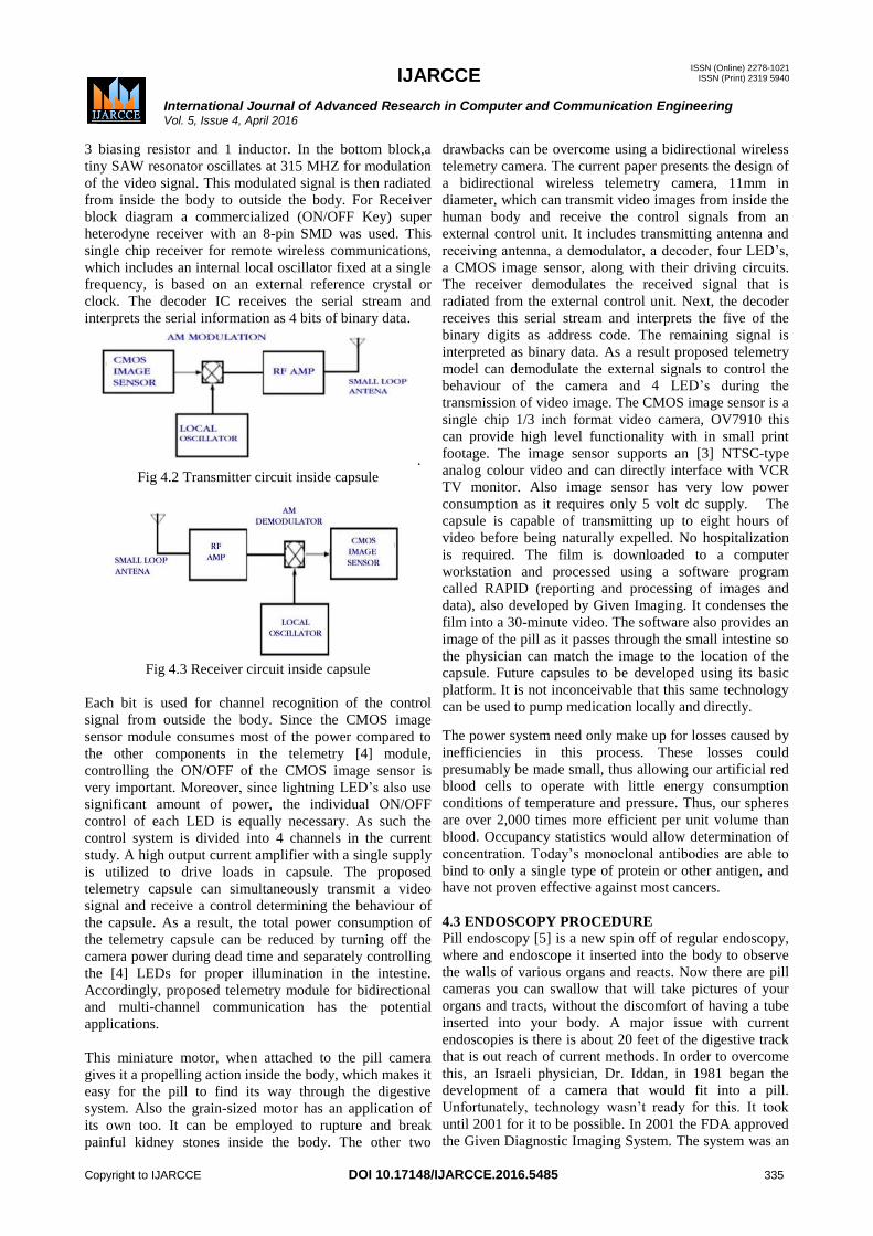
3 biasing resistor and 1 inductor. In the bottom block,a
tiny SAW resonator oscillates at 315 MHZ for modulation
of the video signal. This modulated signal is then radiated
from inside the body to outside the body. For Receiver
block diagram a commercialized (ON/OFF Key) super
heterodyne receiver with an 8-pin SMD was used. This
single chip receiver for remote wireless communications,
which includes an internal local oscillator fixed at a single
frequency, is based on an external reference crystal or
clock. The decoder IC receives the serial stream and
interprets the serial information as 4 bits of binary data.
.
Fig 4.2 Transmitter circuit inside capsule
Fig 4.3 Receiver circuit inside capsule
Each bit is used for channel recognition of the control
signal from outside the body. Since the CMOS image
sensor module consumes most of the power compared to
the other components in the telemetry [4] module,
controlling the ON/OFF of the CMOS image sensor is
very important. Moreover, since lightning LED‟s also use
significant amount of power, the individual ON/OFF
control of each LED is equally necessary. As such the
control system is divided into 4 channels in the current
study. A high output current amplifier with a single supply
is utilized to drive loads in capsule. The proposed
telemetry capsule can simultaneously transmit a video
signal and receive a control determining the behaviour of
the capsule. As a result, the total power consumption of
the telemetry capsule can be reduced by turning off the
camera power during dead time and separately controlling
the [4] LEDs for proper illumination in the intestine.
Accordingly, proposed telemetry module for bidirectional
and multi-channel communication has the potential
applications.
This miniature motor, when attached to the pill camera
gives it a propelling action inside the body, which makes it
easy for the pill to find its way through the digestive
system. Also the grain-sized motor has an application of
its own too. It can be employed to rupture and break
painful kidney stones inside the body. The other two
drawbacks can be overcome using a bidirectional wireless
telemetry camera. The current paper presents the design of
a bidirectional wireless telemetry camera, 11mm in
diameter, which can transmit video images from inside the
human body and receive the control signals from an
external control unit. It includes transmitting antenna and
receiving antenna, a demodulator, a decoder, four LED‟s,
a CMOS image sensor, along with their driving circuits.
The receiver demodulates the received signal that is
radiated from the external control unit. Next, the decoder
receives this serial stream and interprets the five of the
binary digits as address code. The remaining signal is
interpreted as binary data. As a result proposed telemetry
model can demodulate the external signals to control the
behaviour of the camera and 4 LED‟s during the
transmission of video image. The CMOS image sensor is a
single chip 1/3 inch format video camera, OV7910 this
can provide high level functionality with in small print
footage. The image sensor supports an [3] NTSC-type
analog colour video and can directly interface with VCR
TV monitor. Also image sensor has very low power
consumption as it requires only 5 volt dc supply. The
capsule is capable of transmitting up to eight hours of
video before being naturally expelled. No hospitalization
is required. The film is downloaded to a computer
workstation and processed using a software program
called RAPID (reporting and processing of images and
data), also developed by Given Imaging. It condenses the
film into a 30-minute video. The software also provides an
image of the pill as it passes through the small intestine so
the physician can match the image to the location of the
capsule. Future capsules to be developed using its basic
platform. It is not inconceivable that this same technology
can be used to pump medication locally and directly.
The power system need only make up for losses caused by
inefficiencies in this process. These losses could
presumably be made small, thus allowing our artificial red
blood cells to operate with little energy consumption
conditions of temperature and pressure. Thus, our spheres
are over 2,000 times more efficient per unit volume than
blood. Occupancy statistics would allow determination of
concentration. Today‟s monoclonal antibodies are able to
bind to only a single type of protein or other antigen, and
have not proven effective against most cancers.
4.3 ENDOSCOPY PROCEDURE
Pill endoscopy [5] is a new spin off of regular endoscopy,
where and endoscope it inserted into the body to observe
the walls of various organs and reacts. Now there are pill
cameras you can swallow that will take pictures of your
organs and tracts, without the discomfort of having a tube
inserted into your body. A major issue with current
endoscopies is there is about 20 feet of the digestive track
that is out reach of current methods. In order to overcome
this, an Israeli physician, Dr. Iddan, in 1981 began the
development of a camera that would fit into a pill.
Unfortunately, technology wasn‟t ready for this. It took
until 2001 for it to be possible. In 2001 the FDA approved
the Given Diagnostic Imaging System. The system was an

IJARCCE
ISSN (Online) 2278-1021 ISSN (Print) 2319 5940
International Journal of Advanced Research in Computer and Communication Engineering Vol. 5, Issue 4, April 2016
Copyright to IJARCCE DOI 10.17148/IJARCCE.2016.5485 336
11x26mm 4 gram capsule, which contained a colour video
camera, a radio transmitter, 4 LEDs and a battery. The pill
is moved around the body with peristaltic contractions.
Throughout the procedure the patient can perform daily
tasks without discomfort. Throughout the 8-hours, the
images are transmitted to a device about the size of a
walkman. The images are received through special
antenna pads placed on the body. From this the images can
be downloaded to the computer for examination. One
company has put a new twist on the pill camera. Other pill
cameras have their lenses and sensor in the moving
direction, requiring a wide angle lens[2]. The problem
with this is the peripheral regions of the picture become
distorted. So RF Systems developed Sayaka. It is designed
to take picture of the whole surface of the digestive tract.
This is possible by its spinning camera, which takes
pictures in a full 360 degrees. Another thingwith Sayaka
is it is not battery powered. Instead it gets its power
through induction charging. A vest worn by the patient
transmits power, due to a coil in the vest.
Once the pill reaches the intestines it begins to take 30
pictures per second. The walls of the intestine are lit by
florescent and white LEDs. In order to spin the camera
360 degrees, an electromagnet reverses its polarity causing
a permanent magnet to rotate the inner capsule and the
mage sensor 60 degrees every two seconds. A full rotation
takes 12 seconds, which it perfect to get a continuous
picture of the internal wall of the intestine. For it takes the
capsule about 2 minutes to travel an inch within the
intestine. Preparation for a pill camera study requires
fasting for 10-12 hours beforehand to ensure an empty
stomach. Following capsule ingestion, after a brief period
of observation, patients are permitted to leave the
endoscopy centre, with instructions to return within seven
hours, at which time the data recorder will be removed.
During the study, normal activity may be resumed. Light
food is generally permitted beginning four hours after the
capsule is ingested. The capsule is disposable and will
usually pass naturally during a bowel movement within 8-
24 hours. Patients with a history of abdominal surgery,
cardiac pacemaker or difficulty in swallowing should
notify the doctor in advance. [1] Complications are rare
with pill camera studies, and generally occur when there is
an obstruction in the intestinal tract. Notify the doctor if in
the event of abdominal pain, chest pain, fever or vomiting.
Do not undergo an MRI study until the capsule has passed.
Results of the examination will be available after the
captured images have been transferred to a computer and
studied by your doctor. We have a solid track record and a
strong reputation in precision moulded parts, plastic
aspheric lenses and high-precision opt-mechanical
assemblies. Today, we are active in miniature camera-
lenses for mobile and automotive applications, printer
sensor optics, optical storage and high power LED lens
solutions.
4.3.1 Smallest Tethered Endoscope
The PicoEndo [2] endoscope is the smallest tethered
endoscope in the world (4.5mm x 12.0mm). It is also
inexpensive enough to use and discard. It provides a
dramatic cost reduction in equipment requirements from
conventional endoscope or pill camera systems, which can
cost upwards of $30,000 USD. Pico Endo delivers more
images at an improved quality, including images
processed into 3D. The Pico Endo system is applicable to
medical tasks such as photographing the surface of the
esophagus and to applications in any other industry that
needs to place a tiny electronic camera eye in a location
that is difficult to view, such as inspecting the interiors of
assembled engines.
4.3.2Entrance to Exit
The camera-in-a-pill capsule, or pill-cam, measures 2.5cm
by 1.1cm and contains a minuscule digital camera, a light
source, and of course a battery to power it up. However,
the real genius of the pill-cam lies in its tiny radio
transmitter and antenna (also contained in the capsule!)
which enables it to transmit data (pictures!) to a data
recorder that the patient wears strapped around the waist.
From the moment it is swallowed it takes pictures at a rate
of two shots every second, right up until the moment it is
excreted.
4.4 SOME AMAZING FACTS ABOUT THE CAM
PILL
The pill-cam „capsule‟ is about the same size as a large
multi-vitamin tablet, i.e. 2.5cm x 1cm.Two digital images
of the intestine lining are taken every second time taken
for the pill cam‟s entire journey through the body is
approximately 7 hours. Hospitals make use of a computer
software programme to speed up viewing the video. Half
of the pill-cam „capsule‟ consists of batteries [5].The
miniature lens takes pictures from 2-3cm away. The tiny
Perspex dome over the lens ensures that all images taken
are in focus – even when it is touching the wall of the
intestine. The procedure costs about £1000, with the pill-
cam itself costing about half that amount.
The official name of the so-called „pill-cam‟ is the M2A
[5] Capsule Endoscopy, and it was developed by the
Israeli company given Imaging Ltd.The tiniest endoscope
yet takes 30 two-megapixel images per second and
offloads them wirelessly. "Our technology is completely
different from what's available now. This could be the
foundation for the future of endoscopy," said lead author
Eric Seibel, a University on research associate professor of
mechanical engineering. In the past 30 years diagnoses of
oesophageal cancer have more than tripled [3]. The
oesophagus is the section of digestive tract that moves
food from the throat down to the stomach. Oesophageal
cancer often follows a condition called Barrett's
oesophagus, a noticeable change in the oesophageal lining.
Patients with Barrett's oesophagus can be healed, avoiding
the deadly oesophageal cancer. But because internal scans
are expensive most people don't find out they have the
condition until it's progressed to cancer, and by that stage
the survival rate is less than 15 percent. Any screen that
detected whether you had a treatable condition before it
had turned into cancer would save lives."

IJARCCE
ISSN (Online) 2278-1021 ISSN (Print) 2319 5940
International Journal of Advanced Research in Computer and Communication Engineering Vol. 5, Issue 4, April 2016
Copyright to IJARCCE DOI 10.17148/IJARCCE.2016.5485 337
4.4.1 Missile Optical Camera
Only a small percentage of people who get Barrett's
oesophagus, about 5 percent to 10 percent, develop Israeli
military scientist Gabriel Iddan spent years working on
missile technology as the head of the electro-optical
design section of the Rafael Armament Development
Authority at the Ministry of Defence. [3] Iddan had
worked on the seeker, or the "eye" of the missile, which
captures the targets and guides it, and believed the same
technology. While on sabbatical eight years ago in Boston,
Iddan decided to design a tiny capsule containing a guided
missile optical camera that could be swallowed, and would
send images in real time as it traversed a patient's
intestines. But money for the project was scarce.
4.5 DIGESTIVE TRACK
The best of hands the entire small intestine is not
visualized. The visit to attach the sensor pads and swallow
the capsule will take 30 minutes to an hour. You are able
to leave the hospital at this time. The digestive track is aid
with peristaltic activity. The patient comfortably continues
with regular activities throughout the examination without
feeling sensations resulting from the capsule's passage.
4.5.1 Uses
Crohn's Disease.
Mal-absorption Disorders.
Tumours of the small intestine & Vascular Disorders.
Ulcerative Colitis
Medication Related To Small Bowel Injury
4.5.2 Advantages
Biggest impact on the medical industry [5].
Nano robots can perform delicate surgeries
They can also change the physical appearance.
They can slow or reverse the aging process.
Used to shrink the size of components.
Nano technology has the potential to have a positive
effect on the Environment
4.5.3 Drawbacks
It is a revolution, no question about it but the capsule
poses medical risks
1."Unfortunately, patients with gastrointestinal structures
or narrowing are not good candidates for this procedure
due to the risk of obstruction". It might also happen that
the pill camera might not be able to traverse freely inside
digestive system, which may cause the tests to be
inconclusive [5].
2. If there is a partial obstruction in the small intestine,
there is a risk that the pill will get stuck there and a patient
who might have come in for diagnostically reasons may
end up in the emergency room for intestinal obstruction.
3. The pill camera can transmit image from inside to
outside the body. Consequently it becomes impossible to
control the camera behaviour, including the on/off power
functions and effective illuminations inside the intestine
[5]. The first drawback has overcome using another
product manufactured with the help of nanotechnology
which is the rice- grain sized motor. The bidirectional
wireless telemetry camera, 11mm in diameter, can
transmit video images from inside the human body and
receive the control signals from an external control unit. It
include stream transmitting antenna and receiving
antenna, a demodulator, a decoder, four LED‟s, a CMOS
image sensor, along with their driving circuits.The
receiver demodulates the received signal that is radiated
from the external control unit. Next, the decoder receives
this serial stream and interprets the five of the binary digits
as address code. The remaining signal is interpreted as
binary data.
4.5.4 Lighted Flexible Tube
A doctor uses an endoscope, a long, thin, lighted flexible
tube with a small camera on the end. The endoscope is
inserted through the patient‟s mouth and into the
oesophagus. Although the patient is awake during the
procedure, doctors administer sedatives intravenously, and
spray numbing agents into the patient‟s throat to prevent
gagging. Recovery time is one to two hours until the
effects of the sedatives wear off and the patient‟s throat
may be sore for up to two days. Both the PillCam SB and
ESO procedures do not require sedation and can be
administered in a doctor‟s office. Studies have shown
patients undergoing either PillCam procedure have a much
higher level of satisfaction due to procedural convenience
and comfort and immediate recovery. The PillCam SB is
considered the gold standard for detecting diseases of the
small bowel such as Crohn‟s disease and obscure bleeding.
In a study of 106 patients, the sensitivity level of the
PillCam ESO was rated similar to the sensitivity level of a
traditional endoscopy in detecting abnormalities in a
patient‟s oesophagus. PillCam
ESO accuracy is comparable to traditional endoscopy.
Inflammatory Bowel Disease (IBD) is a family of chronic
diseases affecting the intestines. Crohn‟s disease and
ulcerative colitis both fall under the same umbrella and
were once believed to be the same disease. Patients with
IBD experience such symptoms as persistent diarrhoea,
abdominal pain or cramps, fever and weight loss, and
joint, skin, or eye irritations in varying degrees. Some may
not experience all of these symptoms. Patients may also
experience cycles of remission and relapse as the disease
progresses. While Crohn‟s disease is rarely fatal, there is
no cure. Instead, doctors focus on treating the symptoms.
If left untreated, symptoms may worsen, and health
problems such as abscesses, obstruction, malnutrition and
anaemia may occur.
4.5.5 Gastrointestinal Association Data
According to American Gastrointestinal Association data,
approximately 19 million of Americans suffer from
various disorders of the small intestine including bleeding,
Crohn‟s disease, celiac disease, irritable bowel syndrome
and small bowel cancers. Of these 19 million people,
approximately 500,000 people suffer from Crohn‟s
disease.

IJARCCE
ISSN (Online) 2278-1021 ISSN (Print) 2319 5940
International Journal of Advanced Research in Computer and Communication Engineering Vol. 5, Issue 4, April 2016
Copyright to IJARCCE DOI 10.17148/IJARCCE.2016.5485 338
4.6 ESOPHAGEAL VARICES
Gastroesophagealvarices [6] are present in 40-60% of
patients with cirrhosis Haemorrhage from
oesophagealvarices is a leading cause of death in cirrhotic
patients, with mortality rates as high as 50% .Varices are
veins that have become enlarged due to increased pressure.
The increased blood flow causes these fragile blood
vessels to become so stretched that they are susceptible to
breaking and bleeding. Pictures by pill camera of
gastroesophageal endoscopy [7] and path moving of
capsule are as shown in fig 4.4 and fig 4.5 respectively.
Fig.4.4 Images of Gastroesophageal Tract
Fig.4.5 Path of Moving Pill from Digestive System
V. CONCLUSION
5.1 INTRODUCTION
The given endoscopy capsule is a pioneering concept for
medical technology of the 21st century. The endoscopy
system is the first of its kind to be able to provide non-
invasive imaging of the entire small intestine. It has
revolutionized the field of diagnostic imaging to a great
extent and has proved to be of great help to physicians all
over the world. In the near future most of the conventional
manufacturing processes will be replaced with a cheaper
and better manufacturing process “nanotechnology”.
5.2 CONCLUSION
Scientists predict that this is not all nanotechnology is
capable to produce such products. They even foresee that
in the coming decades, with the help of nanotechnology
one can make hearts, lungs, livers and kidneys, just by
providing coal, water and some impurities and even
prevent the aging effect. Nanotechnology has the power to
revolutionize the world of production, but it is sure to
increase unemployment in next generation. This pill
camera technology has glorified biomedical science and
helped doctors to diagnose such a complicated intestinal
bowel in easy way. Use of Pill camera on large scale will
reduce unwanted death rate in upcoming decades. But in
rare case the capsule which is swallowed if does not pass
through body further then, it may need to be removed
endoscopic ally or surgically. So this proposed capsule
endoscopic model has to be further modified after
knowing its disadvantage which occurs while the
transmission of video image.
5.3 FUTURE SCOPE
This pill camera technology in future can be design to
sense temperature, pressure, and various diseases with its
virus present in body. Also it can be made in the form of
programmable chip so that it can work blood cell (WBC‟s
and RBC‟s) reconstruction. This can prevent patients from
surgical operation.
REFERENCES
[1]. New electronics magazine – the site for electronic design
engineers.– www.newelectronics.co.uk – Robotic pill to revolutionise cancer treatment, 12 Nov 2012
[2]. World journal of Gastroenterology: WJG- 2014 Aug 7, published
online.Capsule endoscopy: present status and future expectation by MaheshK. Goenka, & Shounak Mujumdar.
[3]. Web sites:
a. www.sciencedaily.com b. www.dailytech.com/colonprobing+pill+camera
c. www.nanotecnology.com
[4]. Techno Crazed- Pillcam: Swallow a Pill with a Miniature Camera to avoid Colonoscopy-video & news.
[5]. Technical paper on Camera pill endoscopy- Naga Raj.Y
[6]. Technical paper on Emerging Technology- an imaging pill for gastro intestinal endoscopy.
[7]. Technical paper on Wireless capsule endoscopy by P.Swan.
Copyright © 2022 FDOKUMEN