R Plasminogen in cerebrospinal fluid originates from circulating blood
10
RESEARCH Open Access Plasminogen in cerebrospinal fluid originates from circulating blood Anna Mezzapesa 1,2 , Cyrille Orset 2 , Laurent Plawinski 3 , Loic Doeuvre 2 , Sara Martinez de Lizarrondo 2 , Guglielmina Chimienti 1 , Denis Vivien 2 , Alexandre Mansour 2 , Sabrina Matà 4 , Gabriella Pepe 1 and Eduardo Anglés-Cano 5* Abstract Background: Plasminogen activation is a ubiquitous source of fibrinolytic and proteolytic activity. Besides its role in prevention of thrombosis, plasminogen is involved in inflammatory reactions in the central nervous system. Plasminogen has been detected in the cerebrospinal fluid (CSF) of patients with inflammatory diseases; however, its origin remains controversial, as the blood–CSF barrier may restrict its diffusion from blood. Methods: We investigated the origin of plasminogen in CSF using Alexa Fluor 488–labelled rat plasminogen injected into rats with systemic inflammation and blood–CSF barrier dysfunction provoked by lipopolysaccharide (LPS). Near-infrared fluorescence imaging and immunohistochemistry fluorescence microscopy were used to identify plasminogen in brain structures, its concentration and functionality were determined by Western blotting and a chromogenic substrate assay, respectively. In parallel, plasminogen was investigated in CSF from patients with Guillain-Barré syndrome (n = 15), multiple sclerosis (n = 19) and noninflammatory neurological diseases (n = 8). Results: Endogenous rat plasminogen was detected in higher amounts in the CSF and urine of LPS-treated animals as compared to controls. In LPS-primed rats, circulating Alexa Fluor 488–labelled rat plasminogen was abundantly localized in the choroid plexus, CSF and urine. Plasminogen in human CSF was higher in Guillain-Barré syndrome (median = 1.28 ng/μl (interquartile range (IQR) = 0.66 to 1.59)) as compared to multiple sclerosis (median = 0.3 ng/μl (IQR = 0.16 to 0.61)) and to noninflammatory neurological diseases (median = 0.27 ng/μl (IQR = 0.18 to 0.35)). Conclusions: Our findings demonstrate that plasminogen is transported from circulating blood into the CSF of rats via the choroid plexus during inflammation. Our data suggest that a similar mechanism may explain the high CSF concentrations of plasminogen detected in patients with inflammation-derived CSF barrier impairment. Keywords: Blood–cerebrospinal fluid barrier, Inflammation, LPS, Plasminogen Introduction Vessel wall fibrinolytic activity and pericellular proteoly- sis in tissues requires plasminogen binding and trans- formation into plasmin at the surface of fibrin, cells or the extracellular matrix by either the tissue-type plas- minogen activator (tPA) or the urokinase-type plasmino- gen activator (uPA) [1]. The binding of plasminogen to cells or fibrin is a lysine-dependent mechanism that can be inhibited by lysine analogues such as tranexamic acid (TXA). In the vasculature, plasmin formation results in blood clot dissolution [2], whereas in tissues, plasmin is responsible for pericellular proteolysis accompanying cell migration, angiogenesis, wound healing, tissue re- modelling and inflammation [3-5]. Pericellular proteoly- sis in the central nervous system (CNS) is involved in development, regeneration of nervous tissues, neuronal and synaptic plasticity and the inflammatory response [6-9]. In humans, plasminogen deficiency is associated with neural disorders including congenital hydroceph- alus, periventricular nodular heterotopias and Dandy-Walker malformation [10]. Trace amounts of plasminogen have been reported in normal human cerebrospinal fluid (CSF), whereas raised concentrations have been detected in patients with * Correspondence: [email protected] 5 Inserm UMRS1140, Faculty of Pharmaceutical and Biological Sciences, Paris Descartes University, 4 Avenue de l’Observatoire, 75270 Paris, cedex 06, France Full list of author information is available at the end of the article JOURNAL OF NEUROINFLAMMATION © 2014 Mezzapesa et al.; licensee BioMed Central Ltd. This is an Open Access article distributed under the terms of the Creative Commons Attribution License (http://creativecommons.org/licenses/by/4.0), which permits unrestricted use, distribution, and reproduction in any medium, provided the original work is properly credited. The Creative Commons Public Domain Dedication waiver (http://creativecommons.org/publicdomain/zero/1.0/) applies to the data made available in this article, unless otherwise stated. Mezzapesa et al. Journal of Neuroinflammation 2014, 11:154 http://www.jneuroinflammation.com/content/11/1/154
-
Upload
independent -
Category
Documents
-
view
2 -
download
0
Transcript of R Plasminogen in cerebrospinal fluid originates from circulating blood
JOURNAL OF NEUROINFLAMMATION
Mezzapesa et al. Journal of Neuroinflammation 2014, 11:154http://www.jneuroinflammation.com/content/11/1/154
RESEARCH Open Access
Plasminogen in cerebrospinal fluid originatesfrom circulating bloodAnna Mezzapesa1,2, Cyrille Orset2, Laurent Plawinski3, Loic Doeuvre2, Sara Martinez de Lizarrondo2,Guglielmina Chimienti1, Denis Vivien2, Alexandre Mansour2, Sabrina Matà4, Gabriella Pepe1
and Eduardo Anglés-Cano5*
Abstract
Background: Plasminogen activation is a ubiquitous source of fibrinolytic and proteolytic activity. Besides its role inprevention of thrombosis, plasminogen is involved in inflammatory reactions in the central nervous system.Plasminogen has been detected in the cerebrospinal fluid (CSF) of patients with inflammatory diseases; however, itsorigin remains controversial, as the blood–CSF barrier may restrict its diffusion from blood.
Methods: We investigated the origin of plasminogen in CSF using Alexa Fluor 488–labelled rat plasminogeninjected into rats with systemic inflammation and blood–CSF barrier dysfunction provoked by lipopolysaccharide(LPS). Near-infrared fluorescence imaging and immunohistochemistry fluorescence microscopy were used to identifyplasminogen in brain structures, its concentration and functionality were determined by Western blotting and achromogenic substrate assay, respectively. In parallel, plasminogen was investigated in CSF from patients withGuillain-Barré syndrome (n = 15), multiple sclerosis (n = 19) and noninflammatory neurological diseases (n = 8).
Results: Endogenous rat plasminogen was detected in higher amounts in the CSF and urine of LPS-treated animalsas compared to controls. In LPS-primed rats, circulating Alexa Fluor 488–labelled rat plasminogen was abundantlylocalized in the choroid plexus, CSF and urine. Plasminogen in human CSF was higher in Guillain-Barré syndrome(median = 1.28 ng/μl (interquartile range (IQR) = 0.66 to 1.59)) as compared to multiple sclerosis (median = 0.3 ng/μl(IQR = 0.16 to 0.61)) and to noninflammatory neurological diseases (median = 0.27 ng/μl (IQR = 0.18 to 0.35)).
Conclusions: Our findings demonstrate that plasminogen is transported from circulating blood into the CSF of ratsvia the choroid plexus during inflammation. Our data suggest that a similar mechanism may explain the high CSFconcentrations of plasminogen detected in patients with inflammation-derived CSF barrier impairment.
Keywords: Blood–cerebrospinal fluid barrier, Inflammation, LPS, Plasminogen
IntroductionVessel wall fibrinolytic activity and pericellular proteoly-sis in tissues requires plasminogen binding and trans-formation into plasmin at the surface of fibrin, cells orthe extracellular matrix by either the tissue-type plas-minogen activator (tPA) or the urokinase-type plasmino-gen activator (uPA) [1]. The binding of plasminogen tocells or fibrin is a lysine-dependent mechanism that canbe inhibited by lysine analogues such as tranexamic acid
* Correspondence: [email protected] UMRS1140, Faculty of Pharmaceutical and Biological Sciences, ParisDescartes University, 4 Avenue de l’Observatoire, 75270 Paris, cedex 06,FranceFull list of author information is available at the end of the article
© 2014 Mezzapesa et al.; licensee BioMed CenCreative Commons Attribution License (http:/distribution, and reproduction in any mediumDomain Dedication waiver (http://creativecomarticle, unless otherwise stated.
(TXA). In the vasculature, plasmin formation results inblood clot dissolution [2], whereas in tissues, plasmin isresponsible for pericellular proteolysis accompanyingcell migration, angiogenesis, wound healing, tissue re-modelling and inflammation [3-5]. Pericellular proteoly-sis in the central nervous system (CNS) is involved indevelopment, regeneration of nervous tissues, neuronaland synaptic plasticity and the inflammatory response[6-9]. In humans, plasminogen deficiency is associatedwith neural disorders including congenital hydroceph-alus, periventricular nodular heterotopias and Dandy-Walkermalformation [10]. Trace amounts of plasminogen have beenreported in normal human cerebrospinal fluid (CSF), whereasraised concentrations have been detected in patients with
tral Ltd. This is an Open Access article distributed under the terms of the/creativecommons.org/licenses/by/4.0), which permits unrestricted use,, provided the original work is properly credited. The Creative Commons Publicmons.org/publicdomain/zero/1.0/) applies to the data made available in this
Mezzapesa et al. Journal of Neuroinflammation 2014, 11:154 Page 2 of 10http://www.jneuroinflammation.com/content/11/1/154
meningitis [11], subarachnoid haemorrhage [12] and multiplesclerosis [13]. However, the question has not been settled asto whether plasminogen in CSF originates from circulatingblood or is expressed in the CNS, as suggested by the pres-ence of mRNA in mouse brain [14,15] or by its synthesis byrat microglial cells in culture [16].The CNS is protected from the bloodstream by the
blood–brain barrier (BBB) and the blood–CSF barrier[17,18]. The BBB is the barrier separating the braininterstitial fluid compartment from the general circula-tion (endothelial tight junctions, basal lamina of endo-thelial cells and astrocyte pedicles). It selectively limitspenetration of a variety of noxious substances and sup-plies the brain with nutrients. The blood–CSF barrierseparates the CSF compartment from blood throughchoroid plexus epithelial cells, tight junctions, a basalmembrane and the endothelium. It restricts the passageof harmful substances from the blood into the CSF se-creted across the choroid plexus epithelial cells into thebrain’s ventricular system [18]. Because the blood–CSFbarrier is more permeable than the BBB, many plasmaproteins enter the cerebrospinal liquid (through pinocyt-osis or active transport). An impairment of the blood–CSFbarrier thus leads to an increase in the concentration ofproteins in the CSF.Because diffusion of plasma proteins into the CNS is a
selective process, we sought to demonstrate that circu-lating plasminogen may cross the blood–CSF barrierand can be detected in the CSF. For that purpose, westudied control rats and rats with lipopolysaccharide(LPS)-induced systemic inflammation. Our results showthat circulating plasminogen enters the CSF space dur-ing blood–CSF barrier dysfunction induced by systemicinflammation, thus suggesting that plasminogen foundin the CSF of patients with inflammatory neurologicaldisorders originates from circulating blood.
Materials and methodsReagents and proteinsThe enhanced chemiluminescence reagent kit wasobtained from Bio-Rad Laboratories (Hercules, CA,USA). The chromogenic substrate selective for plasmin(methylmalonyl)-hydroxyprolylarginine-para-nitroaniline(CBS0065) was purchased from Stago (Asnières-sur-Seine, France). Escherichia coli LPS serotype 0111:B4,trans-4-(aminomethyl)cyclohexane-1-carboxylic acid (ortranexamic acid, TXA), amiloride and 4′,6-diamidino-2-phenylindole (DAPI) and Evans blue were obtainedfrom Sigma Chemical Co (St Louis, MO, USA). AlexaFluor 488 succinimidyl ester was purchased from LifeTechnologies (Carlsbad, CA, USA). Goat anti-collagentype IV (Col IV) used for immunohistochemical analyseswas obtained from Southern Biotech (Birmingham, AL,USA), and donkey anti-goat antibody F(ab′)2 fragments
linked to tetramethylrhodamine isothiocyanate werepurchased from Jackson ImmunoResearch (West Grove,PA, USA). Glu-plasminogen, plasmin and peroxidase-conjugated monoclonal antibody against plasminogenkringle 1 (CPL15-PO) were prepared and characterizedas described previously [19-21]. A rabbit anti-mouseplasminogen polyclonal antibody was kindly providedby HR Lijnen (University of Leuven, Belgium).
Patient samplingA total of 42 patients with neurological diseases attend-ing the Neurological Clinic of the Careggi UniversityHospital, Florence, Italy, were admitted in this study. In-formed consent was obtained according to the Declar-ation of Helsinki. The Careggi University Hospital ReviewBoard approved the protocol. Diagnoses were based onclinical, laboratory and magnetic resonance imaging dataaccording to the International Classification of Diseases,Tenth Revision, and the Diagnostic and Statistical Man-ual of Mental Disorders, Fourth Edition–Text Revision[22]. Patients with Parkinson’s disease, brain tumour,epilepsy and alcohol or other substance dependencewere excluded. All CSF samples were obtained by lum-bar puncture and were immediately used for routinelaboratory analyses, which included erythrocyte andleucocyte differential cell counts, total protein concen-tration, albumin and immunoglobulin G (IgG) levelsand agarose isoelectric focusing for IgG oligoclonalbands. CSF samples with increased lymphocyte counts(>5 mm3) or with blood contamination (erythrocyteconcentration >50 cells/mm3) were excluded. The CSFto serum albumin quotient (Qalb) was calculated andused to evaluate blood–CSF barrier integrity. Qalb >0.007 was considered a marker of blood–CSF barrierdysfunction [23,24]. The remainder of each sample wasstored in aliquots at −80°C for further analysis.
Animal experimental modelExperiments were performed using male Wistar rats(280 to 350 g) in accordance with the directives of theCouncil of the European Communities (86/609/EEC)and the French Agriculture and Forestry Ministry forhandling animals (decree 87-848).
Purification and labelling of rat plasminogenTo monitor plasminogen uptake by cells and tissuesin vivo in the rat model of inflammation, we usedfluorescence-labelled native plasminogen purified fromrat plasma. Blood was obtained by cardiac puncture ofanaesthetized animals and collected in 0.129 M sodiumcitrate. The plasma was separated from blood by centri-fugation, and plasminogen was isolated by lysine affinityand sieving chromatography as described previously[19]. The purified plasminogen had a relative molecular
Mezzapesa et al. Journal of Neuroinflammation 2014, 11:154 Page 3 of 10http://www.jneuroinflammation.com/content/11/1/154
weight (Mr = 92,000 Da) similar to that of human plas-minogen and was labelled with Alexa Fluor 488 dye (A488-Pg) according to the instructions of the manufacturer.
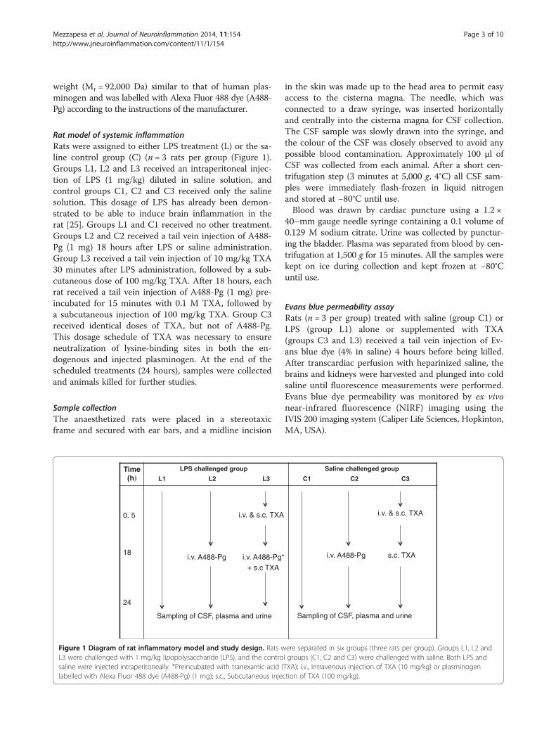
Rat model of systemic inflammationRats were assigned to either LPS treatment (L) or the sa-line control group (C) (n = 3 rats per group (Figure 1).Groups L1, L2 and L3 received an intraperitoneal injec-tion of LPS (1 mg/kg) diluted in saline solution, andcontrol groups C1, C2 and C3 received only the salinesolution. This dosage of LPS has already been demon-strated to be able to induce brain inflammation in therat [25]. Groups L1 and C1 received no other treatment.Groups L2 and C2 received a tail vein injection of A488-Pg (1 mg) 18 hours after LPS or saline administration.Group L3 received a tail vein injection of 10 mg/kg TXA30 minutes after LPS administration, followed by a sub-cutaneous dose of 100 mg/kg TXA. After 18 hours, eachrat received a tail vein injection of A488-Pg (1 mg) pre-incubated for 15 minutes with 0.1 M TXA, followed bya subcutaneous injection of 100 mg/kg TXA. Group C3received identical doses of TXA, but not of A488-Pg.This dosage schedule of TXA was necessary to ensureneutralization of lysine-binding sites in both the en-dogenous and injected plasminogen. At the end of thescheduled treatments (24 hours), samples were collectedand animals killed for further studies.
Sample collectionThe anaesthetized rats were placed in a stereotaxicframe and secured with ear bars, and a midline incision
Time(h)
LPS challenged puorg
L1 L2 L3
0. 5
18
24
i.v. & s.c. TXA
i.v. A488-Pg i.v. A488-Pg* + s.c TXA
Sampling of CSF, plasma and urine
Figure 1 Diagram of rat inflammatory model and study design. Rats wL3 were challenged with 1 mg/kg lipopolysaccharide (LPS), and the controsaline were injected intraperitoneally. *Preincubated with tranexamic acid (labelled with Alexa Fluor 488 dye (A488-Pg) (1 mg); s.c., Subcutaneous inje
in the skin was made up to the head area to permit easyaccess to the cisterna magna. The needle, which wasconnected to a draw syringe, was inserted horizontallyand centrally into the cisterna magna for CSF collection.The CSF sample was slowly drawn into the syringe, andthe colour of the CSF was closely observed to avoid anypossible blood contamination. Approximately 100 μl ofCSF was collected from each animal. After a short cen-trifugation step (3 minutes at 5,000 g, 4°C) all CSF sam-ples were immediately flash-frozen in liquid nitrogenand stored at −80°C until use.Blood was drawn by cardiac puncture using a 1.2 ×
40–mm gauge needle syringe containing a 0.1 volume of0.129 M sodium citrate. Urine was collected by punctur-ing the bladder. Plasma was separated from blood by cen-trifugation at 1,500 g for 15 minutes. All the samples werekept on ice during collection and kept frozen at −80°Cuntil use.
Evans blue permeability assayRats (n = 3 per group) treated with saline (group C1) orLPS (group L1) alone or supplemented with TXA(groups C3 and L3) received a tail vein injection of Ev-ans blue dye (4% in saline) 4 hours before being killed.After transcardiac perfusion with heparinized saline, thebrains and kidneys were harvested and plunged into coldsaline until fluorescence measurements were performed.Evans blue dye permeability was monitored by ex vivonear-infrared fluorescence (NIRF) imaging using theIVIS 200 imaging system (Caliper Life Sciences, Hopkinton,MA, USA).
Saline challenged puorg
C1 C2 C3
i.v. & s.c. TXA
i.v. A488-Pg s.c. TXA
Sampling of CSF, plasma and urine
ere separated in six groups (three rats per group). Groups L1, L2 andl groups (C1, C2 and C3) were challenged with saline. Both LPS andTXA); i.v., Intravenous injection of TXA (10 mg/kg) or plasminogenction of TXA (100 mg/kg).
Mezzapesa et al. Journal of Neuroinflammation 2014, 11:154 Page 4 of 10http://www.jneuroinflammation.com/content/11/1/154
Fluorescence microscopyAfter the rats were killed, parts of the brains were post-fixed with 4% paraformaldehyde in 0.1 M phosphate-buffered saline, pH 7.4, at 4°C for 18 hours, followed by24 hours in 20% sucrose and then frozen in isopentane.For immunocytochemistry, cryostat brain sections (8 μm)were collected on polylysine-coated slides and incubatedovernight with a goat anti-Col IV primary antibody(1:1,500), which was revealed with donkey anti-goatantibody F(ab′)2 fragments linked to tetramethylrho-damine isothiocyanate (1:500). The cells were then coun-terstained with DAPI, mounted with Fluoprep (Dako,Glostrup, Denmark) and observed under an epifluores-cence microscope. Images were digitally captured witha Leica DM6000 microscope-coupled CoolSnap cam-era (Leica Microsystems, Wetzlar, Germany) and visual-ized with METAVUE 5.0 software (Molecular Devices,Sunnyvale, CA, USA). The detection of exogenously ad-ministered A488-Pg in those samples was also studied.
Detection of functional plasminogen in cerebrospinalfluidTransformation of CSF plasminogen into plasmin wasmeasured using uPA (5 IU/ml) and a plasmin-selectivechromogenic substrate (0.75 mM CBS0065), as previ-ously described [26]. In this system, the initial velocity ofp-nitroaniline released from CBS0065 is proportional tothe amount of plasmin(ogen). The ability of TXA to im-pair plasminogen binding to the cell membrane was in-vestigated as described previously [27].
Western blot analysisHuman CSF samples (7 μg of total protein) and rat CSF,urine and plasma (diluted 1:50) samples (10 μl) wereelectrophoresed in 8% SDS-PAGE gel under nonreduc-ing conditions. Proteins were transferred onto a polyvi-nylidene fluoride membrane, and plasminogen wasrevealed using a monoclonal antibody directed againsthuman plasminogen (CPL15-PO, human CSF samples)or a rabbit anti-mouse plasminogen polyclonal antibody(rat samples). The amount of plasminogen was expressedas the number of pixels directly detected using Image-Quant TL 7.0 image analysis software with the Image-Quant LAS 4000 imaging system (GE Healthcare LifeSciences, Pittsburgh, PA, USA) and expressed as nano-grams per microlitre by reference to a known amount ofplasminogen (15 ng, 10 μl) electrophoresed under similarconditions. In rat samples, the amount of plasminogenwas expressed as the number of pixels directly detectedusing the image analysis software.
Statistical analysisData are representative of at least three independent ex-periments and are expressed as median (25th to 75th
interquartile range (IQR)). The Mann–Whitney U testwas used to compare values obtained in treated versuscontrol rats. For human experiments, one-way analysisof variance (ANOVA) was performed to compare the pa-tient groups. Bonferroni’s multiple-comparisons posttestwas run for the pairwise comparison of groups. Statis-tical significance was set at P < 0.05. A specific statisticalpackage for exact nonparametric inference (Stata Statis-tical Software release 9 (2005); StataCorp, College Sta-tion, TX, USA) was used.
ResultsLipopolysaccharide-induced increase in plasminogen inrat cerebrospinal fluidEndogenous plasminogen was detected by Western im-munoblotting in plasma, CSF and urine of both LPS- andsaline-treated control rats (groups L1 and C1, respectively)(Figure 2A). The LPS injection produced no significantvariation in the amount of circulating plasminogen ascompared to controls (0.1-fold increase in injected versuscontrol rats) (Figure 2B). In contrast, we detected higherplasminogen levels in CSF and urine from LPS-treatedrats (fourfold increase in CSF, P = 0.0002; twofold increasein urine, P = 0.0071 (both by Mann–Whitney U test))(Figure 2B). Rat CSF plasminogen was efficiently trans-formed into plasmin; its active concentration appearedto be directly related to the amount of plasminogendetected in CSF samples by Western immunoblotting(P = 0.0045, Mann–Whitney U test) (Figure 2C).
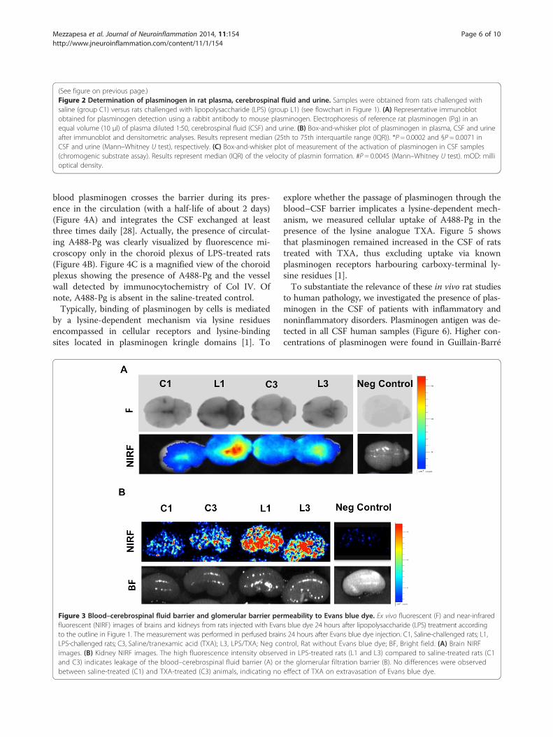
Lipopolysaccharide-induced increase in barrierpermeabilityThe aforementioned data suggest that vascular perme-ability was modified following the LPS injection. Thishypothesis was investigated by testing permeability toEvans blue dye. Ex vivo fluorescent and NIRF imagesshowed higher brain fluorescence in the LPS-treated rats(group L) as compared to controls (group C), indicatingan increase in barrier permeability to the dye due toblood–CSF barrier impairment (Figure 3A). Modifica-tions in the glomerular filtration barrier were also de-tected with the Evans blue test (Figure 3B). Higherkidney fluorescence was observed in LPS-treated rats(group L) as compared to controls (group C). The ad-ministration of TXA (groups C3 and L3) was without ef-fect on barrier permeability.
In vivo evidence of the circulating origin of plasminogenin cerebrospinal fluidTo obtain in vivo evidence of the extravasation of plas-minogen from blood, animals were challenged with ex-ogenous A488-Pg 18 hours after the LPS (group L2) orsaline injection (group C2). A488-Pg appeared in theCSF and urine of all LPS-treated rats, indicating that
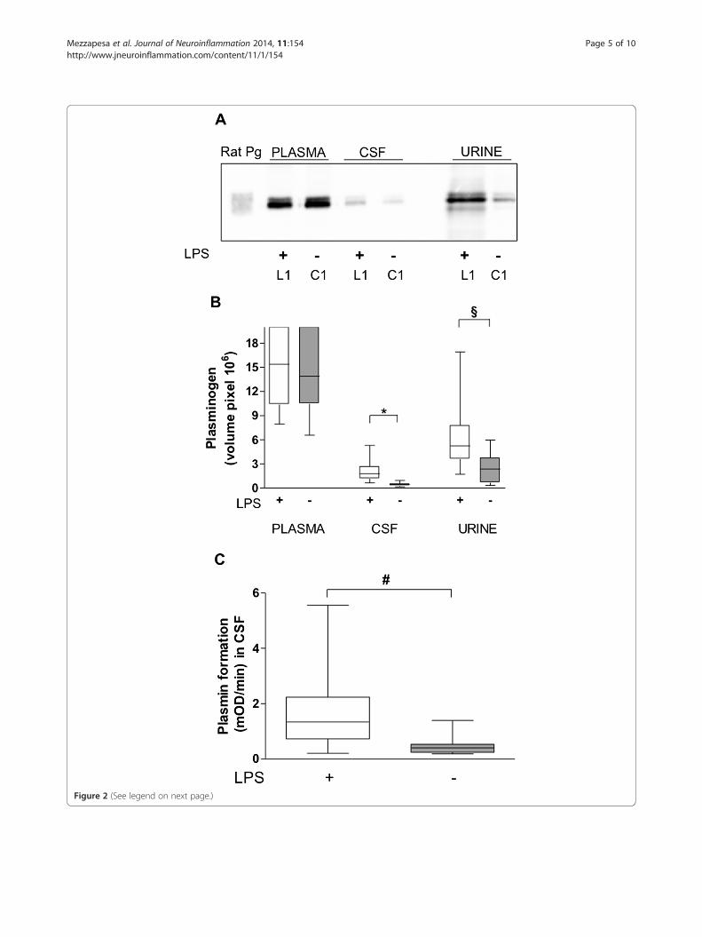
Figure 2 (See legend on next page.)
Mezzapesa et al. Journal of Neuroinflammation 2014, 11:154 Page 5 of 10http://www.jneuroinflammation.com/content/11/1/154
(See figure on previous page.)Figure 2 Determination of plasminogen in rat plasma, cerebrospinal fluid and urine. Samples were obtained from rats challenged withsaline (group C1) versus rats challenged with lipopolysaccharide (LPS) (group L1) (see flowchart in Figure 1). (A) Representative immunoblotobtained for plasminogen detection using a rabbit antibody to mouse plasminogen. Electrophoresis of reference rat plasminogen (Pg) in anequal volume (10 μl) of plasma diluted 1:50, cerebrospinal fluid (CSF) and urine. (B) Box-and-whisker plot of plasminogen in plasma, CSF and urineafter immunoblot and densitometric analyses. Results represent median (25th to 75th interquartile range (IQR)). *P = 0.0002 and §P = 0.0071 inCSF and urine (Mann–Whitney U test), respectively. (C) Box-and-whisker plot of measurement of the activation of plasminogen in CSF samples(chromogenic substrate assay). Results represent median (IQR) of the velocity of plasmin formation. #P = 0.0045 (Mann–Whitney U test). mOD: millioptical density.
Mezzapesa et al. Journal of Neuroinflammation 2014, 11:154 Page 6 of 10http://www.jneuroinflammation.com/content/11/1/154
blood plasminogen crosses the barrier during its pres-ence in the circulation (with a half-life of about 2 days)(Figure 4A) and integrates the CSF exchanged at leastthree times daily [28]. Actually, the presence of circulat-ing A488-Pg was clearly visualized by fluorescence mi-croscopy only in the choroid plexus of LPS-treated rats(Figure 4B). Figure 4C is a magnified view of the choroidplexus showing the presence of A488-Pg and the vesselwall detected by immunocytochemistry of Col IV. Ofnote, A488-Pg is absent in the saline-treated control.Typically, binding of plasminogen by cells is mediated
by a lysine-dependent mechanism via lysine residuesencompassed in cellular receptors and lysine-bindingsites located in plasminogen kringle domains [1]. To
Figure 3 Blood–cerebrospinal fluid barrier and glomerular barrier perfluorescent (NIRF) images of brains and kidneys from rats injected with Evanto the outline in Figure 1. The measurement was performed in perfused brainLPS-challenged rats; C3, Saline/tranexamic acid (TXA); L3, LPS/TXA; Neg coimages. (B) Kidney NIRF images. The high fluorescence intensity observeand C3) indicates leakage of the blood–cerebrospinal fluid barrier (A) orbetween saline-treated (C1) and TXA-treated (C3) animals, indicating no
explore whether the passage of plasminogen through theblood–CSF barrier implicates a lysine-dependent mech-anism, we measured cellular uptake of A488-Pg in thepresence of the lysine analogue TXA. Figure 5 showsthat plasminogen remained increased in the CSF of ratstreated with TXA, thus excluding uptake via knownplasminogen receptors harbouring carboxy-terminal ly-sine residues [1].To substantiate the relevance of these in vivo rat studies
to human pathology, we investigated the presence of plas-minogen in the CSF of patients with inflammatory andnoninflammatory disorders. Plasminogen antigen was de-tected in all CSF human samples (Figure 6). Higher con-centrations of plasminogen were found in Guillain-Barré
meability to Evans blue dye. Ex vivo fluorescent (F) and near-infrareds blue dye 24 hours after lipopolysaccharide (LPS) treatment accordings 24 hours after Evans blue dye injection. C1, Saline-challenged rats; L1,ntrol, Rat without Evans blue dye; BF, Bright field. (A) Brain NIRFd in LPS-treated rats (L1 and L3) compared to saline-treated rats (C1the glomerular filtration barrier (B). No differences were observedeffect of TXA on extravasation of Evans blue dye.
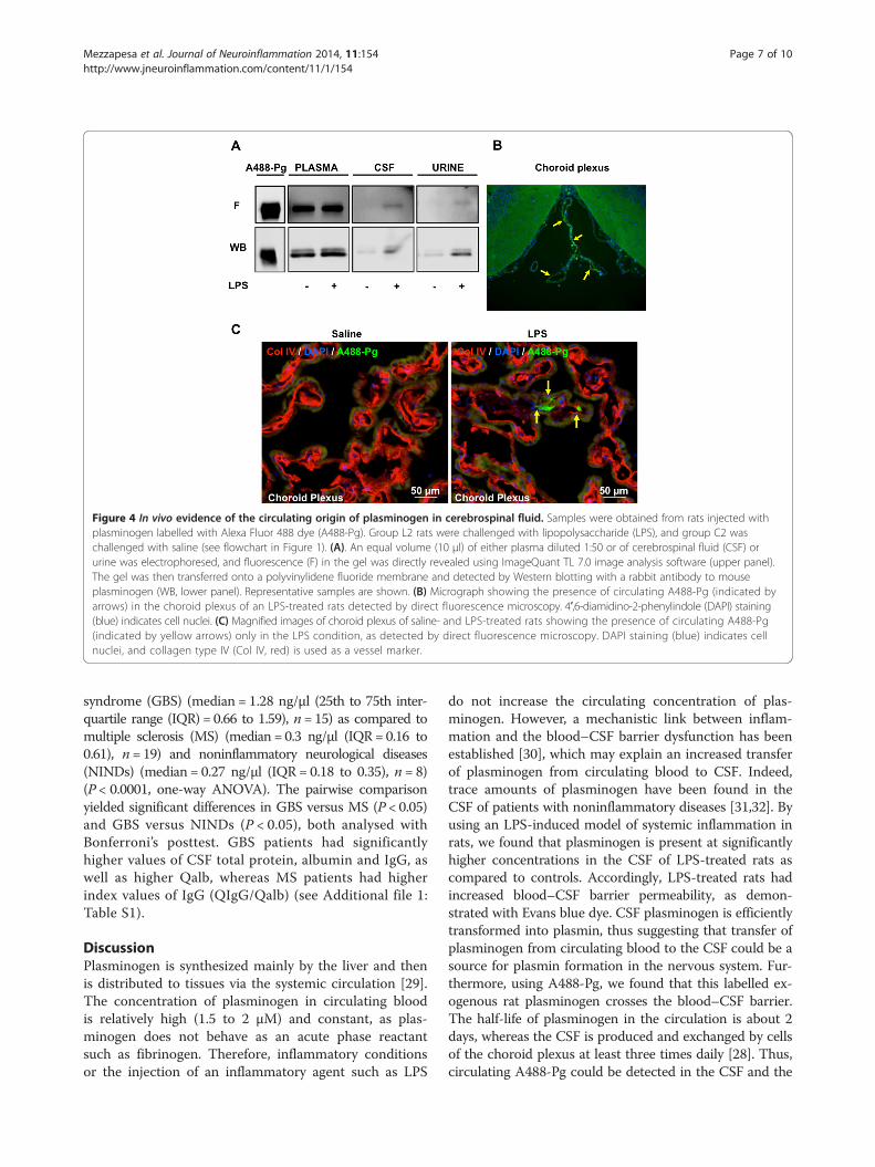
Figure 4 In vivo evidence of the circulating origin of plasminogen in cerebrospinal fluid. Samples were obtained from rats injected withplasminogen labelled with Alexa Fluor 488 dye (A488-Pg). Group L2 rats were challenged with lipopolysaccharide (LPS), and group C2 waschallenged with saline (see flowchart in Figure 1). (A). An equal volume (10 μl) of either plasma diluted 1:50 or of cerebrospinal fluid (CSF) orurine was electrophoresed, and fluorescence (F) in the gel was directly revealed using ImageQuant TL 7.0 image analysis software (upper panel).The gel was then transferred onto a polyvinylidene fluoride membrane and detected by Western blotting with a rabbit antibody to mouseplasminogen (WB, lower panel). Representative samples are shown. (B) Micrograph showing the presence of circulating A488-Pg (indicated byarrows) in the choroid plexus of an LPS-treated rats detected by direct fluorescence microscopy. 4′,6-diamidino-2-phenylindole (DAPI) staining(blue) indicates cell nuclei. (C) Magnified images of choroid plexus of saline- and LPS-treated rats showing the presence of circulating A488-Pg(indicated by yellow arrows) only in the LPS condition, as detected by direct fluorescence microscopy. DAPI staining (blue) indicates cellnuclei, and collagen type IV (Col IV, red) is used as a vessel marker.
Mezzapesa et al. Journal of Neuroinflammation 2014, 11:154 Page 7 of 10http://www.jneuroinflammation.com/content/11/1/154
syndrome (GBS) (median = 1.28 ng/μl (25th to 75th inter-quartile range (IQR) = 0.66 to 1.59), n = 15) as compared tomultiple sclerosis (MS) (median = 0.3 ng/μl (IQR= 0.16 to0.61), n= 19) and noninflammatory neurological diseases(NINDs) (median = 0.27 ng/μl (IQR = 0.18 to 0.35), n = 8)(P < 0.0001, one-way ANOVA). The pairwise comparisonyielded significant differences in GBS versus MS (P < 0.05)and GBS versus NINDs (P < 0.05), both analysed withBonferroni’s posttest. GBS patients had significantlyhigher values of CSF total protein, albumin and IgG, aswell as higher Qalb, whereas MS patients had higherindex values of IgG (QIgG/Qalb) (see Additional file 1:Table S1).
DiscussionPlasminogen is synthesized mainly by the liver and thenis distributed to tissues via the systemic circulation [29].The concentration of plasminogen in circulating bloodis relatively high (1.5 to 2 μM) and constant, as plas-minogen does not behave as an acute phase reactantsuch as fibrinogen. Therefore, inflammatory conditionsor the injection of an inflammatory agent such as LPS
do not increase the circulating concentration of plas-minogen. However, a mechanistic link between inflam-mation and the blood–CSF barrier dysfunction has beenestablished [30], which may explain an increased transferof plasminogen from circulating blood to CSF. Indeed,trace amounts of plasminogen have been found in theCSF of patients with noninflammatory diseases [31,32]. Byusing an LPS-induced model of systemic inflammation inrats, we found that plasminogen is present at significantlyhigher concentrations in the CSF of LPS-treated rats ascompared to controls. Accordingly, LPS-treated rats hadincreased blood–CSF barrier permeability, as demon-strated with Evans blue dye. CSF plasminogen is efficientlytransformed into plasmin, thus suggesting that transfer ofplasminogen from circulating blood to the CSF could be asource for plasmin formation in the nervous system. Fur-thermore, using A488-Pg, we found that this labelled ex-ogenous rat plasminogen crosses the blood–CSF barrier.The half-life of plasminogen in the circulation is about 2days, whereas the CSF is produced and exchanged by cellsof the choroid plexus at least three times daily [28]. Thus,circulating A488-Pg could be detected in the CSF and the
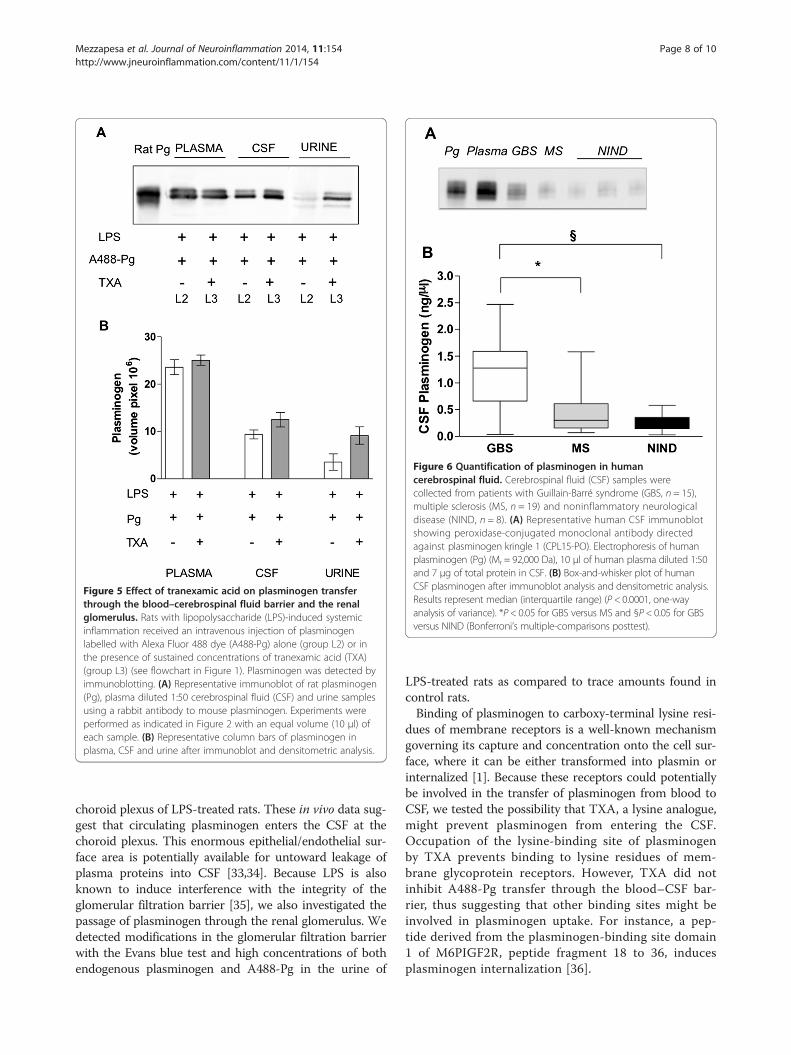
Figure 5 Effect of tranexamic acid on plasminogen transferthrough the blood–cerebrospinal fluid barrier and the renalglomerulus. Rats with lipopolysaccharide (LPS)-induced systemicinflammation received an intravenous injection of plasminogenlabelled with Alexa Fluor 488 dye (A488-Pg) alone (group L2) or inthe presence of sustained concentrations of tranexamic acid (TXA)(group L3) (see flowchart in Figure 1). Plasminogen was detected byimmunoblotting. (A) Representative immunoblot of rat plasminogen(Pg), plasma diluted 1:50 cerebrospinal fluid (CSF) and urine samplesusing a rabbit antibody to mouse plasminogen. Experiments wereperformed as indicated in Figure 2 with an equal volume (10 μl) ofeach sample. (B) Representative column bars of plasminogen inplasma, CSF and urine after immunoblot and densitometric analysis.
Figure 6 Quantification of plasminogen in humancerebrospinal fluid. Cerebrospinal fluid (CSF) samples werecollected from patients with Guillain-Barré syndrome (GBS, n = 15),multiple sclerosis (MS, n = 19) and noninflammatory neurologicaldisease (NIND, n = 8). (A) Representative human CSF immunoblotshowing peroxidase-conjugated monoclonal antibody directedagainst plasminogen kringle 1 (CPL15-PO). Electrophoresis of humanplasminogen (Pg) (Mr = 92,000 Da), 10 μl of human plasma diluted 1:50and 7 μg of total protein in CSF. (B) Box-and-whisker plot of humanCSF plasminogen after immunoblot analysis and densitometric analysis.Results represent median (interquartile range) (P < 0.0001, one-wayanalysis of variance). *P < 0.05 for GBS versus MS and §P < 0.05 for GBSversus NIND (Bonferroni’s multiple-comparisons posttest).
Mezzapesa et al. Journal of Neuroinflammation 2014, 11:154 Page 8 of 10http://www.jneuroinflammation.com/content/11/1/154
choroid plexus of LPS-treated rats. These in vivo data sug-gest that circulating plasminogen enters the CSF at thechoroid plexus. This enormous epithelial/endothelial sur-face area is potentially available for untoward leakage ofplasma proteins into CSF [33,34]. Because LPS is alsoknown to induce interference with the integrity of theglomerular filtration barrier [35], we also investigated thepassage of plasminogen through the renal glomerulus. Wedetected modifications in the glomerular filtration barrierwith the Evans blue test and high concentrations of bothendogenous plasminogen and A488-Pg in the urine of
LPS-treated rats as compared to trace amounts found incontrol rats.Binding of plasminogen to carboxy-terminal lysine resi-
dues of membrane receptors is a well-known mechanismgoverning its capture and concentration onto the cell sur-face, where it can be either transformed into plasmin orinternalized [1]. Because these receptors could potentiallybe involved in the transfer of plasminogen from blood toCSF, we tested the possibility that TXA, a lysine analogue,might prevent plasminogen from entering the CSF.Occupation of the lysine-binding site of plasminogenby TXA prevents binding to lysine residues of mem-brane glycoprotein receptors. However, TXA did notinhibit A488-Pg transfer through the blood–CSF bar-rier, thus suggesting that other binding sites might beinvolved in plasminogen uptake. For instance, a pep-tide derived from the plasminogen-binding site domain1 of M6PIGF2R, peptide fragment 18 to 36, inducesplasminogen internalization [36].
Mezzapesa et al. Journal of Neuroinflammation 2014, 11:154 Page 9 of 10http://www.jneuroinflammation.com/content/11/1/154
Collectively, these findings demonstrate that, duringinflammation, plasminogen is transported from circulat-ing blood into the CSF of rats via the choroid plexus.Interestingly, the high plasminogen CSF content de-tected in LPS-treated versus control rats parallels thesignificantly higher concentration of plasminogen foundin the CSF of patients with blood–CSF barrier dysfunctiondetermined on the basis of a high Qalb. In agreement withpreviously published data [31,32], we found that plas-minogen is present in trace amounts in noninflammatoryCSF samples. However, we found significantly higher con-centrations in the CSF of patients with blood–CSF barrierdysfunction. Our present data are in accord with our pre-vious demonstration of the presence of apolipoprotein(a),a glycoprotein homologous to plasminogen in the CSF ofpatients with blood–CSF barrier dysfunction [37].Although there are limitations to translating results
obtained in experimental animals to human diseases,our data are suggestive of a similar mechanism for plas-minogen transfer from blood to CSF and may be of rele-vance to patients with inflammation-derived CSF barrierimpairment.
ConclusionsWe have demonstrated that plasminogen is increased inthe CSF of patients with a dysfunctional blood–CSF barrier.We have reproduced, in an in vivo rat model of systemicinflammation, similar modifications of barrier function, andwe also have demonstrated that plasminogen from circulat-ing blood enters the CSF space. Altogether our data stronglysuggest that the increase in CSF plasminogen concentrationsdetected in patients obeys a mechanism by which circulatingplasminogen crosses the altered blood–CSF barrier.
Additional file
Additional file 1: Table S1. Study groups and CSF biochemicalparameters.
AbbreviationsA488-Pg: Alexa Fluor 488 plasminogen; CBS0065: (methylmalonyl)-hydroxyprolylarginine-para-nitroaniline; CNS: Central nervous system;CSF: Cerebrospinal fluid; DAPI: 4′,6-diamidino-2-phenylindole; GBS: Guillain-Barré syndrome; MS: Multiple sclerosis; NIND: Noninflammatory neurologicaldisease; NIRF: Near-infrared fluorescence; TXA: Tranexamic acid;uPA: Urokinase-type plasminogen activator.
Competing interestsThe authors declare that they have no competing interests.
Authors’ contributionsAna Mezzapesa performed the research; collected, analysed and interpreteddata; and participated in manuscript drafting. GP and GC analysed andinterpreted data and participated in manuscript drafting. SM recruited patients,collected cerebrospinal fluid, analysed and interpreted data and participated inmanuscript drafting. CO and SM performed animal experiments and analyseddata. DV revised and commented on the manuscript and managed funding. LP,LD and Alexandre Mansour performed chromogenic and immunoblot assaysand participated in the interpretation and discussion of the results. EAC
conceived and designed the study, analysed and interpreted the data, andwrote the manuscript. All authors read and approved the final manuscript.
AcknowledgmentsWe acknowledge financial support from ADISU Puglia, ConsorzioInteruniversità per le Biotecnologie, Italy, and Società Italiana di Biochimica eBiologia Molecolare for the mobility to AM. This study was funded by Inserm(National Institutes for Health and Biomedical Research, France) and theLower Normandy Regional Council.
Author details1Department of Biosciences, Biotechnologies, and Biopharmaceutics,University of Bari, Via Amendola 165/A, 70125 Bari, Italy. 2Inserm U919, GIPCyceron, BP 5229, Boulevard Henri Becquerel, 14074 Caen cedex, Caen,France. 3CNRS UMR 5248 CBMN, Institut Européen de Chimie et Biologie, 2rue Robert Escarpit, 33607 Pessac, France. 4Department of Neurology,University of Florence, Careggi Hospital, Viale Morgagni 85, 50134 Florence,Italy. 5Inserm UMRS1140, Faculty of Pharmaceutical and Biological Sciences,Paris Descartes University, 4 Avenue de l’Observatoire, 75270 Paris, cedex 06,France.
Received: 28 March 2014 Accepted: 14 August 2014
References1. Das R, Pluskota E, Plow EF: Plasminogen and its receptors as regulators of
cardiovascular inflammatory responses. Trends Cardiovasc Med 2010, 20:120–124.2. Rijken DC, Lijnen HR: New insights into the molecular mechanisms of the
fibrinolytic system. J Thromb Haemost 2009, 7:4–13.3. Plow EF, Hoover-Plow J: The functions of plasminogen in cardiovascular
disease. Trends Cardiovasc Med 2004, 14:180–186.4. Li WY, Chong SS, Huang EY, Tuan TL: Plasminogen activator/plasmin system:
a major player in wound healing? Wound Repair Regen 2003, 11:239–247.5. Garcia-Touchard A, Henry TD, Sangiorgi G, Spagnoli LG, Mauriello A,
Conover C, Schwartz RS: Extracellular proteases in atherosclerosis andrestenosis. Arterioscler Thromb Vasc Biol 2005, 25:1119–1127.
6. Sappino AP, Madani R, Huarte J, Belin D, Kiss JZ, Wohlwend A, Vassalli JD:Extracellular proteolysis in the adult murine brain. J Clin Invest 1993,92:679–685.
7. Zhang Y, Pothakos K, Tsirka SA: Extracellular proteases: biological andbehavioral roles in the mammalian central nervous system. Curr Top DevBiol 2005, 66:161–188.
8. Monard D: Cell-derived proteases and protease inhibitors as regulators ofneurite outgrowth. Trends Neurosci 1988, 11:541–544.
9. Gur-Wahnon D, Mizrachi T, Maaravi-Pinto FY, Lourbopoulos A, Grigoriadis N,Higazi AA, Brenner T: The plasminogen activator system: involvement incentral nervous system inflammation and a potential site for therapeuticintervention. J Neuroinflammation 2013, 10:124.
10. Klammt J, Kobelt L, Aktas D, Durak I, Gokbuget A, Hughes Q, Irkec M,Kurtulus I, Lapi E, Mechoulam H, Mendoza-Londono R, Palumbo JS, Steitzer H,Tabbara KF, Ozbek Z, Pucci N, Sotomayor T, Sturm M, Drogies T, Ziegler M,Schuster V: Identification of three novel plasminogen (PLG) gene mutationsin a series of 23 patients with low PLG activity. Thromb Haemost 2011,105:454–460.
11. Newman RL: A method for detecting and estimating plasminogen incerebrospinal fluid. J Clin Pathol 1964, 17:313–315.
12. Vermeulen M, van Vliet HH, Lindsay KW, Hijdra A, van Gijn J: Source offibrin/fibrinogen degradation products in the CSF after subarachnoidhemorrhage. J Neurosurg 1985, 63:573–577.
13. Hammack BN, Fung KY, Hunsucker SW, Duncan MW, Burgoon MP, Owens GP,Gilden DH: Proteomic analysis of multiple sclerosis cerebrospinal fluid. MultScler 2004, 10:245–260.
14. Basham ME, Seeds NW: Plasminogen expression in the neonatal andadult mouse brain. J Neurochem 2001, 77:318–325.
15. Zhang L, Seiffert D, Fowler BJ, Jenkins GR, Thinnes TC, Loskutoff DJ, Parmer RJ,Miles LA: Plasminogen has a broad extrahepatic distribution. ThrombHaemost 2002, 87:493–501.
16. Nakajima K, Tsuzaki N, Nagata K, Takemoto N, Kohsaka S: Production andsecretion of plasminogen in cultured rat brain microglia. FEBS Lett 1992,308:179–182.
Mezzapesa et al. Journal of Neuroinflammation 2014, 11:154 Page 10 of 10http://www.jneuroinflammation.com/content/11/1/154
17. Wolburg H, Paulus W: Choroid plexus: biology and pathology. ActaNeuropathol 2010, 119:75–88.
18. Saunders NR, Ek CJ, Habgood MD, Dziegielewska KM: Barriers in the brain:a renaissance? Trends Neurosci 2008, 31:279–286.
19. Fleury V, Anglés-Cano E: Characterization of the binding of plasminogento fibrin surfaces: the role of carboxy-terminal lysines. Biochemistry 1991,30:7630–7638.
20. Wiman B, Wallén P: Activation of human plasminogen by an insolublederivative of urokinase: structural changes of plasminogen in the courseof activation to plasmin and demonstration of a possible intermediatecompound. Eur J Biochem 1973, 36:25–31.
21. Montes R, Páramo JA, A-Cano E, Rocha E: Development and clinicalapplication of a new ELISA assay to determine plasmin-α2-antiplasmincomplexes in plasma. Br J Haematol 1996, 92:979–985.
22. American Psychiatric Association: Diagnostic and Statistical Manual of MentalDisorders, Volume text revision (DSM-IV-TR). 4th edition. Washington, DC:Author; 2000.
23. Tibbling G, Link H, Ohman S: Principles of albumin and IgG analyses inneurological disorders. I. Establishment of reference values. Scand J ClinLab Invest 1977, 37:385–390.
24. Brettschneider J, Claus A, Kassubek J, Tumani H: Isolated blood–cerebrospinalfluid barrier dysfunction: prevalence and associated diseases. J Neurol 2005,252:1067–1073.
25. Jeong HK, Jou I, Joe EH: Systemic LPS administration induces braininflammation but not dopaminergic neuronal death in the substantianigra. Exp Mol Med 2010, 42:823–832.
26. Bangert K, Thorsen S: Assay of functional plasminogen in rat plasmaapplicable to experimental studies of thrombolysis. Thromb Haemost2000, 84:299–306.
27. Ho-Tin-Noé B, Enslen H, Doeuvre L, Corsi JM, Lijnen HR, Anglés-Cano E: Roleof plasminogen activation in neuronal organization and survival. Mol CellNeurosci 2009, 42:288–295.
28. Brown PD, Davies SL, Speake T, Millar ID: Molecular mechanisms ofcerebrospinal fluid production. Neuroscience 2004, 129:957–970.
29. Bohmfalk JF, Fuller GM: Plasminogen is synthesized by primary cultures ofrat hepatocytes. Science 1980, 209:408–410.
30. Begley DJ, Couraud PO, Greenwood J, Pan W, Perry VH, Persidsky Y,Ransohoff R, Saunders NR, International Brain Barriers Society: Inflammationand the brain barriers. Available at http://www.ibbsoc.org/Reports.htm(accessed 28 August 2014).
31. Wu KK, Jacobsen CD, Hoak JC: Plasminogen in normal and abnormalhuman cerebrospinal fluid. Arch Neurol 1973, 28:64–66.
32. Newman RL, Stewart GT: The use of fibrinolytic activators in meningitisand similar conditions. Arch Dis Child 1965, 40:235–242.
33. Damkier HH, Brown PD, Praetorius J: Cerebrospinal fluid secretion by thechoroid plexus. Physiol Rev 2013, 93:1847–1892.
34. Johanson C, Stopa E, McMillan P, Roth D, Funk J, Krinke G: Thedistributional nexus of choroid plexus to cerebrospinal fluid, ependymaand brain: toxicologic/pathologic phenomena, periventriculardestabilization, and lesion spread. Toxicol Pathol 2011, 39:186–212.
35. Xu C, Chang A, Hack BK, Eadon MT, Alper SL, Cunningham PN: TNF-mediateddamage to glomerular endothelium is an important determinant of acutekidney injury in sepsis. Kidney Int 2014, 85:72–81.
36. Leksa V, Pfisterer K, Ondrovičová G, Binder B, Lakatošová S, Donner C,Schiller HB, Zwirzitz A, Mrvová K, Pevala V, Kutejová E, Stockinger H:Dissecting mannose 6-phosphate-insulin-like growth factor 2 receptorcomplexes that control activation and uptake of plasminogen in cells.J Biol Chem 2012, 287:22450–22462.
37. Pepe G, Chimienti G, Liuzzi GM, Lamanuzzi BL, Nardulli M, Lolli F, Anglés-Cano E,Matà S: Lipoprotein(a) in the cerebrospinal fluid of neurologicalpatients with blood–cerebrospinal fluid barrier dysfunction. Clin Chem2006, 52:2043–2048.
doi:10.1186/s12974-014-0154-yCite this article as: Mezzapesa et al.: Plasminogen in cerebrospinal fluidoriginates from circulating blood. Journal of Neuroinflammation2014 11:154.
Submit your next manuscript to BioMed Centraland take full advantage of:
• Convenient online submission
• Thorough peer review
• No space constraints or color figure charges
• Immediate publication on acceptance
• Inclusion in PubMed, CAS, Scopus and Google Scholar
• Research which is freely available for redistribution
Submit your manuscript at www.biomedcentral.com/submit