Plasminogen activators promote excitotoxicity-induced retinal damage
Upload
khangminh22Category
view
2download
0
Compartment- and context-specific changes intissue-type plasminogen activator (tPA) activityfollowing brain injury and pharmacological stimulationMaithili Sashindranath1,*, Andre Laval Samson1,*, Catherine Eliza Downes2, Peter John Crack2,Andrew John Lawrence3,4, Qiao-Xin Li5,6, Ashley Quan Ping Ng7, Nigel Charles Jones7, Jessica Jade Farrugia1,Eman Abdella1, Jean-Dominique Vassalli8, Rime Madani8 and Robert Lindsay Medcalf1
Tissue-type plasminogen activator (tPA) is a major protease of the central nervous system. Most studies to date have usedin situ- or gel-based zymographic assays to monitor in vivo changes in neural tPA activity. In this study, we demonstratethat the amidolytic assay can be adapted to accurately detect changes in net tPA activity in mouse brain tissues. Usingthe amidolytic assay, we examined differences in net tPA activity in the cerebral cortex, sub-cortical structures andcerebellum in wildtype (WT) and tPA�/� mice, and in transgenic mice selectively overexpressing tPA in neurons. Inaddition, we assessed changes in endogenous net tPA activity in WT mice following morphine administration, epilepticseizures, traumatic brain injury and ischaemic stroke—neurological settings in which tPA has a known functional role.Under these conditions, acute and compartment-specific regulation of tPA activity was observed. tPA also participates invarious forms of chronic neurodegeneration. Accordingly, we assessed tPA activity levels in mouse models of Alzheimer’sdisease (AD) and spinocerebellar ataxia type-1 (SCA1). Decreased tPA activity was detected in the cortex and subcortex ofAD mice, whereas increased tPA activity was found in the cerebellum of SCA1 mice. These findings extend the existinghypotheses that low tPA activity promotes AD, whereas increased tPA activity contributes to cerebellar degeneration.Collectively, our results exemplify the utility of the amidolytic assay and emphasise tPA as a complex mediator of brainfunction and dysfunction. On the basis of this evidence, we propose that alterations in tPA activity levels could be used asa biomarker for perturbations in brain homeostasis.Laboratory Investigation (2011) 91, 1079–1091; doi:10.1038/labinvest.2011.67; published online 25 April 2011
KEYWORDS: alzheimer’s disease; amidolytic assay; head trauma; morphine; seizure; spinocerebellar ataxia; stroke;tissue-type plasminogen activator
Tissue-type plasminogen activator (tPA) and urokinaseplasminogen activator (uPA) are the two main proteasesresponsible for cleaving the inactive plasminogen into activeplasmin. tPA, although best known as an initiator ofintravascular fibrinolysis, is also widely expressed in the centralnervous system (CNS) in which it mediates numerous phy-siological (eg memory, reward and anxiety) and pathological(eg seizure and blood–brain barrier breakdown) processes.1–3
Given these recognised neural roles for tPA, there is needfor an efficient and sensitive assay to measure tPA activity inbrain tissues. There are several existing methods for the de-tection of tPA activity, including zymographic, amidolytic,immunocapture/ELISA-based and clot lysis assays. Althoughall of these assays are optimised and routinely used for thedetection of tPA activity in plasma and other bodily fluids,4–7
most studies to date have used only zymography to monitor
Received 27 October 2010; revised 27 February 2011; accepted 1 March 2011
1Australian Centre for Blood Diseases, Monash University, Melbourne, VIC, Australia; 2Department of Pharmacology, The University of Melbourne, Parkville, VIC,Australia; 3Florey Neurosciences Institute, University of Melbourne, Parkville, VIC, Australia; 4Centre for Neuroscience, University of Melbourne, Parkville, VIC, Australia;5Department of Pathology, University of Melbourne, Parkville, VIC, Australia; 6Centre for Neuroscience, University of Melbourne, Parkville, VIC, Australia; 7Department ofMedicine, Royal Melbourne Hospital, University of Melbourne, Parkville, VIC, Australia and 8Department of Genetic Medicine and Development, Faculty of Medicine,University of Geneva, SwitzerlandCorrespondence: Professor R Medcalf, PhD, Australian Centre for Blood Diseases, Monash University, Level 6, Burnet Building, 89 Commercial Road, Melbourne 3004,VIC, Australia.E-mail: [email protected]
*These authors contributed equally to this work.
Laboratory Investigation (2011) 91, 1079–1091
& 2011 USCAP, Inc All rights reserved 0023-6837/11 $32.00
www.laboratoryinvestigation.org | Laboratory Investigation | Volume 91 July 2011 1079

the in vivo changes in tPA activity in the brain. In this study,we have used a fibrin-dependent amidolytic assay, usedpreviously in other settings,8 to rapidly quantitate changes innet tPA proteolytic activity in mouse brain tissues. Theoptimised amidolytic assay described in this study is pre-ferentially suited to specifically quantitate tPA activity inmouse brain extracts.
By using this assay, we have quantified fluctuations in tPAactivity during mouse models of opiate administration,electrically evoked (kindled) seizures, traumatic brain injury(TBI) and ischaemic stroke. Each of these conditions hasbeen linked with changes in endogenous tPA expression.Following morphine administration, tPA activity levels weresignificantly increased in the cortex, but were reduced in thecerebellum. In contrast, seizure induction triggered a sig-nificant and global increase in tPA activity in all tested neuralcompartments. tPA activity levels were also altered in acuteCNS injury paradigms. Cortical tPA activity was increasedwithin the lesion following TBI, but unexpectedly decreasedfollowing an ischaemic stroke. Neither stroke nor TBI alteredtPA activity levels in the cerebellum.
Further, we used the amidolytic assay to quantifychanges in tPA activity in two models of chronic neurode-generation: the APPswe/PS1dE9 mouse model of Alzheimer’sdisease (AD) and the 154Q/2Q knock-in mouse model ofspinocerebellar ataxia type-1 (SCA1). Strengthening the as-sociation between suppressed tPA activity and increasedamyloid burden, we found that tPA activity was decreasedin the cortex and subcortex of young APPswe/PS1dE9mice. Interestingly, opposite trends arise during cerebellardegeneration, with increased tPA activity present in thecerebella of mice that express a pathogenic form ofAtaxin-1—the mutant protein responsible for SCA1.Increased tPA activity observed in three geneticallydistinct models of cerebellar degeneration9,10 suggests thathigh levels of tPA may causally contribute to cerebellardegeneration.
Although this study did not investigate the physiological orpathological consequences of these changes in tPA expres-sion, that subtle and tight regulation of tPA activity occurredacross such a wide variety of neurological situations high-lights the complex and important neuromodulatory role oftPA in the CNS.
MATERIALS AND METHODSMaterialsS2251 was from Chromogenix AB (Molndal, Sweden).Human plasminogen and plasminogen-depleted humanfibrinogen were from Calbiochem (Merck, Victoria,Australia). Cyanogen bromide-digested fibrinogen (CNBr-fibrinogen) was from American Diagnostica (Stamford, CT,USA). Skim milk powder was from Diploma (Bonlac Foods,Melbourne, Australia). Morphine hydrochloride was fromGlaxo Australia (Boronia, Australia).
AnimalsAll animal procedures were undertaken in accordance with theNational Health and Medical Research Council Code ofPractice for the Care and Use of Animals for ExperimentalPurposes in Australia. Experiments were performed with adultmale mice on the C57/Bl6-J background aged 8–12 weeks(except for APPswe/PS1dE9 mice; see below), and approved bythe appropriate Institutional Animal Ethics Committees. T4mice are transgenic mice that constitutively produce increasedlevels of tPA in post-natal neurons.11 tPA�/� mice12 wereobtained from a homozygote breeding line and backcrossed 13generations. SCA1 and wild-type (WT) littermate mice13 wereobtained from a heterozygote breeding colony. APPswe/PS1dE9 mice were obtained from Jackson Laboratory(#004462; Bar Harbor, ME, USA and bred at the Departmentof Pathology at the University of Melbourne; amyloid pre-cursor protein (APP) gene containing the Swedish mutationand the presenilin (PS1) gene containing a deleted exon-9).These mice express the chimeric mouse/human variant.14
Extraction of Tissue from Naive MiceWild-type, tPA�/� and T4 transgenic mice were transcardiallyperfused with 30ml phosphate-buffered saline (PBS), pH 7.4.Brains were removed and 2-mm coronal sections were pre-pared using a Mouse Brain Blocker (Kopf, Germany). Thehippocampus, striatum, cortex and cerebellum were thendissected from these sections. Each compartment wasweighed and homogenised to a final concentration of 150mgof tissue/ml in PBSþ 1% Triton X-100.
Morphine TreatmentWild-type mice were given a single intraperitoneal injectionof morphine hydrochloride (dissolved in sterile 0.9% saline)at a dose of 20mg/kg. Control mice were injected with thesame volume of a sterile 0.9% saline solution. At 2-hpost-administration, mice were anaesthetised with anintraperitoneal injection of 25% (w/v) urethane (3.3 g/kg),and then transcardially perfused with 30ml of PBS, pH 7.4.The cortex, sub-cortical regions and cerebellum were thendissected out, weighed and homogenised to a final con-centration of 150mg/ml in PBS containing 1% Triton X-100.
Real-Time PCR Quantitative mRNA AnalysisRNA was extracted from PBS-perfused brain tissue using theRNeasy Lipid Tissue kit (Qiagen, Germany). ComplementaryDNA was synthesised using 1 mg of RNA through a reversetranscription reaction (Superscript II, Invitrogen, USA). tPAtranscript levels were determined by one-step qRT-poly-merase chain reaction (PCR) assays using Brilliant II SYBRgreen qRT-PCR master mix (Stratagene) in a Mastercycler epgradient S (Eppendorf, Germany) using the SYBR-greenfluorescence quantification system (Biotools, Spain). Thereactions were incubated in a 96-well optical plate at 951C for10min, followed by 40 cycles of 951C for 15 s and 601 for10min. The threshold cycle (Ct) is defined as the fractional
Changes in tPA after CNS injury or stimulation
M Sashindranath et al
1080 Laboratory Investigation | Volume 91 July 2011 | www.laboratoryinvestigation.org

cycle number at which the fluorescence passes the fixedthreshold.
The primer sequences used were APRT Forward: 50-GACTCCTTCCGAGCTTCCATCC-30 and APRT Reverse: 50-CCTGGAGTCTAGACCTGCGATG-30; tPA Forward: 50-TGGCTCCGACCCATGCTCAGAA-30 and tPA Reverse: 50-TCGCTGCAACTTCGGACAGGCA-30.
Amygdala-Kindling Seizure ModelSurgical procedureBipolar stimulating electrode implantation surgery was per-formed as previously described,15 and modified for mice.Briefly, animals were anaesthetised by injection of a cocktailof ketamine (100mg/kg) and xylazine (20mg/kg), and po-sitioned in a stereotaxic frame. A midline incision was madealong the scalp, and three holes were drilled into the skull.Gold ‘male’ connector electrodes (Farnell In One, ChesterHill, Australia) previously soldered onto nickel alloy jeweller’sscrews were then screwed into the holes to serve as electro-encephalogram (EEG) recording electrodes. Using stereotaxicguidance, a fourth hole was drilled into the skull, and abipolar electrode (Plastics One, Roanoke, VA, USA) wasinserted into the left basolateral amygdala complex (AP:�1.5mm; ML: �3.5mm relative to bregma; DV: �4mmrelative to the dura). Dental cement was applied to the skullto keep electrodes in place, and the skin then sutured aroundthe headpiece. Mice were allowed to recover from surgery forat least 7 days before commencement of electrical kindling.
Electrical amygdala kindlingFollowing recovery, each mouse was randomised to receiveelectrical kindling stimulations or sham stimulations. Forkindled mice, electrical stimulations were applied via thebipolar electrode using an Accupulser Pulse Generator/Sti-mulator (A310, World Precision Instruments, Sarasota, FL,USA) connected to a battery-operated, optically isolated,constant stimulus isolator (A360, World Precision Instru-ments). Stimulations were administered twice daily with atleast 4 h in between stimulations. The stimulations consistedof a 1-s train of 1-ms biphasic square wave pulses at a fre-quency of 60Hz with current intensity of 200 mA. Sham-kindled animals were gently handled, and the stimulatingcable attached to the head piece for 3min twice daily. Elec-trical stimulation, resulting in behavioural seizures weremeasured on the EEG and scored behaviourally using theRacine scale.16 Kindling persisted until each animal reachedthe ‘fully kindled state’ (ie five class V seizures). Once at thispoint, each kindled mouse was left undisturbed for 7 days.This delay was considered crucial to separate the acute short-term effects of a seizure from the effects of seizures experi-enced in the preceding days.
Tissue harvestAt 1 week after kindled mice reached the fully kindled state,each animal received a final electrical stimulation (or sham
stimulation), and the seizure recorded on EEG and gradedfor behavioural class. At 4 h following this stimulation, ani-mals were killed by lethal injection of pentabarb (63mg/kg)and cardiac-perfused with PBS. The brains were then excisedand the contralateral hippocampus, motor cortex andamygdala/piriform cortex regions dissected under a micro-scope, and processed as above.
Mouse Focal Cerebral Ischaemia ModelWildtype mice were subjected to transient cerebral ischaemiainduced by 2 h middle cerebral artery occlusion (MCAo)followed by 30min reperfusion as previously described.17
Mice were anaesthetised with urethane (3.3 g/kg), transcar-dially perfused with PBS, and the brain tissues dissected andhomogenised as described above.
Controlled Cortical Impact (CCI) Model of TraumaticBrain InjuryThe electromagnetic CCI device was assembled as de-scribed.18 Following anaesthesia with 2,2 tribromoethanol(0.5mg/kg) WT mice were placed in a stereotaxic frame(Kopf, Tujunga, CA). A surgical incision was made, the skullwas exposed and a 3-mm diameter circular craniotomy wasperformed with a burr drill over the left parietal cortex, withthe centre at coordinates AP¼�2.0, ML¼ þ 2.0 frombregma. The impactor was positioned at an angle of 201 tothe dural surface and adjusted until the tip was centredwithin the craniotomy. The impactor tip was slowly lowereduntil the tip just contacted the dura. The cortical impact wasinitiated through the graphical user interface of the softwarethat controlled the CCI device. First, there was a retraction ofthe tip of 30mm, and then a downward strike of 31mm(velocity: 5m/s; dwell time 150ms), resulting in a mild tomoderate TBI at an impact depth of 1mm. The exposed sitewas then covered with bone wax, the scalp was sutured andthe animals were allowed to recover on a 371C heat pad. At3 h post-trauma, mice were anaesthetised with urethane(3.3 g/kg), transcardially perfused with PBS, and the braintissues dissected and homogenised as described above.
In all experimental paradigms, homogenisation of 150mgbrain tissue per ml PBS-Triton X-100 consistently producedlysates of 0.25mg of protein per ml as determined by Ther-moFisher Scientific BCA assay (data not shown).
Alzheimer’s Disease (APPswe/PS1dE9) MiceAPPswe/PS1dE9 mice (4-week-old male) and their littermatecontrols were anaesthetised with urethane (3.3 g/kg), andbrain tissues dissected and homogenised as described above.Note, because of animal procedure restrictions, the APPswe/PS1dE9 mice and their littermate controls were not trans-cardially perfused with PBS before tissue collection.
Spinocerebellar Ataxia Type-1 MiceMale SCA-1 mice and their littermate controls were killedby CO2 inhalation, and the brain tissues dissected and
Changes in tPA after CNS injury or stimulation
M Sashindranath et al
www.laboratoryinvestigation.org | Laboratory Investigation | Volume 91 July 2011 1081

homogenised as described above. Note, because of animalprocedure restrictions, the SCA1 mice and their littermatecontrols were not transcardially perfused with PBS beforecollecting tissues.
Amidolytic Assay for Mouse Brain-derived tPAThis method was based on the amidolytic assay initially de-scribed by,8 with modifications for the optimal detection oftPA in mouse brain extracts. Mouse brain lysates were cen-trifuged at 13 000 g for 2min at 41C to pellet cellular debris(use of lysates with no previous centrifugation provided datawith identical trends but greater variance; data not shown). Avolume of 20 ml of supernatant was transferred to an ice-cooled 96-well plate to which S2251 and CNBr-fibrinogenwere added to final concentrations of 2mM and 0.1mg/ml,respectively. Plasminogen was then added to a final con-centration of 0.5 mM and the reactions were made up to afinal volume of 70 ml in PBS. Mineral oil was then placed onthe top of every well. Absorbance at l¼ 405 nm was mea-sured every 2min for 6–8 h at 371C using a fluorescence platereader (BMG Fluostar Optima). As previously described,19
second-order polynomial equations were best-fitted to each‘absorbance at l¼ 405 nm vs time’ curve using GraphPadPrism Version 4.03 software. The second-order coefficient ofeach best-fit polynomial equation was taken as half the initialrate of the amidolytic assay.
Polyacrylamide Gel Electrophoresis (PAGE)-based FibrinZymographySodium dodecyl sulphate (SDS)-PAGE-based fibrin zymo-graphy was performed as described.4 In brief, 10 ml of mousebrain lysates (at 150mg of tissue/ml) were subjected to non-reduced SDS-PAGE. After electrophoresis, gels were washedin 2.5% Triton X-100 for 1.5 h, then placed on top of a fibrin/agarose:plasminogen matrix and incubated in a humidified371C oven until lytic zones were evident. Images were cap-tured at various incubation times using a flatbed documentscanner.
Well-based Fibrin ZymographyFive microlitres of mouse brain lysates (at 150mg of tissue/ml) were added directly to wells (B4mm diameter) punchedfrom a fibrin/agarose:plasminogen matrix prepared as de-scribed above. Gels were then incubated in a humidified 371Coven until lytic zones were evident. Images were captured atvarious incubation times using a flatbed document scanner.
In situ ZymographyIn situ zymography was performed as described.20 In brief,mice were anaesthetised with urethane (3.3 g/kg), transcar-dially perfused with ice-cold PBS, following which theirbrains removed, frozen, and 20 or 40 mm sections preparedusing a cryostat. The overlay mixture (10mM Tris-HCl, pH7.4, 1% (w/v) low-melt agarose, 4% boiled skim milk, 0.1%Triton X-100 and 12.5 mg/ml plasminogen) was prepared at501C and 400 ml was applied evenly onto pre-warmed brainsections on glass slides and glass coverslips added. The slideswere incubated for 4–12 h in a humidified 371C oven untillytic zones were evident. Images were captured under dark-field illumination using an upright Olympus BX50 lightmicroscope (PlanAPO air objective, � 1.25 magnification,numerical aperture 0.04) equipped with a digital camera.
ImmunoblottingSamples were boiled in SDS-loading buffer with dithio-threitol, subjected to SDS-PAGE and transferred onto poly-vinylidene fluoride membranes. Membranes were probedwith primary antibodies (sheep anti-mouse neuroserpin (giftfrom Prof Daniel A Lawrence; Ann Arbor, MI, USA); rabbitanti-mouse PAI-1 (gift from Prof Daniel A Lawrence); ‘4B3’mouse anti-rat PN-1 (gift from Prof Denis Monard; Basel,Switzerland); W02 mouse anti-human b1-1621 to detect APP,mouse tubulin (Sigma T0198) and mouse anti-GAPDH(Millipore MAB374) followed by the appropriate HRP-conjugated secondary antibody. Signals were revealed bychemiluminescence (ThermoScientific).
Statistical AnalysesThe Student’s t-test was used to compare rates of plasmi-nogen activation in Figures 1-4, 5a, 6 and 7. One-way AN-OVA with Newman–Keul’s post-hoc analysis was used tocompare rates of plasminogen activation in Figure 5c andSupplementary Figure S2.
RESULTSAmidolytic AssayOur main objective was to harness a previously describedamidolytic assay8 to enable the accurate determination of nettPA activity in mouse brain extracts. Accordingly, we quan-tified tPA-mediated plasminogen activation in brain tissueextracted from WT, T4 transgenic mice and tPA�/� mice todetermine the reliability, sensitivity and reproducibility of theamidolytic assay. In these experiments, we have expressed the
Figure 1 Comparison of the amidolytic assay and zymographic techniques using tissue-type plasminogen activator (tPA)�/�, wild-type (WT) and T4 mice.
(a) Raw data of an amidolytic assay using cortical lysates from a T4, WT and tPA�/� mouse. (b) PAGE-based fibrin zymography of cortical lysates from two T4,
two WT and two tPA�/� mice. (c) In situ zymography of sagittal brain sections from a T4, WT and tPA�/� mouse. Photographs were taken after 9 h
incubation at 371C. To ensure a comparable degree of lysis, 40 mm section of tPA�/� and WT mouse brains and 20 mm sections of the T4 mouse brain were
used. Abbreviations: P, pallidum, AM, amygdala, CA1-3, cornu ammonis 1-3, DG, dentate gyrus, SN, substantia nigra, CC, cerebellar cortex, PC, piriform
cortex, NA, nucleus accumbens and NC, Neocortex. (d) Nonlinear regression analyses from the amidolytic assays have been expressed as ‘best-fit second-
order coefficients’ (amidolytic assay rate) and were performed on lysates of the cortex, sub-cortical regions and cerebellum of T4, WT and tPA�/� mice.
(e) Fold-change in tPA activity in T4 and tPA�/� relative to WT cortex, sub-cortical regions and cerebellum (n¼ 3 per genotype; data represents
mean±s.e.m.). Data derived from panel d.
Changes in tPA after CNS injury or stimulation
M Sashindranath et al
1082 Laboratory Investigation | Volume 91 July 2011 | www.laboratoryinvestigation.org

rate of change in S2251 hydrolysis (as determined by changesin OD405 nm) in the presence of CNBr-fibrinogen over time asthe ‘best-fit second-order coefficient’. As shown in Figure 1a,
the change in OD405 nm followed a sigmoidal curve. To derivethe amidolytic assay rates from these curves, the firstB20min of the reaction and all values above the inflexion
Changes in tPA after CNS injury or stimulation
M Sashindranath et al
www.laboratoryinvestigation.org | Laboratory Investigation | Volume 91 July 2011 1083

point were excluded as this was found to result in a line ofbest-fit that better approximated the raw data. A second-order polynomial equation (ie aþ bxþ cx2¼ y) was best-fitted to the remaining data using nonlinear regression ana-lysis, in which the second-degree coefficient ‘c’ equates to halfthe rate of the amidolytic assay. The absence of plasminogenactivation in the samples from tPA�/� mice demonstratesthat tPA is the predominant plasminogen activator present inthese brain lysates (Figure 1a). SDS-PAGE-based fibrin zy-mography of cortical lysates from T4, WT and tPA�/� mice(Figure 1b) also confirmed that tPA is the main plasminogenactivator in the mouse brain as urokinase (urokinase plas-minogen activator; B47 kD) was not detected upon in-cubation times shorter than 24 h. Figure 1a also shows thatchanges in the rate of the amidolytic assay correspond tochanges in tPA levels in mouse brain lysates. By applying thisamidolytic assay to quantitate tPA activity in a 1:2 dilutionseries of cortical extracts obtained from a T4 mouse, we wereable to determine a relationship between relative tPA activityand the rate of the amidolytic assay (Supplementary FigureS1). This relationship followed a two-phase exponentialcurve with the equation y¼ 546.5(1�10(�181612*x))�11.13(1�10(�6700000*x)), where x is the rate of plasminogenactivation and y is the dilution factor (in %) of the T4 cor-tical lysate (Supplementary Figure S1). A ratio of the calcu-lated y values provides a fold-change in tPA activity. Forexample, Figure 1d shows the best-fit second-order coeffi-cient, or rate of the amidolytic assay and Figure 1e showsfold-changes in tPA activity that were derived from Figure 1d.In all subsequent figures, both the ‘best-fit second-ordercoefficient’ and the derived ‘fold-change in tPA activity’ arepresented in different panels.
Quantitation of the amidolytic assay shows tPA activity inthe T4 mice was increased by a factor of 17.7, 11.8 and 4.8 inthe cortex, sub-cortical structures and cerebellum, respec-tively, compared with litter-matched WT mice (Figure 1e).The compartmental differences in tPA overexpression in theT4 mice most likely reflects the relative potency of the Thy1.2promoter, which drives strong expression in the hippo-campus and cortex and weaker expression in the cerebellum(using Allen’s on-line brain atlas software v.1.4.2; not shown).
We were also able to use the amidolytic assay to assess nettPA activity in various brain compartments in naive WTC57Bl6 mice, and found that tPA activity is the highest in thecortex (relative to the hippocampus, striatum and cere-bellum; Supplementary Figure S2). We next performed in situzymography on sagittal brain sections from T4, WT andtPA�/� mice (Figure 1c). As expected, high tPA activity levelswere evident in the cerebellar cortex, the hippocampus (CA3and dentate gyrus), the amygdala and the pallidum (Figure1c), with lower activity evident in the substantia nigra of WTmice. Prominent tPA activity was also observed in the pialvessels and meningeal membranes. tPA activity levels weresignificantly elevated in all neural structures in T4 mice, mostnotably in the CA1 and CA2 regions of the hippocampus, the
amygdala, the nucleus accumbens and the piriform areas ofthe cerebral cortex (Figure 1c). In situ zymography on thetPA�/� mouse brain revealed no zones of lysis (Figure 1c),again emphasising that tPA is the predominant plasminogenactivator of the murine CNS.
When comparing the amidolytic assay with SDS-PAGE-and in situ zymography, it was apparent that each assay wascapable of detecting tPA in the mouse brain, however, theamidolytic assay was more sensitive and showed markedlybetter resolution (compare Figure 1e with SupplementaryFigure S3). For instance, whereas the amidolytic assay de-tected a B18-fold difference in tPA activity between thecortex of T4 and WT mice, SDS-PAGE-based fibrin zymo-graphy detected a Bthreefold different and well-based fibrinzymography only a Btwofold difference (compare Figure 1ewith Supplementary Figure S3).
As exemplified in Supplementary Figure S1, we were ableto detect tPA activity even when T4 cortical lysates were di-luted to 0.05% of their original protein concentration, whichwas below the detection limit of the zymographic assaysunder the conditions used (data not shown). Moreover, thehigh throughput and quantitative nature of the amidolyticassay renders it as an attractive method for detecting tPAlevels in the brain.
To demonstrate the utility of the amidolytic assay, wemeasured changes in tPA activity across numerous neurolo-gical situations in which tPA-mediated proteolysis has afunctional bearing: opiate administration (Figure 2), elec-trically evoked seizure (Figure 3), experimental stroke(Figure 4), neurotrauma (Figure 5), AD (Figure 6) andcerebellar degeneration (Figure 7).
Changes in Brain-derived tPA Activity In vivo FollowingCNS StimulationAdministration of morphine for 2 h caused tPA activity toincrease by 10% in the cortex and 23% in the underlying sub-cortical structures (Figure 2). Our findings are consistent withthose of Nagai et al 22 who used SDS-PAGE-basedzymography to show that subcutaneous administration ofmorphine increased tPA activity in numerous compartments ofthe mouse brain at a similar time point.22 Interestingly, weobserved that the single injection of morphine caused tPA ac-tivity in the cerebellum to decrease by approximately 27%compared with saline-treated controls (Figure 2). Using real-time PCR reaction, we also demonstrated that tPA mRNA le-vels followed a similar trend and were marginally higher in thecortex, significantly increased in the sub-cortical structures(Po0.05) and lower in the cerebellum (SupplementaryFigure S4). Hence, morphine-induced changes in tPA activitywere largely mirrored by changes in tPA mRNA abundance,suggesting a direct effect of morphine on tPA gene expression.
As another model of acute CNS stimulation, we inducedseizures in WT mice using the amygdala-kindling model(Figure 3). tPA has been shown to be upregulated followingseizure in rats23 and also implicated in seizure propagation.24
Changes in tPA after CNS injury or stimulation
M Sashindranath et al
1084 Laboratory Investigation | Volume 91 July 2011 | www.laboratoryinvestigation.org

For our seizure experiments, the final electrical stimulationinduced convulsive seizures with secondary generalisation inall mice (class IV/V), and lasted an average of 36.9±4 s(mean±s.e.m.), as measured by review of EEG. At 4 h afterthis seizure, the amidolytic assay showed a 73, 107 and 135%increase in the motor cortex, hippocampus and amygdala/piriform cortex, respectively, (Figure 3). Notably, unlike theother scenarios used in this study, seizure induction caused aglobal, rather than a compartment-specific increase intPA activity.
Changes in Endogenous Brain-derived tPA ActivityFollowing Acute CNS InjuryWe next analysed tPA activity levels in mice that had beensubjected to transient focal cerebral ischaemia, wherebyunilateral ischaemia was induced by MCAo for 2 h followedby a 30min period of reperfusion. As shown in Figure 4,compared with the uninjured contralateral cortex, a 27.5%reduction in net tPA activity was observed in the ipsilateralcortex. The same trend was observed in the sub-corticalstructures (a nonsignificant 15% reduction), whereas tPA
activity was unaltered in the cerebellum following MCAo(Figure 4).
We also assessed changes in tPA activity using the con-trolled cortical impact model of TBI as another form of acutecerebral injury. Interestingly, at 1 and 3 h after TBI in WTmice, tPA activity levels in the ipsilateral cortex increased by28–29% relative to the contralateral cortex, but returned tocontrol levels by 24 h after injury (Figures 5c and d). Noincreases in tPA activity were observed in the ipsilateralcortex of sham-operated animals (Figures 5c and d). As ex-pected, no increases in tPA were observed in the sub-corticalstructures or cerebellum following trauma (Figures 5a and b).
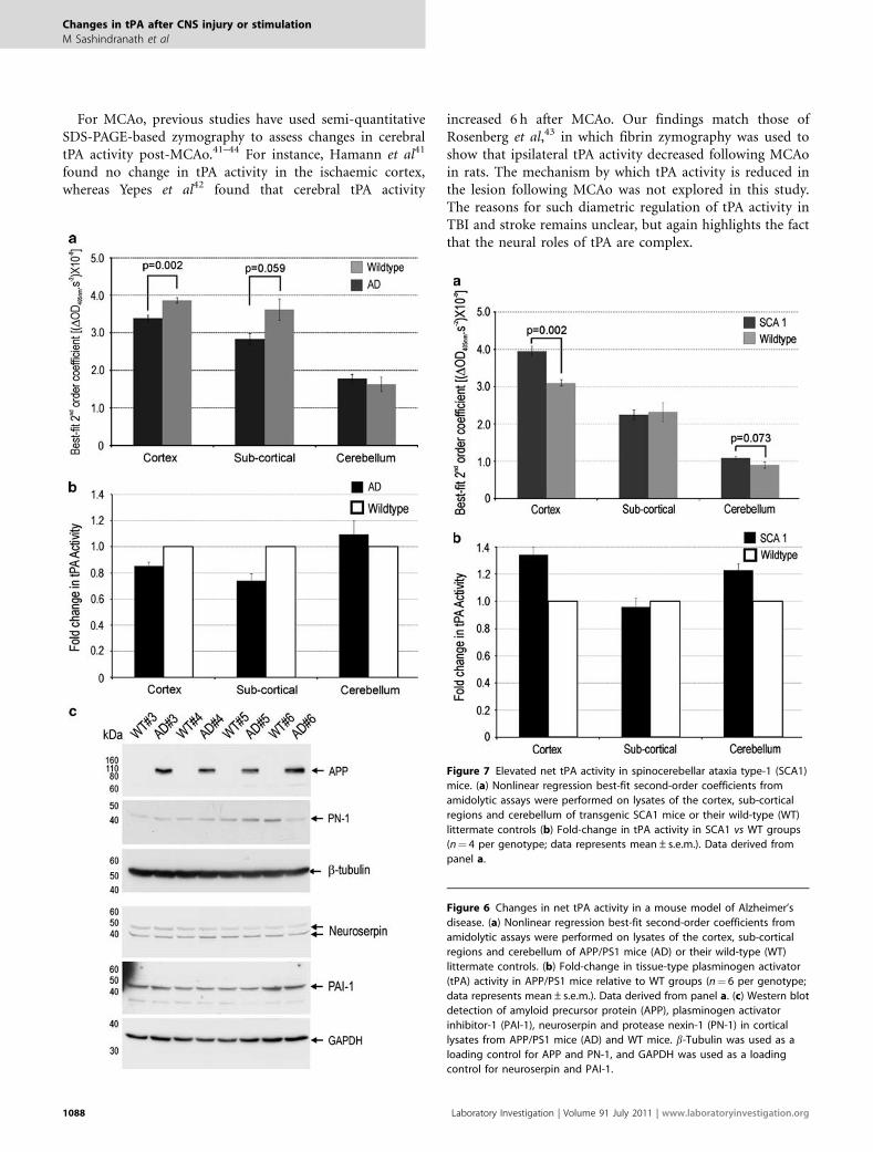
Alteration in Endogenous Brain-derived tPA Activity inChronic Neurodegenerative DiseasesHaving assessed several acute models of CNS injury, we nextwanted to quantitate tPA activity levels in a chronic neuro-degenerative model of AD. Accordingly, we obtained braintissue homogenates from 4-week-old APPswe/PS1dE9 malemice and litter- and age-matched WTmice.14 The amidolyticassay revealed a 15 and 26% decrease in tPA activity in the
Figure 2 Changes in net tissue-type plasminogen activator (tPA) activity
following morphine administration. (a) Nonlinear regression best-fit
second-order coefficients from amidolytic assays were performed on lysates
of the cortex, sub-cortical regions and cerebellum of wildtype (WT) mice at
2 h after intraperitoneal administration of saline or morphine. (b) Fold-
change in tPA activity in morphine vs saline administered groups
(n¼ 5 per treatment group; data represents mean±s.e.m.). Data derived
from panel a.
Figure 3 Changes in net tissue-type plasminogen activator (tPA) activity
following amygdala kindled-seizures. (a) Nonlinear regression best-fit
second-order coefficients from amidolytic assays were performed on lysates
of the cortex, hippocampus and amygdala of wild-type (WT) mice at 4 h
post-seizure. (b) Fold-change in tPA activity in stimulated vs sham groups
(n¼ 11 kindled and n¼ 6 sham; data represents mean±s.e.m.). Data
derived from panel a.
Changes in tPA after CNS injury or stimulation
M Sashindranath et al
www.laboratoryinvestigation.org | Laboratory Investigation | Volume 91 July 2011 1085

cortex and sub-cortex of APPswe/PS1dE9 mice (Figure 6).No significant difference in tPA activity was detected in thecerebellum of APPswe/PS1dE9 mice, a compartment un-affected in APPswe/PS1dE9 mice. tPA activity levels areknown to be influenced by endogenous serine proteaseinhibitors in the brain, including plasminogen activatorinhibitor-1 (PAI-1), protease nexin-1 (PN-1) and neuro-serpin. However, we saw no change in cortical levels of PAI-1,PN-1 or neuroserpin in APPswe/PS1dE9 mice (Figure 6c) at4 weeks of age. As hypothesised by Liu et al,25 elevatedb-amyloid load may directly downregulate tPA levels.
Finally assessment of net tPA activity in brain tissuehomogenates obtained from 8- to 9-week-old SCA1 mice andlitter- and age-matched WT mice revealed a 34% increase incortical tPA activity levels in SCA1 mice (Figure 7). AlthoughtPA activity levels in the sub-cortical structures were un-affected by SCA1, cerebellar net tPA activity was increased by23% in the SCA1 mice (Figure 7). In line with our results,a microarray showed elevated tPA mRNA levels inthe cerebellum of SCA1 mice at 4 and 12 weeks of age(GEO accession # GDS1756). Importantly, the elevation in
cerebellar net tPA activity in SCA1 mice coincides with re-duced motor function as determined by rotarod analysis.13
DISCUSSIONThere are several well-established methods for the detectionof endogenous tPA activity. To date, the vast majority ofresearchers have used zymography to detect in vivo changesin tPA activity in brain tissues; although others have usedS2251 amidolytic assays to measure in vivo changes in tPAactivity.9,26–28 In this study we show that this amidolyticassay, long-used as a sensitive and specific means to detecttPA in biological fluids such as plasma,6,8 is also useful fordetermining in vivo changes in net tPA activity in mousebrain tissue when tested in the presence of its cofactor, fibrin.
The amidolytic assay offers numerous advantages overzymography. For instance, whereas zymographic methods aresemi-quantitative, the amidolytic assay is fully quantitativeover a 250-fold dilution range (Supplementary Figure S1). Inaddition, the resolution of the amidolytic assay is far superiorto that of zymographic techniques (compare Figure 1with Supplementary Figure S3). The amidolytic assay is alsoexquisitely sensitive and can detect low attomole amounts oftPA (data not shown). Furthermore, the assay is rapid andcan be completed within 6 h. The multiwell plate formatof the amidolytic assay also offers considerably higherthroughput than zymographic procedures.
Mouse brain tissue is well-suited for the amidolytic assayas tPA is far more abundant in the brain than urokinaseplasminogen activator.29 Furthermore, the presence of CNBr-fibrinogen accelerates tPA-mediated plasminogen activationmore significantly than urokinase plasminogen activator-mediated plasminogen activation.30 The amidolytic assay,although lacking the ability to resolve activity in fine tissuelocales, can provide information on net tPA activity withinspecific dissectible neural compartments in mice, and showsthat tPA activity is significantly higher in the cortex whencompared with the underlying sub-cortical structures and thecerebellum (Supplementary Figure S2).
One important aspect of the described amidolytic assay isthat the relationship between tPA activity and the rate of theamidolytic assay is not linear, but rather follows a two-phaseexponential association (Supplementary Figure S1). Becauseof this nonlinear association, the amidolytic assay should notbe used as an end-point assay. Instead, we have used theequation: y¼ 546.5(1�10(�181612*x)) �11.13(1�10(�6700000*x))(where x¼ rate of plasminogen activation and y¼dilution in%) to convert experimentally derived changes in the ‘rate ofthe amidolytic assay’ into approximate ‘fold-change in tPAactivity’.
In keeping with tPA having a variety of neuromodulatoryroles, our studies clearly show that tPA activity in the CNS isregulated in a compartment-specific and context-dependentmanner. For example, systemic morphine administrationelicited a 10% increase in cortical tPA activity levels and a27% decrease in cerebellar tPA activity levels (Figure 2). The
Figure 4 Changes in net tPA activity following middle cerebral artery
occlusion (MCAo) and reperfusion. (a) Nonlinear regression best-fit second-
order coefficients from amidolytic assays were performed on lysates of the
cortex, sub-cortical regions and cerebellum of wild-type (WT) mice after 2 h
of MCAo and 30min reperfusion. (b) Fold-change in tPA activity in
ipsilateral vs contralateral groups (n¼ 5 per treatment group; data
represents mean±s.e.m.). Data derived from panel a.
Changes in tPA after CNS injury or stimulation
M Sashindranath et al
1086 Laboratory Investigation | Volume 91 July 2011 | www.laboratoryinvestigation.org

downregulation of cerebellar tPA activity following morphineadministration was unexpected given that morphine haspreviously been shown to upregulate tPA levels in the nucleusaccumbens, striatum and limbic system.22 Furthermore, tPAactivity in the nucleus accumbens is involved in the re-warding and locomotor-stimulating effects of morphine.22
Notably, morphine has recently been identified as anendogenous neuromodulator in the mouse cerebellum,in which GABAergic basket cells release morphine onto them-opioid receptors of Purkinje neurons.31 We hypothesisethat intense activation of Purkinje neurons by exogenousmorphine results in the sharp suppression of net tPA activityin the cerebellum. How this downregulation of cerebellar tPAactivity influences the behavioural effects of morphine abuseis a topic for future studies.
Seizure activity induced by either chemical or electricalstimulation is recognised to upregulate tPA mRNA in hip-pocampus,23 and contributes to seizure propagationthroughout brain regions via a plasmin-independent path-way.24 Further, tPA has been demonstrated to be upregulatedin brain tissue from human epilepsy patients,32 suggestinga possible role in the epileptic disease state. Studies usingtPA�/� mice have also demonstrated that tPA contributes topathological synaptic plasticity associated with epilepsy, in-cluding chemoconvulsant-induced pyramidal cell death33
and mossy fibre sprouting.34 In this study, we show that tPAenzyme activity is upregulated in limbic and cortical areas ofthe contralateral hemisphere at 4 h following electrically
evoked seizures (Figure 3). Notably, one study documents adecrease in tPA activity at 24 h post-seizure.35 Our study,however, is consistent with the reports that demonstrateincreased tPA gene transcription and increased tPA enzymeactivity following seizure.
Investigation into the regulation of tPA activity followingacute cerebral injuries also provided unanticipated findings.Although the primary cause of TBI and stroke are vastlydifferent, the secondary mechanisms of brain infarction areoverlapping. In particular, excitotoxicity, inflammation, freeradical damage and blood–brain barrier breakdown arehallmarks of both TBI and stroke. Moreover, endogenous tPAworsens infarction following both TBI and stroke.36,37 On thebasis of these commonalities, we predicted that endogenoustPA activity would be regulated in a similar manner duringboth stroke and TBI.
However, although cortical net tPA activity increased by29% following TBI (Figure 5), a 26% decrease in tPA activitywas observed following MCAo (Figure 4). We also observedthat the increase in tPA activity after TBI was an acute phe-nomenon, commencing as early as 1 h post-injury and per-sisting till 3 h, but returning to baseline levels at 24 h post-injury. Our findings extend previous studies that documentan increase in tPA antigen in the cerebrospinal fluid of pigswithin 60min after TBI,38 an increase in tPA antigen in braintissue from human TBI autopsy samples39 and an increase intPA mRNA in the rodent cortex at 4 h after TBI (GEO ac-cession # GDS2850 and GDS2851;40).
Figure 5 Changes in net tPA activity following traumatic brain injury (TBI). (a) Nonlinear regression best-fit second-order coefficients from amidolytic assays
were performed on lysates of the cortex, sub-cortical regions and cerebellum of wild-type (WT) mice 3 h after TBI. (b) Fold-change in tPA activity in ipsilateral
vs contralateral groups (n¼ 5 per treatment group; data represents mean±s.e.m.). (c) Time course of the rates of plasminogen activation in the ipsilateral
and contralateral cortical lysates of WT mice 1, 3 and 24 h post-TBI or post-sham operation. Asterisks indicate Po0.05. (d) Fold-change in tPA activity
between ipsilateral vs contralateral groups. Data derived from panel c. (n¼ 5 per treatment group; data represents mean±s.e.m.).
Changes in tPA after CNS injury or stimulation
M Sashindranath et al
www.laboratoryinvestigation.org | Laboratory Investigation | Volume 91 July 2011 1087
For MCAo, previous studies have used semi-quantitativeSDS-PAGE-based zymography to assess changes in cerebraltPA activity post-MCAo.41–44 For instance, Hamann et al41
found no change in tPA activity in the ischaemic cortex,whereas Yepes et al42 found that cerebral tPA activity
increased 6 h after MCAo. Our findings match those ofRosenberg et al,43 in which fibrin zymography was used toshow that ipsilateral tPA activity decreased following MCAoin rats. The mechanism by which tPA activity is reduced inthe lesion following MCAo was not explored in this study.The reasons for such diametric regulation of tPA activity inTBI and stroke remains unclear, but again highlights the factthat the neural roles of tPA are complex.
Figure 6 Changes in net tPA activity in a mouse model of Alzheimer’s
disease. (a) Nonlinear regression best-fit second-order coefficients from
amidolytic assays were performed on lysates of the cortex, sub-cortical
regions and cerebellum of APP/PS1 mice (AD) or their wild-type (WT)
littermate controls. (b) Fold-change in tissue-type plasminogen activator
(tPA) activity in APP/PS1 mice relative to WT groups (n¼ 6 per genotype;
data represents mean±s.e.m.). Data derived from panel a. (c) Western blot
detection of amyloid precursor protein (APP), plasminogen activator
inhibitor-1 (PAI-1), neuroserpin and protease nexin-1 (PN-1) in cortical
lysates from APP/PS1 mice (AD) and WT mice. b-Tubulin was used as a
loading control for APP and PN-1, and GAPDH was used as a loading
control for neuroserpin and PAI-1.
Figure 7 Elevated net tPA activity in spinocerebellar ataxia type-1 (SCA1)
mice. (a) Nonlinear regression best-fit second-order coefficients from
amidolytic assays were performed on lysates of the cortex, sub-cortical
regions and cerebellum of transgenic SCA1 mice or their wild-type (WT)
littermate controls (b) Fold-change in tPA activity in SCA1 vs WT groups
(n¼ 4 per genotype; data represents mean±s.e.m.). Data derived from
panel a.
Changes in tPA after CNS injury or stimulation
M Sashindranath et al
1088 Laboratory Investigation | Volume 91 July 2011 | www.laboratoryinvestigation.org

The tPA/plasmin system has long been hypothesised to bea physiological clearance mechanism for b-amyloid.45,46 Insupport of this notion, previous studies that used SDS-PAGE-based zymography have reported decreased cerebraltPA/plasmin activity in brains of human patients withAD.47–49 A downregulation of tPA activity is also evident inmouse models of AD, with several studies attributing thesuppression of tPA activity to an increase in the expression ofserine protease inhibitors.25,48,50 Although we also show adecrease in tPA activity in the AD mouse brain, no obviousincreases in the expression of the major inhibitors of tPAincluding PAI-1, PN-1 or neuroserpin was observed(Figure 6c). This dissimilarity may be because of the espe-cially young age of the AD mice used in our study, as in-creases in PAI-1 levels25 and decreases in tPA expression51
arise at later stages in development. The suppression of tPAactivity in APPswe/PS1dE9 mice at 4 weeks of age (Figure 6)is of particular interest, as plaques and other hallmarks of ADdo not appear in these mice until approximately 4 monthsof age.52,53 To our knowledge, this is the earliest stage ofdevelopment at which suppressed tPA activity has beenshown in a mouse model of AD, and further implicates lowtPA levels in the progression of AD.
The final situation where we assessed net tPA activity wasthat of cerebellar degeneration. It has previously been shownthat elevated tPA activity precedes cerebellar degeneration inLurcher mice and Nervous mice—two unrelated models ofcerebellar dysfunction.9,10 Experimental evidence from bothof these models led to the postulate that high levels of tPAactivity promote cerebellar degeneration. Accordingly, wemeasured tPA activity levels in transgenic SCA1 mice. SCA1is an autosomal dominant form of spinocerebellar degen-eration.54 The transgenic SCA1 mice used in our study de-velop many of the neurological and histological signs of thehuman disorder including ataxia, Purkinje neuron loss andnuclear inclusion formation.13 At 8 weeks of age, before theonset of Purkinje neuron loss but coincident with motordysfunction,13 we observed that net tPA activity was 23%higher in the cerebellum of SCA1 mice, relative to their lit-termate controls (Figure 7). This is the first documentationof cerebellar tPA activity levels being increased in a model ofhuman cerebellar degeneration and supports the theory thatelevated tPA activity levels promote cerebellar degeneration.Curiously, increased levels of tPA were also detected in thecortex of SCA1 transgenic mice (Figure 7). The cortex ofSCA1 mice displays no significant signs of degeneration,despite prominent SCA1 transgene expression in the cerebralcortical neurons.13 These observations raise the possibilitythat: (1) elevated net tPA activity is not the result of cellinjury but is instead likely due to the presence of pathologicalSCA1 protein, and/or (2) the putative detrimental influenceof high tPA activity may be restricted to the cerebellum inSCA1 mice. If high levels of tPA do indeed contribute tocerebellar degeneration, then the T4 transgenic mice wouldalso be expected to display signs of cerebellar stress such as
motor incoordination and Purkinje neuron loss. This hy-pothesis is currently being tested.
In conclusion, we have harnessed the previously describedfibrin-accelerated amidolytic assay to quantify in vivo chan-ges in mouse brain-derived tPA activity. Using this amido-lytic assay, rapid and differential regulation of tPA activitywas observed in mice between the cortex and cerebellumfollowing systemic morphine administration. Rapid, dia-metric regulation of tPA activity was also observed in thecortex following TBI and stroke. Notably, severe epilepticseizure was the only setting in which a global change in braintPA activity was observed. Thus, on the basis of the elevatedcerebellar tPA activity in SCA1 transgenic mice, we hy-pothesise that high net tPA activity contributes to cerebellardysfunction. This study highlights that a tight and context-dependent regulation underlies the complex neurologicalroles of tPA. On the basis of this exquisite regulation, wealso speculate that alterations in neural net tPA activitylevels could be used as a biomarker for perturbations inbrain stasis.
Supplementary Information accompanies the paper on the Laboratory
Investigation website (http://www.laboratoryinvestigation.org)
ACKNOWLEDGEMENTS
We thank Prof Norman Saunders, Dr Mark Habgood, Dr Simone Beckham
and Mr Adam Galle for constructing the electromagnetic CCI device. We
also thank Ms Amanda Au for assisting in the preparation of mouse brain
lysates following MCAo. Be’eri Niego for constructive comments regarding
the amidolytic assay. Volga Tarlac and Elsdon Storey from the Van Cleef
Roet Centre for Nervous Diseases for providing us with the SCA1 mutant
and wild-type littermate mice, and Prof John Hamilton (Department of
Medicine, Royal Melbourne Hospital) for providing the mice for the seizure
studies. This study was supported by grant numbers 606659 awarded to
RLM and ALS from the National Health and Medical Research Council
(NH&MRC) of Australia and by grant numbers 491152 and 606660 awarded
to RLM from the NH&MRC and D0-34 awarded to RLM from the Victorian
Neurotrauma Initiative. This study was also supported by grant numbers
628391 and G 08M 3821 awarded to PJC from the NH&MRC and the
National Heart Foundation of Australia, respectively, and by grant number
566544 awarded to NCJ from the NH&MRC. AJL and RLM are also supported
with Senior Research Fellowships, and NCJ supported with a Career
Development Award, from the NH&MRC.
DISCLOSURE/CONFLICT OF INTEREST
The authors declare no conflict of interest.
1. Melchor JP, Strickland S. Tissue plasminogen activator in centralnervous system physiology and pathology. Thromb Haemost 2005;93:655–660.
2. Samson AL, Medcalf RL. Tissue-type plasminogen activator: amultifaceted modulator of neurotransmission and synaptic plasticity.Neuron 2006;50:673–678.
3. Yepes M, Roussel BD, Ali C, et al. Tissue-type plasminogen activator inthe ischemic brain: more than a thrombolytic. Trends Neurosci2009;32:48–55.
4. Granelli-Piperno A, Reich E. A study of proteases and protease-inhibitorcomplexes in biological fluids. J Exp Med 1978;148:223–234.
5. Shimada H, Mori T, Takada A, et al. Use of chromogenic substrate S-2251 for determination of plasminogen activator in rat ovaries.Thromb Haemost 1981;46:507–510.
Changes in tPA after CNS injury or stimulation
M Sashindranath et al
www.laboratoryinvestigation.org | Laboratory Investigation | Volume 91 July 2011 1089
6. Eriksson E, Risberg B. Measurement of tissue plasminogen activator inplasma. A comparison of 3 methods and description of a newimproved technique. Thromb Res 1987;46:213–223.
7. Stephens R, Leung KC, Pollanen J, et al. Microplate immunocaptureassay for plasminogen activators and their specific inhibitors.J Immunol Methods 1987;105:245–251.
8. Verheijen JH, Mullaart E, Chang GT, et al. A simple, sensitivespectrophotometric assay for extrinsic (tissue-type) plasminogenactivator applicable to measurements in plasma. Thromb Haemost1982;48:266–269.
9. Lu W, Tsirka SE. Partial rescue of neural apoptosis in the Lurchermutant mouse through elimination of tissue plasminogen activator.Development 2002;129:2043–2050.
10. Li J, Ma Y, Teng YD, et al. Purkinje neuron degeneration in nervous (nr)mutant mice is mediated by a metabolic pathway involving excesstissue plasminogen activator. Proc Natl Acad Sci USA 2006;103:7847–7852.
11. Madani R, Hulo S, Toni N, et al. Enhanced hippocampal long-termpotentiation and learning by increased neuronal expression of tissue-type plasminogen activator in transgenic mice. EMBO J 1999;18:3007–3012.
12. Carmeliet P, Schoonjans L, Kieckens L, et al. Physiologicalconsequences of loss of plasminogen activator gene function inmice. Nature 1994;368:419–424.
13. Watase K, Weeber EJ, Xu B, et al. A long CAG repeat in the mouse Sca1locus replicates SCA1 features and reveals the impact of proteinsolubility on selective neurodegeneration. Neuron 2002;34:905–919.
14. Jankowsky JL, Slunt HH, Ratovitski T, et al. Co-expression of multipletransgenes in mouse CNS: a comparison of strategies. Biomol Eng2001;17:157–165.
15. Salzberg M, Kumar G, Supit L, et al. Early postnatal stress confersenduring vulnerability to limbic epileptogenesis. Epilepsia2007;48:2079–2085.
16. Racine RJ. Modification of seizure activity by electrical stimulation. II.Motor seizure. Electroencephalogr Clin Neurophysiol 1972;32:281–294.
17. Wong CH, Bozinovski S, Hertzog PJ, et al. Absence of glutathioneperoxidase-1 exacerbates cerebral ischemia-reperfusion injury byreducing post-ischemic microvascular perfusion. J Neurochem2008;107:241–252.
18. Onyszchuk G, Al-Hafez B, He YY, et al. A mouse model of sensorimotorcontrolled cortical impact: characterization using longitudinalmagnetic resonance imaging, behavioral assessments and histology.J Neurosci Methods 2007;160:187–196.
19. Niego B, Horvath A, Coughlin PB, et al. Desmoteplase-mediatedplasminogen activation and clot lysis are inhibited by the lysineanalogue tranexamic acid. Blood Coagul Fibrinolysis 2008;19:322–324.
20. Sappino AP, Madani R, Huarte J, et al. Extracellular proteolysis in theadult murine brain. J Clin Invest 1993;92:679–685.
21. Ida N, Hartmann T, Pantel J, et al. Analysis of heterogeneous A4peptides in human cerebrospinal fluid and blood by a newlydeveloped sensitive Western blot assay. J Biol Chem 1996;271:22908–22914.
22. Nagai T, Yamada K, Yoshimura M, et al. The tissue plasminogenactivator-plasmin system participates in the rewarding effect ofmorphine by regulating dopamine release. Proc Natl Acad Sci USA2004;101:3650–3655.
23. Qian Z, Gilbert ME, Colicos MA, et al. Tissue-plasminogen activator isinduced as an immediate-early gene during seizure, kindling and long-term potentiation. Nature 1993;361:453–457.
24. Yepes M, Sandkvist M, Coleman TA, et al. Regulation of seizurespreading by neuroserpin and tissue-type plasminogen activator isplasminogen-independent. [see comments.]. J Clin Invest2002;109:1571–1578.
25. Liu RM, van Groen T, Katre A, et al. Knockout of plasminogen activatorinhibitor 1 gene reduces amyloid beta peptide burden in a mousemodel of Alzheimer’s disease. Neurobiol Aging 2009 (in press).
26. Rogove AD, Lu W, Tsirka SE. Microglial activation and recruitment, butnot proliferation, suffice to mediate neurodegeneration. Cell DeathDiffer 2002;9:801–806.
27. Lu W, Bhasin M, Tsirka SE. Involvement of tissue plasminogenactivator in onset and effector phases of experimental allergicencephalomyelitis. J Neurosci 2002;22:10781–10789.
28. Seeds NW, Basham ME, Ferguson JE. Absence of tissue plasminogenactivator gene or activity impairs mouse cerebellar motor learning. JNeurosci 2003;23:7368–7375.
29. Thewke DP, Seeds NW. The expression of mRNAs for hepatocytegrowth factor/scatter factor, its receptor c-met, and one of itsactivators tissue-type plasminogen activator show a systematicrelationship in the developing and adult cerebral cortex andhippocampus. Brain Res 1999;821:356–367.
30. Lijnen HR, Van Hoef B, Collen D. Influence of cyanogen-bromide-digested fibrinogen on the kinetics of plasminogen activation byurokinase. Eur J Biochem 1984;144:541–544.
31. Muller A, Glattard E, Taleb O, et al. Endogenous morphine in SH-SY5Ycells and the mouse cerebellum. PLoS One 2008;3:e1641.
32. Iyer AM, Zurolo E, Boer K, et al. Tissue plasminogen activator andurokinase plasminogen activator in human epileptogenic pathologies.Neuroscience 2010;167:929–945.
33. Tsirka SE, Gualandris A, Amaral DG, et al. Excitotoxin-induced neuronaldegeneration and seizure are mediated by tissue plasminogenactivator. Nature 1995;377:340–344.
34. Wu YP, Siao CJ, Lu W, et al. The tissue plasminogen activator (tPA)/plasmin extracellular proteolytic system regulates seizure-inducedhippocampal mossy fiber outgrowth through a proteoglycansubstrate. J Cell Biol 2000;148:1295–1304.
35. Lahtinen L, Lukasiuk K, Pitkanen A. Increased expression and activity ofurokinase-type plasminogen activator during epileptogenesis. Eur JNeurosci 2006;24:1935–1945.
36. Mori T, Wang X, Kline AE, et al. Reduced cortical injury and edema intissue plasminogen activator knockout mice after brain trauma.NeuroReport 2001;12:4117–4120.
37. Nagai N, De Mol M, Lijnen HR, et al. Role of plasminogen systemcomponents in focal cerebral ischemic infarction: a gene targeting andgene transfer study in mice. Circulation 1999;99:2440–2444.
38. Armstead WM, Cines DB, Higazie AA. Plasminogen activatorscontribute to age-dependent impairment of NMDAcerebrovasodilation after brain injury. Brain Res Dev Brain Res2005;156:139–146.
39. Dietzmann K, von Bossanyi P, Krause D, et al. Expression of theplasminogen activator system and the inhibitors PAI-1 and PAI-2 inposttraumatic lesions of the CNS and brain injuries following dramaticcirculatory arrests: an immunohistochemical study. Pathol Res Pract2000;196:15–21.
40. Natale JE, Ahmed F, Cernak I, et al. Gene expression profile changes arecommonly modulated across models and species after traumatic braininjury. J Neurotrauma 2003;20:907–927.
41. Hamann GF, Burggraf D, Martens HK, et al. Mild to moderatehypothermia prevents microvascular basal lamina antigen loss inexperimental focal cerebral ischemia. Stroke 2004;35:764–769.
42. Yepes M, Sandkvist M, Wong MK, et al. Neuroserpin reduces cerebralinfarct volume and protects neurons from ischemia-induced apoptosis.Blood 2000;96:569–576.
43. Rosenberg GA, Navratil M, Barone F, et al. Proteolytic cascade enzymesincrease in focal cerebral ischemia in rat. J Cereb Blood Flow Metab1996;16:360–366.
44. Kim JW, Lee SH, Ko HM, et al. Biphasic regulation of tissueplasminogen activator activity in ischemic rat brain and in culturedneural cells: Essential role of astrocyte-derived plasminogen activatorinhibitor-1. Neurochem Int 2010.
45. Kingston IB, Castro MJ, Anderson S. In vitro stimulation of tissue-typeplasminogen activator by Alzheimer amyloid beta-peptide analogues.Nat Med 1995;1:138–142.
46. Jacobsen JS, Comery TA, Martone RL, et al. Enhanced clearance ofAbeta in brain by sustaining the plasmin proteolysis cascade. Proc NatlAcad Sci USA 2008;105:8754–8759.
47. Ledesma MD, Da Silva JS, Crassaerts K, et al. Brain plasmin enhancesAPP alpha-cleavage and Abeta degradation and is reduced inAlzheimer’s disease brains. EMBO Rep 2000;1:530–535.
48. Cacquevel M, Launay S, Castel H, et al. Ageing and amyloid-betapeptide deposition contribute to an impaired brain tissueplasminogen activator activity by different mechanisms. NeurobiolDis 2007;27:164–173.
49. Fabbro S, Seeds NW. Plasminogen activator activity is inhibited whileneuroserpin is up-regulated in the Alzheimer disease brain.J Neurochem 2009;109:303–315.
Changes in tPA after CNS injury or stimulation
M Sashindranath et al
1090 Laboratory Investigation | Volume 91 July 2011 | www.laboratoryinvestigation.org

50. Melchor JP, Pawlak R, Strickland S. The tissue plasminogen activator-plasminogen proteolytic cascade accelerates amyloid-beta (Abeta)degradation and inhibits Abeta-induced neurodegeneration.J Neurosci 2003;23:8867–8871.
51. Roussel BD, Macrez R, Jullienne A, et al. Age and albumin D site-binding protein control tissue plasminogen activator levels: neurotoxicimpact. Brain 2009;132(Part 8):2219–2230.
52. Garcia-Alloza M, Robbins EM, Zhang-Nunes SX, et al. Characterizationof amyloid deposition in the APPswe/PS1dE9 mouse model ofAlzheimer disease. Neurobiol Dis 2006;24:516–524.
53. Ruan L, Kang Z, Pei G, et al. Amyloid deposition and inflammation inAPPswe/PS1dE9 mouse model of Alzheimer’s disease. Curr Alzheimerresearch 2009;6:531–540.
54. Zoghbi HY, Orr HT. Spinocerebellar ataxia type 1. Semin Cell Biol 1995;6:29–35.
Changes in tPA after CNS injury or stimulation
M Sashindranath et al
www.laboratoryinvestigation.org | Laboratory Investigation | Volume 91 July 2011 1091
Copyright © 2022 FDOKUMEN