Routine cerebrospinal fluid (CSF) analysis
14
Huge: “ch04” — 2006/6/29 — 14:54 — page 14 — #1 CHAPTER4 Routine cerebrospinal fluid (CSF) analysis F. Deisenhammer, a A. Bartos, b R. Egg, a N. E. Gilhus, c G. Giovannoni, d S. Rauer, e F. Sellebjerg f Background A great variety of neurological diseases require investigation of the cerebrospinal fluid (CSF) to prove the diagnosis or to rule out relevant differential diagnoses. Objectives To evaluate the theoretical background and provide guidelines for clinical use in rou- tine CSF analysis including total protein, albumin, immunoglobulins, glucose, lactate, cell count, cytological staining, and investigation of infec- tious CSF. Methods Systematic Medline search for the above mentioned variables. Review of appropriate publi- cations by one or more of the task force members. Grading of evidence and recommendations was based on consensus by all task force members. CSF should be analysed immediately after collec- tion. If storage is needed 12 ml of CSF should be partitioned into three to four sterile tubes. a Department of Neurology, Innsbruck Medical University, Austria; b Department of Neurology, Charles University, Prague, Czech Republic; c Department of Clinical Medicine, University of Bergen, Bergen, Norway, and Department of Neurology, Haukeland University Hospital, Bergen, Norway; d Department of Neuroinflammation, Institute of Neurology, University College London, Queen Square, London, UK; e Department of Neurology and Clinical Neurophysiology, Albert-Ludwigs University, Freiburg, Germany; f Department of Neurology, Copenhagen University Hospital, Denmark. Albumin CSF/serum ratio (Q alb ) should be pre- ferred to total protein measurement and normal upper limits should be related to patients’ age. Elevated Q alb is a non-specific finding but occurs mainly in bacterial, cryptococcal, and tubercu- lous meningitis, leptomingeal metastases as well as acute and chronic demyelinating polyneu- ropathies. Pathological decrease of the CSF/serum glu- cose ratio or an increase in lactate concentration indicates bacterial or fungal meningitis or lep- tomeningeal metastases. Intrathecal immunoglobulin G synthesis is best demonstrated by isoelectric focusing followed by specific staining. Cellular morphology (cytological staining) should be evaluated whenever pleocytosis is found or leptomeningeal metastases or pathological bleed- ing is suspected. Computed tomography-negative intrathecal bleeding should be investigated by bilirubin detection. Introduction The cerebrospinal fluid (CSF) is a dynamic, metabolically active substance that has many 14
-
Upload
independent -
Category
Documents
-
view
0 -
download
0
Transcript of Routine cerebrospinal fluid (CSF) analysis

Huge: “ch04” — 2006/6/29 — 14:54 — page 14 — #1
C H A P T E R 4
Routine cerebrospinal fluid (CSF) analysis
F. Deisenhammer,a A. Bartos,b R. Egg,a
N. E. Gilhus,c G. Giovannoni,d S. Rauer,e
F. Sellebjergf
Background A great variety of neurological diseasesrequire investigation of the cerebrospinal fluid(CSF) to prove the diagnosis or to rule out relevantdifferential diagnoses.
Objectives To evaluate the theoretical backgroundand provide guidelines for clinical use in rou-tine CSF analysis including total protein, albumin,immunoglobulins, glucose, lactate, cell count,cytological staining, and investigation of infec-tious CSF.
Methods Systematic Medline search for the abovementioned variables. Review of appropriate publi-cations by one or more of the task force members.Grading of evidence and recommendations wasbased on consensus by all task force members.
CSF should be analysed immediately after collec-tion. If storage is needed 12 ml of CSF shouldbe partitioned into three to four sterile tubes.
aDepartment of Neurology, Innsbruck MedicalUniversity, Austria; bDepartment of Neurology, CharlesUniversity, Prague, Czech Republic; cDepartment ofClinical Medicine, University of Bergen, Bergen,Norway, and Department of Neurology, HaukelandUniversity Hospital, Bergen, Norway; dDepartment of
Neuroinflammation, Institute of Neurology, UniversityCollege London, Queen Square, London, UK;eDepartment of Neurology and ClinicalNeurophysiology, Albert-Ludwigs University, Freiburg,Germany; fDepartment of Neurology, CopenhagenUniversity Hospital, Denmark.
Albumin CSF/serum ratio (Qalb) should be pre-ferred to total protein measurement and normalupper limits should be related to patients’ age.Elevated Qalb is a non-specific finding but occursmainly in bacterial, cryptococcal, and tubercu-lous meningitis, leptomingeal metastases as wellas acute and chronic demyelinating polyneu-ropathies.
Pathological decrease of the CSF/serum glu-cose ratio or an increase in lactate concentrationindicates bacterial or fungal meningitis or lep-tomeningeal metastases.
Intrathecal immunoglobulin G synthesis is bestdemonstrated by isoelectric focusing followed byspecific staining.
Cellular morphology (cytological staining) shouldbe evaluated whenever pleocytosis is found orleptomeningeal metastases or pathological bleed-ing is suspected. Computed tomography-negativeintrathecal bleeding should be investigated bybilirubin detection.
Introduction
The cerebrospinal fluid (CSF) is a dynamic,metabolically active substance that has many
14

Huge: “ch04” — 2006/6/29 — 14:54 — page 15 — #2
Section 2: Investigation 15
Table 4.1 Typical constellation of CSF parameters in some neurological diseases.
Total protein Glucose ratio Lactate Cell count Typical cytology(g/l) (mmol/l) (per 3.2 µl)
Normal valuesa <0.45 >0.4–0.5 <1.0–2.9 <15 MNC
DiseaseAcute bacterialmeningitis
↑ ↓ ↑ >1000 PNC
Viral neuro-infections(meningo/encephalitis)
= /↑ = /↓ = 10–1000 PNC/MNC
Autoimmunepolyneuropathy
↑ = = =
Infectiouspolyneuropathy
↑ = = ↑ MNC
Subarachnoidalhaemorrhage
↑ = = ↑ erythrocytes,macrophages,siderophagesMNC
Multiple sclerosis = = = = /↑ MNCLeptomeningealmetastases
↑ = /↓ NA = /↑ malignant cells,mononuclears
CSF, cerebrospinal fluid; MNC, mononuclear cells; PNC, polymorphonuclear cells.↑/↓, increased/decreased; =, within normal limits; NA, evidence not available. aNormal values aregiven for lumbar CSF in adults.
important functions. It is invaluable as a diag-nostic aid in the evaluation of inflammatory con-ditions, infectious or non-infectious, involvingthe brain, spinal cord, and meninges as well asin CT-negative subarachnoidal haemorrhage andin leptomeningeal metastases. CSF is obtainedwith relative ease by lumbar puncture (LP). Alter-ations in CSF constituents may be similar in dif-ferent pathologic processes and cause difficultiesin interpretation. Combining a set of CSF vari-ables referred to as routine parameters (i.e. deter-mination of protein, albumin, immunoglobulin,glucose, lactate, and cellular changes, as well asspecific antigen and antibody testing for infectiousagents) will increase the diagnostic sensitivity andspecificity.
The aim of this guideline paper was to pro-duce recommendations on how to use this set ofCSF parameters in different clinical settings andto show how different constellations of these vari-ables correlate with diseases of the nervous system(table 4.1) (Brainin et al., 2004).
Search strategy
A Medline search using the search terms cere-brospinal fluid (CSF), immunoglobulin G (IgG)immunoglobulin M (IgM), immunoglobulin A(IgA), and albumin was conducted. Also, the keywords ‘cerebrospinal fluid’ or ‘CSF’ were cross-referenced with ‘glucose’, ‘lactate’, ‘cytology’,‘cell∗ in title’ excluding ‘child∗’. Furthermore, asearch for ‘cerebrospinal fluid’ and ‘immunoglobu-lin’ and ‘diagnosis’ and ‘electrophoresis’ or ‘isoelec-tric focusing’ was performed limited to the timebetween 1 January 1980 and 1 January 2005, andreturned only items with abstracts, and Englishlanguage (274 references). A search for ‘cere-brospinal fluid’ AND ‘infectious’ limited for time(1 January 1980 until now) returned 560 abstracts.Abstracts that primarily did not deal with diagnos-tic issues and infectious CSF (e.g. non-infectiousinflammatory diseases, vaccination, general CSFparameters, pathophysiology, cytokines and ther-apy) were excluded resulting in 60 abstracts.
Huge: “ch04” — 2006/6/29 — 14:54 — page 16 — #3
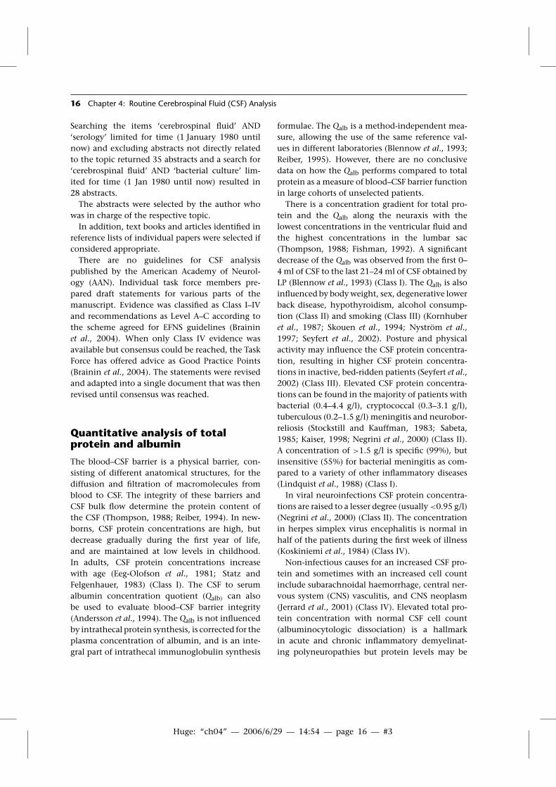
16 Chapter 4: Routine Cerebrospinal Fluid (CSF) Analysis
Searching the items ‘cerebrospinal fluid’ AND‘serology’ limited for time (1 January 1980 untilnow) and excluding abstracts not directly relatedto the topic returned 35 abstracts and a search for‘cerebrospinal fluid’ AND ‘bacterial culture’ lim-ited for time (1 Jan 1980 until now) resulted in28 abstracts.
The abstracts were selected by the author whowas in charge of the respective topic.
In addition, text books and articles identified inreference lists of individual papers were selected ifconsidered appropriate.
There are no guidelines for CSF analysispublished by the American Academy of Neurol-ogy (AAN). Individual task force members pre-pared draft statements for various parts of themanuscript. Evidence was classified as Class I–IVand recommendations as Level A–C according tothe scheme agreed for EFNS guidelines (Braininet al., 2004). When only Class IV evidence wasavailable but consensus could be reached, the TaskForce has offered advice as Good Practice Points(Brainin et al., 2004). The statements were revisedand adapted into a single document that was thenrevised until consensus was reached.
Quantitative analysis of totalprotein and albumin
The blood–CSF barrier is a physical barrier, con-sisting of different anatomical structures, for thediffusion and filtration of macromolecules fromblood to CSF. The integrity of these barriers andCSF bulk flow determine the protein content ofthe CSF (Thompson, 1988; Reiber, 1994). In new-borns, CSF protein concentrations are high, butdecrease gradually during the first year of life,and are maintained at low levels in childhood.In adults, CSF protein concentrations increasewith age (Eeg-Olofson et al., 1981; Statz andFelgenhauer, 1983) (Class I). The CSF to serumalbumin concentration quotient (Qalb) can alsobe used to evaluate blood–CSF barrier integrity(Andersson et al., 1994). The Qalb is not influencedby intrathecal protein synthesis, is corrected for theplasma concentration of albumin, and is an inte-gral part of intrathecal immunoglobulin synthesis
formulae. The Qalb is a method-independent mea-sure, allowing the use of the same reference val-ues in different laboratories (Blennow et al., 1993;Reiber, 1995). However, there are no conclusivedata on how the Qalb performs compared to totalprotein as a measure of blood–CSF barrier functionin large cohorts of unselected patients.
There is a concentration gradient for total pro-tein and the Qalb along the neuraxis with thelowest concentrations in the ventricular fluid andthe highest concentrations in the lumbar sac(Thompson, 1988; Fishman, 1992). A significantdecrease of the Qalb was observed from the first 0–4 ml of CSF to the last 21–24 ml of CSF obtained byLP (Blennow et al., 1993) (Class I). The Qalb is alsoinfluenced by body weight, sex, degenerative lowerback disease, hypothyroidism, alcohol consump-tion (Class II) and smoking (Class III) (Kornhuberet al., 1987; Skouen et al., 1994; Nyström et al.,1997; Seyfert et al., 2002). Posture and physicalactivity may influence the CSF protein concentra-tion, resulting in higher CSF protein concentra-tions in inactive, bed-ridden patients (Seyfert et al.,2002) (Class III). Elevated CSF protein concentra-tions can be found in the majority of patients withbacterial (0.4–4.4 g/l), cryptococcal (0.3–3.1 g/l),tuberculous (0.2–1.5 g/l) meningitis and neurobor-reliosis (Stockstill and Kauffman, 1983; Sabeta,1985; Kaiser, 1998; Negrini et al., 2000) (Class II).A concentration of >1.5 g/l is specific (99%), butinsensitive (55%) for bacterial meningitis as com-pared to a variety of other inflammatory diseases(Lindquist et al., 1988) (Class I).
In viral neuroinfections CSF protein concentra-tions are raised to a lesser degree (usually <0.95 g/l)(Negrini et al., 2000) (Class II). The concentrationin herpes simplex virus encephalitis is normal inhalf of the patients during the first week of illness(Koskiniemi et al., 1984) (Class IV).
Non-infectious causes for an increased CSF pro-tein and sometimes with an increased cell countinclude subarachnoidal haemorrhage, central ner-vous system (CNS) vasculitis, and CNS neoplasm(Jerrard et al., 2001) (Class IV). Elevated total pro-tein concentration with normal CSF cell count(albuminocytologic dissociation) is a hallmarkin acute and chronic inflammatory demyelinat-ing polyneuropathies but protein levels may be

Huge: “ch04” — 2006/6/29 — 14:54 — page 17 — #4
Section 2: Investigation 17
normal during the first week (Segurado et al.,1986; Senevirante, 2000) (Class IV). Total CSFprotein is elevated in 80% of patients with lep-tomeningeal metastases to a median concentrationof 1 g/l with a wide range (Twijnstra et al., 1989)(Class III).
In conclusion, there is Class I evidence thatincreased Qalb and total CSF protein concentra-tions are mainly supportive of bacterial, cryp-tococcal, and tuberculous meningitis as well asleptomingeal metastases. As Qalb or protein isusually not the only CSF investigation the com-bination with other CSF variables will increasethe diagnostic specificity, like albuminocytologicdissociation in Gullain–Barré syndrome.
Quantitative intrathecalimmunoglobulin synthesis
Intrathecal Ig synthesis is found in various, mainlyinflammatory CNS diseases (table 4.2). There is aclose correlation between the Qalb and the CSF-serum IgG concentration quotient (QIgG) which ledto the development of the IgG index (QIgG /Qalb)
(Delpech and Lichtblau, 1972; Ganrot and Laurell,1974; Link and Tibbling, 1977). Reiber’s hyperbolicformula and Öhman’s extended immunoglobu-lin indices are based on the demonstration ofnon-linear relationships between the Qalb and CSF-serum concentration quotients for IgG, IgA andIgM (Öhman et al., 1989 and 1993; Reiber, 1994).
Table 4.2 Percentage of patients in different categories of disease withelevated IgA-index, IgG-index, IgM-index, or non-linear intrathecalsynthesis formula values (data from Schipper et al., 1988; McLean et al.1990; Öhman et al., 1992; Sellebjerg et al., 1996; Korenke et al., 1997).Unexpected increases are more common with the IgA index, IgG indexand IgM index than with corresponding non-linear formulae.
IgG (%) IgA (%) IgM (%)
No inflammatory and no CNS disease <5 <5 <5
Non-inflammatory CNS disease(including degenerativeand vascular diseases) <25a <5 <5
Infections of the nervous system 25–50 25 25Bacterial infections 25–50 25–50 <25Viral infections 25–50 <25 <25Lyme neuroborreliosis 25–50 <25 75
Multiple sclerosis 70–80 <25 <25Clinically isolated syndromes 40–60 <10 <25
Inflammatory neuropathies 25–50a 25–50a 25–50a
Neoplastic disorders (in general) <25a ND NDParaneoplastic syndromes <25 ND NDMeningeal carcinomatosis 25–50 ND ND
Other neuroinflammatory diseases 25–50b NDc ND
CNS, central nervous system; ND, not determined in larger stud-ies using non-linear immunoglobulin formulae. aUsually not associatedwith oligoclonal bands (artefact in presence of barrier impairment);brare in biopsy-proven neurosarcoidosis; cprominent IgA synthesis inadrenoleukodystrophy.

Huge: “ch04” — 2006/6/29 — 14:54 — page 18 — #5
18 Chapter 4: Routine Cerebrospinal Fluid (CSF) Analysis
For the detection of intrathecal IgG synthesis, thedetection of IgG oligoclonal bands is superior tothe IgG index and the non-linear formulae bothin terms of diagnostic sensitivity and specificity.However, the detection of IgG oligoclonal bandsis technically more demanding than the quantita-tive measures, and it has been suggested that in thesetting of suspected multiple sclerosis (MS), oligo-clonal bands analysis may be omitted in patientswith an IgG-index value above 1.1, as almost 100%of such patients turn out to have intrathecally syn-thesized IgG oligoclonal bands (F. Deisenhammer,unpublished data).
In studies comparing CSF findings in patientswith MS and other neurological diseases, non-linear formulae were superior (Öhman et al., 1992;Sellebjerg et al., 1996). Intrathecal IgA, IgG andIgM synthesis formulae may be helpful in discrimi-nating between different infectious diseases of thenervous system (Felgenhauer, 1982; Felgenhauerand Schädlich, 1987) (Class III). However, onestudy suggested that increased values of the Reiberformula do not always reflect intrathecal IgM syn-thesis as increased values were observed in severalpatients with non-inflammatory diseases withoutIgM oligoclonal bands in CSF (Sharief et al., 1990)(Class II). In conclusion, there is no evidence tosupport the routine use of quantitative assessmentof intrathecal immunoglobulin synthesis in thediagnosis of neurological diseases, but in the set-ting of suspected MS the IgG index may be usedas a screening procedure to determine intrathecalIgG synthesis.
Qualitative (oligoclonal)intrathecal IgG synthesis
The detection of intrathecal oligoclonal IgG in theCSF is useful diagnostically, particularly as it is oneof the laboratory criteria supporting the clinicaldiagnosis of MS (McDonald et al., 2001). In addi-tion, it can be used to assist in the diagnosis ofother putative autoimmune disorders of the CNS,such as paraneoplastic disorders and CNS infec-tions (Rauer and Kaiser, 2000; Stich et al., 2003;Storstein et al., 2004).
Using electrophoresis techniques it is possible toclassify the humoral responses according to the
number of antibody clones produced (i.e. mon-oclonal, oligoclonal and polyclonal responses;figure 4.1). Earlier methods have now beensuperseded by the development of the more
1 5
C S C S C S C S C S C S C S C S C S
2 5 4 3 2 2 1
Figure 4.1. IEF immunoblots of the five consensuspatterns of various CSF and serum isoelectric focusingpatterns for local/systemic synthesis. The patternnumber is given above the paired samples.
Type 1 (C-S-): No bands in CSF and serum. Normal.Type 2 (C+S-): Oligoclonal IgG is present in the CSF
with no apparent correspondingabnormality in serum, indicating localintrathecal synthesis of IgG. Typicalexample: MS.
Type 3 (C+>S+): There are IgG bands in both the CSF andserum, with additional bands present inthe CSF. The oligoclonal bands that arecommon to both CSF and serum implya systemic inflammatory response,whereas the bands that are restricted tothe CNS suggest that there is anadditional CNS-only response. Typicalexamples: MS, systemic lupuserythematodes (SLE), sarcoid etc.
Type 4 (C+S+): There are oligoclonal bands present inthe CSF, which are identical to those inserum. This is not indicative of localsynthesis, but rather, the pattern isconsistent with passive transfer ofoligoclonal IgG from a systemicinflammatory response. Typicalexamples: Guillain–Barre syndrome,acute disseminated encephalomyelitis(ADEM) and systemic infections.
Type 5 (Para): There is a monoclonal IgG pattern inboth CSF and serum, the source ofwhich lies outside the CNS. Typicalexamples: Myeloma, monoclonalgammopathy of undeterminedsignificance (MGUS).

Huge: “ch04” — 2006/6/29 — 14:54 — page 19 — #6
Section 2: Investigation 19
sensitive technique of isoelectric focusing (IEF) andimmunofixation (Andersson et al., 1994).
Isoelectric focusing uses a pH gradient to sep-arate IgG populations on the basis of charge,which are then transferred onto a nitro-celluloseor other membrane before immunostaining usingan anti-human immunoglobulin (Keir et al., 1990).Some laboratories continue to use silver staining todetect oligoclonal bands (OCBs) with good results(Blennow and Fredman, 1995).
As CSF is an ultrafiltrate of plasma, it containsimmunoglobulins that are passively transferredfrom the plasma, as well as immunoglobu-lins synthesized locally. Any systemic pattern ofimmunoglobulin production seen in plasma orserum will therefore be mirrored in the CSF. It isimperative that any CSF analysis for oligoclonalbands is accompanied by a paired blood analysis.
An oligoclonal intrathecal IgG antibody responseis not specific. Table 4.3 provides a list with theproportion of cases with oligoclonal bands (for amore detailed list please see McLean et al. (1990)).Local synthesis of oligoclonal bands is thereforenot diagnostic and has to be interpreted in theclinical context. A recently published recommen-dation regarding detection of oligoclonal bandsconcluded as follows (Freedman et al., 2005):
The single most informative analysis is a quali-tative assessment of CSF for IgG, best performedusing IEF together with some form of immun-odetection (blotting or fixation). This qualitativeanalysis should be performed using unconcen-trated CSF and must be compared directly withserum run simultaneously in the same assay inan adjacent track. Optimal runs utilize similaramounts of IgG from paired serum and CSF.Recognised positive and negative controls shouldbe run with each set of samples.
In putative non-infectious inflammatory disor-ders of the CNS there is Class I evidence to supportthe use of CSF IEF for both predictive and diag-nostic testing in the diagnosis of MS. In othernon-infectious inflammatory disorders of the CNSClass II and III evidence exists to support the useof CSF IEF to supplement other diagnostic tests(table 4.3).
CSF glucose concentration,CSF/serum glucose ratio andlactate
As glucose is actively transported across the blood–brain barrier the CSF glucose levels are directlyproportional to the plasma levels and thereforesimultaneous measurement in CSF and blood isrequired. Normal CSF glucose concentration is 50–60% of serum values (Jerrard et al., 2001) (Class IV).A CSF/serum glucose ratio less than 0.4–0.5 is con-sidered to be pathological (Feigin et al., 1992) (ClassIV). CSF glucose takes several hours to equilibratewith plasma glucose; therefore, in unusual cir-cumstances levels of CSF glucose can actually behigher than plasma levels for several hours. DuringCSF storage glucose is degraded. Therefore, glucosedetermination must be performed immediatelyafter CSF collection.
A high CSF glucose concentration has no spe-cific diagnostic importance and is related to anelevated blood glucose concentration, for example,in diabetics.
The behaviour of the CSF/serum glucose ratioin different neurological diseases is shown intable 4.1.
The relevance of CSF lactate is similar to that ofCSF/serum glucose ratio. CSF lactate is indepen-dent of blood concentration (Watson and Scott,1995) (Class IV). The normal value is consideredto be <2.8–3.5 mmol/l (Jordan, 1983) (Class II).Except for mitochondrial disease CSF lactate corre-lates inversely with CSF/serum glucose ratio. Anincreased level can be detected earlier than thereduced glucose concentration.
Decreased CSF/serum glucose ratio or increasedCSF lactate indicate bacterial and fungal infectionsor leptomeningeal metastases.
Cytological examination
Cytological evaluation should be performed within2 h after puncture, preferably within 30 minbecause of a lysis of both red blood cells and whiteblood cells (Steele et al., 1986) (Class IV).
Cerebrospinal fluid leukocytes are usuallycounted in a Fuchs-Rosenthal chamber (volume3.2 µl) and therefore, counts are reported as ‘/3’

Huge: “ch04” — 2006/6/29 — 14:54 — page 20 — #7
20 Chapter 4: Routine Cerebrospinal Fluid (CSF) Analysis
Table 4.3 Inflammatory diseases of the CNS associated with CSF oligoclonal IgGbands (McLean et al., 1990).
Disorder Incidence of oligoclonal Evidencebands (%)
Multiple sclerosis 95 Class Ia
Auto-immuneNeuro-SLE 50 Class IIINeuro-Behcet’s 20 Class IINeuro-sarcoid 40 Class IIIHarada’s meningitis-uveitis 60 Class III
InfectiousAcute viral encephalitis (<7 days) <5 Class IIAcute bacterial meningitis (<7 days) < 5 Class IISubacute sclerosing panencephalitis (SSPE) 100 Class IProgressive rubella panencephalitis 100 Class INeurosyphilis 95 Class INeuro-AIDS 80 Class IINeuro-borrelliosis 80 Class I
Tumour <5 Class IIIHereditaryAtaxia-telangiectasia 60 Class IIIAdrenoleukodystrophy (encephalitic) 100 Class II
CNS, central nervous system; CSF, cerebrospinal fluid; IgG, immunoglobulin G; SLE,systemic lupus erythematodes. aThis is based on studies using the Poser diagnosticcriteria (Poser et al., 1983) that were validated against the original Schumachercriteria (Schumacher et al., 1965). None of these criteria have been validated usingpopulation-based studies. Therefore, it could be argued that the diagnostic ‘goldstandard’ is a flawed standard.
cells to correct for a standard volume of 1 µl.A cytocentrifuge (cytospin), the Sayk sedimen-tation chamber, or membrane filtration can beused to obtain a sufficient number of cells forcytology (Lamers and Wevers, 1995). For cellu-lar differentiation May–Gruenwald–Giemsa stain-ing is widely used but specific methods may beperformed, especially for the detection of malig-nant cells (Roma et al., 2002; Adam et al., 2001)(Class II).
Lymphocytes and monocytes at the restingphase and occasionally ependymal cells are foundin normal CSF.
An increased number of neutrophilic granulo-cytes can be found in bacterial and acute viralCNS infections (Spanos et al., 1989; Adam, 2001)
(Class II). In the postacute phase a mononucleartransformation occurs.
Upon activation lymphocytes can enlarge orbecome plasma cells indicating an unspecificinflammatory reaction (Adam, 2001; Zeman et al.,2001) (Class IV). Resting monocytes enlarge anddisplay vacuoles when activated. Macrophages arethe most activated monocytes. These cell forms canoccur in a great variety of diseases.
Erythrophages occur 12–18 h after haemor-rhage. Siderophages containing haemosiderin areseen as early as 1–2 days after haemorrhage andmay persist for weeks. Macrophages containinghaematoidin (crystallized bilirubin) degraded fromhaemoglobin may appear about 2 weeks afterbleeding and are a sign of a previous subarachnoid

Huge: “ch04” — 2006/6/29 — 14:54 — page 21 — #8
Section 2: Investigation 21
Table 4.4 List of infectious agents responsible for the vast majority of infectious CNS diseases.
Pathogen Symptoms, Comments Recommended diagnosticmethod∗
BacteriaShould be considered in first lineNeisseria meningitides Microscopy, cultureStreptococcus pneumoniae Microscopy, cultureHaemophilus influenzae Rare due to vaccination Microscopy, cultureStaphylococcus aureus Neurosurgical intervention, trauma Microscopy, cultureEscherichia coli Newborns Microscopy, cultureBorrelia burgdorferi sensu lato SerologyTreponema pallidum Syphilis in the past SerologyMycobacterium tuberculosis PCRa, culture, positive
tuberculin test
Should be considered especially in immunosuppressed patientsActinobacter species CultureBacteroides fragilis CultureJC-virus Progressive multifocal leukoencephalopathy PCRListeria monocytogenes Microscopy, cultureNocardia asteroides Microscopy (modified
Ziehl-Neelsen stain andculture from brain biopsy)
Pasteurella multocida CultureStreptococcus mitis Culture
Should be considered in special situationsBrucella spp. Ingestion of raw milk (products) from cows,
sheep or goatsCulture
Campylobacter fetus Microscopy, cultureCoxiella burnetti (Q-fever) Contact with infected parturient animals
(sheep, goat, cattle) or inhalation of dustcontaminated by the excrements of infectedanimals or ticks
Serology
Leptospira interrogans Exposure to contaminated water or rodenturine
Culture, serology
Mycoplasma pneumoniae Children and young adults SerologyRickettsia Tick exposure, exanthema Serologycoagulase-negative staphylococci Patients with ventricular shunts or drainages Culturegroup B streptococci (preterm) newborns Microscopy, cultureTropheryma whipplei (M. Whipple) Patients with gastrointestinal symptoms
(malabsorption)PCR
VirusesShould be considered in first lineHerpes simplex virus (HSV) type 1 and 2 PCR, serologyVaricella–Zoster virus (VZV) PCR, serologyEnteroviruses (Echovirus, PCR, serologyCoxsackievirus A, B)Human immunodeficiency PCR, serologyvirus (HIV) type 1 and 2
Continued

Huge: “ch04” — 2006/6/29 — 14:54 — page 22 — #9
22 Chapter 4: Routine Cerebrospinal Fluid (CSF) Analysis
Table 4.4 Continued.
Pathogen Symptoms, Comments Recommended diagnosticmethod∗
Epstein–Barr virus (EBV) Lymphadenitis, splenomegaly PCRCytomegalovirus (CMV) Very rare in immunocompetent patients PCR
Should be considered in special situationsAdenovirus Children and young adults PCR, culture, antigen detectionHuman T-cell leukaemia virus type I (HTLV-I) Spastic paraparesis SerologyInfluenza - and SerologyParainfluenza virusLymphocytic Serologychorio-meningitis (LCM)Mumps virus SerologyPoliovirus Flaccid paresis PCRRabies virus Contact with rabies-infected animals PCR from CSF, root of hair,
corneaRotavirus Diarrhoea, febrile convulsions in children Antigen detection in stool
specimensRubella virus SerologySandfly Fever Endemic region: Italy Serology
FungiAspergillus fumigatus Antigen detection in CSF,
where required culture frombrain biopsy
Cryptococcus neoformans Antigen detection in CSF, indiaink stain, less sensitive thanantigen detection, culture
ParasitesToxoplasma gondii CSF: PCR, serology; brain
biopsy: PCRStrongyloides stercoralis Pathogen detection in stool
The following pathogens should be considered in acute myelitis [Recommendation Level B]: HSV type 1 and 2 (PCR), VZV(PCR), Enteroviruses (PCR), Borrelia burgdorferi sensu latu (serology, AI), HIV (serology), tick-borne encephalitis virus (only inendemic areas) (serology, AI). aNested PCR technique has been shown to be substantially more sensitive and specific thanconventional single step PCR techniques (Takahashi et al., 2005).
bleeding (Adam, 2001) (Class IV). However, spec-trophotometry of CSF involving bilirubin quanti-tation has been recommended as the method ofchoice to prove CT-negative subarachnoid bleed-ing up to 2 weeks after onset (UK National ExternalQuality Assessment Scheme for ImmunochemistryWorking Group, 2003).
Lipophages indicate CNS tissue destruction.The presence of macrophages without detectableintracellular material is a non-specific finding,
occurring in disc herniation, malignant meningealinfiltration, spinal tumours, head trauma, stroke,MS, vasculitis, infections and subarachnoid haem-orrhage (Adam, 2001) (Class IV).
Eosinophils are normally not present in CSF. Thepresence of 10 or more eosinophils/µl in CSF oreosinophilia of at least 10% of the total CSF leuko-cyte count is associated with a limited number ofdiseases, including parasitic infections, and coc-cidioiodomycosis. It can occur in malignancies

Huge: “ch04” — 2006/6/29 — 14:54 — page 23 — #10
Section 2: Investigation 23
and react to medication and ventriculoperitonealshunts (Lo Re, 2003). Malignant CSF cells indicateleptomeningeal metastases. False positive resultsoften occur when inflammatory cells are mistakenfor tumour cells or due to contamination withperipheral blood (Twijnstra et al., 1987). False neg-ative detection of malignant cells on cytologicexamination of CSF is common. Factors increas-ing the detection rate of malignant cells includea volume of at least 10.5 ml and repeating thisprocedure once if the cytology is negative. Thedetection rate of 50–70% after the first investiga-tion can be increased to 85–92% after a secondpuncture (Glantz et al., 1998) (Class III). FurtherLPs will only slightly increase the diagnostic sensi-tivity (Wasserstrom et al., 1982; Kaplan et al., 1990)(Class III).
In conclusion, cell count is generally usefulbecause most of the indications for CSF analysisinclude diseases that are associated with elevatednumbers of various cells. Cytological staining canbe helpful in distinguishing CNS diseases when thecell count is increased.
Investigation of infectious CSF
There are many small to medium-sized studiesinvestigating the diagnostic sensitivity and speci-ficity of tests for various infectious agents but nocontrolled study evaluating a work-up of infec-tious CSF in general. Therefore, there are no validdata on the indication, sensitivity and specificityof microbiological procedures in general (i.e. howto proceed with CSF in obvious CNS infections).Existing proposals for the general work-up of infec-tious CSF are based on clinical practice and theo-retically plausible procedures (Schlossberg, 1990;Kniehl et al., 2001; Kaiser, 2002).
There is a great number of methods for antigenor specific antibody detection and their use dependmainly on the type of antigen (table 4.4).
In neuroinfections specific antigen or antibodydetection should be performed depending onthe clinical presentation and the results of basicCSF analysis. The formula for the estimation ofthe relative intrathecal synthesis of specific anti-bodies in the CSF (Antibody Index [AI]) is as
follows:
Estimation of intrathecal synthesis of specific antibod-ies in the CSF (Antibody Index [AI])
Antibody ratio = Antibody − concentrationCSF
Antibody − concentrationserum
IgG ratio = IgG − concentrationCSF
IgG − concentrationserum
AI = Antibody ratio/IgG ratio(positive > 1, 5)
Cerebrospinal fluid polymerase chain reaction canbe performed rapidly and inexpensively and hasbecome an integral component of diagnostic med-ical practice. A patient with a positive PCR result is88 times more likely to have a definite diagnosis ofviral infection of the CNS as compared to a patientwith a negative PCR result. A negative PCR resultcan be used with moderate confidence to rule outa diagnosis of viral infection of the CNS (the prob-ability of a definite viral CNS infection was 0.1 incase of a negative PCR result compared to a pos-itive PCR result) (Jeffery et al., 1997). It shouldbe considered that false negative results are mostlikely if the CSF sample is taken within the first3 days after the illness or 10 days and more after theonset of the disease (Davies et al., 2005; Kennedy,2005).
In general, PCR is indicated in the followingsituations:
when microscopy, culture or serology is insensi-tive or inappropriate;
when culture does not yield a result despiteclinical suspicion of infectious meningitis/meningoencephalitis; and
in immunodeficient patients.
R E C O M M E N D AT I O N S
CSF should be analysed immediately (i.e. <1 h)after collection. If storage is required forlater investigation this can be done at 4–8◦C(short term) or at −20◦C (long term). Only
continued

Huge: “ch04” — 2006/6/29 — 14:54 — page 24 — #11
24 Chapter 4: Routine Cerebrospinal Fluid (CSF) Analysis
protein components and RNA (after appropriatepreparation) can be analysed from stored CSF(Good Practice Point).
The Level B recommendation regarding CSFpartitioning and storage states that 12 ml of CSFshould be partitioned into three to four steriletubes. It is important that the CSF is not allowedto sediment before partitioning. Store 3–4 ml at4◦C for general investigations, cultivation andmicroscopic investigation of bacteria and fungi,antibody testing, polymerase chain reaction(PCR), and antigen detection. Larger volumes(10–15 ml) are necessary for certain pathogenslike Mycobacterium tuberculosis, fungi or parasites.
Normal CSF protein concentration should berelated to the patient’s age (higher in the neonateperiod and after age of 60 years) and the site ofLP (Level B). Exact upper normal limits of proteinconcentration differ according to the techniqueand the examining laboratory.
The Qalb should be preferred to total proteinconcentrations, partly because reference levelsare more clearly defined and partly because it isnot confounded by changes in other CSF proteins(Level B).
The glucose concentration in CSF should berelated to the blood concentration. Therefore CSFglucose/serum ratio is preferable. Pathologicalchanges in this ratio or in lactate concentrationare supportive for bacterial or fungal meningitis orleptomeningeal metastases (Level B).
Intrathecal IgG synthesis can be measured byvarious quantitative methods, but at least forthe diagnosis of MS the detection of oligoclonalbands by appropriate methods is superior to anyexisting formula (Level A). Patients with otherdiseases associated with intrathecal inflammation,for example, patients with CNS infections, mayalso have intrathecal IgA and IgM synthesis asassessed by non-linear formulae (Reiber hyperbolicformulae or extended indices), which shouldbe preferred to the linear IgA and IgM indices(Level B).
Cellular morphology (cytological staining)should be evaluated whenever pleocytosis is foundor leptomeningeal metastases or pathological
bleeding is suspected (Level B). If cytology is incon-clusive in case of query CSF bleeding measurementof bilirubin is recommended up to 2 weeks after theclinical event.
For standard microbiological examination sedi-mentation at 3000×g for 10 min is recommended(Level B). Microscopy should be performed usingGram or methylene blue, Auramin O or Ziehl-Nielsen (M. tuberculosis), or Indian ink stain (Cryp-tococcus). Depending on the clinical presentationincubation with bacterial and fungal culture mediacan be useful. Anaerobic culture media are recom-mended only if there is suspicion of brain abscess. Aviral culture is generally not recommended. A list ofinfectious agents and their association with differ-ent diseases as well as the recommended methodof detection is provided in table 4.4. The results ofbacterial antigen detection have to be interpretedwith respect to the microscopical CSF investiga-tion and culture results. It is not routinely recom-mended in cases of negative microscopy. A diag-nosis of bacterial nervous system infection basedon antigen detection alone is not recommended(risk of contamination).
Conflicts of interestThe authors have reported no conflicts of interest.
Acknowledgment
We are grateful to Professor Christian Bogdan(Director of the Department for Microbiologyand Hygiene, Albert Ludwigs-Universität Freiburg,Germany) and to Professor Rüdiger Dörries (Headof the Department of Virology, Institute ofMedical Microbiology und Hygiene Ruprecht-Karls-Universität Heidelberg, Germany) for crit-ical review of the microbiological part of themanuscript (infectious CSF).
ReferencesAdam P, Taborsky L, Sobek O et al. (2001). Cerebrospinal
fluid. Adv. Clin. Chem. 36:1–62.

Huge: “ch04” — 2006/6/29 — 14:54 — page 25 — #12
Section 2: Investigation 25
Andersson M, Alvarez-Cermeño J, Bernadi G et al. (1994).Cerebrospinal fluid in the diagnosis of multiple scle-rosis: a consensus report. J Neurol Neurosurg Psychiatry57:897–902.
Blennow K, Fredman P, Wallin A, Gottfries C-G,Långström G, Svennerholm L (1993). Protein analy-ses in cerebrospinal fluid. I. Influence of concentrationgradients for proteins on cerebrospinal fluid/serumalbumin ratio. Eur Neurol 33:126–128.
Blennow K, Fredman P (1995). Detection of cerebrospinalfluid leakage by isoelectric focusing on polyacrylamidegels with silver staining using the PhastSystem. ActaNeurochir 136:135–139.
Brainin M, Barnes M, Baron JC, Gilhus NE, Hughes R,Selmaj K, Waldemar G (2004). Guidance for the prepa-ration of neurological management guidelines by EFNSscientific task forces–revised recommendations 2004.Eur J Neurol 11:577–581.
Davies NW, Brown LJ, Gonde J, Irish D, Robinson RO,Swan AV, Banatvala J, Howard RS, Sharief MK, Muir P(2005). Factors influencing PCR detection of virusesin cerebrospinal fluid of patients with suspectedCNS infections. J Neurol Neurosurg Psychiatry 76:82–87.
Delpech B, Lichtblau E (1972). Étude quantitative desimmunoglobulines G et de l’albumine du liquidecephalo rachidien. Clin Chim Acta 37:15–23.
Donald PR, Malan C, van der Walt A (1983). Simultaneousdetermination of cerebrospinal fluid glucose and bloodglucose concentrations in the diagnosis of bacterialmeningitis. J Pediatr 103:413–415.
Eeg-Olofson O, Link H, Wigertz A (1981). Concentrationsof CSF proteins as a measure of blood brain barrier func-tion and synthesis of IgG within the CNS in ’normal’subjects from the age of 6 months to 30 years. ActaPaediatr Scand 70:167–170.
Feigin RD, McCracken GH Jr, Klein JO (1992). Diagno-sis and management of meningitis. Pediatr Infect Dis J11:785–814.
Felgenhauer K (1982). Differentiation of the humoralimmune response in inflammatory diseasesof the central nervous system. J Neurol 228:223–237.
Felgenhauer K, Schädlich H-J (1987). The compartmen-tal IgM and IgA response within the central nervoussystem. J Neurol Sci 77:125–135.
Freedman MS, Thompson EJ, Deisenhammer F et al.(2005) Recommended standard of cerebrospinal fluidanalysis in the diagnosis of multiple sclerosis. ArchNeurol 62:865–870.
Fishman RA (1992). Cerebrospinal Fluid in Diseases of theNervous System. Philadelphia, PA: W.B. Saunders, 1992.
Ganrot K, Laurell C-B (1974). Measurement of IgG andalbumin content of cerebrospinal fluid, and its inter-pretation. Clin Chem 20:571–573.
Genton B, Berger JP (1990). Cerebrospinal fluid lactatein 78 cases of adult meningitis. Intensive Care Med16:196–200.
Glantz MJ, Cole BF, Glantz LK, Cobb J, Mills P, Lekos A,Walters BC, Recht LD (1998). Cerebrospinal fluid cytol-ogy in patients with cancer: minimizing false-negativeresults. Cancer 82:733–739.
Jeffery KJ, Read SJ, Peto TE, Mayon-White RT,Bangham CR (1997). Diagnosis of viral infectionsof the central nervous system: clinical interpretationof PCR results. Lancet 349:313–317.
Jerrard DA, Hanna JR, Schindelheim GL (2001). Cere-brospinal fluid. J Emerg Med 21:171–178.
Jordan GW, Statland B, Halsted C (1983). CSF lac-tate in diseases of the CNS. Arch Intern Med 143:85–87.
Kaiser R (1998). Neuroborreliosis. J Neurol 245:247–255.Kaiser R (2002). Entzündliche und infektiöse Erkrankun-
gen. In: Hufschmidt A, Lücking CH, eds. NeurologieCompact, Leitlinie für Klinik und Praxis. Georg ThiemeVerlag, Stuttgart.
Kaplan JG, DeSouza TG, Farkash A, Shafran B, Pack D,Rehman F, Fuks J, Portenoy R (1990). Leptomeningealmetastases: comparison of clinical features and labora-tory data of solid tumors, lymphomas and leukemias.J Neurooncol 9:225–229.
Keir G, Luxton RW, Thompson EJ (1990). Isoelec-tric focusing of cerebrospinal fluid immunoglobu-lin G: an annotated update. Ann Clin Biochem 27:436–443.
Kennedy PG (2005). Viral encephalitis. J Neurol252:268–272.
Kniehl ER, Dörries HK, Geiss B, Matz D,Neumann-Häfelin HW, Pfister H, Prange D, Schlüter B,Spellerberg FB, Specker (2001). MiQ 17: Qualitätsstan-dards in der mikrobiologisch-infektiologischen Diagnostik.Mauch H, Lütticken R eds. München, Jena: Urban &Fischer, 2001.
Korenke GC, Reiber H, Hunneman DH, HanefeldF (1997). Intrathecal IgA synthesis in X-linkedcerebral adrenoleukocystrophy. J Child Neurol 12:314–320.
Kornhuber J, Kaiserauer, CH, Kornhuber AW,Kornhuber ME (1987). Alcohol consumption andblood-cerebrospinal fluid barrier dysfunction in man.Neurosci Lett 79:218–222.
Koskiniemi M, Vaheri A, Taskinen E (1984). Cerebrospinalfluid alterations in herpes simplex virus encephalitis.Rev Infect Dis 6:608–618.

Huge: “ch04” — 2006/6/29 — 14:54 — page 26 — #13
26 Chapter 4: Routine Cerebrospinal Fluid (CSF) Analysis
Lamers KJB, Wevers RA (1995). Cerebrospinal fluid diag-nostics: biochemical and clinical aspects. Klin BiochemMetab 3:63–75.
Lindquist L, Linne T, Hansson LO, Kalin M, Axelsson G(1988). Value of cerebrospinal fluid analysis inthe differential diagnosis of meningitis: a study in710 patients with suspected central nervous sys-tem infection. Eur J Clin Microbiol Infect Dis 7:374–380.
Link H, Tibbling G (1977). Principles of albumin andIgG analyses in neurological disorders. III. Evaluationof IgG synthesis within the central nervous system inmultiple sclerosis. Scand J Clin Lab Invest 37:397–401.
Lo Re V 3rd, Gluckman SJ (2003). Eosinophilic meningi-tis. Am J Med 114:217–223.
McDonald WI, Compston A, Edan G et al. (2001). Recom-mended diagnostic criteria for multiple sclerosis: guide-lines from the international panel on the diagnosis ofmultiple sclerosis. Ann Neurol 50:121–127.
McLean BN, Luxton RW, Thompson EJ (1990). A study ofimmunoglobulin G in the cerebrospinal fluid of 1007patients with suspected neurological disease using iso-electric focusing and the Log IgG-Index. A comparisonand diagnostic applications. Brain 113:1269–1289.
Negrini B, Kelleher KJ, Wald ER (2000). Cerebrospinalfluid findings in aseptic versus bacterial meningitis.Pediatrics 105:316–319.
Nyström E, Hamberger A, Lindstedt G, Lundquist C,Wikkelsö (1997). Cerebrospinal fluid proteins in sub-clinical and overt hypothyroidism. Acta Neurol Scand95:311–314.
Öhman S, Forsberg P, Nelson N, Vrethem M (1989). Animproved formula for the judgement of intrathecallyproduced IgG in the presence of blood brain barrierdamage. Clin Chim Acta 181:265–272.
Öhman S, Ernerudh J, Forsberg P, Henriksson A, vonSchenck H, Vrethem M (1992). Comparison of sevenformulae and isoelectrofocusing for determination ofintrathecally produced IgG in neurological diseases.Ann Clin Biochem 29:405–410.
Öhman S, Ernerudh J, Forsberg P, von Schenck H,Vrethem M (1993). Improved formulae for the judge-ment of intrathecally produced IgA and IgM in thepresence of blood CSF barrier damage. Ann Clin Biochem30:454–462.
Poser CM, Paty W, Scheinberg LC et al. (1983). Newdiagnostic criteria for multiple sclerosis: guidelines forresearch protocols. Ann Neurol 13:227–231.
Rauer S, Kaiser R (2000). Demonstration of anti-HuD specific oligoclonal bands in the cerebrospinalfluid from patients with paraneoplastic neurolog-ical syndromes. Qualitative evidence of anti-HuD
specific IgG-synthesis in the central nervous system.J Neuroimmunol 111:241–244.
Reiber H (1994). Flow rate of cerebrospinal fluid (CSF) – aconcept common to normal blood-CSF barrier functionand to dysfunction in neurological diseases. J Neurol Sci122:189–203.
Reiber H (1995). External quality assessment in clinicalneurochemistry: survey of analysis for cerebrospinalfluid (CSF) proteins based on CSF/serum quotients. ClinChem 41:256–263.
Reiber H, Thompson EJ, Grimsley G et al. (2003). Qual-ity Assurance for Cerebrospinal Fluid Protein Analysis:International Consensus by an Internet-Based GroupDiscussion. Clin Chem Lab Med 41:331–337. Availableat: www.teamspace.net/CSF.
Roma AA, Garcia A, Avagnina A, Rescia C, Elsner B(2002). Lymphoid and myeloid neoplasms involvingcerebrospinal fluid: comparison of morphologic exam-ination and immunophenotyping by flow cytometry.Diagn Cytopathol 27:271–275.
Sabetta JR, Andriole VT (1985). Cryptococcal infectionof the central nervous system. Med Clin North Am69:333–344.
Schipper HI, Bardosi A, Jacobi C, Felgenhauer K (1988).Meningeal carcinomatosis: origin of local IgG produc-tion in the CSF. Neurology 38:413–416.
Schlossberg D (1990). Infections of the Nervous System.Springer-Verlag, Berlin.
Schumacher FA, Beebe GW, Kibler RF et al. (1965). Prob-lems of experimental trials of therapy in multiplesclerosis. Ann NY Acad Sci 122:552–568.
Segurado OG, Kruger H, Mertens HG (1986). Clini-cal significance of serum and CSF findings in theGuillain-Barre syndrome and related disorders. J Neurol233:202–208.
Sellebjerg F, Christiansen M, Rasmussen LS, Jaliachvili I,Nielsen PM, Frederiksen JL (1996). The cerebrospinalfluid in multiple sclerosis. Quantitative assessmentof intrathecal immunoglobulin synthesis by empiricalformulae. Eur J Neurol 3:548–559.
Seneviratne U (2000). Guillain-Barre syndrome. PostgradMed J 76:774–782.
Seyfert S, Kunzmann V, Schwertfeger N, Koch HC,Faulstich A (2002). Determinants of lumbar CSF proteinconcentration. J Neurol 249:1021–1026.
Sharief MK, Keir G, Thompson EJ (1990). Intrathe-cal synthesis of IgM in neurological diseases:a comparison between detection of oligoclonalbands and quantitative estimation. J Neurol Sci 96:131–143.
Skouen JS, Larsen JL, Vollset SE (1994). Cerebrospinalfluid protein concentrations related to clinical findings

Huge: “ch04” — 2006/6/29 — 14:54 — page 27 — #14
Section 2: Investigation 27
in patients with sciatica caused by disk herniation.J Spinal Disord 7:12–18.
Spanos A, Harrell FE Jr, Durack DT (1989). Differen-tial diagnosis of acute meningitis. An analysis of thepredictive value of initial observations. JAMA 262:2700–2707.
Statz A, Felgenhauer K (1983). Development of the blood-CSF barrier. Develop Med Child Neurol 25:152–161.
Steele RW, Marmer DJ, O’Brien MD, Tyson ST, Steele CR(1986). Leukocyte survival in cerebrospinal fluid. J ClinMicrobiol 23:965–966.
Stich O, Graus F, Rasiah C, Rauer S (2003). Qualita-tive evidence of anti-Yo-specific intrathecal antibodysynthesis in patients with paraneoplastic cerebellardegeneration. J Neuroimmunol 141:165–169.
Stockstill MT, Kauffman CA (1983). Comparison ofcryptococcal and tuberculous meningitis. Arch Neurol40:81–85.
Storstein A, Monstad SE, Honnorat J, Vedeler CA (2004).Paraneoplastic antibodies detected by isoelectric focus-ing of cerebrospinal fluid and serum. J Neuroimmunol155:150–154.
Takahashi T, Nakayama T, Tamura M, Ogawa K, Tsuda H,Morita A, Hara M, Togo M, Shiota H, Suzuki Y,Minami M, Ishikawa H, Miki K, Shikata E, Takahashi S,Kuragano T, Matsumoto K, Sawada S, Mizutani T(2005). Nested polymerase chain reaction for assessingthe clinical course of tuberculous meningitis. Neurology64:1789–1793.
Thompson EJ (1988). The CSF Proteins: A BiochemicalApproach. Amsterdam, Netherlands: Elsevier.
Twijnstra A, Ongerboer de Visser BW, van Zanten AP(1987). Diagnosis of leptomeningeal metastasis. ClinNeurol Neurosurg 89:79–85.
Twijnstra A, Ongerboer de Visser BW, van Zanten AP,Hart AA, Nooyen WJ (1989). Serial lumbar and ventric-ular cerebrospinal fluid biochemical marker measure-ments in patients with leptomeningeal metastases fromsolid and hematological tumors. J Neurooncol 7:57–63.
UK National External Quality Assessment Scheme forImmunochemistry Working Group. (2003) Nationalguidelines for analysis of cerebrospinal fluid for biliru-bin in suspected subarachnoid haemorrhage. Ann ClinBiochem 40:481–488.
van Oostenbrugge RJ, Twijnstra A (1999). Presentingfeatures and value of diagnostic procedures in lep-tomeningeal metastases. Neurology 53:382–385.
Wasserstrom WR, Glass JP, Posner JB (1982). Diagno-sis and treatment of leptomeningeal metastases fromsolid tumors: experience with 90 patients. Cancer49:759–772.
Watson MA, Scott MG (1995). Clinical utility of bio-chemical analysis of cerebrospinal fluid. Clin Chem41:343–360.
Zeman D, Adam P, Kalistova H, Sobek O, Andel J, Andel M(2001). Cerebrospinal fluid cytologic findings in multi-ple sclerosis. A comparison between patient subgroups.Acta Cytol 45:51–59.


![Recognizing acute delirium as part of your routine [RADAR]](https://static.fdokumen.com/doc/165x107/632322e9887d24588e0485f5/recognizing-acute-delirium-as-part-of-your-routine-radar.jpg)