1α,25(OH)2D3-dependent modulation of Akt in proliferating and differentiating C2C12 skeletal muscle...
12
1a,25(OH) 2 D 3 -Dependent Modulation of Akt in Proliferating and Differentiating C2C12 Skeletal Muscle Cells Claudia G. Buitrago, Nadia S. Arango, and Ricardo L. Boland * Departamento de Biologı ´a, Bioquı ´mica y Farmacia, Universidad Nacional del Sur, 8000 Bahı ´a Blanca, Argentina ABSTRACT We previously reported that 1a,25-dihydroxy-vitamin D 3 [1a,25(OH) 2 D 3 ] induces non-transcriptional rapid responses through activation of Src and MAPKs in the skeletal muscle cell line C2C12. In the present study we investigated the modulation of Akt by the secosteroid hormone in C2C12 cells at proliferative stage (myoblasts) and at early differentiation stage. In proliferating cells, 1a,25(OH) 2 D 3 activates Akt by phosphorylation in Ser473 in a time-dependent manner (5–60 min). When these cells were pretreated with methyl-beta-cyclodextrin to disrupt caveolae microdomains, hormone-induced activation of Akt was suppressed. Similar results were obtained by siRNA silencing of caveolin-1 expression, further indicating that hormone effects on cell membrane caveolae are required for downstream signaling. PI3K and p38 MAPK, but not ERK1/2, participate in 1a,25(OH) 2 D 3 activation of Akt in myoblasts. The involvement of p38 MAPK in Akt phosphorylation by the hormone probably occurs through MAPK-activated protein kinase 2 (MK2), which is activated by the steroid. In addition, the participation of Src in Akt phosphorylation by 1a,25(OH) 2 D 3 was demonstrated using the inhibitor PP2 and antisense oligodeoxynucleotides that suppress Src expression. We also observed that PI3K participates in hormone-induced proliferation. During the early phase of C2C12 cell differentiation 1a,25(OH) 2 D 3 also increases Akt phosphorylation and activates Src. Of relevance, Src and PI3K are involved in Akt activation and in MHC and myogenin increased expression by 1a,25(OH) 2 D 3 . Altogether, these data suggest that 1a,25(OH) 2 D 3 upregulates Akt through Src, PI 3 K, and p38 MAPK to stimulate myogenesis in C2C12 cells. J. Cell. Biochem. 113: 1170– 1181, 2012. ß 2011 Wiley Periodicals, Inc. KEY WORDS: 1a,25(OH) 2 D 3 ; C2C12 MUSCLE CELLS; Akt AND MYOGENESIS A kt (also called PKB) is a serine/threonine kinase family with key implications in proliferation, survival, differentiation, and viability of muscle cells [Ceci et al., 2004; Guttridge, 2004]. The mechanism of activation of Akt is complex and has not been fully elucidated [Zhong et al., 2008]. In response to growth factors, Akt upregulation depends on previous PI3K activation. PI3K comprises enzymes which phosphorylate phosphatidylinositol 4,5-bispho- sphate (PIP 2 ) to form phosphatidylinositol 3,4,5-trisphosphate (PIP 3 ). Binding of Akt N-terminal pleckstrin homology domain to PIP 3 causes its translocation to the plasma membrane. Once there, Akt activation occurs when 3-phosphoinositide-dependent kinase-1 (PDK1) phosphorylates it in Thr308 and PDK2 (whose identification remains unclear), phosphorylates its Ser473 residue [Ueki et al., 2000; Vanhaesebroeck and Alessi, 2000; Hajduch et al., 2001]. Additional protein kinases, such as integrin-linked kinase-1, can modulate the activity of Akt through their ability to phosphorylate it on Ser473 [Troussard et al., 2006]. Once activated, maintenance of Thr308 phosphorylation of Akt does not appear to be required to keep it active [Yamada et al., 2001], and Akt retains its activity through autophosphorylation at Ser473 [Toker and Newton, 2000]. Cholesterol and sphingomyelin-enriched lipid rafts are plasma membrane microdomains that concentrate a plethora of molecules involved in signal transduction. Caveolae are specialized lipid rafts organized by the cholesterol-binding proteins, caveolins, which have participation in cellular signaling [reviewed by Chidlow and Sessa, 2010]. Of relevance, caveolae intact structure is required for Akt activation by angiotensin II [Ushio-Fukai et al., 2001]. In vascular smooth muscle cells (VSMCs), caveolae and caveolin-1 (cav-1) expression are involved in integrin-mediated activation of Akt [Sedding et al., 2005]. Up to the present, the contribution of caveolae or cav-1 to modulate Akt activity in skeletal muscle cells has not been investigated. Other molecules have also been implicated in the activation of Akt in different tissues. Kettritz et al. [2002] reported that p38 MAPK-dependent MAPK-activated protein kinase-2 (MK-2) can function as PDK2 and cause phosphorylation of Akt at Ser473 in neutrophils. Furthermore, in osteoclasts, Src has been shown to be Journal of Cellular Biochemistry ARTICLE Journal of Cellular Biochemistry 113:1170–1181 (2012) 1170 Additional Supporting Information may be found in the online version of this article. Grant sponsor: Consejo Nacional de Investigaciones Cientı ´ficas y Te ´cnicas (CONICET); Grant sponsor: Universidad Nacional del Sur, Argentina. *Correspondence to: Dr. Ricardo L. Boland, Departamento de Biologı ´a, Bioquı ´mica y Farmacia, Universidad Nacional del Sur, San Juan 670, 8000 Bahı ´a Blanca, Argentina. E-mail: [email protected] Received 2 November 2011; Accepted 3 November 2011 DOI 10.1002/jcb.23444 ß 2011 Wiley Periodicals, Inc. Published online 17 November 2011 in Wiley Online Library (wileyonlinelibrary.com).
Transcript of 1α,25(OH)2D3-dependent modulation of Akt in proliferating and differentiating C2C12 skeletal muscle...

1a,25(OH)2D3-Dependent Modulation of Akt inProliferating and Differentiating C2C12 Skeletal Muscle Cells
Claudia G. Buitrago, Nadia S. Arango, and Ricardo L. Boland*
Departamento de Biologıa, Bioquımica y Farmacia, Universidad Nacional del Sur, 8000 Bahıa Blanca, Argentina
ABSTRACTWe previously reported that 1a,25-dihydroxy-vitamin D3 [1a,25(OH)2D3] induces non-transcriptional rapid responses through activation of
Src and MAPKs in the skeletal muscle cell line C2C12. In the present study we investigated the modulation of Akt by the secosteroid hormone
in C2C12 cells at proliferative stage (myoblasts) and at early differentiation stage. In proliferating cells, 1a,25(OH)2D3 activates Akt by
phosphorylation in Ser473 in a time-dependent manner (5–60min). When these cells were pretreated with methyl-beta-cyclodextrin to
disrupt caveolae microdomains, hormone-induced activation of Akt was suppressed. Similar results were obtained by siRNA silencing of
caveolin-1 expression, further indicating that hormone effects on cell membrane caveolae are required for downstream signaling. PI3K and
p38 MAPK, but not ERK1/2, participate in 1a,25(OH)2D3 activation of Akt in myoblasts. The involvement of p38 MAPK in Akt
phosphorylation by the hormone probably occurs through MAPK-activated protein kinase 2 (MK2), which is activated by the steroid. In
addition, the participation of Src in Akt phosphorylation by 1a,25(OH)2D3 was demonstrated using the inhibitor PP2 and antisense
oligodeoxynucleotides that suppress Src expression. We also observed that PI3K participates in hormone-induced proliferation. During the
early phase of C2C12 cell differentiation 1a,25(OH)2D3 also increases Akt phosphorylation and activates Src. Of relevance, Src and PI3K are
involved in Akt activation and in MHC and myogenin increased expression by 1a,25(OH)2D3. Altogether, these data suggest that
1a,25(OH)2D3 upregulates Akt through Src, PI3K, and p38 MAPK to stimulate myogenesis in C2C12 cells. J. Cell. Biochem. 113: 1170–
1181, 2012. � 2011 Wiley Periodicals, Inc.
KEY WORDS: 1a,25(OH)2D3; C2C12 MUSCLE CELLS; Akt AND MYOGENESIS
A kt (also called PKB) is a serine/threonine kinase family with
key implications in proliferation, survival, differentiation,
and viability of muscle cells [Ceci et al., 2004; Guttridge, 2004]. The
mechanism of activation of Akt is complex and has not been fully
elucidated [Zhong et al., 2008]. In response to growth factors, Akt
upregulation depends on previous PI3K activation. PI3K comprises
enzymes which phosphorylate phosphatidylinositol 4,5-bispho-
sphate (PIP2) to form phosphatidylinositol 3,4,5-trisphosphate
(PIP3). Binding of Akt N-terminal pleckstrin homology domain to
PIP3 causes its translocation to the plasma membrane. Once there,
Akt activation occurs when 3-phosphoinositide-dependent kinase-1
(PDK1) phosphorylates it in Thr308 and PDK2 (whose identification
remains unclear), phosphorylates its Ser473 residue [Ueki et al.,
2000; Vanhaesebroeck and Alessi, 2000; Hajduch et al., 2001].
Additional protein kinases, such as integrin-linked kinase-1, can
modulate the activity of Akt through their ability to phosphorylate it
on Ser473 [Troussard et al., 2006]. Once activated, maintenance
of Thr308 phosphorylation of Akt does not appear to be required
to keep it active [Yamada et al., 2001], and Akt retains its
activity through autophosphorylation at Ser473 [Toker and Newton,
2000].
Cholesterol and sphingomyelin-enriched lipid rafts are plasma
membrane microdomains that concentrate a plethora of molecules
involved in signal transduction. Caveolae are specialized lipid rafts
organized by the cholesterol-binding proteins, caveolins, which
have participation in cellular signaling [reviewed by Chidlow and
Sessa, 2010]. Of relevance, caveolae intact structure is required
for Akt activation by angiotensin II [Ushio-Fukai et al., 2001]. In
vascular smooth muscle cells (VSMCs), caveolae and caveolin-1
(cav-1) expression are involved in integrin-mediated activation of
Akt [Sedding et al., 2005]. Up to the present, the contribution of
caveolae or cav-1 to modulate Akt activity in skeletal muscle cells
has not been investigated.
Other molecules have also been implicated in the activation of
Akt in different tissues. Kettritz et al. [2002] reported that p38
MAPK-dependent MAPK-activated protein kinase-2 (MK-2) can
function as PDK2 and cause phosphorylation of Akt at Ser473 in
neutrophils. Furthermore, in osteoclasts, Src has been shown to be
Journal of CellularBiochemistry
ARTICLEJournal of Cellular Biochemistry 113:1170–1181 (2012)
1170
Additional Supporting Information may be found in the online version of this article.
Grant sponsor: Consejo Nacional de Investigaciones Cientıficas y Tecnicas (CONICET); Grant sponsor: UniversidadNacional del Sur, Argentina.
*Correspondence to: Dr. Ricardo L. Boland, Departamento de Biologıa, Bioquımica y Farmacia, Universidad Nacionaldel Sur, San Juan 670, 8000 Bahıa Blanca, Argentina. E-mail: [email protected]
Received 2 November 2011; Accepted 3 November 2011 � DOI 10.1002/jcb.23444 � � 2011 Wiley Periodicals, Inc.
Published online 17 November 2011 in Wiley Online Library (wileyonlinelibrary.com).

involved upstream in Akt phosphorylation, and in VSMCs Src
mediates PI3K/Akt activation [Wong et al., 1999; Gang et al., 2010].
Of our interest, 1a,25(OH)2D3 actions on Akt activity in non-
tumorigenic cells have been scarcely investigated. In keratinocytes,
the hormone exerts its protective actions against apoptosis
regulating the PI3K/Akt survival pathway [De Haes et al., 2004].
Zhang and Zanello [2008] published that anti-apoptotic effects of
the hormone in osteoblasts occur through non-genomic activation
of a VDR/PI3K/Akt survival pathway. Furthermore, 1a,25(OH)2D3
modulates Akt activity in human sperm, extending the role of this
hormone beyond its conventional physiological actions [Aquila
et al., 2009]. Results from our laboratory have shown that the sex
steroid hormone 17b-estradiol exerts anti-apoptotic effects in
C2C12 proliferative skeletal muscle cells through modulation of the
PI3K/Akt pathway [Vasconsuelo et al., 2008]. In addition, it was
previously reported that Akt activation is related to survival events
in C2C12 differentiated cells [Fujio et al., 2001; Conejo et al., 2002].
In these myotubes, 1a,25(OH)2D3 improves the free-fatty-acid-
induced insulin resistance through Akt participation [Zhou et al.,
2008]. Nevertheless, the signaling molecules involved in Akt
activation by 1a,25(OH)2D3 have not been studied neither in
myoblasts (proliferating C2C12 cells) and nor in differentiated
C2C12 cells (myotubes). It is important to point out that C2C12 cells
live in a proliferation state indefinitely, like satellite myoblasts
present in mature muscle, until an adequate stimulus promotes its
differentiation to muscle fibres. Considering this and the lack of
information about regulation of Akt in these cells, the aim of the
present work is to investigate 1a,25(OH)2D3-dependent modulation
of Akt activity in proliferating and differentiated C2C12 cells.
MATERIALS AND METHODS
CHEMICALS
Dulbecco’s modified Eagle’s medium (DMEM) low glucose, with
L-glutamine and HEPES, without phenol red, was from US Biological
(Swampscott, MA). Fetal bovine serum (FBS), horse serum (HS),
1a,25(OH)2D3 and methyl-beta-cyclodextrin (MbCD) were from
Sigma-Aldrich Co. (St. Louis, MO). Primary antibodies anti-
caveolin-1, anti-myogenin and anti-myosin heavy chain (MHC),
secondary antibodies goat anti-rabbit and goat anti-mouse horse
radish peroxidase-conjugated IgG, and caveolin-1 siRNA were
purchased from Santa Cruz Biotechnology (Santa Cruz, CA). Anti-
phospho Akt, anti-Akt, anti-c-Src, anti-phospho (Tyr 416) c-Src,
anti-phospho (Thr 334) MAPK-activated protein kinase 2 (MK-2), and
anti-actin antibodies were acquired from Cell Signaling Technology,
Inc. (Beverly, MA). Wortmannin and LY294002 were from Alomone
Laboratories (Jerusalem, Israel). Antisense oligodeoxynucleotides
(ODNs) were synthesized by the DNAgency (Malvern, PA). Enhanced
Chemiluminiscence Plus Western blotting detection reagents were
from GE Healthcare (Anaheim, CA). Protein size markers were from
Amersham Biosciences (Piscataway, NJ). The CellTiter 961 AQueous
Non-Radioactive Cell Proliferation Assay was from PROMEGA
Corporation (Madison, WI). The C2C12 cell line (CRL1772) was
provided by ATCC (American Type Culture Collection,Manassas, VA).
U0126 and SB203580were from Tocris Cookson Ltd (Bristol, UK). PP2
was from Calbiochem-Novabiochem Corp. (La Jolla, CA).
CULTURE OF PROLIFERATING C2C12 CELLS
The murine skeletal muscle cell line C2C12 is a good model system
for studying myogenesis. These myoblasts proliferate in medium
containing fetal serum with high growth factor concentrations
(growth medium, GM), but are induced to differentiate upon
incubation of preconfluent cells in medium with low growth factor
quantities (horse serum). C2C12 cells were seeded at an appropriate
density (120,000 cells/cm2) in Petri dishes (100mm diameter) with
DMEM without phenol red, supplemented with 10% FBS (GM
medium) and antibiotic-antimycotic solution at 378C under a
humidified atmosphere (95% air/5% CO2). Undifferentiated cells
cultured for 2 days were used to perform the experiments during the
proliferation stage. Before each treatment, cells were deprived of
serum for 60min. During this preincubation the cells were exposed
to inhibitors when indicated in experiments. All cell treatments were
carried out in phenol red-free medium without serum.
DIFFERENTIATION OF C2C12 MYOBLASTS
To promote myoblast differentiation, GM of C2C12 cells grown up to
70% of confluence was replaced by DMEM without phenol red,
supplemented with 1% HS (differentiation medium, DM) and cells
were used after 24, 48, and 72 h of culture. As for proliferative
myoblasts (2.2), cells were preincubated without serum for 60min in
the presence of inhibitors when indicated followed by treatments in
phenol red-free and serum-free medium.
We obtained digital images by immunofluorescence where whole
cells were green stained with anti-actin antibody and nuclei were
visualized by DAPI, at 40� objective magnification. As shown in the
micrografs of muscle cell cultures, under the above conditions, a
differentiation pattern of the C2C12 mouse cell line typical of the
onset of myogenesis was observed [Kubo, 1991]. Differentiation
of C2C12 myoblasts was revealed by their morphological changes
such as alignment, elongation and fusion of mononucleated cells
to multinucleated myotubes after switching cells from GM to DM
media. By day 2 in GM, non-confluent proliferating C2C12 cells
were polygonal and had only one nucleus (A). By day 4 in DM, 70% of
C2C12 cells were elongated, multinucleated and aligned, indicative
of cell fusion (B), whereas a subpopulation of cells remained
undifferentiated as reserve cells [Yoshida et al., 1998]. Accompa-
nying these morphological changes, the expression of muscle-
specific proteins, myogenin and MHC was upregulated (please see
Fig. 8, Results Section). MHC, a marker for mature muscle cells, was
only detected in myotubes, as reported before [Sun et al., 2005].
MTS PROLIFERATION ASSAY
The CellTiter 961 Aqueous Non-Radioactive Cell Proliferation
Assay is a homogeneous, colorimetric method for determining the
number of viable cells in proliferation, cytotoxicity, or chemo-
sensitivity assays. This assay is based upon the use of solutions of
a novel tetrazolium compound [3-(4,5-dimethylthiazol-2-yl)-5-(3-
carboxymethoxyphenyl)-2-(4-sulfophenyl)-2H-tetrazolium, inner
salt; MTS] and an electron coupling reagent (phenazine metho-
sulfate; PMS). MTS is bioreduced by cells into a formazan product
that is soluble in tissue culture medium. The conversion of MTS into
formazan is accomplished by dehydrogenase enzymes found in
metabolically active cells. The quantity of aqueous soluble formazan
JOURNAL OF CELLULAR BIOCHEMISTRY 1a,25(OH)2D3 ACTIVATES Akt IN C2C12 CELLS 1171

product as measured by the amount of 490 nm absorbance is directly
proportional to the number of living cells in culture.
Ninety-six-well tissue culture plates with 1,200 cells/well in
antibiotic-free normal GM supplemented with 10% FBS were
incubated at 378C in a CO2 incubator for 24 h. Then, the medium was
removed and replaced by 1% FBS supplemented medium for 4 h.
Cells were incubated with 10�9M 1a,25(OH)2D3 or its vehicle
(isopropanol, 0.001%) for the indicated times. The medium was
aspirated and MTS was added following manufacturer’s instructions
and after 1 h the absorbance at 490 nm was measured.
SDS–PAGE AND IMMUNOBLOTTING
Cells were lysed using a buffer made of 50mM Tris–HCl pH 7.4,
150mM NaCl, 0.2mM Na2VO4, 2mM EDTA, 25mM NaF, 1mM
PMSF, 1% NP40, leupeptin 20mg/ml, and aprotinin 20mg/ml.
Lysates were collected by aspiration and centrifuged at 12,000g
during 15min. The protein content of the supernatant was
quantified by the Bradford procedure [Bradford, 1976]. Lysate
proteins dissolved in Laemmli sample buffer [Laemmli et al., 1970]
were separated on SDS–polyacrylamide (10%) gels and electro-
transferred to polyvinylidene difluoride (PVDF) membranes.
Membranes were blocked 1 h at room temperature in TBST buffer
(50mM Tris–HCl, pH 7.4, 200mM NaCl, 0.1% Tween-20) containing
5% dry milk. Membranes were subjected to immunoblotting using
different primary antibodies overnight at 48C. Membranes were
then washed three times in TBST, incubated in TBST containing 1%
dry milk with peroxidase-conjugated secondary antibody for 1 h at
room temperature and washed again three times with TBST. Next,
membranes were visualized using an enhanced chemiluminiscent
technique (ECL) according to the manufacturer’s instructions.
Images were obtained with a GS-700 Imaging Densitomer from
Bio-Rad (Hercules, CA) by scanning at 600 dpi. Bands were
quantified using the Molecular Analyst program (Bio-Rad). To strip
the membranes for reprobing with other antibodies, the membranes
were washed 10min in TBST, incubated in stripping buffer (62.5mM
Tris–HCl, pH 6.8, 2% SDS, and 50mM mercaptoethanol) for 30min
at 558C, washed 10min in TBST and then blocked and blotted as
described above.
TRANSFECTION OF SMALL INTERFERING RNA (siRNA)
Six-well tissue culture plates with 2� 105 cells/well in antibiotic-
free normal GM supplemented with FBS were incubated at 378C in
a CO2 incubator until the cells were 60–80% confluent (usually 24 h).
Then the following solutions were used. Solution A: for each
transfection, 5ml of siRNA duplex (i.e., 0.5mg siRNA) into 100ml
siRNA transfection medium. Solution B: for each transfection, 5ml
of siRNA transfection reagent into 100ml siRNA transfection
medium. Afterwards, solution A was directly added to solution B,
mixed gently and incubated 30min at room temperature. Cells
were washed with 2ml of siRNA transfection medium. For each
transfection, 0.8ml siRNA transfection medium containing the
siRNA mixture (Solution Aþ Solution B) were added and the cells
incubated 6 h at 378C in a CO2 incubator. The transfection mixture
was removed and replaced with normal GM. Cells were incubated
for an additional 18 h until used for treatments.
TRANSFECTION OF OLIGODEOXYNUCLEOTIDES
Transfections with ODNs against c-Src mRNA and ODNs with
scramble sequence were performed using Lipofectin according to the
manufacturer’s instructions. As in previous studies [Capiati et al.,
2000, 2001], ODNs were incubated with Lipofectin in DMEMwithout
serum for 15min at room temperature. Plates of subconfluent cells
were washed to remove serum before addition of ODN–Lipofectin
mixtures and incubation was performed for 12 h at 378C. The ODNsolution was removed, DMEM (1% serum) was added and the plates
were placed into a metabolic incubator for a further 24 h. Control
treatments including DMEM or Lipofectin only were also carried
out. Dose- and time-response curves for Lipofectin and ODN were
previously optimized [Buitrago et al., 2002]. The following ODN
sequence with phosphorothioate linkages throughout the entire
molecule was used: anti-(c-Src), 50-CACCACCATGGGGAGCAGCA-30 (antisense against the 95–114 nucleotide sequence containing theAUG region from Gallus gallus c-Src mRNA).
STATISTICAL ANALYSIS
Statistical significance of the data was evaluated using Student’s
t-test [Snedecor and Cochran, 1967] and probability values below
0.05 (P< 0.05) were considered significant. Results are expressed
as means� standard deviation (SD) from the indicated set of
experiments.
1172 1a,25(OH)2D3 ACTIVATES Akt IN C2C12 CELLS JOURNAL OF CELLULAR BIOCHEMISTRY
RESULTS
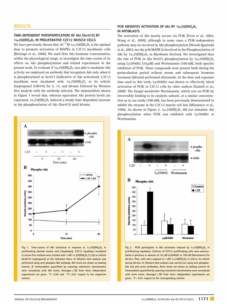
TIME-DEPENDENT PHOSPHORYLATION OF Akt (Ser473) BY
1a,25(OH)2D3 IN PROLIFERATIVE C2C12 MUSCLE CELLS
We have previously shown that 10�9M 1a,25(OH)2D3 is the optimal
dose to promote activation of MAPKs in C2C12 myoblastic cells
[Buitrago et al., 2006]. We used then this hormone concentration,
within the physiological range, to investigate the time course of its
effects on Akt phosphorylation and related experiments in the
present work. To evaluate if 1a,25(OH)2D3 was able to modulate Akt
activity we employed an antibody that recognizes Akt only when it
is phosphorylated in Ser473 (indicative of Akt activation). C2C12
myoblasts were incubated with 1a,25(OH)2D3 or its vehicle
(isopropanol 0.001%) for 5, 15, and 60min followed by Western
blot analysis with the antibody selected. The immunoblots shown
in Figure 1 reveal that, whereas equivalent Akt protein levels are
expressed, 1a,25(OH)2D3 induced a steady time-dependent increase
in the phosphorylation of Akt (Ser473) until 60min.
PI3K MEDIATES ACTIVATION OF Akt BY 1a,25(OH)2D3
IN MYOBLASTS
The activation of Akt mostly occurs via PI3K [Frost et al., 2002;
Wang et al., 2009], although in some cases a PI3K-independent
pathway may be involved in Akt phosphorylation [Woods Ignatoski
et al., 2003; see the p38 MAPK Is Involved in the Phosphorylation of
Akt by 1a,25(OH)2D3 in Myoblasts Section]. We investigated then
the role of PI3K in Akt Ser473 phosphorylation by 1a,25(OH)2D3
using Ly294002 (10mM) and Wortmannin (100 nM), both specific
inhibitors of PI3K. These compounds were present both during the
preincubation period without serum and subsequent hormone
treatment (60min) performed afterwards. At the dose and exposure
time used in this work, Ly294002 was shown to effectively block
activation of PI3K in C2C12 cells by other authors [Gamell et al.,
2008]. The fungal metabolite Wortmannin, which acts on PI3K by
irreversibly binding to its catalytic subunit, at a similar concentra-
tion as in our study (100 nM), has been previously demonstrated to
inhibit the enzyme in the C2C12 muscle cell line [Milasincic et al.,
1996]. As shown in Figure 2, 1a,25(OH)2D3 did not stimulate Akt
phosphorylation when PI3K was inhibited with Ly294002 or
Wortmannin.
Fig. 1. Time-course of Akt activation in response to 1a,25(OH)2D3 in
proliferating skeletal muscle cells (myoblasts). C2C12 myoblasts incubated
in serum-free medium were treated with 1 nM 1a,25(OH)2D3 (1,25) or vehicle
(0.001% isopropanol) at the indicated times. A: Western blot analysis was
performed using anti-phospho-Akt antibody. Akt levels are shown as loading
control. B: Immunoblots quantified by scanning volumetric densitometry
were normalized with Akt levels. Averages� SD from three independent
experiments are given. �P< 0.05 and ��P< 0.01 respect to the respective
control.
Fig. 2. PI3K participates in Akt activation induced by 1a,25(OH)2D3 in
proliferating myoblasts. Cultures of C2C12 proliferating cells were preincu-
bated in presence or absence of 10mM Ly294002 or 100 nM Wortmannin for
60min. Then, cells were exposed to 1 nM 1a,25(OH)2D3 (1,25) or its vehicle
during 60min. A: Western blot analysis was carried out using anti-phospho-
Akt and anti-actin antibodies. Actin levels are shown as loading control. B:
Immunoblots quantified by scanning volumetric densitometry were normalized
with actin levels. Averages� SD from three independent experiments are
given. �P< 0.01 respect to the corresponding control.
JOURNAL OF CELLULAR BIOCHEMISTRY 1a,25(OH)2D3 ACTIVATES Akt IN C2C12 CELLS 1173

CAVEOLAE DISRUPTION BY MbCD TREATMENT AND SILENCING OF
CAVEOLIN-1 EXPRESSION ABOLISH Akt PHOSPHORYLATION
INDUCED BY 1a,25(OH)2D3 IN C2C12 MYOBLASTS
As mentioned before, intact caveolae microdomains are required for
1a,25(OH)2D3-dependent activation of Src and MAPKs in C2C12
myoblasts [Buitrago and Boland, 2010]. Therefore, we examined the
participation of caveolae in the phosphorylation of Akt triggered
by the hormone. Cells were preincubated with MbCD to disrupt
caveolae structure before exposure to the steroid as previously
described [Buitrago and Boland, 2010]. Figure 3A shows that
cholesterol removal from the plasma membrane suppressed the
stimulation of Akt phosphorylation on Ser473 by 1a,25(OH)2D3,
implying that intact caveolae are required for hormone modulation
of Akt activity.
It has been established that cav-1 protein is essential for caveolae
maintenance [Rothberg et al., 1992]. To corroborate the caveolae
requirement in Akt activation, we used a previous experiment where
we significantly reduced cav-1 expression in C2C12 cells using a
specific siRNA [Buitrago and Boland, 2010]. The re-probing of these
membranes shows that Cav-1 silencing blocked Akt phosphorylation
induced by 1a,25(OH)2D3, proving again the importance of caveolae
in the upregulation of Akt activity by the hormone (Fig. 3B).
p38 MAPK IS INVOLVED IN THE PHOSPHORYLATION OF Akt BY
1a,25(OH)2D3 IN MYOBLASTS
We previously reported that MAPK pathways are activated by
1a,25(OH)2D3 in C2C12 myoblasts [Ronda et al., 2007]. In view of
these results and evidence demonstrating that p38 MAPK is
involved in Akt (Ser473) phosphorylation in VSMCs [Ushio-Fukai
et al., 2001] and human mammary epithelial (HME) cells [Woods
Ignatoski et al., 2003], we investigated the role of ERK1/2 and p38
MAPK in Akt activation in response to the hormone in C2C12
myoblasts. For this purpose, we used U0126 and SB203580 to
inhibit ERK1/2 and p38 MAPK, respectively. Various doses of each
inhibitor were previously tested to establish its most effective
concentration (data not shown). As seen in Figure 4, 10mM
SB203580 markedly blocked both basal and 1a,25(OH)2D3-
induced Akt phosphorylation in Ser473, whereas 20mM U0126
did not exert a significant effect. These data reveal that p38
MAPK and not ERK1/2 mediates hormone activation of Akt.
Exposure of cells to H2O2 (50mM, 10min) did not affect the
phosphorylation of Akt, as previously reported in VSMCs [Ushio-
Fukai et al., 2001], whereas incubation with 10% FBS for 5min
activated Akt in C2C12 muscle cells, as reported for fibroblasts
[Lim, 2008].
Fig. 3. Caveolae and caveolin-1 are implicated in hormone-dependent Akt activation in proliferating myoblasts. A: Cultures of C2C12 myoblasts were preincubated in
presence or absence of 4 nM MbCD for 30min. Then, cells were treated with 1 nM 1a,25(OH)2D3 (1,25) or vehicle during 60min in fresh free-serum medium. Western blot
analyses were carried out using anti-phospho Akt and anti-actin antibodies. Actin levels are shown as loading control. Immunoblots quantified by scanning volumetric
densitometry were normalized with actin levels. Averages� SD from three independents experiments are given. �P< 0.01 respect to its respective control. B: Cells were
transfected with control-siRNA or cav-1-siRNA as described in the Materials and Methods Section. Twenty-four hours later cells were exposed to 1 nM 1a,25(OH)2D3 or its
vehicle during 60min. Western blot analyses were performed using anti-cav-1, anti-phospho-Akt, and anti-Akt antibodies. Total Akt levels are shown as loading control.
Immunoblots quantified by scanning volumetric densitometry were normalized with Akt levels. Averages� SD from three independent experiments are given. �P< 0.01 respect
to the corresponding control.
1174 1a,25(OH)2D3 ACTIVATES Akt IN C2C12 CELLS JOURNAL OF CELLULAR BIOCHEMISTRY

STIMULATION OF MAPK-ACTIVATING PROTEIN KINASE-2 (MK-2)
BY 1a,25(OH)2D3
Our result above shows that p38 MAPK is implicated in Akt
activation by 1a,25(OH)2D3 in proliferating C2C12 cells. It has been
reported that p38 MAPK-dependent MAPK-activated protein kinase
2 (MK-2) phosphorylates Akt in neutrophils [Kettritz et al., 2002].
Also, the p38 MAPK/MK-2 pathway mediates Akt stimulation by
angiotensin II in VSMCs [Taniyama et al., 2004]. We previously
demonstrated that 1a,25(OH)2D3 rapidly activates MK-2 (at 1min) in
C2C12 cells [Buitrago et al., 2006]. In view that, in the preceding
experiments C2C12 cells were exposed to 1a,25(OH)2D3 for 60min
we investigated the changes in MK-2 Thr 334 phosphorylation
(activation) at this hormone treatment interval. C2C12 myoblasts
were preincubated with 10mMU0126 or 20mM SB203580 to inhibit
ERK1/2 and p38 MAPK, respectively. Our results show that
1a,25(OH)2D3-induced MK-2 activation is dependent on p38
MAPK, whereas ERK1/2 seems not to be involved (Fig. 5). These
data suggest then that p38 MAPK-dependent MK-2 mediates Akt
activation by 1a,25(OH)2D3.
Src MEDIATES Akt PHOSPHORYLATION INDUCED BY 1a,25(OH)2D3
IN PROLIFERATING SKELETAL MUSCLE CELLS
Src is a key molecule in the regulation of several signal transduction
pathways. Regarding the present work, it has been demonstrated
that Src mediates PI3K/Akt activation in endothelial cells exposed to
estrogen [Haynes et al., 2003] and more recently in IGF-I-stimulated
VSMCs [Gang et al., 2010]. Interestingly, there is evidence that
1a,25(OH)2D3 activates Src in proliferating C2C12 cells [Buitrago
and Boland, 2010]. To investigate the role of Src in the stimulation of
Akt by the hormone in myoblasts we used PP2, a specific inhibitor
for all members of the Src family. Various doses of the inhibitor were
tested to establish its most effective concentration (data not shown).
As observed in Figure 6A,B, Akt phosphorylation induced by the
steroid was completely inhibited when the cells were incubated with
25mM PP2. To confirm the participation of Src in hormone-
dependent Akt (Ser473) phosphorylation, we markedly diminished
the expression of c-Src by transfecting cells with a pool of antisense
ODNs against c-Src mRNA (Fig. 6C, upper blot). Decreased Src
expression abolished the phosphorylation of Akt induced by
Fig. 4. Involvement of p38 MAPK and not ERK1/2 in 1a,25(OH)2D3-
dependent Akt phosphorylation in proliferating skeletal muscle cells. C2C12
myoblasts were preincubated with or without 10mM U0126 or 20mM
SB203580 for 60min. Then, cells were treated with 1 nM 1a,25(OH)2D3(1,25) or vehicle during 60min. As positive and negative control of Akt
activation, cells were exposed to FBS (10%, 5min) and H2O2 (50mM,
10min), respectively. A: Western blot assays were carried out using anti-
phospho-Akt and anti-Akt antibodies. B: Akt levels are shown as loading
control. Immunoblots quantified by scanning volumetric densitometry were
normalized with Akt levels. Averages� SD from three independent experi-
ments are given. �P< 0.01 respect to 1a,25(OH)2D3 without inhibitor.
Fig. 5. 1a,25(OH)2D3 modulates p38 MAPK-dependent MK-2 in C2C12
myoblasts. C2C12 proliferating myoblasts were preincubated with or without
10mM U0126 and 20mM SB203580 for 60min. Then, cells were treated with
1 nM 1a,25(OH)2D3 (1,25) or vehicle for 60min. Myoblasts were also exposed
to FBS and H2O2 as mentioned in this legend. A: Western blot assays were
performed with anti-phospho-MK-2 and anti-Akt antibodies. Akt levels are
shown as loading control. B: Immunoblots quantified by scanning volumetric
densitometry were normalized with Akt levels. Averages� SD from three
independent experiments are given. �P< 0.05 respect to 1a,25(OH)2D3 with-
out inhibitor.
JOURNAL OF CELLULAR BIOCHEMISTRY 1a,25(OH)2D3 ACTIVATES Akt IN C2C12 CELLS 1175

1a,25(OH)2D3 (Fig. 6C, center blot). The data from these experiments
provide strong evidence indicating that Src is necessary for Akt
activation by the hormone in myoblasts.
1a,25(OH)2D3 STIMULATION OF C2C12 MYOBLAST CELL
PROLIFERATION IS DEPENDENT ON PI3K
Knowing that PI3K has been involved in C2C12 cell proliferation
[Spangenburg and Booth, 2002], we tested if PI3K and/or Src are
implicated in the mitogenic effects of 1a,25(OH)2D3 on these muscle
cells. First, to determine changes in proliferation in C2C12 myoblast
cultures stimulated with the hormone, we used a MTS proliferation
assay as described in the Materials and Methods Section.
Figure 7A shows that 24 h of treatment with 1a,25(OH)2D3
stimulates C2C12 cell proliferation by 40%. Using the PI3K inhibitor
Ly294002 during the 24 h incubation with the hormone, the increase
in proliferation was no longer observed (Fig. 7B). Our data indicate
then that PI3K is involved in the mitogenic action of 1a,25(OH)2D3
on C2C12 myoblasts.
1a,25(OH)2D3 STIMULATES PHOSPHORYLATION OF Akt (Ser473)
AND THE EXPRESSION OF MYOSIN HEAVY CHAIN (MHC) AND
MYOGENIN IN THE EARLY STAGE OF DIFFERENTIATION OF
C2C12 CELLS
Consistent with the idea that Akt signaling is essential for myoblast
differentiation [Wilson et al., 2004; Wilson and Rotwein, 2007], it
was recently demonstrated that API-2, an Akt inhibitor, blocks
C2C12 myoblast cell differentiation [Shu and Houghton, 2009].
Our laboratory described for first time that 1a,25(OH)2D3
regulates chick myoblast differentiation [Capiati et al., 1999].
Subsequently, Endo et al. [2003] demonstrated that the VDR is
Fig. 6. Src mediates Akt phosphorylation induced by 1a,25(OH)2D3 in
proliferative skeletal muscle cells. A: Proliferating C2C12 myoblasts were
preincubated with or without 25mM PP2. As positive and negative control
of Akt activation, cells were exposed to FBS (10%, 5min) and H2O2 (50mM,
10min), respectively. Then, cultures were treated with 1 nM 1a,25(OH)2D3
(1,25) or vehicle for 60min. Western blot assays were carried out using anti-
phospho-Akt and anti-actin antibodies. Actin levels are shown as loading
control. B: Immunoblots quantified by scanning volumetric densitometry were
normalized with actin levels. Averages� SD from three independents experi-
ments are given. �P< 0.05 respect to hormone without PP2. C: Cells were
transfected with sense (S) or antisense (AS) c-Src ODNs as described in the
Materials and Methods Section. Eighteen hours later cells were exposed to
1 nM 1a,25(OH)2D3 or its vehicle during 60min. Western blot analysis was
performed using anti-Src, anti-phospho-Akt, and anti-actin antibodies. Actin
levels are shown as loading control. The immunoblots are representative from
three independent experiments.
Fig. 7. PI3K is involved in 1a,25(OH)2D3-dependent C2C12 myoblasts pro-
liferation. A: C2C12 myoblasts were exposed to 1 nM 1a,25(OH)2D3 (1,25) or
vehicle for 14, 24, and 48 h. At these times, MTS proliferation assays were
carried out using a kit according to manufacturer’s instructions. Percentage
(%) of proliferation was calculated from five independent experiments.
Averages� SD are given. �P< 0.01 respect to the respective control.
B: C2C12 proliferating cells were preincubated with or without 10mM
Ly294002. Then, cultures were treated with 1 nM 1a,25(OH)2D3 or vehicle
for 24 h. At this time, MTS proliferation assays were carried out. Proliferation
(%) was calculated from five independent experiments. Averages� SD are
given. �P< 0.05 respect to 1a,25(OH)2D3 without inhibitor.
1176 1a,25(OH)2D3 ACTIVATES Akt IN C2C12 CELLS JOURNAL OF CELLULAR BIOCHEMISTRY

necessary for normal skeletal muscle development and the correct
expression of myoregulatory transcription factors in mice. Except
the work of the latter authors, the action of 1a,25(OH)2D3 on the
differentiation of mammalian skeletal muscle cells has not been
investigated. To gain insights into the regulatory mechanism of
this process by 1a,25(OH)2D3, the effects of the hormone on Akt
activation during C2C12 myoblast differentiation were studied,
evaluating the expression of MHC andmyogenin, marker proteins of
early muscle cell maturation. C2C12 cells grown in DM for 24,
48, and 72 h were stimulated with 1a,25(OH)2D3 or its vehicle
isopropanol every 24 h. Figure 8 shows that, although the expression
pattern of total Akt was not altered, phosphorylation of Akt Ser473
augments during differentiation, as previously reported [Fujio et al.,
2001; Conejo et al., 2002]. 1a,25(OH)2D3 further increases Akt
(Ser473) phosphorylation and the expression levels of MHC and
myogenin at the three time intervals studied. These results suggest
that the hormone stimulates myoblast differentiation in an Akt-
dependent manner.
1a,25(OH)2D3 PROMOTES ACTIVATION OF Src AT EARLY STEPS OF
C2C12 CELL DIFFERENTIATION. SRC AND PI3K MEDIATE MHC AND
MYOGENIN EXPRESSION AND PHOSPHORYLATION OF AKT
INDUCED BY THE HORMONE
It has been previously demonstrated that the differentiation of L6 rat
skeletal muscle cells involves a PTPa-mediated Src pathway [Lu
et al., 2002]. In the C2C12 cell line, Src could also be involved in the
enhancement of FAK activity and in the activation of downstream
pathways which lead to the differentiation of myoblastic cells
[Clemente et al., 2005]. We investigated here the role of Src in
1a,25(OH)2D3-dependent enhancement of Akt activation during the
early stages of C2C12 differentiation. As Figure 9A shows, hormone
treatment for 48 h induces activation of Src, which was efficaciously
inhibited by 25mM PP2. C2C12 proliferating cells (grown in GM)
and C2C12 differentiating cells (in DM) do not show differences
in basal Src phosphorylation levels (Fig. 9A, lanes 1 and 2). In
differentiating cells exposed for 48 h to the hormone, the expression
Fig. 8. 1a,25(OH)2D3 promotes Akt phosphorylation and the expression of
MHC and myogenin in early steps of C2C12 cell differentiation. C2C12
myoblasts induced to differentiate as described in the Materials and Methods
Section were incubated with 1 nM 1a,25(OH)2D3 (1,25) or vehicle for 24, 48,
and 72 h. Western blot assays were carried out using anti-phospho-Akt, anti-
myosin heavy chain (MHC), anti-myogenin, and anti-Akt antibodies. Akt bands
correspond to loading controls showing that protein levels do not significantly
change in any tested condition. The immunoblots are representative from three
independent experiments.
Fig. 9. At early stages of C2C12 cell differentiation, 1a,25(OH)2D3 stimu-
lates Src phosphorylation. Src and PI3K participate in MHC and myogenin
expression and Akt activation induced by the hormone. A: C2C12 proliferating
myoblasts (lane 1) and C2C12 differentiating cells (lanes 2–6) were incubated
with or without 25mM PP2 and exposed to 1 nM 1a,25(OH)2D3 (1,25) or
vehicle for 48 h. As positive control of Src activation, 10% FBS (5min) was
used. Western blot assays were carried out using anti-phospho-Src (Tyr416)
and anti-actin antibodies. The immunoblots showed are representative from
three independent experiments. B: C2C12 differentiating cells were incubated
with or without 25mM PP2 or 100 nM Wortmannin and exposed to 1 nM
1a,25(OH)2D3 or vehicle for 48 h. Western blot assays were carried out using
anti-phospho-Akt, anti-MHC, anti-myogenin, and anti-actin antibodies. The
immunoblots showed are representative from three independent experiments.
C: Immunoblots quantified by scanning volumetric densitometry were nor-
malized with actin levels. Averages� SD from three independents experiments
are given. �P< 0.05 respect to the respective control.
JOURNAL OF CELLULAR BIOCHEMISTRY 1a,25(OH)2D3 ACTIVATES Akt IN C2C12 CELLS 1177

of MHC and myogenin as well as Akt activation were suppressed by
PP2 and Wortmannin (Fig. 9B). These results suggest that Src and
PI3K act upstream Akt activation which is involved in MHC and
myogenin expression.
DISCUSSION
Myogenesis involves withdrawal of myoblasts from the cellular
cycle, subsequent expression of myotube-specific genes and
formation of multinucleated myotubes [McKinsey et al., 2002;
Buckingham et al., 2003; Parker et al., 2003]. This process is largely
regulated by the myogenic basic helix-loop-helix family of
transcription factors (myogenin, MyoD, myf5, and MRF4) and
MEF2, which regulate the expression of many muscle-specific
genes, such as the MHC [Olson et al., 1995]. The presence of
myogenin guarantees MHC expression and correct myotube
development, demonstrating that myogenin acts early determining
myoblasts to be differentiated into myotubes [Davie et al., 2007]. In
mature skeletal muscle, a pool of satellite myoblasts stay quiescent
until a tissue injury triggers their proliferation and subsequent
differentiation to replace the loss of functional muscle fibers.
Akt is a key molecule involved in signaling pathways which
regulate myogenesis. On the one hand, this kinase takes part in the
control of skeletal muscle cell proliferation [Frost et al., 2002; Glass,
2003; Guttridge, 2004]. Specifically, proliferation of C2C12
myoblasts induced by insulin depends on Akt activity [Conejo
and Lorenzo, 2001]. On the other hand, related to the skeletal muscle
differentiation process, PI3K/Akt signaling modulates muscle gene
expression during myogenic differentiation [White, 2003]. Of
relevance, PI3K has been shown to participate in MHC expression
induced by insulin in C2C12 cells [Sumitani et al., 2002] and the
IGF-PI3K-Akt signaling pathway regulates myogenin expression in
normal myogenic cells [Xu and Wu, 2000]. Therefore, Akt plays an
important role in muscle proliferation and differentiation, control-
ling the number, size and survival of muscle cells. However, the
signaling pathways underlying the participation of Akt in the
regulation of these processes inmuscle have not been investigated in
detail.
Some of the actions of 1a,25(OH)2D3 are related to the normal
development of skeletal muscle [Endo et al., 2003], in keeping with
the importance of this hormone in the control of skeletal muscle
metabolism and contractility [Boland, 1986; Boland et al., 2005;
Buitrago et al., 2009]. Of relevance, physiological doses of
1a,25(OH)2D3 stimulate proliferation of chicken myoblasts and
also their differentiation into myotubes [Capiati et al., 1999]. Our
study shows that treatment with 1 nM 1a,25(OH)2D3 for 24 h
promotes proliferation of murine C2C12 cells in a PI3K-dependent
manner. In addition, at the proliferative cell stage, 1a,25(OH)2D3
induces Akt (Ser473) phosphorylation, implying its activation, in a
time-dependent way. The time course of Akt activation by the
hormone was similar to that observed in fibroblasts by Park et al.
[2003]. Also, 17b-estradiol elicits a similar pattern of Akt
phosphorylation in endometrial cancer cells [Guoa et al., 2006]
and hypoxia has been shown to activate Akt at comparable times in
artery endothelium [Chen and Meyrick, 2004]. In agreement with
other results reporting that Akt activation in muscle is PI3K-
dependent [Vasconsuelo et al., 2008; Gorelick-Feldman et al., 2010],
we report here the involvement of PI3K in hormone-induced Akt
activation. These data were obtained using LY 294002 and
Wortmannin, two different blockers of PI3K. Regarding the use
of both pharmacological inhibitors, their specificity and possible
undesirable effects deserve consideration. LY 294002 was reported
to inhibit all isoforms of PI3K but not to affect other kinases such as
PKC and PKA, MAPK family, S6 kinase and Src kinases [Vlahos
et al., 1994]. Wortmannin, although affecting myosin light chain
kinase, is widely recognized as a selective and specific PI3K inhibitor
too, with reportedly no effects on PKC and protein-tyrosine kinases
like Src [Powis et al., 1994].
We asked ourselves which other mechanisms, independent of
PI3K, could be used by 1a,25(OH)2D3 to activate Akt. In this study,
experiments with C2C12 myoblasts in which caveolae were
disrupted by MbCD treatment or caveolin-1 expression was silenced
with specific siRNA, revealed that Akt phosphorylation induced by
1a,25(OH)2D3 was abolished, demonstrating that caveolae and cav-
1 are required in Akt Ser473 phosphorylation by the hormone. In
agreement with this interpretation, it was previously observed that
cav-1 and caveolae participates in Akt activation in messangial cells
[Zhang et al., 2007] and in VSMCs [Sedding et al., 2005]. Moreover,
it was reported that caveolin-enriched microdomains play a crucial
role in the mechanism by which ceramide impairs the activation of
Akt in adipocytes and in cultured rat skeletal-muscle cells [Hajduch
et al., 2008]. Akt function is complex and it has been also related to
p38 MAPK-dependent MAPKAPK-2 (MK-2) activation [Taniyama
et al., 2004]. Our results show that p38 MAPK inhibition abolishes
Akt and MK-2 activation induced by the hormone, suggesting that
the sequential cascade of p38 MAPK/MK-2/Akt is modulated by
1a,25(OH)2D3 in proliferating C2C12 cells. In earlier work we
reported that in these cells Src is required for p38 MAPK activation
by the hormone [Buitrago et al., 2006]. In myoblasts and myotubes
of L6 and L8 cell lines, Src appears to upregulate Akt activation by
insulin [Jacob et al., 2009]. In the present investigation we obtained
evidence that Src is needed for hormone-induced Akt activation
both in C2C12 myoblasts and in C2C12 differentiating cells, by
inhibiting either its activity or its expression. To inhibit Src, the
Fig. 10. Proposed schematic diagram of Akt activation by 1a,25(OH)2D3 in
C2C12 skeletal muscle cells. 1a,25(OH)2D3 (1,25) may act through caveolae
microdomains and after that, the hormone triggers rapid signaling cascades
including activation of PKC/PTPa/Src/p38 MAPK/MK2 leading to Akt activa-
tion. An alternative mechanism of regulation of Akt implicates PI3K action
which could be activated by Src action.
1178 1a,25(OH)2D3 ACTIVATES Akt IN C2C12 CELLS JOURNAL OF CELLULAR BIOCHEMISTRY

selective inhibitor PP2 is widely used [Hanke et al., 1996]. The
inhibitory action of this compound on 1a,25(OH)2D3-induced Akt
phosphorylation is most likely to be the result of its main
pharmacological effect on the corresponding Src kinase family
rather than an unspecific effect. Supporting this contention, we
could abolish Akt phosphorylation by the hormone using Src
antisense ODNs which successfully blocked Src expression in C2C12
cells.
Modulation of Akt by 1a,25(OH)2D3 was also investigated in the
early stages of differentiation of C2C12 cells. It has been shown that
MHC and myogenin expression requires Akt activation in chicken
embryo myoblasts induced to differentiation [Jiang et al., 1999].
Moreover, PI3K-dependent MHC and myogenin expression in
C2C12 cells was previously reported [Xu and Wu, 2000; Sumitani
et al., 2002]. Correlated to this information, this study revealed that
the steroid hormone 1a,25(OH)2D3 increases MHC and myogenin
expression and also activates Akt in differentiating myoblasts. Of
relevance, Src and PI3K were required for 1a,25(OH)2D3 to
upregulate MHC and myogenin expression and Akt phosphorylation
in these cells. In accord with our data, Lu et al. [2002] previously
informed that Src activation is needed for C2C12 muscle cell
differentiation. However, it has been observed that the Src inhibitor
SU6656 has no effect on MHC and myogenin expression in C2C12
cells after 6 days of initiation of the differentiation program [Lim
et al., 2007]. We speculate that Src activation is necessary in the
early stages of C2C12 myoblast differentiation (first 3 days) but
might be dispensable in mature muscle fibers.
Finally, the data obtained in this work lead us to conclude that
Akt may represent a key intermediate in 1a,25(OH)2D3 regulation of
myoblast proliferation, survival and myogenic differentiation. One
important finding in this study is that Src takes part in Akt-mediated
1a,25(OH)2D3 regulation of signaling pathways involved in skeletal
muscle proliferation and differentiation. Moreover, our results
suggest the operation of another mechanism of Akt activation that is
independent of PI3K action, that is, the involvement of p38MAPK in
Akt phosphorylation by the hormone through MK2 (Fig. 10).
ACKNOWLEDGMENTS
This research was supported by grants from the Consejo Nacional deInvestigaciones Cientıficas y Tecnicas (CONICET) and the Uni-versidad Nacional del Sur, Argentina.
REFERENCES
Aquila S, Guido C, Middea E, Perrotta I, Bruno R, PellegrinoM, Ando S. 2009.Human male gamete endocrinology: 1alpha, 25-dihydroxyvitamin D3
(1a,25(OH)2D3) regulates different aspects of human sperm biology andmetabolism. Reprod Biol Endocrinol 30:140–143.
Boland R. 1986. Role of vitamin D in skeletal muscle function. Endocr Rev7:434–448.
Boland R, Buitrago C, De Boland AR. 2005. Modulation of tyrosine phos-phorylation signalling pathways by 1alpha,25(OH)2-vitamin D3. TrendsEndocrinol Metab 16:280–287.
Bradford M. 1976. A rapid and sensitive method for the quantitation ofmicrogram quantities of protein utilizing the principle of protein-dye bind-ing. Anal Biochem 72:248–254.
Buckingham M, Bajard L, Chang T, Daubas P, Hadchouel J, Meilhac S,Montarras D, Rocancourt D, Relaix F. 2003. The formation of skeletal muscle:From somite to limb. J Anat 20:259–268.
Buitrago C, Boland R. 2010. Caveolae and caveolin-1 are implicated in1alpha,25(OH)2-vitamin D3-dependent modulation of Src, MAPK cascadesand VDR localization in skeletal muscle cells. J Steroid Biochem Mol Biol121:169–175.
Buitrago C, Gonzalez Pardo V, Russo de Boland A. 2002. Nongenomic actionof 1 alpha,25(OH)2-vitamin D3. Activation of muscle cell PLC gammathrough the tyrosine kinase c-Src and PtdIns 3-kinase. Eur J Biochem269:2506–2515.
Buitrago C, Ronda A, de Boland A, Boland R. 2006. MAP kinases p38 and JNKare activated by the steroid hormone 1alpha,25(OH)2-vitamin D3 in theC2C12 muscle cell line. J Cell Biochem 97:698–708.
Buitrago C, Milanesi L, Ronda A, Vasconsuelo A, Boland R. 2009. 1,25(OH)2-vitamin D3 and 17b-estradiol: Two steroid partners acting in skeletal muscle.Curr Med Chem 3:159–168.
Capiati D, Tellez-Inon M, Boland R. 1999. Participation of protein kinase Calpha in 1,25-dihydroxy-vitamin D3 regulation of chick myoblast prolifera-tion and differentiation. Mol Cell Endocrinol 153:39–45.
Capiati D, Vazquez G, Tellez-Inon T, Boland R. 2000. Antisense oligonucleo-tides targeted against protein kinase c alpha inhibit proliferation of culturedavian myoblasts. Cell Prolif 33:307–315.
Capiati D, Vazquez G, Boland R. 2001. Protein kinase C alpha modulatesthe Ca2þ influx phase of the Ca2þ response to 1alpha,25-dihydroxy-vitamin-D3 in skeletal muscle cells. Horm Metab Res 33:201–206.
Ceci M, Ross J, Jr., Condorelli G. 2004. Molecular determinants of thephysiological adaptation to stress in the cardiomyocyte: A focus on AKT.J Mol Cell Cardiol 37:905–912.
Chen J, Meyrick B. 2004. Hypoxia increases Hsp90 binding to eNOS viaPI3K-Akt in porcine coronary artery endothelium. Lab Invest 84:182–190.
Chidlow J, Jr., Sessa C. 2010. Caveolae, caveolins, and cavins: Complexcontrol of cellular signalling and inflammation. Cardiovasc Res 86:219–225.
Clemente C, Corat M, Saad S, Franchini K. 2005. Differentiation of C2C12myoblasts is critically regulated by FAK signaling. Am J Physiol Regul IntegrComp Physiol 289:R862–R870.
Conejo R, Lorenzo M. 2001. Insulin signaling leading to proliferation,survival, and membrane ruffling in C2C12 myoblasts. J Cell Physiol 187:96–108.
Conejo R, de Alvaro C, Benito M, Cuadrado A, Lorenzo M. 2002. Insulinrestores differentiation of Ras-transformed C2C12 myoblasts by inducingNF-kappaB through an AKT/P70S6K/p38-MAPK pathway. Oncogene21:3739–3753.
Davie J, Cho J, Meadows E, Flynn J, Knapp J, Klein W. 2007. Target geneselectivity of the myogenic basic helix-loop-helix transcription factor myo-genin in embryonic muscle. Dev Biol 311:650–664.
De Haes P, Garmyn M, Carmeliet G, Degreef H, Vantieghem K, Bouillon R,Segaert S. 2004. Molecular pathways involved in the anti-apoptotic effect of1,25-dihydroxyvitamin D3 in primary human keratinocytes. J Cell Biochem93:951–967.
Endo I, Inoue D, Mitsui T, Umaki Y, Akaike M, Yoshizawa T, Kato S,Matsumoto T. 2003. Deletion of vitamin D receptor gene in mice resultsin abnormal skeletal muscle development with deregulated expression ofmyoregulatory transcription factors. Endocrinology 144:5138–5144.
Frost R, Nystrom G, Lang C. 2002. Regulation of IGF-I mRNA and signaltransducers and activators of transcription-3 and -5 (Stat-3 and -5) by GH inC2C12 myoblasts. Endocrinology 143:492–503.
Fujio Y, Mitsuuchi Y, Testa J, Walsh K. 2001. Activation of Akt2 inhibitsanoikis and apoptosis induced by myogenic differentiation. Cell Death Differ8:1207–1212.
JOURNAL OF CELLULAR BIOCHEMISTRY 1a,25(OH)2D3 ACTIVATES Akt IN C2C12 CELLS 1179

Gamell C, Osses N, Bartrons R, Ruckle T, Camps M, Rosa J, Ventura F. 2008.BMP2 induction of actin cytoskeleton reorganization and cell migrationrequires PI3-kinase and Cdc42 activity. J Cell Sci 121:3960–3970.
Gang X, Xinchun S, Yashwanth R, Laura M, David C. 2010. Hyperglycemia-induced p66shc inhibits insulin-like growth factor I-dependent cell survivalvia impairment of Src kinase-mediated phosphoinositide-3 kinase/AKTactivation in vascular smooth muscle cells. Endocrinology 151:3611–3623.
Glass D. 2003. Molecular mechanisms modulating muscle mass. Trends MolMed 9:344–350.
Gorelick-Feldman J, Cohick W, Raskin I. 2010. Ecdysteroids elicit arapid Ca2þ flux leading to Akt activation and increased protein synthesisin skeletal muscle cells. Steroids 75:632–637.
Guoa R, Li-HuiWei A, Zheng Tu, Peng-Ming S, Jian-LiuW, Dan Z, Xiao-PingL, Jian-Min T. 2006. 17b-Estradiol activates PI3K/Akt signaling pathway byestrogen receptor (ER)-dependent and ER-independent mechanisms inendometrial cancer cells. J Steroid Biochem Mol Biol 99:9–18.
Guttridge D. 2004. Signaling pathways weigh in on decisions to make orbreak skeletal muscle. Curr Opin Clin Nutr Metab Care 7:443–450.
Hajduch E, Litherland G, Hundal H. 2001. Protein kinase B (PKB/Akt): A keyregulator of glucose transport. FEBS Lett 492:199–203.
Hajduch E, Turban S, Le Liepvre X, Le Lay S, Lipina C, Dimopoulos N, DugailI, Hundal H. 2008. Targeting of PKCzeta and PKB to caveolin-enrichedmicrodomains represents a crucial step underpinning the disruption in PKB-directed signalling by ceramide. Biochem J 410:369–379.
Hanke J, Gardner J, Dow R, Changelian P, Brissette W, Weringer E, Pollok B,Connelly P. 1996. J Biol Chem 271:695–701.
Haynes P, Li L, Sinha D, Russell K, Hisamoto K, Baron R, Collinge M, Sessa W,Bender J. 2003. Src kinase mediates phosphatidylinositol 3-kinase/Akt-dependent rapid endothelial nitric-oxide synthase activation by estrogen.J Biol Chem 278:2118–2123.
Jacob AI, Horovitz-Fried M, Aga-Mizrachi S, Brutman-Barazani T, Okhri-menko H, Zick Y, Brodie C, Sampson S. 2009. The regulatory domain ofprotein kinase C delta positively regulates insulin receptor signaling. J MolEndocrinol 44:155–169.
Jiang B, Aoki M, Zheng J, Li J, Vogt P. 1999. Myogenic signaling ofphosphatidylinositol 3-kinase requires the serine-threonine kinase Akt/pro-tein kinase B. Proc Natl Acad Sci USA 96:2077–2081.
Kettritz R, Choi M, Butt W, Rane M, Rolle S, Luft F, Klein J. 2002.Phosphatidylinositol 3-kinase controls antineutrophil cytoplasmic antibo-dies—Induced respiratory burst in human neutrophils. J Am Soc Nephrol13:1740–1749.
Kubo Y. 1991. Comparison of initial stages of muscle differentiation in ratand mouse myoblastic and mouse mesodermal stem cell lines. J Physiol442:743–759.
Laemmli U, Beguin F, Gujer-Kellenberger G. 1970. A factor preventing themajor head protein of bacteriophage T4 from random aggregation. J Mol Biol47:69–85.
Lim S. 2008. Superoxide and Akt reactivation via inactivation of PTEN,Chapter 3. Singapur, Asia: Scholarbank National University of Singapore.
LimMJ, Seo YH, Choi KJ, Cho CH, Kim BS, Kim YH, Lee J, Lee H, Jung CY, HaJ, Kang I, Kim SS. 2007. Suppression of c-Src activity stimulates muscledifferentiation via p38 MAPK activation. Arch Biochem Biophys 465:197–208.
Lu H, Shah P, Ennis D, Shinder G, Sap J, Le-Tien H, Fantus IG. 2002. Thedifferentiation of skeletal muscle cells involves a protein-tyrosine phospha-tase-alpha-mediated C-Src signaling pathway. J Biol Chem 277:46687–46695.
McKinsey T, Zhang C, Olson E. 2002. Signaling chromatin to make muscle.Curr Opin Cell Biol 14:763–772.
Milasincic D, Calera M, Farmer S, Pilch P. 1996. Stimulation of C2C12myoblast growth by basic fibroblast growth factor and insulin-like growth
factor 1 can occur via mitogen-activated protein kinase-dependentand -independent pathways. Mol Cell Biol 16:5964–5973.
Olson E, Perry M, Schulz R. 1995. Regulation of muscle differentiation by theMEF2 family of MADS box transcription factors. Dev Biol 172:2–14.
Park C, Schneider I, Haugh J. 2003. Kinetic analysis of platelet-derivedgrowth factor receptor/phosphoinositide 3-kinase/Akt signaling in fibro-blasts. J Biol Chem 278:37064–37072.
Parker M, Seale P, Rudnicki M. 2003. Looking back to the embryo: Definingtranscriptional networks in adult myogenesis. Nat Rev Genet 4:497–507.
Powis G, Bonjouklian R, Berggren M, Gallegos A, Abraham R, Ashendel C,Zalkow L, Matter W, Dodge J, Grindey G. 1994. Wortmannin, a potent andselective inhibitor of phosphatidylinositol-3-kinase. Cancer Res 54:2419–2423.
Ronda A, Buitrago C, Colicheo A, de Boland A, Roldan E, Boland R. 2007.Activation of MAPKs by 1alpha,25(OH)2-vitamin D3 and 17beta-estradiol inskeletal muscle cells leads to phosphorylation of Elk-1 and CREB transcrip-tion factors. J Steroid Biochem Mol Biol 103:462–466.
Rothberg K, Heuser J, Donzell W, Ying Y, Glenney J, Anderson R. 1992.Caveolin, a protein component of caveolae membrane coats. Cell 68:673–682.
Sedding D, Hermsen J, Seay U, Eickelberg O, Kummer W, Schwencke C,Strasser R, Tillmanns H, Braun-Dullaeus R. 2005. Caveolin-1 facilitatesmechanosensitive protein kinase B (Akt) signaling in vitro and in vivo.Circ Res 96:635–642.
Shu L, Houghton P. 2009. Houghton the mTORC2 complex regulates terminaldifferentiation of C2C12 myoblasts. Mol Cell Biol 29:4691–4700.
Snedecor G, Cochran W. 1967. Statistical methods, 6th edition. Ames,IA, U.S.A.: The Iowa State University Press.
Spangenburg E, Booth F. 2002. Multiple signaling pathways mediate LIF-induced skeletal muscle satellite cell proliferation. Am J Physiol Cell Physiol283:C204–C211.
Sumitani S, Goya K, Testa JR, Kouhara H, Kasayama S. 2002. Akt1 and Akt2differently regulate muscle creatine kinase and myogenin gene transcriptionin insulin-induced differentiation of C2C12 myoblasts. Endocrinology 143:820–828.
Sun L, Trausch-Azar JS, Ciechanover A, Schwartz A. 2005. Ubiquitin-proteasome-mediated degradation, intracellular localization, and proteinsynthesis of MyoD and Id1 during muscle differentiation. J Biol Chem280:26448–26456.
Taniyama Y, Ushio-Fukai M, Hitomi H, Rocic P, Kingsley M, Pfahnl C, WeberD, Wayne A, Griendling K. 2004. Role of p38 MAPK and MAPKAPK-2 inangiotensin II-induced Akt activation in vascular smooth muscle cells. Am JPhysiol Cell Physiol 287:C494–C499.
Toker A, Newton A. 2000. Akt/protein kinase B is regulated by autopho-sphorylation at the hypothetical PDK-2 site. J Biol Chem 275:8271–8274.
Troussard A, McDonald P, Wederell E, Mawji N, Filipenko N, Gelmon K,Kucab J, Dunn S, Emerman J, Bally M, Dedhar S. 2006. Preferential depen-dence of breast cancer cells versus normal cells on integrin-linked kinase forprotein kinase B/Akt activation and cell survival. Cancer Res 66:393–403.
Ueki K, Yamauchi T, Tamemoto H, Tobe K, Yamamoto-Honda R, Kaburagi Y,Akanuma Y, Yazaki Y, Aizawa S, Nagai R, Kadowaki T. 2000. Restoredinsulin-sensitivity in IRS-1-deficient mice treated by adenovirus-mediatedgene therapy. J Clin Invest 105:1437–1445.
Ushio-Fukai M, Hilenski L, Santanam N, Becker P, Ma Y, Griendling K,Alexander R. 2001. Cholesterol depletion inhibits epidermal growth factorreceptor transactivation by angiotensin II in vascular smooth muscle cells:Role of cholesterol-rich microdomains and focal adhesions in angiotensin IIsignalling. J Biol Chem 276:48269–48275.
Vanhaesebroeck B, Alessi D. 2000. The PI3K-PDK1 connection: More thanjust a road to PKB. Biochem J 346:561–576.
1180 1a,25(OH)2D3 ACTIVATES Akt IN C2C12 CELLS JOURNAL OF CELLULAR BIOCHEMISTRY

Vasconsuelo A, Milanesi L, Boland R. 2008. 17Beta-estradiol abrogatesapoptosis in murine skeletal muscle cells through estrogen receptors: Roleof the phosphatidylinositol 3-kinase/Akt pathway. J Endocrinol 196:385–397.
Vlahos C, Matter W, Hui K, Brown R. 1994. J Biol Chem 269:5241–5248.
Wang W, Fridman A, Blackledge W, Connelly S, Wilson I, Pilz R, Boss G.2009. The phosphatidylinositol 3-kinase/Akt cassette regulates purinenucleotide synthesis. J Biol Chem 284:3521–3528.
White M. 2003. Insulin signaling in health and disease. Science. 302:1710–1711.
Wilson E, Tureckova J, Rotwein P. 2004. Permissive roles of phosphatidylinositol 3-kinase and Akt in skeletal myocyte maturation. Mol. Biol. Cell15:497–505.
Wilson E, Rotwein P. 2007. Selective control of skeletal muscle differentia-tion by Akt1. J. Biol. Chem 282:5106–5110.
Wong B, Besser D, Kim N, Arron J, Vologodskaia M, Hanafusa H,Choi Y. 1999. TRANCE, a TNF family member, activates Akt/PKBthrough a signaling complex involving TRAF6 and c-Src. Mol Cell 4:1041–1049.
Woods Ignatoski K, Livant D, Markwart S, Grewal N, Ethier S. 2003. The roleof phosphatidylinositol 30-kinase and its downstream signals in erbB-2-mediated transformation. Mol Cancer Res 1:551–560.
Xu Q, Wu Z. 2000. The insulin-like growth factor-phosphatidylinositol3-kinase-Akt signaling pathway regulates myogenin expression in normalmyogenic cells but not in rhabdomyosarcoma-derived RD cells. J Biol Chem275:36750–36757.
Yamada T, Katagiri H, Asano T, Inukai K, Tsuru M, Kodama T, Kikuchi M, OkaM. 2001. 3-Phosphoinositide-dependent protein kinase 1, an Akt1 kinase, isinvolved in dephosphorylation of Thr-308 of Akt1 in Chinese hamster ovarycells. J Biol Chem 276:5339–5345.
Yoshida N, Yoshida S, Koishi K, Masuda K, Nabeshima Y. 1998. Cellheterogeneity upon myogenic differentiation: Down-regulation of MyoDand Myf-5 generates ‘reserve cells’. J Cell Sci 111:769–779.
Zhang X, Zanello L. 2008. Vitamin D receptor-dependent 1 alpha,25(OH)2-vitamin D3-induced anti-apoptotic PI3K/AKT signaling in osteoblasts. J BoneMiner Res 23:1238–1248.
Zhang B, Peng F, Wu D, Ingram A, Gao B, Krepinsky J. 2007. Caveolin-1phosphorylation is required for stretch-induced EGFR and Akt activation inmessangial cells. Cell Signal 19:1690–1700.
Zhong D, Liu X, Schafer-Hales K, Marcus AI, Khuri FR, Sun SY, Zhou W.2008. 2-Deoxyglucose induces Akt phosphorylation via a mechanism inde-pendent of LKB1/AMP-activated protein kinase signaling activation orglycolysis inhibition. Mol Cancer Ther 7:809–817.
Zhou Q, Hou F, Guo Z, Liang M, Wang G, Zhang X. 2008. 1,25-Dihydrox-yvitamin D improved the free fatty-acid-induced insulin resistance incultured C2C12 cells. Diabetes Metab Res Rev 24:459–464.
JOURNAL OF CELLULAR BIOCHEMISTRY 1a,25(OH)2D3 ACTIVATES Akt IN C2C12 CELLS 1181