Introducci´n o Introducci´n.......... o Simulaci´n en la Industria. o Definici´n

An N-acetylglucosaminyltransferase of the Golgi apparatus of the yeastSaccharomyces cerevisiae that can modify N-linked glycans
Takehiko Yoko-o1, Christine A.R. Wiggins, JuÈrgen Stolz2,Sew Y. Peak-Chew, and Sean Munro3
MRC Laboratory of Molecular Biology, Hills Road, CambridgeCB2 2QH, UK
Received on January 22, 2003; revised on February 26, 2003; accepted onFebruary 26, 2003
The yeast Saccharomyces cerevisiae is widely regarded asbeing only capable of producing N-linked glycans with high-mannose structures. To investigate the glycan structuresmade in different mutant strains, we made use of a reporterprotein consisting of a version of hen egg lysozyme that con-tains a single site for N-linked glycosylation. Mass spectro-metry analysis of the attached glycans revealed that a largeproportion contained an unexpected extra mass correspond-ing to a single N-acetylhexosamine residue. In addition, theglycosylated lysozyme was recognized by an N-acetylgluco-samine specific lectin. The genome of S. cerevisiae containsan uncharacterized open reading frame, YOR320c, that isrelated to a known N-acetylglucosaminyltransferase. Dele-tion of this ORF resulted in the disappearance of the extramass on the N-linked glycans and loss of lectin binding. Weshow that the protein encoded by YOR320c (which we termGnt1p) is localized to the Golgi apparatus and has GlcNAc-transferase activity in vitro. The physiological role of Gnt1p isunclear because mutants lacking the protein show no obviousgrowth or cell wall defects. Nonetheless, these results indicatethat heterologous glycoproteins expressed in yeast can receiveN-glycans with structures other than high mannose. In addi-tion, they indicate that the lumen of the yeast Golgi containsUDP-GlcNAc, which may facilitate reconstitution of highereukaryotic N-glycan processing.
Key words: glycosylation/GNT1/Golgi/N-acetylglucosaminyl-transferase/yeast
Introduction
N-linked glycans are based on a core structure that isattached to nascent glycoproteins as they are translocatedinto the endoplasmic reticulum (ER). This core is trimmed
during protein folding to produce GlcNAc2Man8±9
structures that are then modified by enzymes in the Golgiapparatus in a manner that varies widely between speciesand even between individual cell types and proteins within agiven species. In mammals several mannoses are removedbefore the generation of a diversity of complex structurescontaining such sugars as N-acetylglucosamine (GlcNAc),galactose, fucose, and sialic acid. In contrast, the yeastSaccharomyces cerevisiae does not trim the ER-derivedN-glycan but extends it further to make one of two generalstructures (Dean, 1999; Munro, 2001). These are a core-typestructure, containing just a few extra residues, that is foundon the glycoproteins of internal membranes and a mannanstructure that consists of a long branched polymer of ~200mannoses that is attached to many proteins of the cell walland periplasmic space. Analysis of the core-type and man-nan structures from both individual proteins and from bulkyeast cell wall protein has consistently found that theyare made up entirely of mannose or phosphomannose inaddition to the GlcNAc2Man8±9 core (Ballou et al., 1990;Hernandez et al., 1992; Nakanishi-Shindo et al., 1993;Olivero et al., 2000; Peat et al., 1961; Trimble and Atkinson,1986). A large number of yeast mutants with defects inGolgi glycosylation have been isolated, which has allowedthe identification of many (if not all) of the mannosyltrans-ferases involved in Golgi processing. In addition, suchmutants have revealed the transporters and other enzymesnecessary to provide the Golgi lumen with nucleotidesugars and ion cofactors (Antebi and Fink, 1992; Deanet al., 1997).
Despite these differences from mammalian glycoproteinprocessing, yeast has attracted considerable interest as asystem for the secretion of heterologous proteins. The fold-ing environment of the yeast ER appears very similar to thatof mammalian cells, and yeasts are genetically tractable andhave low-cost growth requirements. The mannan structurerepresents a limitation because it is highly antigenic, but justas it is attached to only a subset of endogenous proteins, it isnot attached to all exogenous proteins. The basis of thisselectivity is not understood, but it has meant that bothnonglycosylated and also glycosylated recombinant pro-teins with and without mannan have all been successfullysecreted from yeasts. These include a hepatitis vaccine thatreceives no N-linked glycans in yeast (McAleer et al., 1984)and a recombinant granulocyte-macrophage stimulatingfactor that receives some O-linked sugars (but no mannan),which are in widespread clinical use. In addition, secretionof recombinant proteins has been investigated in mutantsthat lack mannan addition (Ip et al., 1992; Kang et al., 1998;Kniskern et al., 1994), or in other yeasts, such as Pichiapastoris, and filamentous fungi in which the mannan chain
1Present address: Research Center for Glycoscience, National Instituteof Advanced Industrial Science and Technology, AIST Central 6,Higashi, Tsukuba 305-8566, Japan2Present address: Lehrstuhl fuÈr Zellbiologie und Pflanzenphysiologie,UniversitaÈt Regensburg, UniversitaÈ tsstr. 31, D-93040 Regensburg,Germany3To whom correspondence should be addressed;e-mail: [email protected]
Glycobiology vol. 13. no. 8 # Oxford University Press 2003; all rights reserved. 581
Glycobiology vol. 13 no. 8 pp. 581±589, 2003DOI: 10.1093/glycob/cwg063
by guest on October 17, 2014
http://glycob.oxfordjournals.org/D
ownloaded from
is shorter or more frequently absent (Bretthauer andCastellino, 1999; Maras et al., 1999; Murphy et al., 1998;Scorer et al., 1993; Zhu et al., 1997).
To understand more about the mechanism by which onlysome glycoproteins receive mannan we have examined theglycosylation of a simple reporter protein based on hen egglysozyme. This protein is not normally glycosylated, butwhen a site for N-linked glycan is introduced by the mutationG49N, the resulting protein is glycosylated and then receivesa mannan structure when expressed in yeast (Nakamuraet al., 1993). The first modification step that is specific tothe mannan pathway is the addition of an a-1,6-linked poly-mer by mannan-polymerase I (M-Pol I), a complex of twomannosyltransferases Mnn9p and Van1p (Hernandez et al.,1989; Jungmann and Munro, 1998; Jungmann et al., 1999).Both of these proteins contain a DxD motif, a feature con-tained in many families of nucleotide-sugar using glycosyl-transferases and shown to form part of the active site (Unligiland Rini, 2000; Wiggins and Munro, 1998). We have foundthat mutations in the DxD motif of either of Mnn9p orVan1p block mannan addition, even though the complexremains intact (Stolz and Munro, 2002). Lysozyme-G49Nexpressed from these two mutants had a slightly differentmobility, suggesting that the two mutant complexes hadretained differing residual activity. To investigate thisfurther, the N-linked glycans on lysozyme-G49N were exam-ined by mass spectrometry (MS). We report that the glycansfrom the two different mutants did differ in size, but in bothcases most of the glycan structures contained an unexpectedextra mass. We show that this is apparently a GlcNAc resi-due and that its attachment requires a previously uncharac-terized and unanticipated GlcNAc-transferase that is presentin the yeast Golgi apparatus.
Results
MS analysis of the glycans attached tolysozyme-G49N
To follow the Golgi processing of N-linked glycans wepreviously used a reporter protein consisting of a glycosy-lated version of hen egg lysozyme (lysozyme-G49N) (Stolzand Munro, 2002). This has the advantage that it is a smallprotein with just one N-glycan addition site, so any altera-tion in the gel mobility of the protein should reflect analteration in glycan structure. When lysozyme-G49N isexpressed in yeast it receives a mannan chain on the singleN-linked glycan (Nakamura et al., 1993). However, inmutant strains in which either of the Golgi enzymes Van1por Mnn9p are inactivated by mutation of their catalytic siteDxD motifs (strains mnn9-AxD or van1-AxD) mannansynthesis is blocked as expected (Stolz and Munro, 2002).The mobility of the lysozyme-G49N produced by mnn9-AxD was slightly faster than that from van1-AxD, suggest-ing that Mnn9p might add the first residue of the mannanbackbone. To examine this in more detail, the lysozyme-G49N from these strains was separated by sodium dodecylsulfate±polyacrylamide gel electrophoresis, the glycansremoved by digestion with endoglycosidase F (endo F),and then examined by MS. To simplify analysis, the strainsalso lacked the Mnn1p a-1,3-mannosyltransferase that adds
terminal residues to both core-type and mannan structures(Alvarado et al., 1990).
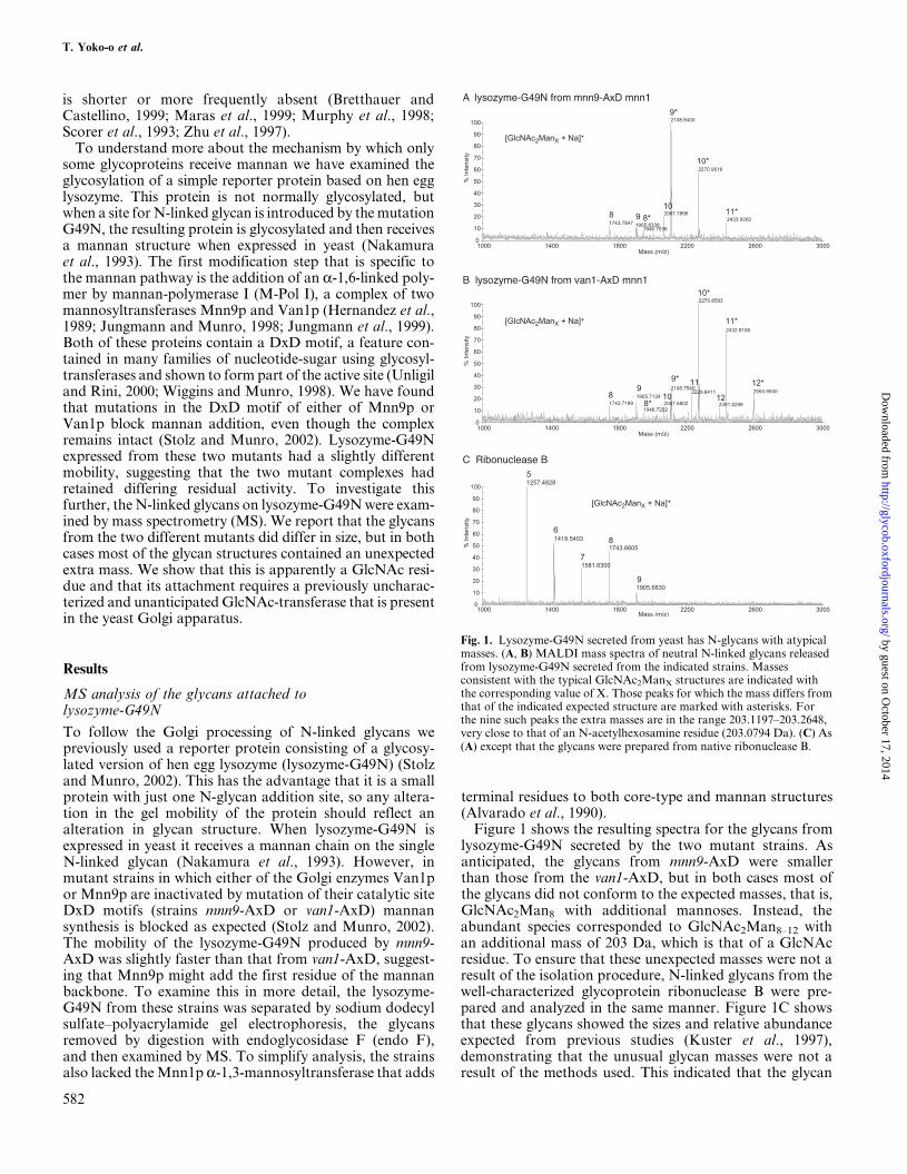
Figure 1 shows the resulting spectra for the glycans fromlysozyme-G49N secreted by the two mutant strains. Asanticipated, the glycans from mnn9-AxD were smallerthan those from the van1-AxD, but in both cases most ofthe glycans did not conform to the expected masses, that is,GlcNAc2Man8 with additional mannoses. Instead, theabundant species corresponded to GlcNAc2Man8±12 withan additional mass of 203 Da, which is that of a GlcNAcresidue. To ensure that these unexpected masses were not aresult of the isolation procedure, N-linked glycans from thewell-characterized glycoprotein ribonuclease B were pre-pared and analyzed in the same manner. Figure 1C showsthat these glycans showed the sizes and relative abundanceexpected from previous studies (Kuster et al., 1997),demonstrating that the unusual glycan masses were not aresult of the methods used. This indicated that the glycan
Fig. 1. Lysozyme-G49N secreted from yeast has N-glycans with atypicalmasses. (A, B) MALDI mass spectra of neutral N-linked glycans releasedfrom lysozyme-G49N secreted from the indicated strains. Massesconsistent with the typical GlcNAc2ManX structures are indicated withthe corresponding value of X. Those peaks for which the mass differs fromthat of the indicated expected structure are marked with asterisks. Forthe nine such peaks the extra masses are in the range 203.1197±203.2648,very close to that of an N-acetylhexosamine residue (203.0794 Da). (C) As(A) except that the glycans were prepared from native ribonuclease B.
T. Yoko-o et al.
582
by guest on October 17, 2014
http://glycob.oxfordjournals.org/D
ownloaded from
on lysozyme-G49N from these strains carries the additionof a single residue that does not appear to be mannose.
The open reading frame YOR320c encodes a putativeGlcNAc-transferase
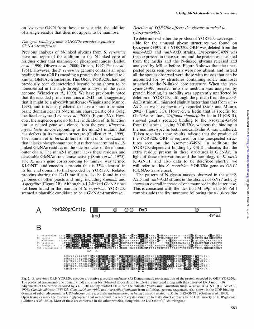
Previous analyses of N-linked glycans from S. cerevisiaehave not reported the addition to the N-linked core ofresidues other that mannose or phosphomannose (Ballouet al., 1990; Olivero et al., 2000; Orlean, 1997; Peat et al.,1961). However, the S. cerevisiae genome contains an openreading frame (ORF) encoding a protein that is related to aknown GlcNAc-transferase. This ORF, YOR320c, had notpreviously been characterized beyond being shown to benonessential in the high-throughput analysis of the yeastgenome (Winzeler et al., 1999). We have previously notedthat the encoded protein contains a DxD motif, suggestingthat it might be a glycosyltransferase (Wiggins and Munro,1998), and it is also predicted to have a short transmem-brane domain near its N-terminus characteristic of a Golgilocalized enzyme (Levine et al., 2000) (Figure 2A). How-ever, the sequence gave no further indication of its functionuntil a related gene was cloned from the yeast Kluyvero-myces lactis as corresponding to the mnn2-1 mutant thathas defects in its mannan structure (Guillen et al., 1999).The mannan of K. lactis differs from that of S. cerevisiae inthat it lacks phosphomannose but rather has terminal a-1,2-linked GlcNAc residues on the side branches of the mannanouter chain. The mnn2-1 mutant lacks these residues anddetectable GlcNAc-transferase activity (Smith et al., 1975).The K. lactis gene corresponding to mnn2-1 was termedKl-GNT1 and encodes a protein that is 33% identical inits lumenal domain to that encoded by YOR320c. Relatedproteins sharing the DxD motif can also be found in thegenomes of other yeasts and fungi including Candida andAspergillus (Figure 2B). Although a-1,2-linked GlcNAc hasnot been found in the mannan of S. cerevisiae, YOR320cseemed a plausible candidate to be a GlcNAc-transferase.
Deletion of YOR320c affects the glycans attached tolysozyme-G49N
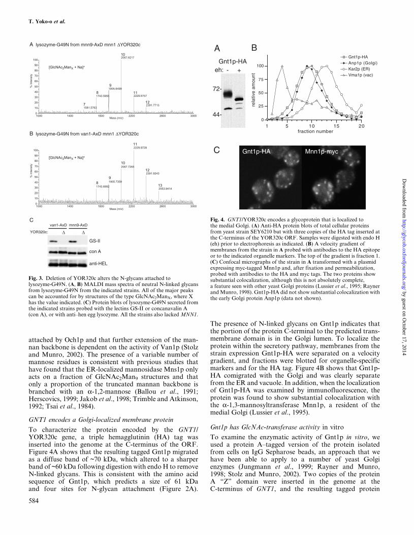
To determine whether the product of YOR320c was respon-sible for the unusual glycan structures we found onlysozyme-G49N, the YOR320c ORF was deleted from themnn9-AxD and van1-AxD strains. Lysozyme-G49N wasthen expressed in these strains, and the protein was isolatedfrom the media and the N-linked glycans released andanalyzed by MS as before. Figure 3 shows that the unex-pected peaks seen previously were now absent, and insteadall the species observed were those with masses that can beaccounted for by structures containing solely mannosesattached to the N-linked core structure. When the lyso-zyme-G49N secreted into the medium was analyzed byprotein blotting, its mobility was apparently unaffected bydeletion of YOR320c, although the protein from the mnn9-AxD strain still migrated slightly faster than that from van1-AxD, as we have previously reported (Stolz and Munro,2002) (Figure 3C). However, a lectin that is specific forGlcNAc residues, Griffonia simplicifolia lectin II (GS-II),showed greatly reduced binding to the lysozyme-G49Nfrom the strains lacking YOR320c, whereas the binding tothe mannose-specific lectin concanavalin A was unaltered.Taken together, these results indicate that the product ofthe YOR320c ORF is required for the unexpected struc-tures seen on the lysozyme-G49N. In addition, theYOR320c-dependent binding by GS-II indicates that theextra residue present in these structures is GlcNAc. Inlight of these observations and the homology to K. lactisKl-GNT1, and also data to be described shortly, wewill refer to this S. cerevisiae YOR320c gene as GNT1(GlcNAc-transferase).
The pattern of N-glycan masses observed in the mnn9-AxD and van1-AxD strains in the absence of GNT1 activityshows an overall increase of one mannose in the latter case.This is consistent with the idea that Mnn9p in the M-Pol Icomplex adds the first mannose following the a-1,6-residue
Fig. 2. S. cerevisiae ORF YOR320c encodes a putative glycosyltransferase. (A) Diagrammatic representation of the protein encoded by ORF YOR320c.The predicted transmembrane domain (tmd) and sites for N-linked glycosylation (circles) are indicated along with the conserved DxD motif. (B)Alignments of the protein encoded by YOR320c and by related ORFs from the indicated yeasts and filamentous fungi. K. lactis, Kl-GNT1 (Guillen et al.,1999); Candida albicans, IPF6425; Colletotrichum trifolii and Aspergillus fumigatus from unfinished genome sequences. Also shown is the UDP-bindingdomain of rabbit glycogenin, a UDP-glucose using glycosyltransferase noted as being distantly related to K. lactis Kl-GNT1p (Guillen et al., 1999).Open triangles mark the residues in glycogenin that were found in a recent crystal structure to make direct contacts to the UDP moiety of UDP-glucose(Gibbons et al., 2002). Most of these are conserved in the other proteins, along with the DxD motif (filled triangles).
A Golgi GlcNAc-transferase in S. cerevisiae
583
by guest on October 17, 2014
http://glycob.oxfordjournals.org/D
ownloaded from
attached by Och1p and that further extension of the man-nan backbone is dependent on the activity of Van1p (Stolzand Munro, 2002). The presence of a variable number ofmannose residues is consistent with previous studies thathave found that the ER-localized mannosidase Mns1p onlyacts on a fraction of GlcNAc2Man9 structures and thatonly a proportion of the truncated mannan backbone isbranched with an a-1,2-mannose (Ballou et al., 1991;Herscovics, 1999; Jakob et al., 1998; Trimble and Atkinson,1992; Tsai et al., 1984).
GNT1 encodes a Golgi-localized membrane protein
To characterize the protein encoded by the GNT1/YOR320c gene, a triple hemagglutinin (HA) tag wasinserted into the genome at the C-terminus of the ORF.Figure 4A shows that the resulting tagged Gnt1p migratedas a diffuse band of ~70 kDa, which altered to a sharperband of ~60 kDa following digestion with endo H to removeN-linked glycans. This is consistent with the amino acidsequence of Gnt1p, which predicts a size of 61 kDaand four sites for N-glycan attachment (Figure 2A).
Fig. 3. Deletion of YOR320c alters the N-glycans attached tolysozyme-G49N. (A, B) MALDI mass spectra of neutral N-linked glycansfrom lysozyme-G49N from the indicated strains. All of the major peakscan be accounted for by structures of the type GlcNAc2ManX, where Xhas the value indicated. (C) Protein blots of lysozyme-G49N secreted fromthe indicated strains probed with the lectins GS-II or concanavalin A(con A), or with anti±hen egg lysozyme. All the strains also lacked MNN1.
)
Fig. 4. GNT1/YOR320c encodes a glycoprotein that is localized tothe medial Golgi. (A) Anti-HA protein blots of total cellular proteinsfrom yeast strain SEY6210 but with three copies of the HA tag inserted atthe C-terminus of the YOR320c ORF. Samples were digested with endo H(eh) prior to electrophoresis as indicated. (B) A velocity gradient ofmembranes from the strain in A probed with antibodies to the HA epitopeor to the indicated organelle markers. The top of the gradient is fraction 1.(C) Confocal micrographs of the strain in A transformed with a plasmidexpressing myc-tagged Mnn1p and, after fixation and permeabilization,probed with antibodies to the HA and myc tags. The two proteins showsubstantial colocalization, although this is not absolutely complete,a feature seen with other yeast Golgi proteins (Lussier et al., 1995; Raynerand Munro, 1998). Gnt1p-HA did not show substantial colocalization withthe early Golgi protein Anp1p (data not shown).
The presence of N-linked glycans on Gnt1p indicates thatthe portion of the protein C-terminal to the predicted trans-membrane domain is in the Golgi lumen. To localize theprotein within the secretory pathway, membranes from thestrain expression Gnt1p-HA were separated on a velocitygradient, and fractions were blotted for organelle-specificmarkers and for the HA tag. Figure 4B shows that Gnt1p-HA comigrated with the Golgi and was clearly separatefrom the ER and vacuole. In addition, when the localizationof Gnt1p-HA was examined by immunofluorescence, theprotein was found to show substantial colocalization withthe a-1,3-mannosyltransferase Mnn1p, a resident of themedial Golgi (Lussier et al., 1995).
Gnt1p has GlcNAc-transferase activity in vitro
To examine the enzymatic activity of Gnt1p in vitro, weused a protein A±tagged version of the protein isolatedfrom cells on IgG Sepharose beads, an approach that wehave been able to apply to a number of yeast Golgienzymes (Jungmann et al., 1999; Rayner and Munro,1998; Stolz and Munro, 2002). Two copies of the proteinA `̀ Z'' domain were inserted in the genome at theC-terminus of GNT1, and the resulting tagged protein
T. Yoko-o et al.
584
A
eh: - +
72-
44-0
25
50
75
100
rela
tive
amou
nt
5 10 1 0fraction number
Vma1p (vac)Kar2p (ER)Anp1p (Golgi)Gnt1p-HA
1
Gnt1p-HA
B
C Gnt1p-HA Mnn1p-myc
5 2
by guest on October 17, 2014
http://glycob.oxfordjournals.org/D
ownloaded from

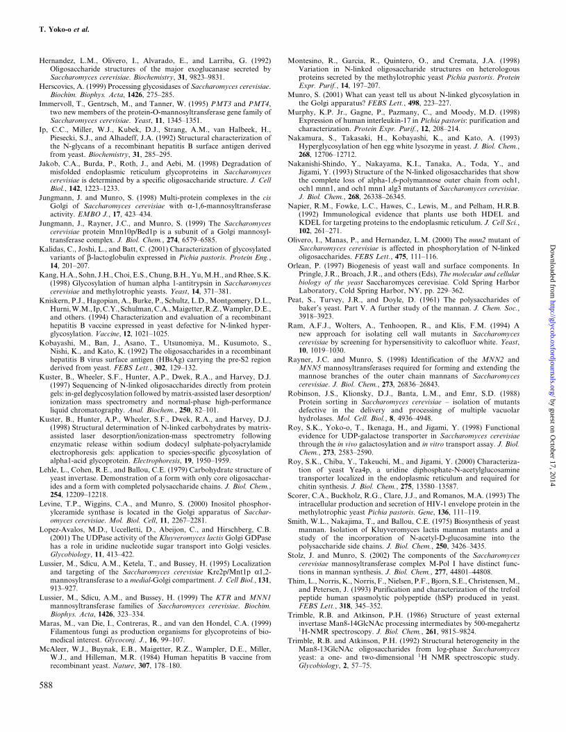
(Gnt1p-ZZ) was isolated from detergent-solubilized cells.Previous studies on the K. lactis Kl-GNT1 gene had foundthat a mutation in the gene correlated with the loss of atransferase activity from cell lysates that could be measuredusing UDP-GlcNAc and the acceptor a-1,3-manno-biose (Guillen et al., 1999). Thus, Gnt1p-ZZ immobilizedon IgG Sepharose beads was incubated with UDP-[3H]GlcNAc and a-1,3-mannobiose, and the productswere separated by thin-layer chromatography (TLC).Figure 5A shows that there was some hydrolysis of theUDP-[3H]GlcNAc that was independent of substrate butwas dependent on Gnt1p-ZZ because it was not seen withbeads isolated from an untagged control strain. Suchnucleotide sugar hydrolysis has been reported in a previousin vitro analysis of yeast glycosyltransferases (Doering,1999) and may reflect the in vitro conditions not being aprecise replica of the intra-Golgi milieu. Nonetheless, in thepresence of the acceptor a labeled product was also pro-duced demonstrating that Gnt1p has GlcNAc-transferaseactivity in vitro. As shown in Figure 5B, this activityrequired the divalent cation manganese, as has beenobserved for the activity of the K. lactis GlcNAc transfer-ase and many other DxD-containing glycosyltransferases(Smith et al., 1975). Examination of different acceptorsshowed a preference for a-1,3-mannobiose over other sim-ple mannose-containing substrates (Figure 5C). However,the activity toward the larger GlcNAc2Man9 N-linked corestructure showed a lower Km than that seen for a-1,3-mannobiose (0.07 mM versus 6.0 mM; data not shown).
Discussion
In this article we report that a heterologous glycoproteinexpressed in S. cerevisiae receives an unexpected residue onits N-linked glycans that appears to be GlcNAc. This mod-ification depends on the presence of a previously uncharac-terized ORF, GNT1/YOR320c, which we show encodesa Golgi-localized glycoprotein that has GlcNAc-transferaseactivity in vitro. Presently the linkage formed by Gnt1p hasnot been defined, although the Gnt1p relative in K. lactisis required for the addition of an a-1,2-linked GlcNAc(Guillen et al., 1999; Smith et al., 1975). Indeed, we cannotat this stage exclude the formal possibility that in vivo Gnt1pcarries out a GlcNAc-transferase reaction that is required forthe activity of a second, unknown enzyme that is responsiblefor the addition of the GlcNAc observed on lysozyme-G49N.However, the simplest interpretation of our results is thatGnt1p is itself the GlcNAc-transferase that is responsible fordirectly modifying the N-linked glycan on lysozyme-G49N invivo. In any case, these results have implications both foryeast cell biology and for the use of yeast as a system for theexpression of recombinant glycoproteins.
Protein glycosylation in the yeast S. cerevisiae has beenextensively studied for many decades, and this has revealedmuch of the enzymology of both Golgi and ER pathways ofglycosylation, with the latter in particular being ofdirect relevance to mammalian systems (Aebi and Hennet,2001; Dean, 1999; Orlean, 1997). The structure of N-linkedglycans in yeast was initially addressed by examining total
Fig. 5. In vitro assay of the GlcNAc-transferase activity of Gnt1p. (A) Autoradiogram of a TLC separation of the products of a transferase reactioncontaining UDP-[3H]GlcNAc and IgG Sepharose beads that had been incubated in lysates from a strain expressing Gnt1p-ZZ, or from a strain withno tagged protein (control). Acceptor a-1,3-mannobiose was present as indicated (a-1,3mb). A product can be seen that is dependent on the presence ofboth Gnt1p-ZZ and acceptor (arrow), and the TLC was run in the direction indicated (tlc). (B) Transfer by Gnt1p-ZZ of radiolabel from UDP-[3H]GlcNActo neutral products in the presence of varying amounts of divalent manganese and with or without acceptor. The assay was as in A, except that total neutralproducts were counted and reactions performed in duplicate with means and standard deviation indicated. Acceptor-dependent transfer requires thepresence of Mn2�, with an apparent Km of 2 mM. (C) Transfer of radiolabel from UDP-[3H]GlcNAc to the indicated acceptors by Gnt1p-ZZ. Transfer wasassayed as in B, with acceptors at 2 mM, except for GlcNAc2Man9 (20 mM). Production of neutral products with acceptors other than a-1,3-mannobioseand GlcNAc2Man9 was not significantly above that seen with no acceptor, with this background (7100 cpm) having been subtracted from the counts shown.
A Golgi GlcNAc-transferase in S. cerevisiae
585
Gnt1p-ZZcontrol
0
2000
4000
6000
8000
0 2 4 6 8 10MnCl2 (mM)
activ
ity (
cpm
)
2mM α-1,3-mannobiose
no acceptor
α-1,3mb:
tlc
- + - +
0
1000
2000
3000
4000
5000
α-methyl-mannoside
α-1,2-
mannobiose
α-1,3- α-1,6- GlcNAc2-Man9
activ
ity (
cpm
)A B C
by guest on October 17, 2014
http://glycob.oxfordjournals.org/D
ownloaded from

mannan released from cell walls (Peat et al., 1961). Furtherstudies then examined the oligosaccharides attached toa number of endogenous proteins, including invertase, car-boxypeptidase Y, and exoglucanase from both wild-typecells and those with mutants in mannan synthesis (Ballouet al., 1990; Hernandez et al., 1992; Lehle et al., 1979;Trimble and Atkinson, 1986). These studies have produceda consistent structure of yeast N-linked glycans that is basedsolely on mannose and phosphomannose, and we have notbeen able to find a single report suggesting the addition offurther N-acetylhexosamine residues beyond the twoGlcNAc residues found in the core structure. Although itis possible that minor species may have been missed or werenot fully resolved by separation methods based on high-performance liquid chromatography (HPLC), it seemsinconceivable that the Gnt1p-dependent modification is auniversal feature of yeast N-linked glycans that has so farescaped detection. Indeed, we observed no difference in thebinding of the GS-II lectin to total cellular proteins or tofixed cells when wild-type and Dgnt1 cells were compared(data not shown). This suggests that if Gnt1p does modifyendogenous N-linked glycans, then it either acts on only asmall percentage of proteins or only under special conditions.
The phenotype of yeast lacking GNT1 has provided fewclues as to likely function. The Dgnt1 cells showed nochange in sensitivity to caffeine, calcofluor white, or hygro-mycin, all of which have increased toxicity toward strainswith cell wall defects (Dean, 1995; Ram et al., 1994), andthere was no change in the mobility of invertase or increasedsecretion of the ER resident protein Kar2p (data notshown). It is possible that the normal substrate of theprotein is not N-glycans, and it is perhaps noteworthy thatGNT1 is located in the genome next to the PMT3 gene thatencodes a protein O-mannosyltransferase (Immervoll et al.,1995). However, no GlcNAc has been found in the O-linkedsugars from S. cerevisiae (Lussier et al., 1999). Nonetheless,the conservation of the gene in diverse yeasts and filamen-tous fungi, such as Candida, K. lactis, and Aspergillus,suggests that it must serve a function that is not highlyspecies-specific. Of course, in K. lactis the protein appearsto provide the GlcNAc in the mannan branches (Guillenet al., 1999; Smith et al., 1975). However, the other yeasts donot have this sugar in their mannan, so perhaps Kl-GNT1pin K. lactis was only recruited recently to mannan bio-genesis. Mannan covers the outer surface of the yeast cellwall, and the structure of its branches varies greatly betweenyeast species, presumably reflecting an evolutionary pres-sure to evade hydrolytic enzymes and toxins, and in the caseof pathogenic yeasts, neutralizing antibodies.
Irrespective of the in vivo role of this protein, the resultsdescribed herein have possible implications for the use ofS. cerevisiae as an expression system for recombinantglycoproteins. The Golgi-specific modification of N-linkedglycans in yeast is clearly very different than that seen inmammals. However, the fact that yeast appear to have thecapability to supply UDP-GlcNAc to the lumen of theirGolgi means that converting yeast to make mammalian-type structures may require less engineering than previouslyanticipated. Yeast have already been found to haveendogenous machinery capable of supplying UDP-GlcNAcand UDP-GalNAc to the lumen of the ER and Golgi,
respectively (Roy et al., 1998, 2000). Indeed, the use ofUDP-GlcNAc in the Golgi lumen by Gnt1p may providean explanation for why S. cerevisiae has been found to havethe capacity to degrade both GDP and UDP in the Golgilumen when the only nucleotide sugar previously found tobe required by endogenous Golgi glycosyltransferases wasGDP-mannose (Abeijon et al., 1993; Gao et al., 1999;Lopez-Avalos et al., 2001).
Another implication of these findings is that not all het-erologous glycoproteins expressed in yeast can be assumedto receive solely high-mannose structures on their N-linkedglycans. S. cerevisiae has been tested as an expression sys-tem for a wide range of glycoproteins, including potentialvaccines and therapeutic proteins. In many cases therecombinant glycoproteins receive mannan addition, andattempts have been made to avoid this by the use of mnn9mutants or other yeasts. The N-glycans attached to some ofthese heterologous proteins have been examined in detail,including those from a glycosylated version of hepatitissurface antigen and from human trefoil factor expressed inS. cerevisiae (Ip et al., 1992; Kniskern et al., 1994;Kobayashi et al., 1992; Thim et al., 1993) and b-lactoglo-bulin and tick antigens expressed in Pichia (Kalidas et al.,2001; Montesino et al., 1998). In these cases the glycansfound conformed to the expected high-mannose structures,although in some cases this conclusion was based on the useof HPLC, which has a size resolution that is not as high asthat of MS. However, the fact that Gnt1p appears to be ableto efficiently modify lysozyme-G49N in vivo, and GlcNAc2
Man9 in vitro means that it seems possible that other hetero-logous glycoproteins could also be modified. It is not incon-ceivable that the presence of this extra residue could alterthe circulation properties or the susceptibility to immuno-logical responses of the resulting glycoprotein. Thus, itseems important to consider the Golgi addition of GlcNAcas a potential variable in the use of S. cerevisiae and otheryeasts and fungi as expression systems for therapeutic gly-coproteins. The apparent lack of effect on viability of dele-tion of the GNT1 gene at least provides a simple means toremove the modification if this is desired.
Materials and methods
Yeast strains and plasmids
Yeast strains were based on the parental strain SEY6210(MATa ura3-52 leu2-3,112 his3-D200 trp1-D901 lys2-801suc2D9) (Robinson et al., 1988). Strains lacking MNN1and having AxD mutations in the genomic copies ofMNN9 or VAN1 were as described (Stolz and Munro,2002), and the YOR320c coding region was deleted inthese by polymerase chain reaction (PCR)±based homolo-gous recombination using Saccharomyces pombe his5�
(Wach et al., 1997). The GNT1 ORF was tagged with HAat the C-terminus using PCR-based homologous recombi-nation and plasmid p3xHA-HIS5 (Jungmann et al., 1999).A similar approach was used for protein A±tagging Gnt1pfor isolation for enzyme assays, except kanMX-based plas-mid pFZ was used (Whyte and Munro, 2001), and theparental strain was the multiply protease deficient strainc13-ABYS 86 (MATa pra1-1 prb1-1 prc1-1 cps1-3 ura3D5
T. Yoko-o et al.
586
by guest on October 17, 2014
http://glycob.oxfordjournals.org/D
ownloaded from

leu2-3,112 his3) (Heinemeyer et al., 1991). Lysozyme-G49Nwas expressed from the 2 m plasmid pVT100-U-HELG49N(Stolz and Munro, 2002) and triple myc-tagged Mnn1pfrom its own promoter in a CEN plasmid (Wiggins andMunro, 1998).
Protein localization
Fractionation of yeast membranes on sucrose velocitygradients and localization of proteins by immunofluores-cence were as described previously (Levine et al., 2000).Monoclonal antibodies against the HA epitope (3F10;Roche, Lewes, UK), Kar2p (2E7) (Napier et al., 1992),Vma1p (Molecular Probes, Eugene, OR), and rabbit poly-clonal antibodies against Anp1p (Jungmann and Munro,1998) and the myc-epitope (Santa Cruz Biotechnology,Santa Cruz, CA), were detected with species-specificsecondary antisera labeled with fluorophores or peroxidase(Amersham Biosciences, Piscataway, NJ), and the latterwas detected by chemiluminescence (Amersham Bios-ciences). For lectin blotting, biotinylated GS-II or concana-valin A (Vector Laboratories, Burlingame, CA) were usedto probe blots at 0.25 mg/ml in phosphate buffered saline,0.1% Tween-20, 200 mM CaCl2, and 200 mM MgCl2, fol-lowed by peroxidase-avidin (1 mg/ml; Vector Laboratories).
MS analysis of N-linked glycans
Lysozyme-G49N was isolated from the medium of strainsharboring plasmid pVT100-U-HELG49N by ion exchangechromatography (Stolz and Munro, 2002). The N-glycansfrom typically 25 mg of protein were released by in geldigestion with endo F, followed by cleanup and MS asdescribed previously (Kuster et al., 1997, 1998). Matrix-assisted laser desorption/ionization (MALDI) MS wasperformed on a PerSeptive Biosystems (Framingham,MA) Voyager-DE STR instrument.
In vitro assays of GlcNAc transferase activity
Protein A±tagged Gnt1p was precipitated from detergentlysates of spheroplasts using IgG Sepharose essentially asdescribed previously (Rayner and Munro, 1998), exceptthat 1% Triton X-100 was used as the detergent, and afterbinding and washing, the beads were washed into 50 mM4-morpholine propane sulfonic acid (MOPS)±NaOH(pH 7.5). GlcNAc transferase activity was assayed in 50-mlreactions containing 20 ml beads (prepared from thelysate of 200 mg of cells) and 50 mM MOPS-NaOH (pH7.5), 5 mM MnCl2, 0.24 mM (0.5 mCi) UDP-[3H]GlcNAc(41.6 Ci/mmol; New England Nuclear, Boston, MA), andacceptor. The mixture was shaken gently for 3 h at 30�Cand, after addition of 200 ml water, applied to a 0.9-mlcolumn of Dowex 1-X8 in the acetate form, the neutralreaction products eluted with 1.0 ml water, and the radioac-tivity quantified by scintillation counting. Analysis of pro-ducts by TLC was as described previously (Doering, 1999).
Acknowledgments
We are indebted to David Harvey for advice on the MS ofN-glycans. Takehiko Yoko-o was supported by a 1-yearfellowship from the Science and Technology Agency of
Japan and JuÈrgen Stolz by an EMBO long-term fellowship(ALTF 495-1999).
Abbreviations
Endo F, endoglycosidase F; ER, endoplasmic reticulum;GS-II, Griffonia simplicifolia lectin II; HA, hemagglu-tinin; HPLC, high-performance liquid chromatography;MALDI, matrix-assisted laser desorption/ionization;MOPS, 4-morpholine propane sulfonic acid; M-Pol I,mannan-polymerase I; MS, mass spectrometry; ORF, openreading frame; PCR, polymerase chain reaction; TLC,thin-layer chromatography.
References
Abeijon, C., Yanagisawa, K., Mandon, E.C., Hausler, A., Moremen, K.,Hirschberg, C.B., and Robbins, P.W. (1993) Guanosine diphos-phatase is required for protein and sphingolipid glycosylation inthe Golgi lumen of Saccharomyces cerevisiae. J. Cell Biol., 122,307±323.
Aebi, M. and Hennet, T. (2001) Congenital disorders of glycosylation:genetic model systems lead the way. Trends Cell Biol., 11, 136±141.
Alvarado, E., Ballou, L., Hernandez, L.M., and Ballou, C.E. (1990)Localization of a1-3-linked mannoses in the N-linked oligosaccharidesof Saccharomyces cerevisiae mnn mutants. Biochemistry, 29,2471±2482.
Antebi, A. and Fink, G.R. (1992) The yeast Ca2�-ATPase homologue,PMR1, is required for normal Golgi function and localizes in a novelGolgi-like distribution. Mol. Biol. Cell, 3, 633±654.
Ballou, L., Hernandez, L.M., Alvarado, E., and Ballou, C.E. (1990)Revision of the oligosaccharide structures of yeast carboxypeptidaseY. Proc. Natl Acad. Sci. USA, 87, 3368±3372.
Ballou, L., Hitzeman, R.A., Lewis, M.S., and Ballou, C.E. (1991)Vanadate-resistant yeast mutants are defective in protein glycosyla-tion. Proc. Natl Acad. Sci. USA, 88, 3209±3212.
Bretthauer, R.K. and Castellino, F.J. (1999) Glycosylation of Pichiapastoris-derived proteins. Biotechnol. Appl. Biochem., 30, 193±200.
Dean, N. (1995) Yeast glycosylation mutants are sensitive to aminoglyco-sides. Proc. Natl Acad. Sci. USA, 92, 1287±1291.
Dean, N. (1999) Asparagine-linked glycosylation in the yeast Golgi.Biochim. Biophys. Acta, 1426, 309±322.
Dean, N., Zhang, Y.B., and Poster, J.B. (1997) The VRG4 gene is requiredfor GDP-mannose transport into the lumen of the Golgi in the yeast,Saccharomyces cerevisiae. J. Biol. Chem., 272, 31908±31914.
Doering, T.L. (1999) A unique alpha-1,3 mannosyltransferase ofthe pathogenic fungus Cryptococcus neoformans. J. Bacteriol., 181,5482±5488.
Gao, X.D., Kaigorodov, V., and Jigami, Y. (1999) YND1, a homologue ofGDA1, encodes membrane-bound apyrase required for Golgi N- andO-glycosylation in Saccharomyces cerevisiae. J. Biol. Chem., 274,21450±21456.
Gibbons, B.J., Roach, P.J., and Hurley, T.D. (2002) Crystal structure ofthe autocatalytic initiator of glycogen biosynthesis, glycogenin. J. Mol.Biol., 319, 463±477.
Guillen, E., Abeijon, C., and Hirschberg, C.B. (1999) The genes for theGolgi apparatus N-acetylglucosaminyltransferase and the UDP-N-acetylglucosamine transporter are contiguous in Kluyveromyces lactis.J. Biol. Chem., 274, 6641±6646.
Heinemeyer, W., Kleinschmidt, J.A., Saidowsky, J., Escher, C., andWolf, D.H. (1991) Proteinase yscE, the yeast proteasome/multicatalytic-multifunctional proteinase: mutants unravel its functionin stress induced proteolysis and uncover its necessity for cell survival.EMBO J., 10, 555±562.
Hernandez, L.M., Ballou, L., Alvarado, E., Tsai, P.K., and Ballou, C.E.(1989) Structure of the phosphorylated N-linked oligosaccharides fromthe mnn9 and mnn10 mutants of Saccharomyces cerevisiae. J. Biol.Chem., 264, 13648±13659.
A Golgi GlcNAc-transferase in S. cerevisiae
587
by guest on October 17, 2014
http://glycob.oxfordjournals.org/D
ownloaded from
Hernandez, L.M., Olivero, I., Alvarado, E., and Larriba, G. (1992)Oligosaccharide structures of the major exoglucanase secreted bySaccharomyces cerevisiae. Biochemistry, 31, 9823±9831.
Herscovics, A. (1999) Processing glycosidases of Saccharomyces cerevisiae.Biochim. Biophys. Acta, 1426, 275±285.
Immervoll, T., Gentzsch, M., and Tanner, W. (1995) PMT3 and PMT4,two new members of the protein-O-mannosyltransferase gene family ofSaccharomyces cerevisiae. Yeast, 11, 1345±1351.
Ip, C.C., Miller, W.J., Kubek, D.J., Strang, A.M., van Halbeek, H.,Piesecki, S.J., and Alhadeff, J.A. (1992) Structural characterization ofthe N-glycans of a recombinant hepatitis B surface antigen derivedfrom yeast. Biochemistry, 31, 285±295.
Jakob, C.A., Burda, P., Roth, J., and Aebi, M. (1998) Degradation ofmisfolded endoplasmic reticulum glycoproteins in Saccharomycescerevisiae is determined by a specific oligosaccharide structure. J. CellBiol., 142, 1223±1233.
Jungmann, J. and Munro, S. (1998) Multi-protein complexes in the cisGolgi of Saccharomyces cerevisiae with a-1,6-mannosyltransferaseactivity. EMBO J., 17, 423±434.
Jungmann, J., Rayner, J.C., and Munro, S. (1999) The Saccharomycescerevisiae protein Mnn10p/Bed1p is a subunit of a Golgi mannosyl-transferase complex. J. Biol. Chem., 274, 6579±6585.
Kalidas, C., Joshi, L., and Batt, C. (2001) Characterization of glycosylatedvariants of b-lactoglobulin expressed in Pichia pastoris. Protein Eng.,14, 201±207.
Kang, H.A., Sohn, J.H., Choi, E.S., Chung, B.H., Yu, M.H., and Rhee, S.K.(1998) Glycosylation of human alpha 1-antitrypsin in Saccharomycescerevisiae and methylotrophic yeasts. Yeast, 14, 371±381.
Kniskern, P.J., Hagopian, A., Burke, P., Schultz, L.D., Montgomery, D.L.,Hurni,W.M.,Ip,C.Y.,Schulman,C.A.,Maigetter,R.Z.,Wampler,D.E.,and others. (1994) Characterization and evaluation of a recombinanthepatitis B vaccine expressed in yeast defective for N-linked hyper-glycosylation. Vaccine, 12, 1021±1025.
Kobayashi, M., Ban, J., Asano, T., Utsunomiya, M., Kusumoto, S.,Nishi, K., and Kato, K. (1992) The oligosaccharides in a recombinanthepatitis B virus surface antigen (HBsAg) carrying the pre-S2 regionderived from yeast. FEBS Lett., 302, 129±132.
Kuster, B., Wheeler, S.F., Hunter, A.P., Dwek, R.A., and Harvey, D.J.(1997) Sequencing of N-linked oligosaccharides directly from proteingels: in-gel deglycosylation followed by matrix-assisted laser desorption/ionization mass spectrometry and normal-phase high-performanceliquid chromatography. Anal. Biochem., 250, 82±101.
Kuster, B., Hunter, A.P., Wheeler, S.F., Dwek, R.A., and Harvey, D.J.(1998) Structural determination of N-linked carbohydrates by matrix-assisted laser desorption/ionization-mass spectrometry followingenzymatic release within sodium dodecyl sulphate-polyacrylamideelectrophoresis gels: application to species-specific glycosylation ofalpha1-acid glycoprotein. Electrophoresis, 19, 1950±1959.
Lehle, L., Cohen, R.E., and Ballou, C.E. (1979) Carbohydrate structure ofyeast invertase. Demonstration of a form with only core oligosacchar-ides and a form with completed polysaccharide chains. J. Biol. Chem.,254, 12209±12218.
Levine, T.P., Wiggins, C.A., and Munro, S. (2000) Inositol phosphor-ylceramide synthase is located in the Golgi apparatus of Sacchar-omyces cerevisiae. Mol. Biol. Cell, 11, 2267±2281.
Lopez-Avalos, M.D., Uccelletti, D., Abeijon, C., and Hirschberg, C.B.(2001) The UDPase activity of the Kluyveromyces lactis Golgi GDPasehas a role in uridine nucleotide sugar transport into Golgi vesicles.Glycobiology, 11, 413±422.
Lussier, M., Sdicu, A.M., Ketela, T., and Bussey, H. (1995) Localizationand targeting of the Saccharomyces cerevisiae Kre2p/Mnt1p a1,2-mannosyltransferase to a medial-Golgi compartment. J. Cell Biol., 131,913±927.
Lussier, M., Sdicu, A.M., and Bussey, H. (1999) The KTR and MNN1mannosyltransferase families of Saccharomyces cerevisiae. Biochim.Biophys. Acta, 1426, 323±334.
Maras, M., van Die, I., Contreras, R., and van den Hondel, C.A. (1999)Filamentous fungi as production organisms for glycoproteins of bio-medical interest. Glycoconj. J., 16, 99±107.
McAleer, W.J., Buynak, E.B., Maigetter, R.Z., Wampler, D.E., Miller,W.J., and Hilleman, M.R. (1984) Human hepatitis B vaccine fromrecombinant yeast. Nature, 307, 178±180.
Montesino, R., Garcia, R., Quintero, O., and Cremata, J.A. (1998)Variation in N-linked oligosaccharide structures on heterologousproteins secreted by the methylotrophic yeast Pichia pastoris. ProteinExpr. Purif., 14, 197±207.
Munro, S. (2001) What can yeast tell us about N-linked glycosylation inthe Golgi apparatus? FEBS Lett., 498, 223±227.
Murphy, K.P. Jr., Gagne, P., Pazmany, C., and Moody, M.D. (1998)Expression of human interleukin-17 in Pichia pastoris: purification andcharacterization. Protein Expr. Purif., 12, 208±214.
Nakamura, S., Takasaki, H., Kobayashi, K., and Kato, A. (1993)Hyperglycosylation of hen egg white lysozyme in yeast. J. Biol. Chem.,268, 12706±12712.
Nakanishi-Shindo, Y., Nakayama, K.I., Tanaka, A., Toda, Y., andJigami, Y. (1993) Structure of the N-linked oligosaccharides that showthe complete loss of alpha-1,6-polymannose outer chain from och1,och1 mnn1, and och1 mnn1 alg3 mutants of Saccharomyces cerevisiae.J. Biol. Chem., 268, 26338±26345.
Napier, R.M., Fowke, L.C., Hawes, C., Lewis, M., and Pelham, H.R.B.(1992) Immunological evidence that plants use both HDEL andKDEL for targeting proteins to the endoplasmic reticulum. J. Cell Sci.,102, 261±271.
Olivero, I., Manas, P., and Hernandez, L.M. (2000) The mnn2 mutant ofSaccharomyces cerevisiae is affected in phosphorylation of N-linkedoligosaccharides. FEBS Lett., 475, 111±116.
Orlean, P. (1997) Biogenesis of yeast wall and surface components. InPringle, J.R., Broach, J.R., and others (Eds), The molecular and cellularbiology of the yeast Saccharomyces cerevisiae. Cold Spring HarborLaboratory, Cold Spring Harbor, NY, pp. 229±362.
Peat, S., Turvey, J.R., and Doyle, D. (1961) The polysaccharides ofbaker's yeast. Part V. A further study of the mannan. J. Chem. Soc.,3918±3923.
Ram, A.F.J., Wolters, A., Tenhoopen, R., and Klis, F.M. (1994) Anew approach for isolating cell wall mutants in Saccharomycescerevisiae by screening for hypersensitivity to calcofluor white. Yeast,10, 1019±1030.
Rayner, J.C. and Munro, S. (1998) Identification of the MNN2 andMNN5 mannosyltransferases required for forming and extending themannose branches of the outer chain mannans of Saccharomycescerevisiae. J. Biol. Chem., 273, 26836±26843.
Robinson, J.S., Klionsky, D.J., Banta, L.M., and Emr, S.D. (1988)Protein sorting in Saccharomyces cerevisiae ± isolation of mutantsdefective in the delivery and processing of multiple vacuolarhydrolases. Mol. Cell. Biol., 8, 4936±4948.
Roy, S.K., Yoko-o, T., Ikenaga, H., and Jigami, Y. (1998) Functionalevidence for UDP-galactose transporter in Saccharomyces cerevisiaethrough the in vivo galactosylation and in vitro transport assay. J. Biol.Chem., 273, 2583±2590.
Roy, S.K., Chiba, Y., Takeuchi, M., and Jigami, Y. (2000) Characteriza-tion of yeast Yea4p, a uridine diphosphate-N-acetylglucosaminetransporter localized in the endoplasmic reticulum and required forchitin synthesis. J. Biol. Chem., 275, 13580±13587.
Scorer, C.A., Buckholz, R.G., Clare, J.J., and Romanos, M.A. (1993) Theintracellular production and secretion of HIV-1 envelope protein in themethylotrophic yeast Pichia pastoris. Gene, 136, 111±119.
Smith, W.L., Nakajima, T., and Ballou, C.E. (1975) Biosynthesis of yeastmannan. Isolation of Kluyveromyces lactis mannan mutants and astudy of the incorporation of N-acetyl-D-glucosamine into thepolysaccharide side chains. J. Biol. Chem., 250, 3426±3435.
Stolz, J. and Munro, S. (2002) The components of the Saccharomycescerevisiae mannosyltransferase complex M-Pol I have distinct func-tions in mannan synthesis. J. Biol. Chem., 277, 44801±44808.
Thim, L., Norris, K., Norris, F., Nielsen, P.F., Bjorn, S.E., Christensen, M.,and Petersen, J. (1993) Purification and characterization of the trefoilpeptide human spasmolytic polypeptide (hSP) produced in yeast.FEBS Lett., 318, 345±352.
Trimble, R.B. and Atkinson, P.H. (1986) Structure of yeast externalinvertase Man8-14GlcNAc processing intermediates by 500-megahertz1H-NMR spectroscopy. J. Biol. Chem., 261, 9815±9824.
Trimble, R.B. and Atkinson, P.H. (1992) Structural heterogeneity in theMan8-13GlcNAc oligosaccharides from log-phase Saccharomycesyeast: a one- and two-dimensional 1H NMR spectroscopic study.Glycobiology, 2, 57±75.
T. Yoko-o et al.
588
by guest on October 17, 2014
http://glycob.oxfordjournals.org/D
ownloaded from
Tsai, P.K., Frevert, J., and Ballou, C.E. (1984) Carbohydrate structure ofSaccharomyces cerevisiae mnn9 mannoprotein. J. Biol. Chem., 259,3805±3811.
Unligil, U.M. and Rini, J.M. (2000) Glycosyltransferase structure andmechanism. Curr. Opin. Struct. Biol., 10, 510±517.
Wach, A., Brachat, A., AlbertiSegui, C., Rebischung, C., and Philippsen, P.(1997) Heterologous HIS3 marker and GFP reporter modules forPCR-targeting in Saccharomyces cerevisiae. Yeast, 13, 1065±1075.
Whyte, J.R. and Munro, S. (2001) The Sec34/35 Golgi transport complexis related to the exocyst, defining a family of complexes involved inmultiple steps of membrane traffic. Dev. Cell, 1, 527±537.
Wiggins, C.A.R. and Munro, S. (1998) Activity of the yeast MNN1 a-1,3-mannosyltransferase requires a motif conserved in many other familiesof glycosyltransferases. Proc. Natl Acad. Sci. USA, 95, 7945±7950.
Winzeler, E.A., Shoemaker, D.D., Astromoff, A., Liang, H.,Anderson, K., Andre, B., Bangham, R., Benito, R., Boeke, J.D.,Bussey, H., and others. (1999) Functional characterization of theS. cerevisiae genome by gene deletion and parallel analysis. Science,285, 901±906.
Zhu, X.P., Wu, S.X., and Letchworth, G.J. (1997) Yeast-secreted bovineherpesvirus type 1 glycoprotein D has authentic conformational struc-ture and immunogenicity. Vaccine, 15, 679±688.
A Golgi GlcNAc-transferase in S. cerevisiae
589
by guest on October 17, 2014
http://glycob.oxfordjournals.org/D
ownloaded from
Copyright © 2022 FDOKUMEN