
Human 'parietal reach region' encodes visual stimulus coordinates, not movement direction, during...
10
Human Parietal ‘‘Reach Region’’ Primarily Encodes Intrinsic Visual Direction, Not Extrinsic Movement Direction, in a Visual--Motor Dissociation Task Juan Fernandez-Ruiz 1,2,3 , Herbert C. Goltz 1,4,5 , Joseph F.X. DeSouza 1,3 , Tutis Vilis 1,6 and J. Douglas Crawford 1,3,7 1 Canadian Institutes of Health Research Group on Action and Perception, 2 Departamento de Fisiologı´a, Facultad de Medicina, Universidad Nacional Auto´ noma de Me´ xico, Me´ xico, 3 Centre for Vision Research, York University, Toronto, Ontario, Canada M3J 1P3, 4 Imaging Research Labs, Robarts Research Institute, London, Ontario N6A 5K8, Canada, Departments of 5 Psychology and 6 Physiology and Pharmacology, University of Western Ontario, London, Ontario, Canada N6A 5C1, and 7 Department of Psychology, York University, Toronto, Ontario, Canada M3J 1P3 Posterior parietal cortex (PPC) participates in the planning of visuospatial behaviors, including reach movements, in gaze- centered coordinates. It is not known if these representations encode the visual goal in retinal coordinates, or the movement direction relative to gaze. Here, by dissociating the intrinsic retinal stimulus from the extrinsic direction of movement, we show that PPC employs a visual code. Using delayed pointing and event- related functional magnetic resonance imaging, we identified a cluster of PPC regions whose activity was topographically (con- tralaterally) related to the direction of the planned movement. We then switched the normal visual--motor spatial relationship by adapting subjects to optical left/right reversing prisms. With prisms, movement-related PPC topography reversed, remaining tied to the retinal image. Thus, remarkably, the PPC region in each hemisphere now responded more for planned ipsilateral pointing movements. Other non-PPC regions showed the opposite world- or motor-fixed pattern. These findings suggest that PPC primarily encodes not motor commands but movement goals in visual coordinates. Keywords: pointing, precuneus, reaching, reversing prism adaptation, visuomotor learning, visuomotor transformation Introduction When we reach for a visual stimulus, the brain initially codes the target in eye-fixed retinal coordinates but the final output command is coded in muscle coordinates. A central problem in neuroscience is how and where the central nervous system achieves the transformation between these visual and motor representations (Soechting and Flanders 1992; Pouget and Snyder 2000; Andersen and Buneo 2002; Crawford et al. 2004). Perhaps the most fundamental, yet unanswered, question that visuomotor scientists can ask is at what stage in the transformation is the transition made from a visual representa- tion to a motor representation? Primate neurophysiology has revealed a network for visuo- motor transformations that includes the posterior parietal (PPC) and premotor cortices (Kalaska and Crammond 1992; Colby and Goldberg 1999; Snyder 2000; Andersen and Buneo 2002). The PPC can be further divided into several functional subregions (Colby and Goldberg 1999; Andersen and Buneo 2002). In particular, the lateral intraparietal (LIP) sulcus encodes in- tended saccades (Snyder et al. 1997; Dickinson et al. 2003), whereas the medial intraparietal region often called the ‘‘parietal reach region’’ (PRR), is more active during arm move- ments (Snyder et al., 1997;Batista et al., 1999; Calton et al., 2002). Both regions encode the target and the action in contralateral space relative to the gaze pointing direction, that is, in a gaze- centered frame like the retina (Duhamel et al. 1992; Stricanne et al. 1996; Batista et al. 1999; Colby and Goldberg 1999). Recent functional magnetic resonance imaging (fMRI) ex- periments suggest that human PPC probably has a similar func- tional organization. Human PPC contains a region (or regions), perhaps analogous to monkey LIP that selectively code for con- tralateral saccades (Sereno et al. 2001; Medendorp et al. 2003; Schluppeck et al. 2005). Some regions within PPC are also acti- vated during contralateral pointing movements and are modu- lated by eye position in a manner consistent with the physiology of monkey PRR (DeSouza et al. 2000; Medendorp et al. 2003; Medendorp, Goltz, Crawford, et al. 2005; Medendorp, Goltz, Vilis, 2005). Specifically, Connolly et al. (2003) have identified a dorso-medial area of PPC within the precuneus that they called the human PRR. Moreover, some of these saccade and pointing regions, like the corresponding monkey regions (Duhamel et al. 1992; Batista et al. 1999), appear to represent space in a gaze-centered frame, and update these representations when the eyes rotate (Medendorp et al. 2003; Merriam et al. 2003). This has recently been confirmed causally: Optic ataxia patients with PPC damage show reach deficits in a gaze-centered frame that is updated with each saccade (Khan, Pisella, Rossetti, et al. 2005; Khan, Pisella, Vighetto, et al. 2005). Thus, spatial activity in human PRR is not tied to the initial retinal input but instead provides a dynamic, gaze-centered representation (Khan, Pisella, Vighetto, et al. 2005). However, the more basic question remains unanswered: It is not known if this gaze-centered signal encodes the visual goal of the movement (upstream from the vision-to-motor transforma- tion) (Gottlieb and Goldberg 1999) or the direction of the movement (downstream from this transformation) (Kalaska 1996; Eskandar and Assad 1999; Zhang and Barash 2000). This fundamental question has practical importance because PRR is one of the regions that is currently being investigated as a possible neural interface for prosthetic devices (Musallam et al. 2004). This is a difficult question to address experimentally because the visual goal and movement direction are normally aligned. When subjects are asked to look or point in the direction opposite to the visual target (‘‘antisaccades’’ and ‘‘antipointing’’) neural activity sometimes shows a transient visual response but ends up aligned with the movement direction (Kalaska 1996; Eskandar and Assad 1999; Connolly et al. 2000; Zhang and Barash 2000). This shows that PPC activity can be dissociated from the visual stimulus. However, these antipointing and antisaccade tasks instruct the subject to imagine a goal opposite Cerebral Cortex October 2007;17:2283--2292 doi:10.1093/cercor/bhl137 Advance Access publication January 10, 2007 Ó The Author 2007. Published by Oxford University Press. All rights reserved. For permissions, please e-mail: [email protected] at Hospital for Sick Children Hospital Library on July 29, 2014 http://cercor.oxfordjournals.org/ Downloaded from
Transcript of Human 'parietal reach region' encodes visual stimulus coordinates, not movement direction, during...

Human Parietal ‘‘Reach Region’’ PrimarilyEncodes Intrinsic Visual Direction, NotExtrinsic Movement Direction, in aVisual--Motor Dissociation Task
Juan Fernandez-Ruiz1,2,3, Herbert C. Goltz1,4,5, Joseph F.X.
DeSouza1,3, Tutis Vilis1,6 and J. Douglas Crawford1,3,7
1Canadian Institutes of Health Research Group on Action and
Perception, 2Departamento de Fisiologıa, Facultad de
Medicina, Universidad Nacional Autonoma de Mexico, Mexico,3Centre for Vision Research, York University, Toronto,
Ontario, Canada M3J 1P3, 4Imaging Research Labs, Robarts
Research Institute, London, Ontario N6A 5K8, Canada,
Departments of 5Psychology and 6Physiology and
Pharmacology, University of Western Ontario, London,
Ontario, Canada N6A 5C1, and 7Department of Psychology,
York University, Toronto, Ontario, Canada M3J 1P3
Posterior parietal cortex (PPC) participates in the planning ofvisuospatial behaviors, including reach movements, in gaze-centered coordinates. It is not known if these representationsencode the visual goal in retinal coordinates, or the movementdirection relative to gaze. Here, by dissociating the intrinsic retinalstimulus from the extrinsic direction of movement, we show thatPPC employs a visual code. Using delayed pointing and event-related functional magnetic resonance imaging, we identified acluster of PPC regions whose activity was topographically (con-tralaterally) related to the direction of the planned movement. Wethen switched the normal visual--motor spatial relationship byadapting subjects to optical left/right reversing prisms. With prisms,movement-related PPC topography reversed, remaining tied to theretinal image. Thus, remarkably, the PPC region in each hemispherenow responded more for planned ipsilateral pointing movements.Other non-PPC regions showed the opposite world- or motor-fixedpattern. These findings suggest that PPC primarily encodes notmotor commands but movement goals in visual coordinates.
Keywords: pointing, precuneus, reaching, reversing prism adaptation,visuomotor learning, visuomotor transformation
Introduction
When we reach for a visual stimulus, the brain initially codes the
target in eye-fixed retinal coordinates but the final output
command is coded in muscle coordinates. A central problem in
neuroscience is how and where the central nervous system
achieves the transformation between these visual and motor
representations (Soechting and Flanders 1992; Pouget and
Snyder 2000; Andersen and Buneo 2002; Crawford et al.
2004). Perhaps the most fundamental, yet unanswered, question
that visuomotor scientists can ask is at what stage in the
transformation is the transition made from a visual representa-
tion to a motor representation?
Primate neurophysiology has revealed a network for visuo-
motor transformations that includes the posterior parietal (PPC)
and premotor cortices (Kalaska and Crammond 1992; Colby and
Goldberg 1999; Snyder 2000; Andersen and Buneo 2002). The
PPC can be further divided into several functional subregions
(Colby and Goldberg 1999; Andersen and Buneo 2002). In
particular, the lateral intraparietal (LIP) sulcus encodes in-
tended saccades (Snyder et al. 1997; Dickinson et al. 2003),
whereas the medial intraparietal region often called the
‘‘parietal reach region’’ (PRR), is more active during arm move-
ments (Snyder et al., 1997;Batista et al., 1999; Calton et al., 2002).
Both regions encode the target and the action in contralateral
space relative to the gaze pointing direction, that is, in a gaze-
centered frame like the retina (Duhamel et al. 1992; Stricanne
et al. 1996; Batista et al. 1999; Colby and Goldberg 1999).
Recent functional magnetic resonance imaging (fMRI) ex-
periments suggest that human PPC probably has a similar func-
tional organization. Human PPC contains a region (or regions),
perhaps analogous to monkey LIP that selectively code for con-
tralateral saccades (Sereno et al. 2001; Medendorp et al. 2003;
Schluppeck et al. 2005). Some regions within PPC are also acti-
vated during contralateral pointing movements and are modu-
lated by eye position in a manner consistent with the physiology
of monkey PRR (DeSouza et al. 2000; Medendorp et al. 2003;
Medendorp, Goltz, Crawford, et al. 2005; Medendorp, Goltz,
Vilis, 2005). Specifically, Connolly et al. (2003) have identified
a dorso-medial area of PPC within the precuneus that they
called the human PRR.
Moreover, some of these saccade and pointing regions, like
the corresponding monkey regions (Duhamel et al. 1992; Batista
et al. 1999), appear to represent space in a gaze-centered frame,
and update these representations when the eyes rotate
(Medendorp et al. 2003; Merriam et al. 2003). This has recently
been confirmed causally: Optic ataxia patients with PPC damage
show reach deficits in a gaze-centered frame that is updated
with each saccade (Khan, Pisella, Rossetti, et al. 2005; Khan,
Pisella, Vighetto, et al. 2005). Thus, spatial activity in human PRR
is not tied to the initial retinal input but instead provides a
dynamic, gaze-centered representation (Khan, Pisella, Vighetto,
et al. 2005).
However, the more basic question remains unanswered: It is
not known if this gaze-centered signal encodes the visual goal of
the movement (upstream from the vision-to-motor transforma-
tion) (Gottlieb and Goldberg 1999) or the direction of the
movement (downstream from this transformation) (Kalaska
1996; Eskandar and Assad 1999; Zhang and Barash 2000). This
fundamental question has practical importance because PRR is
one of the regions that is currently being investigated as
a possible neural interface for prosthetic devices (Musallam
et al. 2004).
This is a difficult question to address experimentally because
the visual goal and movement direction are normally aligned.
When subjects are asked to look or point in the direction
opposite to the visual target (‘‘antisaccades’’ and ‘‘antipointing’’)
neural activity sometimes shows a transient visual response but
ends up aligned with the movement direction (Kalaska 1996;
Eskandar and Assad 1999; Connolly et al. 2000; Zhang and
Barash 2000). This shows that PPC activity can be dissociated
from the visual stimulus. However, these antipointing and
antisaccade tasks instruct the subject to imagine a goal opposite
Cerebral Cortex October 2007;17:2283--2292
doi:10.1093/cercor/bhl137
Advance Access publication January 10, 2007
� The Author 2007. Published by Oxford University Press. All rights reserved.
For permissions, please e-mail: [email protected]
at Hospital for Sick C
hildren Hospital L
ibrary on July 29, 2014http://cercor.oxfordjournals.org/
Dow
nloaded from

to the stimulus, so they probably do not truly dissociate the goal
from the movement.
A better way to specifically dissociate the retinal coordinates
of the goal from the corresponding motor command is to train
subjects to point while looking through optical reversing prisms
(Kohler 1962; Sugita 1996). A recent study has shown that this
produces a simple visuomotor adaptation that does not gener-
alize to other motor tasks (Marotta et al. 2005). Because this
optical manipulation reverses the normal spatial contingency
between the proximal retinal stimulus and the direction of
movement, we can now ask if human PRR encodes the intrinsic
direction of the goal in retinal coordinates, or the extrinsic
direction of movement relative to gaze.
Here we localized parietal areas active during a pointing task
in an initial fMRI block design experiment. Subsequently, using
an event-related fMRI design, we mapped those areas active
during 2 specific components of the pointing task: the spatial
memory delay and the motor pointing. Finally, we tested the
activity of those areas in the reversing prism condition.
Materials and Methods
ParticipantsNine right handed subjects participated in these experiments (3 females
and 6 males, 29.6 ± 5.4 year, mean ± standard deviation [SD]). All subjects
provided informed written consent and experimental protocols were
approved by the York University Human Participants Review Sub-
committee and the University of Western Ontario Ethics Review Board.
Behavioral Paradigms
Block Design Behavioral Paradigm
Block design scans were employed to find parietal regions of interest
(ROIs) related to finger pointing and saccades (Fig. 1). Before each scan,
subjects were instructed to make either right index finger pointing
movements or saccades. With the left eye covered by a cardboard
occluder, subjects fixated a central cross with the right eye. Then,
a peripheral dot target was presented for 250 ms to either the left or
right at random horizontal eccentricities between 10� and 25�. Sub-sequently, a band of distractors (70� horizontal 3 8� vertical; angular
eccentricity of the individual dot elements, 0.8�; density, 0.14 dots per
square degree) flashed at 5 Hz for 2.5 s, during which the subjects
maintained central fixation. Then, at distractor offset, subjects made the
previously cued movement—finger pointing or saccade—to the re-
membered target location and immediately back to center. During the
pointing scans, subjects were instructed to maintain central gaze
fixation at all times. During the saccade scans, subjects were instructed
to maintain visibility of their right index finger and keep it directed at
the fixation cross at all times. Pointing movements consisted of finger
movements, such that the right index finger pointed to the remembered
target location. Topographic saccade regions were determined using 3
scans. Each scan had 10 epochs of 20 s each. Within each scan, 5
leftward and rightward saccade epochs were alternated with fixation
epochs. Scans to determine pointing topography had the same structure
described above. For both saccade and pointing scans the time between
successive movements was 5 s, resulting in 4movement trials per epoch.
Each scan included 2 initial and 2 ending fixation epochs, each 20 s in
length. The starting movement direction was varied from scan-to-scan.
Subjects were required to maintain sight of the tip of their right index
finger at all times because during the event-related fMRI reversing prism
adaptation experiment which followed, it would be necessary for
subjects to see their finger in order to acquire the adaptation.
Event-Related Design Behavioral Paradigm
Subsequent to the identification of topographically organized areas of
PPC for saccades and pointing via the block design experiment, event-
related scans were employed. This approach allowed analysis of the
response periods separately, most importantly the memory delay period
in isolation. This allowed study of parietal activity during the memory
delay without contamination of the signals by visual or motor activity.
With the left eye covered by a cardboard occluder, subjects fixated
a central cross for 4 s with the right eye and with the right index finger
pointing to it (see Fig. 2A) (Snyder et al. 1997; Batista et al. 1999; Sereno
et al. 2001). Then, a peripheral target dot was presented for 250 ms. The
dot appeared to either the left or right at random horizontal eccentric-
ities between 5� and 12�, thereby instructing the direction and
amplitude of the pointing movement to follow. Subsequently, a band
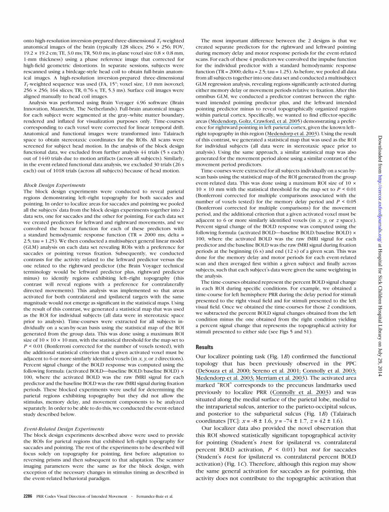
Figure 1. The block design localizer delayed-movement task. (A) Subjects were cued to either point or make a saccade before the onset of each scan. Subsequently, subjectsfixated a central cross and pointed to the same cross with their index finger. A peripheral dot was presented for 250 ms. After this target disappeared, a horizontal band ofdistractors dots blinked for 2.5 s. At distractor offset, subjects made a fast pointing or saccade movement to the remembered target location and immediately made a movementback to center. The time between successive movements was 5 s. Subjects made no movements during the fixation and delay periods. (B) Medial view of the left hemisphereshowing pointing activations within the precuneus. The activity shown is the result of the GLM contrast of rightward movementsþ, leftward movements� for 6 subjects. The colorbar shows the t-values associated with the activated voxels. The ‘‘P’’ value shown is Bonferroni corrected and is associated with a t-statistic. (C) Percent signal change in the BOLDresponse within the precuneus ROI during pointing (top) and during saccades (bottom) while making ipsilateral or contralateral movements. *P# 0.01 in a Student’s paired t-test.Error bars are standard error of the mean (SEM).
2284 PRR Codes Visual Direction of Intended Movement d Fernandez-Ruiz et al.
at Hospital for Sick C
hildren Hospital L
ibrary on July 29, 2014http://cercor.oxfordjournals.org/
Dow
nloaded from

of distractors (70� horizontal 3 8� vertical; angular eccentricity of the
individual dot elements, 0.8�; density, 0.14 dots per square degree)
flashed at 5 Hz for 11.75 s, during which the subjects maintained central
fixation (this memory period was designed to isolate activity related to
the stored visuospatial movement plan (Sereno et al. 2001; Medendorp
et al. 2003). At distractor offset, subjects made the instructed pointing
movement with the right index finger to the remembered target
location and immediately pointed back to center while maintaining
continuous fixation. This movement period was given 4 s in the analysis,
and was followed by an additional 6 s of control fixation to allow the
blood oxygen level--dependent (BOLD) response to return to baseline
levels. The next target was presented after another 4 s of central
fixation. The whole trial lasted 26 s. On average, we recorded 24
leftward, and 24 rightward trials per subject in this condition. Subjects
were instructed to maintain central gaze fixation at all times while
keeping sight of the tip of their right index finger. The starting pointing
direction was varied from scan-to-scan for each subject.
Prism Event-Related Behavioral Paradigm
The prism task was identical to the event-related paradigm described
above except that the subjects looked through a left--right reversing
dove prism 25 3 105.7 3 25 mm. The reversing prism was carefully
centered on the eye to prevent tonic positional shifts of the fixation
point (as occurs with displacing (wedge) prisms—not used here—
which produce positional shifts when fixating a centered point). Prism
training was performed with subjects positioned normally within the
MRI, after acquisition of fMRI data for control pointing was complete. No
scanning was conducted during the training period, given the need to
communicate with the subjects and the fact that the adaptation took
a small number of trials, and had large variability across subjects that it
was unlikely to produce statistically significant results on analysis of the
fMRI signals. It was necessary for subjects to see their finger moving
through the prism in order to produce the stimulus for visuomotor
adaptation (see Fig. 2B). Training was continued until subjects attained
10 consecutively correct trials. fMRI recordings were then resumedwith
the prism still in place so that the visuomotor reversal wasmaintained for
the remainder of the experiment. Performancewasmonitored online via
an infrared-sensitive charge coupled device video camera at the back of
the magnet bore which provided an unobstructed view of both the
subject’s right index finger and the visual stimulus displayed on the top
of the magnet bore. An additional behavioral experiment was done in 6
subjects in a task identical to that reported here but performed outside
of the MRI bore (Fig. 2C). Eye tracking was not performed inside the
magnet because the prism apparatus used completely occluded the eyes,
however, ocular responses outside the magnet using conventional eye
tracking apparatus confirm that the subjects were capable of maintain-
ing fixation and produce correct pointing responses while doing
our task (Medendorp et al. 2003). Moreover, in-magnet eye tracking
was used in a similar prior study of topography and antisaccades
(Medendorp, Goltz and Vilis 2005).
Imaging MethodsData were collected with a 4.0-Tesla Varian/Siemens hybrid whole-body
imaging system. To maximize the signal-to-noise ratio of our recordings
we used an occipito-parietal radio-frequency quadrature surface coil
(15.5 3 11.5-cm elements), which was centered over the posterior
parietal lobe. The size of the coil elements and the placement of the
subjects’ heads with respect to this coil limited the recordings to the
back half of the brain because it was designed to image only occipital
and parietal regions effectively, with a rapid fall-off of signal outside its
physical margins. In an additional experiment we tried repeating these
procedures with a ‘‘full head coil’’ to simultaneously image parietal and
frontal cortices but the head coil used did not yield sufficient signal to
demonstrate the basic left--right topography required for these experi-
ments. More trials or a newer style phased array coil would likely make
this possible.
Subjects viewed stimuli generated with Macromedia Flash MX Pro-
fessional 2004 software (San Francisco, CA) and presented using
a computer connected to an Avotec SV-6021 LCD projector (resolution
1024 3 768 pixels, refresh rate, 60 Hz). The stimulus images were
projected off a first-surface mirror onto the top of the magnet bore in
front of the subject’s eyes. Seventeen contiguous axial-oblique slices
were used to image the entire parietal cortex as well as portions of the
occipital lobe. Functional data were obtained using navigator echo-
corrected T2*-weighted segmented spiral imaging (time echo [TE],
15 ms; flip angle [FA], 40�; field of view [FOV], 19.2 3 19.2 cm; volume
acquisition time, 2 s [time repetition (TR) = 1 s 3 2 shots]; in-plane voxel
size, 3 3 3 mm; thickness, 4 mm). Functional data were superimposed
Figure 2. (A) Event-related delayed-movement task. Subjects fixated a central cross for 4 s. Then a peripheral dot target was presented for 250 ms toward the right or left atrandom horizontal eccentricities between 10� and 25�. After this target disappeared, a horizontal band of distractor dots flashed on and off at 5 Hz for 11.75 s. Subsequently,subjects made a rapid pointing movement with the right index finger to the remembered target location and then immediately pointed back to center. The total trial length was 26 s.(B) Viewpoint from the subject’s perspective. The prism-pointing task was identical to the previous task except that the subjects had to point while looking through a left--rightreversing prism. When looking through the prism the subject’s view of the index finger and the actual movements that they made appeared to be reversed. This reversed view of thepointing finger caused subjects to adapt their pointing movements to the opposite direction (shown here) after the first few incorrect trials. The diagrams within the boxes arerenderings of the subject’s view through the prism. The circles represent the remembered location where the subject had to point in order to perform the task correctly. (C)Behavioral data collected from 6 subjects in a task identical to that reported here but performed outside of the MRI bore. The figure shows the mean number of correct responseswhile pointing without (diamonds) and with prism (circles). The data are binned in blocks of 10 trials each. Errors bars are SEM.
Cerebral Cortex October 2007, V 17 N 10 2285
at Hospital for Sick C
hildren Hospital L
ibrary on July 29, 2014http://cercor.oxfordjournals.org/
Dow
nloaded from
onto high-resolution inversion-prepared three-dimensional T1-weighted
anatomical images of the brain (typically 128 slices; 256 3 256; FOV,
19.2 3 19.2 cm; TE, 3.0 ms; TR, 50.0 ms; in-plane voxel size 0.8 3 0.8 mm,
1-mm thickness) using a phase reference image that corrected for
high-field geometric distortions. In separate sessions, subjects were
rescanned using a birdcage-style head coil to obtain full-brain anatom-
ical images. A high-resolution inversion-prepared three-dimensional
T1-weighted sequence was used (FA, 15�; voxel size, 1.0 mm isovoxel;
256 3 256; 164 slices; TR, 0.76 s; TE, 5.3 ms). Surface coil images were
aligned manually to head coil images.
Analysis was performed using Brain Voyager 4.96 software (Brain
Innovation, Maastricht, The Netherlands). Full-brain anatomical images
for each subject were segmented at the gray--white matter boundary,
rendered and inflated for visualization purposes only. Time-courses
corresponding to each voxel were corrected for linear temporal drift.
Anatomical and functional images were transformed into Talairach
space to obtain stereotaxic coordinates for the ROIs. All scans were
screened for subject head motion. In the analysis of the block design
functional data, we excluded from further analysis 44 trials (5 s each)
out of 1440 trials due to motion artifacts (across all subjects). Similarly,
in the event-related functional data analysis, we excluded 30 trials (26 s
each) out of 1018 trials (across all subjects) because of head motion.
Block Design Experiments
The block design experiments were conducted to reveal parietal
regions demonstrating left--right topography for both saccades and
pointing. In order to localize areas for saccades and pointing we pooled
all the subjects’ data from the block design experiments together into 2
data sets, one for saccades and the other for pointing. For each data set
we created predictors for leftward and rightward movements, and we
convolved the boxcar function for each of these predictors with
a standard hemodynamic response function (TR = 2000 ms; delta =2.5; tau = 1.25). We then conducted a multisubject general linear model
(GLM) analysis on each data set revealing ROIs with a preference for
saccades or pointing versus fixation. Subsequently, we conducted
contrasts for the activity related to the leftward predictor versus the
one related to the rightward predictor (the Brain Voyager technical
terminology would be leftward predictor plus, rightward predictor
minus) to identify regions exhibiting left--right topography (this
contrast will reveal regions with a preference for contralaterally
directed movements). This analysis was implemented so that areas
activated for both contralateral and ipsilateral targets with the same
magnitude would not emerge as significant in the statistical maps. Using
the result of this contrast, we generated a statistical map that was used
as the ROI for individual subjects (all data were in stereotaxic space
prior to analysis). Time-courses were extracted for all subjects in-
dividually on a scan-by-scan basis using the statistical map of the ROI
generated from the group data. This was done using a maximum ROI
size of 10 3 10 3 10 mm, with the statistical threshold for the map set to
P < 0.01 (Bonferroni corrected for the number of voxels tested), with
the additional statistical criterion that a given activated voxel must be
adjacent to 6 or more similarly identified voxels (in x, y, or z directions).
Percent signal change of the BOLD response was computed using the
following formula: (activated BOLD—baseline BOLD/baseline BOLD) 3
100, where the activated BOLD was the raw fMRI signal for each
predictor and the baseline BOLDwas the raw fMRI signal during fixation
periods. These blocked experiments were useful for determining the
parietal regions exhibiting topography but they did not allow the
stimulus, memory delay, and movement components to be analyzed
separately. In order to be able to do this, we conducted the event-related
study described below.
Event-Related Design Experiments
The block design experiments described above were used to provide
the ROIs for parietal regions that exhibited left--right topography for
saccades and pointing. The rest of the experiments to be described will
focus solely on topography for pointing, first before adaptation to
reversing prisms and then subsequent to that adaptation. The scanner
imaging parameters were the same as for the block design, with
exception of the necessary changes in stimulus timing as described in
the event-related behavioral paradigm.
The most important difference between the 2 designs is that we
created separate predictors for the rightward and leftward pointing
during memory delay and motor response periods for the event-related
scans. For each of these 4 predictors we convolved the impulse function
for the individual predictor with a standard hemodynamic response
function (TR = 2000; delta = 2.5; tau = 1.25). As before, we pooled all data
from all subjects together into one data set and conducted amultisubject
GLM regression analysis, revealing regions significantly activated during
either memory delay or movement periods relative to fixation. After this
omnibus GLM, we conducted a predictor contrast between the right-
ward intended pointing predictor plus, and the leftward intended
pointing predictor minus to reveal topographically organized regions
within parietal cortex. Specifically, we wanted to find effector-specific
areas (Medendorp, Goltz, Crawford, et al. 2005) demonstrating a prefer-
ence for rightward pointing in left parietal cortex, given the known left--
right topography in this region (Medendorp et al. 2003). Using the result
of this contrast, we generated a statistical map that was used as the ROI
for individual subjects (all data were in stereotaxic space prior to
analysis). Using the same approach, a similar statistical map was also
generated for the movement period alone using a similar contrast of the
movement period predictors.
Time-courses were extracted for all subjects individually on a scan-by-
scan basis using the statistical map of the ROI generated from the group
event-related data. This was done using a maximum ROI size of 10 3
10 3 10 mm with the statistical threshold for the map set to P < 0.01
(Bonferroni corrected for multiple comparisons associated with the
number of voxels tested) for the memory delay period and P < 0.05
(Bonferroni corrected for multiple comparisons) for the movement
period, and the additional criterion that a given activated voxel must be
adjacent to 6 or more similarly identified voxels (in x, y, or z space).
Percent signal change of the BOLD response was computed using the
following formula: (activated BOLD—baseline BOLD/baseline BOLD) 3
100, where the activated BOLD was the raw fMRI signal for each
predictor and the baseline BOLDwas the raw fMRI signal during fixation
periods at the beginning (6 s) and end (12 s) of a given scan. This was
done for the memory delay and motor periods for each event-related
scan and then averaged first within a given subject and finally across
subjects, such that each subject’s data were given the same weighting in
the analysis.
The time-courses obtained represent the percent BOLD signal change
in each ROI during specific conditions. For example, we obtained a
time-course for left hemisphere PRR during the delay period for stimuli
presented to the right visual field and for stimuli presented to the left
visual field. Once we obtained the time-courses for those 2 conditions,
we subtracted the percent BOLD signal changes obtained from the left
condition minus the one obtained from the right condition yielding
a percent signal change that represents the topographical activity for
stimuli presented to either side (see Figs 5 and S1).
Results
Our localizer pointing task (Fig. 1B) confirmed the functional
topology that has been previously observed in the PPC
(DeSouza et al. 2000; Sereno et al. 2001; Connolly et al. 2003;
Medendorp et al. 2003; Merriam et al. 2003). The activated area
marked ‘‘ROI’’ corresponds to the precuneus landmarks used
previously to localize PRR (Connolly et al. 2003) and was
situated along the medial surface of the parietal lobe, medial to
the intraparietal sulcus, anterior to the parieto-occipital sulcus,
and posterior to the subparietal sulcus (Fig. 1B) (Talairach
coordinates [TC]: x = –8 ± 1.6, y = –74 ± 1.7, z = 42 ± 1.6).
Our localizer data also provided the novel observation that
this ROI showed statistically significant topographical activity
for pointing (Student’s t-test for ipsilateral vs. contralateral
percent BOLD activation, P < 0.01) but not for saccades
(Student’s t-test for ipsilateral vs. contralateral percent BOLD
activation) (Fig. 1C). Therefore, although this region may show
the same general activation for saccades as for pointing, this
activity does not contribute to the topographic activation that
2286 PRR Codes Visual Direction of Intended Movement d Fernandez-Ruiz et al.
at Hospital for Sick C
hildren Hospital L
ibrary on July 29, 2014http://cercor.oxfordjournals.org/
Dow
nloaded from

forms the basis for the experiments below. Henceforth, we will
refer to this region as human PRR.
For our main paradigm we used an event-related fMRI design
that allowed us to analyze the topographical response during the
memory andmovement periods of our task separately. The TC of
the ROI from our localizer data were used to guide selection of
the ROI in the event-related data but the precise extent of the
ROI was described in the event-related design experiments
section. Figure 3 plots the results of statistical contrast of right
intended pointing plus, left intended pointing minus data over
a posterior--dorsal--medial view of the left hemisphere visualized
on an ‘‘inflated brain.’’ This contrast is the approach that we have
used previously to demonstrate topography (Medendorp et al.
2003) and effector specificity (Medendorp, Goltz, Crawford,
et al. 2005). Figure 3(A) shows that during the 12-s memory
interval of the task, the only region that was significantly
spatially selective (Student’s t-test for ipsilateral vs. contralateral
percent BOLD activation; t (peak value) = 8, df = 5460, P[cor] <
0.01) was the ROI corresponding to PRR, that is, left PRR was
significantlymore activated for rightward than leftward pointing.
To contextualize the activation in PRR with the concurrent
activity in the surrounding visual and visuomotor areas, we
relaxed the statistical threshold for the same GLM contrast and
observed the broader activity during the memory delay period
(Fig. 3B). This revealed a network of areas with trends toward
spatial selectivity that activated while PRR was active. These
regions included one lateral to the intraparietal sulcus that,
based on previous studies, we have labeled LIP (TC: x = –26 ±1.1, y = –68 ± 2.9, z = 41 ± 1.3) (Sereno et al. 2001; Medendorp
et al. 2003; Schluppeck et al. 2005). Similarly, we labeled more
posterior ‘‘visual’’ regions as a putatively V7 homologue (TC
x = –27 ± 0.9, y = –79 ± 1.4, z = 29 ± 1), and V3 (TC) x = –28 ± 0.5,
y, –83 ± 1.8, z = 14.6 ± 0.9) (Shipp et al. 1995; Schluppeck
et al. 2005). We also observed a more anterior parietal area of
activation that corresponded anatomically to Brodmann’s area
7 (BA7) (TC x = –12 ± 0.8, y = 60 ± 1.1, z = 57 ± 1.6), which is
thought to function in the storage of motor sequences in spatial
working memory during finger movements (Sadato et al. 1996).
All of these regions were more activated for contralateral
pointing than ipsilateral pointing. These areas’ coordinates
agree with previous studies (Sereno et al. 2001; Connolly et al.
2003; Merriam et al. 2003).
We did a mirror image analysis for right parietal cortex and
qualitatively found the same results, that is, a similar network of
ROIs was activated but now with the contrast leftward pointing
movements plus, rightward pointing movements minus (see Fig.
S2 for single subject examples from 2 individuals). However,
right cortical activation never reached Bonferroni-corrected
significance because our subjects always used their right hand.
Previously we showed that the maximal cortical activation for
pointing in the delayed pointing task occurred in the cortical
hemisphere that is contralateral to both the goal and the
effector (here the right hand) (Medendorp, Goltz, Crawford,
et al. 2005). Consistent with this, the only area with significant
directionally selective activation during the memory interval
was PRR in the left hemisphere (Fig. 3A). Therefore, we will
focus our analysis on the statistically significant left hemisphere
parietal responses.
When a similar analysis was performed on the movement
phase of the task, a different pattern of activation emerged (Fig.
3C). PRR remained active but activity in the other parietal areas
decreased, consistent with previous findings (Connolly et al.
2003). During the movement phase, another region located
along the angular gyrus (AG) (TC, x = –39 ± 0.5, y = –65 ± 2.5, z =40 ± 0.6) became significantly activated. AG has previously been
associated with manual pointing, tool use, and other visuospatial
functions (Astafiev et al. 2003; Johnson-Frey et al. 2005). Other
more frontal cortical areas were probably also activated during
the movement phase but could not be imaged in our experi-
ment (see Methods for explanation).
Pointing with Reversing Prisms
After completing data acquisition in the normal delayed point-
ing task, we trained subjects to point with the prism placed in
front of their right eye. Complete adaptation (involving all
visuospatial behaviors) to reversing prisms takes considerable
time but in a previous behavioral experiment we found that
rapid adaptation can be achieved by employing a very simple set
of horizontal visual targets (Marotta et al. 2005). This type of
reversing prism adaptation does not appear to be achieved
Figure 3. Cortical ROIs. Rear-medial views of the left hemisphere are rendered as ‘‘inflated brains,’’ that is, with unfolded sulci (darker areas) and gyri (lighter areas). Locations ofthe cortical activation (group-averaged fMRI BOLD responses were determined using GLM regression analysis in 9 subjects) across 2 different task periods in the event-related task.Statistical significance of the cortical activation is proportional to the color scale shown for each hemisphere (t-scores). (A) The topographical activation obtained during the delayperiod before pointing, when the target had been projected to the contralateral side (GLM predictor contrast of rightward intended movementsþ, leftward intended movements�,P\0.01). (B) Same as (A) but at a reduced threshold (t-value of 2.2 instead of 5.2) to view a broader functional network. (C) Similar to (A) but data collected during the movementperiod (t-value of 3.2). N.S. 5 not significant, V3 5 Visual area 3, V7 5 visual area 7, IPS 5 Intraparietal sulcus.
Cerebral Cortex October 2007, V 17 N 10 2287
at Hospital for Sick C
hildren Hospital L
ibrary on July 29, 2014http://cercor.oxfordjournals.org/
Dow
nloaded from
through a general cognitive reversal strategy because the
learned pointing response does not generalize to other visuo-
spatial behaviors, such as perceived orientation (Marotta et al.
2005). The subjects took around 30 trials to completely adapt to
the reversing prisms (Fig. 2C). Once adapted, subjects reported
that they were unaware of the adaptation. Here, as confirmed in
our video analyses within the scanner and in our quantitative
behavioral controls (Fig. 2C), subjects also showed a very rapid
pointing adaptation (within approximately thirty pointing
movements, on average). Subsequent to this adaptation, we re-
sumed our fMRI recordings with the prism in place for com-
parison between the normal data and the optically reversed
data.
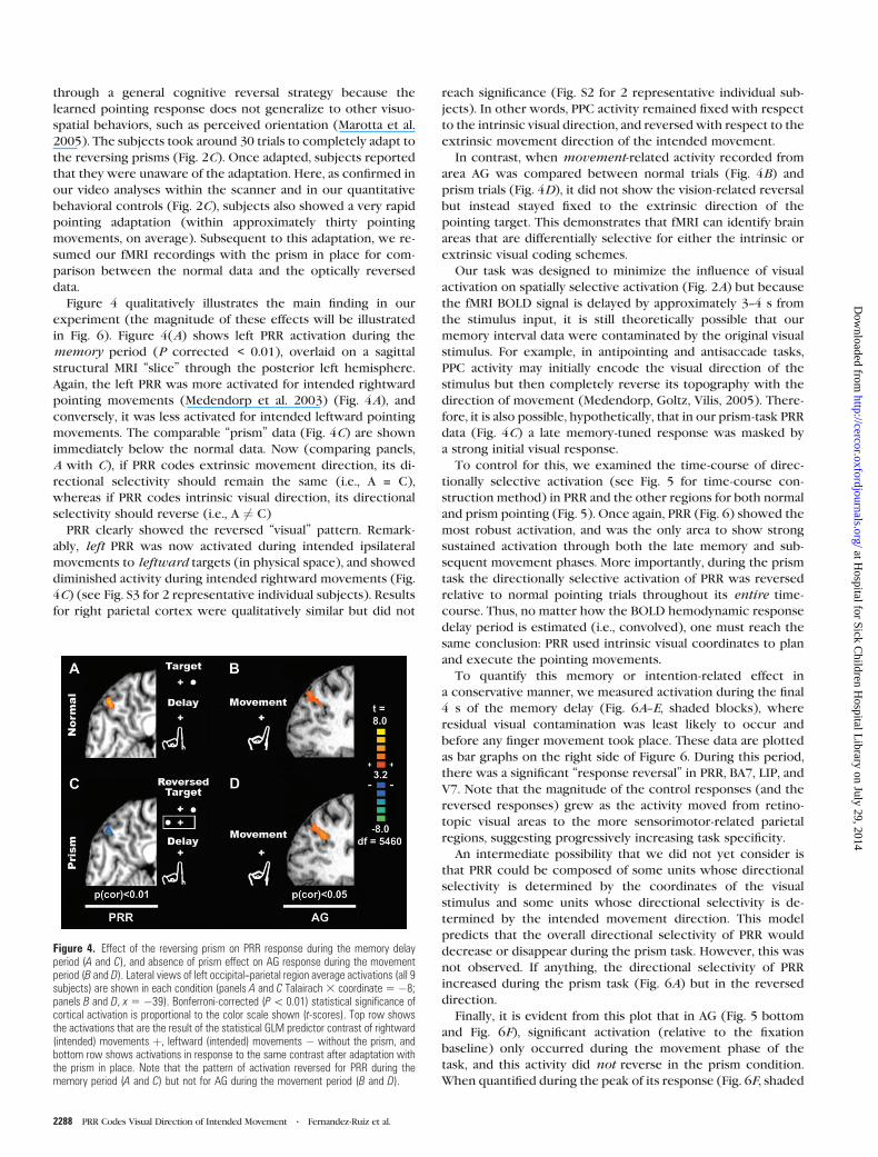
Figure 4 qualitatively illustrates the main finding in our
experiment (the magnitude of these effects will be illustrated
in Fig. 6). Figure 4(A) shows left PRR activation during the
memory period (P corrected < 0.01), overlaid on a sagittal
structural MRI ‘‘slice’’ through the posterior left hemisphere.
Again, the left PRR was more activated for intended rightward
pointing movements (Medendorp et al. 2003) (Fig. 4A), and
conversely, it was less activated for intended leftward pointing
movements. The comparable ‘‘prism’’ data (Fig. 4C) are shown
immediately below the normal data. Now (comparing panels,
A with C), if PRR codes extrinsic movement direction, its di-
rectional selectivity should remain the same (i.e., A = C),
whereas if PRR codes intrinsic visual direction, its directional
selectivity should reverse (i.e., A 6¼ C)
PRR clearly showed the reversed ‘‘visual’’ pattern. Remark-
ably, left PRR was now activated during intended ipsilateral
movements to leftward targets (in physical space), and showed
diminished activity during intended rightward movements (Fig.
4C) (see Fig. S3 for 2 representative individual subjects). Results
for right parietal cortex were qualitatively similar but did not
reach significance (Fig. S2 for 2 representative individual sub-
jects). In other words, PPC activity remained fixed with respect
to the intrinsic visual direction, and reversed with respect to the
extrinsic movement direction of the intended movement.
In contrast, when movement-related activity recorded from
area AG was compared between normal trials (Fig. 4B) and
prism trials (Fig. 4D), it did not show the vision-related reversal
but instead stayed fixed to the extrinsic direction of the
pointing target. This demonstrates that fMRI can identify brain
areas that are differentially selective for either the intrinsic or
extrinsic visual coding schemes.
Our task was designed to minimize the influence of visual
activation on spatially selective activation (Fig. 2A) but because
the fMRI BOLD signal is delayed by approximately 3--4 s from
the stimulus input, it is still theoretically possible that our
memory interval data were contaminated by the original visual
stimulus. For example, in antipointing and antisaccade tasks,
PPC activity may initially encode the visual direction of the
stimulus but then completely reverse its topography with the
direction of movement (Medendorp, Goltz, Vilis, 2005). There-
fore, it is also possible, hypothetically, that in our prism-task PRR
data (Fig. 4C) a late memory-tuned response was masked by
a strong initial visual response.
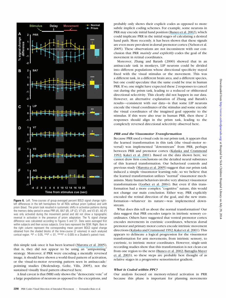
To control for this, we examined the time-course of direc-
tionally selective activation (see Fig. 5 for time-course con-
struction method) in PRR and the other regions for both normal
and prism pointing (Fig. 5). Once again, PRR (Fig. 6) showed the
most robust activation, and was the only area to show strong
sustained activation through both the late memory and sub-
sequent movement phases. More importantly, during the prism
task the directionally selective activation of PRR was reversed
relative to normal pointing trials throughout its entire time-
course. Thus, no matter how the BOLD hemodynamic response
delay period is estimated (i.e., convolved), one must reach the
same conclusion: PRR used intrinsic visual coordinates to plan
and execute the pointing movements.
To quantify this memory or intention-related effect in
a conservative manner, we measured activation during the final
4 s of the memory delay (Fig. 6A--E, shaded blocks), where
residual visual contamination was least likely to occur and
before any finger movement took place. These data are plotted
as bar graphs on the right side of Figure 6. During this period,
there was a significant ‘‘response reversal’’ in PRR, BA7, LIP, and
V7. Note that the magnitude of the control responses (and the
reversed responses) grew as the activity moved from retino-
topic visual areas to the more sensorimotor-related parietal
regions, suggesting progressively increasing task specificity.
An intermediate possibility that we did not yet consider is
that PRR could be composed of some units whose directional
selectivity is determined by the coordinates of the visual
stimulus and some units whose directional selectivity is de-
termined by the intended movement direction. This model
predicts that the overall directional selectivity of PRR would
decrease or disappear during the prism task. However, this was
not observed. If anything, the directional selectivity of PRR
increased during the prism task (Fig. 6A) but in the reversed
direction.
Finally, it is evident from this plot that in AG (Fig. 5 bottom
and Fig. 6F), significant activation (relative to the fixation
baseline) only occurred during the movement phase of the
task, and this activity did not reverse in the prism condition.
When quantified during the peak of its response (Fig. 6F, shaded
Figure 4. Effect of the reversing prism on PRR response during the memory delayperiod (A and C), and absence of prism effect on AG response during the movementperiod (B and D). Lateral views of left occipital--parietal region average activations (all 9subjects) are shown in each condition (panels A and C Talairach3 coordinate5 �8;panels B and D, x 5 �39). Bonferroni-corrected (P\ 0.01) statistical significance ofcortical activation is proportional to the color scale shown (t-scores). Top row showsthe activations that are the result of the statistical GLM predictor contrast of rightward(intended) movements þ, leftward (intended) movements � without the prism, andbottom row shows activations in response to the same contrast after adaptation withthe prism in place. Note that the pattern of activation reversed for PRR during thememory period (A and C) but not for AG during the movement period (B and D).
2288 PRR Codes Visual Direction of Intended Movement d Fernandez-Ruiz et al.
at Hospital for Sick C
hildren Hospital L
ibrary on July 29, 2014http://cercor.oxfordjournals.org/
Dow
nloaded from

block), AG showed preference for the same pointing direction
(or world-fixed target direction) both with and without prism.
Discussion
Our event-related fMRI study is the first to show that human
PRR—and indeed the entire cluster of human PPC regions
associated with manual pointing—shows directionally selective
activity that is fixed with respect to the intrinsic retinal
coordinates of the remembered goal (Gottlieb and Goldberg
1999), not its physical location in space or the extrinsic
direction of the movement. In contrast, another area, AG, ap-
peared to encode the physical location of the goal or the
extrinsic direction of the movement. For technical reasons (see
experimental procedures) we were not able to simultaneously
record spatially selective activation in frontal cortex but it is
likely that areas like ventral premotor cortex or dorsolateral
prefrontal cortex might show a similar extrinsic pattern (Kakei
et al. 2001; Hagler and Sereno 2006). The final stage would be
the transformation from extrinsic movement plans to intrinsic
muscle commands (Kalaska and Crammond 1992; Kakei et al.
2001).
Data Interpretation
We think our main ROI is task specific because it only showed
topography during pointing movements, not saccades. More-
over, our anatomic characterization of this region agrees with
previous descriptions of human PRR (Connolly et al. 2003). We
also observed a progression of the delay-period activation from
early occipital areas to our putative LIP, which ultimately peaks
in ‘‘PRR’’ (Fig. 6). This is consistent with previous claims that
PRR is task specific for manual reaching and pointing (Calton
et al. 2002; Connolly et al. 2003). However, because we did not
directly contrast this activation to activations for attention
paradigms, we cannot directly exclude the possibility that
we were measuring a spatially selective attention response
(Merriam et al. 2003; Silver et al. 2005). If so, this would not
change our major conclusion but would simply modify it to
state that attentional responses in human PRR encode the ret-
inal coordinates of the goal as opposed to the motor coordi-
nates of the movement. However, because we and others have
found effector-specific activations for similar PPC regions in
this and similar paradigms (Medendorp, Goltz, Crawford, et al.
2005), we do not believe that attention alone can explain our
results.
Our PRR results cannot likely be explained away as simple
residual visual activation from the stimulus. First, because
remapping and antisaccade/pointing paradigms have already
shown that reach-related activity in parietal cortex is not locked
to the initial visual stimulus (Batista et al. 1999; Medendorp et al.
2003; Khan, Pisella, Vighetto, et al. 2005). Second, in our
experiment, PRR showed a robust reversal of activation with
the prism that grew during the first half of the 12-second
memory delay and was sustained through the second half of this
interval right through the actual movement. Conversely, the
world-fixed activation observed in AG during the movement
phase could not be a trivial by-product of viewing the
movement because this would have led to the visual reversal
pattern. Finally, PRR and AG showed opposite response patterns
in the presence of the prism adaptation. This negates the
possibility that the PRR result is the trivial product of sensory
input and hemodynamics.
A related question is whether subjects were pointing toward
a mentally reversed image, as we have argued is the case in
antipointing tasks. Our previous experiments suggest that
subjects are not consciously aware of the transformation in
Figure 5. Top. Event-related percent BOLD signal change difference across time (s) in the PRR ROI in the left hemisphere. (A) Time-courses with error bars (SEM) for stimulipresented in the contralateral (right) visual field (blue diamonds), and ipsilateral (left) visual field (red squares). (B) Same as (A) but while viewing through the reversing prism afteradaptation. (C) Time-course of the resulting percent BOLD signal change difference in PRR after subtracting contralateral activity from ipsilateral activity in normal (yellow diamonds)and prism (light blue squares) conditions. The colored areas in (A) and (B) represent the magnitude shown in (C). Bottom. Same as above but in AG. This method was used toproduce all % signal change difference time-courses shown in Figure 6.
Cerebral Cortex October 2007, V 17 N 10 2289
at Hospital for Sick C
hildren Hospital L
ibrary on July 29, 2014http://cercor.oxfordjournals.org/
Dow
nloaded from
this simple task once it has been learned (Marotta et al. 2005),
that is, they did not appear to be using an ‘‘antipointing’’
strategy. Moreover, if PRR were encoding a mentally reversed
image, it should have shown a world-fixed pattern of activation,
or the visual-to-motor reversing pattern seen in antisaccade/
pointing studies (Medendorp, Goltz, Vilis, 2005), not the
sustained visually fixed pattern observed here.
A final caveat is that fMRI only shows the ‘‘democratic vote’’ of
a large population of neurons as opposed to the exceptions, and
probably only shows their explicit codes as opposed to more
subtle implicit coding schemes. For example, some neurons in
PRRmay encode initial hand position (Buneo et al. 2002), which
could implicate PRR in the initial stages of calculating a desired
hand path. More recently, it has been shown that these signals
are even more prevalent in dorsal premotor cortex (Nelson et al.
2005). These observations are not inconsistent with our con-
clusion that PRR mainly and explicitly codes the goal of the
movement in retinal coordinates.
Moreover, Zhang and Barash (2000) showed that in an
antisaccade task in monkeys, LIP neurons could be divided
into different populations whose directional specificity stayed
fixed with the visual stimulus or the movement. This was
a different task, in a different brain area, and a different species,
but one could speculate that the same could be true in human
PRR. If so, one might have expected these 2 responses to cancel
out during the prism task, leading to a reduced or obliterated
directional selectivity. This clearly did not happen in our data.
However, an alternative explanation of Zhang and Barash’s
results—consistent with our data—is that some LIP neurons
encode the visual coordinates of the stimulus and some encode
the visual coordinates of the imagined goal opposite to the
stimulus. If this were also true in human PRR, then these 2
responses should align in the prism task, leading to the
completely reversed directional selectivity observed here.
PRR and the Visuomotor Transformation
Because PRR used a visual code in our prism task, it appears that
the learned transformation in this task (the visual--motor re-
versal) was implemented ‘‘downstream’’ from PRR, perhaps
between PRR and premotor cortex (Kalaska and Crammond
1992; Kakei et al. 2001). Based on the data shown here, we
cannot draw firm conclusions on the detailed neural substrates
of this learned transformation. Our behavioral controls and
previous study (Marotta et al. 2005) suggest that our prism task
induced a simple visuomotor learning rule, so we believe that
the learned transformation utilizes ‘‘normal’’ visuomotor mech-
anisms. Many human behaviors involve very abstract visuomotor
transformations (Gorbet et al. 2004). But even if this trans-
formation had a more complex ‘‘cognitive’’ nature, this would
not change our main conclusion. Either way, PRR explicitly
encoded the retinal direction of the goal, and the new trans-
formation—whatever its nature—was implemented down-
stream.
What does this tell us about the normal transformation? Our
data suggest that PRR encodes targets in intrinsic sensory co-
ordinates. Others have suggested that ventral premotor cortex
encodes movements in extrinsic coordinates, whereas dorsal
premotor and primary motor cortex encode intrinsic movement
directions (Kalaska and Crammond 1992; Kakei et al. 2001). This
appears to delineate a logical progression for the visuomotor
transformation for arm movements, from intrinsic sensory, to
extrinsic, to intrinsic motor coordinates. However, single-unit
recording studies show that this transformation is not clean-cut
from one region to the next (Buneo et al. 2002; Battaglia-Mayer
et al. 2003), so these steps are probably best thought of as
relative stages in a progressive sensorimotor gradient.
What is Coded within PPC?
Our analysis focused on memory-related activation in PRR
because this phase is important for planning movements
Figure 6. Left. Time-courses of group-averaged percent BOLD signal change right--left differences in the left hemisphere for all ROIs without prism (yellow) and withprism (blue). The prism task resulted in systematic shifts in activation patterns duringthe memory delay period in areas PRR (A), BA7 (B), LIP (C), V7 (D), and V3 (E). AG (F)was only activated during the movement period and did not show a topographicreversal in activation in the presence of prism adaptation. The % signal changedifference was calculated according to Figures 5 and S1. Data were averaged firstwithin subjects and then across subjects. Error bars represent the SEM. Right. Bars inthe right column represent the corresponding mean percent BOLD signal changeobtained from the shaded blocks of the time-course (2 volumes) in each analyzedcortical region. *P\ 0.05, **P\ 01, ***P # 0.005 in a Student’s paired t-test.
2290 PRR Codes Visual Direction of Intended Movement d Fernandez-Ruiz et al.
at Hospital for Sick C
hildren Hospital L
ibrary on July 29, 2014http://cercor.oxfordjournals.org/
Dow
nloaded from

(Andersen and Buneo 2002) and was the least likely period to be
contaminated by visual signals in our paradigm. PRR may also be
involved in the online monitoring of feedback signals during
movements (Pisella et al. 2000; Prablanc et al. 2003). If so, our
data would suggest that this is also done in retinal coordinates
but this supposition cannot be directly tested using our method
because of the rapidity of individual movements relative to the
temporally delayed fMRI signals and the possibility of visual
contamination during the movement phase.
As confirmed here, PPC also plays an important role in storing
target representations for intended movements when the
original visual stimulus is no longer present (Snyder et al.
1997; Andersen and Buneo 2002). Moreover, we know from
previous studies that this memory-related activity is not hard-
wired to retinal input because its activity is updated during eye
movements (Batista et al. 1999; Medendorp et al. 2003; Khan,
Pisella, Rossetti, et al. 2005) and can reverse (with the excep-
tion of some neurons) during antipointing/saccade paradigms
(Kalaska 1996; Eskandar and Assad 1999; Connolly et al. 2000;
Zhang and Barash 2000; Medendorp, Goltz, Vilis, 2005). The
novel finding in the current study is that the directionally
selective PPC activity measured with fMRI can also be com-
pletely dissociated from movement direction.
Taken together with our new results, these findings suggest
that specific PPC regions like PRR primarily encode neither
vision nor movement per se but rather something more
intermediate and abstract: the spatial goal of the movement in
retinal coordinates. This places PPC at a point intermediate
between visual and motor codes. This provides another piece of
information in the understanding of the highly complex work-
ings of the visuomotor transformation and contributes valuable
constraint information in the quest to model parietal function
(Crawford et al. 2004), interpreting data from patients with
parietal damage (Khan, Pisella, Rossetti, et al. 2005), and using
PRR signals as neural inputs to drive prosthetic devices in
complex behavioral paradigms (Musallam et al. 2004).
Supplementary Data
Supplementary material can be found at: http://www.cercor.
oxfordjournals.org/.
Notes
Conflict of Interest: None declared.
Address correspondence to Dr J. Douglas Crawford, York Centre for
Vision Research, York University, 4700 Keele Street, Toronto, Ontario,
Canada M3J 1P3. Email: [email protected].
References
Andersen RA, Buneo CA. 2002. Intentional maps in posterior parietal
cortex. Annu Rev Neurosci. 25:189--220.
Astafiev SV, Shulman GL, Stanley CM, Snyder AZ, Van Essen DC, Corbetta
M. 2003. Functional organization of human intraparietal and
frontal cortex for attending, looking, and pointing. J Neurosci. 23:
4689--4699.
Batista AP, Buneo CA, Snyder LH, Andersen RA. 1999. Reach plans in eye-
centered coordinates. Science. 285:257--260.
Battaglia-Mayer A, Caminiti R, Lacquaniti F, Zago M. 2003. Multiple levels
of representation of reaching in the parieto-frontal network. Cereb
Cortex. 13:1009--1022.
Buneo CA, Jarvis MR, Batista AP, Andersen RA. 2002. Direct visuomotor
transformations for reaching. Nature. 416:632--636.
Calton JL, Dickinson AR, Snyder LH. 2002. Non-spatial, motor-specific
activation in posterior parietal cortex. Nat Neurosci. 5:580--588.
Colby CL, Goldberg ME. 1999. Space and attention in parietal cortex.
Annu Rev Neurosci. 22:319--349.
Connolly JD, Andersen RA, Goodale MA. 2003. fMRI evidence for a ‘pari-
etal reach region’ in the human brain. Exp Brain Res. 153:140--145.
Connolly JD, Goodale MA, Desouza JF, Menon RS, Vilis T. 2000. A
comparison of frontoparietal fMRI activation during anti-saccades
and anti-pointing. J Neurophysiol. 84:1645--1655.
Crawford JD, Medendorp WP, Marotta JJ. 2004. Spatial transformations
for eye-hand coordination. J Neurophysiol. 92:10--19.
DeSouza JF, Dukelow SP, Gati JS, Menon RS, Andersen RA, Vilis T. 2000.
Eye position signal modulates a human parietal pointing region
during memory-guided movements. J Neurosci. 20:5835--5840.
Dickinson AR, Calton JL, Snyder LH. 2003. Nonspatial saccade-specific
activation in area LIP of monkey parietal cortex. J Neurophysiol.
90:2460--2464.
Duhamel JR, Colby CL, Goldberg ME. 1992. The updating of the rep-
resentation of visual space in parietal cortex by intended eye
movements. Science. 255:90--92.
Eskandar EN, Assad JA. 1999. Dissociation of visual, motor and predictive
signals in parietal cortex during visual guidance. Nat Neurosci.
2:88--93.
Gorbet DJ, Staines WR, Sergio LE. 2004. Brain mechanisms for preparing
increasingly complex sensory to motor transformations. Neuro-
image. 23:1100--1111.
Gottlieb J, Goldberg ME. 1999. Activity of neurons in the lateral intra-
parietal area of the monkey during an antisaccade task. Nat Neurosci.
2:906--912.
Hagler DJ, Jr, Sereno MI. 2006. Spatial maps in frontal and prefrontal
cortex. Neuroimage 29:567--77.
Johnson-Frey SH, Newman-Norlund R, Grafton ST. 2005. A distributed
left hemisphere network active during planning of everyday tool use
skills. Cereb Cortex 15:681--95.
Kakei S, Hoffman DS, Strick PL. 2001. Direction of action is represented
in the ventral premotor cortex. Nat Neurosci. 4:1020--1025.
Kalaska JF. 1996. Parietal cortex area 5 and visuomotor behavior. Can J
Physiol Pharmacol. 74:483--498.
Kalaska JF, Crammond DJ. 1992. Cerebral cortical mechanisms of
reaching movements. Science. 255:1517--1523.
Khan AZ, Pisella L, Rossetti Y, Vighetto A, Crawford JD. 2005.
Impairment of gaze-centered updating of reach targets in bilateral
parietal-occipital damaged patients. Cereb Cortex. 15:1547--1560.
Khan AZ, Pisella L, Vighetto A, Cotton F, Luaute J, Boisson D, Salemme R,
Crawford JD, Rossetti Y. 2005. Optic ataxia errors depend on
remapped, not viewed, target location. Nat Neurosci. 8:418--420.
Kohler I. 1962. Experiments with goggles. Sci Am. 206:62--72.
Marotta JJ, Keith GP, Crawford JD. 2005. Task-specific sensorimotor
adaptation to reversing prisms. J Neurophysiol. 93:1104--1110.
Medendorp WP, Goltz HC, Crawford JD, Vilis T. 2005. Integration of
target and effector information in human posterior parietal cortex
for the planning of action. J Neurophysiol. 93:954--962.
Medendorp WP, Goltz HC, Vilis T. 2005. Remapping the remembered
target location for anti-saccades in human posterior parietal cortex.
J Neurophysiol. 94:734--740.
Medendorp WP, Goltz HC, Vilis T, Crawford JD. 2003. Gaze-centered
updating of visual space in human parietal cortex. J Neurosci.
23:6209--6214.
Merriam EP, Genovese CR, Colby CL. 2003. Spatial updating in human
parietal cortex. Neuron. 39:361--373.
Musallam S, Corneil BD, Greger B, Scherberger H, Andersen RA. 2004.
Cognitivecontrol signals for neural prosthetics. Science. 305:258--262.
Nelson MJ, Pesaran B, Andersen RA. 2005. Dorsal premotor neurons
encode the relative position of the hand and the eye. Society for
Neuroscience Abstract Program No. 363.14.
Pisella L, Grea H, Tilikete C, Vighetto A, Desmurget M, Rode G, Boisson
D, Rossetti Y. 2000. An ‘automatic pilot’ for the hand in human
posterior parietal cortex: toward reinterpreting optic ataxia. Nat
Neurosci. 3:729--736.
Pouget A, Snyder LH. 2000. Computational approaches to sensorimotor
transformations. Nat Neurosci. 3(Suppl):1192--1198.
Prablanc C, Desmurget M, Grea H. 2003. Neural control of on-line
guidance of hand reaching movements. Prog Brain Res. 142:155--170.
Cerebral Cortex October 2007, V 17 N 10 2291
at Hospital for Sick C
hildren Hospital L
ibrary on July 29, 2014http://cercor.oxfordjournals.org/
Dow
nloaded from

Sadato N, Campbell G, Ibanez V, Deiber M, Hallett M. 1996. Complexity
affects regional cerebral blood flow change during sequential finger
movements. J Neurosci. 16:2691--2700.
Schluppeck D, Glimcher P, Heeger DJ. 2005. Topographic organization
for delayed saccades in human posterior parietal cortex. J Neuro-
physiol. 94:1372--1384.
Sereno MI, Pitzalis S, Martinez A. 2001. Mapping of contralateral space in
retinotopic coordinates by a parietal cortical area in humans.
Science. 294:1350--1354.
Shipp S, Watson JD, Frackowiak RS, Zeki S. 1995. Retinotopic maps in
human prestriate visual cortex: the demarcation of areas V2 and V3.
Neuroimage. 2:125--132.
Silver MA, Ress D, Heeger DJ. 2005. Topographic maps of visual spatial
attention in human parietal cortex. J Neurophysiol. 94:1358--1371.
Snyder LH. 2000. Coordinate transformations for eye and arm move-
ments in the brain. Curr Opin Neurobiol. 10:747--754.
Snyder LH, Batista AP, Andersen RA. 1997. Coding of intention in the
posterior parietal cortex. Nature. 386:167--170.
Soechting JF, Flanders M. 1992. Moving in three-dimensional space:
frames of reference, vectors, and coordinate systems. Annu Rev
Neurosci. 15:167--191.
Stricanne B, Andersen RA, Mazzoni P. 1996. Eye-centered, head-
centered, and intermediate coding of remembered sound locations
in area LIP. J Neurophysiol. 76:2071--2076.
Sugita Y. 1996. Global plasticity in adult visual cortex following reversal
of visual input. Nature. 380:523--526.
Zhang M, Barash S. 2000. Neuronal switching of sensorimotor trans-
formations for antisaccades. Nature. 408:971--975.
2292 PRR Codes Visual Direction of Intended Movement d Fernandez-Ruiz et al.
at Hospital for Sick C
hildren Hospital L
ibrary on July 29, 2014http://cercor.oxfordjournals.org/
Dow
nloaded from