Intranasal IFN-γ gene transfer protects BALB/c mice against respiratory syncytial virus infection
Upload
independentCategory
view
1download
0
189Vet. Res. 35 (2004) 189–197© INRA, EDP Sciences, 2004DOI: 10.1051/vetres:2004003
Original article
Detection of bovine respiratory syncytial virus in experimentally infected balb/c mice
Renata Servan ALMEIDA, Helena Gallicchio DOMINGUES, Lia Treptow COSWIG, Regina Celia Freitas D’ARCE, Rodrigo Franco de CARVALHO, Clarice Weis ARNS*
Laboratório de Virologia Animal, Departamento de Microbiologia e Imunologia, Instituto de Biologia, Universidade Estadual de Campinas – UNICAMP, 13081-970 Campinas, SP, Brazil
(Received 4 March 2003; accepted 18 September 2003)
Abstract – The present study used an RT-nested-PCR and an immunohistochemistry assay to detectbovine respiratory syncytial virus in tissues from experimentally infected balb/c mice. As a firststep, Chicken Embryo Related (CER) cell monolayers infected with the BRSV-25-BR strainisolated in Brazil were used for antigen production. Then, the infected lung and tracheal tissues offemale balb/c mice were collected on 3, 5, 7 and 10 days post-infection and submitted to bothtechniques. Primers specific to F and G genes that amplify fragments of 481 bp and 371 bp,respectively, were used. The BRSV detection was not successful in all of the animals tested. Thegenomic fragment of the G gene from the organs of some infected mice on all analyzed post-infection days was amplified. However, in the RT-nested-PCR corresponding to the F gene, it wasnot possible to observe any amplified fragment. This was probably due to the higher sensitivity ofthe developed technique to amplify the fragment corresponding to the G gene compared to the Fgene. Moreover, only three of the lungs collected five days post-infection were positive byimmunohistochemistry. To the author’s knowledge, this is the first study reporting bovinerespiratory syncytial virus detection in balb/c mice after experimental inoculation.
bovine respiratory syncytial virus / RT-nested-PCR / immunohistochemistry / mice /experimental infection
1. INTRODUCTION
The bovine respiratory syncytial virus(BRSV) is an agent that belongs to theParamyxoviridae family, the Pneumovirusgenus, being one of the pathogens of higherimportance in respiratory diseases [23], andresponsible for significant economic lossesin commercial cattle production [7]. Spe-cific antibodies for the BRSV are morefrequently detected in animals up to six
months of age [18]. In individuals of twoweeks to three months of age, the clinicalmanifestation of the illness is inverselyrelated to the level of maternal specificserum antibodies conferred to these animals[16]. These maternal antibodies also inter-fere with the serological diagnosis and withthe response to vaccination.
Laboratory diagnosis of BRSV is gener-ally based on criteria such as virus isolation,BRSV antigen detection in the suspected
* Corresponding author: [email protected]
190 R.S. Almeida et al.
tissues, serologic methods or histopatho-logical examination. There are however,frequent difficulties related to this due to itslimited growth in cell cultures or in themajority of experimental animals, and tothe lability of the virus. Hence, the isolationof the virus in cell cultures is a procedureless indicated for the diagnosis of BRSVinfections, even in clinical samples contain-ing high virus concentration.
Currently, the diagnosis of BRSV infec-tions is more commonly performed throughthe detection of specific antibodies bysero-diagnostic methods such as the serumneutralization test, complement fixationtest, indirect immunofluorescence test andELISA [29, 30]. Although the serum neu-tralization test is the standard gold test forthe detection of specific antibodies againstBRSV, difficulties arise regarding the timeneeded to obtain the results, which takes anaverage of 5 to 7 days [12]. Furthermore,there are reports of asymptomatic infec-tions by the Human Respiratory SyncytialVirus (HRSV), which are serologically unde-tectable [10]. In analogy to the HRSV, sinceBRSV and HRSV are antigenetically relatedof the up to 80% in proteins N, M, M2 andF [31], is possible to speculate that part ofthe infections by BRSV cannot be diagnosedthrough the detection of specific serumantibodies. This probably occurs due to thesimple fact that these animals do notdevelop antibodies in levels high enough tobe detected by conventional techniques. Inaddition, there is evidence indicating theexistence of latent infections, since thevirus has been isolated from asymptomaticanimals, as well as from animals infectedseven months before the appearance of thesymptoms [1].
There is an urgent need, therefore, for aquick, sensitive and specific method for thediagnosis of infections by the BRSV virus.Several authors have been working on thedevelopment of techniques to simplify andto increase the sensitivity of BRSV infec-tion diagnosis. The reverse transcription-PCR assay has shown high speed, sensitiv-
ity and specificity in great parts of theseworks [20–22, 27, 28], providing a valuabletool for diagnostic and epidemiologicalpurposes.
Experimental animals represent a usefulalternative for experiments with virusesand other microorganisms, mainly when itis not possible to work with their naturalhost. Collins et al. [10] have pointed outthat a wide range of animals, includingmarmosets, nonhuman primates, ferrets,mink, chinchillas, guinea pigs, cotton ratsand mice, could be experimentally infectedwhen RSV is inoculated directly into therespiratory tract. Small experimental ani-mals have been used as models of lowerrespiratory tract infection with RSV [5, 8,9, 15, 17, 19, 24, 25]. The present studydetected BRSV in experimentally infectedbalb/c mice by RT-nested-PCR and immu-nohistochemistry.
2. MATERIALS AND METHODS
2.1. Virus
The Brazilian BRSV-25-BR strain, iso-lated in the Laboratorio de VirologiaAnimal do Institute de Biologia da Univer-sidade Estadual de Campinas (UNICAMP)[2], was used in the present study. Thisstrain was isolated from samples ofnasotracheal secretions collected from liveanimals from the Southern and Southeast-ern Brazilian States, inoculated succes-sively in MDBK (Madin Darby BovineKidney) and CER (Chicken EmbryoRelated) cells.
2.2. Cells
The cell cultures used were from thecontinuous cell line of CER. The cells werecultivated in bottles of 75 cm2, with 1.5 ×10⁵ cells/mL (initial concentration) in min-imal essential Eagle medium (MEME)supplemented with 10% BRSV-free fetalcalf serum (FCS) and kept at 37 °C.
Detection of BRSV in mice 191
2.3. Antigen production
For the production of the viral antigenused in the mice infection and submitted tothe extraction of the viral RNA for RT-nested-PCR, cellular monolayers, wereinoculated with 104.3 DICC50/mL of theBRSV-25-BR strain. The bottles were incu-bated at 37 °C and the cells were observeddaily under an inverted light microscope(Axiovert 100, Carl Zeiss, Oberkochen,Germany) in order to detect a cytopathiceffect (CPE). Then, the infected cells weresubmitted to RNA extraction or scraped andthe supernatant was clarified through cen-trifugation, aliquoted and stored at –70 °Cuntil use in the mice infection.
2.4. Animal infection
During the experiment, all mice werekept in the laboratory animal house of theDepartamento de Microbiologia e Immun-ologia do Instituto de Biologia da UNI-CAMP. The infected animals were installedseparately from negative control animals.
Four-week-old specific-pathogen-free(SPF) female balb/c mice, purchased fromthe Centro Multidisciplinar de InvestigaçãoBiológica of UNICAMP (CEMIB-UNI-CAMP), kept behind barriers, were used inthis study. The animals were distributedinto two groups, one containing 16 animalsand the other, a negative control, containing8 animals. All animals of the first groupwere intranasally inoculated with 100 µL ofthe viral antigen with an infectious dose of104.3 DICC50/mL, using a micropipette.The negative control animals received anamount of supernatant CER cells free ofvirus. The animals were observed daily forclinical signs. The samples of lung and tra-cheal tissues of the infected (4 animals) andcontrol group (2 animals), collected 3, 5,7 and 10 days post-infection (dpi), weresubmitted to immunohistochemistry andRT-nested-PCR assays. These inoculationswere performed in three repetitions, in dif-ferent periods of time, resulting in a total of48 virus-infected and 24 mock-infectedanimals.
2.5. Histopathology
Tissue samples were fixed in 4% para-formaldehyde (Sigma-Aldrish Co., Mis-souri, USA), embedded in paraffin wax, cutin 4 µm thick sections, and mounted onmicroscope slides. For a routine histologi-cal examination, the sections were stainedwith haematoxylin and eosin (HE). Thesame protocol was used for section prepa-ration for the immunohistochemistry assay.
2.6. Immunohistochemistry (IHC)
Immunohistochemistry was carried outat the Royal Veterinary and AgriculturalUniversity, Copenhagen, Denmark. Briefly,the sections were de-paraffinated by heat-ing up to 70 °C for 10–15 min and baths ofxylene and ethanol. After a pre-treatmentwith 0.018% protease for 5 min at roomtemperature, the sections were blockedwith 5% normal pig serum and incubatedovernight at 4 °C with a polyclonal bovine-serum against BRSV in a 1:600 dilution(primary antibody). After this, the sectionswere incubated with 0.9% streptavidin(DAKO, Glostrup, Denmark) and follow-ing with 0.9% biotinylated alkaline phos-phatase for 30 min at 37 °C (DAKO). Fastred (KemEnTec, Copenhagen, Denmark)was used for 10–15 min to visualize thepositive reactions. The sections were coun-ter-stained with Harris haematoxylin andmounted in Glycergel (DAKO) for lightmicroscopy.
2.7. RNA extraction
The RNA extraction from approxi-mately 100 mg of tissue samples wascarried out using phenol guanidine isothio-cyanate (TRIzol, InvitrogenTM, Carlsbad,California, USA) and chloroform. The pre-cipitation of the total RNA was performedwith 100% ethanol followed by centrifu-gation. The pellet was washed with 70%ethanol and diluted in 25 µL of watercontaining 0.1% of diethyl pyrocarbonate(DEPC, Sigma-Aldrish Co., Missouri, USA).
192 R.S. Almeida et al.
Positive controls have been processedusing the virus multiplied in CER cell cul-tures. The RNA extracted from non-infected mice tissue was used as the nega-tive control.
2.8. RT-nested-PCR
The technique was standardized to amplifya fragment of 481 bp corresponding to a partof the F gene and 371 bp corresponding toa part of the G gene of the BRSV, usingouter and inner primers described by Vilceket al. [28].
The different RT-nested-PCR steps, fromRNA extraction until the second amplifica-tion round, have been processed in differentrooms to avoid contamination problems.
2.8.1. cDNA synthesis
For the synthesis of the cDNA, the com-mercial kit, SuperScriptTM II Rnase HReverse Transcriptase (InvitrogenTM, Cal-ifornia, USA) was used according to themanufacturers’ recommendations and prim-ers B2A (F gene) or B6A (G gene) designedby Vilcek et al. [28].
2.8.2. PCR reactions
For a final volume of 50 µL, the follow-ing reagents were added in a PCR tube:5 µL 10× amplification buffer [200 mMTris-HCl (pH 8.4), 500 mM KCl]; 2 µLdNTP 10 mM; 1 µL primer B1 (F gene;10 pmoles) or B5A (G gene; 10 pmoles);1 µL primer B2A (F) or B6A (G); 0.6 µL ofthe enzyme PlatinumTM Pfx DNA Poly-merase 2.5 U/µL (InvitrogenTM, Califor-nia, USA); 1 µL of MgSO4 (50 mM); 2 µLcDNA and sterilized distilled water withDEPC until reaching the final volume. Themixture was heated for 2 min at 94 °C forthe initial denaturation. The amplificationwas carried out in a thermal cycler PCRSystem 9700 (Gene Amp, Applied Biosys-tems, Perkin-Elmer, California, USA). Thecycling program consisted of denaturation
at 94 °C for 30 s, annealing at 55 °C for 30 sand elongation at 72 °C for 1 min. Twenty-five cycles of amplification were carriedout. The second PCR was done using 2 µLof the first PCR product, substituting theprimers for B3 and B4A (F gene) or B7Aand B8 (G gene). After initial denaturationat 94 °C for 2 min, the thermal cycle con-sisted of denaturation at 94 °C for 30 s,annealing at 62 °C for 45 s and elongationat 72 °C for 1 min. Thirty-five cycles ofamplification were carried out. The lastcycle of elongation was prolonged for7 min in both PCR reactions.
RT-nested-PCR reactions were submit-ted to electrophoresis in 0.7% agarose gelcontaining 0.5 mg of ethidium bromide/mL, run at 110 V for 60 min. The molecularsizes of the products were compared withthose of a 50 bp ladder (InvitrogenTM,California, USA). The ethidium bromide-stained bands were visualized by UV lightand recorded with the equipment ImageMaster VDS (Amersham Biosciences, Buck-inghamshire, England).
2.9. RT-nested-PCR sensitivity analysis
The cDNA synthesized from viral RNAextracted from infected CER cells, both forthe F gene and G gene, was quantifiedby spectrophotometry, submitted to serialdilutions (10–1 to 10–3) and amplified byRT-nested-PCR.
3. RESULTS
3.1. Animal infections
The balb/c infected mice did not presentapparent clinical signs during this experi-ment. However, macroscopic lesions wereobserved in the pulmonary tissue of one ofthe infected and sacrificed animals with10 dpi, characterized for intense hemor-rhage. Histopathological analysis demon-strated infiltration of mononuclear cells in

Detection of BRSV in mice 193
the lungs with intense thickening of alveo-lar walls and mucosal oedema, periarterialoedema, mononuclear infiltrate in laminapropria, multifocal proliferative tracheitisand hyperplasia of the mucus glands in thetracheae (data not shown).
3.2. Immunohistochemistry
A positive result was detected only inthree lung samples extracted from micefive days post-infection. The positive reac-tion was present in bronchial epithelial cells(Fig. 1).
3.3. RT-nested-PCR
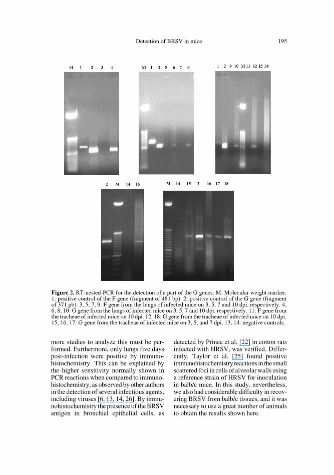
Positive results were obtained with theamplifications of the fragment correspond-ing to a part of the G gene, from RNAextracted from 2 lungs and 2 tracheae col-lected on 3 dpi; 2 lungs and 1 trachea col-lected on 5 dpi; 1 lung and 1 tracheacollected on 7 dpi and 2 lungs and 2 tracheaecollected on 10 dpi. In the RT-nested-PCR
corresponding to the F gene, however, itwas not possible to observe any amplifiedfragment (Tab. I and Fig. 2).
3.4. Nested-PCR sensitivity analysis
The technique amplified the fragmentcorresponding to the G gene of the materialdiluted up to 10-2 (containing 1225 µg/mLof cDNA), while the fragment correspond-ing to the F protein could only be amplifiedfrom pure material (1 197.5 µg/mL; Fig. 3).
4. DISCUSSION
Several small experimental animalshave been used as models of infections withRSV. In this study, balb/c mice were electedto be infected with the BRSV-25-BR straindue to the impossibility of maintaininglarge experimental animals – such as calves– in our institution. Several authors haveused this lineage mice during experimentscarried out with HRSV [4, 11, 17, 24, 25]
Figure 1. A, B and C: Immunohistochemistry of 3 infected mice lungs sacrificed 5 days post-infection. The red coloration represents the infected cells (see www.edpsciences.org/vetres for acolour version of this figure). D: negative control (magnified 20×).

194 R.S. Almeida et al.
and BRSV [4]. According to Kumar et al.[17], balb/c mice provide a good modelfor the study of infection by HRSV, inwhich the infection duration and the pulmo-nary pathology resembles those in humanbeings. Viral replication was detected inbalb/c mice experimentally infected withRSVs through tests of immunofluorescence[24], in situ hybridization [11] and ELISA[4, 17]. Bastien et al. [3, 4] obtained virustitles in balb/c mice challenged with HRSVand BRSV during experiments with syn-thetic peptides to confer protection againstthese viruses. Notwithstanding, some authorsmentioned the great difficulty to get BRSVdetection after the infection of mice. Theycomment that some reasons for this phe-nomenon may exist, since many strains donot grow well in mice and this would seri-ously reduce the possibility of viral nucleicacid detection. However, intranasal inocu-lation of balb/c mice with live BRSVinduces similar levels of serum antibodiesas those detected in mice infected withHRSV (G. Taylor, Institute for AnimalHealth, Compton, UK, personal communi-cation) and BRSV could undergo anabortive infection in the lungs of mice(A. Easton, University of Warwick, Coven-try, UK, personal communication). Tayloret al. [24] have also concluded that thegrowth of the HRSV strain in different celllines affects its infectivity for mice.
In the present study, no clinical signswere observed in the animals, in agreementwith the findings of some authors [4, 11, 17,
24]. However, it was possible to detectBRSV in mice tissue by RT-nested-PCRand immunohistochemistry which, to theauthor’s knowledge, was the first time thatthis virus was detected in balb/c mice. TheF gene was chosen as the target for the RT-nested-PCR because it is one of the mostconservative RSV genes and, consequently,represents a good alternative to be used forvirus detection in calves with unknown his-tory about this virus. The G gene, however,is described as the more variable portion ofthe RSV genome, and can be used in thefuture in Brazilian field sample character-ization. Therefore, these genes are goodchoices to study the BRSV in our country.The RT-nested-PCR was able to amplify afragment corresponding to a part of theG gene from the RNA extracted from thelungs and tracheae collected 3, 5, 7 and10 dpi but only from one or two infectedmice each day. However, in the amplifica-tions corresponding to the F gene, it was notpossible to observe any amplified fragment.The RT-nested-PCR sensitivity, up to onehundred times superior in the amplificationof the G gene fragment as compared to theF gene fragment, may explain the amplifi-cation of only the first one of the infectedmice tissues. Probably, the viral RNAextracted from this material was enoughonly to allow the amplification of the G geneand not the F gene. Although there are noreports of similar results in the literature,this phenomenon may have occurred due toinherent conditions of the primers. Even so,
Table I. The results of RT-nested-PCR of BRSV F and G genes from the tracheae and lungs ofexperimentally infected balb/c mice.
Days post-inoculation (dpi)
No. F gene amplified/total animals
No. G gene amplified/total animals Positive total
Trachea Lung Trachea Lung
3 days 0/12 0/12 2/12 2/12 45 days 0/12 0/12 1/12 2/12 37 days 0/12 0/12 1/12 1/12 210 days 0/12 0/12 2/12 2/12 4Positive total 0 0 6 7 13
Detection of BRSV in mice 195
more studies to analyze this must be per-formed. Furthermore, only lungs five dayspost-infection were positive by immuno-histochemistry. This can be explained bythe higher sensitivity normally shown inPCR reactions when compared to immuno-histochemistry, as observed by other authorsin the detection of several infectious agents,including viruses [6, 13, 14, 26]. By immu-nohistochemistry the presence of the BRSVantigen in bronchial epithelial cells, as
detected by Prince et al. [22] in cotton ratsinfected with HRSV, was verified. Differ-ently, Taylor et al. [25] found positiveimmunohistochemistry reactions in the smallscattered foci in cells of alveolar walls usinga reference strain of HRSV for inoculationin balb/c mice. In this study, nevertheless,we also had considerable difficulty in recov-ering BRSV from balb/c tissues, and it wasnecessary to use a great number of animalsto obtain the results shown here.
Figure 2. RT-nested-PCR for the detection of a part of the G genes. M: Molecular weight marker.1: positive control of the F gene (fragment of 481 bp). 2: positive control of the G gene (fragmentof 371 pb). 3, 5, 7, 9: F gene from the lungs of infected mice on 3, 5, 7 and 10 dpi, respectively. 4,6, 8, 10: G gene from the lungs of infected mice on 3, 5, 7 and 10 dpi, respectively. 11: F gene fromthe tracheae of infected mice on 10 dpi. 12, 18: G gene from the tracheae of infected mice on 10 dpi.15, 16, 17: G gene from the tracheae of infected mice on 3, 5, and 7 dpi. 13, 14: negative controls.

196 R.S. Almeida et al.
Although the present study permittedthe detection of BRSV in experimentallyinfected balb/c mice, this procedure showeddifficulties. The results here obtained dem-onstrate that the use of balb/c mice withoutany artifice can be laborious and oftenunsuccessful. The use of immunodepressedmice could probably represent a good alter-native in order to obtain better results withexperimental models to BRSV. Moreover,in our study, only one cell line type andvirus strain were tested. It is important toemphasize that studies using more cell linesand other virus strains to complement ourunedited data, should be carried to analyzethe real application condition of this partic-ular experimental animal.
ACKNOWLEDGEMENTS
We appreciate the financial support ofFAPESP throughout the research. We also thankDr. Birgitte Viuff from the Royal Veterinary and
Agricultural University and Dr. Lars Erik Larsenfrom the Danish Veterinary Institute, Copenha-gen, Denmark, for the opportunity to perform theimmunohistochemistry assays, and the technicalassistance of Joyce Camargo and Geneci Davi.
REFERENCES
[1] Ames T.R., The epidemiology of BRSVinfection, Vet. Med. Sci. 88 (1993) 881–885.
[2] Arns C.S., Campalans J., Costa S.C.B.,Domingues H.G., D’Arce R.C.F., AlmeidaR.S., Characterization of bovine respiratoryvirus isolated in Brazil, Braz. J. Med. Biol.Res. 36 (2003) 213–218.
[3] Bastien N., Taylor G., Thomas L.H., WyldS.G., Simard C., Trudel M., Immunizationwith a peptide derived from the G glycopro-tein of bovine respiratory syncytial virus(BRSV) reduces the incidence of BRSV-associated pneumonia in the natural host,Vaccine 15 (1997) 1385–1390.
[4] Bastien N., Trudel M., Simard C., Completeprotection of mice from respiratory syncytialvirus infection following mucosal delivery ofsynthetic peptide vaccine, Vaccine 17 (1999)832–836.
[5] Bourgeois C., Bour J.B., Aho L.S., Pothier P.,Prophylactic administration of a complemen-tarity-determining region derived from a neu-tralizing monoclonal antibody is effectiveagainst respiratory syncytial virus infectionin balb/c mice, J. Virol. 72 (1998) 807–810.
[6] Brainard J.A., Greenson J.K., Vesy C.J., TesiR.J., Papp A.C., Snyder P.J., Western L.,Prior T.W., Detection of cytomegalovirus inliver transplant biopsies. A comparison oflight microscopy, immunohistochemistry,duplex PCR and nested PCR, Transplantation57 (1994) 1753–1757.
[7] Bryson D.G., McConnel S., McAliskey M.,McNulty M.S., Ultrastructural features ofalveolar lesions in induced respiratory syncy-tial virus pneumonia of calves, Vet. Pathol.28 (1991) 286–292.
[8] Cannon M.J., Microplaque immunoperoxi-dase detection of infectious respiratory syn-cytial virus in the lungs of infected mice, J.Virol. Methods 16 (1987) 293–301.
[9] Cannon M.J., Openshaw P.J., Askonas B.A.,Cytotoxic cells clear virus but augment lungpathology in mice infected with respiratorysyncytial virus, J. Exp. Med. 168 (1988)1163–1168.
[10] Collins P.L., McIntosh K., Chanock R.M.,Respiratory syncytial virus, in: Fields B.N.(Ed.), Virology, Lippincott-Raven Publish-ers, Philadelphia, 1996, pp. 1313–1351.
Figure 3. Analysis of the sensitivity of RT-nested-PCR. 1: pure cDNA of the F gene(1225 µg/mL). 2: cDNA 1:10 dilution of theF gene. 3: cDNA 1:100 dilution of the F gene.4: cDNA 1:1000 dilution of the F gene. M: molec-ular weight marker. 5: pure cDNA of the G gene(1197.5 µg/mL). 6: cDNA 1:10 dilution of theG gene. 7: cDNA 1:100 dilution of the G gene.8: cDNA 1:1000 dilution of the G gene.
Detection of BRSV in mice 197
[11] Cook P.M., Eglin R.P., Easton A.J., Patho-genesis of pneumovirus infections in mice:detection of pneumonia virus of mice andhuman respiratory syncytial virus mRNA inlung of infected mice by in situ hybridization,J. Gen. Virol. 79 (1998) 2411–2417.
[12] Dubovi E.J., Diagnosing BRSV infection: alaboratory perspective, Vet. Med. Sci. (1993)888–893.
[13] Gressens P., Langston C., Mitchell W.J.,Martin J.R., Detection of viral DNA in neona-tal herpes encephalitis autopsy tissue by solu-tion-phase PCR: comparison with pathologyand immunohistochemistry, Brain Pathol. 3(1993) 237–250.
[14] Held T.K., Kruger D., Switala A.R., Beyer J.,Kingreen D., Busemann C., Janitschke K.,Siegert W., Diagnosis of toxoplasmosis inbone marrow transplant recipients: compari-son of PCR-based results and immunohis-tochemistry, Bone Marrow Transplant. 25(2000) 1257–1262.
[15] Johnson R.A., Prince G.A., Siffin S.C.,Horswood R.L., Chanock R.M., Respiratorysyncytial virus infection in cyclophosphamide-treated cotton rats, Infect. Immun. (1982) 369–373.
[16] Kimman T.G., Zimmer G.M., Westenbrink F.,Mars J., van Leeuwen E., Epidemiologicalstudy of bovine respiratory syncytial virusinfections in calves; influence of maternal anti-bodies on the outcome of disease, Vet. Rec.123 (1988) 104-109.
[17] Kumar M., Behera A.K., Matsuse H., LockeyR.F., Mohapatra S.S., Intranasal IFN-γ genetransfer protect BALB/c mice against respira-tory syncytial virus infection, Vaccine 18(2000) 558–567.
[18] Mohanty S.B., Ingling A.L., Lillie M.G.,Experimentally induced respiratory syncytialviral infection in calves, Am. J. Vet. Res. 36(1975) 417–419.
[19] Murphy B.R., Prince G.A., Lawrence L.A.,Croen K.D., Collins P.L., Detection of respi-ratory syncytial virus (RSV) infected cells byin situ hybridization in the lungs of cotton ratsimmunized with formalin-inactivated virusor purified RSV F and G glycoprotein subunitvaccine and challenged with RSV, Virus Res.16 (1991) 153–162.
[20] Oberst R.D., Hays M.P., Hennessy K.J., StineL.C., Evermann J.F., Kelling C.L., Identify-ing bovine respiratory syncytial virus byreverse transcription-polymerase chain reac-tion and oligonucleotide hybridization, J.Clin. Microbiol. 31 (1993) 1237–1240.
[21] Paton A.W., Paton J.C., Lawrence J.,Goldwater P.N., Harris R.J., Rapid detectionof respiratory syncytial virus in nasopharyn-geal aspirate by reverse transcription and
polymerase chain reaction amplification, J.Clin. Microbiol. 30 (1992) 901–904.
[22] Prince G.A., Jenson A.B., Horswood R.L.,Camargo E., Chanock R.M., The pathogene-sis of respiratory syncytial virus infection incotton rats, Am. J. Pathol. 43 (1984) 649–655.
[23] Samal S., Zamora M., Nucleotide sequenceanalysis of a matrix and small hydrophobicprotein dicistronic mRNA of bovine respira-tory syncytial virus demonstrates extensivesequence divergence of the small hydropho-bic protein from that of human respiratorysyncytial virus, J. Gen. Virol. 72 (1991)1715–1720.
[24] Taylor G., Stott E.J., Hughes M., CollinsA.P., Respiratory syncytial virus in mice,Infect. Immun. 43 (1984) 649–655.
[25] Taylor G., Stott E.J., Bew M., Fernie B.F.,Cote P.J., Collins A.P., Hughes M., Jebbett J.,Monoclonal antibodies protect against respi-ratory syncytial virus in mice, Immunology52 (1984) 137-142.
[26] Tegtmeier C., Angen O., Ahrens P., Compar-ison of bacterial cultivation, PCR, in situhybridization and immunohistochemistry astools for diagnosis of Haemophilus somnuspneumonia in cattle, Vet. Microbiol. 76(2000) 385–394.
[27] Valarcher J., Bourhy H., Gelfi J., SchelcherF., Evaluation of a nested reverse transcrip-tion-PCR assay based of the nucleoproteingene of diagnosis of spontaneous and experi-mental bovine respiratory syncytial virusinfection, J. Clin. Microbiol. 37 (1999) 1858–1862.
[28] Vilcek S., Elvander M., Ballagi-Pordany A.,Belak S., Development nested PCR assay fordetection of bovine respiratory syncytialvirus in clinical samples, J. Clin. Microbiol.32 (1994) 2225–2231.
[29] Westenbrink F., Brinkhof J.M.A., StraverP.J., Quak J., De Leeuw P.W., Comparison ofa newly developed enzyme-linked immun-osorbent assay with complement fixation andneutralization tests for serology of bovinerespiratory syncytial virus infection, Res.Vet. Sci. 38 (1985) 334–340.
[30] Westenbrink F., Kimman T.G., Immunoglobu-lin M-specific enzyme-linked immunosorbentassay for serodiagnosis of bovine respiratorysyncytial virus infections, Am. J. Vet. Res. 48(1987) 1132–1137.
[31] Zamora M., Samal S.K., Sequence analysisof M2 mRNA of bovine respiratory syncytialvirus obtained from an F-M2 dicistronicmRNA suggests structural homology withthat on human respiratory syncytial virus, J.Gen. Virol. 73 (1992) 737–741.
Copyright © 2022 FDOKUMEN